7 minute read
HEART TESTS FOR EVERY BODY. When and why you


HEART TESTS FOR EVERY BODY
CARDIAC CHECKUPS CAN BE YOUR KEY TO PROTECTING YOUR HEALTH. HERE’S A GUIDE TO SOME OF THE MOST COMMON TESTS.

You may know that heart disease kills one in four Americans and is the nation’s leading cause of death. Fortunately, many heart conditions can be prevented or treated—especially if you catch them or their warning signs early with appropriate tests.
“Start by getting regular checkups with your primary care physician, who can listen to your heart, order screening tests and assess your risks,” says cardiologist Danny Wang, MD, of Robert Wood Johnson University Hospital (RWJUH) Rahway. If screening tests fi nd reasons for concern, you may be referred to a cardiologist for further testing to investigate underlying conditions and DANNY WANG, MD determine next steps. BLOOD PRESSURE WHAT IT IS: A measurement of the force that blood exerts on arteries as your heart pumps. HOW IT’S DONE: A healthcare professional places a cuff around your upper arm, infl ates it to compress an artery, then slowly releases it while monitoring your pulse. WHY IT’S IMPORTANT: High blood pressure trigg ers no symptoms but greatly increases your risk of heart disease.
WHEN YOU MAY NEED MORE
TESTS: Healthy adults should get blood pressure checked at least once a year, but your doctor may check more oft en if your reading is higher than 120/80 or you have risk factors.


BLOOD GLUCOSE WHAT IT IS: A gauge of blood sugar levels that reflect the presence or risk of diabetes. HOW IT’S DONE: A variety of blood tests assess glucose levels; some require fasting. WHY IT’S IMPORTANT: Untreated diabetes substantially increases your risk of heart disease, but elevated blood glucose that’s caught early can be reversed.
WHEN YOU MAY NEED MORE
TESTS: Your doctor may test blood glucose more frequently if you’re overweight or if you have additional cardiac risk factors like high blood pressure or cholesterol.
Your heart doesn’t beat just for you. Get it checked. To fi nd an RWJUH Rahway cardiac specialist, call 888.724.7123 888.724.7123 or visit www.rwjbh.org/heart. www.rwjbh.org/heart. LIPID PROFILE WHAT IT IS: A blood test that checks circulating levels of fatty substances such as cholesterol (total, LDL and HDL) and triglycerides. HOW IT’S DONE: A healthcare professional uses a small needle to draw blood into a vial that’s sent to a lab for analysis. Fasting may be required before the test. WHY IT’S IMPORTANT: High levels of LDL cholesterol and triglycerides boost your risk of heart disease, while HDL is protective.
WHEN YOU MAY NEED MORE
TESTS: Healthy adults should get a lipid profi le every four to six years, but your doctor may order screenings more oft en if your numbers are worrisome or you have risk factors like a family history of heart disease.


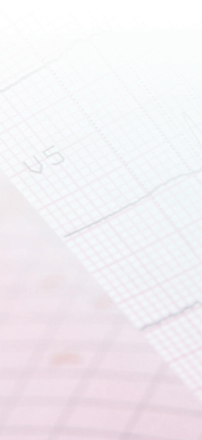

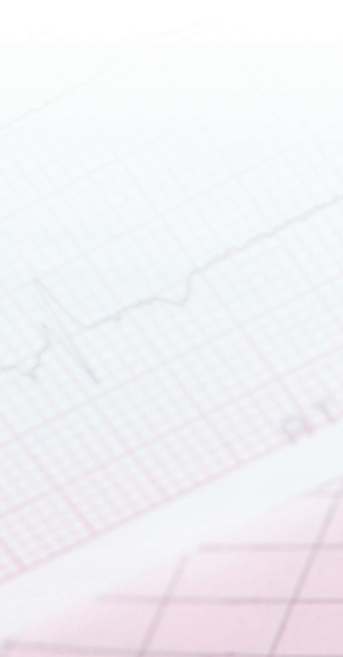
ELECTROCARDIOGRAM (EKG or ECG) WHAT IT IS: A measurement of electrical activity in the heart as it beats. HOW IT’S DONE: Electrodes affixed to your chest, arms and legs convey your heart’s electrical signals through wires to a computer. WHY IT’S IMPORTANT: Abnormal electrical activity can indicate conditions such as irregular heartbeat, clogged arteries, heart damage, heart failure or a heart attack.
WHEN YOU MAY NEED MORE
TESTS: Cardiac electrical activity can vary throughout the day, but an EKG only offers a minuteslong snapshot. A portable Holter monitor can record electrical readings over 24 to 48 hours for a more complete picture.
ECHOCARDIOGRAM WHAT IT IS: An ultrasound scan that generates measurements as well as still and moving images of the heart’s interior, including its chambers, blood vessels, valves and blood fl ow. HOW IT’S DONE: An instrument called a transducer creates images as it’s moved over the skin of your chest and torso. WHY IT’S IMPORTANT: Obtaining information about your heart’s size, shape, movement, strength and function can reveal problems including faulty valves, structural abnormalities, heart attack damage, infl ammation and heart failure.
WHEN YOU MAY NEED MORE
TESTS: If a standard echocardiogram doesn’t provide enough detail, you may need an invasive transesophageal echocardiogram (TEE), in which you’re sedated and a transducer is inserted down your throat to a position closer to the heart.


EXERCISE STRESS TEST WHAT IT IS: A way for your doctor to know how your heart responds to exertion. HOW IT’S DONE: You walk on a treadmill or pedal a stationary bike while your heart rate, blood pressure and electrical rhythms are tracked. WHY IT’S IMPORTANT: Th e stress test allows a doctor to see if enough blood fl ows to your heart as you get more active, whether your heart rhythms are normal and more.
WHEN YOU MAY NEED MORE
TESTS: More tests may be needed if the test results are unclear; if you have other risk factors for heart disease that raise concern; or to confi rm results that indicate coronary artery disease. CHEST X-RAY WHAT IT IS: A black-and-white, 2D image that shows your bones, lungs and heart. HOW IT’S DONE: You stand between a machine that generates X-rays and a plate that captures the image. WHY IT’S IMPORTANT: Chest X-rays can reveal heart-related lung conditions such as fl uid resulting from congestive heart failure, problems with blood vessels near the heart and abnormalities in the heart’s size and shape.
WHEN YOU MAY NEED MORE
TESTS: If needed, your doctor may order further imaging tests that reveal details not visible on an X-ray, such as internal heart structures. NUCLEAR STRESS TEST WHAT IT IS: Similar to the exercise stress test, but with images. HOW IT’S DONE: A small amount of radioactive dye is injected, and then two sets of images are taken with a special camera, one while the patient is at rest and another aft er exertion. WHY IT’S IMPORTANT: May be recommended if an exercise stress test doesn’t pinpoint the cause of symptoms.
WHEN YOU MAY NEED MORE
TESTS: If results indicate blockages or damage, a coronary angiogram, also known as cardiac catheterization, may be done. Note: Patients who can’t do exercise for the test may get a
pharmacological nuclear stress test,
in which a medication is injected to mimic the eff ects of exercise.


CT SCAN WHAT IT IS: An imaging method called computed tomography (CT) in which X-rays taken from multiple angles produce detailed, 3D images of the heart and its arteries. HOW IT’S DONE: You lie on a table that slides into a large, tunnel-like machine in which X-ray beams rotate around you. WHY IT’S IMPORTANT: CT scans can reveal coronary artery plaque buildups that threaten the heart, along with valve problems, infl ammation and pumping defi ciencies.
WHEN YOU MAY NEED MORE
TESTS: If your doctor is concerned about your exposure to X-ray radiation or needs greater accuracy for specifi c conditions, you may receive a magnetic resonance imaging (MRI) test to take detailed images
using magnets and radio waves.


CARDIAC CATHETERIZATION WHAT IT IS: A diagnostic procedure in which a cardiologist inserts a thin tube called a catheter into a blood vessel (typically in the groin) and threads it to your heart to obtain images or samples. HOW IT’S DONE: Th e procedure typically takes place in a hospital catheterization (cath) lab under light sedation. You may be injected with a dye that makes blood vessels more visible. WHY IT’S IMPORTANT: Cath images can show if blood vessels supplying the heart are narrowed or blocked, and a procedure to open them may be done during the same catheterization.
WHEN YOU MAY NEED MORE
TESTS: If you need a procedure, such as open-heart surgery, more tests may be required to prepare for your operation.