3 minute read
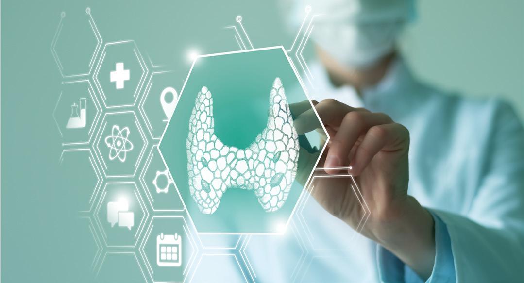
AI and the thyroid gland


Artificial intelligence and the thyroid gland
Artificial intelligence (AI) is an emerging technology which is increasingly being used in thyroid medicine, enabling the transition from population-based to individualised medicine.
Advertisement
In the era of personalised medicine, risk stratification before, during and after treatment is important. It is a combination of technologies that mimic human interaction − it corresponds to human perception.
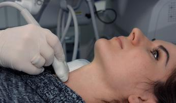
At the same time, thyroid ultrasonography (TUS) is increasing worldwide, resulting in the identification of more thyroid nodules, and a growing number of fine needle aspirations (FNA). There are several problems in the care of patients with thyroid nodules: inconsistent assessment by the ultrasonographer, uncertainty in cytopathologic diagnosis, difficulty in differentiating follicular neoplasms, and inaccurate prognosis. Therefore, the final step in characterising a thyroid nodule is pathohistological analysis, which is sometimes unnecessary.
Application of AI in TUS
TUS is the recommended imaging modality for patients with thyroid nodules, because it is inexpensive, effective and does not require radiation. Several features on TUS indicate an increased risk of malignancy: solid composition, hypoechogenicity, irregular margin, microcalcification, and a shape that is taller than it is wide. However, these features cannot confirm or exclude a diagnosis of thyroid cancer.
The agreement of ultrasonographers between centres in evaluating these features is not very satisfactory. Thyroid Imaging Reporting and Data Systems (TI-RADS) are of great value as a risk stratification system in papillary thyroid cancer, but less so in other malignancies.
AI uses TUS images, cell smears and tissue sections to extract morphological, textural and molecular features. This information is fed into the AI classifier to improve its performance and optimise the workflow of thyroid cancer diagnosis and treatment. AI applications are of increasing interest in reducing the number of invasive clinical procedures.
©iStock/alexey_ds
Application of AI in cytopathological evaluation of FNA
FNA is one of the most important preoperative examinations for the evaluation of thyroid nodules. The Bethesda System for Reporting Thyroid Cytopathology (TBSRTC) is a state-ofthe-art decision-making tool for clinicians. In clinical practice, 15−30% of thyroid nodules continue to be classified as indeterminate nodules. Recent studies showed excellent agreement between machine learning models and cytologists in predicting malignancy (malignancy risk of TBSRTC III determined by machine learning was much lower than manual classification: 4.2 versus 18.8%). Morphological and genetic classifications supported by AI models are quite accurate in distinguishing malignant from benign thyroid nodules. AI as a second opinion reduced the number of FNA procedures by 27%.
Conclusion
AI systems learn on a case-by-case basis. Because human tissue is characterised by high heterogeneity and variability between and within subjects, no finite training set can fully represent the diversity of cases that may occur in clinical practice.
Extensive research is still needed to improve the generalisability and accuracy of AI-based models (the major limitation being the small amount of data used to develop and validate predictive models). Therefore, the use of AI applications alone for diagnosis in clinical practice should continue to be avoided. However, AI applications can improve the accuracy of thyroid diagnosis, especially for physicians in training. AI applications may be integral to the procedure and should be interpreted according to routine clinical practice.
Karin Zibar Tomšić
Croatia
FURTHER READING
- Bini et al. 2021 Cancers13 4740 (https://doi.org/10.3390/cancers13194740).
- Li et al. 2021 Frontiers in Oncology10 604051 (https://doi.org/10.3389/fonc.2020.604051).


