Issue 46 Autumn 2021
Endocrine Views Opinion and news from the European Society of Endocrinology
Bone: the latest endocrine insights In this issue The long-lasting endocrine impact of COVID-19 Pituitary adenomas in pregnancy: new guidance
ISSN 2752-8510 (print) ISSN 2752-8529 (online)
CONTENTS & EDITORIAL
In this issue
Editorial
Society News
It is with pride and excitement that I write this editorial: the first for Endocrine Views. This is a major step in the evolution of a stimulating forum for topical debate and discussion, as well as a means for ESE to increase your involvement in the Society’s work to advance our discipline.
03 ECE 2022: let’s meet again in Milan! Welcome to Endocrine Views 04 ESE membership review, From the ESE office, Centres of Special Interest, Make your nominations 05 Your President’s perspective: ESE at 15
Interview
06 Meet Jérôme Bertherat, ESE President-Elect
Inside science
07 Osteomorphs: a new cell entity
Debate
08 PTH replacement improves quality of life 09 PTH replacement can induce long term problems
Features
10 Addressing the denosumab rebound effect 11 FGF23: a new therapeutic target in bone disease 12 Calcifediol treatment and COVID-19 13 COVID-19: long term endocrine complications
Editors’ Selection
14 Vitamin D special issue for Endocrine Connections 15 Pituitary adenomas in pregnancy: new ESE guideline
At the Back...
16 The latest from EYES, plus Diary dates
Editor: Justo Castaño, Spain Email: justo@uco.es Co-Editor: Martin Reincke, Germany Email: ese.president@ese-hormones.org Editorial Board: Adrian Clark, UK Olaf Dekkers, The Netherlands Karim Meeran, UK Stavroula Paschou, Greece Marija Pfeifer, Slovenia Walter Vena, Italy Victoria Withy, ESE Office Maria Chiara Zatelli, Italy Guest Editors: Peter Kamenický, France Martina Rauner, Germany Managing Editor: Caroline Brewser Design: Qube Design Associates Website: www.ese-hormones.org ©2021 European Society of Endocrinology
2
The views expressed by the contributors are not necessarily those of ESE. This document is available on the ESE website, www.ese-hormones.org ESEndocrinology EuropeanSocietyofEndocrinology The addresses used to mail this issue of Endocrine Views were supplied by the members of ESE and are stored in Bioscientifica’s database for future use. If you do not wish to receive further mailings, please advise info@euroendo.org ESE thanks the following for their support. Premium Corporate Members: Ipsen, Pfizer, Recordati Rare Diseases, Takeda Corporate Members: Advanced Accelerator Applications, Alexion Pharma GmbH, Amryt Pharmaceuticals, Chiasma, Diurnal, HRA Pharma, Kyowa Kirin International, Novo Nordisk Supporters: Crinetics Pharmaceuticals, Isotopen Technologien Munchen AG, Soleno Therapeutics
Appropriately, on page 5, our President talks about the current ESE strategic review, which will set the goals for the next 5 years. Martin Reincke reminds us of our Society’s huge progress and the many successful initiatives of the 15 years since ESE was founded. None of these would have been possible without the support of you, the members. This issue is the first to benefit from the involvement of Guest Editors within the Editorial Board. The immense contributions of Martina Rauner and Peter Kamenický, ESE Focus Area Leads for Calcium and Bone, have enabled us to bring you cutting edge research in this field, and highly contested debate. Our cover is graced with a vibrant image from the work of Michelle McDonald, Natalie Sims, Tri Phan and Peter Croucher. Find their insights into the private life of the osteomorph, a newly characterised bone cell type, on page 7. Pages 8 and 9 host our first debate. Unlike patients affected by other hormone deficiencies, individuals lacking parathyroid hormone do not receive hormone replacement as first-line treatment. Read the arguments for and against its use, presented by Heide Siggelkow and Peter Kamenický. Anne Sophie Sølling and Bente Langdahl address the rebound effect seen after withdrawal of osteoporosis drug denosumab on page 10. On page 11, Agnès Linglart looks at FGF23 as a potential therapeutic target in metabolic bone disease. COVID-19 will regrettably be with us for some time. On page 12, Roger Bouillon, José López Miranda and José Manuel Quesada Gómez explain how rapid improvement of patients’ vitamin D status using calcifediol offers hope in disease management. On page 13, Manel Puig Domingo and Mónica Marazuela summarise the latest understanding of the virus’s wide-reaching effects on endocrinology and metabolism. Alongside these hot topics, we also bring you up to date with plans for ECE 2022, including the long-awaited return to a face-to-face meeting in Milan, Italy (page 3). We get to know ESE President-Elect Jérôme Bertherat on page 6, and we welcome the latest ESE Clinical Practice Guideline, courtesy of Anton Luger and colleagues, on page 15. Finally, I thank Endocrine Views’ hard-working Editorial Board and production team for sharing their vision for the future of publishing and making it a reality. Here you see the first steps on our journey of transformation.
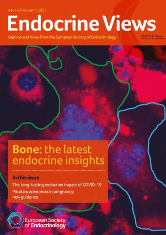
Justo P Castaño Editor, Endocrine Views Cover image: Live-cell imaging of osteoclast fusion post-sRANKL stimulation in vitro. Cells labelled with wheat germ agglutinin-AlexaFluor 488 (red pseudocolour) and Hoechst (cyan). Reproduced under CC BY licence (http://creativecommons.org/licenses/by/4.0) from McDonald et al. https://doi.org/10.1016/j.cell.2021.02.002 ©2021 The Authors. Find out more on page 7 of this issue.
ECE 2022
SOCIETY NEWS
21–24 May 2022 Milan, Italy
www.ese-hormones.org/ece-2022
Award lecturers at ECE 2022 Geoffrey Harris Award AJ van der Lely The Netherlands European Journal of Endocrinology Award Roland Stimson UK
Milan welcomes ECE 2022 Plans for the 24th European Congress of Endocrinology (ECE) on 21–24 May 2022 in Milan, Italy, are well underway. The Congress will be the first time since ECE 2019 in Lyon, France, that our endocrine community has the chance to meet in person, due to the COVID-19 pandemic. As such, it will be not only a celebration of our discipline but an opportunity to celebrate being together once more. ECE 2022’s Local Organising Committee (LOC) Chair is none other than the Society’s Past President, Andrea Giustina, who is looking forward to warmly welcoming us all to Milan in May. The ECE Programme Organising Committee (POC), along with the LOC, have developed a diverse, engaging and vibrant programme to shape the Congress, coupled with many opportunities to network and collaborate. See www.esehormones.org/ece-2022 for details.
Take part at home
We know that you may be unable to join us in person in Milan. This is why, for the first time, ECE 2022 will offer you two options to engage with the Congress: • in person in Milan • from your home with ECE @Home. So, while we hope to see you in person, we will welcome you in the way that suits you best, as your contribution and engagement are what makes our Congresses so special and memorable. More information about the two registration options is available at www.ese-hormones.org/ece-2022. Issue 46 Autumn 2021
Endocrine Views Opinion and news from the European Society of Endocrinology
ISSN 2752-8510 (print) ISSN 2752-8529 (online)
Take part in Milan Delights await all those who come to ECE 2022 in person. Milan is Italy’s second largest city. Founded by the Celts in 600 BCE, it is home to the largest opera house in Europe, the Teatro alla Scala, as well as to the San Siro stadium − the so-called ‘La Scala’ of football! Situated in the Lombardy region, Milan is known for its gastronomic delights. It’s also famous for its colourful art scene and is the capital of Italian fashion. Italy is also home to two endocrine societies: the Italian Association of Clinical Endocrinologists and the Italian Endocrine Society, both of which have a long and rich history in our field.
Bone: the latest endocrine insights In this issue The long-lasting endocrine impact of COVID-19 Pituitary adenomas in pregnancy: new guidance
European Hormone Medal (joint winners) Josef Köhrle Germany Peter Rossing Denmark Clinical Endocrinology Trust Award Alberto Pereira The Netherlands Transatlantic Alliance Award Shlomo Melmed USA
Submit your abstracts
Abstract submission and registration open this Autumn – and we cannot wait to read your abstracts to find out more about what you have been working on over the last year. Even more importantly, we cannot wait to see you at ECE 2022! Riccarda Granata ESE Congress Committee Chair Beata Kos-Kudła POC Clinical Lead Carlos Dieguez POC Basic Lead Andrea Giustina LOC Chair
We are delighted to announce that Shlomo Melmed is the recipient of the inaugural Transatlantic Alliance Award. This joint ESE and Endocrine Society initiative recognises an international leader in endocrinology who has made significant advances through collaboration in both the USA and Europe.
Welcome to Endocrine Views
Endocrine Views is the new name for ESE News, reflecting a refreshed focus on the latest opinion and debate in endocrinology and related fields. You will still find your Society’s latest initiatives and plans within these pages, alongside an evolving forum for discussion and communication amongst endocrinologists worldwide. Topical content will be developed in close liaison with the ESE Focus Area Leads. In this issue, the Editorial Board are delighted to have worked with Peter Kamenický and Martina Rauner from the Calcium and Bone Focus Area, to present the latest developments and debates in this field. The icons shown below will tag articles according to their ESE Focus Area or other area of relevance. These are the first stages in a programme of development over the next few years. Let us have your thoughts and ideas via info@euro-endo.org.
Adrenal and Cardiovascular Endocrinology
Diabetes, Obesity, Metabolism and Nutrition
Events
Publications
Awards
Education
Grants
Reproductive and Developmental Endocrinology
Calcium and Bone
Endocrine related cancer
Pituitary and Neuroendocrinology
Research
COVID-19
Environmental Endocrinology
Policy and Advocacy
Thyroid
3
SOCIETY NEWS
Ensuring your membership works well for you The ESE Membership Committee has been reviewing the Society’s membership offering, to ensure it continues to meet all members’ needs, throughout their career. An update on the first part of this work featured in ESE News issue 45. Since then, you will have received an email about the membership group you have been placed in. This also contained details about how to make any amendments, if necessary. The next stage of the review has, importantly, worked to ensure that the benefits you can access and the activities we highlight to you are relevant to your career stage and to your area of work. Over the next few months, you will see these changes becoming apparent – through the
membership information on the ESE website, in your membership area and also in the email communications you receive from the Society. The first of these will be your ESE 2022 membership renewal reminder. Many individual benefits will be grouped under the following five headings: 1. Awards 2. Grants 3. Information 4. Journals 5. Sharing knowledge You can find all the details about the benefits which are tailored to your specific membership category and career stage in the membership section of the ESE website (www.
ese-hormones.org/about-us/ membership). Please take a moment to read the information and refresh yourself on the many benefits that ESE membership offers. We will continue to highlight benefits and activities to you that we think will interest you and help you get the most out of your membership. These exciting, positive developments will help ESE remain your supportive partner throughout your career. Anton Luger and Jérôme Bertherat ESE Membership Committee Co-Chairs Martin Reincke ESE President
Register your endocrine Elections in 2022 centre today! The ESE Centres of Special Interest (CoSI) database provides information on endocrine centres engaged with research, education and healthcare across Europe. Why should your centre join the CoSI database? • For increased visibility on the European map of endocrinology • To ease communication with neighbouring or regional centres • For more opportunities for exchange of young career or established endocrinologists
• For increased opportunities to join clinical collaborative efforts • To search for partners for your projects and initiatives • To benefit from sharing best practice. Apply and find more details at www.ese-hormones.org/ ecas-cosi.
You will soon have a chance to help shape the future of European endocrinology by nominating two members of the ESE Executive Committee. Elections in spring 2022 will replace members who are stepping down at the ends of their very productive terms of office: ESE Secretary Mónica Marazuela (Spain) and Congress Committee Chair Riccarda Granata (Italy). Basic scientists are particularly encouraged to make nominations, to ensure a good balance of representation at the highest level of your Society.
You will receive an email shortly with details of how to make your nominations.
2023 Awards Make your nominations for the 2023 ESE Awards by 28 February 2022. You can find details, including the criteria and nomination process, at www.ese-hormones.org/ grants-and-awards/awards.
From the ESE Office As our 5-year strategic period from 2017 to 2021 nears its end, we are excited to be undertaking a new strategic review. We are also proud of our great progress against our original plans. Our major goals for membership growth have been met. The membership of ESE has grown by more than 34%, to reach our 5-year target of over 5000 (reaching 5009, to be exact, in May 2021!). The percentage of early career members has also grown, from 24 to 30%, which was another major objective. Early career members are our life blood and the future of the Society; they will always be a key focus for ESE. Our immensely expanded activities within policy and
4
advocacy, and the creation of an internal structure to support them, are the embodiment of another substantial change that has happened in line with our strategy. These include the publication of the ESE White Paper Hormones in European Health Policies: How endocrinologists can contribute towards a healthier Europe (launched in May 2021), our extensively increased profile in Brussels, and the introduction of an external agency to support these very specialist activities. Strengthening our journals has been another focus. European Journal of Endocrinology’s increase in impact factor to 6.664 this year was very significant, along with the increase to 3.335 for our open access
journal Endocrine Connections. We are also pleased to have developed our educational activities extensively, particularly online, with an expanded number of events and the launch of the European exam. Last but not least, we cannot forget our wonderful Congress – our meeting point. It transformed well to a digital event, and everyone was justifiably proud of its success. Online is, however, not a permanent replacement for face-to-face, and we look forward to seeing you in Milan in May next year! The Executive Committee are hard at work on our strategy for the next period, and evaluating how ESE can best support you. As always, get in touch if there is anything you would like to discuss.
Helen Gregson Chief Executive Officer, ESE helen.gregson@ese-hormones.org
SOCIETY NEWS
A role model for innovation ESE President Martin Reincke reflects on your Society’s rapid evolution over the last 15 years and its many achievements, as our focus and strategy for the coming years take shape. This is a year for celebration: ESE is 15 years old. Launched in 2006, our Society is still young – in human terms it would be in puberty. But, instead of being in the middle of a hormonal storm of confusion, we are well on track to achieve our goals: namely, shaping the future of endocrinology in Europe, leading to substantial steps forward in science, knowledge and hormone health.
for endocrinologists worldwide. And, together with the Society for Endocrinology, we launched the open access journal Endocrine Connections and it began to fly.
2006-2009: In the early years, ESE had to establish itself as a society. The focus was developing our annual Congress (ECE) as the leading European endocrine congress, ensuring it stayed absolutely current and highly attractive. ESE also continued to develop its top quality postgraduate education and training for all career stages in clinical practice and basic research.
Since 2016: ESE has recruited its own professional team, starting with our Chief Executive Officer, Helen Gregson. ESE had previously been managed by Bioscientifica but, from this point, their role in ESE’s association management reduced. Bioscientifica still provides ESE with conference organisation and journal publishing services. The ESE staff has now grown to a team of nine. ESE organised its activities into eight Focus Areas, to guarantee balanced, in-depth representation of all clinical and scientific areas. The Society worked continuously on its financial sustainability, with a reserve fund in place to ensure continuation of its activities in economically difficult times.
2010-2013: Our young endocrinologists organised themselves as EYES (now the ESE Young Endocrinologists and Scientists): a powerhouse of ideas driving innovation. The ESE Clinical Practice Guideline programme was launched, providing scientifically founded statements, vital for the daily care of patients. The foundation of the ESE Council of Affiliated Societies (ECAS) strengthened the relationship between ESE and our National Affiliated Societies, ensuring that affiliation is mutually advantageous and synergistic. Our lead journal, European Journal of Endocrinology, developed as a highly competitive international journal, providing a primary source of knowledge
2018-2021: ESE has increasingly turned towards public health topics, inspired by our experience that many themes relevant to hormones can only be addressed by a continuous policy and advocacy strategy, with regular activities at the level of the EU. Our major project, Mapping Endocrinology in Europe, provided information about endocrinologists across the continent. After 2 years of consultation, ESE presented its essence in the form of a White Paper: Hormones in European Health Policies – How endocrinologists can contribute to a healthier Europe. Endorsed by 45 national endocrine societies and seven European and international specialist societies, this in-depth
Some selected steps in our rapid evolution
‘Our Society has the essential ingredients for this desirable development at hand: a highly dedicated crowd of talented, motivated and inspired members who support us at all levels and in all our activities: YOU!’ 2006
2007
2008
2009
2010
2011
2012
2013
statement provides a European endocrinology policy focus around the areas of obesity, rare endocrine diseases, endocrinology and cancer, and endocrine disruptors. With ESE’s activities growing, the number of committees and task forces also grew. ESE membership exceeded 5000 for the first time, with total membership through representation by the National Affiliated Societies and the ESE Advocacy Representation Scheme (EARS) now over 20 000. The tragedy of the COVID-19 pandemic forced ESE to hold ECE 2020 and ECE 2021 virtually, but with excellent participation and success. Physical committee meetings and courses switched to a digital format. The pandemic has presented our Society with a heavy stress test, but we can proudly say that we have passed it so far.
The future As you can see, ESE has developed into a pretty good scientific society over the last 15 years. The question is, can an already very good society become even better, possibly to become a great one, in the future? The ambition for this is clearly out there! We believe that our Society has the essential ingredients for this desirable development at hand: a highly dedicated crowd of talented, motivated and inspired members who support us at all levels and in all our activities: YOU! Your critical input and continuous support are currently, and will remain, the most important resources of ESE. The Executive Committee is currently working on the Society’s strategy for the next 5 years, considering many topics and goals, such as inclusion, innovation, equality, diversity, specialised societies, strategic office location, the ESE Foundation, the ESE Academy and international outreach. And there may be other areas where ESE has the potential to improve in the future. Please let us know your ideas and visions. I recently came across the Jim Collins bestseller From Good to Great: Why Some Societies Make the Leap ... and Others Don’t. This was good reading, and an
2014
2015
2016
2017
interesting source of inspiration for an ambitious society. Here are the seven principles, which he considers essential, and which I have adapted a little bit: 1. Leadership: be driven to do what is best for the society. 2. Get the right people ‘on the bus’. 3. Face the facts, including the unpleasant and disappointing ones, but never give up. 4. Hedgehog concept: focus is key! ‘The fox knows many things, but the hedgehog knows one big thing.’ 5. Culture of discipline: don’t forget to ‘rinse the cottage cheese’. 6. Technology accelerators: use technology to accelerate growth. 7. Finally, if you follow 1 to 6, your organisation may become a flywheel: the additive effects of many small initiatives become synergistic. I am convinced that ESE is well on course to ‘becoming a flywheel’, able to shape the future of endocrinology in Europe and beyond. Ultimately, ESE must turn into a pan-European organisation at all levels. Our strategic Executive Committee session this autumn saw us reflect on our strengths and weaknesses, to develop focus and strategy for the coming years. This will bring achievements according to our ambitions. Martin Reincke ESE President Twitter: @EsePresident
2018
2019
2020
2021
5
INTERVIEW
An interview with
Jérôme Bertherat Jérôme Bertherat is President-Elect of ESE. We recently took the opportunity to talk to him about his enthusiasm for endocrinology and for the Society, and his perspective on the future of our field. How did you become interested in endocrinology?
I was initially attracted by neurology and chose PitiéSalpêtrière Hospital in Paris, a well known neurology centre since the time of Charcot. My interest in endocrinology probably stemmed from a blend of two things: outstanding lectures on the physiology and anatomy of the pituitary and hypothalamus, given by great teachers in large amphitheatres in my first years of medical school, and, soon after that, seeing patients with pituitary disorders at the hospital. I was then fascinated by the multiple and systemic consequences of hormone dysregulation, with both a somatic and psychiatric impact. Further motivation came from the observation, as a student, of the great efficiency of treatments in the endocrinology department in contrast with the neurology department. Besides, I really enjoyed subsequent research training in a laboratory, where I studied somatostatin receptors in the brain. At this time, somatostatin analogues appeared as a new treatment for acromegaly. This convinced me to remain in the field of endocrinology.
Who has inspired you most?
I will answer with a title: ‘My American Uncle’. It is a movie directed by Alain Resnais that I loved when I was a student. It is a fiction on human behaviour with the famous French actors Gérard Depardieu and Nicole Garcia, mixed with famous old movie scenes and comments by neurobiologist Henri Laborit. It attempts to explain the mechanisms of dominant and defensive human behaviour. I recently got a DVD to watch it again, after many years, with my three children, because they study cinema, psychology and psychiatry. I was convinced it would perfectly match their centres of interest. Regrettably, I did not feel the same enthusiasm any more...
What might you have studied if not medicine?
Medical school was a very late pick. My original dilemma was between cinema, agronomy, and political science. These may seem very diverse, but the approach was similar for me: first observe to understand, and then be able to provide something to help others. The choice of medical school was mostly based on the idea that I could always change back to the
other activities afterwards, while medical training took too long to start later. In fact, I even took a first year of cinema school during my second year of medical school. But then medical school became too demanding to proceed with cinema. I compensated by studying brain somatostatin receptors by a new light microscopic autoradiography method; that produced spectacular photographs!
What areas of endocrinology are you most interested in?
As a clinician, I’m interested in general endocrinology with a specific focus on pituitary and adrenal diseases and, as a ‘translational’ researcher, in pituitary and adrenal diseases causing hormone excess. I’m currently working on Cushing’s syndrome, the genetics of adrenocortical tumours and cAMP signalling alterations in pituitary and adrenocortical tumours. In our team, we like to merge the results of genomics studies and cell biology, in order to understand these diseases better as well as to classify them.
What achievements are you most proud of?
My three children and my wife’s novels – all of which are actually the outcomes of her work!
What are we most likely to find you doing on a day off? If it is a real full day off, I will have enough time to go to the countryside and take care of the garden, where I grow mostly fruit and vegetables.
What are the greatest challenges for endocrinology?
Clinical teaching with residents and students, Endocrinology Department, Cochin Hospital, Paris, France.
6
Endocrinology has been very successful in treating dysregulation of endocrine glands, even if many situations still need improvement. This progress is now reaching a high level of specialisation in diverse sub-domains. One challenge is to motivate, train and maintain enough specialised healthcare professionals (physicians, nurses, psychologists,
surgeons, radiologist, biologists...) to have the expertise available for all patients. In this regard, patients’ education and the general population’s knowledge are also important. Apart from diseases of a specific endocrine gland, many conditions such as obesity, diabetes, metabolic disorders, cancer, cardiovascular diseases, ageing, etc., are linked to the actions and dysregulation of hormones. The COVID-19 pandemic has also clearly shown that these conditions are major determinants of the outcome of infectious diseases. Environmental problems also interfere with hormones’ actions. The major challenge for endocrinology is the recognition of this important and specific role of hormones, and therefore of endocrinology itself. This acknowledgement is the key to motivate, support, and encourage healthcare professionals and researchers to tackle these conditions, by studying hormones, by developing and using specific preventive and therapeutic interventions, based on this knowledge.
How can ESE best support its members?
ESE has many very successful methods for achieving this. Communication is a major support, both between the members of the endocrine community and between them and the various stakeholders, including health authorities. Appropriate education at each stage of members’ careers and in each type of activity is also important.
What will the pandemic’s legacy be?
It’s main impact on medical care and everyday life will probably be the use of virtual communication. The impact of mRNA for therapy in general will be very interesting to follow.
What are your words of advice for early career endocrinologists?
Be aware that hormones are vital molecules that you perfectly know. Do not hesitate to do what you like most: passion is the key for successful development of rewarding activities.
What else would you like to say?
I am very grateful to ESE’s members for trusting me as President-Elect. It is a pleasure to interact with all ESE members and the endocrine community in general, to promote and support endocrinology, so that we all develop and participate in projects that benefit the community, and especially patients.
INSIDE SCIENCE
Osteomorphs: a new cell entity Identification of this new cell type within the osteoclast lineage has already started to provide a new perspective on a clinical challenge. Osteoclasts are the only cells capable of resorbing bone mineral and matrix. These long-lived bone resident cells form through the fusion of mononuclear precursor cells and adhere strongly to the bone surface. This creates a tightly sealed zone, enclosing the acidic microenvironment needed to demineralise and break down bone matrix. Bone resorption is essential for maintaining levels of circulating minerals and optimal skeletal structure and strength. In adulthood, bone resorption continuously renews the skeleton by removing bone that is then replaced by a balanced level of bone formation. If unbalanced, skeletal pathologies arise, including osteoporosis. Current anti-resorptive agents inhibit osteoclast function or formation. However, recent developments suggest that an intermediate cell type, the ‘osteomorph’ could also be targeted therapeutically, and may determine patients’ response after anti-resorptive treatment ceases.
The discovery of osteomorphs Until recently, the life cycle of the osteoclast was documented to reach 2−3 weeks,1 at which point the cells would undergo apoptosis. Recently, tracking of osteoclast populations in mice revealed their persistence for up to 6 months.2 This finding was surprising and begged the question, how could this be achieved? In vitro studies as long ago as 1949 described long filaments between osteoclast cell bodies, and suggested that large osteoclasts may undergo fission to produce smaller individual cells.3 In 1981, this observation was supported by histological evidence. After blocking osteoclast activity using calcitonin in rats, Baron et al. observed an acute increase in the number of small osteoclasts with few nuclei, concluding that this must have occurred through osteoclast fission.4 It was not until 2012 that this concept of osteoclast fission was confirmed in vitro in real time, using live cell imaging.5 However, it had never been observed in vivo. This changed in March 2021, when evidence of osteoclasts undergoing fission in real time inside living bone tissue was published in Cell.6 By combining bone marrow from mice genetically modified to have osteoclast precursors carrying either red or green fluorescence, real time fusion of these precursor cells to form both red and green osteoclasts could be observed. What’s more, this enabled the tracking of osteoclasts as they underwent fission using cutting edge two-photon microscopy to image deep into intact murine bone. Not only were osteoclasts captured undergoing cell fission, the resulting smaller individual osteoclasts were shown to fuse with other osteoclasts, sometimes almost immediately after their fission from the parent cell. This revealed that osteoclasts do not always undergo apoptosis, but can be recycled. Since this intermediate cell can change its shape and ‘morph’ with other cells, it was named the ‘osteomorph’.
The role of osteomorphs What is the nature of these osteomorphs, and how do they differ from osteoclasts? Single cell RNA sequencing of osteomorphs confirmed that, at the level of gene expression, osteomorphs are different to osteoclasts and osteoclast precursors. This indicates that the osteomorph is a novel cell of the osteoclast lineage. When these osteomorph-defining genes were examined in 40 mouse lines with single gene deletions of osteomorph genes,7 deletions of 17 of these genes were associated with changes in bone mass, structure or strength. This highlights osteomorphs’ potential importance in maintaining skeletal integrity. Are these genes important in human skeletal disease? It seems so. When genes that define osteomorphs were examined in human datasets, 22 were shown to cause skeletal dysplasias, and many were associated with estimated bone mineral density in the UK Biobank study. This indicates that osteomorph genes may play a role in skeletal diseases and could provide targets for novel therapeutics. Although further studies are required to determine the role of osteomorphs in skeletal pathologies, this work also provided evidence of
Live-cell imaging of osteoclast cell fission in vitro. Reproduced under CC BY licence (http://creativecommons.org/licenses/by/4.0) from McDonald et al.6 https://doi.org/10.1016/j.cell.2021.02.002 ©2021 The Authors.
a role for osteomorphs and osteoclast recycling in patients’ response to current bone-targeted drugs.
Anti-resorptive therapeutics Anti-resorptive therapeutics target osteoclasts, and exist in two classes: bisphosphonates (BPs), which reduce osteoclast activity, and the antiRANKL antibody denosumab (DMAB), which inhibits their formation. These agents have been used for decades to prevent further bone loss in patients with osteoporosis. However, in the case of DMAB, when patients cease treatment, rebound bone loss and increased risk of vertebral fracture are observed.8−10 This is a challenge for patient management. To explore the mechanisms underlying this rebound bone loss, osteoclast fusion, fission and recycling were examined using intravital imaging, where osteoclast formation was inhibited with osteoprotegerin, the decoy receptor for RANKL, which mimics the action of DMAB (OPG:Fc). During OPG:Fc treatment, osteoclast movement slowed and there were very few fission, fusion and recycling events. Instead, osteomorphs and osteoclast precursors accumulated in the bone marrow space. When OPG:Fc therapy was withdrawn, these ‘waiting’ osteomorphs and osteoclast precursors rapidly re-fused, increasing osteoclast formation and stimulating rapid bone loss. This may explain what occurs following DMAB withdrawal, and raises the possibility that targeting osteoclast recycling could provide new approaches to prevent bone loss in this situation. Such an approach could complement the current method of transitioning patients onto BP therapy when DMAB is ceased, which has been inconsistent in its ability to protect patients from rebound bone loss and increased risk of vertebral fractures.11 The osteomorph’s identification as a component of the osteoclast lineage and its inclusion in the repertoire of bone resident cells has already started to provide a new view on a clinical challenge. In the coming decades, there will be much more to learn about this new cell, which could lead to new ways of understanding the processes of bone development, growth and degeneration, and could lead to osteomorph-centred therapies for bone diseases. Michelle M McDonald, Natalie A Sims, Tri G Phan, Peter I Croucher Garvan Institute of Medical Research, Sydney, NSW and St Vincent’s Institute of Medical Research, University of Melbourne, Victoria, Australia REFERENCES 1. Stavros et al. 2000 Endocrine Reviews 21 115–137. 2. Jacome-Galarza et al. 2019 Nature 568 541–545. 3. Hancox 1949 Journal of Physiology 110 205–206. 4. Baron & Vignery 1981 Metabolic Bone Disease & Related Research 2 339–346. 5. Jansen et al. 2012 Calcified Tissue International 90 515–522. 6. McDonald et al. 2021 Cell 184 1330–1347. 7. Freudenthal et al. 2016 Journal of Endocrinology 231 R31–R46. 8. Cummings et al. 2018 Journal of Bone & Mineral Research 33 190–198. 9. McClung et al. 2017 Osteoporosis International 28 1723–1732. 10. Tsourdi et al. 2017 Bone 105 11–17. 11. Sølling et al. 2020 Journal of Bone & Mineral Research 35 1858–1870.
7
DEBATE
A matter of debate
Hypoparathyroidism treatment is the subject of our first Endocrine Views debate. Conventional treatment uses active vitamin D analogues and calcium supplements, but these do not fully replace the actions of parathyroid hormone (PTH), and may lead to complications and decreased quality of life. PTH replacement is usually a second-line treatment, when disease is poorly managed by the convential approach, and presents safety concerns. Here, Heide Siggelkow and Peter Kamenický present the pros and cons of each treatment regimen.
PTH replacement improves quality of life In the treatment of hypoparathyroidism (hypoPT), we do not replace the missing hormone, as we do for all other endocrine diseases. The current approach is to supplement with calcium and active vitamin D compounds, with or without genuine vitamin D and/or magnesium. However, this does not restore the normal physiology of parathyroid hormone (PTH)-regulated calcium metabolism. Under this conventional therapy, patients often report a number of symptoms and complaints, including physical, emotional and neurocognitive symptoms, besides the typical clinical symptoms of hypocalcaemia (e.g. muscle cramps, tingling in the extremities and perioral numbness). Meanwhile, the significance of the patients’ well-being has been acknowledged.1−3 Many studies have analysed quality of life (QoL) in patients with hypoPT on conventional therapy using validated generic or specific questionnaires, and clearly demonstrated an impairment in ‘joie de vivre’.4−6
What influences quality of life?
To better understand and consistently improve the lives of patients suffering from hypoPT, several studies investigated the factors influencing QoL. In addition to the existing organ involvement and co-morbidities, questionnaires identified disease aetiology, duration, gender, age and current medication as having an impact on daily life, as well as several biochemical parameters. The only positive effect on QoL demonstrated to date in all the different studies resulted from the use of hormonal treatment in the form of PTH.6,7 This indicates that the missing PTH could be causal in patients’ ill-being. We must take the impairment in QoL seriously, as QoL in hypoPT is found to be generally comparable with or lower than scores reported for patients with other long term diseases, such as heart disease, haematological disorders, diabetes and cancer. Besides the misery in our patients’ daily lives, the evident decrease in work productivity impairs social performance. However, many physicians, even endocrinologists, still consider these patients as well treated and well adjusted under the current approach. The decrease in QoL is not yet understood, with a number of study patients reaching the biochemical reference values corresponding to guidelines. The target value for serum calcium, at or even below the lower end of the reference range, is seen as the key to reducing long term complications, but might well be too low. Alternatively, other changes in mineral metabolism using conventional therapy (e.g. high phosphate levels) may interfere with the equilibrium of other organ systems, including the brain. However, symptoms and complaints seem to be partially influenced by calcium levels, suggesting that higher levels support overall general health, and reduce cardiovascular risk and neuropsychological dysfunction. Certainly, on conventional therapy, higher calcium levels are associated with high phosphate levels and thus a high calcium−phosphate product, regarded as the most problematic risk factor in several disease complications. Conversely, phosphate levels on PTH therapy are low, owing to the phosphaturic effect of PTH; the concern associated with increasing phosphate is no longer justified. Furthermore, as stated above, there is discussion as to whether the absence of the hormone itself could be responsible for the impairment to daily life.
8
Evidence for PTH’s positive effect
If hormonal treatment positively influences QoL, what data do we have to support this notion? In rare diseases, such as hypoPT, double blind prospective studies are not easy to perform, given the risk of hyper- or hypocalcaemia during treatment. However, some data exist on the effect of hormone treatment on QoL.6 PTH(1−34) administered twice a day improved SF-36 scores at 6 months, and QoL remained generally above baseline at 2 years.8,9 Treatment with recombinant human PTH(1−84) was investigated in open label and controlled studies using the SF-36 validated generic questionnaire. A clear improvement was demonstrated compared with baseline after 1, 5 and 8 years, but not in all aspects of the initial deterioration. PTH therapy in a double blind, placebo-controlled study was shown to have no benefit, with a fixed, rather high dose of PTH without reduction of calcium or active vitamin D supplements, possibly owing to the development of hypercalcaemia.10 In a multinational, double blind, placebo-controlled, phase 3 trial, QoL assessed on randomisation and at 24 weeks by SF-36 improved in the PTH-treated group compared with controls, although overall differences between groups were only borderline significant.11 Interestingly, low baseline QoL scores and higher calcium and calcitriol doses predicted greater improvement in response to PTH therapy, suggesting that patients with poor QoL and those requiring high doses of oral supplements are more likely to benefit from PTH therapy.12 Very recently, QoL improved significantly in the mental and physical component score of the SF-36 after only 4 weeks of treatment in a controlled, blinded trial using a long-acting form of PTH(1−34), despite a relatively small sample size (n=44; n=13 controls).7 Hence, hormone therapy is still the only factor demonstrating positive effects on QoL. Hormone therapy with the most physiological replenishment of PTH should be made available faster and at lower cost, and hence to more of our patients with hypoPT, to alleviate their symptoms and unburden their lives. It is now time to consider adapting to a dose high enough to reach calcium levels in the centre of the target range, rather than the lower end, while keeping a watchful eye on phosphate levels. Of course, with this kind of treatment regimen, regular and thorough treatment controls for organ involvement and complications remain obligatory. Heide Siggelkow University Medical Center, Clinic of Gastroenterology, Gastrointestinal Oncology and Endocrinology, and MVZ Endokrinologikum Göttingen, Göttingen, Germany REFERENCES 1. Bollerslev et al. 2015 European Journal of Endocrinology 173 G1−G20. 2. Bollerslev et al. 2019 European Journal of Endocrinology 181 P1−P19. 3. Mannstadt et al. 2017 Nature Reviews Disease Primers 3 17055. 4. Siggelkow et al. 2020 Clinical Endocrinology 92 159−168. 5. Buttner et al. 2020 Journal of Clinical Endocrinology & Metabolism doi: 10.1210/clinem/dgaa597. 6. Vokes 2019 Bone 120 542−547. 7. Khan et al. 2021 Journal of Clinical Endocrinology & Metabolism doi: 10.1210/clinem/dgab577. 8. Santonati et al. 2015 Journal of Clinical Endocrinology & Metabolism 100 3590−3597. 9. Palermo et al. 2018 Journal of Clinical Endocrinology & Metabolism 103 271−280. 10. Sikjaer et al. 2014 Osteoporosis International 25 1717−1726. 11. Vokes et al. 2018 Journal of Clinical Endocrinology & Metabolism 103 722−731. 12. Tabacco et al. 2019 Journal of Clinical Endocrinology & Metabolism 104 2748−2756.
DEBATE
PTH replacement can induce long term problems Replacing parathyroid hormone (PTH) in hypoparathyroidism (hypoPT) by subcutaneous administration of recombinant human PTH(1–84) has been approved as an adjunct to conventional treatment by the US Food and Drug Administration, and conditionally by the European Medicines Agency. It is approved for use in adult patients whose disease is refractory to conventional treatment.1,2 In many countries, including several in Europe, PTH(1−84) is not available. Instead, teriparatide, a PTH(1–34) fragment, is used (administered once or twice daily).3 PTH(1–34) has been authorised for use in the USA and Europe only for the treatment of osteoporosis in postmenopausal women and, more recently, in men at high risk of fractures, with restricted duration of therapy of 18−24 months. It is used in inadequately controlled hypoPT ‘off-label’. However, data on long term consequences of these treatments, especially on bone structure and metabolism and on kidney function, are limited.
Effects of PTH therapy on bone
Bone turnover is low in patients with hypoPT, either untreated or receiving conventional therapy. In contrast, it is often stimulated by once or twice daily administration of PTH(1−34) or PTH(1−84), with markers of bone remodelling being elevated.3-6 PTH therapy has prevalent anabolic effects on trabecular bone and catabolic effects on cortical bone.7 Histomorphometry studies using iliac crest biopsies in patients receiving PTH(1−84) up to 8 years and PTH(1−34) up to 18 months showed an increase in cancellous bone volume and trabecular number, and a parallel increase in cortical porosity.4,6 These effects of PTH treatment on bone may depend on the baseline skeletal structure, skeletal dynamics, compartments and menopausal status.8 We have recently reported severe pain and major scintigraphic staining in a young woman with autoimmune polyendocrine syndrome type 1 receiving PTH(1−34).9 In this patient, PTH(1−34) administered at 20µg once- to twice-daily adequately controlled hypocalcaemia over 5.5 years, with a mean albumin-adjusted serum calcium of 2.11mmol/l. However, the patient complained of increasing pain, mainly in her peripheral joints and long bones. We found low bone density of the distal radius and increased markers of bone formation. Whole body bone scintigraphy visualised precocious and intense bone uptake of the radiotracer in the peripheral joints and in the axial skeleton. This image, compatible with a super bone scan observed in patients with hyperparathyroidism, suggested overstimulation of bone cells by PTH. PTH was therefore discontinued. Joint pain disappeared 48 hours later, and the bone scan normalised after 1 year.9 The discrepancy between the abnormally increased metabolic bone activity and perfectly adequate biochemical control may be related to the pharmacokinetics of the drug, administered intermittently twice daily, and raises safety concerns regarding bone metabolism in these patients. Whether bone pain identifies patients in whom PTH replacement induced skeletal hyperparathyroidism remains to be established.
Discontinuation of PTH
Discontinuation of long term PTH therapy requires special attention and should be done prudently and gradually. Requirements for active vitamin D derivatives and oral (and eventually i.v.) calcium supplements may be transiently higher than before PTH initiation.7 This is because bone turnover is no longer stimulated by PTH and returns to low levels, and consequently a massive transfer of calcium and phosphate to the bone tissue may occur, reproducing the pathophysiology of the so-called ‘hungry bone syndrome’, which occurs in some patients with primary hyperparathyroidism after parathyroidectomy.7 This state is characterised by hypocalcemia and (very) low 24-hour urinary calcium excretion, despite parenteral calcium substitution.
PTH therapy in hypercalciuria
Conventional treatment often leads to absorptive hypercalciuria, resulting from the lack of PTH-driven reabsorption of calcium in the distal convoluted and connecting tubule. These patients are at high risk of developing kidney stones and nephrocalcinosis. Two principal therapeutic options may be considered in patients with hyperparathyroidism who develop hypercalciuria on conventional therapy: thiazide diuretics and PTH replacement therapy.10 Thiazide diuretics are supposed to reduce urinary calcium excretion by increasing proximal calcium reabsorption, resulting in lower urinary concentrations of stone-forming calcium salts. They are orally administered and are far less expensive than PTH replacement. However, they cause a renal loss of sodium chloride, potassium and magnesium. Unfortunately, no study has so far compared the benefits and risks of thiazide diuretics with PTH replacement. PTH therapy is expected to lower calciuria, as it replaces the physiological PTH action on distal tubular calcium reabsorption. However, studies with PTH(1–34) and PTH(1–84) have inconsistently shown reduction of hypercalciuria.7 Whether PTH therapy reduces the development of nephrocalcinosis or kidney stones is also not clear. In fact, treatment with PTH(1−34) seems to reduce urinary citrate excretion. As urinary citrate promotes the solubility of urinary calcium, PTH(1−34)-induced hypocitraturia may, in fact, be a risk factor for renal calcifications.11 Finally, one publication suggested one benefit of PTH(1−84) was to slow the progression of a decline in glomerular filtration rate, but more data are necessary to draw clear conclusions on the preservation of renal function.12 The currently available PTH replacement therapy, with its specific pharmacokinetics, is not a panacea in patients with hypoPT, and should be indicated with prudence. Long term consequences of PTH replacement for bone and kidney warrant further investigation. Peter Kamenický Université Paris-Saclay, Inserm, Physiologie et Physiopathologie Endocriniennes, APHP, Hôpital Bicêtre, Service d’Endocrinologie et des Maladies de la Reproduction, Centre de Références des Maladies Rares du Métabolisme du Calcium et du Phosphate, Le Kremlin Bicêtre, France REFERENCES 1. Mannstadt et al. 2013 Lancet Diabetes & Endocrinology 1 275−283. 2. Brandi et al. 2016 Journal of Clinical Endocrinology & Metabolism 101 2273−2283. 3. Winer et al. 2003 Journal of Clinical Endocrinology & Metabolism 88 4214−4220. 4. Gafni et al. 2012 Journal of Bone & Mineral Research 27 1811−1820. 5. Rubin et al. 2016 Journal of Clinical Endocrinology & Metabolism 101 2742−2750. 6. Rubin et al. 2018 Journal of Bone & Mineral Research 33 1931−1939. 7. Gafni & Collins 2019 New England Journal of Medicine 380 1738−1747. 8. Cipriani et al. 2018 Journal of Bone & Mineral Research 33 2132−2139. 9. Goujard et al. 2020 Lancet 395 1304. 10. Bollerslev et al. 2015 European Journal of Endocrinology 173 G1−G20. 11. Gafni et al. 2018 Journal of Bone & Mineral Research 33 1741−1747. 12. Chen et al. 2020 Journal of Clinical Endocrinology & Metabolism 105 e3557−e3565. 13. Madej et al. 2014 Nucleic Acids Research 42 D297−D303.
Protein structure of human PTH(1−84) with solvent accessible sites shown by ball and stick. Graphic created using Molecular Modelling Database (MMDB).13
9
FEATURE ARTICLE
Addressing the denosumab rebound effect Discontinuation of the osteoporosis drug denosumab (DMAB) leads to rapid activation of bone resorption. Anne Sophie Sølling and Bente Langdahl discuss the latest research and recommendations. DMAB effectively prevents osteoporotic fractures.1 It has a positive benefit−risk ratio for up to 10 years in postmenopausal women with osteoporosis.2 Discontinuation leads to rapid activation of bone resorption, a phenomenon named ‘rebound activation’.3 McDonald et al. showed that inhibition of receptor activator of nuclear factor kappa B ligand (RANKL) by DMAB in mice leads to fission of osteoclasts into osteomorphs, and that the rapid activation of resorption after discontinuing DMAB was at least partly explained by fusion of these osteomorphs into active osteoclasts.4 Whether this is true in humans, and whether this process is independent of DMAB treatment duration, remain to be shown. The increases in bone resorption and subsequent bone loss have, in several case series, been associated with increased risk of vertebral and multiple vertebral fractures.5,6 A post-hoc analysis from the FREEDOM and FREEDOM extension studies also showed an increased risk of multiple vertebral fractures in patients discontinuing DMAB.7
Examining possibilities for prevention
Reid and colleagues published one of the first observational studies on zoledronate (ZOL) subsequent to long term DMAB.8 Substantial bone loss was seen at the spine (−10%) and total hip (−8%) in women who stopped DMAB after 7 years of treatment (n=6), despite one infusion of ZOL 6 months after the last DMAB.
AfterDmab study
Here, 57 postmenopausal women who received short term DMAB (2.0−2.4 years) were randomised to two injections of DMAB (n =30) or a single infusion of ZOL (n =27).9 In those receiving ZOL, bone mineral density (BMD) at the spine and hip increased towards month 12 (lumbar spine (LS)BMD 1.7%), but then decreased towards month 24 (LSBMD compared with baseline −0.1%). ZOL-treated women with a BMD T-score greater than −2.5 at month 24 (n =23) were invited to participate in a 1-year extension trial.10 LSBMD continued to decrease below baseline values (−2.0%); however, femoral neck BMD did not change significantly. Both procollagen type I N-terminal propeptide (PINP) and C-terminal cross-linked telopeptide (CTX) increased within the postmenopausal reference range from baseline to month 24. During the extension study, PINP decreased slightly, whereas CTX increased continuously.
ZOLARMAB study
In this study, 60 postmenopausal women and men >50 years of age discontinued long term treatment with DMAB (4.6 years). They were randomised to ZOL 6 or 9 months after the last DMAB injection or at an individual timepoint when bone turnover was increased (the OBS group).11,12 Participants were retreated with ZOL if CTX increased >1.24µg/l or BMD decreased ≥5%. One year after ZOL, LSBMD had decreased by 4.5% and total hip BMD by 3.1%, but BMD was maintained at all sites during the second year of the study. There was no difference between the three groups regarding changes in BMD 1 and 2 years after ZOL. Before the initial ZOL treatment, CTX increased rapidly to very high levels in the two groups with delayed infusion of ZOL (the 9-month and OBS groups); this was prevented in the 6-month group. Nearly half the study population fulfilled the criteria for retreatment with ZOL at some point during the study (mostly in the 6-month group). Based on the CTX levels in the 6-month group and the reported vertebral fractures in patients discontinuing DMAB,5 treatment with an antiresorptive 6 months after the last DMAB seems the most attractive approach.
10
Young patient with low fracture risk
DMAB treatment generally not recommended
DMAB treatment for short duration (≤2.5 years) and low fracture risk
Switch to oral BPs for 12−24 months or administer ZOL for 1−2 years, depending on re-evaluation of BTMs and BMD
DMAB treatment for long duration (>2.5 years) and/or high fracture risk
Continue DMAB for up to 10 years (individualised decision after that point)
Recommendations for sequential treatment and follow-up in patients discontinuing DMAB. Reproduced in amended form from Tsourdi et al.14 https://doi.org/10.1210/ clinem/dgaa756 (Oxford University Press) ©2020 The Authors.
Switch to ZOL: • Begin 6 months after last DMAB injection and measure BTMs 3 and 6 months later. Consider repeated infusion of ZOL if BTMs persistently increased • If BTMs are not available, give ZOL 6 and 12 months after last DMAB injection • If ZOL is not an option due to unavailability, patient preference or intolerance, treat with oral BPs for 12−24 months, depending on re-evaluation of BTMs and BMD.
In line with this, Lyo et al. investigated fracture risk associated with delayed DMAB injections based on an UK primary care database. They found that delaying DMAB more than 16 weeks was associated with an increased risk of vertebral and possibly major osteoporotic fractures.13
Identifying the best approach
These studies and additional observational studies were reviewed in the European Calcified Tissue Society position paper on DMAB discontinuation.14 The data suggest that treatment duration is a key determinant of the severity of the rebound phenomenon and bone loss. Based on this observation and a single study15 demonstrating that alendronate maintains BMD after 1 year of DMAB treatment, the following recommendations were made for patients discontinuing DMAB. 1. Patients treated for ≤2.5 years with DMAB: switch to oral bisphosphonates (BPs) for 12−24 months or ZOL for 1−2 years, depending on bone turnover markers (BTMs) and BMD. 2. Patients treated for >2.5 years with DMAB: switch to ZOL. Give the first infusion 6 months after the last DMAB injection. Measure BTMs 3 and 6 months later. Consider repeated ZOL infusions if BTMs are increased. If BTMs are unavailable, administer ZOL twice: 6 and 12 months after the last DMAB injection. The BMD target in a patient discontinuing DMAB should probably be slightly higher than the treatment target, since bone loss should be expected. Rebound activation seems limited to the first year after DMAB discontinuation.12 However, depending on the quantity of BPs given to control the rebound activation, these patients are probably not protected against bone loss in the longer term, as is the case in patients treated with BPs for longer. Continued monitoring of BMD is therefore recommended, with retreatment if needed. Anne Sophie Sølling and Bente Langdahl Department of Endocrinology and Internal Medicine, Aarhus University Hospital, Denmark REFERENCES 1. Cummings et al. 2009 New England Journal of Medicine 361 756–765. 2. Bone et al. 2017 Lancet Diabetes & Endocrinology 5 513–523. 3. Bone et al. 2011 Journal of Clinical Endocrinology & Metabolism 96 972–980. 4. McDonald et al. 2021 Cell 184 1330−1347. 5. Anastasilakis et al. 2017 Journal of Bone & Mineral Research 32 1291–1296. 6. Popp et al. 2016 Osteoporosis International 27 1917–1921. 7. Cummings et al. 2018 Journal of Bone & Mineral Research 33 190–198. 8. Reid et al. 2017 Calcified Tissue International 101 371–374. 9. Anastasilakis et al. 2019 Journal of Bone & Mineral Research 34 2220–2228. 10. Makras et al. 2020 Bone 138 115478. 11. Sølling et al. 2020 Journal of Bone & Mineral Research 35 1858−1870. 12. Sølling et al. 2021 Journal of Bone & Mineral Research 36 1245−1254. 13. Lyu et al. 2020 Annals of Internal Medicine 173 516−526. 14. Tsourdi et al. 2021 Journal of Clinical Endocrinology & Metabolism 106 264–281. 15. Freemantle et al. 2012 Osteoporosis International 23 317–326.
FEATURE ARTICLE
FGF23: a new therapeutic target for metabolic bone disease Blocking the action of fibroblast growth factor 23 (FGF23) is a promising therapy for X-linked hypophosphataemia and similar conditions, as Agnès Linglart explains. Phosphate is as important as calcium in building bones. It also has many other critical key physiological roles, including in membrane composition and nucleotide structure. It is stored in bone, yet phosphate homeostasis is controlled by the kidneys through the actions of two major hormones: parathyroid hormone and fibroblast growth factor 23 (FGF23). Both tend to decrease tubular phosphate reabsorption, and hence the serum phosphate level. FGF23 is one of the most important factors in controlling serum phosphate level through reabsorption of phosphate in the renal proximal tubule. It is expressed by many cell types, including osteocytes. The intact form of the protein elicits its actions through binding to FGF receptors (FGFRs). In the presence of co-receptors such as KLOTHO, the affinity of intact FGF23 for FGFRs increases tremendously, leading to decreased expression of the renal sodium phosphate co-transporters NaPi2a and NaPi2c in the proximal tubule, suppressed expression of 1α-hydroxylase and increased expression of vitamin D 24-hydroxylase. When FGF23 is produced in excess, phosphate leaks through the kidney into the urine; hence, serum phosphate is below the normal range. In addition, permanently elevated levels of FGF23 have a strong inhibitory effect on 1,25-dihydroxyvitamin D (1,25(OH)2D) synthesis, reducing both phosphate and calcium absorption through the gut.
X-linked hypophosphataemia
The most frequent form of genetic rickets is caused by mutations in PHEX, a gene on the X chromosome encoding an endopeptidase which is expressed in osteoblasts and odontoblasts. This rare disease is named X-linked hypophosphataemia (XLH; OMIM#300550). Molecular defects in PHEX lead to the accumulation of peptides or fragments of peptides of the SIBLING-family (also called ASARM peptides) and increased production of circulating FGF23. Therefore, patients who carry a PHEX mutation usually present with low serum phosphate, mildly decreased serum calcium, phosphate wasting and reduced 1,25(OH)2D levels. The abnormal phosphate level, the defect in 1,25(OH)2D synthesis and the accumulation of ASARM peptides lead to an impaired mineralisation of the skeleton that presents in children as rickets and insufficient growth, and in adults as osteomalacia and odontomalacia. The clinical presentation evolves towards the accumulation of complications including fractures, pseudofractures, osteoarthritis, enthesopathies, craniosynostosis and related neurological consequences, obesity and metabolic syndrome, hyperparathyroidism, nephrocalcinosis, hearing impairment, teeth abscesses and periodontal disease, limited function, progressive decline and severe handicap. For years, this disease was treated only during growth with phosphate supplements and vitamin D analogues, aimed at addressing the phosphate wasting and the insufficient 1,25(OH)2D production. The efficacy and tolerance of this therapy were quite variable in reports from the literature, depending on the doses administered, the expertise of the team in charge and, probably, the genotype of the patient. The medical needs of children and adults affected by XLH were undoubted.
FGF23 as a therapeutic target
The discovery of FGF23 in the early 2000s, along with the proof of concept in Hyp mice that antibodies against FGF23 rescue phosphate reabsorption through the kidney, endogenous 1,25(OH)2D synthesis and rickets, led to the development of new therapeutic avenues targeting the FGF23 pathway.1 Clinical trials and reports have shown that use of anti-FGF23 antibodies (named burosumab) allows near normalisation of serum phosphate and phosphate renal tubular reabsorption, significant improvement in the endogenous synthesis of 1,25(OH)2D and improvement of rickets in all affected children with XLH. Surprisingly, growth velocity is only minimally improved. This therapeutic effect is sustained over more than 3 years, associated with a satisfactory safety profile.
Skeletal consequences of increased FGF23 levels in XLH in a child (left) and a young adult who was not treated during childhood (right).
Interestingly, when compared with the combination of phosphate supplements and vitamin D analogues, anti-FGF23 antibody treatment appears more efficient in healing rickets, improving alkaline phosphatase levels and increasing global muscular function, evaluated through the 6-minute walk test.2−4 In adults with XLH, limiting the actions of FGF23 also gives very encouraging results. In addition to rectifying serum phosphate and urinary phosphate reabsorption, it leads to a significant improvement in osteomalacia on bone biopsies and, more importantly, efficient fracture healing and improved muscular function.5,6 In exceptional cases, adult patients present with mesenchymal tumours that produce significant amounts of FGF23, leading to a marked phenotype with hypophosphataemia, phosphate wasting and osteomalacia with fractures. If the tumour is not removable, targeting FGF23 production has been shown to improve patient health.
Looking to the future
Several questions remain unanswered. Other strategies are under development to downregulate FGF23 signalling in patients with XLH; one is gene therapy, using the liver as a platform to deliver an FGFR inhibitor.7 Finally, some pathologic consequences of PHEX mutations have not been addressed, such as the accumulation of ASARM peptides, which have a strong inhibitory effect on mineralisation. Endocrinology of the bone is ‘under development’. XLH is one example of what has become achievable in a short period of time, from the discovery of FGF23 to targeting its pathway! Agnès Linglart APHP, Endocrinologie et Diabète de l’Enfant, Hôpital Bicêtre Paris Saclay, Le Kremlin Bicêtre, France
REFERENCES 1. Aono et al. 2009 Journal of Bone & Mineral Research 24 1879−1888. 2. Carpenter et al. 2018 New England Journal of Medicine 378 1987−1998. 3. Whyte et al. 2018 Journal of Bone & Mineral Research 33 1383−1393. 4. Imel et al. 2019 Lancet 393 2416−2427. 5. Insogna et al. 2018 Journal of Bone & Mineral Research 33 1383−1393. 6. Insogna et al. 2019 Journal of Bone & Mineral Research 34 2183−2191. 7. Zhukouskaya et al. 2021 Endocrine Abstracts 73 OC6.2.
11
FEATURE ARTICLE
Calcifediol treatment and COVID-19 Retrospective multicentre study
Roger Bouillon
José López Miranda
José Manuel Quesada Gómez
As our understanding of the pathophysiology of SARSCoV-2 develops, Roger Bouillon, José López Miranda and José Manuel Quesada Gómez describe their recent studies into the importance of vitamin D status in combatting the most serious effects of the virus. A low vitamin D status was suggested as a correctable risk factor for SARS-CoV-2 infection, offering a potential approach to mitigate the effects of severe disease. The lung, endothelium and immune cells are all known target tissues of the vitamin D endocrine system (VDES) and several plausible mechanisms link a poor VDES with the severity of COVID-19 infection.1−4 Recently, our teams have conducted three studies showing that treatment with calcifediol (25(OH)D3) could reduce the risk of morbidity and mortality in patients with COVID.5−7 We decided to use calcifediol to rapidly improve patients’ vitamin D status, administering a rather high oral dose (0.532mg) on the first day, with 0.266mg on days 3 and 7 and then weekly until discharge or ICU admission. Calcifediol has a high intestinal absorption rate, even in cases with malabsorption. It is more hydrophilic than cholecalciferol, and avoids hepatic passage, so it restores normal 25(OH)D values more quickly and predictably than cholecalciferol, providing the appropriate substrate for the synthesis of 1,25(OH)2D almost immediately after oral administration.
Pilot study
First, a controlled pilot trial in 76 hospitalised participants with COVID-195 concluded that administration of calcifediol dramatically reduced the need for ICU admission (1 out of 50 patients; 2%) compared with the control group (13 out of 26; 50%). This difference remained significant when using multivariate logistic regression to calculate the adjusted odds ratio, correcting for two risk factors (hypertension and type 2 diabetes mellitus) which were significantly more prevalent in the control group. The odds ratio for ICU admissions remained 0.03 (95% CI 0.003−0.25). The power of the study did not allow evaluation of a potential effect on mortality.
Observational cohort study
A larger observational cohort study included patients admitted to COVID-19 wards of Hospital del Mar, Barcelona, Spain.6 Out of 838 patients, 447 received calcifediol upon admission, using a similar schedule to the pilot study mentioned above, while 391 were not treated at this point. Calcifediol was used for all patients admitted to one of five wards, whereas subjects admitted to three other wards did not receive calcifediol (assignment to a ward was based on availability of beds). Treatment was otherwise similar and there were no significant baseline differences in patient characteristics. Among those treated on admission with calcifediol, 4.5% required ICU admission, compared with 21% in the untreated group. Logistic regression of calcifediol treatment on ICU admission, adjusted by age, gender, linearised 25(OH)D levels at baseline and co-morbidities, showed that treated patients had a reduced odds ratio to require ICU (0.13; 95% CI 0.07−0.23). Moreover, 4.7% treated with calcifediol at admission died compared with 15.9% of non-treated subjects, with an adjusted odds ratio of 0.21 (95% CI 0.10−0.43).
12
Finally, we reported a retrospective study of patients hospitalised for COVID-19 in five hospitals in southern Spain.7 Patients at one hospital had the option of receiving calcifediol (as in the pilot study), whereas this option was not available in the other hospitals. General treatment was otherwise very similar. In-hospital mortality during the first 30 days was 17.5%. The odds ratio of death for patients receiving calcifediol (mortality rate 5%) was 0.22 (95% CI 0.08−0.61), compared with patients not receiving such treatment (mortality rate 20%; P=0.0005). In the multivariable logistic regression model, there were significant differences in mortality between patients who received calcifediol and those who did not: odds ratio 0.104 (95% CI 0.027−0.404). In conclusion, we found that early calcifediol administration reduced the need for ICU admission and, most importantly, significantly reduced the overall mortality risk for Spanish patients hospitalised for COVID-19.
Implications of the available evidence
25(OH)D deficiency is highly prevalent worldwide, and even more so in patients with COVID-19 when compared with the general population. The observational data and those derived from the pilot clinical trial study described above strongly suggest that rapid correction of such deficiency by calcifediol may decrease disease severity, as evidenced by a reduced need for intensive care and a decreased risk of mortality. A recent randomised controlled trial in Brazil8 used a large bolus of vitamin D and did not find an improvement in the outcome of COVID-19, but these patients were probably less vitamin D-deficient than our Spanish patients. Whether these different results are due to the use of calcifediol versus vitamin D, or due to the choice of a bolus dose of vitamin D (known to be less efficient in preventing upper respiratory infections in general), or other patient characteristics, is unknown. Further large, randomised trials are needed to validate these findings. In the meantime, we recommend rapid correction of 25(OH)D deficiency in all subjects who have been potentially exposed to SARS-CoV-2. This costeffective and widely available treatment could have positive implications for the management of COVID-19 worldwide, particularly in developing countries. Roger Bouillon Clinical and Experimental Endocrinology, KU Leuven, Belgium José López Miranda Hospital Universitario Reina Sofia, Instituto Maimónides de Investigación Biomédica de Córdoba (IMIBIC), Universidad de Córdoba, CIBEROBN, Spain José Manuel Quesada Gómez Instituto Maimónides de Investigación Biomédica de Córdoba (IMIBIC), CIBERFES, Spain
‘This cost-effective and widely available treatment could have positive implications for the management of COVID-19 worldwide.’ REFERENCES 1. Quesada-Gomez et al. 2020 Journal of Steroid Biochemistry & Molecular Biology 202 105719. 2. Bilezikian et al. 2020 European Journal of Endocrinology 183 R133−R147. 3. Bassatne et al. 2021 Metabolism 119 154753. 4. Wang et al. 2021 Clinical Endocrinology doi: 10.1111/cen.14540. 5. Entrenas Castillo et al. 2020 Journal of Steroid Biochemistry & Molecular Biology 203 105751. 6. Nogues et al. 2021 Journal of Clinical Endocrinology & Metabolism doi: 10.1210/clinem/dgab405. 7. Alcala-Diaz et al. 2021 Nutrients 13 1760. 8. Murai et al. 2021 JAMA 325 1053−1060.
FEATURE ARTICLE
Long term endocrine−metabolic complications of COVID-19 The full implications of infection with SARS-CoV-2 are only gradually coming to light. Manel Puig-Domingo and Mónica Marazuela summarise the latest understanding of the virus’s wide-reaching effects on the endocrine system. The COVID-19 pandemic has had a tremendous impact on our lives, at the personal, social and professional levels. As endocrinologists (like colleagues in other specialties), many of us have had to directly care for COVID patients. However, at the same time, this pandemic has shown how valuable our contribution is, as specialists in endocrine and metabolic diseases, in treating these patients and contributing to alleviate their ill health and preserve their lives.
A characteristic endocrine phenotype
These last 18 months have shown that patients with COVID-19 present a characteristic endocrine phenotype. The coexistence of diabetes, obesity and hypovitaminosis D plays a key role, as these factors predispose patients to a worse disease outcome, with huge implications for the prevention and, mostly, for the management of the disease. This evidence appeared after the first 6 months of the pandemic, as the endocrine community tried to maintain glycaemic control as well as possible, according to the gold standard of care. Moreover, direct islet cell lesions have been detected in post-mortem studies, indicating that these patients are not just at higher risk of developing type 2 diabetes due to an unfavourable metabolic profile, but also due to the direct, deleterious effects of the virus upon β cells.
Profound nutritional impact
In addition, new metabolic and nutritional evidence appeared in the subsequent months of 2020, regarding patients who, after severe disease and a hospital stay, presented with a profound nutritional impact. This included significant weight loss and muscle mass impairment approaching a sarcopenic state. So far, the nutritional implications of the acute phase of the disease are at least as great as we have previously experienced in severe cases of infectious disease, even in patients requiring an ICU stay. Patients suffer a profound state of anorexia associated with dysgeusia, which contributes additionally to the hypercatabolic infectious state. As a consequence, if action is not taken from the earliest days of a hospital stay, massive weight loss may arise. The observed effect on muscle mass contributes to an impairment of respiratory function and precipitates a need for ventilatory assistance. Those surviving ICU have a severe decrease in muscle mass and strength which, accompanied by a rebound hyperphagic state, ensures a rapid increase in adipose deposits. The net result is that this post-COVID situation leaves the patient at a higher risk of metabolic syndrome, as the nutritional profile has moved towards a reduced lean compartment plus a higher adipose mass. These findings have prompted a change in the clinical recommendations, further to what was proposed initially, even by the expert scientific societies in their recommendations at the beginning of the pandemic. A large cohort of more than 5000 surviving patients is being followed up at Germans Trias Hospital in Badalona, Spain, in order to elucidate the metabolic evolution of these individuals as well as other topics waiting for answers. Dysgeusia/ageusia/anosmia is present in about 80% of patients experiencing early COVID-19 variants, and less frequently in those with the delta variant, one of the worst mutations in terms of infectibility and aggressiveness. These symptoms also influence nutritional behaviour, as a lack of smell and taste decreases appetite. In survivors over 70 years of age, non-recovery to normal gustative and olfactive function may contribute to anorexia associated with ageing and, thus, further impair nutritional status. The causative mechanisms were initially oro-naso-pharyngeal lesions directly related to the coronavirus, but consistent information now indicates that central involvement related to microhaemorrhages plays a major role. Recovery may take months in a substantial number of people, being more rapid in young individuals.
Despite defeating COVID and leaving the ICU, a patient’s greatly reduced muscle mass means many weeks of rehabilitation and use of a wheelchair.
Vitamin D, immunity and bone health
In older COVID patients, a subset of the population generally affected by chronic vitamin D deficiency, this latter condition seems to be consistently associated with a worse prognosis. This is not just related to age per se, but to vitamin D depletion, which has important immunocompetent functions. An Italian study at San Raffaele Hospital in Milan has demonstrated how important vitamin D deficiency is in these patients, and has also shown that costal and vertebral fractures are present with an unexpectedly high prevalence. In the same way that a long stay in the hospital has notable deleterious consequences for muscle mass and strength, the immobilisation and insufficient nutritional support, including vitamin D, can impact bone health during the months after hospital discharge. Close follow-up of these patients will be required, in order to assess progression of osteoporosis.
Wider endocrine implications
Other endocrine conditions under evaluation as consequences of COVID-19 infection include potential pathogenetic roles for the coronavirus in subacute thyroiditis, adrenalitis and hypophysitis. Viral material has been detected in these glands, and typical inflammatory lesions have been found at post mortem and in clinical cases, consistent with transitory subacute thyroiditis and pituitary insufficiency, with relatively concordant features on magnetic resonance imaging. New outbreaks of COVID-19, caused by different mutational variants, are appearing in different geographical locations. The local environmental characteristics may interact differently with the coronavirus’s mechanisms of damage. The number of patients has been and is so phenomenal that evaluation of medium and long term endocrine consequences will be ongoing. To help the many patients, we, as the endocrine community, will need to remain vigilant and smart, to achieve our goal of combatting all the COVID-related consequences for the endocrine system. Manel Puig-Domingo Germans Trias i Pujol Research Institute and University Hospital, Badalona, Spain Mónica Marazuela La Princesa University Hospital and Research Institute, Universidad Autonoma Madrid, Spain
13
EDITORS’ SELECTION
Vitamin D Special Issue Guest Editors: Josef Köhrle, Susan Lanham-New and Martina Rauner The journal’s first special issue covers the latest developments in the field of vitamin D, marking the centenary of the discovery of its relationship with ultraviolet light. It will provide clinicians and researchers with a comprehensive update from around the globe. Invited articles include Acute respiratory tract infections and vitamin D interactions: implications for COVID-19 Adrian Martineau UK Vitamin D requirement in normal health and disease Christopher Gallagher USA Vitamin D and liver function James Fleet USA Autoimmune disease and interconnections with vitamin D Martin Hewison USA Global differences in vitamin D biochemical status and dietary intake Kevin Cashman Ireland UV exposure interactions and vitamin D metabolism Ann Webb UK
‘Few topics better represent the ethos of this journal that seeks to promote the “connections” within and beyond classical endocrinology.’ Adrian Clark, Editor-in-Chief ‘It is clear that more research on vitamin D is necessary to understand its many functions better and to unravel its role in connecting endocrine systems.’ Martina Rauner ‘This special issue will connect endocrine knowledge on established calcitriol actions with new information on its beneficial role in metabolic disorders as well as in prevention of infections and certain cancers.’ Josef Köhrle
See https://ec.bioscientifica.com/page/VitaminD 14
EDITORS’ SELECTION
Pituitary adenomas in pregnancy EJE
Clinical & translational endocrinology from around the globe
The new ESE Clinical Practice Guideline on functioning and non-functioning pituitary adenomas in pregnancy has recently been published in European Journal of Endocrinology.1 The idea for an ESE Clinical Practice Guideline on the diagnosis and treatment of pituitary adenomas in pregnancy originated several years ago, as no recent international guidance on the topic was available at that time. Pregnancies are rare in women with pituitary adenomas, due to impairment of the pituitary−gonadal axis, either because of prolactin or cortisol excess or because of the mass effect of the adenomas. Therefore, only small series or case reports have been published on several aspects. In addition, the diagnosis and evaluation of the development of pituitary adenomas during pregnancy is complicated by the increase in size of the pituitary gland and hormonal changes that occur in normal pregnancy. In compiling the guideline, we reviewed the effects of medical therapies and surgical interventions on the outcome for pregnant women and their fetuses. We aimed to provide guidance for doctors and women with pituitary adenomas from preconception throughout pregnancy and up to delivery and breastfeeding.
Working Group membership
After receiving the approval of the ESE Clinical Committee, and the appointment of Olaf Dekkers and myself as Chairs, a range of experts was selected to form the Working Group. They represented the various disciplines that are primarily confronted with the concerns and medical problems of women with pituitary adenomas before and during pregnancy. Besides the two Chairs, the group consisted of endocrinologists, an endocrine nurse, an obstetrician/gynaecologist, a neurosurgeon, a paediatric endocrinologist and a specialist in methodology. The working group had two in-person meetings in February 2019 (see photo) and November 2019, and otherwise communicated by phone and email.
The most important questions
At the first face-to-face meeting in Vienna, Austria, we formulated 21 clinical questions regarding diagnosis, treatment and outcome related to pregnant women with a pituitary adenoma. The Working Group then rated their relevance; the top five were each the subject of a systematic literature search and review. These five selected clinical topics were: 1. Safety profile of medical treatment of prolactinomas in pregnancy 2. Safety profile of medical treatment of acromegaly in pregnancy 3. Safety profile of medical treatment of Cushing’s disease in pregnancy 4. Incidence of tumour growth in pregnancy 5. Safety of pituitary surgery in pregnancy. More than 1900 reports were identified in the literature, of which less than 10% met the inclusion criteria for further analyses.
Reaching the recommendations
At our second face-to-face meeting, in Leiden, The Netherlands, 46 recommendations were formulated: 14 general recommendations, 5 for non-functioning adenomas, 10 each for prolactinomas and acromegaly and 7 for Cushing’s disease. The rationale for these recommendations was first provided by subgroups. They were then commented upon and discussed extensively by all Working Group members before the final wording was accepted. Minority positions were acknowledged.
(L−R) Working Group members Leonie Broersen (The Netherlands), Nienke Biermasz (The Netherlands), Rasa Verkauskiene (Lithuania), Anton Luger (Austria), Fahrettin Kelestimur (Turkey), Philippe Chanson (France), Gesthimani Mintziori (Greece), Susan Webb (Spain), Olaf Dekkers (The Netherlands), Jens Otto Jørgensen (Denmark) and Beverly MK Biller (USA) at the first face-to-face Working Group meeting. Other group members Michael Buchfelder (Germany), Sofia Llahana (UK), Dominique Maiter (Belgium) and Felice Petraglia (Italy) were not able to attend.
GRADE (Grading of Recommendations Assessment, Development and Evaluation) was used as methodological basis. The quality of the evidence behind the recommendations was classified as very low (+), low (++), moderate (+++), or strong (++++). Not all recommendations were formally graded: only those addressing our initial clinical questions that were based on a systematic review and formal evidence syntheses. ‘Recommend’ was used for strong recommendations and ‘suggest’ for weak recommendations.
A broad consultation process
A draft of the guideline was first reviewed by four experts in the field: Felipe Casanueva (Spain), Sophie Christin-Maitre (France), Thomas Graillon (France) and Marija Pfeifer (Slovenia). It was then distributed to all ESE members for comment. In addition, the Endocrine Society, the European Reference Network on Rare Endocrine Conditions (Endo-ERN), the European Neuroendocrine Association, the European Association of Neurosurgical Societies and patient groups were invited to give feedback. All comments and suggestions were discussed and implemented as thought appropriate by the Working Group. In addition to the ESE Clinical Practice Guideline, a patient information leaflet will be available shortly, based on the recommendations. You can find the guidelines and supporting material at www.ese-hormones.org/ esecpg-pituitaryadenomas. I thank all members of the Guideline Working Group for their constant constructive input and the pleasant atmosphere in which all discussions occurred throughout the entire process of guideline production, as well as ESE for supporting this project. We hope that this guideline will benefit patients, nurses and doctors from various specialties involved in the care of women with pituitary adenomas, from the stage of considering pregnancy throughout delivery and breastfeeding. Anton Luger on behalf of the Guideline Working Group REFERENCE 1. Luger et al. 2021 European Journal of Endocrinology 185 G1–G33.
15
SOCIETY NEWS
Embracing the EYES spirit
Save the date For more information about any ESE event see www.ese-hormones.org.
EuroPit 2021
22–24 November 2021 Annecy, France
ESE Clinical Update on Obesity 29 November–1 December 2021 Online
ESE Spotlight on Science 2 December 2021 Online
45th Symposium on Hormones and Cell Regulation The 8th ESE Young Endocrinologists and Scientists (EYES) Meeting took place online, due to the pandemic, on 3−5 September 2021. Any limitations posed by the virtual environment were overcome and the event was a great success. It was organised by an amazing Local Organising Committee from the University of Birmingham, UK. More than 240 delegates participated from 41 countries across Europe, the USA, Asia and Africa. The very high quality programme featured lectures by early career and senior speakers. Practical workshops covered topics such as academic career development and the peer review process (with experienced Editors), and sessions examined research funding opportunities from the European Research Council and ESE. Many young investigators had the opportunity to present the results of their endocrine research and have fruitful discussions with other participants from all areas of endocrinology, diabetes and metabolism. Existing and novel EYES initiatives were highlighted, such as the EYES Newsletter, Coffee Connections and the Clinical and Research Observership Programmes.
ESE President-Elect Jérôme Bertherat and CEO Helen Gregson also took part, presenting views from ESE for the early career community. As EYES co-Chairs, we are delighted that more and more colleagues from around the world welcome the EYES Meeting every year. Even when distanced, we could enjoy the spirit of science, endocrine practice, networking, collaboration and international friendship. In fact, this IS the EYES spirit! We work hard to nurture it, to strengthen the potential of EYES and to increase young endocrinologists’ and scientists’ opportunities for academic development, irrespective of gender, ethnicity, religion, or social or economic background. Remember to visit the poster platform at https://sites.google.com/view/ eyes2021/delegate-information/ visit-the-poster-hall, where all oral communications and posters remain available on demand to all registered delegates. Ensure you embrace the EYES spirit soon at www.ese-hormones.org/eyes. Eva Coopmans and Lina Paschou EYES co-Chairs
Observership programmes provide support The EYES Committee is working hard to provide new career and networking opportunities for early career investigators across Europe. These include the EYES Clinical and Research Observership Programmes (COP/ROP), which allow early career investigators to grow and learn during a 1-month stay in various European endocrine centres of special interest (see ESE News issue 43). In its first year, the EYES COP was a resounding success. Of the 26 applicants, 10 were awarded observerships, three of which were funded by ESE and seven were self-funded by the participants. Due to the COVID-19 pandemic, these observerships will take place in the second half of 2021. In 2021/2022, early career investigators undertaking basic or translational research will benefit from the new EYES ROP. We thank the
16
ESE Science Committee, led by Chair Martin Fassnacht, for supporting this initiative. Applications for both EYES COP and EYES ROP open in November 2021. You can find application criteria and documents at www.ese-hormones.org/eyes-cop. The EYES COP and ROP are surely destined to become an integral part of the European endocrinology programme, as a small but important step towards unification and standardisation of the panEuropean endocrinology curriculum. Antoan Stefan Šojat EYES Committee Member and Observership Programme Lead
23–26 March 2022 Mont Ste Odile, France
ECE 2022 24th European Congress of Endocrinology
21–24 May 2022 Milan, Italy
Deadlines 30 November 2021
ESE Short-Term Fellowship Application deadline 28 February 2022
ESE Awards: • Geoffrey Harris Award • European Journal of Endocrinology Award • Clinical Endocrinology Trust Award • European Hormone Medal • Jens Sandahl Christiansen Award Nomination deadline
Join us at the
9th EYES Meeting Zagreb, Croatia | September 2022