5 minute read
Exploring 3D printing and extracellular vesicles

from Endocrine Views (ESE News) Spring 2024 (News and Views European Society of Endocrinology) Issue 53

Barbara Zavan contemplates how generating functional 3D-printed organoids may offer promise for transplants of the future.
The escalating global shortage of transplantable organs constitutes a formidable challenge, characterised by an ever-growing demand that dramatically surpasses the insufficient supply of willing organ donors.
Despite the urgency of the matter, progress in increasing the number of donors and actual transplantations remains disappointingly minimal. Recent strides at the intersection of biology and engineering, propelled by innovations in bioprinting, regenerative medicine and materials science, have paved the way for ground-breaking developments in the creation of biological tissue. However, the transition from the conceptualisation of 3D-printed constructs to their practical clinical applications is fraught with an array of complex challenges.
The need for new technology
Traditional tissue engineering methods, while showcasing potential in the generation of artificial organs, hinge on the placement of living cells and biologically active substances onto a porous scaffold. While effective in tissue repair, this approach encounters limitations, such as inconsistent cell distribution, low cell density, and the notable challenge of integrating vascular and neural networks.
The advent of 3D bioprinting, an additive manufacturing technique, has revolutionised tissue engineering by providing the means for the precise and customisable construction of biological structures, effectively overcoming the constraints inherent in traditional methods. Bioprinted models stand as superior representations of human physiology, adept at mimicking intricate 3D microenvironments, thus surmounting the limitations posed by 2D cell cultures and animal models.
The process of bioprinting
The bioprinting process unfolds in three pivotal phases: pre-bioprinting, bioprinting and post-bioprinting. In the pre-bioprinting phase, a digital design is meticulously crafted from a biological model obtained through a tissue biopsy, often augmented by the aid of computed tomography or magnetic resonance imaging scans. These 2D images serve as the blueprint for subsequent model creation, and the careful selection and cultivation of cells in a suitable medium yield bioink − an indispensable component for the subsequent bioprinting stage.
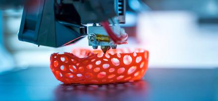
The bioprinting phase is a particularly intricate process involving the loading of bioink into a bioprinter, guided by the software’s 2D design, to fabricate a 3D structure on a scaffold. The complexity of this stage arises from the necessity of forming diverse cell types that faithfully mimic the targeted tissues and organs. Bioink, systematically deposited layer by layer, comprises living cellular components, growth-supporting substances and a reinforcing framework such as hydrogel, derived from either natural or synthetic biomaterials. The choice of bioink is a critical consideration, taking into account its physicochemical attributes, biocompatibility and potential for large-scale production.
Our experience
In the course of our preliminary results, the focal point shifted towards obtaining organoids for the purpose of studying extracellular communication through exosomes. Organoids, aptly described as ‘in vitro mini-organs,’ are meticulously crafted by isolating cells from tissues or differentiating them from stem cells, thereby forming functional 3D structures that authentically mimic the complexities of original tissues. The process of organoid generation delves into the early biological concept of cell dissociation and reaggregation. Given the limitations of 2D adherent cell culture in representing the original structure and function of organs, 3D organoids have risen to prominence as a superior model for clinical applications in various organs compared with their 2D counterparts.

Our project started with the digestion of a mouse thyroid gland, leading to the isolation of cells. These cells, subsequently embedded in a bioink based on hyaluronic acid, underwent the intricate process of 3D bioprinting. The resulting diminutive constructs were then cultured in vitro for a span of up to 3 weeks, facilitating the seamless reorganisation of cells into compact, organic-like clusters. The analysis of gene expression profiles, conducted through sec mRNA, unequivocally confirmed the sustained maintenance of an adult phenotype in the cells. Following this, our research trajectory pivoted towards the isolation and comprehensive characterisation of extracellular vesicles (EVs) produced within these intricate organic structures.
The crucial role of extracellular vesicles
Extracellular vesicles, fulfilling a pivotal role in the exciting realm of endocrinology, represent a novel dimension of intercellular communication. Released by diverse cell types, these membrane-bound structures serve as messengers traversing the extracellular space, ferrying proteins, lipids and nucleic acids. In the world of endocrinology, EVs facilitate the transfer of signalling molecules between endocrine cells, thereby wielding influence over hormonal regulation and cellular responses.
A particularly profound aspect of EVs in endocrinology lies in their ability to mediate long-distance communication between glands and tissues, seamlessly complementing the traditional conduits of hormonal transmission. EVs contribute significantly to the fine-tuning of endocrine processes, delivering bioactive molecules − including microRNAs and hormones − to target cells, thereby orchestrating the modulation of gene expression and cellular functions. The study of EVs in endocrinology not only sheds new light on our understanding of diseases such as diabetes, thyroid disorders and hormonal imbalances, but also unveils promising avenues for diagnostic and therapeutic interventions. The cargo carried by these vesicles serves as a reflective mirror, offering valuable insights into the physiological state of the originating cells.
In conclusion, EVs emerge as a captivating frontier in endocrinological research, unravelling the intricacies of intercellular communication and extending insights beyond the confines of traditional hormonal pathways. Delving deeper into EV-mediated signalling holds the promise of groundbreaking advancements in both the understanding and treatment of endocrine disorders, showcasing the potential for transformative breakthroughs in the field. As the tapestry of knowledge surrounding EVs unfolds, the prospect of ushering in a new era of precision medicine for endocrine conditions becomes increasingly apparent.
Barbara Zavan Department of Translational Medicine, University of Ferrara, Italy
1. Jeon et al. 2023 Advanced Drug Delivery Reviews https://doi.org/10.1016/j.addr.2023.114959
2. Ogundipe et al. 2022 Tissue Engineering Part A https://doi.org/10.1089/ten.TEA.2021.0221
3. Chae et al. 2021 Gland Surgery https://doi.org/10.21037/gs-21-264
4. Delcorte et al. 2022 Biomedicines https://doi.org/10.3390/biomedicines10102585
5. Chen et al. 2023 International Reviews of Immunology https://doi.org/10.1080/08830185.2022.2057482


