5 minute read
Idiopathic intracranial hypertension: a complex

from Endocrine Views (ESE News) Autumn 2022 (News and Views European Society of Endocrinology) Issue 49

IIH: a complex metabolic disease?
Growing evidence suggests that idiopathic intracranial hypertension (IIH) is a systemic metabolic disease. This recent study examined the glucocorticoid (GC) metabolome associated with the condition.1
Advertisement
Idiopathic intracranial hypertension is an increasingly prevalent condition due to the obesity epidemic. It primarily affects women of reproductive age who have obesity.2
People with IIH present with raised intracranial pressure (ICP), chronic headaches and papilloedema which, if left untreated, can cause blindness. Recent evidence suggests that IIH is a disease of metabolic dysfunction; those with the condition have abdominal obesity, with insulin and leptin resistance in excess of that conferred by obesity.3 Adipose tissue in IIH is transcriptionally primed for increased calorie intake, with a unique depot-specific lipogenic profile.3 Additionally, women with IIH have a distinct androgen excess phenotype, consisting of increased serum and cerebrospinal fluid testosterone.4 This metabolic phenotype probably underpins the recently identified clinical findings of a higher risk of cardiovascular disease and type 2 diabetes mellitus compared with control subjects with obesity.2 Additionally, the finding of decreased birth rates in IIH, with increased risk of gestational diabetes and pre-eclampsia, may stem from underlying metabolic dysfunction.5
Excess GCs are a known determinant of metabolic disease. Indeed, GCs have previously been implicated in the pathophysiology of IIH; a significant correlation has been observed between the change in 11β-hydroxysteroid dehydrogenase type 1 (11β-HSD1) activity and the change in ICP.6 No studies have investigated the basal GC phenotype in IIH so, in this recent research, we aimed to define the IIH GC metabolome and determine the origin of any findings.1
Multiple complementary studies
We took meticulously phenotyped women with IIH, and controls matched for age, sex and body mass index.1 We compared their urinary steroid profiles using mass spectrometry. In our randomised control trial of bariatric surgery in IIH, we assessed the urinary steroid profile of the participants at baseline and 12 months.7 Additionally, adipose biopsies from patients with IIH and controls were incubated with cortisone to assess 11β-HSD1 activity.
Our findings
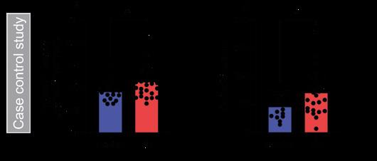
We demonstrated that people with IIH have increased 11β-HSD1 activity and increased urinary cortisol compared with controls (see Figure). In addition, 5α-reductase activity, a marker of GC clearance, was increased in IIH. No other steroids that we assessed were found to be altered in patients with IIH. There was no increase in total GC excretion, indicating no overt GC excess. This suggests a tissue-specific dysfunction, rather than adrenal or hypothalamic–pituitary–adrenal axis dysfunction.
Weight loss has been proven to induce remission of IIH. Urinary steroid analysis before and after weight loss demonstrated that, following bariatric surgery, there was a reduction in 11β-HSD1 activity. This change in 11β-HSD1 activity correlated with change in lumbar puncture opening pressure, which is a measure of IIH disease activity. This could suggest that GCs have a role to play in the regulation of ICP in the context of IIH.
Subsequently, subcutaneous adipose analysis showed that patients with IIH have a 300% higher 11β-HSD1 activity compared with controls.
In conclusion
Taken together, our data suggest that people with IIH have increased 11β-HSD1 activity both systemically and in adipose tissue. This supports growing evidence that IIH is a complex metabolic disease and not just a neuro-ophthalmological disease. In the future, an endocrine approach to management may become increasingly pertinent.
Connar SJ Westgate, Susan P Mollan and Alexandra J Sinclair Institute of Metabolism and Systems Research, University of Birmingham, and University Hospitals Birmingham NHS Foundation Trust, UK
REFERENCES
1. Westgate et al. 2022 European Journal of Endocrinology187 323−333. 2. Adderley et al. 2019 JAMA Neurology76 1088−1098. 3. Westgate et al. 2021 JCI Insight6 e145346. 4. O’Reilly et al. 2019 JCI Insight4 e125348. 5. Thaller et al. 2022 BJOG doi: 10.1111/1471-0528.17241. 6. Sinclair et al. 2010 Journal of Clinical Endocrinology & Metabolism95 5348−5356. 7. Mollan et al. 2021 JAMA Neurology78 678−686.
Insights from the Editor
Idiopathic intracranial hypertension (the former label ‘benign’ has, quite rightly, been dropped) is severe and debilitating. Disordered GC metabolism and, particularly, altered 11β-HSD1 activity have been implicated in its aetiology, but the picture may be complex. 11β-HSD1 catalyses interconversion of inactive corticosterone and active cortisol. It is believed generally to activate GC in vivo, though, in the correct conditions, it can catalyse inactivation. Activating activity is increased in choroid plexus in IIH, but an 11β-HSD1 antagonist only minimally lowered intracranial pressure. Moreover, active Cushing’s syndrome is not clearly associated with IIH, while numerous reports describe IIH on acute GC lowering or chronic deficiency. This careful study adds further pieces to the puzzle by addressing 11β-HSD1 activity in peripheral tissues in IIH, showing increased systemic activity, and markedly increased activity in adipose tissue. Levels were reduced by weight loss, and correlated with reduced intracranial pressure. This suggests that disordered GC metabolism may be important, not only in IIH itself, but also in the associated metabolic abnormalities. This creates new opportunities for treatment, though the discrepant observation of IIH in GC deficiency emphasises the importance of drilling down on 11β-HSD1 activity tissue by tissue. More studies are awaited with interest.
Robert Semple, Deputy Editor, EJE

IMAGE REFERENCE: Case control study demonstrating increased (A) 11β-HSD1 activity and (B) urinary cortisol in IIH. 5α-THF+THF/THE, 5α-tetra hydocortisol+tetrahydrocortisol/ tetrahydrocortisone; *P<0.05. Reproduced under CC BY 4.0 licence (https:// creativecommons.org/licenses/by/4.0) from Westgate et al. 1 https://doi.org/10.1530/ EJE-22-0108 ©The Authors 2022.


