In this issue: 02 Guest editorial 12 Your SVS Is endovascular Meet the dominance 2026 SVS undermining surgical election training, or are candidates trainees adapting? 14 The Outpatient 08 S CVS Payment for the new LER Reducing unwanted codes: It’s not just about care through better the work communication
MAY 2026 Volume 22 Number 4
THE OFFICIAL NEWSPAPER OF THE
www.vascularspecialistonline.com
STUDY REVEALS HIGHER PAD DETECTION RATES IN WOMEN DESPITE SIMILAR RISK PROFILES By Killian Meara
WOMEN MAY BE MORE LIKELY than men to have undiagnosed peripheral artery disease (PAD) at the time of screening, despite meeting similar risk criteria, according to new data from a community-based screening program targeting underserved populations. The study analyzed participants in the CHAMPIONS initiative, a program designed to deliver free cardiovascular and limb-focused screening in vascular deserts, or areas where access to specialty care is limited. “Sex-based disparities in vascular disease are not new,” said Leigh Ann O’Banion, MD, senior author on the study and associate clinical professor of surgery at the University of California, San Francisco Fresno. “They are consistently described in registry, claims and health system data, where females with PAD are more likely to be diagnosed later, present with less typical symptoms and experience worse functional and limb outcomes. The limitation is that most of those datasets capture patients only after they have entered the health care system.” The retrospective analysis included 736 participants screened between 2022 and 2025. Investigators used guideline-based criteria to classify participants as at risk for PAD and performed toe
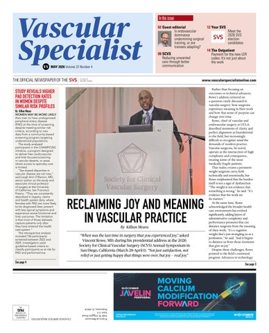
RECLAIMING JOY AND MEANING IN VASCULAR PRACTICE “When was the last time in surgery that you experienced joy,” asked Vincent Rowe, MD, during his presidential address at the 2026 Society for Clinical Vascular Surgery (SCVS) Annual Symposium in San Diego, California (March 28-April 1). “Not just satisfaction, not relief or just getting happy that things were over, but joy — real joy.”
Rather than focusing on outcomes or technical advances, Rowe’s address centered on a question rarely discussed in vascular surgery: how surgeons experience meaning in their work and how that sense of purpose can change over time. Rowe, chief of vascular and endovascular surgery at UCLA, described moments of clarity and perfect alignment as foundational to the field, but increasingly difficult to recognize amid the demands of modern practice. Vascular surgeons, he noted, operate at the intersection of high complexity and consequence, treating some of the most medically fragile patients. That reality creates a persistent weight surgeons carry, both technically and emotionally, but Rowe emphasized that the burden itself is not a sign of dysfunction. “The weight is not evidence that something is wrong,” he said. “It’s evidence that the work we do matters.” At the same time, Rowe acknowledged the broader health care environment has evolved significantly, adding layers of administrative complexity and performance pressures that can distance surgeons from the meaning of their work. “It’s a cognitive weight that’s just strangling us as a profession,” he said. “And it begins to distance us from those moments that give us joy.” Despite these challenges, Rowe pointed to the field’s continued progress. Advances in technology
See page 4
See page 3
PERIPHERAL
MOVING CALCIUM MODIFICATION In the US: Rx only. Prior to use, please reference the Important Safety Information www.shockwavemedical.com/ IFU for more information on indications, contraindications, warnings, precautions and adverse events. Please contact your local Shockwave representative for specific country availability. SPL 79741 Rev. A
Vascular Specialist 9400 W. Higgins Road, Suite 315 Rosemont, IL 60018
IM
PAID
CHANGE SERVICE REQUESTED
PRESORTED STANDARD MAIL U.S. POSTAGE
2
Vascular Specialist | May 2026
GUEST EDITORIAL Medical Editor Malachi Sheahan III, MD Associate Medical Editors Bernadette Aulivola, MD | O. William Brown, MD | Elliot L. Chaikof, MD, PhD | Carlo Dall’Olmo, MD | Alan M. Dietzek MD, RPVI, FACS | John F. Eidt, MD | Robert Fitridge, MD | Dennis R. Gable, MD | Linda Harris, MD | Krishna Jain, MD | Larry Kraiss, MD | Joann Lohr, MD | James McKinsey, MD | Joseph Mills, MD | Erica L. Mitchell, MD, MEd, FACS | Leila Mureebe, MD | Frank Pomposelli, MD | David Rigberg, MD | Clifford Sales, MD | Bhagwan Satiani, MD | Larry Scher, MD | Marc Schermerhorn, MD | Murray L. Shames, MD | Niten Singh, MD | Frank J. Veith, MD | Robert Eugene Zierler, MD Resident/Fellow Editor Saranya Sundaram, MD Executive Director SVS Kenneth M. Slaw, PhD Senior Director for Public Affairs and Advocacy Megan Marcinko, MPS Managing Editor Killian Meara kmeara@vascularsociety.org Assistant Editor Maria Gifford Design Madison Spadafino Advertising Nicole Schmitz nschmitz@vascularsociety.org Letters to the editor vascularspecialist@vascularsociety.org
Published by the Society for Vascular Surgery
Vascular Specialist is the official newspaper of the Society for Vascular Surgery and provides the vascular specialist with timely and relevant news and commentary about clinical developments and about the impact of healthcare policy. The ideas and opinions expressed in Vascular Specialist do not necessarily reflect those of the Society. The Society for Vascular Surgery will not assume responsibility for damages, loss, or claims of any kind arising from or related to the information contained in this publication, including any claims related to the products, drugs, or services, or the quality or endorsement of advertised products or services, mentioned herein. | The Society for Vascular Surgery headquarters is located at 9400 W. Higgins Road, Suite 315, Rosemont, IL 60018. | POSTMASTER: Send changes of address (with old mailing label) to Vascular Specialist, Subscription Services, 9400 W. Higgins Road, Suite 315, Rosemont, IL 60018. | RECIPIENT: To change your address, e-mail vascularspecialist@ vascularsociety.org | For missing issue claims, e-mail vascularspecialist@vascularsociety.org | Vascular Specialist (ISSN 1558-0148) is published by the Society for Vascular Surgery. | Printed by Ironmark | ©Copyright 2025 by the Society for Vascular Surgery
Follow Vascular Specialist on socials vascularspecialist vascular-specialist-online vascularonline
The kids are alright By Benjamin J Pearce, MD
“I know I gotta get away and I know if I don’t, I’ll go out of my mind; better leave her behind with the kids, they’re alright, the kids are alright.” - Pete Townshend, The Who
Mankind harnesses the power of fire; the assembly line makes automobile travel feasible for the common person and aneurysms can be repaired with endografts. Maybe the latter does not quite have the impact on mankind as a whole but within the sphere of vascular surgeons, it certainly ranks up there with Neil Armstrong toeing the surface of the moon. And, as with all disruptive innovation (thanks Professor Christensen), there are always, always, unintended consequences. In the case of EVAR, this has come at the expense of the single greatest badge of honor carried by a vascular surgeons — the open aneurysm repair. At the American College of Surgeons meeting in 2017, our colleagues from Michigan used Medicare claims data to demonstrate that in a four-year period (2010-2014), graduating vascular trainees saw a decline in median open AAA repairs of 10 to 6.4 and, further, that half (egads!) would graduate with less than five open AAA in their training. Much like the first person splashed with mud by a Model T, surgeons shouted at the passing endovascular revolution that we must put a stop to this menace. And the mudstained clamoring persists to this day. But as a wise man once said, repeatedly, are we worrying about a problem we don’t have? First, what is the number of open aneurysm repairs needed to be considered “proficient” at the procedure? Is it really the 10 in 2010? 20? 30? What are the components of aneurysm repair that are so critical to perform this hallmark feat of mankind? Is it the evaluation of a CT to know if it can be done and where the problems may lie? In the exposure of proximal and distal control? The ability to move the case along to avoid unnecessary insensible fluid loss
and exposure of the patient to those dastardly foes sitting at the head of the bed doom scrolling Instagram while the patient slowly gets more and more tachycardic? Lots of questions and many of the answers are uncomfortably gray and likely surgeon specific. I can only speak to my own experience. According to the ACGME case logs, I performed 42 open aneurysms of all types during my two-year fellowship (this doesn’t include the cases done as a surgery resident as those are not available online — evidently they burned all that parchment years ago). I can tell you for a fact as sure as the sun will rise tomorrow in the East and
I did not sleep a wink the night before I did my first open aneurysm repair as faculty. Or my second, third and so on. And despite doing more open aortic cases in six months than I did in those two years of fellowship, I continue to learn things about aneurysm repair even to this day. Coach K is the greatest NCAA basketball coach of all time, I did not sleep a wink the night before I did my first open aneurysm repair as faculty. Or my second, third and so on. And despite doing more open aortic cases in six months than I did in those two years of fellowship, I continue to learn things about aneurysm repair even to this day. One wise man taught me a nifty trick to staple the diaphragm when opening the chest and a different one told me never to operate on the aorta without talking to some family member first. A wise woman gave me some critical advice on suture technique and conduit choice in fixing a ruptured aneurysm in a 14-year-old. I haven’t really slept hard before any of those cases. I would posit that proficiency in aneurysm repair is moving target and aspirational. IMHO (as the kids say), there is also a point at which you can only learn so much aortic surgery as a trainee. At some point, the hard lessons about retroaortic renal veins, mycotic fields and safely dissecting in redo cases comes with being the person whose name is on the consent. The good news is that we have help coming on the horizon. The advent of the entrustable professional activity (EPA) paradigm should provide a framework for providing this level of autonomy to trainees at high volume centers to have some of that critical attending level experience while still having a support structure to make mistakes. Which brings us to our second point, not everyone is missing out on open aortic experience — and that is ok. In response to the outcry about trainees possibly graduating with less than five (egads! again) open aneurysm repairs, centers
3
www.vascularspecialistonline.com
FROM THE COVER:
RECLAIMING JOY AND MEANING IN VASCULAR PRACTICE continued from page 1 and technique have expanded treatment options, allowing surgeons to care for increasingly complex patients without worsening outcomes. “We’re treating the same patients that are sicker and we’re treating them with the same morbidity and mortality,” he said. “This is something that we should really be proud of.” However, Rowe argued professional success itself can contribute to a gradual erosion of fulfillment. “The erosion of joy rarely occurs when we’re struggling,” he said. “Perhaps most importantly, it doesn’t occur in failure, it occurs in our success. It occurs when the practice is thriving. When we have outcomes that are strong and responsibilities are expanding. It’s in that space, the very professional achievement, that the distance can begin to emerge.”
“The weight is not evidence that something is wrong, it’s evidence that the work we do matters.” VINCENT ROWE, MD
Rowe also discussed burnout and cited data showing a substantial portion of surgeons and nearly half of trainees experience symptoms. “It’s rarely something that occurs dramatically,” he said. “It’s something that seems to drift away from us and take that joy away from our profession.
SCVS
New data support tailored revascularization strategies for patients with CLTI By Maria Gifford A new study comparing autologous vein bypass to endovascular intervention as the initial revascularization strategy for patients with chronic limb-threatening ischemia (CLTI) requiring infrapopliteal treatment found when patients were carefully matched for comorbidities, outcomes between the two approaches were largely similar, pointing to the importance of individualized treatment selection. The findings were presented at the 2026 Society for Clinical Vascular Surgery (SCVS) Annual Symposium in San Diego, California (March 28-April 1), offering a real-world counterpoint to the sometimes-conflicting data from landmark randomized trials. “The results of the BEST-CLI and BASIL-2 trials were somewhat contradictory and a number of studies have since been published to see the real-world experience,” said Hasan Dosluoglu, MD, senior author on the study and chief of the division of vascular surgery at the State University of New York. “Our experience reflects all comers to our practice with full follow-up in a vascular surgeon-led program in a VA setting who adopted advanced endovascular interventions early.” The study drew from 20 years of institutional experience from January 2004 through March 2024, capturing patients treated under a fully individualized revascularization philosophy. Bypass was selected in just 22% of cases, with endovascular intervention used for the remainder, reflecting a patient-by-patient decision-making framework grounded in clinical presentation, comorbidities, anatomic characteristics and autologous vein graft availability. In practice, that individualization played out across a wide clinical spectrum. “There were patients who had complex anatomy and had endovascular revascularization,
and there were patients who had COPD and CKD who had bypass,” said Dosluoglu. On unmatched analysis, the bypass group fared better than those who underwent endovascular-first revascularization. But when patients were propensity-matched for comorbidities, the gap narrowed considerably, with outcomes proving comparable between the two strategies despite residual differences in disease complexity and patient age that persisted even among matched pairs. A particularly striking finding from the matching process itself illuminated just how different the two patient populations tend to be. Only 21% of patients in the endovascular group could be successfully matched to bypass patients, compared to 72% of patients in the open surgery group — a disparity reflecting how profoundly comorbidity burden shapes real-world treatment selection. “The overall experience suggests that patients who were elected for bypass did better than patients who had endovascular first,” said Dosluoglu. “However, when matched for comorbidities, we found that the outcomes aren’t that different, despite the remaining differences in matched groups such as complexity of disease and age.” The findings carry practical implications for how CLTI programs are structured. Rather than defaulting to a specialty-driven or reimbursement-influenced approach, the data support a patient-centered model in which each modality is deployed where it fits best. “The optimum revascularization should not be determined by the individual provider’s specialty or type of reimbursement but should be very much centered on the individual patient,” said Dosluoglu. “Bypass has a definite role in these patients and it’s probably appropriate as the first line in about 20% of patients in our experience, which
A sense that the day has become a little more transactional than meaningful.” To address this, Rowe said that re-centering the patient, honoring the profession’s weight and rediscovering one’s “why” are three key strategies for reconnecting with purpose. He also encouraged surgeons to embrace the emotional impact of difficult cases rather than suppress it. “The question is not whether vascular surgery is hard — it is,” said Rowe. “The question is whether we’ll practice it aligned with the reason we chose it. Because if we do, even though the weight is still there, even though the work doesn’t get any easier, it becomes something we can carry with meaning. And in those moments, that feeling comes back. Not every day, not every case, but enough. Enough to remind us why we chose this.”
involves a vascular surgeon-only practice with no financial gain with either approach.” While that 20% figure may not translate uniformly across all centers, it offers a working benchmark that any CLTI program can use to gauge whether it’s performing too many or too few bypass procedures. Dosluoglu argued that the field needs to broaden how it measures success in this patient population. “We need more studies on patient-centric outcomes such as quality of life, wound healing and maintenance of independent living rather than just amputation-free survival,”
said Dosluoglu. He added that the impact of reimbursement structures on outcomes also warrants dedicated study. “Deciding on the optimum revascularization strategy is a highly individualized process and open versus endovascular versus hybrid options aren’t competitive but complementary,” said Dosluoglu. “Treat the patient, with the least damage and maximum benefit.”
vasculearn.vascular.org
NEW! Automatic ABS Credit Transfer for JVS and JVS-VL Articles
Start earning CME for articles now!
Explore the new CME opportunity with the Journal of Vascular Surgery.
4
Vascular Specialist | May 2026
GUEST EDITORIAL THE KIDS ARE ALRIGHT
➽
such as Boston University, Duke, UAB and the Cleveland Clinic have all published articles demonstrating that open aortic volumes are holding steady at more than 20 open repairs and, optimistically, are increasing if one incudes EVAR explant and open repair of occlusive disease at levels commensurate to the “golden age” of vascular fellowship before EVAR dominance. Some may argue that these papers represent a “flex” (as the kids say) and not “the real world” experience of vascular surgeons. However, Medicare claims data would argue otherwise. Consistently, EVAR represents > 80% of aneurysm repairs in the United States and that number has only risen to match the ability to rise up the aorta with branched and fenestrated devices. As recent perusing of SVS Connect reveals, we actually all agree that being able to fix aneurysms open is the right thing to do for many patients, especially those with challenging anatomy and many years at risk. Even ChatGPT will tell you that EVAR dominates open repair in the short term by both complications and cost, but in the long game the costs of reintervention and surveillance combined with the complications and late aortic mortality makes open repair the more durable choice for patient living eight years. These cases should and can be done open. A solid open, non-re-operative aneurysm repair is a great case for a newly minted vascular surgeon to
continued from page 2
Absolutely, the role of the vascular surgeon in the modern American community is to be there to save a life when a trocar goes missing. Or to expose a spine so an orthopedist doesn’t biopsy the cava. I’ve got news for those not currently operating at major trauma centers; the modern vascular trainee is doing that, a lot. do — with their senior partner. It is my understanding that this has been the modus operandi for community practice since the inception of the vascular certificate in special training. All the other problems, mycotic, thoracoabdominal and infected prior repairs, are best suited in centers who do these cases routinely anyway. Medicare claims and common sense would indicate that that is already being done. Which brings us to our last point, you learn a lot more than just fixing an aneurysm when you do an open aneurysm repair. Amen and pass the communion plate.
Absolutely, the role of the vascular surgeon in the modern American community is to be there to save a life when a trocar goes missing. Or to expose a spine so an orthopedist doesn’t biopsy the cava. I’ve got news for those not currently operating at major trauma centers; the modern vascular trainee is doing that, a lot. This isn’t your parent’s general surgery residency anymore. They have had their own disruptive events between non-operative trauma protocols and using the robot to do everything including entering their case logs from what I gather. The control and reconstruction of blood vessel injury is increasingly falling to the vascular team and almost exclusively the “intra-op” consult for bleeding. Fortunately, this isn’t just the “back in my day” rantings of an aging aortic surgeon, our colleagues at LSU just presented this very data at the recent meeting of the SCVS. They looked at all things pertaining to blood vessels in the retroperitoneum — open AAA, thoraco-abdominal, aortobifems, mesenteric, bypass, spine exposures and trauma. Comparing trainees from 2021-2024 for the modern cohort against 2007-2010 as controls, the modern vascular integrated resident had 52.3 open aortic cases, the modern fellow 48.6 and the historic fellows 47.7. So, maybe as The Who said, the kids are alright after all. Maybe the issue lies with how we see it, or as Cheap Trick would say, “Mommy’s alright, Daddy’s alright, they just seem a little weird.”
FROM THE COVER:
STUDY REVEALS HIGHER PAD DETECTION RATES IN WOMEN DESPITE SIMILAR RISK PROFILES continued from page 1
brachial index (TBI) testing in those individuals. The primary objective was to evaluate whether sex was associated with being labeled at risk and with having PAD detected through objective hemodynamic testing. The program was designed with a prevention-focused approach rather than identifying candidates for procedures. “None of these participants were being screened to determine whether they needed invasive intervention,” said O’Banion. “The goal is prevention: to detect atherosclerotic disease earlier, identify cardiovascular risk and intervene with education, medical therapy and risk factor modification to halt progression — long before latestage disease and revascularization enter the picture.” The most notable finding was a disconnect between risk classification and actual disease detection. “Males and females were similarly likely to meet guideline-based criteria for being at-risk for PAD,” said Guistinna Tun, first author on the study. “Yet, among those who were tested and at risk, females were more likely to have PAD based on TBI. PAD was confirmed in 10.9% of at-risk females versus just 2.8% of at-risk males. It’s a stark difference.” The findings also highlight the role of socioeconomic disadvantage, which was more prevalent among women and may contribute to delayed diagnosis. “Females in this cohort carried substantial socioeconomic disadvantages,” said Tun. “They were less likely to be employed. Even if they were employed, they made the least annual income compared to men. They were more likely to report grade school as being their highest level of education. This matters because social risk and access barriers shape whether risk factors are being treated early and whether symptoms lead to testing.”
“We need better strategies for earlier detection in females, whether it be through education or reshaping how clinicians interpret leg symptoms and how our community understands PAD.” LEIGH ANN O’BANION, MD
O’Banion said the data suggest a need to rethink how PAD risk is evaluated in women, particularly those who present with atypical symptoms. “Females are more likely to have leg symptoms that do not fit the classic claudication story and those symptoms are more likely to be attributed to nonvascular causes,” she said. “The result is a lower index of suspicion, fewer objective tests and a longer runway before diagnosis. Our data suggest that even when males and females meet similar riskbased screening thresholds, females may already have more established atherosclerotic disease when objective testing finally happens.” Community-based screening programs like CHAMPIONS may help address these disparities by improving access to early detection. “In vascular deserts, the barriers are not subtle: geographic distance to specialty care, limited access to noninvasive testing, language barriers, financial insecurity and inconsistent primary care access,” said O’Banion. “When the system requires multiple steps before a patient reaches objective testing, the patients most likely to fall through are the ones with the fewest resources.” Tun said the findings ultimately reinforce the importance of early detection and prevention. “We need better strategies for earlier detection in females, whether it be through education or reshaping how clinicians interpret leg symptoms and how our community understands PAD,” she said. “We also need more community screenings. In terms of taking the extra step, it would be being able to partner with a reliable bridge to longitudinal care. Something that often comes up is that we get screening results, there’s something wrong, but what do we do next? How do we ensure that our patients are being connected with care and that they’re being taken care of beyond the day of the screening?”
VISIT US AT BOOTH 306
LOUD. CLEAR. CORDLESS. Vascular therapeutics have evolved over the past 50 years, why hasn’t your Doppler?
VascuChek is the first cordless, handheld vascular Doppler FDA-cleared for evaluating intraoperative, subcutaneous blood flow. With two integrated high-definition speakers and a NEW Bluetooth® external speaker, VascuChek delivers loud and clear audio feedback even in the busiest OR.
Schedule a demo at VascuChek.com or call 866-484-9307
R
Our disposable surgical probe tip features an aseptic sheath to cover the transceiver and is FDA-approved for use in the sterile field.
6
Vascular Specialist | May 2026
ADVERTORIAL | SPONSORED BY SHOCKWAVE PERIPHERAL IVL
Advancing below-the-knee intervention in CLTI with Shockwave Javelin Peripheral IVL
Charles Bailey, MD, George Adams, MD and Vanessa Adams, BS, share their experience with Shockwave Javelin, the first-of-its-kind forward intravascular lithotripsy (IVL) technology that is enabling further interventional treatment for patients suffering from chronic limb-threatening ischemia (CLTI).
P
eripheral artery disease (PAD) affects millions worldwide and is associated with reduced blood flow to the legs, impaired quality of life and increased risk of cardiovascular events. CLTI is the most advanced stage of PAD and carries substantial rates of amputation and mortality. Managing heavily calcified or severely narrowed lesions remains a persistent clinical challenge, particularly when device delivery is limited. The Shockwave Javelin Peripheral IVL catheter is designed on a novel Forward IVL Platform to modify calcium in these complex lesions, enabling treatment in vessels where conventional devices may be difficult to advance and supporting a broader range of endovascular interventions.
CASE REPORT 1 Patient presentation A 67-year-old man returned to the vascular clinic with left foot nocturnal rest pain and recurrence of a superficial ulceration to the tip of Distal AT and the fourth toe. Medical DP arteries history was notable for diabetes mellitus, hypertension, hyperlipidemia, congestive heart failure with reduced ejection fraction (LVEF 40-45%) and prior tobacco abuse. Surgical history included a prior left leg femoral-to-proximal anterior tibial artery (AT) prosthetic bypass graft (August 2025). In-office vascular lab testing revealed absence of great toe pressure and interval occlusion of the distal AT and dorsal pedal (DP) arteries (Fig 1). His bypass graft remained patent,
though with diminished peak-systolic velocities throughout. Given the clinical findings of CLTI (CLTI-ischemic rest pain, recurrent digital ulceration) and a threatened surgical bypass graft at risk of thrombosis with diminished ingraft velocities and distal outflow tract occlusion, the patient was consented for a left lower extremity angiogram with possible endovascular intervention. The goal of revascularization included improved forefoot perfusion for CLTI, and to improve the outflow tract to support bypass graft long-term assisted patency.
2
Procedural details With pre-procedural imaging evidence of distal AT and infra-malleolar occlusions, traversed intraluminal using an 0.014 an antegrade ultrasound-guided ipsilateral Hydrophilic guidewire with a 3.5g tip left femoral percutaneous access was load and supportive 2.3-Fr. by 90-cm performed. Diagnostic angiogram crossing catheter. The wire and catheter confirmed patency of the combination were advanced femoral-to-proximal AT across the pedal-plantar loop prosthetic bypass graft, and positioned in the proximal without anastomotic or in-graft plantar artery branches, with stenoses. The AT was patent wire exchanged for a 0.014 along its proximal and mid hydrophobic guidewire with a segments, with an occlusion soft 0.7g-0.8g tip load to support noted above the malleolus and device delivery. In consideration extending into the DP artery of small caliber vessels and and pedal-plantar loop. Bypass medial calcifications throughout graft patency was maintained occluded segments, the decision via a distal branch with was made to perform plaque collateral flow perfusion to the and vessel wall modification Shockwave Javelin in the AT with the Shockwave Javelin level of the posterior tibial and around the pedal loop artery (PT) bifurcation into Peripheral IVL catheter. From medial (MPA) and lateral plantar (LPA) experience, the competitive advantage of branches (Fig 2). A 5-Fr. by 55-cm sheath Javelin over traditional plain angioplasty was advanced. Systemic heparinization balloons in this type of anatomy includes was administered and activated clotting its low crossing profile and ability to times monitored (ACT>250 seconds). safely modify plaque beyond the tip,9,10 creating a small The distal AT and DP lesions were lumen (1.5mm) similar to native vessel reference diameters and enabling further treatment. All the available 120 pulses were administered across the occlusive segment, with an even distribution of pulses delivered during antegrade Post Shockwave Javelin angio passage and retrograde catheter retrieval. A post-lithotripsy angioplasty was
1
performed throughout with use of a 1.5mm balloon across the distal DP and pedal-plantar loop and a 2mm balloon angioplasty of the distal AT and proximal DP. Completion imaging demonstrating restored patency through the distal AT, DP and pedal-plantar loop intervention sites noting enhanced opacification of branch vessels throughout all levels of the foot, without noted residual stenosis, contrast extravasation or dissection. Vascular physical exam findings with restored multiphasic Final angio post-Shockwave doppler signal Javelin and PTA at distal AT and DP vessels. Clinically, the patient noted complete resolution of ischemic rest pain symptoms with continued healing of digital ulcers. Surveillance arterial duplex at one-month postoperatively demonstrated patency of the DP with multiphasic waveform and restoration of a great toe pressure (71mmHg) adequate for wound healing in CLTI patient (Fig 2). CHARLES BAILEY is a board-certified vascular surgeon at Emory Healthcare and an assistant professor of surgery at Emory University School of Medicine. He specializes in limb preservation, peripheral arterial disease and complex vascular care.
7
www.vascularspecialistonline.com
ADVERTORIAL | SPONSORED BY SHOCKWAVE PERIPHERAL IVL CASE REPORT 2
Procedural Details
Roughly one to three million patients suffer from life limiting claudication in the United States8 and approximately 5-10% of these patients evolve to critical limb ischemia,4,5 increasing their risk for amputation. Understanding that many lesions belowthe-knee are chronic total occlusions comprised of calcium,6,7 we effectively utilized the Shockwave Javelin Forward IVL device to modify the calcific plaque and cross the chronic total occlusion (CTO), thus reducing the risk of dissection, perforation and ultimately amputation.
Patient presentation A 79-year-old male with a history of coronary artery disease, diabetes, hyperlipidemia, chronic obstructive pulmonary disease and hypertension presented with a failing to heal left great toe wound. He stated that over the last year his exercise capacity has significantly decreased because of a cramp like pain in both calves to the point he could not walk more than 100 yards without having to stop. This issue peaked approximately three months ago when he stumped his left great toe on the edge of the bed opening a wound. He was seen and treated by podiatry with little success in healing the wound. His left toe brachial index (TBI) was 0.2 with blunted monophasic waveforms — therefore, referred for vascular consultation. Considering the location of the wound and the angiosome related artery1,2,3 our focus for endovascular intervention was the anterior tibial artery (ATA). Abdominal aortography and runoff performed of the left lower extremity showed a widely patent left common iliac, external iliac, common femoral, superficial femoral, deep femoral and popliteal arteries. Below-the-knee (Fig 1 and 2), the AT artery showed a 99% proximal stenosis and occlusion abovethe-ankle. The posterior tibial (PT) artery showed an occlusion above-the-ankle. The peroneal artery extended above the ankle providing collaterals flow to the distal PT and the medial and lateral plantar arteries. There was little to no flow to the left great toe for healing.
1
2
Endovascular intervention followed, a 5F 90cm sheath was placed to the level of the left popliteal artery and anticoagulating with heparin achieving and maintaining an activated clotting time >250 seconds. We crossed the proximal AT stenosis with a guidewire and performed percutaneous transluminal angioplasty with a 3x200mm noncompliant balloon (Fig 3) for two minutes, resulting in <20% residual and no flow limiting dissections. We then crossed the distal left AT occlusion with a 0.14, 300 cm length hydrophilic coated 3 guidewire to the left great toe digital artery. A low profile 1x15mm balloon would not pass across the distal AT occlusion and therefore chose to use Shockwave Javelin. We slowly advanced the Forward IVL device to the level of the left digital artery utilizing the entire 120 pulses (Fig 4), modifying the calcium and creating a small lumen. Successful percutaneous transluminal angioplasty (PTA) was performed using a 1x15mm balloon. We then used adjunctive 4 non-compliant balloon angioplasty starting with a 2mm followed by a 2.5mm balloon to effectively dilate the vessel from the left digital artery to the distal AT resulting in less than 20% residual and improved flow to the toe wound (Fig 5). 5 The patient’s left great toe wound successfully healed within a period of two weeks with the aid of podiatry. Utilizing the angiosome concept1,2,3 allowed us to select the vessel which supplied direct in-line flow to the wound and the AT artery, which promoted healing. GEORGE ADAMS is a board-certified interventional cardiologist at UNC Rex Hospital and a clinical associate professor of medicine at the University of North Carolina at Chapel Hill. He specializes in complex peripheral vascular interventions, amputation prevention, and advanced cardiovascular care. VANESSA ADAMS is an undergraduate at NC State and on the research staff at UNC Rex Hospital.
“We effectively utilized the Shockwave Javelin Forward IVL device to modify the calcific plaque and cross the chronic total occlusion (CTO), thus reducing the risk of dissection, perforation and ultimately amputation.”
References 1. Taylor GI, Corlett RJ, Ashton MW. The Functional Angiosome: Clinical Implications of the Anatomical Concept. Plastic and Reconstructive Surgery. 2017. 2. Attinger CE, Evans KK, Bulan E, Blume P, Cooper P. Angiosomoes of the Foot and Ankle and Clinical Implications for Limb Salvage: Reconstruction, Incisions, and Revascularization. Plastic and Reconstructive Surgery. 2006. 3. Taylor GI, Pan WR. Angiosomes of the Leg: Anatomic Study and Clinical Implications. Plastic and Reconstructive Surgery. 1998. 4. Farber A. Chronic Limb Threatening Ischemia. The New England Journal of Medicine. 2018. 5. Farber A, Eberhardt RT. The Current State of Critical Limb Ischemia: A Systematic Review. JAMA Surgery. 2016. 6. Mcdermott MM. Peripheral Arterial Disease in the Legs. The New England Journal of Medicine. 2026. 7. Hamur H, Onk OA, Vuruskari E, et al. Determinants of Chronic Total Occlusion in Patients with Peripheral Arterial Occlusive Disease. Angiology. 2017. 8. Criqui M, et al. Lower Extremity Peripheral Artery Disease: Contemporary Epidemiology, Management Gaps, and Future Directions: A Scientific Statement from the American Heart Association. Circ. Volume 144, Number 9. July 2021. 9. Corl JD, Clair D, Mwipatayi P, et al. FORWARD PAD IDE/Feasibility Studies: Primary Endpoint Analysis of a Novel Non-Balloon-Based Peripheral IVL Catheter. JACC: Cardiovascular Interventions. Published online November 4, 2024. doi:10.1016 jcin.2024.10.035. 10. Corl J, VIVA Late Breaking Clinical Trial 2024.
Charles Bailey and George Adams are paid consultants of Shockwave Medical. The views expressed are those of the authors and not necessarily those of Shockwave Medical. Shockwave Javelin Peripheral IVL Catheter Safety Information In the United States: Rx only. Indications for Use — The Shockwave Medical IVL System with the Javelin Peripheral IVL Catheter is intended for lithotripsy- enabled modification and crossing of calcified lesions in the peripheral vasculature, including the iliac, femoral, ilio-femoral, popliteal, and infra-popliteal arteries, prior to final treatment. Not for use in coronary, carotid, or cerebral vasculature. Additionally, not for use in pulmonary vasculature in the U.S. and New Zealand. Contraindications — Do not use if unable to pass 0.014″ (0.36 mm) guidewire across the treatment site-Not intended for treatment of in-stent restenosis or in coronary, carotid, cerebral or pulmonary arteries. Warnings — Only to be used by physicians who are familiar with interventional vascular procedures— Physicians must be trained prior to use of the device— Use the generator in accordance with recommended settings as stated in the Operator’s Manual. Precautions — Avoid applying acoustic pressure pulses while IVL window is not filled with sterile saline— Appropriate anticoagulant therapy should be administered by the physician— Decision regarding use of distal protection should be made based on physician assessment of treatment lesion morphology. Adverse effects — Possible adverse effects consistent with standard angioplasty include-Access site complications -Allergy to contrast or blood thinner- Arterial bypass surgery—Bleeding complications— Death— Fracture of guidewire or device—Hypertension/Hypotension— Infection/sepsis—Placement of a stent—renal failure— Shock/pulmonary edema—target vessel stenosis or occlusion— Vascular complications. Risks unique to the device and its use— Allergy to catheter material(s)— Device malfunction or failure. Prior to use, please reference the Instructions for Use for more information on indications, contraindications, warnings, precautions and adverse events. www.shockwavemedical.com/IFU © Shockwave Medical, Inc. All Rights Reserved. SPL 80483 Rev. A.
8
Vascular Specialist | May 2026
SCVS
REDUCING UNWANTED CARE THROUGH BETTER COMMUNICATION By Killian Meara Current communication practices often fail to help patients and their families understand if surgery is right for them, argued Margaret “Gretchen” Schwarze, MD, during a distinguished visiting professor address at the 2026 Society for Clinical Vascular Surgery (SCVS) Annual Symposium in San Diego, California (March 28-April 1). While clinicians are doing the best they can with the framework they were given, she said it does not adequately prepare patients for what surgery may actually involve. Schwarze, professor of surgery at the University of Wisconsin School of Medicine and Public health, illustrated the problem through the case of a frail woman in her late 70s who underwent surgery for a thoracoabdominal aneurysm after was told she faced a 50% chance of death, a 60% chance of dialysis and an 80% chance of being stuck on a ventilator.
After the surgery, Schwarze said the patient was doing fairly well in the ICU. However, when the family came in and saw how she looked — puffy all over, lines of drip behind her, tubes everywhere — they said it was not what she would have wanted and made the difficult decision to withdraw lifesupporting treatment. “If I had to describe to you what happened to this patient, I would tell you that she received unwanted care,” said Schwarze. “If I had to explain to you why it occurred, I would say that the way all of us have been taught to talk to patients about the consequences of surgery, using probabilistic language, is not helping our patients and families understand whether surgery is right for them.” Schwarze said families often hear those numbers and conclude that the patient still has a meaningful chance of returning to baseline, even when surgeons know that’s
“Surgery can only do four things. It can help you live longer, it can help you feel better, it can prevent a disability or it can make a diagnosis. That’s it.” MARGARET “GRETCHEN” SCHWARZE, MD
very unlikely. “I know there’s not a person in this room who would believe that was true,” said Schwarze. “My concern is we are doing as well as we can with what we’ve been taught, but it doesn’t help our patients and families figure out whether surgery is right for them.” To address that gap, Schwarze described a communication framework developed in her lab called “best-case, worst-case.” The approach combines a simple graphic aid with scenario planning, asking surgeons to describe best-case, worst-case and most likely outcomes for each treatment option. “It’s this idea of how to manage uncertainty,” said Schwarze. “We don’t know how this will go, but we actually have a pretty good idea of what it looks like when things do go well. We want to use a story to describe that narrative.” She said surgeons also need to be more explicit about what surgery is actually intending to accomplish. In her research recording surgeon-patient conversations, she found clinicians often spend most of their time discussing anatomy and operative technique, while failing to name the actual goal of surgery. “Surgery can only do four things,” she said. “It can help you live longer, it can help you feel better, it can prevent a disability or it can make a diagnosis. That’s it.” Instead, Schwarze said interventions are frequently described as a way to “fix” a problem, which allows patients to attach their own hopes to it. “It is a very easy sell,” she said. “The problem with it being a very easy sell is it’s really hard to walk it back when you don’t think surgery will help them. Fix-it language creates this idea that
people will be normal again after we simply fix the problem. It’s very hard to get out of that language.” Ultimately, Schwarze said recognizing emotion during conversations is something surgeons need to do better. “What we could all do as surgeons is think about what to say when a patient says to you, ‘Isn’t there anything else you can do,’” she said. “The answer is not, ‘Well, sure, I can do a fourth time redo bypass below-the-knee with PTFE.’ The reason that’s not the answer is because that’s not going to meet anybody’s goal. What should you say instead? ‘I wish. I’m on your side.’”
MARGARET “GRETCHEN” SCHWARZE, MD
SCVS
TCAR may offer lower neurological risk than CEA in older symptomatic patients By Maria Gifford A large retrospective analysis of more than 350,000 patients drawn from the Vascular Quality Initiative (VQI) database found transcarotid artery revascularization (TCAR) maintained a more favorable neurological risk profile compared with carotid endarterectomy (CEA) in older symptomatic patients, with the advantage growing more pronounced as age increased. The findings were presented at the 2026 Society for Clinical Vascular Surgery (SCVS) Annual Symposium in San Diego (March 28–April 1). “The question was, for the older population and those presenting with symptomatic carotid disease, which modality is better: TCAR or CEA,” said Faisal Aziz, MD, senior author on the study and chief of the division of vascular surgery at Penn State Health. The researchers looked at 22 years of VQI data from 2003 to 2025, with patients stratified by their symptoms and age group. The study captured outcomes across more than 265,000 CEA patients and 93,000 TCAR patients. Primary endpoints included postoperative stroke and mortality. Among symptomatic patients who underwent CEA, the overall risk of a neurological event was 2.6%, with that risk climbing as patient age increased. Among TCAR patients, the rise in neurological risk with advancing age was minimal, and on adjusted analysis,
TCAR was associated with significantly lower odds of postoperative neurological events compared with CEA. The advantage was seen most among the oldest patients. “Both are acceptable options for treating patients with carotid disease,” said Aziz. “However, when dealing with patients 80 years or older who are symptomatic, it’s better to treat them with TCAR instead of CEA.” The study’s scope and dataset size lend it particular weight in a field where evidence specific to elderly and symptomatic patients has historically been limited. The landmark NASCET trial established in the 1990s that surgical treatment cuts stroke risk dramatically in symptomatic patients, from roughly 25% with medical management alone to approximately 9% with CEA. Yet which surgical approach is optimal for highrisk subgroups has remained less settled. The recently published CREST-2 trial focused exclusively on asymptomatic patients and did not include TCAR as a study arm, leaving a meaningful gap that this analysis addresses. “This study answers questions that were not answered in that trial,” said Aziz. “The true value of this study is for patients who are symptomatic.” The large patient volume, while a product of retrospective registry design rather than randomization,
is itself a meaningful strength of the analysis. “In looking at more than 350,000 patients, we found subtle differences which are very important,” said Aziz. “The patient volume is so high that we were able to get results which would not be possible with smaller sample sizes.” The study offers practical direction for clinicians treating patients in this population. “It’s clinical judgment,” said Aziz. “We rely a lot on clinical judgment while making these decisions. It’s food for thought that for elderly patients who are presenting with symptoms of stroke, we should consider TCAR instead of endarterectomy if they’re deemed appropriate candidates based on their anatomy.” Aziz pointed to the need for a prospective, randomized trial that would directly compare all three major approaches in the same study. “To date, we have no randomized controlled trial comparing TCAR with CEA or transfemoral carotid stenting,” said Aziz. “Hence the value of studies like this. They’re not level one evidence for randomized controlled trials, but they’re very powerful studies and they show the statistical difference between one and the other. The next step would be doing a randomized controlled trial and include TCAR, CEA and transfemoral stenting.”
9
www.vascularspecialistonline.com
VAM
From the airwaves to the OR: Michael Smerconish to deliver VAM26 keynote address By Maria Gifford Michael Smerconish, a nationally recognized political commentator, SiriusXM radio host, CNN contributor, bestselling author and vocal advocate for civil discourse in a polarized America, will deliver the annual keynote address at the upcoming 2026 Vascular Annual Meeting (VAM26) in Boston, Massachusetts ( June 10-13). Smerconish is best known for his decades-long career in broadcast journalism and political commentary, first as a Philadelphia television personality, then as a nationally recognized voice on radio and cable news. A trained attorney, he has built a reputation as one of media’s more reliably independent voices, a registered centrist who has spent three decades watching American polarization intensify from a front-row seat. That perspective, he argues, has only grown more urgent as the national conversation has hardened into something fewer people feel they can enter. He said his path to that front-row seat was deliberate. “I had unique political experiences at a young age that led to my being invited to provide political commentary for Philadel-
phia-based network affiliates,” Smerconish said. “As I completed my legal training and began to practice law, I always had in mind the desire to become a talk show host who happened to be a lawyer, instead of a lawyer who worked part-time as a talk show host. When the opportunity presented itself to make that change, I didn’t hesitate and never looked back.” In recent years, Smerconish has turned that platform toward The Mingle Project, a social initiative rooted in data and social science that aims to counter the self-sorting and ideological siloing he believes is fracturing American life. Grounded in Harvard research showing that cross-class friendships meaningfully improve life outcomes, the project promotes face-to-face conversation across political and social divides as both a personal and community remedy. Smerconish describes it as both a diagnosis and a prescription. “Michael’s commitment to open, respectful dialogue aligns perfectly with the spirit of the Mingle Project and the values the SVS strives to uphold,” said SVS president Keith Calligaro, MD. “In choosing him as our VAM26 keynote speaker, I was drawn to his work with
the Mingle Project and its focus on fostering meaningful, civil conversation, an approach that resonates strongly today.” Smerconish said he sees a clear parallel between the misinformation challenges that plague media and those that physicians face in the exam room every day. “I imagine that vascular surgeons — like radio and television presenters — are constantly combatting false information,” he said. “The ease with which bad data can spread online is a threat to all of our professions. The challenge for us all is to earn people’s trust with accurate information, compassion and empathy.” The message of connection, credibility and the responsibility to cut through noise is
what Smerconish intends to bring to VAM26. He’s also well aware of the delicate balance any political speaker must strike with a group of medical professionals. “I have a message with political implications that, despite our polarizing times, will not alienate anyone in the room,” he said. “That’s quite a needle to thread, but I will do it in Boston.” For VAM26 attendees, Smerconish said to show up ready to listen, because he’s bringing something specific. “There is a fix for what ails the nation,” he said. “It’s a community prescription that I will offer, based on data and anecdotal assessment — and it can be reduced to one word that starts with the letter ‘M.’”
“I have a message with political implications that, despite our polarizing times, will not alienate anyone in the room. That’s quite a needle to thread, but I will do it in Boston.” MICHAEL SMERCONISH
Brought to you by:
June 10-13, Boston, MA
INTRODUCING THE SPECIAL LECTURES AND SPEAKERS
Don’t Wait!
Register Today
E. Stanley Crawford Forum
Frank J. Veith Distinguished Lecture
Wesley Moore Endowed Lecture in Vascular Surgery
Yao Research Award
Linda Harris, MD, DFSVS Moderator
Anahita Dua, MD Speaker
Peter Schneider, MD Speaker
Michael Fassler, MD Speaker
Perclose™ ProStyle™
Suture-Mediated Closure and Repair System
Contact your Vascular Closure Specialist today!
Keep your options open before, during, and after each procedure. • Choose whether to deploy the Perclose™ sutures before the procedure (pre-close technique) or after the procedure. • Maintain guide wire access, even after deployment, keeping all therapeutic options open to you during the procedure. • Re-puncture the same access site either immediately or in the near-term since suture-mediated closure has no re-access restrictions after the procedure. Perclose™ ProStyle™ SMCR System – Instructions for Use (IFU). Refer to IFU for additional information.
See Important Safety Information on page 11.
INDICATIONS The Perclose™ ProStyle™ Suture-Mediated Closure and Repair System is indicated for the percutaneous delivery of suture for closing the common femoral artery and vein access sites of patients who have undergone diagnostic or interventional catheterization procedures. The Perclose™ ProStyle™ SMCR System is indicated for closing the common femoral vein in single or multiple access sites per limb. The Perclose™ ProStyle™ SMCR System is used without or, if required, with adjunctive manual compression. For access sites in the common femoral artery using 5F to 21F sheaths. For arterial sheath sizes greater than 8F, at least two devices and the pre-close technique are required. For access sites in the common femoral vein using 5F to 24F sheaths. For venous sheath sizes greater than 14F, at least two devices and the pre-close technique are required.
Information contained herein for DISTRIBUTION in the U.S. ONLY. www.cardiovascular.abbott ©2024 Abbott. All rights reserved. MAT-2412640 v1.0 Page 1 of 2
IMPORTANT SAFETY INFORMATION Perclose™ ProStyle™ Suture-Mediated Closure and Repair (SMCR) System
INDICATIONS The Perclose™ ProStyle™ Suture-Mediated Closure and Repair System is indicated for the percutaneous delivery of suture for closing the common femoral artery and vein access sites of patients who have undergone diagnostic or interventional catheterization procedures. The Perclose™ ProStyle™ SMCR System is indicated for closing the common femoral vein in single or multiple access sites per limb. The Perclose™ ProStyle™ SMCR System is used without or, if required, with adjunctive manual compression. For access sites in the common femoral artery using 5F to 21F sheaths. For arterial sheath sizes greater than 8F, at least two devices and the pre-close technique are required. For access sites in the common femoral vein using 5F to 24F sheaths. For venous sheath sizes greater than 14F, at least two devices and the pre-close technique are required. CAUTION Federal law restricts this medical device to sale by or on the order of a physician (or allied healthcare professionals, authorized by, or under the direction of, such physicians) who is trained in diagnostic and / or interventional catheterization procedures and who has been trained by an authorized representative of Abbott. Prior to use, the operator must review the Instructions for Use and be familiar with the deployment techniques associated with the use of this device. During closure of access sites using a procedural sheath greater than 8F, it is recommended that a vascular surgeon or a surgeon with vascular training be available in case surgical conversion to control bleeding and to repair the vessel is needed. CONTRAINDICATIONS There are no known contraindications to the use of this device. WARNINGS Do not use the Perclose™ ProStyle™ SMCR System if the packaging or sterile barrier has been previously opened or damaged or if the components appear to be damaged or defective. DO NOT RESTERILIZE OR REUSE. The Perclose™ ProStyle™ SMCR System is intended for single use only. Do not use the Perclose™ ProStyle™ SMCR System if the sterile field has been broken where bacterial contamination of the sheath or surrounding tissues may have occurred, since such a broken sterile field may result in infection. Do not use the Perclose™ ProStyle™ SMCR System if the puncture site is located above the most inferior border of the inferior epigastric artery (IEA) and / or above the inguinal ligament based upon bony landmarks, since such a puncture site may result in a retroperitoneal hematoma. Perform a femoral angiogram to verify the location of the puncture site. Note: This may require both a right anterior oblique (RAO) and left anterior oblique (LAO) angiogram to adequately visualize where the sheath enters the femoral vessel. Do not use the Perclose™ ProStyle™ SMCR System in arterial or venous access if the puncture is through the posterior wall or if there are multiple punctures in the same access site, since such punctures may result in a hematoma or retroperitoneal bleed. Do not use the Perclose™ ProStyle™ SMCR System if the puncture site is located in the superficial femoral artery or the profunda femoris artery, or the bifurcation of these vessels, since such puncture sites may result in a pseudoaneurysm, intimal dissection, or an acute vessel closure (thrombosis of small artery lumen). Perform a femoral angiogram to verify the location of the puncture site. Note: This may require both a right anterior oblique (RAO) and left anterior oblique (LAO) angiogram to adequately visualize where the sheath enters the femoral vessel.
PRECAUTIONS 1. Prior to use, inspect the Perclose™ ProStyle™ SMCR System to ensure that the sterile packaging has not been damaged during shipment. Examine all components prior to use to verify proper function. Exercise care during device handling to reduce the possibility of accidental device breakage. 2. As with all catheter-based procedures, infection is a possibility. Observe sterile technique at all times when using the Perclose™ ProStyle™ SMCR System. Employ appropriate groin management, as per hospital protocol, post-procedure, and post-hospital discharge to prevent infection. 3. Use a single wall puncture technique. Do not puncture the posterior wall of the vessel in arterial and venous access. 4. Do not deploy the Perclose™ ProStyle™ Device at an elevated angle against resistance as this may cause a cuff miss or device breakage. 5. There are no reaccess restrictions if previous arteriotomy / venotomy repairs were achieved with Abbott Medical SMC or SMCR systems. 6. If significant blood flow is present around the Perclose™ ProStyle™ Device, do not deploy needles. Remove the device over a 0.038" (0.97 mm) (or smaller) guide wire and insert an appropriately sized sheath. 7. Prior to depressing the plunger to advance the needles, stabilize the device by the body to ensure the foot is apposed to the vessel wall and the device does not twist during deployment. Twisting (torquing) of the device could lead to needle deflection resulting in a cuff miss. Do not use excessive force or repeatedly depress the plunger. Excessive force on the plunger during deployment could potentially cause breakage of the device, which may necessitate intervention and / or surgical removal of the device and vessel repair. 8. Do not apply excessive force to the lever when opening the foot and returning the foot to its original position down to the body of the device. Do not attempt to remove the device without closing the lever. Excessive force on the lever or attempting to remove the device without closing the lever could cause breakage of the device and / or lead to vessel trauma, which may necessitate intervention and / or surgical removal of the device and vessel repair. 9. Do not advance or withdraw the Perclose™ ProStyle™ Device against resistance until the cause of that resistance has been determined. Excessive force used to advance or torque the Perclose™ ProStyle™ Device should be avoided, as this may lead to significant vessel damage and / or breakage of the device, which may necessitate intervention and / or surgical removal of the device and vessel repair. 10. If excessive resistance in advancing the Perclose™ ProStyle™ Device is encountered, withdraw the device over a 0.038" (0.97 mm) (or smaller) guide wire and reinsert the introducer sheath or use manual compression. 11. Remove the Perclose™ ProStyle™ sheath before tightening the suture. Failure to remove the sheath prior to tightening the suture may result in detachment of the tip of the sheath. 12. Care should be taken to avoid damage to the suture from handling. Avoid crushing damage due to application of surgical instruments such as clamps, forceps or needle holders. 13. For catheterization procedures using a 5F – 8F procedural sheath, use manual compression in the event that bleeding from the femoral access site persists after the use of the Perclose™ ProStyle™ SMCR System to obtain hemostasis. 14. For catheterization procedures using a procedural sheath > 8F, use manual compression, compression assisted devices, surgical repair, and / or other appropriate treatment methods in the event that bleeding from the femoral access site persists after the use of the Perclose™ ProStyle™ SMCR System to obtain hemostasis.
15. For catheterization procedures using a procedural sheath > 8F, where the operating physician is not a vascular surgeon, it is recommended that a vascular surgeon or a surgeon with vascular training be available during the procedure to perform any necessary vascular surgical intervention. 16. If the Perclose™ ProStyle™ Device is used to close and repair multiple access sites in the same vessel, space the access sites apart adequately to minimize sheath-device interference. POTENTIAL ADVERSE EVENTS Potential adverse events associated with use of vessel closure devices may include, but are not limited to, the following: • Allergic reaction or hypersensitivity to device components • Vascular access complications which may require transfusion or vessel repair, including: • Anemia • Aneurysm • Arteriovenous fistula • Bleeding / hemorrhage / re-bleeding • Bruising • Hematoma • Embolism • Inflammation • Intimal tear / dissection • Perforation • Pseudoaneurysm • Retroperitoneal hematoma / bleeding • Scar formation • Wound dehiscence • Cardiac arrhythmias (including conduction disorders, atrial and ventricular arrhythmias) • Atrial arrhythmias • Ventricular arrhythmias • Femoral artery / venous complications which may require additional intervention, including: • Arterial / venous stenosis • Arterial / venous occlusion • Arteriovenous fistula • Intimal tear / dissection • Ischemia distal to closure site • Nerve injury • Numbness • Thrombus formation • Vascular injury • Venous thromboembolism (including deep vein thrombosis, pulmonary embolism, post-procedure pulmonary embolism) • Infection – local or systemic • Pain • Hemodynamic instability: • Hypotension / hypertension • Vasovagal episode • Death • Device complications • Device failure • Device malfunction
CAUTION: This product is intended for use by or under the direction of a physician. Prior to use, reference the Instructions for Use, inside the product carton (when available) or at manuals.eifu.abbott for more detailed information on Indications, Contraindications, Warnings, Precautions and Adverse Events. This material is intended for use with healthcare professionals only. Illustrations are artist’s representations only and should not be considered as engineering drawings or photographs. Photos on file at Abbott. Information contained herein for DISTRIBUTION in the U.S. ONLY. Abbott 3200 Lakeside Dr., Santa Clara, CA 95054 USA, Tel: 1.800.227.9902 ™ Indicates a trademark of the Abbott Group of Companies. www.cardiovascular.abbott ©2024 Abbott. All rights reserved. MAT-2412640 v1.0
Page 2 of 2
12
Vascular Specialist | May 2026
YOUR SVS
Don’t forget to cast your votes in the 2026 SVS elections Voting for the 2026 Society for Vascular Surgery (SVS) election begins on May 21 and will be open through June 11 at 2 p.m. Central Time. In this year’s election, eligible members will elect the next SVS vice president and treasurer, as well as vote on a bylaw referendum related to governance structure. Voting in the election gives members the opportunity to influence the future of the SVS and the vascular surgery community at large, as these roles are key to the leadership and future direction of the society. Election results will be announced at the Annual Business Meeting on June 12 at 5:15 pm Eastern Time (third floor, ballroom A). Early active, active and legacy SVS members in good standing are eligible to vote. This year, there are two candidates for vice president and three for treasure. The vice president serves a one-year term, followed by a year as president-elect. They then assume the role of president. The treasurer serves a two-year term. Because there are more than two candidates for treasure, the SVS executive board process requires rank order voting. This means a voter will rank candidates in order of preference, from one (first choice) to three (third choice). SVS members can rank as many or as few candidates as they would like but must rank at least one unless they wish to abstain from voting. No two candidates may receive the same ranking.
Before voting on the bylaw referenda, members should review the proposed bylaw changes and rationale related to it. The primary change would transition the current Strategic Board of Directors to a new Vascular Leadership Council, focused on identifying and addressing key strategic issues impacting vascular surgery and fostering collaboration across vascular societies. The SVS urges all voting members to participate in the election, as results are often close.
Meet the Candidates
Vice President
MICHAEL S. CONTE, MD
Conte is professor and chief of vascular and endovascular surgery at the University of California, San Francisco (UCSF) and holds the E.J. Wylie Chair in Vascular Surgery. He serves as co-director of the UCSF Heart and Vascular Center and program director of the vascular surgery fellowship and integrated residency. Conte is a past president of the Western Vascular Society and past Chair of the AHA Council on Peripheral Vascular Diseases. He has served SVS and other entities in leadership roles directly impacting the specialty of vascular surgery.
WILLIAM P. SHUTZE, MD
Schutze is board-certif ied in vascular surgery, general surgery and surgical critical care and primary practices at The Heart Hospital in Plano, Texas. He previously served as co-director of the hospital’s Aortic Program until 2022. He was also program director for the vascular
surgery fellowship at Baylor University Medical Center from 2013 to 2015. In 1997, he co-founded Texas Vascular Associates, which now includes 24 vascular surgeons and three advanced nurse practitioners that serve 19 hospitals across six counties in the greater Dallas metro area.
Treasurer
BERNADETTE AULIVOLA, MD
Aulivola is a professor of surgery at Loyola University Chicago Stritch School of Medicine, director of the division of vascular surgery and endovascular therapy at Loyola University Health System (LUHS) and serves as medical director of the LUHS Vascular Noninvasive Lab.
KWAME AMANKWAH, MD
Amankwah is chief of vascular and endovascular surgery at the University of Connecticut, director of the vascular surgery board of the American Board of Surgery and serves as vice president of NESVS. He was previously chief of interventional radiology at the Syracuse VA Medical Center.
ANIL HINGORANI, MD
Hingorani is a vascular surgeon at the Vascular Institute of New York in Brooklyn and a clinical professor at NYU Langone Brooklyn. He previously served as chair of the SVS/ACS Outpatient Vascular Verification Program and was awarded the Castle Connolly Regional Top Doctor in 2019. Learn more about the election at: vascular.org/Election..
YOUR SVS
BETTER TOGETHER: SVS APP SECTION This year, the Society for Vascular Surgery (SVS) launched the transition of the Physician Assistant (PA) Section to the Advanced Practice Provider (APP) Section, creating a forum to recognize our APPs, including Advanced Practice Registered Nurses (APRN) and PAs. This expansion mirrors our practice as we work side by side to care for our patients and have similar needs for education and professional development. We have had some initial collaboration endeavors, most notably at VAM, with only positive results. This will only enhance education, networking, best-practice sharing and long-term connections that make us better together. The new section has hit the ground running by improving existing programs, creating new education opportunities and serving on SVS Committees. The APP section at VAM this year is shaping up
to be the best yet, collaborating with the Society for Vascular Nursing (SVN) with robust APP lead lectures and hands-on case-based sessions. We are revamping the PA Section case studies and incorporating clinical algorithms to complement. The group is working on an APP vascular competency document in partnership with SVN and the Society for Vascular Medicine (SVM) APP Leadership Committee. We have a new podcast series in the works. We have APP liaisons to the leadership development program planning group and APP representatives on the SVS clinical practice group. Finally, in collaboration with SVN, we are launching the Vascular Care Team Speakers Bureau. If you have a special interest and have experience speaking locally, nationally, or internationally, you may be a good fit, so check it out!
The APP Section is open to APRNs and PAs. If you are a PA and a members of SVS, email sections@vascularsociety.org to join the APP Section. If you are an APRN and a member of SVN, email sections@vascularsociety.org to join the APP Section.
24TH ANNUAL IVC MIAMI 2026
INTERNATIONAL VASCULAR CONGRESS
JEAN PANNETON, MD CONGRESS CO-CHAIRMAN
DR. JOSE ALMEIDA CONGRESS CHAIRMAN
A Venous Legacy. A Vascular Future.
Scan for website
EVENT DATE: MAY 28 - 30, 2026 LOCATION: MIAMI MARRIOTT BISCAYNE BAY
IVC Miami News! IVC is now in a partnership with the University Miami Department of Surgery.
One Congress, Four Distinct Learning Paths.
Venous Track Superficial to Deep Arterial Track Ultrasound Track Hands-on with live patients Fellows Track Scholarships available
To Exhibit or Sponsor: Call Tracey Reid, CMP-HC 305-928-4669 or email Treid@btscme.com
More information:
www.ivcmiami.com
14
Vascular Specialist | May 2026
COMMENT&ANALYSIS THE OUTPATIENT
Payment for the new LER codes: It’s not just about the work
By David Han, MD
The new Current Procedural Terminology (CPT) codes and associated values for lower extremity revascularization (LER) took effect Jan. 1, 2026. These 46 new codes represent a significant update from the last time these codes were revised in 2011. As noted in the Vascular Specialist article from the January/February 2026 issue, the new code set provides discrete coding for the complexity of the lesion, greater granularity within the femoralpopliteal territory and the addition of codes for the inframalleolar territory as well as intravascular lithotripsy (IVL). The work values for these codes were proposed by a multidisciplinary team led by the Society for Vascular Surgery (SVS) advisors to the American Medical Association Relative Value Update Committee (RUC). These values were accepted by the Centers for Medicare and Medicaid Services (CMS) and published as part of the CMS Final Rule for 2026. These work values were in large part determined as a result of surveys taken by practicing specialists (primarily cardiologists, radiologists, and vascular surgeons) who practice in sites of service including hospitals, ambulatory care facilities and outpatient provider-owned labs. The new code revision updates the LER codes to reflect contemporary practice. Recognizing the heterogeneity of the population undergoing intervention allows separation of each code into straightforward and complex. Our belief was that for each code, the work value should go up for complex interventions and down for straightforward interventions. The final result was that of the 16 legacy
codes that were separated into 32 straightforward and complex codes, 27 did exactly that. Of the remaining five, two had no change, two went down and one went up. However, reimbursement is not based solely on the work values alone, but rather the sum of the work, practice expense and professional liability
FACILITY AND NONFACILITY PAYMENTS FOR PFS:
DIRECT PRACTICE EXPENSE INPUTS:
“While some codes had efficiencies introduced as a result of separating interventions into straightforward and complex, others saw appropriate increases as a result of the typical use of new technology.” values associated with each CPT code. For those providers who are directly responsible for the practice expense costs, it is no secret that these costs easily eclipse the costs for the physician work associated with each intervention. CMS uses the terms facility and nonfacility to account for the different practice expense values associated with payment in these different environments. For those that are interested in seeing the facility and nonfacility payments for the entire Physician Fee Schedule (PFS) they can be downloaded at the link below. Practice expense values have two components: indirect and direct. Direct practice expense accounts for clinical labor, supplies and equipment.
Please note that this is not a comprehensive explanation of the CMS PFS and is only meant for illustrative purposes as it relates to practice expense inputs.
Indirect practice expense accounts for everything else such as overhead and nonclinical labor (administrative staff, etc.). Indirect practice expense is attributed to each code in a formulaic manner which is beyond the scope of this discussion. Contemporary practice for LER has clearly evolved from 2011. The 2026 update allowed us to account for new supplies and equipment that represent evolving technology and typical use. As a result, while some codes had efficiencies introduced as a result of separating interventions into straightforward and complex, others saw appropriate increases as a result of the typical use of new technology. For example, the straightforward femoral-popliteal stent code saw a decrease of roughly 30% compared to the legacy code, while the complex code saw an increase of just over 50% compared to the legacy code. This was a result of a more contemporary assessment of the practice expenses needed to perform these procedures. For anyone interested in seeing the entire list of direct practice expense inputs in the PFS they can be found by downloading “CY 2026 PFS Final Rule
Direct PE Inputs” from the link below. Finally, the addition of IVL as a Category 1 CPT code allows reimbursement in the nonfacility setting for the practice expense costs associated with its use. Worth noting is that there are only codes for IVL in the iliac and femoral-popliteal territories given that at the time of submission, there was insufficient data to recommend CPT codes for tibial IVL. The SVS Coding Committee is currently reviewing more recently available data to support submission and valuation of tibial IVL. In summary, 2026 brought in a significant overhaul of the LER codes and their valuations. While a significant amount of effort was brought to bear regarding appropriate work valuation, the direct practice expense inputs represent a significant component of reimbursement in the nonfacility setting. My thanks to our SVS Coding Committee and in particular the RUC and CPT teams who remain committed to ensuring fair and appropriate recognition of the work we all do every day in every care setting. Please continue to fill out the surveys as they arise so that we can properly represent all of you.
Brought to you by:
Looking for CME credit opportunities? Earn up to 102 CME credits.
Learn on desktop or the mobile app.
15
www.vascularspecialistonline.com
CLINICAL&DEVICENEWS FDA awards IDE for non-surgical replacement venous valve study THE U.S. FOOD AND DRUG ADMINISTRATION (FDA) has awarded an investigational device exemption (IDE) for a pivotal study investigating a non-surgical replacement venous valve, enVVeno Medical announced in a release. The Transcatheter Venous Valve Endoprosthesis (TAVVE) pivotal study evaluate the enVVe system for patients with severe deep chronic venous insufficiency (CVI) and is slated to begin later this year. The first stage of the TAVVE study will include 10 patients whose 30-day safety results will be submitted to the FDA for review. The patients will continue to be followed as a separate cohort throughout the study to gather safety and efficacy data. The second stage of the study will enroll 220 patients, with 165 patients receiving the enVVe valve and 55 patients randomized into a control arm who will receive standard of care treatment. The TAVVE study will enroll patients at up to 40 clinical sites across the U.S. and will include vascular surgeons, interventional radiologists and interventional cardiologists.
Compiled by Killian Meara
Key features of the enVVe system include a minimally invasive procedure without general anesthesia or hospital stay, a self-expanding biocompatible nickel-titanium frame, geometry accommodating vein dynamics, three valve sizes for fit, a mono-cusp porcine pericardium leaflet, a low 13Fr delivery profile and an over-the-wire, single-stage pull delivery system approach. “We have been working very closely with the FDA to ensure that the TAVVE study is structured to provide everything that the FDA wants to see in evaluating the safety and efficacy of enVVe and that we are in complete alignment,” Robert Berman, CEO of enVVeno Medical, said in a release. “We have also been busy speaking with clinical sites that are interested in participating in the TAVVE study. A transcatheter-based non-surgical replacement venous valve has always been considered the “holy-grail” for patients with severe CVI and the interest and enthusiasm among physicians and their clinical teams has been extremely high. We are excited to be able to officially start the process of selecting and activating clinical sites and look forward to the first implantation procedure using the enVVe System.”
“We have been working very closely with the FDA to ensure that the TAVVE study is structured to provide everything that the FDA wants to see in evaluating the safety and efficacy of enVVe and that we are in complete alignment.”
THE U.S. FOOD AND DRUG ADMINISTRATION (FDA) has granted De Novo Authorization to the XplantR device, Hjarta Care announced in a release. XplantR is a is a purposebuilt, single-use device designed for the safe and efficient explantation of aortic endografts used in endovascular aortic repair (EVAR). The De Novo Authorization for XplantR was supported by multicenter simulated-use validation data that demonstrated 100% atraumatic extraction and vessel preservation. In the release. Hjarta Care said the authorization establishes a new device classification for aortic endograft explantation tools. “For too long, vascular surgeons have been forced to improvise during one of the most technically demanding and highest-risk procedures we perform,” Kellie R. Brown, MD, professor of vascular and endovascular surgery at the Medical College of Wisconsin and coinventor of XplantR, said in a release. “The XplantR provides, for the first time, a standardized, atraumatic approach to endograft explantation — one that was designed around how these grafts actually integrate with the aortic wall and the unique dangers they present during removal.” Image Copyright © Hjarta Care
INVERA INFUSION DEVICE GRANTED FDA 510(K) CLEARANCE
GORE® EXCLUDER® Conformable AAA Endoprosthesis
THE U.S. FOOD AND DRUG ADMINISTRATION HAS GRANTED 510(K) clearance for the InVera infusion device, InVera Medical said in a release. The new minimally invasive device was designed to help physicians deliver more effective treatment to patients with chronic venous disease (CVD). The InVera infusion device is the first that mechanically prepares the vein wall to enhance sclerosant infusion to deeper layers, enabling a procedure that combines both safety and effectiveness. The device provides a more comfortable, effective, nonthermal and non-implant alternative for patients with CVD. It features a 5Fr catheter with a novel helical coil to mechanically prepare the vein wall by disrupting the inner media layer for enhanced infusion of sclerotherapy agents. “There is a clear clinical need for a non-thermal procedure that is both less invasive and highly effective for CVD,” Nigel Phelan, chief medical officer and co-Founder of InVera, said in a release. “Existing thermal options involve many painful injections, risk skin and nerve injury and often lead to significant bruising, swelling and prolonged recovery periods for patients making them poorly suited to the Office Based Lab (OBL) setting. Current non-thermal methods can be ineffective, painful, or burdened by the risk of permanent glue implantation. The InVera infusion device provides a safer, less invasive and effective alternative that can benefit millions of patients.”
“Current non-thermal methods can be ineffective, painful, or burdened by the risk of permanent glue implantation. The InVera infusion device provides a safer, less invasive and effective alternative that can benefit millions of patients.”
Hjarta Care gets FDA De Novo Authorization for EVAR XplantR device
Refer to Instructions for Use at eifu.goremedical.com for a complete description of all applicable indications, warnings, precautions and contraindications for the markets where this product is available. INDICATIONS FOR USE: The GORE® EXCLUDER® Conformable AAA Endoprosthesis is intended to exclude the aneurysm from the blood circulation in patients diagnosed with infrarenal abdominal aortic aneurysm (AAA) disease and who have appropriate anatomy as described below: Adequate iliac/femoral access; Infrarenal aortic neck treatment diameter range of 16-32 mm and a minimum aortic neck length of 10 mm; Proximal aortic neck angulation ≤ 90°; Iliac artery treatment diameter range of 8-25 mm and iliac distal vessel seal zone length of at least 10 mm. Aortic Extender Endoprosthesis and Iliac Extender Endoprosthesis Components are intended to be used after deployment of the GORE® EXCLUDER® Conformable AAA Endoprosthesis. These extensions are intended to be used when additional length and/or sealing for aneurysmal exclusion is desired. CONTRAINDICATIONS: The GORE® EXCLUDER® Conformable AAA Endoprosthesis is contraindicated in: patients with known sensitivities or allergies to the device materials. All components of the GORE® EXCLUDER® Conformable Endoprosthesis contain expanded polytetrafluoroethylene (ePTFE), fluorinated ethylene propylene (FEP), nitinol (nickel-titanium alloy) and gold. Patients with systemic infection who may be at increased risk of endovascular graft infection. Products listed may not be available in all markets. © 2026 W. L. Gore & Associates, Inc. All rights reserved. All trademarks referenced are trademarks of either a member of the Gore group of affiliated companies or their respective owners. “Together, improving life” mark and design are trademarks of a Gore company. 26AR1002-EN01 FEBRUARY 2026 MAT-0015-1
26AR1002-EN01-EXCC-Indications-JVS-BW-Ad-RESIZE.indd 1
4/8/26 11:19 AM
GORE® EXCLUDER® Conformable AAA Endoprosthesis
Empowering EVAR precision to a whole new degree
Treating complex aortic anatomy demands confidence in your device. The GORE® EXCLUDER® Conformable AAA Endoprosthesis is specifically engineered for conformability, adaptability, and reliability — the only EVAR device proven in ≤ 90o and ≥ 10 mm necks.1 Experience a new degree of control goremedical.com/products/excluder-conformable 1. Rhee RY, et al. Early results from the pivotal trial substudy of the GORE EXCLUDER Conformable Endoprosthesis in angulated necks. Journal of Vascular Surgery. 2025; 81(2): 342-350. W. L. Gore & Associates, Inc.
Flagstaff, Arizona 86004
Products listed may not be available in all markets. © 2026 W. L. Gore & Associates, Inc. All rights reserved. All trademarks referenced are trademarks of either a member of the Gore group of affiliated companies or their respective owners. “Together, improving life” mark and design are trademarks of a Gore company. 26AR1003-EN01 FEBRUARY 2026 MAT-0012-1 Please see accompanying prescribing information in this journal.