9 minute read
A true friend indeed: How dog cancer research helps treat human patients

Our close companionship with dogs means that our lifestyle and environment affect their health. Cancer, for example, is also common in our four-legged friends. Canine patients receive treatment at the Vetsuisse Faculty, but they also take part in studies that help researchers improve and develop therapies and also find ways to predict cancers in humans – improving health for both them and us.
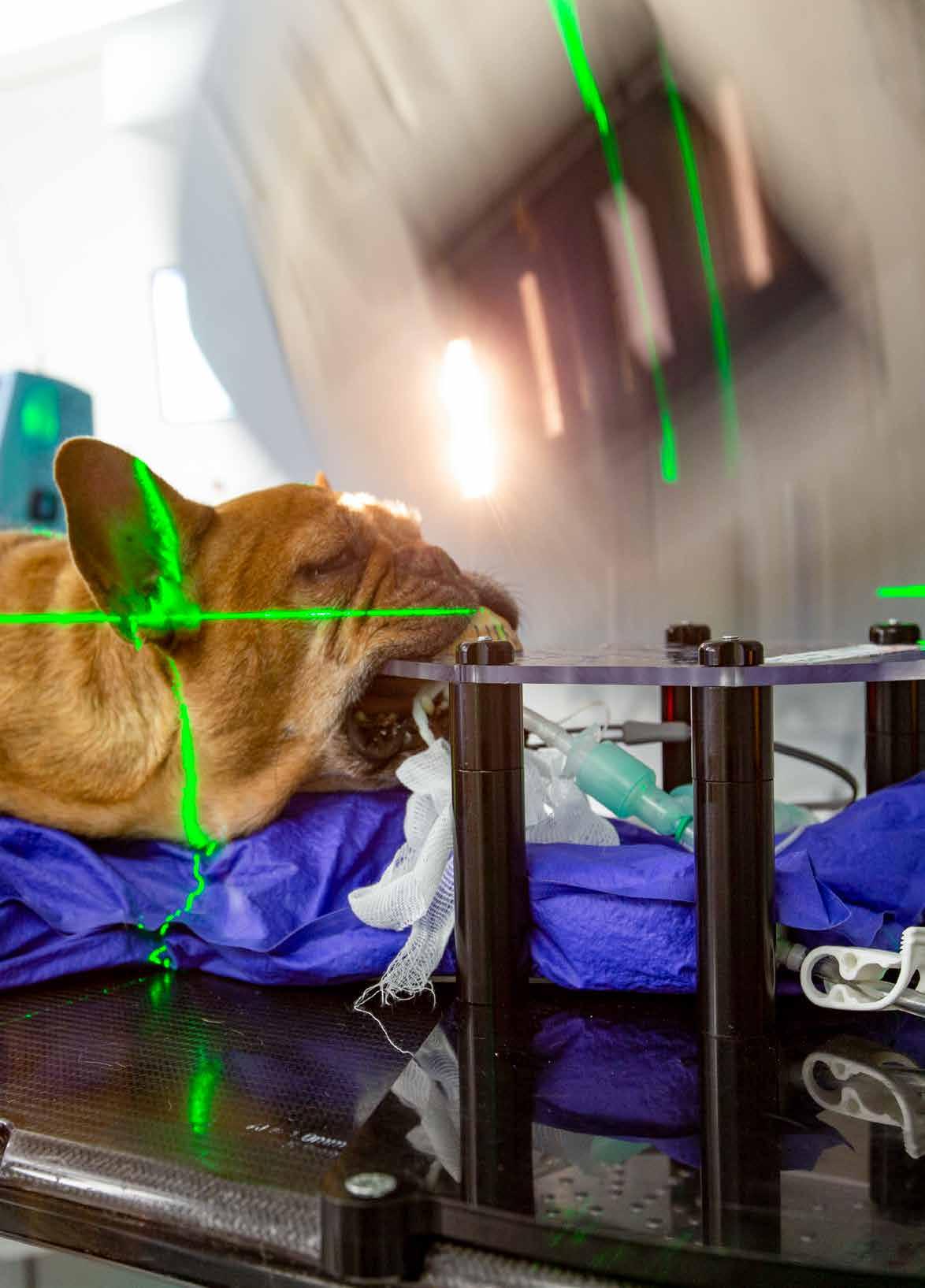
Luna, a cute 8-year-old female French Bulldog, is standing on a treatment table in the Division of Radiation Oncology at the Vetsuisse Faculty. Veterinarian Chris Staudinger administers an anaesthetic through a catheter in her left leg. Slowly, Luna lies down to sleep. She is about to undergo radiation treatment, which hopefully will destroy or at least reduce the size of the tumour that sits between her cerebellum and the delicate brain stem area.
Dogs suffer from cancer roughly as frequently as humans. “About 20 to 30 percent of the dogs we have in radiation treatment have a brain tumour,” says Carla Rohrer Bley, head of oncology at the Department of Small Animals at the Veterinary Hospital. The centre is renowned throughout Europe for the treatment of brain cancer, and some patients come in from other European countries to be treated here.
“When a dog is diagnosed with a brain tumour, typically their neurologic condition is bad. Without an antitumour treatment, they may live for only six or eight more weeks, all the while suffering from symptoms such as epileptic seizures, balance problems or stupor,” says Rohrer Bley. Luna, for example, sways slightly when she is walking. To destroy the tumorous cells that cause her symptoms, veterinarians use radiation therapy. First, the animal’s body is precisely positioned so that a state-of-the-art device — the same used on human patients — can deliver highenergy X-ray radiation with the utmost accuracy, so as to prevent damage of the surrounding healthy brain.
Luna’s radiation treatment takes a mere two minutes. Afterwards, vet Staudinger wraps her in a warm blanket while she slowly wakes up. A few moments later, she is up on her paws and hungry, barking to make her needs heard.
Improved treatment In an ongoing research project, Rohrer Bley and her team aim to make radiotherapy more effective by targeting small areas of tumours with higher radiation levels. Conventionally, the radiation dose is distributed homogeneously throughout the tumour tissue. The total dose is limited by what the surrounding healthy tissue can tolerate without being damaged. However, in some parts of the tumour, a higher dose might be desirable to better destroy cancerous cells. Therefore, Rohrer Bley and her team plan to deliver a higher dose to areas of the tumour that are not adjacent to sensitive organs. This would allow them to treat each animal with its individual highest dose without increasing the risk of severe side effects.
In a first step, the researchers will simulate the effect of this radiation plan in a computer model. Next, they will compare the effects of the regular protocols to these higher-dosed, heterogeneous treatments on the canine patients. In order to direct the high-energy X-rays accurately to certain areas of the tumour while avoiding others, a high level of expertise in physics and treatment planning skills are needed. That’s why the Vetsuisse researchers have teamed up with medical physicists who optimise radiation therapy and radiation devices in human medicine. The outcomes of this interdisciplinary team effort will potentially benefit animal and human patients alike.
Deep dive into cancer cells The researchers at the Vetsuisse Faculty are also working to better understand how cancers grow and develop: Why do cells suddenly start to multiply uncontrollably? How can this growth be predicted, and which molecules in these cells could be suitable targets for new drugs? As model animals for many types of cancer in humans, dog patients can help to answer these questions (see box).
One of the researchers involved is Franco Guscetti from the Institute of Veterinary Pathology. As a pathologist, he regularly examines cancerous tissue that has been removed during surgery to determine if a tumour has been completely excised. But, he also uses these tissue samples to better understand how cancer grows and to find new therapies. Specifically, Guscetti and his team have investigated canine oral squamous cell carcinoma (COSCC), a malignant tumour that develops in the mouth and infiltrates the surrounding tissue, sometimes including the bone.
Cancer cells are reprogrammed to no longer produce proteins in the same way as normal cells. Using a method called RNA-sequencing, the researchers identified genes
affected by this reprogramming that had a role in cancer growth and development. Because COSCC in dogs is very similar to human head and neck squamous cell carcinoma (HNSCC), the researchers compared their results with pre-existing genetic data from humans and found that the canine and the human cancer cells were very similar in their reprogramming. In cooperation with Enni Markkanen from the Institute of Veterinary Pharmacology and Toxicology, the team pinpointed two genes named CDK4 and CDK6, which are affected in both the human and the canine cancer variant. This indicated that dogs suffering from this type of tumour could be treated with a drug already in use in human breast cancer therapy.
A biobank to boost research Pathologists all over Switzerland increasingly store tumourous tissue in biobanks together with information such as the diagnosis and sample origin, preparation, and type. Because of the data’s high value in research, Guscetti is currently establishing a biobank information system to manage samples collected at the Vetsuisse Faculty. “These samples will then be accessible to a wide community of researchers and offer the possibility to address a variety of research questions,” says Guscetti.
His colleague, Markkanen, also uses surgically removed tumour tissue from dogs in her research. Originally, the young group leader wanted to work as a veterinarian at the clinic. After all, her passion for helping animals extends to her private life, which she and her family share with several horses, three dogs, a cat and three geckos. Last year, she nursed over 70 hedgehogs that were sick or injured. In addition to her veterinary work, however, Markkanen discovered her enthusiasm for research that aims to understand how the tissue surrounding cancer cells can drive tumour growth.
“Typically, researchers look for these growth drivers inside the cancer cells,” says Markkanen. The role of surrounding normal tissue, called cancer-associated stroma (CAS), is often overlooked, she says, even though it may influence whether an emerging tumour growth is able to establish itself in a cellular environment. For example, the stroma contains a variety of cells, including immune cells, which can release substances that inhibit the growth of cancer cells or even kill them. However, tumour cells know some tricks as well and are able to manipulate their surroundings to become more hospitable. To find out how exactly the tumour cells achieve this feat, Markkanen and her co-workers have focused on breast cancer as an example. This is not only the most common cancer in women but also in female dogs, about 24 percent of whom develop it. Predicting a tumour’s aggressiveness Markkanen and her colleagues put thin slices of canine breast cancer tissue samples under a microscope and used a fine laser to separate stroma from cancer tissue. Using RNA-sequencing to compare CAS to normal stroma from the same patients, they found specific genes that appear to influence tumour growth. Currently the researchers are investigating the function of those genes more closely. “We want to understand how exactly the stroma influences the tumour and whether specific genes have a positive or a negative effect on its growth,” explains Markkanen.
Making a prognosis of whether a lump of tissue will develop into a vicious tumour is also important in human medicine, but the first step towards such a prognosis is to understand how tumours grow and develop on a molecular level. Once researchers know which genes or molecules set off the chain reaction of unchecked cell division, they can use these genes and molecules as markers to watch out for in tissue samples.
So far, Markkanen and her team found that the genetic make-up in stroma cells was different in benign breast cancers compared to malignant, and the genetic changes happening in the stroma of dogs are strikingly similar to those in humans. Based on these results, scientist can now go on to identify biomarkers for diagnosis and prognosis of breast cancer in women as well.
Prognosis of tumour aggressiveness is likewise important for prostate cancer, the third-leading cause of cancer death in men. Although it can be detected by a blood test, the majority of prostate cancers are not aggressive at the time of initial diagnosis and do not require immediate intervention. Not treating these cancers minimizes overtreatment, but it also runs the risk of missing early intervention options for cancers that turn out to be aggressive. “We need to find ways for early prediction of a tumour’s aggressiveness,” says molecular biologist Raffaella Santoro from the Department of Molecular Mechanisms of Disease.
A revolution for prostate cancer Santoro’s research is focused on how healthy prostate tissue turns into a tumour. However, scientists have not yet managed to grow prostate tumours in a lab in order to study them, so Santoro and her team had to come up with an entirely different solution: They use prostate cells from mice and grow them into a 3D structure, thus constructing mini-organs, or so-called prostate organoids. “Having such a laboratory prostate cancer model is truly a revolution,” says Santoro.

Group leader Enni Markkanen reviews the results of an RNA-sequencing run. This method enables her to pinpoint genetic features of different cancer variants.
Using such a prostate cancer model, she can now mimic the cancer by manipulating the model organ’s genetic make-up. For example, the deletion of a gene called PTEN turns the organoid into cancerous tissue. Santoro and her team were also able to identify another gene called TIP5 that is closely linked with aggressive prostate cancer. In the future, it might be used as an early prognostic biomarker to facilitate treatment choices in human prostate cancer patients.
Finding such prognostic markers and optimising therapies will also help dogs like the French Bulldog Luna. After her radiation therapy session, she is done for the day and waits for her owner to pick her up. Typically, treatments stretch out over ten sessions. “After therapy, canine brain tumour patients typically enjoy two more years of life, mostly without any symptoms,” says Rohrer Bley. “In nearly all cases, 97 percent actually, we see a long-lasting improvement.”
Why dogs are a good model for cancer in humans
1. Dogs intimately share our environment and lifestyle, be it passive cigarette smoke at home, pesticides used in the garden, or excessive UV radiation from sunlight. Hence, factors that cause cancer in humans also do in dogs.
2. In comparison to laboratory models such as rats and mice, cancers in dogs are not induced on purpose. As in humans, tumours in dogs arise spontaneously and have a high degree of variability.
3. Similarly, dogs have a functioning immune system, whereas the immune system in laboratory animals is suppressed.
4. Cancers have a genetic component. Such genetic predispositions also exist in dogs and can be retained in some breeds.
5. The anatomy and physiology of cancers in dogs and humans are similar. For example, canine cancers are similar in size to those of humans.
