UNIVERSITY OF ROCHESTER MEDICINE | SCHOOL OF MEDICINE & DENTISTRY | 2026 VOLUME 2
TINY SENSOR ...
HUGE
... POTENTIAL
Tissue-on-a-Chip Aims to Revolutionize Clinical Trials
Integrative Oncology: A Fresh New Tool in Cancer Care and Education
On the cover
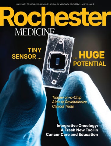
One of the many tissue-on-a-chip devices being developed as part of a multi-department collaboration in URochester research labs. The technology could make clinical trials significantly faster and safer. Photo by J. Adam Fenster
POINT OF VIEW File photo
THROWBACK
Match Day So, what did Match Day look like 50 years ago? If you don’t look too closely at the haircuts and bold stripes, not that different from today. The same emotions, excitement, joy, and sense of anticipation define the day: then and now. See inside this issue for a glimpse at this year’s celebration.
ROCHESTER MEDICINE | 2026 – VOL. 2 1
In this issue, you’ll notice the use of our new brand: University of Rochester Medicine. This new name reflects a defining strength of our growing academic health system: the powerful integration of our education, research, and patient care missions in what has evolved into an eight-hospital health system with 3,500 doctors and other clinical providers caring for patients in more than 300 locations. But our missions are more than complementary. They are deeply interconnected. Our cover story is a great example. “Tissue-on-a-chip” technology is being developed to run tests of in-development drugs—in part without human subjects. This has the potential to bring medications to market more quickly while minimizing risk to humans or laboratory animals. Eventually, this could have major implications for personalized medicine, with patient-derived cells determining which drug will be most effective for individuals based on their genetic makeup. We are one of a handful of institutions awarded major grants to perfect this technology, but our work didn’t come out of a single lab dedicated to the breakthrough idea. Instead, the key researchers came together from different disciplines and different schools at the university, and their varying skill sets have made us uniquely qualified to develop this technology. That’s the power of our One Rochester philosophy.
David Linehan, MD CEO, University of Rochester Medical Center Dean, School of Medicine & Dentistry Senior Vice President for Health Sciences
Yet it was a student’s passion that provided the early momentum. And a group of students, on their own, would create an international conference and invite faculty members to speak. That kind of enthusiasm is unusual and encouraging. And it can only happen where education is entwined with research and patient care. It’s a powerful combination. Our other feature story shows a similar convergence of missions, in a very different realm. Dr. Alissa Huston in Hematology/Oncology is using evidence-based research to train medical students and residents in integrative care, a logical extension of our institution’s legacy as the leaders in the biopsychosocial model of medicine. Eventually, she hopes to offer a formalized fellowship training program. Equipping our current and future practitioners with new skills and research solutions, all with the goal of improving patients’ health, is a key part of the vision for our academic medical center. Here in Rochester, it’s an idea that’s a century old. But as we find new ways to work together and learn together, the potential for important impact in patient care continues to grow. – David
What do you think? Rochester Medicine welcomes letters from readers. The editor reserves the right to select letters for publication and to edit for style and space.
Read more Rochester Medicine RochesterMedicine.urmc.edu
Brief letters are encouraged.
Submit Class Notes RochesterMedicineMagazine@urmc.rochester.edu
RochesterMedicineMagazine@urmc.rochester.edu
2 ROCHESTER MEDICINE | 2026 – VOL. 2
Write to Us Rochester Medicine magazine University of Rochester Medicine 601 Elmwood Avenue Box 643 Rochester, New York 14642
CONTENTS 4
Cover Story 10 19
Feature Story 20
Rochester Rounds The Mighty, Tiny Tissue-on-a-Chip Revolution Mentorship, Rochester Style A Different Kind of Care: Integrative Oncology Brings a New Approach to Treatment
26
Match Day: The Envelope, Please
28
Commencement: Crossing the Finish Line
30
For Ever Better Update
34
Faculty News
38
Philanthropy
40
Class Notes and News
41
In Memoriam
p. 10 p. 20
Rochester Medicine is published by:
Editor
Mark Liu
For questions or comments, contact:
The University of Rochester Medicine
Art Direction & Design
Karen Ver Steeg
Dept. of Alumni Relations and Advancement
Department of Communications, in
Staff Writers
Bethany Bushen Jim Miller
300 East River Road, Rochester, NY 14627
Relations and Advancement for the School
Production Manager
Bethany Bushen
of Medicine & Dentistry.
Feature Photography
J. Adam Fenster John Schlia Matt Wittmeyer
conjunction with the Department of Alumni
for the School of Medicine and Dentistry Phone 800.333.4428 585.273.5954 Fax 585.461.2081
To comment on this issue, e-mail: RochesterMedicineMagazine@urmc.rochester.edu
Contributors
Sydney Burrows Barbara Ficarra Kellie Fraver Scott Hesel Mark Michaud Leslie Orr Susanne Pallo Kelsie Smith-Hayduk
Find us on facebook at:
Director of Communications
Lori Barrette
www.facebook.com/urochestermed ROCHESTER MEDICINE | 2026 – VOL. 2 3
ROCHESTER ROUNDS
New Method Allows Precise Targeting of the Brain’s Neural Pathways By Mark Michaud
A new study in Cell Reports Methods describes an elusive method to control specific communication pathways in the brain. “This study provides us a new way to precisely target how brain regions communicate,” said Kuan Hong Wang, PhD, senior author of the study. “Instead of affecting broad areas, we can now control specific pathways, offering a clearer view of the circuits behind complex behavior and brain disorders.” Using the refined, light-based technique of optogenetics, the team of scientists at the University of Rochester Medicine Del Monte Institute for Neuroscience worked with the brain of the common marmoset, a small primate widely used in neuroscience, to target only the neurons that connect one brain region to another. They could then either activate or silence those same cells on demand.
The cerebral cortex depends on long-distance connections that link specialized areas into coordinated networks. But traditionally, scientists have had limited tools for selectively turning these specific connections “on” or “off” in animal models that most closely resemble the human brain. Optogenetics uses light to control cells genetically modified to respond to it, enabling researchers to selectively activate or inhibit specific neurons. This new method brings us closer to understanding how distributed brain networks support higher-order functions such as perception, decisionmaking, and social behavior. In the long term, tools like this will also help clarify how disruptions in specific brain circuits contribute to neurological and psychiatric disorders and guide the development of more targeted treatments.
Many Cured of ‘Incurable’ Disease after Clinical Trial By Susanne Pallo
Robert Oman has been cancer-free for 20 years, thanks to a longstanding clinical trial conducted at the University of Rochester Medicine Wilmot Cancer Institute. And 70 percent of patients who had advanced-stage follicular lymphoma survived at least 15 years after completing a standard treatment regimen of immunotherapy and a chemotherapy combination known as CHOP. A new analysis of long-term data from the trial, published in JAMA Oncology, shows that 42 percent of treated patients were functionally cured—defined as having no chance of lymphoma recurring during their expected lifespan. For a disease long deemed incurable due to its propensity to recur, that’s a game-changer. In the study, the rate of disease relapse fell from 6.8 percent in the first five years after treatment to only 0.6 percent between 15 and 20 years. “When we started this trial 25 years ago…we expected the CHOP chemoimmunotherapy to be better than the standard treatment of the day,” said Wilmot Director Jonathan W. Friedberg, MD, MMSc, who led the new study. “But we never expected it would be curative. The fact that a subset of patients achieved cure is truly remarkable.” Oman, who finished treatment in 2006, has never shown any sign of recurrence—an outcome that would have been highly unlikely if he hadn’t participated in the trial. His father’s experience with lymphoma decades ago was very different. “My dad was in his 60s when he had it,” said the younger Oman. “He lived for 14 years, but he took seven rounds of chemo. It always came back. It would be like two years, and the cancer always came back.” For the younger Oman, 65, of Campbell, N.Y., surgery to remove affected lymph nodes seemed to work, but follicular 4 ROCHESTER MEDICINE | 2026 – VOL. 2
Three generations of the Oman family, together thanks to a clinical trial Robert Oman (front left) enrolled in 20 years ago. lymphoma returned in 2005 along with a tumor on his kidney. That’s when his oncologist at Guthrie Corning Hospital referred him to Wilmot for the phase 3 clinical trial, which was conducted by the SWOG Cancer Research Network, a clinical trials group funded by the National Cancer Institute. Unlike his father, Oman had only minor complications, missing just a few days at his job on a Cornell University potato farm while maintaining his own farm throughout treatment. Oman is still active and says he has lived a normal life. Now, his sons are grown, and he’s been able to watch two of his grandchildren graduate from high school in recent years. “I was extremely lucky and blessed to be able to see our first two grandchildren grow up, and now we’re starting a new chapter in our life with our newest grandson, born last October,” he said.
ROCHESTER ROUNDS
Discovery of Unexpected Food Source for Tumors Could Lead to New Treatment By Leslie Orr
URochester Medicine researchers have discovered an antioxidant, glutathione, that cancer cells appear to devour as fuel, opening new pathways for investigation and a drug that can restrict the way tumors use this nutrient. The top-tier scientific journal Nature published the study. Isaac Harris, PhD, of the Department of Biomedical Genetics, and a team from the Wilmot Cancer Institute conducted the research. Nutrients are often scarce in the tissue and cells surrounding tumors. But cancer cells have produced an effective strategy to take in nutrients. This is where glutathione, a powerful pro-tumor nutrient that Harris has been studying, came into focus. “Cancer cells and normal cells potentially use different food sources,” Harris explained, “and we discovered how cancer cells, specifically, break down this antioxidant and use it as fuel.” The new role for glutathione is notable, Harris said, because until now, most scientists who study glutathione have focused on the way it can prevent or repair cell damage, rather than its ability to feed cancer cells. “Maybe we need to re-examine the pantry that cancer relies on and look at things that we never thought could actually be used as food for tumors,” Harris said. “There are additional complex metabolites that others are looking at, so we’re potentially opening a whole new interest into how cancer cells acquire nutrients and how to block that activity.” The body makes glutathione naturally, but it is also widely sold as an antioxidant supplement accompanied by pro-health claims, which is in contrast to more nuanced and cautionary messages from the National Cancer Institute about additives and nutrients. “It’s important to understand how cancer hijacks certain substances that we may think of as harmless,” Harris said, emphasizing that antioxidants can be a double-edged sword. For example, last year, Jeevisha Bajaj, PhD, a colleague of Harris’s, discovered that taurine, another antioxidant available in foods, supplements, and energy drinks, drives the growth of leukemia cells. Her work was also reported in Nature. Earlier, the Harris team, in collaboration with Tom Campbell, MD (Res ’13), and Erin Campbell, MD (Res ’12) discovered how a whole-food plant-based diet may reduce pro-tumor fuel sources in the body. It laid the groundwork for the current study, exploring the complex links between antioxidants, health, and cancer. The latest research goes many steps further: Researchers analyzed breast tumor samples from individuals who had donated their tissue to Wilmot’s Biobank. By examining the fluid inside these tumors, researchers found abundant storage of glutathione, confirming that tumors are aggressively consuming it as a nutrient source. The team also found they could slow tumor growth by blocking the cancer’s ability to use glutathione. This discovery may apply more broadly to other types of cancer, Harris said, since preliminary research shows that many tumors consume glutathione. Harris emphasized that although the antioxidant glutathione is linked to cancer, it does not mean that individuals should stop eating antioxidant-rich whole foods.
Isaac Harris, PhD (center), with lab members Fabio Hecht, PhD (left), and Marco Zocchi, PhD. “Eating a balanced diet with fruits and vegetables is important,” Harris said. “But people should be cautious about taking supplements in general, particularly glutathione. Taking a pill that is unregulated by the FDA and has a high concentration of glutathione can present risks.” In the current study, Harris’s team used advanced technology to search for therapies that could inhibit a tumor’s ability to use glutathione. Researchers identified a hit—a drug that was developed nearly a decade ago. University of Rochester chemist Tom Driver, PhD, the Robert K. Boeckman Jr., and Mary H. Delton Family Distinguished Professor in Organic Chemistry, and Joshua Munger, PhD, professor of Biochemistry and Biophysics and an expert in cancer cell metabolism, are exploring new ways to improve that drug and to pinpoint precise proteins involved in feeding glutathione to tumors. Other plans include testing combinations of anti-cancer drugs in the context of dietary changes that could improve cancer outcomes. The goal is to develop novel therapies that kill tumors without impacting healthy cells. ROCHESTER MEDICINE | 2026 – VOL. 2 5
ROCHESTER ROUNDS
Low-Cost Interventions Lead to Major Reduction in Opioid Prescriptions after Surgery By Jim Miller
A novel team approach, based on a culture change in doctor-patient communication, hoped to achieve a 20 percent reduction in opioid prescriptions at discharge. “But the strategies we deployed actually reduced it by 67 percent, and they did so without any increase in patient-reported concern about pain,” said University of Rochester Medicine endocrine surgeon Jacob Moalem, MD, the study’s lead author. “One surgeon stated that prior to this project, he never would have imagined sending certain patients home without an opioid prescription, but the team now does it routinely.” The study, published in the Journal of the American College of Surgeons, is the first report of a low-cost, scalable, and selfsustaining opioid stewardship program spanning all perioperative phases. Opioid overprescriptions after surgery remain extremely common nationwide, even as studies show that most of what’s prescribed goes unused, leaving staggering amounts of medication left over for potential misuse. Evidence also shows that new, persistent opioid use is seen even after limited postsurgical exposure. Moalem and his co-authors deployed patient- and staffdirected educational materials, electronic medical record modifications, and a dashboard that tracked and analyzed opioid prescribing across the Department of Surgery. They followed 6,619 opioid-naïve adult patients undergoing one of 15 elective procedures through acute care, colorectal, thoracic, transplant, and surgical oncology divisions. “The culture change was the secret ingredient here,” said Moalem, also a professor of Surgery and Medicine in the School of Medicine & Dentistry. “We empowered the care team with all the information they needed to make an informed decision on a prescription that matched the patient’s needs, as opposed to simply defaulting to a standardized quantity of opioids.” For patients, the message is simple: Some discomfort after surgery is normal and expected, and the goal for postoperative pain management should be to reduce the discomfort to a level that does not interfere with normal activities of daily life. We should not aim to eliminate all discomfort after surgery, Moalem said. “Most patients generally understand that opioids are not a panacea and that they come with downsides, but Jacob Moalem, MD patients may also
6 ROCHESTER MEDICINE | 2026 – VOL. 2
be scared, anxious, and expect that their post-surgical pain will be treated,” he added. “Balancing those factors is where the conversation with your doctor comes in, and where there is an opportunity to set proper expectations and to explore opioid alternatives. When doctors and patients work together on that, we see the best results.” Other results from the study: • The reduction in median discharge opioid prescriptions was achieved across 14 of the 15 procedures studied, with 10 operations reaching a median prescription discharge quantity of zero. • Patients had 3.4-fold higher odds of being discharged without opioid medications and a reduction in the likelihood of receiving a refill or a prescription for opioids after discharge. • Patients discharged with an opioid prescription received an average of eight fewer pills. Specific interventions implemented by the study authors: • Badge tags showing recommended prescription quantities for common operations were distributed to all residents and advanced practice providers. • Distribution of prescribing data, benchmarked against prescribing goals that surgeons set for themselves, to division chiefs via monthly dashboards. • Electronic medical record updates, including displays of a patient’s opioid administrations over the 48 hours prior to discharge; a window encouraging the prescription of opioid alternatives; a streamlined opioid prescription panel promoting lower preferred quantities; and an advisory panel requiring justification for patients being discharged with an opioid prescription despite not having received opioids in the hospital in the prior 48 hours. The program is replicable. The interventions are low-cost and easy to implement, and several other University of Rochester Medicine surgical departments are already using the toolkit and seeing positive results. Moalem said he and his co-authors will next look at whether the improvements can be sustained over time and at the extent to which the suggested interventions work in other departments. A recent presentation by surgery resident Mitchell Breitenbach (MD ’23) at the Academic Surgical Congress is a positive sign. In a follow-up study on patients undergoing laparoscopic cholecystectomy, the department’s baseline prescribing was well within national recommendations, but the interventions reduced postoperative prescriptions by 48 percent—amounting to an annual savings of approximately 10,000 pills—with no change in the frequency of patient phone calls for inadequate pain management.
ROCHESTER ROUNDS
How a Small Environmental Project Grew into a National Model By Mark Michaud
Taking it to the beach: Members of the Lake Ontario MicroPlastics Center in action. In 2019, University of Rochester biomedical engineer Jim McGrath, PhD, received pilot support to apply nanomembranes in detecting plastic particles in water samples—a growing publichealth concern. The support came from the National Institute of Environmental Health Sciences Core Center (EHSC) at URochester, the second-oldest such center in the country. McGrath had pioneered ultrathin silicon nitride nanomembranes for biomedical applications. URochester partnered with the City of Rochester Water Bureau to conduct the first systematic analysis of microplastics in the city’s drinkingwater system, sampling water at various points along its 35-mile journey from the Hemlock Lake reservoir to the University. The research revealed the potential of the nanomembranes to filter and characterize microplastics, including particles small enough to be absorbed by the human body. As the work unfolded, McGrath’s team helped establish the Microplastics Working Group of Western New York, which brought together scientists from the URochester and Rochester Institute of Technology as well as community stakeholders to share methods, data, and emerging concerns. The group became a launching pad for new microplastics research, with EHSC pilot awards to URochester researchers
Jacques Robert, PhD; Lisa DeLouise, PhD; and Allison Elder, PhD (Flw ’00) to study how microplastics interact with skin, developmental pathways, hormone and immune systems, and other biological processes. It also fostered discussion of how inhaled microplastics might affect human health. This line of research, led by Elder, builds on EHSC’s decades of research on inhaled particles. The Working Group also strengthened ties with regional partners engaged in Great Lakes science and research on plastics. That, in turn, served as the basis for the Lake Ontario MicroPlastics Center (LOMP) in 2024, which is co-directed by Katrina Korfmacher, PhD. LOMP serves as a research hub and a regional convening point—supporting discovery science, advancing communityengaged research, and informing solutions to reduce exposure to microplastics across the Great Lakes, which hold more than 20 percent of the world's surface freshwater. What began as a small pilot project connecting a university lab and a city water system has grown into a national model for transdisciplinary environmental health research. As environmental challenges intensify, these partnerships will continue to guide the region toward evidence-based, community-driven solutions. ROCHESTER MEDICINE | 2026 – VOL. 2 7
ROCHESTER ROUNDS
Gender Differences in Brain Immune Cells Could Lead to Personalized Alzheimer’s Treatment By Kelsie Smith-Hayduk
Two-thirds of the more than seven million Americans who have Alzheimer’s disease are women. “But we still do not have a great understanding of why this is the case,” says M. Kerry O’Banion, MD, PhD (Flw ’91), professor of Neuroscience and Neurology. “We can only improve any possible treatment or prevention of this disease if we know the why, when, and where these differences are occurring.” New research led by O’Banion and Neuroscience graduate student Lia Calcines-Rodríguez at the University of Rochester Medicine Del Monte Institute for Neuroscience shows that the immune cells in the brain, known as microglia, act differently in the male and female Alzheimer’s brain. They appear to cause residual harm in the female brain. As described in a recent paper in the Journal of Neuroinflammation, the researchers discovered that in mice, when microglia respond to amyloid-β plaques that accumulate in the brain in Alzheimer’s disease, female microglia express more interferon-related genes. Interferons help combat viral infections, but their role in Alzheimer’s disease is unknown. Researchers believe that as the microglia consume the amyloid-β plaques, they may be exposed to DNA or RNA and mistake it for a virus; this may cause the cells to release interferon. This research also found that female microglia leave behind larger and more irregular plaques, which damage more neuronal connections than those in the male brain. Now, researchers aim to understand whether these differences between male and female microglia could provide a path to alter the disease course. The team sees the interferon signaling in microglia as a potential sex-specific, personalized treatment to combat Alzheimer’s.
Lia Calcines-Rodríguez
25 Years of Extending Lives as the Area’s Only Heart Failure and Transplant Center By Leslie White
Every morning, John Lefrois feels grateful to open his eyes. “It’s a daily reminder that I’m lucky to be alive.” LeFrois is Strong Memorial Hospital's longest-surviving heart transplant recipient. He received his heart 25 years ago—the second recipient out of more than 400 through the University of Rochester Medicine Advanced Heart Failure program. Strong Memorial is Upstate New York’s only site for heart transplantation, eliminating the need for patients throughout the region to travel long distances. For LeFrois, along with hundreds like him, the gift of life has granted him precious time to raise his children, walk his daughters down the aisle, and share his passion for fishing with his six grandchildren. “Patients put trust in their care teams to restore their health,” said Peter Knight, MD (Flw ’87), chief of Cardiac Surgery. “We take great pride in helping them not only recover but thrive.” John Lefrois 8 ROCHESTER MEDICINE | 2026 – VOL. 2
ROCHESTER ROUNDS
Farm-Living Families Develop Earlier Immunity Through Breast Milk By Scott Hesel
Children who grow up in farming communities have long been known to develop far fewer allergies than their urban peers. A new University of Rochester Medicine study offers a possible reason: Their immune systems may mature faster, and breast milk appears to play an important supporting role. In a longitudinal birth cohort comparing infants from Old Order Mennonite (OOM) farming families in New York’s Finger Lakes region with those from urban and suburban families in Rochester, researchers found that farm-exposed babies had more “experienced” B cells and higher levels of protective antibodies during the first year of life. The study, published in Science Translational Medicine, was led by Kirsi Järvinen-Seppo, MD, PhD, chief of Pediatric Allergy and Immunology at URochester Medicine’s Golisano Children’s. According to Järvinen-Seppo, the study shows that OOM children “seem better equipped, earlier in life, to handle foods and other exposures without overreacting.” The team followed mothers and babies from pregnancy through the first year of life, collecting cord blood, infant blood, stool, saliva, and human milk samples. Farm-exposed infants had more memory and IgG+ B cells, suggesting earlier maturation of the antibody-producing system. They also had higher levels of IgG and IgA antibodies in blood, saliva, and stool and higher IgA levels in human milk from their mothers. Taken together, the data point to a more robust and active antibody system in infants growing up in a traditional farming lifestyle. One notable finding involved egg-specific antibodies and the development of egg allergy, one of the most common food allergies in young children. The researchers measured eggtargeting IgG4 and IgA antibodies in infant blood and human milk, and tracked which infants later developed egg allergy.
“We saw a continuum: The more egg-specific antibodies in breast milk, the less likely babies were to develop egg allergy,” Järvinen-Seppo said. “We cannot prove causality from this study, but the association is compelling.” Breastfeeding has long been associated with protection against atopic dermatitis and wheezing illnesses in infancy, but evidence that it prevents food allergy has been less consistent. Järvinen-Seppo believes this may be because not all breast milk is the same. “Our data suggest there may be particular benefit when mothers have high levels of food-specific antibodies in their milk,” she said. “Not every mother does, and that could help explain why results have been mixed on the association between breast feeding and food allergy.” OOM families typically raise their own chickens and eat eggs frequently. That repeated exposure appears to boost mothers’ antibody levels against egg proteins, which then show up both in the bloodstream and in breast milk. “Just as an infection or a vaccine can boost your antibody levels, regularly eating certain foods could do the same,” Järvinen-Seppo said. The study also found that OOM infants were born with higher cord blood levels of IgG and IgG4 antibodies to dust mites and horses, reflecting their mothers’ exposures, while urban infants had higher antibodies to peanuts and cats. The researchers are now leading a randomized clinical trial with participants assigned either to regularly eat egg and peanuts during late pregnancy and early breastfeeding or to avoid them. The study will track mothers’ antibody levels and the development of food allergies in their infants. “Ultimately,” Järvinen-Seppo said, “our goal is to translate what we learn from these communities into safe, practical strategies for all families.” ROCHESTER MEDICINE | 2026 – VOL. 2 9
The Mighty, Tiny Tissue-on-a-Chip Revolution It promises to make drug development quicker and safer.
Here’s how Rochester researchers have gained a leading role.
By Barbara Ficarra 10 ROCHESTER MEDICINE | 2026 – VOL. 2
W
of fruitful research to their names, but all three describe tissuehen he was a kid, Ben Miller used to read a lot on-a-chip as the biggest development in their careers. of science fiction. “I don’t anymore,” he said, They see the coming change profoundly affecting “because I’m living it.” academia, industry, and medicine. And soon. Miller is part of a group of researchers The flaws in mouse experiments and clinical trials make developing small, bioengineered devices—with core elements them unsustainable as stand-alone models for pre-clinical drug measured in micrometers—to mimic human cells and organs. testing, said McGrath, the William R. Kenan Jr. Professor of This tissue-on-a-chip technology promises to revolutionize Biomedical Engineering. “Ninety percent of all drug trials fail,” medical research trials as we know them today. he said, noting the U.S. pharmaceutical industry spends well The hope is to improve on—and one day even replace— over $100 billion on drug trials every year, amounting to vast mouse experiments that have been the standard of scientific sums spent on failed trials. inquiry for the last 100 years. When perfected, the technology Even the rare successful trial is lengthy. “In the best could limit animal testing as well as speed clinical trials and of all worlds, a drug makes it to market after 13 to 15 avoid the potential catastrophe of a human trial that causes years of development,” said Miller, a Dean’s Professor unforeseen harm to participants. of Dermatology at Rochester with joint The worldwide pursuit of tissue-on-aappointments in Biomedical Engineering, chip devices that are reliable and robust Biochemistry and Biophysics, Optics, enough to replicate human biology and Materials Science. has been accelerated in the U.S. The lack of humanthrough federal mandates, (From left) Ben Miller, relevant data slows the along with grants to Hani Awad, and James McGrath: process. a handful of select collaborating changed everything. “Animal studies academic research do not sufficiently powerhouses predict a drug’s including Duke, effects,” explained Harvard, Johns Awad, the Donald Hopkins, and the and Mary Clark University of Distinguished Rochester. Professor in URochester Orthopaedics actually started and a professor late. But in this of Biomedical race to redefine Engineering. how diseases Differences in are studied and reactions between how emerging mice and humans treatments are are a problem, along tested, a small with the cost, labor, group of Rochester and time to care for living researchers and students animal subjects. And inbred now has an inside track. research mice lack the genetic Looking at how the diversity of humans. work has developed here, it’s Frequently, drugs have been not surprising that URochester is a shown to be safe and effective in animal leader in this field; in several crucial ways, trials but not in human subjects. it could only have happened here. Pushed by those failures and pulled by the promise of emerging technologies, the FDA issued a 2025 mandate Big Change Coming—Quickly to reduce and then largely replace animal testing for drug Tissue chips are bioengineered, microfluidic devices development by 2029. It earmarked $150 million to develop, test, embedded with human cells in a way that keeps them alive for and standardize human-based research models such as tissuedays or even weeks. They mimic physical, biological, and even on-a-chip, organoids, AI, and computer modeling. mechanical environments of human tissue and organs so that The NIH has now established a new Office of Research scientists can observe cell interactions and responses when Innovation, Validation, and Application (ORIVA) to reaffirm its studying disease or testing potential cures. This could largely commitment to advancing scientifically valid alternatives to replace current testing for disease modeling, drug discovery, animal testing, and it has a “Complement Animal Research toxicology screening, and even personalized medicine. in Experimentation” program to facilitate New Approach University of Rochester researchers Hani Awad, James Methodologies (NAM) development. McGrath, and Miller are leading federally funded tissue-ona-chip research projects on the River and Health Sciences URochester is in the thick of all this. Early work included campuses. Combined, these researchers have nearly a century research by Ben Frisch, PhD, assistant professor of Pathology ROCHESTER MEDICINE | 2026 – VOL. 2 11
A photonic wafer with sensor chips used in microphysiological systems to monitor the effects of infection and treatment on organ models.
and Laboratory Medicine, to build tissue chips for studying acute myeloid leukemia, and Lisa DeLouise, PhD, associate professor in Dermatology and in Biomedical Engineering, to identify drugs to protect salivary glands from radiation damage during cancer treatment. In 2020, URochester became one of 10 NIH-funded research centers around the nation for testing tissue chips’ potential to simulate clinical trials. It received $4 million to develop a tissue chip to test therapies for tendon injury. In 2024, NIH awarded URochester a $7.5-million grant to establish the Translational Center for Barrier Microphysiological Systems (TraCe-bMPS) in partnership with Duke University to build FDA-qualified drug development tools. Powered by URochester technologies and industry and academic partnerships, the project is using tissue chips on Rochester-built platforms to study human models in the muscle-immune system, neuroinflammatory injury, joint-lining remodeling, immune cell trafficking, and the blood-retinal barrier. The same year, URochester earned a three-year contract valued at up to $7.1 million from the Biomedical Advanced Research and Development Authority (BARDA), within the Department of Health and Human Services, to model respiratory disease effects on the brain and test drugs to prevent and treat symptoms. This will open a new way to study the effects on the brain from long COVID and viruses such as influenza. Progress is already taking the work outside the lab—and attracting industry.
Made in Rochester
McGrath, Miller, and Awad have founded Rochester-based chip-manufacturing companies that are producing more than 5,000 tissue chips and more than 50,000 photosensors per year, supplying 12 URochester labs and nine others around the world. These labs have at least 35 chip projects underway, and the number keeps growing. URochester has developed some game-changing advantages with its approach. McGrath’s nanomembrane, for one, is the thinnest available, which helps the cells behave more naturally. Thicker materials used by other researchers give a cloudy aspect to cells seen through a microscope, but “Jim’s nanomembrane is optically transparent … as if it is not there,” Awad said. And with such a thin layer, there’s no interference from a membrane between cells. “We can embed Ben’s photonic sensors in the chip to measure what these cells are releasing in real time.” Many chips on the market are made of a stretchy, elastomeric material that can leach or absorb substances used in testing. URochester has developed chips that use silicon nanomembranes and photonic sensors in a hard plastic shell, 12 ROCHESTER MEDICINE | 2026 – VOL. 2
minimizing the leaching problem. The chips are also modular, so a multitude of users can customize them for different areas of research. The chips give researchers insight into human cell behavior that they never had before. They have channels to simulate blood flow across tissue or to introduce pathogens such as viruses to trigger an immune response in the cells, or a biologic agent that can affect an inflammatory response, or a drug in development to test its benefit or toxicity. The chips can be engineered with posts to have changeable structures in order to alter the amount of mechanical force in the microenvironment. This “stretches” the cells to simulate the breathing motions of a lung, the shear forces of blood flow, or the mechanical stress on a tendon. “Modularity in the design is absolutely a competitive advantage,” McGrath said. “Our tissue chips are very different than anything else out there—it’s very difficult for others to reproduce what we do. And this is all Rochester homegrown technology.” The pharmaceutical industry has noticed. Abbvie, Pfizer, Bausch & Lomb, and ApoGlyx have a vested interest in streamlining the drug development process and are working with URochester; Abbvie and Bausch & Lomb also provide funding. Non-pharma industry partnerships include manufacturers and system developers such as SiMPore, Aline, Phlotonics, SiObex, and AIM Photonics. Academic partnerships are surging, as well. Duke University and Rochester Institute of Technology are part of the consortium in the TraCe project; other academic centers involved with URochester include the University of Michigan, University of Oregon, Georgia Tech, and Indiana University. The sheer volume of research at multiple sites means standardizing of processes is essential. A team led by Joan Adamo, PhD, director of the Office of Regulatory Support in the Clinical and Translational Science Institute, is developing quality systems as a blueprint for researchers to follow. More than 60 standard operating procedures (SOPs) have been published to date. SOPs for tissue chip research are vital to the integrity of findings and another form of leadership in an emerging industry. “This is such a new approach, we are having to invent as we go,” Adamo said. “We don’t have 30 to 40 years of standard practice for this type of research.” The amount of work is testament to the amount of potential so many see. Human-equivalent chips, Awad said, “would create safer and more effective drugs faster. … Predicting failed drug candidates early in the development process would save pharmaceutical companies tons of money and potentially shorten the development process from 13 or 15 years to maybe 7 to 10.” But there is a long road ahead. The chips need to use a few hundred cells to accurately replicate the human body’s response, all while keeping cells alive long enough to get meaningful results. A way to analyze and catalog the voluminous data from testing must be perfected. And as with all manufacturing, scaling up is a challenge.
This specialized inkjet printer shoots microscopic drops of antibody solutions onto the sensor chips before they’re integrated into tissue chips. ROCHESTER MEDICINE | 2026 – VOL. 2 13
(Above) Assembly fixtures
guide components for the modular tissue chip.
URochester researchers are racing to find solutions, and in many critical aspects they are at the head of the pack. That in itself is remarkable, considering that the roots of this technology began elsewhere. In Rochester, it took several fortunate twists of fate and even a failed experiment to leap ahead. And it might not have happened at all if not for a student who connected some dots and changed several people’s lives.
The Johnnys-Come-Lately
In 2018, graduate student John Cognetti started working in Miller’s lab. But he also worked in McGrath’s lab. And something clicked. He described it this way: “I said, ‘I like what Jim’s doing with microphysiological systems, but I like Ben’s sensors too; I would like to put them together because it seems like most of the models out there don’t really do much in the way of sensing.’” Everyone agreed it was a good idea. “So then I spent the next five or six years trying to get it to work,” he said. Cognetti had to address many engineering details in order to get technologies to work together. He had to adapt what he knew about microfluidics to work with Miller’s sensors, which use laser light to measure cell activity. He also spent a lot of time figuring out how to affix McGrath’s nanomembrane to the chip, eventually turning to a precision craft cutter. “I like to say my PhD is in arts and crafts,” Cognetti said. “I did a lot of cutting and taping and gluing.” At the time, tissue-on-a-chip had been in development for nearly two decades. Concepts emerged in the late 1990s, and by 14 ROCHESTER MEDICINE | 2026 – VOL. 2
A tissue chip platform with an open well— one of the variations for different needs.
2010, Harvard’s Wyss Institute had developed the first, functioning human lung-on-a-chip. Within a few years, companies like Emulate began selling ready-made chips to researchers. In other words, URochester was a relative newcomer. Yet the pieces were in place. “Tissue-on-a-chip is an area my lab has been interested in forever—in some ways going back to when I was a postdoc,” Miller said. The collaboration between labs was the catalyst. In 2020, Miller and McGrath were using their separate inventions to develop early COVID tests. They hadn’t collaborated much with each other or with Awad, but the three had intersecting interests in tissue chips. That same year, the NIH was calling for applications from researchers to develop tissue chips that could model the microenvironment of a particular disease and perform a clinical trial for it. Awad saw the opportunity. But there was a major problem: The deadline was in 14 days. “Of course, we were Johnny-come-latelies to the party,” Awad said. “But I fired up an email to Ben and Jim.” The decision to throw their hats into the ring with the deadline looming? “Oh, yeah, that was crazy,” McGrath said. “That was a Hani Awad special.” The three crammed months’ worth of work into two weeks, submitting an application to create a tissue chip to model tendon fibrosis after injury. It worked. They were one of 10 applicants nationally to win funding.
“Not a bad result, and the best two weeks of work I’ve ever done, because it brought the three of us together as partners,” Awad said. “I recognized when we got the grant that this is the moment I reinvented my career.” John Cognetti It’s also the moment that their thriving research entities crystallized into one. “It really brought the three of us together,” McGrath said. “It is hopefully going to lead to a lot of good for the University.” He cited work to build out the TraCe center into “an institute that will enable the University to be a world leader in this space.” In very short order, the researchers have set that stage. “A lot of institutions that have come here wish they had what we have in terms of the technology advantages of our chips and our experience employing these materials,” McGrath said.
A Louis Pasteur Moment
Chance favors the prepared mind, Pasteur said. Case in point, the breakthrough nanomembranes came about by accident. And the photonic sensors were developed for a different purpose altogether. The nanomembranes were born out of a lab project gone wrong: A post-doc, Chris Striemer, was trying to make a photonic conductor out of a silicon layer. ROCHESTER MEDICINE | 2026 – VOL. 2 15
Secret Weapon: Enthusiastic Students By Barbara Ficarra
From the grad student who made a key connection between two labs to the post-doc who failed at making a photo conductor and, instead, accidentally spurred the creation of a groundbreaking nanomembrane, students have been a key catalyst in tissue chip development at URochester. “Without the students’ work, we would not be anywhere near where we are now,” said Jim McGrath, PhD. Dozens of students in labs run by McGrath, Ben Miller, and Hani Awad have had the learning opportunity of a lifetime—and have made important contributions along the way. Tissue chips, says McGrath, “are the stuff that PhD students’ theses are made of.” But it’s not just the quality or volume of the students’ work. Miller points to a kind of enthusiasm and initiative that speaks to how much promise the field holds. “It’s exciting to me when a student comes to my office with an idea that’s not mine,” Miller said. “It’s unusual for a younger student.” John Cognetti (PhD ’22) strengthened the connection between Miller’s lab and McGrath’s in memorable fashion, calling a meeting with the two and collaborator Harris Gelbard, MD, PhD, director of the Center for Neurotherapeutics Discovery. In the meeting, Cognetti led a PowerPoint presentation on combining sensors and nanomembranes. As Miller recalled it, Cognetti told the faculty members, “I’m doing this for my PhD project. We’re here to see who will be my advisor.” That kind of initiative pervades URochester’s tissue chip efforts. URochester students have the advantage of rotating through multiple labs in their PhD preparation, and they have furthered that collaboration by forming the Microphysiological Systems (MPS) Network. “A lot of students are working on tissue chip research, but there wasn’t really a good way for us to communicate and work together,” 16 ROCHESTER MEDICINE | 2026 – VOL. 2
said Isabelle Linares, a sixth-year biomedical engineering PhD candidate who serves as co-president with Katie Daniel, also a sixth-year biomedical engineering PhD candidate. The group aims to build a community to support the development of graduate student trainees, with faculty and academic colleagues from throughout the region welcomed. They meet monthly to discuss research, share ideas and inspiration, and build professional connections. In 2024, on their own, students Katie Daniel developed an international symposium that took place in Rochester. Organized by Azmeer Sharipol and Kevin Ling, it brought together 100 colleagues from academic research sites, pharmaceutical companies, and manufacturers. The second version, held in May 2026 and entirely student-run, grew to 130 participants. It included academic and industry sponsors and guest speakers and panelists from RIT, Cornell, and industry leaders such as AstraZeneca and AbbVie. This kind of student initiative shows Isabelle Linares the immense value of learning outside the lab, Miller said. “The level of engagement and advocacy by students in this has been extraordinary. I’ve never seen anything like it in my career.”
“He kept making it thinner and thinner and thinner to help visualize silicon crystals,” said McGrath, “but when he baked it at a thousand degrees (Celsius), in addition to the silicon crystals, he saw nanopores. The pores ruined it for his application, but Chris was smart enough to realize the material could have utility.” So he brought it to McGrath’s lab. “We designed some experiments to show that the membranes were really exceptional at filtering molecules,” said McGrath. So exceptional that the results were published in Nature. “That changed our lives and we went all in. I pivoted my career to focus on applications of this new material.” It took nearly a decade of work to enhance the raw material, understand it, and make it stable enough for lab use. Blood-brain barrier researchers began reaching out to McGrath to try it. “So I knew there was value in the material for those researchers, and we built it as a tissue chip for them. It’s a relationship that continues to this day.” Likewise, the photonic sensors were not originally built for tissue chips. Miller, who has been at URochester for 30 years, had been developing sensors that proved useful as a diagnostic tool during COVID. They’re now used in tissue chips to enable real-time measurement of cell activity. Each has eight to 18 different individual microring resonators that use tiny beams of light to detect a specific cell activity—when the cell starts secreting something in response to a drug or toxin, the device gives a reading to note the change. Manufactured in Albany, the photosensors arrive as 11,000 individual units fabricated on a single disc the size of a vinyl LP record. In the lab, the team then builds the tissue chips with components tailored to the needs of each experiment. “We use the nanomembranes to culture the cells and circulating media in the device to keep them alive,” Miller said. The photosensors measure and record cell activity in real time, unlike other chips that require disassembly and an end to the experiment to determine how cells reacted. The researchers seed the chips with a variety of cell types. A brain chip, for example, uses different types of donated cells from different parts of a person’s brain. “You’ve got admittedly a very simple model,” Miller said, “but a model of that person now on a chip.” That model brings a clear benefit. “We have been able to see differences from donor to donor in drug response for an anti-inflammatory. And that starts to be super exciting because the way drug development is done now, you don’t see any of that until the phase one clinical trial,” Miller said. “Doing that on chips, you already have an idea of how it’s going to work before it’s actually used in part of a human being.” This has implications for theranostics, a personalized approach to medicine that combines diagnostic imaging and targeted therapy specific to an individual’s diagnosis. “We could take that a step further,” Miller said. “You can have the theranostic procedure but then also test a panel of different drugs for a person to find the most effective drug for their condition and genetic makeup.” For the promise of personalized medicine, tissue-chip technology would enable research projects to solicit donor cells from a true cross-section of the population to achieve better outcomes. Males, females, pregnant people, all age ranges and races could be represented in any clinical trial by donating a small
tissue sample, which would give researchers a picture of how a potential drug would work for a variety of patients. And potentially, each person could contribute donor cells for a personalized study to see which cancer drug could be most effective for them.
Chips Changing Research Now
Tissue-on-a-chip “is big because it can be applied to any problem,” said Frisch of Pathology and Laboratory Medicine. Frisch is using tissue chips to model leukemia; this is especially useful because cancer medications can be very toxic to human tissues. Using chips has important advantages over current, singlecell studies in a dish, Frisch said. “Cells on a dish are not getting any of the interaction or communication that they normally get from other cells in their environment. And that can fundamentally alter the way they behave and the way they respond to therapies.” In a dish, he explained, it’s easy to figure out what can kill cells and control their growth. “But they often respond completely differently when they have that support in an environment that they’re used to growing in. With tissue chips, we can provide a lot more of that interaction … and they will respond more like what they would in their native environment in the human body.” The tissue chips he uses can simulate blood flow through vessels and the exchange of nutrients such as oxygen and carbon dioxide across a cell layer, out to the tissue and back. He also has built an extracellular matrix made of fibroid gel and other
Ben Frisch, PhD, uses tissue chips in cancer research. ROCHESTER MEDICINE | 2026 – VOL. 2 17
In 2017, DeLouise of Dermatology and Biomedical Engineering received a $3.8 million NIH grant to support her invention of microbubble array technology. Circular 3-D chambers are an ideal environment in which to culture and study groups of cells, and the cupped shape of her device facilitates cell interaction. She’s using it to study preventive treatments for salivary gland radiation damage sustained by head and neck cancer patients. It’s another example of expanding the breadth of what is discoverable—a key focus of URochester’s vision. The TraCe project, for example, shows the value of URochester’s emphasis on linking different types of tissue chips. “Our modular platform design grew out of the fact that we were building different chips to meet different needs,” McGrath said. “I realized that we couldn’t support collaborations with a single chip geometry, so we created a platform where the parts could be redesigned and ‘snapped’ together by non-engineering laboratories.” What that means for the future is where things begin to look more like science fiction.
Your Own Personal Chip?
Ben Miller holds a photosensor consisting of 11,000 individual units fabricated on a single disc. cell types critical for normal blood production and cancer cells. “All of that, on one chip,” he said. The hope is to save time, money, and patients’ efforts by testing different possible therapies on tissue chips first, then choosing the most effective to take into clinical trials. But it’s not just an advantage in cancer research. Awad sees the same thing with tendon-on-a-chip technology that URochester is developing: “We shouldn’t be looking at that one cell particularly—we should be looking at the microenvironment within which it lives.” In the tendon chip, a collagen hydrogel simulates an injured tendon’s microenvironment, which includes tendon cells and macrophages. The gel keeps cells in their “scar” microenvironment. A top compartment has the nanomembrane containing the single-layer endothelial cells that line blood vessels, allowing researchers to simulate circulating blood cells. The nanomembranes are the building block of Rochester’s chips, and they are a game-changer. “It is interest from others that affirms that our platform has unique value,” McGrath said. “No one is approaching tissue-on-achip this way.” But just like the importance of needing a microenvironment for better testing, there will be no “one chip fits all” solution. “When the NIH began to think about replacing animal models, they weren’t just thinking about tissue chips,” Awad said. “It’s really a multitude of complementary technologies.”
18 ROCHESTER MEDICINE | 2026 – VOL. 2
As the research is developed, Awad said, “We can think about potentially creating your own tissue chip made out of your own cells.” He imagines taking a skin biopsy, then using fibroblast cells from the connective tissue and inducing them into stem cells. On your chip, those cells could grow into whatever is needed. “We can create your immune cells, we can create your cartilage, bone, whatever, right? So the technology is here, we have protocols, and we’ve been perfecting them. It ultimately comes down to scale. Can we scale this up to enable these kinds of experiments?” Scaling these tiny tissue chips requires thinking big. “We need colleagues to become partners and drive the adoption of these tissue chips,” Awad said. “We think this can be accomplished by establishing an institute for microphysiological systems at the University of Rochester to do work similar to institutes at powerhouses like the Whitehead Institute.” The risk is losing ground. “We certainly get a lot of interest from other universities and researchers wanting to take aspects of our technology and integrate it into theirs,” Miller said. “The really key thing is continuing to build these models and validate them against whole animal models and human disease and health. That’s something that needs to happen here.” Already, the pace of URochester’s acceleration into more advanced tissue-chip systems has been possible only because of the multiple grants it has received. That, and a relentless sharing of ideas. “I think it’s a particular strength of the University of Rochester: We’re a culture of collaboration,” Miller said. “My lab and Hani’s are at the medical center, and Jim’s lab is just across the street. We have all these different people we can talk to all the time. For 30 years, I’ve been saying this to people: We recognize that we’re a relatively small place and for us to have an impact, we know that we have to work together. So we’re motivated to do that … We all want to work together.” RM
Mentorship, Rochester-Style Video Series Showcases a Key SMD Strength
The School of Medicine & Dentistry is known for its culture of mentorship and collaboration. But what does that look like in action? A video series of short vignettes, Learners on Location, gives a glimpse into how working together and growing together make an important difference in the lives of students and faculty members. By following mentors and trainees into meaningful places around the city, the series also highlights the connections, community, and experiences that define today’s training environment. Here are summaries of a few recent episodes. To view them, scan the QR code or search “Learners on Location” on YouTube.
‘It Was a Lot About Life’ Mentorship Beyond Medicine Watch faculty mentor David Paul (MD ’16, Flw ’21 ’24, Res ’23), assistant professor of Neurosurgery, and third-year medical student Bryan Redmond discuss their evolving connection, which began even before Redmond’s acceptance into medical school. They also offer insights into the medical school experience and the benefits of living, working, and taking part in the Rochester community.
Learning Through ‘True Translational Research' Mentor Richard Burack, MD, PhD, and mentee Hannah Bell (MD ’24, PhD ’24), discuss the extraordinary speed of medical innovation and SMD’s robust graduate medical education program, which offers students the opportunity to make an impact on the lives of those suffering from disease. Drs. Burack and Bell, both from the Department of Pathology and Laboratory Medicine, also share insights on the cultural and recreational benefits of living and working in the Rochester region.
Supporting the Whole Scientist In this episode, assistant professor Calvin Cole, PhD, and fifthyear PhD candidate Zach Sechrist show how mentorship goes beyond the lab. Their approach—grounded in wellness, growth, and purpose—demonstrates how personal well-being can lead to meaningful scientific results. Together, they’re advancing cancer cachexia wasting-syndrome research while building a foundation for long-term success.
ROCHESTER MEDICINE | 2026 – VOL. 2 19
A Different Kind of Care Integrative Oncology Gives Patients (and Physicians) a New Approach to Treatment By Sydney Burrows
Alissa Huston, MD (Res ’03), is bringing evidence-based approaches to integrative oncology.
20 ROCHESTER MEDICINE | 2026 – VOL. 2
When Julia Schlossman (MD ’25) entered medical school, she thought she might want to specialize in oncology. What she didn’t expect was that practicing yoga alongside patients, developing nutrition resources, and helping launch a culinary medicine course would shape her path. In her third year at the University of Rochester School of Medicine & Dentistry, Schlossman attended a talk by Alissa Huston, MD (Res ’03), co-medical director of the URochester Medicine Pluta Integrative Oncology & Wellness Center. Huston described a model of cancer care that goes beyond standard treatment—one that integrates evidence-based wellness strategies into each patient’s personalized care plan. “I found everything Huston spoke about to be so interesting, especially the ways in which integrative wellness empowers patients to have a sense of controI during a time when there is so much uncertainty. I thought it would be amazing to shadow her in my fourth year of medical school.” A cancer diagnosis can be overwhelming—physically, emotionally, and mentally. Complex treatment plans, major changes to daily life, and uncertainty about the future can leave patients feeling a loss of control. This is where integrative oncology can play a critical role. For Huston and her colleagues, integrative oncology begins—and ends—with science. Its foundation in research is what separates integrative wellness from some other approaches. It’s an important distinction, especially as more patients come in with questions about information they’ve heard from friends and family, social media, or other sources that may or may not help— and may even hurt. Patients and medical students are discovering the benefits of integrative oncology therapies. And more and more research is demonstrating the importance of this innovative approach in cancer treatment.
A Different Approach to Cancer Care
Huston is triple board-certified in oncology, hematology, and integrative medicine. She is the first integrative oncology specialist at URochester Medicine. Huston’s first experience in oncology began with a customized elective at URochester Medicine. During her residency here, Huston was drawn to caring for oncology patients. An attending physician, seeing her interest, created a breast-cancer elective for Huston that allowed her to work with different providers and aspects of oncologic care that otherwise would not have been part of her studies. “I think that’s also what we created in the Center—that touchpoint of creating something where students from all the disciplines we train here have the opportunity to experience and work in a space that might be different than what they initially envisioned their career trajectory to be,” she said. For Huston, a career in oncology was the next step. Her interest in integrative oncology evolved as she consistently met questions from patients about what they could be doing during treatment to help with healing. Huston had found immense value in the mentorship of faculty during her time in residency. After a fellowship at the University of Pittsburgh School of Medicine, she returned to
Rochester with hopes of providing similar support for the next generation of providers. When Schlossman inquired about studying at the Integrative Oncology Center, which was created by the Wilmot Cancer Institute in partnership with the Pluta Cancer Foundation, Huston realized there was a need and an opportunity to expand learning. Ahead of Schlossman’s final year at SMD, Huston developed an elective that allows medical students to work alongside her and a multidisciplinary team. Students gain experience not only in oncology care, but also with certified clinical exercise oncology physiologists, registered oncology dietitians, art and music therapists, and mind-body and meditation instructors. “It gave me a better understanding of the patient experience. I think about it every day,” Schlossman said. “The integrative center gives patients the tools to make lifestyle modifications to feel better, connect with each other, and cope with their disease.”
Whole Person Not Patient
At the Integrative Oncology & Wellness Center, evidencebased therapies work in tandem with traditional cancer treatments such as surgery, chemotherapy, and radiation. Patients receive oncology care through the URochester Medicine Wilmot Cancer Institute, while the Integrative Oncology Center supports symptom management through offerings such as: • Nutrition counseling and cooking programs run by registered oncology dietitians • Mindfulness, meditation, and stress-reduction classes • Art and music therapy • URochester’s ReNEW exercise oncology clinic (Recharge, Revive, Relax, Nutrition, Exercise, and Wellness), which is run by certified clinical exercise oncology physiologists • Mindfulness-based therapies, including yoga, qigong, and tai chi chuan • Touch therapies, including acupuncture and oncology massage These programs, led by specialists in their fields, help patients stay connected to their bodies and maintain a sense of agency throughout treatment. Individual and group sessions are available, allowing patients to choose the resource that’s best for them. This patient-centered philosophy shapes everything at the Center. Jane Beck, who attends the Center’s qigong classes regularly, said the offering gave her a supportive space during a difficult time. “I really needed emotional support, and the classes gave it to me,” she said. “The people there are so encouraging. You feel more like a whole person and not just a patient, not just a sick person.”
Beck has found immense value in the Center’s holistic approaches as well as in the sense of community that comes from taking class alongside others who may share similar experiences. She appreciated not only the faculty and staff but also the inclusion of medical students in the classes. “I really enjoyed having them there, and I was impressed they were participating,” Beck said. “I want more doctors out there who understand what it’s like to be a patient. It gives them the perspective of being able to recommend this to people.”
ROCHESTER MEDICINE | 2026 – VOL. 2 21
Research That Drives Care
There was a time when medicine had fewer distinctions: care was care. The fact that integrative oncology care is being accepted into the standard course of treatment is recognition of how many factors go into a person’s health. “It isn’t alternative care,” Huston said. “It’s evidence-based, cancer-directed treatment. Integrative oncology therapies help manage symptoms alongside standard care.” Karen Mustian, PhD (MPH ’09), associate director of Population Science at Wilmot Cancer Institute, works closely with Huston to advance this field. “Huston ensures her patients have access to clinical trials and emerging therapies,” Mustian said. “She brings integrative medicine into the context of cancer treatment in a way that truly sets us apart at Wilmot.” Recent nationwide research led by Mustian and URochester Medicine colleague Po-Ju Lin, PhD, examined how exercise affects fatigue and cognitive symptoms during chemotherapy. The study, a nationwide phase III randomized clinical trial funded by NCI, showed that patients who followed a six-week, structured, home-based exercise prescription maintained their usual activity levels while receiving their chemotherapy, leading to less fatigue and cognitive impairment. This research, featured in the March 2026 issue of the Journal of the National Comprehensive Cancer Network, has the potential to greatly improve the experiences of patients in chemotherapy, who frequently experience extreme fatigue and are unable to continue their normal daily activities. The American Society of Clinical Oncology (ASCO) chose another study conducted by Mustian, alongside faculty member Yuri Choi, PhD, to showcase at their 2026 annual meeting. The nationwide study discovered that gentle, restorative yoga improves insomnia among cancer survivors. Improving sleep is especially important for cancer survivors because severe fatigue can increase their risk of developing other health conditions. Huston and Mustian have worked with the Society for Integrative Oncology and ASCO to create evidence-based guidelines for the use of integrative oncology therapies. Their research supports using integrative oncology treatments to help with pain, anxiety, depression, and fatigue that are caused by cancer treatments. The work reflects a long-standing commitment at URochester Medicine to study and develop therapies that reduce the side effects of cancer treatment. Since the 1970s, Wilmot Cancer Institute faculty have participated in extensive research that has led to radical improvements in care, including the establishment of anti-emetic drug protocols for managing chemo-induced nausea and vomiting. When such protocols involve patients who would otherwise be too nauseous to continue their treatment, they save lives.
Little Things, Big Impact
The student experience at the Integrative Oncology Center goes beyond clinical learning. “You’re with patients throughout their entire cancer journey,” Schlossman said. “Huston forms deep, meaningful relationships. It’s the little things—she knows how to connect.” Huston’s approach centers on creating manageable, personalized care plans that include follow-up touch points. “We start with one or two services,” she said. “We’re here to support, not overwhelm.” The services are incorporated into the patient’s treatment plan, showing up in scheduling in the MyChart patient portal. Accessibility is central to Huston’s care and the mission of the Integrative Oncology Center. Because of the support of the Pluta Cancer Foundation, all classes and programs are offered at no cost to patients. Patients can have in-person or virtual individual appointments, participate in in-person or virtual classes, access recorded sessions, and use educational resources developed by the care team. One example is a patient-education guide, recently developed by Huston and Schlossman, on using nutrition to help manage side effects such as nausea and insomnia. In a digital world filled with conflicting health information, Huston helps patients navigate what’s credible. “Patients come in with questions from social media and podcasts,” she said. “We help them understand what’s safe and effective—while making sure they feel heard.”
The Challenging Conversations
One of the most common conversations Huston has with patients is about what they could be doing outside of the standard care to help themselves. Frequent questions revolve around supplements. Rather than dismissing well-intentioned ideas, Huston takes the time to understand why the person is interested in a particular supplement. She then tries to suggest evidence-based, integrative oncology approaches that don’t pose potential risk. If, for example, patients are looking to take something to help with fatigue, there are five or six integrative oncology options with strong scientific support that they can consider. “That becomes an opportunity to share,” she said. “It’s important to have an open place to ask those questions, to help the individual navigate. A lot of times, their explanation leads to options that we can suggest that have the best scientific evidence.” For students, these challenging conversations are essential to witness. It’s a chance to observe how best to navigate such discussions with compassion, all while presenting factual data and recommendations. “It’s difficult counseling patients because you want to gain trust,” Schlossman said of observing Huston with her patients. “And as their physician, your goal is to guide them and help them make decisions that they
Julia Schlossman, MD
22 ROCHESTER MEDICINE | 2026 – VOL. 2
Kalese Goodwin, an MD candidate at SMD, converses wtih Huston.
ROCHESTER MEDICINE | 2026 – VOL. 2 23
Students don’t just observe, they participate. Here, MD student Kalese Goodwin joins the group of patients learning the ukulele. Patients say the sessions are joyful and that the connection with other people is good for their overall health.
feel good about. Huston does a great job of doing research on certain supplements and works hard on the language you use.”
Beyond Rochester
Interest in integrative oncology is growing nationwide, and Huston is helping shape its future beyond the Rochester region. Huston learned much of her integrative wellness training from the integrative medicine fellowship at the University of Michigan. While integrative oncology for patients has been available for years, no formalized, fellowship training programs existed until more recently. Huston was the first fellow at Michigan to focus on integrative oncology. Suzie Zick, ND, MPH, co-director of the fellowship, describes Huston as a “trailblazer.” She and Huston continued to work closely together on numerous committees for the Society for Integrative Oncology after Huston graduated. Outside of their institutions, both are working to advance
24 ROCHESTER MEDICINE | 2026 – VOL. 2
research, clinical guidelines, and medical education for integrative medicine. “In my ideal world, integrative medicine and integrative oncology are medicine,” she said. “We should be blending them in, and it should be a consistent part of what students learn.” Threading integrative medicine into existing medical education is part of Huston’s vision. She recently co-authored a chapter in a textbook, Comprehensive Integrative Oncology, one of the first clinical textbooks in the field, with Susan Dent, MD, a colleague at URochester Medicine. Their work explores an integrative approach to cardiooncology, an emerging area focused on protecting heart health during cancer treatment. Many cancer treatments carry cardiovascular risk on their own, and often patients have personal risk factors for heart conditions. Dent and Huston examined where these risks merge and how to navigate symptoms of different cancer treatments.
Music therapist Leigh A. Kirby, MS, MT-BC, leads the music class. The Center has more than a dozen integrative practitioners.
In their chapter, they focus on the importance of lifestyle interventions, such as exercise and nutrition, while managing standard-of-care treatment recommendations. Zick, co-editor of the textbook, sees Huston as a stand-out not only in her academic approach but also as a mentor and physician. “Patient care and educating are very different, and she can do both seamlessly, transferring and giving her full effort,” she said. “She’s driven to be excellent and to reach the goals she puts in front of her.” At URochester Medicine, Huston’s goals include continuing to expand educational opportunities for learners at every stage— from medical students to residents—including an IO (integrative oncology) Ambassador Training program, which teaches bedside integrative skills to nurses and Advanced Practice Partners. She is developing a formal integrative care elective to bring this approach more fully into medical training.
Her work also focuses on improving the patient experience. In collaboration with the Integrative Oncology Center, Huston is helping to develop new digital tools, including an interactive mobile app, while continuing to refine resources that support patients throughout treatment. As the research evidence mounts, integrative medicine continues to support oncology patients as well as those with other chronic conditions—all while improving overall well-being and helping physicians understand what their patients need. Now a resident at Boston Medical Center, Schlossman continues to apply to her work each day what she learned from Huston. “As physicians, we can recommend interventions, but understanding what patients are going through is what makes the difference,” she said. “Stepping outside the medicine and being able to meet your patients where they’re at—that’s what being a doctor is about.” RM
ROCHESTER MEDICINE | 2026 – VOL. 2 25
The Envelope, Please Match Day Launches Med Students on Their Next Chapters By Barbara Ficarra
As big moments in life go, it’s hard to beat Match Day. Noon on Friday, March 20, will go down as a pivotal life event for Class of 2026 School of Medicine and Dentistry members. They gathered in a packed-to-the-rafters Class of ’62 Auditorium alongside family and friends who cheered them on. The big news came in the small envelopes they opened as the clock struck 12: brief letters that told them where they matched. The room rocked with joyous shrieks, hugs, and handshakes. “It’s a very exciting time for students—the culmination of all their efforts, where they will train, and what specialty they will pursue,” said David R. Lambert, senior associate dean for Medical School Education and professor of Medicine at SMD. This year, approximately 28,000 students from Rochester and across the country were pursuing a residency spot. (SMD students who applied to residency programs in ophthalmology, urology, and U.S. military programs learned earlier this year where they will be training.) “Match Day has become a way for me to reflect each year on how much progress we’ve made in medicine over the years
Members of the SMD Class of 2026 will train in 22 different states and the District of Columbia. Forty-two will stay in New York State, and of those, 30 will get training at University of Rochester Medicine. States with the most SMD graduates after New York are: • Pennsylvania: 11 • California: 9 • Massachusetts: 5 • Connecticut and Maryland: 4 each 26 ROCHESTER MEDICINE | 2026 – VOL. 2
and how that impacts the lives of both physicians and their patients,” said David Linehan, MD, CEO of URMC and Dean of SMD. He noted advances made since his Match Day in 1990, such as improved pancreatic cancer survival rates and the development of mRNA. “Our work is far from done, and we need fresh ideas and new innovations” that will come from today’s trainees in the decades to come.
A Century of Training
Match Day 2026 was also notable because it coincided with SMD’s centennial. SMD’s first class had 20 men and two women. In contrast, the 105-member class has an almost equal number of men and women in its ranks; more than 60 identify as non-Caucasian, and 10 percent are historically underrepresented in medicine. Over the past century, the school has evolved “in response to new content, new teaching and assessment methods, and the needs of society as well,” Lambert said. “We have a very robust system by which a student can do a year of research, and we have expanded international training opportunities. We have fully integrated clinical science and research and maintained training in biopsychosocial medicine that was developed here in Rochester, which distinguishes the medical training we provide.” And those qualities make their mark. Lambert said, “Feedback from the residency programs that get SMD students remark on their ability to engage with patients and see things in a very holistic way, with excellent clinical-reasoning skills.” This was one of the most successful Match Day events SMD has ever had, Lambert added. His parting message to students on Match Day about the residency journey ahead of them: “You think it’s going to be great. It’s going to be even greater than you anticipate. Residency is challenging, sure, but it’s a great challenge, because you are learning and growing and helping your patients. It all comes back to that.”
They will branch out for training in 25 different specialties. Top among them are: • • • • • •
Internal Medicine Anesthesiology Family Medicine Emergency Medicine Child Neurology and Neurology Psychiatry
ROCHESTER MEDICINE | 2026 – VOL. 2 27
Scenes from Commencement 2026 More than 500 graduates from across University of Rochester Medicine crossed the stage in May. They leave with the knowledge, research experience, and clinical training to make a true difference around the world. The School of Medicine & Dentistry ceremony took place in Kodak Hall at the Eastman School of Music, with special guest speaker John (Jack) Rowe (’70 MD, ’02 Hnr), Julius B. Richmond Professor of Health Policy and Aging at the Columbia University Mailman School of Public Health and former chairman and CEO of Aetna. The SMD Master’s Degree and Advanced Certificate Ceremony took place in Kilbourn Hall at Eastman, with special guest speaker Phuong-Lan Nguyen, MD, assistant professor, Department of Pathology and Laboratory Medicine at University of Rochester Medicine.
28 ROCHESTER MEDICINE | 2026 – VOL. 2
ROCHESTER MEDICINE | 2026 – VOL. 2 29
FOR BRILLIANT MINDS LANDMARK GIFT TO UROLOGY
CAMPAIGN MOMENTUM For Ever Better: The Campaign for the University of Rochester is already making a real difference for our students, for our patients, and for our world. The historic campaign seeks to raise $1.75 billion by 2030 while expanding engagement with our alumni, parents, and friends.
Vinod Luthra—an engineer, business developer, and founder of Barrington Residential—and Vinay Luthra, MD, have committed $2 million to establish an endowed fund at University of Rochester Medicine. The Vinod and Vinay Luthra Family Distinguished Professorship in Urology will provide lasting support for faculty leadership and help recruit and retain leading physician-scientists who advance discovery, train the next generation, and improve patient outcomes. “Our family is honored to make this gift,” says Vinod Luthra. “Advances in urologic cancer research have the potential to impact many individuals and families, and we hope this professorship will help accelerate that progress. We are grateful to give back to a community that has given our family so much,” adds Vinay Luthra. “We are immensely grateful to the Luthras for their vision and partnership,” says Jean V. Joseph, MD, MBA, the Winfield W. Scott Chair in Urology. “Endowed professorships like this are powerful tools for supporting excellence in academic medicine.”
FOR BOUNDLESS OPPORTUNITIES DONOR SUPPORT OPENS DOORS
Every student deserves the opportunity to learn, grow, and succeed. For Knickole Bergman ’24M (MS), a PhD student at the School of Medicine & Dentistry, the Robert L. and Mary L. Sproull Fellowship is more than a prestigious honor—it helps make her research career possible. Bergman’s interest in environmental medicine and toxicology began with classroom experiences that revealed the human consequences of chemical exposure. That early curiosity grew into a commitment to maternal health and environmental justice, driving her to help address a field that remains too often understudied and misunderstood. By easing financial strain, the fellowship allows Bergman to focus more fully on her research, share her work at conferences, and continue building a future dedicated to improving health for mothers and children. “I really want to contribute to research that improves our understanding of maternal health,” she says. “The support and recognition of the fellowship have strengthened my competitiveness for other fellowships, which are instrumental to me as a scientist.”
30 ROCHESTER MEDICINE | 2026 – VOL. 2
FOR NEXT-GENERATION SPACES A HISTORIC HOSPITAL EXPANSION
The University is undertaking its largest capital project ever—the expansion of Strong Memorial Hospital—an investment in the future of healthcare for the Greater Rochester region. The project will add over 650,000 square feet of new clinical space, anchored by a nine-story patient tower with more than 100 private inpatient rooms and new space for advanced treatments and future operating suites. It will also triple the size of Strong’s Emergency Department, adding 200-plus examination, treatment, and observation stations, along with expanded psychiatric emergency services to better support patients and families. Fueling this major building project is generous support from alumni, grateful patients, and community partners—including some of the project’s largest gifts from the Building Brighter Days Foundation, Peter J. Landers ’83 (MS) and Kathleen E. Landers ’82, Martin and Darcy Mucci, and Donald and Leslie Tomeny.
FOR THRIVING COMMUNITIES LEAVING CANCER IN THE DUST
Wilmot Warrior Weekend, presented by West Herr Automotive Group, returns to Rochester September 19–20, 2026, bringing the community together in the fight against cancer. Hosted at the Xerox Campus in Webster, this premier charity event draws patients, families, clinicians, and supporters to the weekend’s Wilmot Breakaway Rides, with cycling routes up to 75 miles, as well as a run, walk, and a Spin Zone option. In just a few short years, the event has grown into a vibrant community celebration—raising more each year, from over $350,000 at its start to $780,000 in 2025. Proceeds support cancer research, clinical trials, and survivorship programs at Wilmot, the region’s only National Cancer Institute-designated cancer center. For many participants, the event is personal. “I do this … to honor my husband’s memory so one day families won’t have to face a cancer diagnosis,” says Team Captain Cyndi Lembo, who walks to honor her late husband, Marty.
MELIORA FOR LIFE Be a part of the For Ever Better campaign: · Make a gift of any size. · Build community by joining one of the University’s networks. · Show your pride and advocate for URochester.
We’d love to see you at these events:
· Wilmot Warrior Weekend, September 19–20 · Meliora Weekend, October 1–4 · Golisano Children’s Gala, October 17
Looking for more ways to support our patients and families?
· Volunteer for Meals on Wheels · Support hospice care · Participate in a community event like a benefit walk or golf tournament, or host your own community fundraiser
Your connection to the University and Meliora is for life. Learn more at everbetter.rochester.edu.
SPONSORED BY THE OFFICE OF ADVANCEMENT ROCHESTER MEDICINE | 2026 – VOL. 2 31
CAMPAIGN NEWS BEYOND THE EXAM ROOM From a three-room clinic to today’s Flaum Eye Institute, Donald A. Grover ’62, ’66M (MD), ’73M (Res) has helped shape ophthalmology at the University of Rochester. Today, he and his wife, Karen, endow professorships and support training, extending a legacy of mentorship, access, and care.
HEALTHIER SMILES Jack Howitt ’56, ’62 (Pdc), DDS, and Harriette Howitt have established an endowed professorship at the Eastman Institute for Oral Health—its first ever. The Jack and Harriette Howitt Professor of Dentistry will provide permanent support to a faculty leader, help recruit and retain talent, and advance dental education, research, and patient care.
HUMANITY FORWARD Arnold Lisio ’56, ’61M (MD) and Anne Moore Lisio, MD, have created an endowed visiting professor/ postdoctoral scholar fund within the School of Arts & Sciences and the Humanities Center. This gift builds on the couple’s longstanding support of the humanities, Italian studies, and modern languages and cultures at the University.
SUPPORTING FUTURE LEADERS Thomas G. Rodenhouse ’73M (Res), a faculty member with the University’s academic health system, has established the Howard J. Silberstein Pediatric Neurosurgery Fellowship Fund in honor of a recognized clinician, innovative researcher, and respected colleague.
ADDING UP SUPPORT Eric T. Lincke ’54, ’57M (MD) has established the John F. Randolph Professorship in Mathematics within the School of Arts & Sciences. The endowed gift honors Randolph—Lincke’s late mentor—and played a key role in recruiting Maria (Masha) Gordina as the inaugural holder of the professorship and chair of the mathematics department.
EXCELLENCE IN MEDICINE Lainie Friedman Ross, MD, PhD, has been named the inaugural Mark & Lois Taubman Distinguished Professor of Health Humanities and Bioethics. This new professorship was established through the generosity of Mark Taubman, former CEO of the Medical Center and dean of the School of Medicine & Dentistry; Lois Taubman, JD; and Rachel D. Kohler, a social impact entrepreneur and investor.
SPONSORED BY THE OFFICE OF ADVANCEMENT 32 ROCHESTER MEDICINE | 2026 – VOL. 2
For the bold ideas that begin with possibility and become progress for humanity. At the University of Rochester, our researchers push boundaries to launch bold initiatives, advance technologies, and pursue cures that redefine care and change lives.
FROM VISIONARY SCIENCE TO CURES THAT SAVE LIVES Join us in a campaign that turns curiosity into discovery, and discovery into solutions. everbetter.rochester.edu ROCHESTER MEDICINE | 2026 – VOL. 2 33
FA C U LT Y N E W S
FAC U LT Y N E W S
Constine Earns Coveted Pediatric Oncology Award Staff report
Louis S. Constine, MD, FASTRO, FACR, received the highest honor given by the American Society of Clinical Oncology (ASCO). Constine was among a handful of ASCO Special Award recipients from around the country and the first radiation oncologist to receive the honor. The Society presented the awards at its 2026 ASCO Annual Meeting. The awards recognize researchers, educators, advocates, and leaders for their career-defining excellence and contributions to cancer progress around the globe. “We are thrilled to honor the ASCO Special Award recipients, whose exceptional leadership and dedication serve as an inspiration to the entire oncology community. Each award recipient is a trailblazer in oncology,” said Lynn Schuchter, MD, FASCO, past president of ASCO and chair of the Joint Special Awards Selection Committee. Constine is the Philip Rubin Professor of Radiation Oncology and Pediatrics (triple-boarded) in Radiation Oncology at the University of Rochester Medicine Wilmot Cancer Institute. A pioneer in cancer survivorship, Constine has made seminal contributions to understanding normal tissue damage from cytotoxic therapies in adults and children. He leads Pediatric Normal Tissue Effects in the Clinic (PENTEC), an international consortium that investigates the vulnerability of pediatric cancer patients to radiation and chemotherapy toxicities as a function of their developmental status.
He served as the first director of the Judy DiMarzo Cancer Survivorship Program at Wilmot and has chaired or been an advisor in international committees on survivorship/ late effects and lymphomas as well as in the ASCO Survivorship Advisory Group. He also led a task force to compile Louis S. Constine, MD, FASTRO, FACR radiation normal tissue dose constraints for COG protocols, was a member of the United Nations task force collating the adverse effects of radiation on children, and is the recipient of an NIH merit award. “For the survivor of cancer,” Constine says, “the world is full and each day is a celebration. For the physician, each patient is an inspiration. For the person fighting cancer, each day is precious and must be faced with courage.”
Juliet Emamaullee Named Inaugural Transplant Research Director By Mark Michaud
University of Rochester Medicine has named Juliet Emamaullee, MD, PhD, the inaugural research director of the Transplant Institute. A nationally recognized surgeon-scientist, Emamaullee will lead efforts to expand transplant research, strengthen clinical trials, and accelerate the translation of discovery science into improved patient care. The creation of this role reflects the rapid growth of Rochester’s transplant programs and the launch of the Transplant Institute as a unifying structure for clinical care, research, and education. “Dr. Emamaullee brings a rare combination of scientific rigor, translational vision, and experience building research programs alongside busy clinical services,” said Roberto HernandezAlejandro, MD, chief of the Division of Transplantation. “Transplant is a team effort, and her track record of mentorship, collaboration, and funding success will help elevate everyone around her.” Emamaullee will also mentor faculty, residents, fellows, and trainees. “I was drawn to Rochester because of the strength of the clinical transplant program and the clear investment in research and collaboration,” she said. “There is a unique opportunity here… 34 ROCHESTER MEDICINE | 2026 – VOL. 2
to bring modern immunology and precision approaches directly into transplant care. That level of alignment is rare.” Emamaullee came to Rochester from the Keck School of Medicine of the University of Southern California, where she served as associate chief, Division of Clinical Research, Department of Surgery. She completed her PhD and MD degrees at the University of Alberta, followed by residency training in general surgery at Emory University and a fellowship in abdominal organ transplant/HPB surgery at the University of Alberta. According to Tanya Mayadas (PhD ’89), director of the URochester Medicine Institute for Immunological Sciences and Chair of Microbiology and Immunology, “Dr. Emamaullee’s expertise in systems immunology and spatial profiling will create powerful collaborations across immunology, oncology, and Juliet Emamaullee, surgery that have immediate MD, PhD potential to change patient care.”
FA C U LT Y N E W S
Randall J. Kimple Named Chair of Radiation Oncology By Kellie Fraver
Randall J. Kimple, MD, PhD, MBA, FASTRO, has joined University of Rochester Medicine as Philip Rubin Professor and Chair of the Department of Radiation Oncology. Kimple serves on Wilmot Cancer Institute’s executive committee, acting as a liaison between the cancer center and other institutes across URochester, and will be a member of Wilmot’s Cancer Microenvironment Research Program. Yuhchyau Chen, MD, PhD, stepped down as chair this spring after more than 16 years of leadership and remains on the faculty. Kimple served as a radiation oncologist at the University of Wisconsin Carbone Comprehensive Cancer Center (UWCCC) beginning in 2010 and was a professor in the Department of Human Oncology at the University of Wisconsin School of Medicine and Public Health for two years. He has held multiple senior leadership roles in cancer research, education, and departmental administration, including serving as co-leader of the Imaging and Radiation Sciences Program at UWCCC and spearheading key efforts in faculty development and graduate education. Within the Department of Human Oncology, he provided strategic leadership as associate chair for Business and Faculty Development, with an additional affiliate appointment in Medical Physics. Originally from western Michigan, Kimple completed his undergraduate studies at Michigan State University in East Lansing, earning a bachelor’s degree in environmental science and management. He earned both his Doctor of Medicine and Doctor of Philosophy in Pharmacology from the University of North Carolina, followed by a residency in radiation oncology at UNC Hospitals and a postdoctoral research fellowship focused on cancer biology. He joined the University of Wisconsin School of Medicine and Public Health in 2010 as faculty and as the Kaye Postdoctoral Fellow in Head and Neck Cancer Research. "Dr. Kimple is an outstanding physician-scientist whose leadership, clinical expertise, and commitment to translating research to patient care make him an exceptional choice to lead Radiation Oncology at Wilmot,” said David Linehan, MD, CEO of URMC and dean of SMD. “Dr. Kimple’s vision aligns perfectly with our institutional priorities to advance discovery, strengthen multidisciplinary care, and provide patients access to the most innovative treatments available through clinical trials.” “I am thrilled that Dr. Kimple will be joining our team at Wilmot,” said Jonathan W. Friedberg, MD, MMSc, director of Wilmot Cancer Institute. “As a nationally funded cancer researcher, he brings a clear and innovative vision for the future of radiation oncology, an essential component of comprehensive cancer care.” Kimple’s clinical and research focus is on patients with head and neck cancers, particularly those with locally advanced or recurrent disease, which remain challenging to treat and require a multidisciplinary, team-based approach. His research includes a novel cell therapy for radiationinduced salivary gland damage and the development of next-generation radiopharmaceuticals that use engineered antibodies to deliver targeted radiation for both imaging and treatment. “This is a department with tremendous clinical strengths and a deep commitment to providing the very best care to the patients we have the privilege of serving,” said Kimple. “I was especially drawn to the focus on caring for patients across the entire region—there is a real opportunity here to deliver world-class cancer care throughout the 27 counties Wilmot serves in Upstate New York. Wilmot’s recent NCI designation reflects both the strength of the institution and the alignment across University of Rochester Medicine around the importance of cancer care and research, and that makes this an exciting place to be.”
Randall J. Kimple, MD, PhD
ROCHESTER MEDICINE | 2026 – VOL. 2 35
FA C U LT Y N E W S
Nedergaard Elected to the National Academy of Inventors By Mark Michaud
Maiken Nedergaard MD, DMSc, co-director of the University of Rochester Center for Translational Neuromedicine, has been elected a 2025 international fellow of the National Academy of Inventors (NAI). It is the highest professional distinction awarded solely to inventors. The honor recognizes academic innovators whose work has led to patented technologies, translational breakthroughs, and broad societal impact. Nedergaard’s selection reflects more than a decade of groundbreaking research that has fundamentally reshaped basic neuroscience, reframed public discussions about sleep and brain health, and opened avenues for entirely new classes of therapies for neurological disorders. In 2012, Nedergaard and colleagues first described the glymphatic system, a network of fluid channels that clears metabolic waste from the brain, primarily during deep sleep. This discovery resolved long-standing questions in neuroscience and catalyzed a new field focused on how sleep, vascular health, and cellular activity interact to protect the brain. More recently, the group identified the role of lymphatic vessels located in the membranes surrounding the brain, which carry waste out to the body’s broader lymphatic system. Together, these interconnected pathways form a plumbing system in the brain now implicated in conditions ranging from Alzheimer’s and Huntington’s disease to traumatic brain injury and migraines. Research at the University of Rochester, Nedergaard’s second lab at the University of Copenhagen, and around the world has expanded rapidly ever since, revealing that:
Maiken Nedergaard MD, DMSc
36 ROCHESTER MEDICINE | 2026 – VOL. 2
• Deep, restorative sleep maximizes waste clearance, while poor sleep or common over-the-counter sleep aids may impede it. • Rhythmic expansion and contraction of blood vessels help propel cerebrospinal fluid through the brain. • Certain proteins guide the flow of fluid; disruptions to their function are linked to impaired clearance. • Glymphatic efficiency declines with age and after traumatic brain injury, increasing vulnerability to neurodegeneration. Nedergaard’s team is now examining these components in greater detail, building a comprehensive map of the system and how its breakdown contributes to disease. Now, scientists are beginning to see opportunities to harness these findings for new therapies. Early studies in animals suggest that several factors influence the brain’s ability to effectively flush out waste, including the health of lymphatic vessels surrounding the brain, the function of key water channels, and the stability of normal sleep rhythms. By improving these processes, researchers have been able to enhance the brain’s ability to clear toxic proteins linked to Alzheimer’s, Huntington’s disease, and other neurodegenerative disorders. Some teams are exploring ways to strengthen the brain’s lymphatic pathways to help move waste more efficiently, while others are looking at how to boost the activity of Aquaporin-4 protein to improve fluid flow through brain tissue. Restoring normal patterns of sleep, which is when glymphatic function is naturally strongest, after a brain injury has also shown promise in reactivating the system. Even noninvasive physical techniques that gently stimulate fluid movement in the head and neck have produced encouraging results in mouse models. Researchers are also investigating medications that could support healthier brain fluid movement, including repurposed drugs that appear to improve circulation or stabilize key components of the glymphatic network. Although these approaches are still in early stages, they point toward a future in which enhancing the brain’s natural cleanup system may offer a novel pathway for preventing or slowing neurological disease.
FA C U LT Y I N M E M O R I A M
Remembering James Aquavella, Pioneer of Sight-Giving Surgeries By Jim Miller
James Aquavella, MD, didn’t just blaze trails—he made it possible for other people to see them. Sunsets, too. And the faces of loved ones. The pioneering ophthalmic surgeon, whose nearly 70-year career helped transform the field, died May 14 at age 94, a year after his retirement. He is remembered as a devoted doctor, forward-thinking researcher, mentor to many current faculty, and strong benefactor of the university who had a particular interest in fine art. Aquavella was a founder and past president of the Cornea Society and a director of the Eye Bank Association of America. He was also a consultant to the Food and Drug Administration and Tissue Banks International and was a past president of the Contact Lens Association of Ophthalmology. He was internationally recognized as lecturer, textbook author, and researcher with more than 350 publications to his name. His research interests included cornea wound healing and keratoprosthesis (artificial cornea transplantation), which restored vision for many patients who did not respond to traditional corneal transplants. Gullapalli Rao, MBBS, adjunct professor of Ophthalmology, said Aquavella led the way in cornea and cataract surgeries, therapeutic contact lenses, and refractive surgery. He was the first in New York and among the first nationwide to offer ambulatory surgeries and was the first in the U.S. to perform artificial cornea transplants for infants. He was “a surgeon par excellence, a superb clinician, a natural leader, passionate about innovation and employing best practices, an outstanding mentor, and a caring individual,” Rao said. Aquavella was the first fellowshiptrained corneal surgeon in the U.S. He earned his bachelor’s degree from Johns Hopkins in 1952 and his MD from the University of Naples in 1957. He came to Rochester in 1961 and practiced at Park Avenue, Park Ridge, St. Mary’s, and Highland hospitals before joining SMD in 1977 as a clinical associate professor of Ophthalmology. From 1982 to 1999, he served as director of SMD’s Corneal Research
Laboratory. Since 2015, he had held the Catherine E. Aquavella Distinguished Professorship in Ophthalmology at University of Rochester Medicine—a professorship he endowed in honor of his late wife. “For many within our department, he was more than a colleague or teacher—he was a trusted mentor, a guiding presence, and a dear friend,” said David DiLoreto (PhD ’95, MD ’97), chair of the Department of Ophthalmology and director of the Flaum Eye Institute. “His dedication to patients and to the advancement of ophthalmology established a standard to which we all continue to aspire.” Steven Feldon, MD, MBA, professor and chair emeritus of Ophthalmology, said Aquavella developed one of the first climate-controlled labs in the country for the study of dry eye. Krystel Huxlin, MD, who holds the James V. Aquavella Professor of Ophthalmology, began working closely with him in 2004 when she started to do research. “When I think of Dr. James Aquavella—my friend, colleague and mentor—I will always see what he created, championed, taught,” she said. “I believe this is true of all of us who knew him personally, and just as importantly,
of those who did not. He created legacy on many fronts and as such, he will always be part of our ophthalmic practice, research history and institutional accomplishments. Personally though, I will miss his wit, his fathomless wisdom, his worldliness and his simple daily presence in our lives. Safe travels, Jim.” Aquavella donated $4 million to the University in 2013 in honor of his late wife, funding the endowed professorships named in the couple’s honor. Catherine Aquavella was a nurse, educator, and administrator who helped build the foundation for URochester Medicine’s Flaum Eye Institute. The couple’s interest in art spurred additional philanthropy. They donated 15 works from 17th- and 18th-century Europe and America to the Memorial Art Gallery. “Looking back over history, the greatest scientists—from people like Galileo to DaVinci to Isaac Newton— were all devoted to the advancement of their fields as well as to art, music, and the humanities overall,” Aquavella said after one donation. “Such a commitment contributes to a more complete image of an individual, as the humanities provide a lens through which to see and appreciate the world and a foundation to better understand the human experience along with the richness and beauty of it.” ROCHESTER MEDICINE | 2026 – VOL. 2 37
PHILANTHROPY
Dr. Alexander Levitan (MD ’63) A Legacy of Compassion, Innovation, and Global Impact
Alexander Levitan (MD ’63), a renowned physician, humanitarian, and philanthropist, passed away in March 2026 at the age of 86. Over his distinguished career as an internist and oncologist, Levitan devoted his life to the practice of medicine and to helping others. One of his core beliefs was, “Give to others and it will enrich you.” This was reflected in the many lives he touched, including medical students at the University of Rochester. “Al believed that a medical school education was a profound privilege. He made it possible for students from around the world to apply to our school and then supported these scholarship recipients by welcoming them into his family,” says David Linehan, MD, CEO of the University of Rochester Medical Center and Dean of the School of Medicine & Dentistry. “He and his wife, Lucy, are among the greatest benefactors of our medical student experience.” In 2006, Dr. and Mrs. Levitan established the Levitan Family Endowed Scholarship, which provides full financial support for a student in each entering MD class who has lived outside the U.S. for 15 years or more. Grounded in their conviction that education is one of the most powerful and enduring assets a person can attain, the scholarship supports students whose future work will strengthen communities around the world. The Levitans also created a fellowship fund to support students’ national and international research experiences. “Their extraordinary generosity has created a far-reaching legacy, transforming our school and enriching the experience of each medical class now and in the future,” says Linehan. A graduate of Cornell University, Levitan went on to earn his medical degree from the URochester School of Medicine & Dentistry in 1963. He practiced internal medicine and oncology in Minneapolis for more than 30 years. He was a pioneer in oncology and clinical hypnosis and taught for two decades at the University of Minnesota and presented more than 165 workshops worldwide, sharing his expertise and passion for advancing healthcare. Through his lifelong dedication to medicine, education, and service, Levitan leaves a lasting global legacy that will continue to shape future physicians and the communities they serve.
38 ROCHESTER MEDICINE | 2026 – VOL. 2
From medical training to a lasting impact A shared commitment to support future discovery and care Being a physician is a gift. Rochester gave me the foundation to help so many people—and now we want to help the next generation.” WILLIAM “BILL” FEASTER ’76M (MD) AND SANDI FEASTER Murrieta, CA | Wilson Society Members
For more than four decades, William “Bill” Feaster ’76M (MD) dedicated his medical career to helping children and families thrive. Inspired by the compassion and humanism he experienced at the University of Rochester, Bill and his wife Sandi chose to include the University in their estate plans— establishing funds that will help future physicians and researchers continue improving lives.
To learn more about bequest intentions and other planned giving methods, contact the Office of Trusts, Estates & Gift Planning (800) 635-4672 • (585) 275-8894 • rochester.giftplans.org • giftplanning@rochester.edu
”
CLASS NOTES AND NEWS
C L A S S N OT E S
Stuart B. Bauer (MD ’68) retired in February after nearly 49 years as a pediatric urologist at Boston
Children’s Hospital and professor of Surgery (Urology) at Harvard Medical School. A pioneer in the care of children with myelodysplasia, Bauer reflects on a storied career improving pediatric urologic care worldwide and credits his education at the University of Rochester SMD as a foundation for his success.
John Richert (MD ’70, RES ’72) had the distinction of delivering the 2025 annual Murray B. Bornstein Memorial Lecture at Dartmouth Medical School. The lecture series recognizes distinguished leaders in medicine and provides a forum for reflecting on advances in clinical care, medical research, and education. Richert also is continuing his biotech consulting work. Dennis A. Clements III (MD ’72) (pictured here with family at his retirement celebration) is the 2026 recipient of Duke University’s Kyle J. Cavanaugh Award for Leadership and Service. Clements, a distinguished pediatrician and global health leader, was honored for his dedication to medical education, mentorship, and improving child health worldwide.
Kenneth A. Jones (MD ’89, RES ’93) has co-authored a memoir with
Carol W. Malach titled Where the Silence Ends: Echoes of a Childhood, reflecting, in part, on his experience at the School of Medicine & Dentistry. The book traces his journey from a challenging childhood to a career in medicine, highlighting themes of resilience, mentorship, and the role of his University of Rochester medical education in shaping his path as a physician.
Edward J. Fox (’91, MD ’95) shares that he was recognized as the Edwards P. Schwentker Professor of Orthopaedics, an endowed faculty position at Penn State College of Medicine. The professorship celebrates his excellence and leadership in the field.
40 ROCHESTER MEDICINE | 2026 – VOL. 2
CLASS NOTES AND NEWS
Christian M. Capitini (MD ’02) has accepted the role as director of the University of Wisconsin
Carbone Cancer Center. Capitini, a leader in stem cell transplant and cellular immunotherapy, is the first pediatric oncologist to lead the NCI-designated center.
William Gensheimer (MD ’10, RES ’14) shared that the
School of Medicine & Dentistry Class of 2010 held its inaugural ski reunion at Attitash Mountain in New Hampshire this February. The group spent the weekend enjoying time on the slopes together, making the most of the winter
(From left) Bill Gensheimer, Larry Donahue, Matt Malek (MD ’11), Ed Sze, Harvey and Rachel Williams (MD ’13), Ben Faustich (Not pictured, Kurt Hofmann 06–08)
IN MEMORIAM
The School of Medicine & Dentistry expresses its sympathy to the loved ones of alumni and friends who have passed away. (Through May 1) Hildegard Messenbaugh (MD ’66), January 10, 2026. Alvin L. Ureles (MD ’45), December 14, 2025. Robert H. Lobliner (MS ’68), December 21, 2025. Ruth A. Lawrence (MD ’49, RES ’58), October 12, 2025. James M. Boyle (MD ’69), November 16, 2025. Austin R. Leve (’49, MD ’53, RES ’58), November 4, 2025. Clifton T. Furukawa (MD ’70), February 22, 2026. Hugh D. Van Liew (MS ’53, PHD ’56), December 24, 2025. Samuel M. Hazlett (MD ’70), January 11, 2026. Clifton E. Peterson (MD ’54), February 20, 2026. Brock H. Powell (MD ’55, MS ’59), February 4, 2026. Steven A. Newman (MD ’71), February 7, 2026. Mario Sparagana (MD ’55, Flw ’59), October 20, 2025. Kenneth S. Lane (MD ’72), January 27, 2026. Bruce H. Hudson (RES ’57), October 19, 2025. Edward N. Bailey (RES ’73), January 28, 2026. Charles P. Graceffo (MD ’58), January 24, 2026. Deming L. Payne (RES ’75), October 14, 2025. Robert O. McClintock (MS ’58), December 21, 2025. Richard P. White (MD ’75, RES ’77), November 10, 2025. Thomas G. Wise (RES ’75), November 4, 2025. Norman J. Marieb (MD ’59), January 9, 2026. Walter J. Psoter (PDC ’80, RES ’80), December 11, 2025. Roger J. Boulay (RES ’61), March 10, 2026. Laurence A. Savett (MD ’61), November 13, 2025. Brian D. Dailey (MD ’81), October 27, 2025. Thomas A. Delaney (MD ’62), March 17, 2026. Craig G. Hall (’77, MS ’83, MD ’84), February 21, 2026. Donald E. Hultquist (’56, PhD ’62), November 5, 2025. Jane T. Hickok (MS ’84), October 5, 2025. Gary M. Kawczynski (’85, MD ’89), November 15, 2025. Marvin Lederman (’59, MD ’63), November 19, 2025. Robert P. Burns (MS ’90, PHD ’95), January 23, 2026. Robert F. Ritchie (MD ’60), October 5, 2025. Peter W. Crane (’96, MBA ’06, Cert Health Services ’18), Alexander A. Levitan (MD ’63), March 29, 2026. John E. Reuter (MA ’63, PhD ’68, RES ’13), December 29, 2025. March 13, 2026. James M. Bothwell (MD ’97, RES ’02), March 11, 2026. Edward J. Miller (PhD ’64), October 31, 2025. Eric D. Lovullo (MS ’07, PhD ’10), December 28, 2025. Howard J. Schneider (MD ’64), February 16, 2026. Stefani R. Patten (RES ’24), December 27, 2025. James H. Chesebro (MD ’66, RES ’68), February 26, 2026. ROCHESTER MEDICINE | 2026 – VOL. 2 41
NONPROFIT ORG U.S. POSTAGE
PA I D
ROCHSTER NY PERMIT 780 University of Rochester Medicine 601 Elmwood Avenue, Box 643 Rochester, NY 14642
Wilmot Breakaway Ride Warrior Walk + Run Sept 19–20, 2026 Wilmot Warrior Weekend is our premier charity event, offering cycling, Spin Zone, a walk, and run in the Finger Lakes region. Our warriors come together and raise money for the Wilmot Cancer Institute to find advances which move us toward more cures.
Register Today! warriorweekend.urmc.edu
presenting sponsor
C4 ROCHESTER MEDICINE | 2026 – VOL. 2