University of Rochester Medicine | Ernest J. Del Monte Institute for Neuroscience Vol. 29 - 2026
John J. Foxe, PhD
Kilian J. and Caroline F. Schmitt Chair in Neuroscience
Director, Ernest J. Del Monte Institute for Neuroscience
Professor & Chair, Department of Neuroscience
Director, Golisano Intellectual and Development Disabilities Institute
Assummer settles in, I am reminded of how exquisite this time of year is in the Finger Lakes region. The outdoor wonders that envelop us here are remarkable.
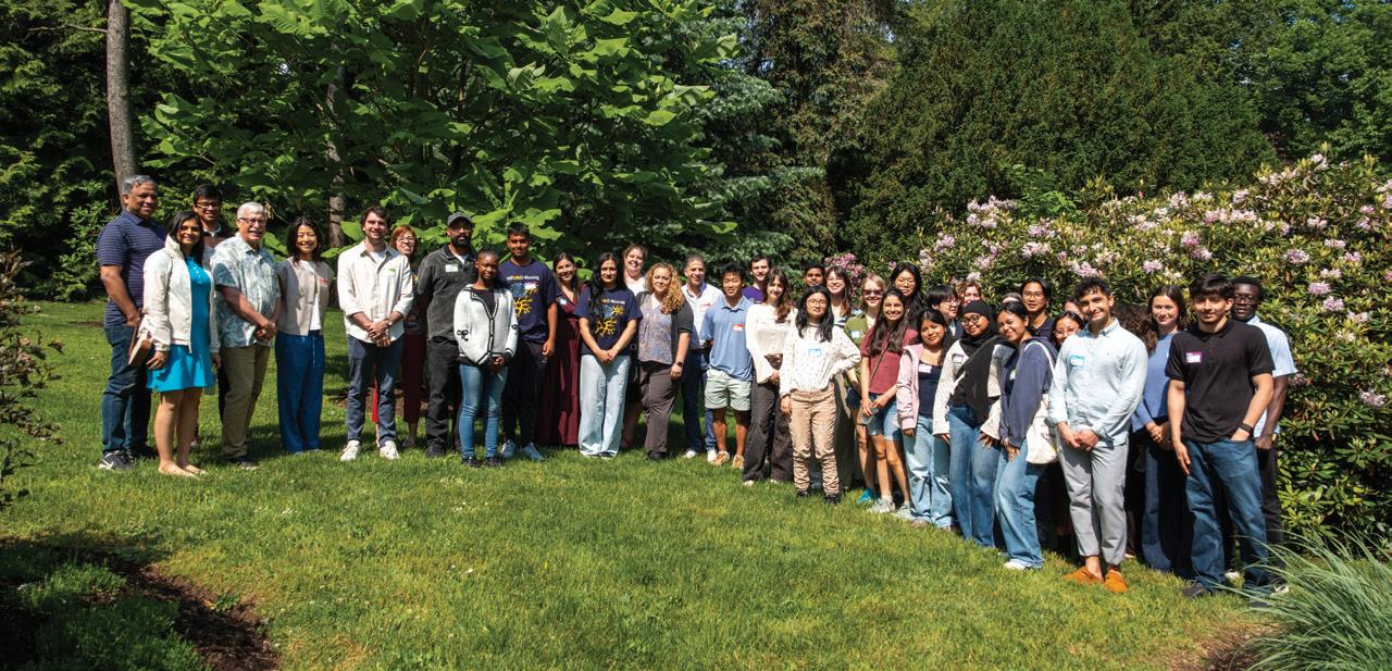
Indoors, the Neuroscience Commission is well into educating future scientists. Local high school students in our NEURO-RocHS program are busy navigating the research process and tackling new challenges as they take on a unique research project that they will present during a poster session. And scholars of our NEUROCITY programs, who are undergraduate students from the City University of New York (CUNY) system, are immersed in the University of Rochester. Living on campus, working in labs, and taking the time to explore the region. These students are also working on a project that they will present at the end of summer at URochester, and again at the Society for Neuroscience conference in November. All our summer scholar students are our future; they will mark the next 250 years of our country’s scientific engine, helping to move science forward, elevate research, and improve the lives of all people.
The more people we have at the forefront of care and investigation, the better we will be able to positively impact the lives of patients and their families.
In April, our annual Neuroscience Graduate Retreat highlighted the impressive research of our graduate students. Dozens of students working in labs across URochester presented on a myriad of neuroscience-related research. This program has continued to recruit highly motivated and skilled scientists from around the world who come to URochester and elevate our research, foster new collaborations, and bring a sense of curiosity to our labs and across our campus. During the retreat, I was privileged to sit down with the keynote speaker, Dr. Anne Hart, professor and chair of Neuroscience from Brown University, for a new episode of Neuroscience Perspectives. She drives home the important role of basic science in research and in our understanding of the human brain.
SCAN TO LISTEN TO NEUROSCIENCE PERSPECTIVES WITH DR. ANNE HART TODAY!
ON THE COVER:
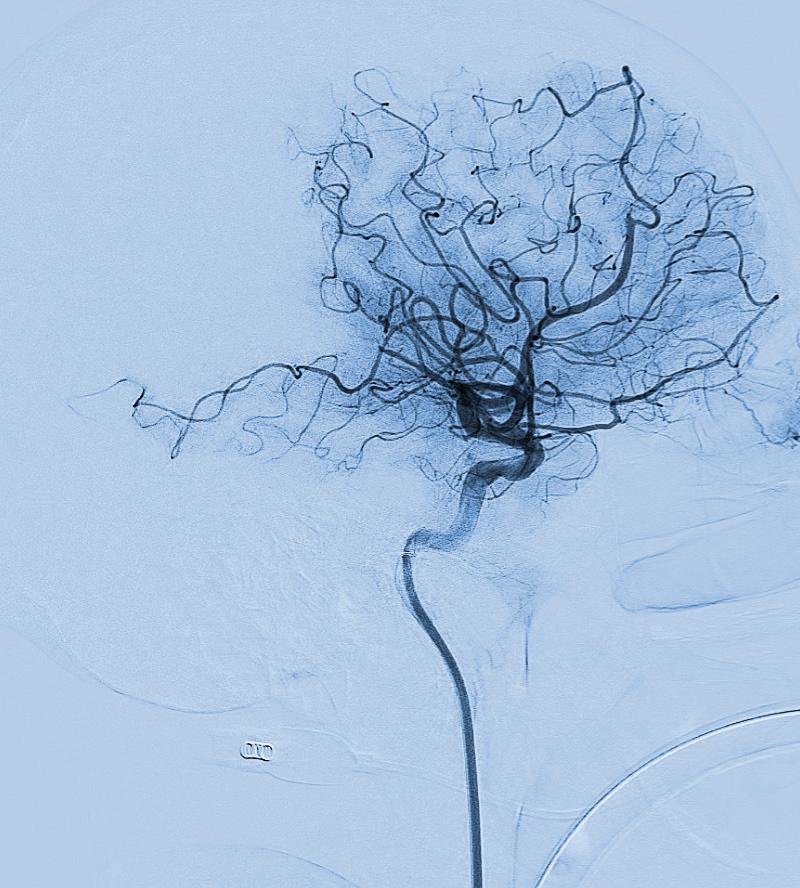
A pre-treatment brain scan of an acute stroke caused by a blockage in the third segment of the middle cerebral artery. Image provided by the Bender Lab.
I took great pride to see the Golisano Children’s Alliance grow again. Fifteen hospitals across the country are now part of this network dedicated to improving children's lives. We are so proud to be the first institution to take the lead in this alliance. Clinicians from the first Golisano Children’s are key partners in the Golisano Intellectual and Developmental Disabilities Institute, a pillar for some of the most vulnerable members of our community.
There is so much good happening in science, medicine, and education, despite the constant swirl of misinformation and the strain on funding resources. It motivates me and should motivate all of us to keep pressing on and moving science forward. Because, despite roadblocks today, there will be science tomorrow.
In Science,
John J. Foxe, PhD
Del Monte Institute for Neuroscience Executive Committee
John Foxe, PhD Chair, Department of Neuroscience
Bradford Berk, MD, PhD Professor of Medicine, Cardiology
Robert Dirksen, PhD Chair, Department of Pharmacology & Physiology
Diane Dalecki, PhD Department of Biomedical Engineering
Jennifer Harvey, MD Chair, Department of Imaging Sciences
Robert Holloway, MD, MPH Chair, Department of Neurology
Paige Lawrence, PhD Chair, Department of Environmental Medicine
Hochang (Ben) Lee, MD Chair, Department of Psychiatry
Shawn Newlands, MD, PhD, MBA Chair, Department of Otolaryngology
Webster Pilcher, MD, PhD Chair, Department of Neurosurgery
Steven Silverstein, PhD Professor, Department of Psychiatry
Duje Tadin, PhD
Interim Dean, School of Arts & Sciences
Editor/Writer
Kelsie Smith Hayduk
Kelsie_Smith-Hayduk@ urmc.rochester.edu
Contributors
Mark Michaud, Bethany Bushen
Feature Photography
John Schlia Photography J. Adam Fenster
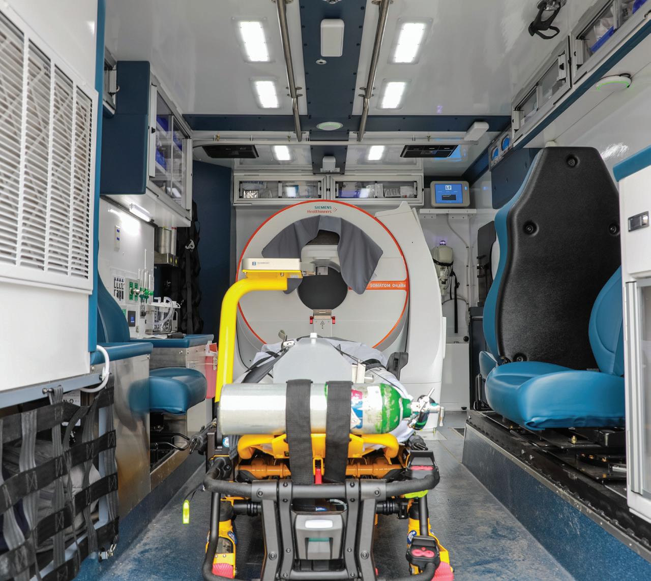
Inside University of Rochester Medicine Mobile Stroke Unit. It is staffed with specialized, stroke-trained personnel, equipped with a CT scanner, and stocked with essential medications necessary for prompt stroke diagnosis and treatment on site, including clot-busting drugs. Volume 29
2 LATEST RESEARCH
Why our brain needs sleep, how our focus shifts, and how our neurons work together as we learn are among the latest URochester neuroscience research findings.
4 THE NEUROSCIENCE-DRIVEN APPROACH TO STROKE
URochester researchers and clinicians use mobile emergency treatment, regional telemedicine networks, and research, including understanding brain plasticity, rehabilitation, sleep, and clot biology to redefine stroke care and recovery.
8
FACULTY Q&A
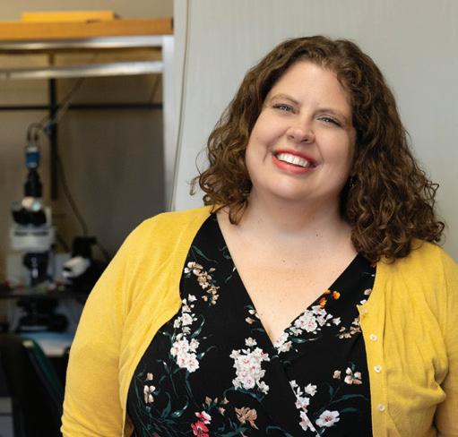
Chigusa Kurumada, PhD, is an associate professor of Brain and Cognitive Sciences at URochester. Her research studies how language is used to share thoughts and intentions.
9
POSTDOC SPOTLIGHT
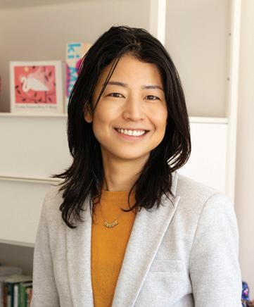
Minju Kim, PhD, is a postdoctoral fellow in the Cognitive Neurophysiology Laboratory at the School of Medicine & Dentistry. Her research focuses on the intersection of cognition and motor function.
Brain Rhythms May Cause Shift in Focus
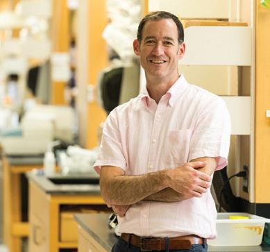
University of Rochester Medicine researchers find that you may not be as focused as you think. Associate Professor of Neuroscience Ian Fiebelkorn, PhD, discovered that our attention is on a cycle, shifting seven to ten times per second. He and postdoctoral fellow Zach Redding, PhD ’24, published this research in PLOS Biology. This rhythmic occurrence may be crucial for survival, as it prevents us from becoming overly focused on one thing in our environment. It could help us to see a car backing up in a parking lot while we search for where we parked, or to duck to avoid a low-hanging tree branch on a walk while watching a kid ride a bike. But these windows that shift our attention could also make us more susceptible to distractions, like screens, digital alerts, and other visual stimuli. These frequent and innate windows for shifting attention may make it easier to be pulled away from a task. This may provide a glimpse into what may cause someone with ADHD to be hyper-focused or have increased distractibility.
Shedding Light on the Brain: New Method Controls Neural Pathways
Understanding how the brain works requires more than studying single regions in isolation. The cerebral cortex depends on long-distance connections that link specialized areas into coordinated networks. But scientists have had limited tools for selectively turning these specific connections “on” or “off” in animal models that most closely resemble the human brain.
Using a refined viral and light-based technique called optogenetics, neuroscientist Kuan Hong Wang, PhD, was able to target only the neurons that connect one brain region to another—and then either activate or silence those same cells on demand. Optogenetics is a method that uses light to control cells genetically modified to respond to it. In neuroscience, it enables researchers to selectively activate or inhibit specific neurons.
The new method, published in Cell Reports Methods, brings us closer to understanding how distributed brain networks support higher-order functions such as perception, decision-making, and social behavior.
How Sleep, Waste Clearance, and Dementia May Be Linked
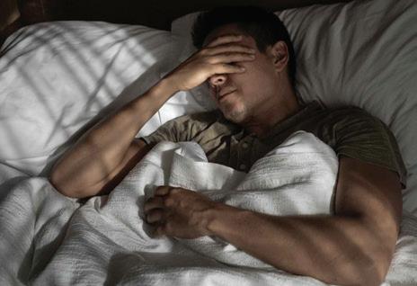
Why are conditions such as chronic stress, depression, cardiovascular disease, fragmented sleep, and aging all associated with a higher risk of dementia? In a new review piece in Science, URochester Medicine neuroscientist Maiken Nedergaard, MD, DMSc, proposes that many of these seemingly different conditions may converge on the same biological problem: disruption of a sleep-dependent brain rhythm that helps clear waste from the brain.
The article presents a new way of thinking about sleep, not simply as a period of rest, but as a highly organized biological state that coordinates brain chemistry, blood vessel movement, and cerebrospinal fluid flow to support the brain’s nightly cleaning process. The piece also points to a potential biomarker, heart rate variability, which can already be tracked with consumer wearables, as a simple, noninvasive way to assess sleep-related brain health and identify people at increased risk for cognitive decline.
AI Reveals How the Brain Clears Harmful Waste
Using MRI scans and AI tools, Professor Douglas Kelley, PhD, and colleagues developed a new approach to measure fluid flow linked to diseases such as Alzheimer’s. Physics-informed AI determined fluid flow velocities from MRI data and videos of dye spreading across brain tissue over time, and researchers built neural networks to deduce how fast the fluid flows and how permeable the brain tissue is.
The results, published in Science Advances, showed two main ways that the glymphatic system, the brain’s waste-clearing system, washes away particles in the brain, such as the amyloid beta proteins linked to Alzheimer’s disease, and one of these ways is much faster than the other. The fast flow of the glymphatic system’s waterlike fluid moves at a few microns per second around the brain’s open regions, such as the surface between the skull and the brain, while the slower flow of the waterlike fluid trickles through the brain’s deep tissue at a rate about 50 times slower. Eventually, researchers hope to compare fluid flow in healthy and sick brains, as well as in young and old brains, with aspirations to study circulation in humans.
Learning Makes Brain Cells Work Together, Not Apart
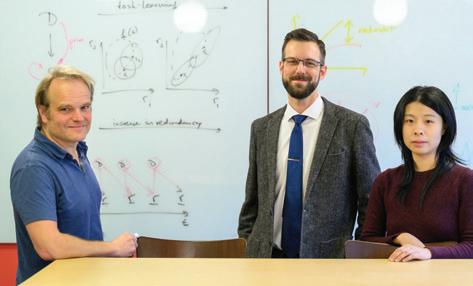
A study challenges a long-standing theory in neuroscience and could reshape how scientists think about perception, learning disorders, and artificial intelligence. URochester researchers tracked the activity of the same small networks of neurons in the visual cortex over several weeks as subjects learned to tell apart different visual patterns. They discovered that before learning, neurons mostly worked independently. But as subjects honed their visual skills, the neurons started to behave more like a well-trained sports team, communicating and working together in a coordinated way. Importantly, this coordinated effect only appeared when subjects were actively performing a task and making decisions based on what they saw. When they passively looked at the same images without needing to respond, the effect disappeared. The most important neurons for the task showed the biggest boost in coordination, especially at the moments when decisions were made. But these are flexible, not permanent, changes. The researchers believe these shifts are guided by feedback signals from higher-level brain areas, allowing neurons to adjust their behavior on the fly, depending on the task.
From Dish to Brain: Researchers Chart Human Glial Cell Maturation
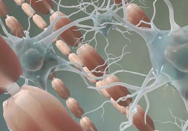
URochester researchers developed a new method showing that human glial progenitor cells (GPCs) are a promising and safe cell product for transplantation. The research also defines the transcriptional and epigenetic signatures of these cells as they mature into astrocytes and oligodendrocytes, two essential support cell types in the brain. Previously, transplanting pluripotent stem cells directly into mice or humans has been shown to be dangerous, causing a teratoma to form, a benign tumor that can cause severe neurological complications by occupying critical space in the brain. In this study, led by John Mariani, PhD, and published in Nature Communications, the GPCs showed no pluripotent signatures in culture, and mature identity persisted after transplantation into mice, indicating that the cells were highly specified, functional, and safe.
This transplantation model may have implications for disorders such as multiple sclerosis, leukodystrophies, and Huntington’s disease, in which myelination, the insulating process that helps nerve cells communicate efficiently, is disrupted.
From left: Ralf Haefner, PhD, Adam Snyder, PhD, and graduate student Shizhao Liu are authors of this research published in Science. (University of Rochester photo / J. Adam Fenster)
(University of Rochester photo / J. Adam Fenster)
Douglas Kelley, PhD
REDEFINING STROKE CARE & RECOVERY THROUGH NEUROSCIENCE & INNOVATION
By Mark Michaud
Every minute after a stroke, nearly two million brain cells die. But increasingly, researchers and clinicians are demonstrating that stroke care is not only about restoring blood flow in the first critical hours; it is also about understanding how the brain repairs, reorganizes, and recovers in the weeks and months that follow.
At URochester Medicine, that work spans the entire continuum of stroke care, from mobile emergency treatment and regional telemedicine networks to research exploring brain plasticity, rehabilitation, clot biology, and the roles of sleep and circadian rhythms in recovery. These efforts are helping shape a more personalized and neuroscience-driven approach to stroke treatment and recovery.
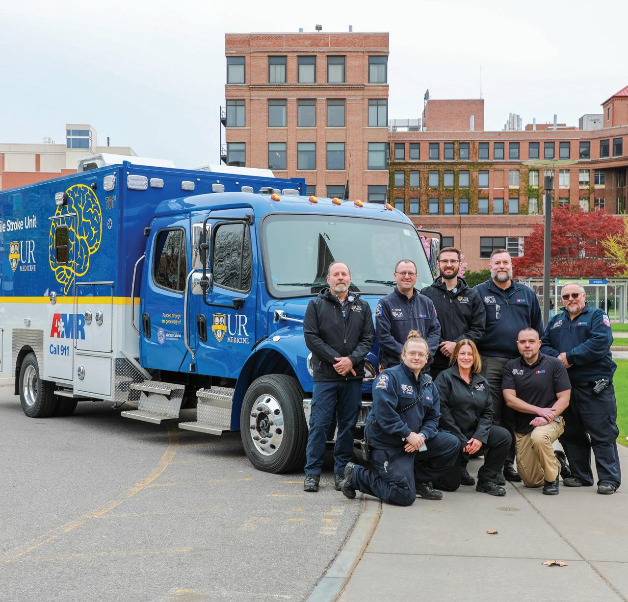
From left: Frank Post, Nate Coons, Joshua Lesperance, Jason Burgett, Nathan Young, Lena Brockmann, Zachary Callahan, and Justin Connolly are members of URochester Medicine/AMR Mobile Stroke Unit team.
Building a Regional System of Stroke Care
URochester Medicine’s clinical stroke program is built around a regional system of care that begins before patients reach the hospital and extends through emergency treatment, intensive care, rehabilitation, and long-term recovery. At the center of that system is the Comprehensive Stroke Center at Strong Memorial Hospital, which includes the Neuromedicine ICU and Acute Brain Injury Rehabilitation Unit and brings together specialists in stroke neurology, neurosurgery, neurocritical care, rehabilitation medicine, and imaging.
One of the program’s most visible innovations is the Mobile Stroke Unit (MSU). Introduced in 2018, the specially equipped ambulance functions as an emergency department on wheels, carrying a CT scanner, point-of-care laboratory testing, telemedicine capability, and clot-busting medications directly to patients in the field. Earlier this year, URochester Medicine expanded the program with a second MSU, extending the reach of rapid stroke treatment across the region.
“The Mobile Stroke Unit allows us to deliver specialized stroke care at the earliest possible moment, before a patient even reaches the hospital,” said neurosurgeon Tarun Bhalla, MD, PhD, director of the MSU program. “The second unit builds on years of field experience, expands access to rapid diagnosis and treatment, when every minute can mean the difference between recovery and disability.”
The Telestroke program extends that reach even further. Led by neurologist Adam Kelly, MD, the service provides 24/7 remote stroke and neurological consultations for 16 hospitals across upstate New York and now fields more than 4,000 calls annually. Through video consultations and rapid review of imaging and clinical data, stroke specialists help determine which patients require advanced interventions—such as thrombectomy, ICU care, or complex vascular procedures—and which can remain at their local hospital.
“By understanding hy how the brain adapts after injury, scientists are developing therapies that are more personalized.”
the region,” said neurologist Curt Benesch, MD, medical director of the Comprehensive Stroke Center. “We’re not just treating the stroke in front of us; we’re constantly working on how to prevent the next one, expand acute treatment options, and make sure that evidence-based care reaches every bedside.”
Relearning Movement After a Stroke
Stroke recovery does not end when a patient leaves the hospital.
Increasingly, researchers at URochester Medicine are showing that the brain retains a remarkable ability to adapt, relearn, and reorganize after injury—a property known as plasticity— and that understanding those mechanisms may transform rehabilitation.
The model strengthens stroke expertise while preserving highly specialized resources for the patients who need them most. It also creates a powerful platform for research by connecting investigators to a large, diverse patient population and by accelerating the translation of new discoveries into clinical care.
“What makes our stroke program unique is that we cover the entire spectrum—from basic neuroscience to innovative recovery strategies, clinical trials, and a highvolume stroke care network that reaches patients across
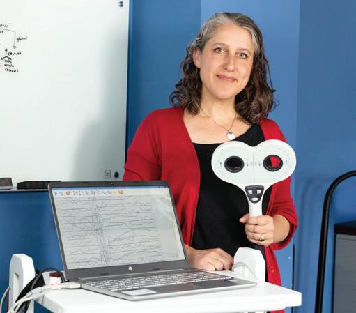
In the Movement and Plasticity Lab, stroke neurologist Ania Busza, MD, PhD, studies how the brain relearns movement after stroke and how rehabilitation can better harness that recovery process. Her work focuses on motor system plasticity, the way practice and experience reshape surviving brain circuits after injury. “Recovery after stroke isn’t just about getting stronger,” Busza said. “It’s about helping the brain learn how to use the remaining circuits it still has.”
For decades, rehabilitation often followed a relatively simple principle: more therapy leads to better outcomes. But researchers now recognize that stroke patients develop very different motor control problems depending on which neural pathways are damaged. Some struggle with delayed
Ania Busza, MD, PhD
movement, others with fatigue or involuntary activation of opposing muscles.
“Two people may both have weakness after a stroke, but the underlying control problem may be completely different,” Busza said. “If we understand those differences, we can start designing rehabilitation that is much more personalized and targeted.”
Her lab combines neuroscience, imaging, biomechanics, engineering, and rehabilitation science to better understand those differences. In one project, patients use faint electrical signals from weakened muscles to control a simple video game through surface electromyography (EMG). Even patients unable to lift their arms can participate. The system helps researchers track how motor impairments evolve during the first months after stroke and identify early warning signs of long-term disability.
Researchers are also studying whether interventions such as vagal nerve stimulation, transcranial magnetic stimulation, and other neuromodulation approaches can safely amplify the brain’s natural plasticity and improve recovery.
The broader goal is to understand not only whether patients recover, but also how recovery occurs within the brain itself. Modern neuroscience has changed how researchers think about that process. Rather than the brain simply “rewiring” around damage, scientists now believe recovery depends on whether alternative, partially redundant circuits can be strengthened and refined through practice.
“Practice after stroke is really a form of learning,” Busza said. “The brain is constantly adapting based on experience, and rehabilitation is trying to shape that adaptation in a productive way.”
Restoring Vision After a Stroke
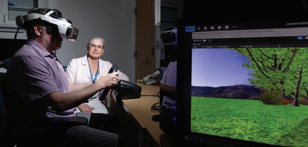
Krystel Huxlin, PhD, is studying another often-overlooked consequence of stroke: vision loss. Her research focuses on patients whose strokes damage the visual cortex in the occipital lobe, leaving them partially blind. Historically, many of these patients were told little could be done. “There has been a longstanding belief that vision loss after stroke is permanent and untreatable,” Huxlin said. “Our work has shown that it is not true.”
Huxlin’s Visual Perceptual Restoration Laboratory has demonstrated that intensive visual training can help patients recover some visual abilities in damaged portions of the visual field. The approach works much like physical therapy for movement recovery, in which repeated, carefully structured practice trains surviving brain circuits to process visual information more effectively.
“We’re essentially retraining the visual system,” Huxlin said. “The damaged brain still has some capacity to change, but you have to engage it in the right way.”
The work has implications beyond vision rehabilitation. Huxlin says it reflects a major shift in neuroscience itself. A few decades ago, the idea that the adult brain could meaningfully reorganize after injury remained controversial. Today, plasticity is recognized as a lifelong feature of the nervous system.
Her lab now uses advanced imaging, eye tracking, stateof-the-art virtual reality headsets, and brain stimulation to better understand the mechanisms behind visual recovery and make rehabilitation more accessible to patients at home.
Together, the work of Busza, Huxlin, and their collaborators reflects a larger transformation in stroke
Krystal Huxlin, PhD
research. By understanding how the brain adapts after injury, scientists are developing therapies that are more personalized, more targeted, and increasingly grounded in the biology of recovery itself.
What Stroke Clots Can Reveal
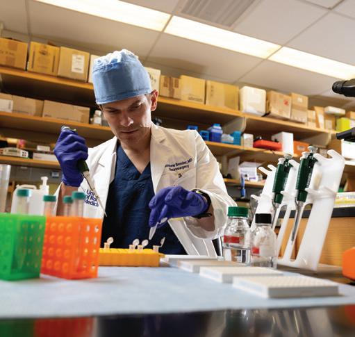
To neurosurgeon Matthew T. Bender, MD, the clot removed during a stroke procedure is more than medical waste—it is a source of critical information about why strokes happen and how patients respond to treatment.
Bender’s research focuses on the molecular composition of stroke clots, particularly in patients with large-vessel occlusion strokes treated with thrombectomy. By analyzing proteins and inflammatory markers within these clots, his team hopes to better understand why some strokes are harder to treat and why outcomes vary dramatically between patients.
“Some stroke researchers find the clots themselves to be bland or uninteresting,” Bender said. “We feel the opposite very much.”
His work has identified proteins linked to more difficult clot removal, worse recovery, and complications such as hemorrhage. The long-term goal is to develop blood-based biomarkers that could help physicians tailor stroke treatment to individual patients.
Conducting that work at a high-volume stroke center is critical. URochester Medicine performs more than 200 thrombectomies each year, giving researchers access to blood and clot samples that would otherwise be discarded.
“Those biospecimens are invaluable,” he said. “They allow us to study how clots form in real human stroke patients and develop better ways to break them down.”
The Role of Sleep, Timing, and the Brain’s 'Cleaning System'
Stroke has traditionally been viewed as a problem of blocked blood flow, but emerging research suggests it may also be a disorder of timing.
Neuroscientist Lauren Hablitz, PhD, studies how sleep, circadian rhythms, and the brain’s glymphatic system—a network that clears waste from the brain during sleep— influence stroke recovery. The glymphatic system, first discovered in the lab of URochester Medicine neuroscientist Maiken Nedergaard, MD, DMSc, appears to become impaired after stroke, potentially allowing inflammatory molecules and cellular waste to accumulate.
“Our central question became: if timing is broken after stroke, can we improve recovery by reinforcing the biological clock?” Hablitz said.
Using mouse models, researchers tested interventions designed to strengthen circadian rhythms, including timed feeding and experimental clock-targeting drugs. Both approaches improved glymphatic function, reduced inflammation, and improved recovery after stroke.
Rather than viewing recovery solely as an inflammation problem, Hablitz believes researchers may also need to consider the brain’s ability to clear harmful signals and metabolic waste. “If the system responsible for clearing signaling molecules is not working properly,” she said, “everything builds up.”
The findings suggest that relatively simple interventions— such as timed eating, light exposure, or therapies that improve sleep and circadian health—could one day become part of stroke rehabilitation strategies.
Matthew Bender, MD
Lauren Hablitz, PhD
Q&A with Chigusa Kurumada, PhD
Chigusa Kurumada, PhD, is an associate professor of Brain and Cognitive Sciences at URochester. Kurumada first came to the University for her postdoctoral training. She has a PhD in Linguistics from Stanford University, an MA in Language and Information Sciences from the University of Tokyo, and a BA in Cross-Cultural Studies from Kyoto Prefectural University. Her research studies how we understand language. Kurumada is also dedicated to her students. As a mentor, she co-founded URochester Women in the Neurosciences (URWINS) to support scientists at all career levels.
Please summarize your research.
My research studies how language is used to share thoughts and intentions. In our daily conversations, we communicate information at a remarkable speed—on average, about 2.5 words every second—and convey many layers of meaning. There is also significant variability in how people use language: factors such as age, gender, accent, and language background can affect our voices and how we speak. I am interested in how we can still understand each other, despite all the variability and time pressure.
How did you become interested in your field?
Since I was a young child, I have been fascinated with how language works. When a news anchor made a small mistake, I was always happy to point it out to my parents. I also liked thinking about how knock-knock jokes (or their Japanese equivalents) and other wordplay work, or how irony and sarcasm can be so powerful in conversation. The pivotal moment in my early scientific life was when I was a college student and worked as a volunteer Japanese language teacher at two primary schools in Australia. I was
fascinated by how young children learn to speak, sometimes in more than one language. That’s when I decided to pursue a PhD in linguistics.
What brought you to the URochester?
The Department of Brain and Cognitive Sciences has long been a world leader in psycholinguistics and language development, two fields that have strongly influenced my research. I was also drawn to Rochester’s collaborative culture, where researchers regularly work across disciplines to solve complex problems. The opportunity to collaborate with colleagues from various areas of cognitive science— like language and vision—and to co-mentor students with innovative ideas was particularly exciting. More than a decade later, that interdisciplinary spirit remains one of my favorite aspects of being at URochester.
What is your favorite piece of advice?
I received some particularly helpful advice from a senior colleague in the field. She told me that there is no single “right” way to be a successful researcher. Her advice helped me refocus on building a research program that reflects my own goals, values, and strengths—and it continues to guide how I lead my lab today.
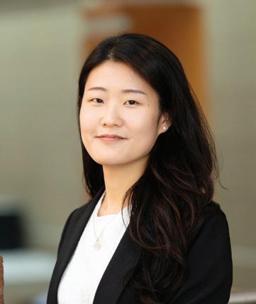
Minju Kim, PhD, is a postdoctoral fellow in the Cognitive Neurophysiology Laboratory (CNL) at URochester School of Medicine & Dentistry. She received her BS in electrical engineering and a PhD in human factors engineering from Ulsan National Institute of Science and Technology (UNIST) in South Korea. She completed doctoral research in the lab of Sung-Phil Kim, PhD, one of the pioneers of braincomputer interface (BCI) in Korea. Today, her research focuses on this intersection of cognition and motor function to hopefully inform the development of better treatments and technologies for older adults and people with neurological conditions.
“My research asks what happens in the brain when people walk and think at the same time,” said Kim, whose interest in the human mind and behavior began long before she started studying the brain. “I’ve always been curious about why people think and behave the way they do. That curiosity led me to pick up books about the brain. Reading about how the brain works was captivating, and I became interested in how technology could be used not only to understand the brain but also to help people in practical ways. That interest eventually led me to engineering.”
As a postdoc at URochester, Kim can extend her research into clinical collaborations. Working with her lab’s principal investigators, John Foxe, PhD, and Ed Freedman, PhD, Kim uses mobile brain/body imaging
(MoBI) to understand better how neurological diseases like Parkinson’s affect a person’s ability to walk and complete a simple task. “I investigate how the interference between cognitive and motor function arises in those populations using brain recordings and movement measurements. My long-term goal is to develop technologies and assessments that can support people who have difficulty moving or interacting with their environments by combining the knowledge and experience I gain at URochester with my background in BCI.”
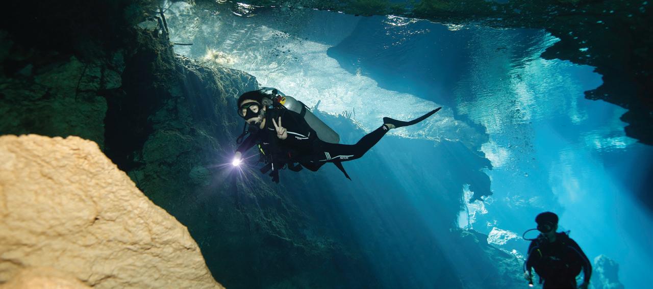
Outside of the lab, Kim enjoys exploring the Rochester area. “I love visiting parks, lakes, trails, and small villages around the region.” She also enjoys time away from land. Scuba diving into, as she puts it—a different world underwater. “In some ways, scuba diving feels similar to research for me. If you pay attention to subtle signals, you can discover things that are usually hidden from view.”