UNIVERSITY OF MICHIGAN
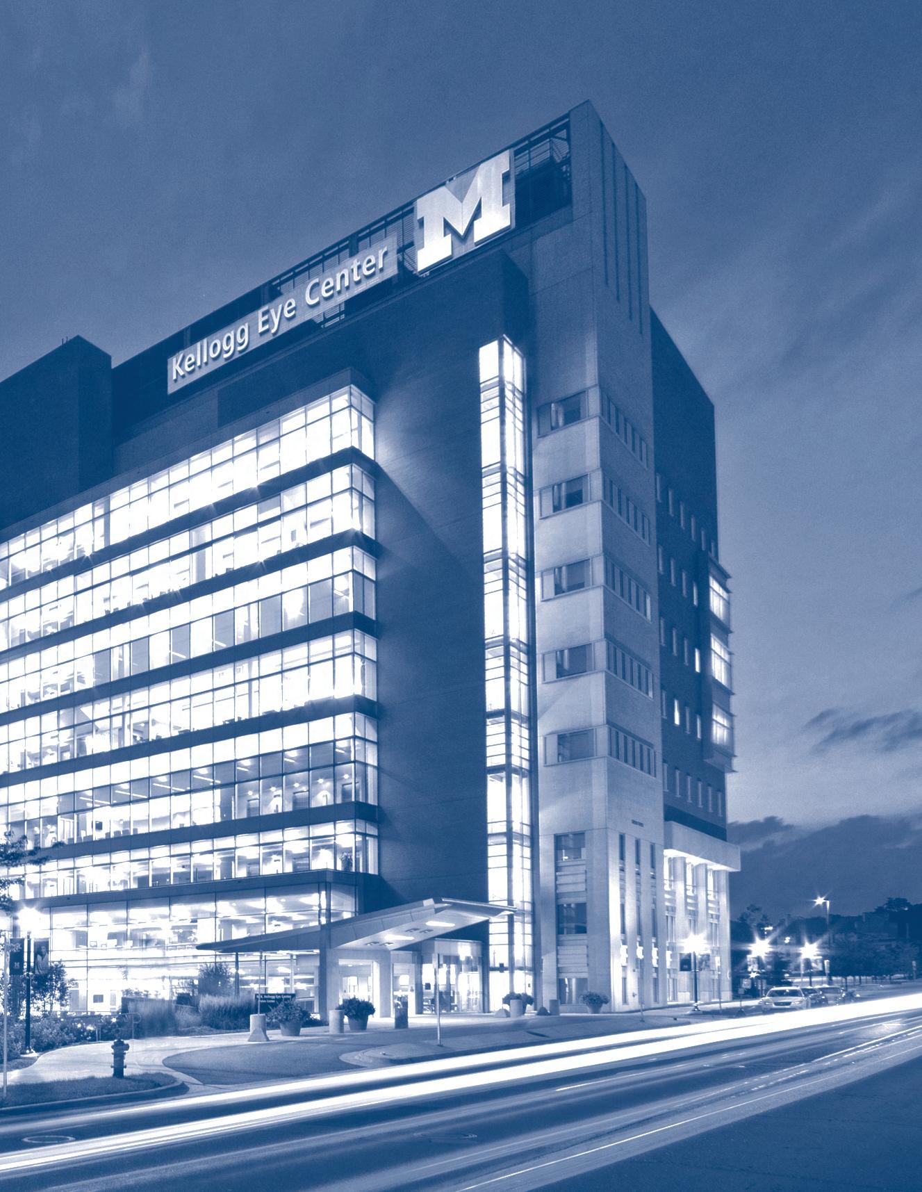
W.K. KELLOGG EYE CENTER


An Out-of-This-World Perspective on Residency from one of Forbes’ Thirty-Under-Thirty
19 Kellogg Post-Doc Receives Prestigious NIH grant
20 Kellogg PGY4 Sole Resident on ACGME Residency Program Review Committee
21 Pre-Med Awarded NIH Research Supplement 22 2023-2024 Heed Fellows
Philanthropic Support
33 The Alan Sugar, M.D., Research Professorship
34 Honoring the Visionary Leadership of Paul P. Lee, M.D., J.D.
35 The Edna H. Perkiss Research Professorship Improving Vision Worldwide
36 Partnership Between U-M Med School, Business School, and the Kellogg Eye Center Drives Latest Kenya Initiative
Kellogg Updates
Alumni Highlights
38 First-Year and Graduating Residents 39 Departing Clinical Fellows 40 New Faculty 41 Faculty Honors and Recognition
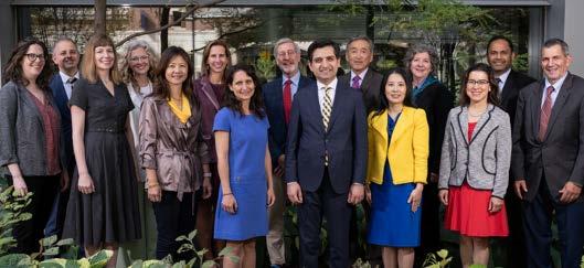
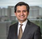
After marking 150 years of ophthalmology at the University of Michigan last year, the Kellogg Eye Center has kicked off our second sesquicentennial with another year of amazing achievements.
If there’s a common theme to our 2023 report, it’s the power of teamwork. Within our center, across the University and around the globe, our faculty have cemented longstanding partnerships and forged new, sometimes surprising ones to tackle the most vexing clinical and research questions, and to prepare the next generation of leaders in ophthalmology and vision science.
From the rarest inherited retinal dystrophies to the most severe ocular surface injuries, when presented with complex clinical challenges, team care is the standard of care at Kellogg. One example shared here is a baby who, at less than 12 hours old, was rushed to U-M with acute ophthalmic and systemic issues. Kellogg specialists in pediatric ophthalmology, genetics, retina, glaucoma and cornea came together to diagnose her rare genetic syndrome and perform the surgeries that will give her the best possible chance at vision.
This year’s research highlights include the renewal of four major institutional grants, the recruitment of new faculty, and the opening of our new image-guided medical robotics lab—all evidence that Kellogg is essentially built for team science. With the resources and support only available at a top academic eye center, our investigators initiated numerous novel projects in 2023, in partnership with experts in data analytics, artificial
intelligence, gene sequencing bioengineering, psychiatry and more.
In Kellogg’s model of ophthalmic education, teamwork is taught by example. Students, residents and fellows are encouraged to collaborate in pursuit of their own unique areas of interest. That approach resulted in a remarkably diverse group of learners in 2023, including a medical student helping with a vision clinic expansion in Kenya, and a resident who contributed to a textbook devoted to spaceflight-associated neuroocular syndrome, a condition which, as the name implies, is exclusive to astronauts.
I joined the Kellogg faculty in 2002, and not a day has passed since that I haven’t marveled at the compassion, talent and collegiality that fuel this amazing place. As a national search for our next leader continues, it is my honor to serve as Interim Chair.

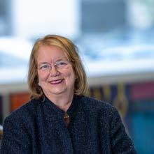
Shahzad I. Mian, M.D. Interim Chair and Professor, University of Michigan Department of Ophthalmology and Visual Sciences
Dr. Lee Assumes Expanded Role at Michigan Medicine
The spirit of teamwork that drives Kellogg was driven by Emeritus Chair Paul Lee, M.D., J.D. Dr. Lee captained the Kellogg team from 2012 to 2022. In his new role as Senior Associate Dean of Clinical Affairs in the U-M Medical School and Executive Director of the U-M Medical Group, his talent and vision are now shaping the delivery of clinical care across all of University of Michigan Health.
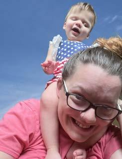
Aspen Schuelke was born March 1, 2022, in Jackson, Michigan, 40 miles west of Ann Arbor. Right away, doctors had several concerns, especially about her eyes.
Aspen’s eyes appeared small and cloudy. Her intraocular pressure (IOP) was very high, which had caused protruding scar tissue to develop on her right eye.
Less than 12 hours after her birth, Aspen was transferred to U-M C.S. Mott Children’s Hospital. There, she would be cared for by a multidisciplinary team, including a number of experts from Kellogg.
Her Kellogg team would eventually include childhood glaucoma specialist Adam Jacobson, M.D., pediatric retina specialist Cagri Besirli, M.D., Ph.D., cornea specialist Shahzad Mian, M.D., and ophthalmic genetics specialist Lev Prasov, M.D., Ph.D.
“Along with severe corneal issues and glaucoma, Aspen was diagnosed with aniridia, a disorder that results in eyes without irises,” explains Dr. Jacobson. “Her other doctors identified additional issues, including trouble taking nourishment, poor muscle tone, and a heart defect.”
Dr. Jacobson wondered if one syndrome might be behind Aspen’s many medical challenges. “One clue pointing to a genetic explanation is that her mother has anterior segment dysgenesis, or ASD,” he says. In ASD, the tissue in the front of the eye does not develop normally, increasing the risk of glaucoma and cataracts. “Allison has a relatively mild form of glaucoma, which
“
SHE SEEMS TO TRACK THINGS BETTER, ESPECIALLY BLACK AND WHITE OR HIGH CONTRAST OBJECTS. MORE IMPORTANTLY, SHE CONTINUES TO THRIVE.
SHE’S A LOVING, LOVABLE, HAPPY-GO-LUCKY BABY, WHO INSPIRES US EVERY DAY.
“
Allison Schuelke
has been monitored here at Kellogg for years.”
He consulted with Dr. Prasov, who evaluated daughter and mother in the Kellogg Multidisciplinary Ophthalmic Genetics Clinic (MOGC) together with the pediatric genetics team of Dr. Amanda Pritchard, M.D., and genetic counselor Adelyn Beil, M.S., CGC, M.P.H. Genetic testing identified that both carry a mutation in the gene FOXC1. “Aspen has a form of ASD associated with FOXC1 called Axenfeld-Rieger Syndrome,” explains Dr. Prasov. “Allison, who had never had genetic testing before, was found to carry the same FOXC1 variant, but in her case it has far less impact.”
While the genetic evaluation proceeded, the immediate challenge was stabilizing Aspen’s IOP. “Patients with this type of glaucoma do not tend to respond well to medications, and often require surgical intervention,” says Dr. Jacobson. “We first attempted to insert tubes to lower the pressure, but in such small, underdeveloped eyes, there was no room to safely place them. We instead performed a laser procedure, which has successfully kept the intraocular pressure controlled.”
In her first months of life, Aspen underwent multiple exams under anesthesia so that the team could monitor her IOP, the condition of her retinas, and the progressive scarring on her corneas. By the spring of 2023, the group was debating whether Aspen should receive a cornea transplant.
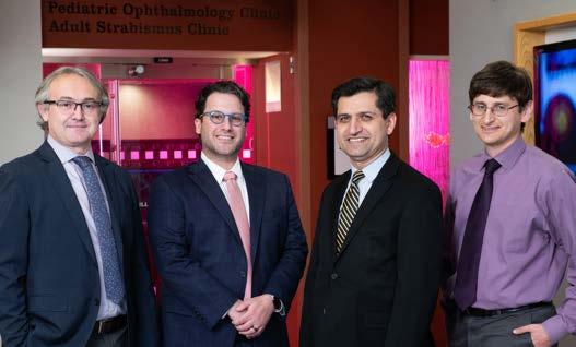
“There’s a difficult risk/reward calculation when considering cornea transplantation for a child,” explains Dr. Mian. “On top of the general risks of surgery under anesthesia, corneal grafts are more likely to fail or be rejected in young patients.
“Those risks need to be balanced with the potential benefit to the child’s vision,” he continues. “When a clouded cornea blocks light from entering the retina, the visual pathways between the retina and the brain don’t develop properly. Delay the transplant too long, and you may miss the window for improving vision.”
“
In June 2023, after extensive consultation with the care team and Aspen’s parents, Dr. Mian operated on Aspen’s right eye, removing the enlarged, opaque cornea with the lens embedded in it, and transplanting a new cornea.
dad have been essential members of the team from day one.”
There is a long road ahead for Aspen and her family. In addition to monitoring her IOP, which may one day require additional surgery, a procedure is being planned on the right eye to further reduce the scarring under the cornea of the right eye. If possible, the left eye may eventually receive a cornea transplant.
PROVIDING THE COMPLEX CARE
ASPEN NEEDS IS TRULY A TEAM EFFORT, AND ASPEN’S MOM AND DAD HAVE BEEN ESSENTIAL MEMBERS OF THE TEAM FROM DAY ONE.
“
Shahzad Mian,
“While we understood the risks, we really advocated for the procedure,” says Allison Schuelke. “To give Aspen the best possible chance at vision, we need her developing brain to receive as much visual input as possible.”
“Providing the complex care Aspen needs is truly a team effort,” says Dr. Mian, “and Aspen’s mom and
As Team Aspen at Kellogg addresses the ocular aspects of Aspen’s Axenfeld-Rieger Syndrome, Team Aspen at Mott continues to tackle the systemic challenges of the syndrome. Not yet two years old, Aspen has already endured countless specialty clinic evaluations, therapies and procedures to improve her cardiovascular, digestive and musculoskeletal health.
M.D.
Nonetheless, at the Schuelke home, there is plenty of room for optimism. “We’ve noticed small improvements in Aspen’s vision,” Allison says. “She seems to track things better, especially black and white or high contrast objects. More importantly, she continues to thrive. She’s a loving, lovable, happy-go-lucky baby, who inspires us every day.”
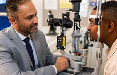
“
THERE’S A VICIOUS CYCLE OF VISION LOSS AND MENTAL DISTRESS. DISTRESS TENDS TO INCREASE AS VISION DECREASES, AND THE MORE PATIENTS STRUGGLE WITH EMOTIONAL ISSUES, THE LESS LIKELY THEY ARE TO FULLY BENEFIT FROM LVR.
“
K. Thiran Jayasundera, M.D., M.S.
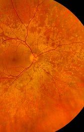
Inherited retinal diseases (IRDs) are a group of rare retinal diseases that damage the light-sensitive cells in the retina, causing progressive vision loss.
Different IRDs result in different low vision (LV) challenges. LV refers to vision problems that cannot be fully resolved with corrective lenses, medical or surgical therapies. Depending on their diagnosis, IRD patients may experience worsening central vision or peripheral vision, color blindness, night blindness, light sensitivity, blurred vision, or difficulty seeing details.
Retina specialist K. Thiran Jayasundera, M.D., M.S., is dedicated to improving vision outcomes for IRD patients. In clinical practice, he often refers patients to Kellogg’s Low Vision and Visual Rehabilitation Service. The service provides comprehensive low vision rehabilitation (LVR) tailored to individual patients’ needs, combining technologies, devices, environmental modifications and skills training to help maximize their remaining vision and maintain independence in daily living.
Dr. Jayasundera’s latest research project focuses on the mental health of IRD patients. The two-part project is funded by an NIH R01 grant.
“Understandably, many patients coping with worsening vision also struggle with anxiety and depression,” he explains. “There’s a vicious cycle of vision loss and mental distress. Distress tends to increase as vision decreases, and the more patients struggle with emotional issues, the less likely they are to fully benefit from LVR.”
The first objective of the project is to study the interplay between functional vision impairments and
emotional distress in patients with IRDs. Participants will undergo functional vision testing and general depression screening. They, will also complete two surveys designed specifically for IRD patients to self-report their vision disabilities, anxiety and depression. Both surveys were developed and validated by Dr. Jayasundera.
The project’s second component is a clinical trial of a therapeutic intervention. Participants will receive a targeted combination of LVR and psychotherapy using a template technique called emotion regulation therapy (ERT). ERT integrates components of cognitive-behavioral, acceptance, dialectical, mindfulness and experiential therapies. Developed by David Fresco, Ph.D., Co-Investigator on the R01, ERT has been proven effective in reducing anxiety and depression by addressing the underlying factors that worsen distress.
“Based on the IRD diagnosis, we know how to individualize a patient’s LVR,” he explains. “This study adds a component of psychotherapy, also individualized to patients’ reported symptoms. We hypothesize that when combining tailored psychotherapy with tailored vision rehabilitation, each will bolster the other. With relief from their anxiety and depression symptoms, patients will be better able to comply withparticipate in rehabilitation, and achieve the best possible vision results.”
If shown effective in this clinical trial, this approach could be adapted for other vision-related conditions that evoke distress.
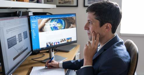
More than half of the 14 million people in the U.S. living with vision impairment (VI) or blindness are age 65 and older. Racial and ethnic minorities are disproportionately affected, and the number of people suffering from VI is anticipated to double by 2050 as our population ages. Social determinants of health (SDoH) like healthcare access and economic stability play a large role in shaping who is impacted.
Older adults with VI are thought to be at elevated risk for many adverse outcomes, including dementia, admission to a long-term care facility, and even death.
a battery of objective measures of visual function to gauge respondents’ distance and near vision and contrast sensitivity.
Dr. Ehrlich played a pivotal role in developing, testing and validating those measures. He can now begin analyzing the two years of available data, adding an additional year’s data annually throughout the grant’s five-year duration. He will be assisted by Kellogg investigators Lindsey De Lott, M.D., M.S., and Angela Elam, M.D., M.P.H., and by colleagues at the U-M Institute for Social Research, where he also has a faculty appointment.
“
UNTIL NOW, WE HAVEN’T HAD NATIONALLY REPRESENTATIVE LONGITUDINAL DATA IN THE U.S. ON THE CONNECTION BETWEEN VI, SDOH AND CRITICAL LATE-LIFE OUTCOMES.
Kellogg clinician-scientist Joshua Ehrlich, M.D., M.P.H., is painting a more precise picture of the impact of age-associated vision loss. A leader in vision-related population and health services research in the U.S. and around the world, Dr. Ehrlich has been awarded a National Institutes of Health (NIH) R01 grant to analyze newlycollected data on the vision health of U.S. seniors— a data set he helped develop.
The project has three aims. First, to describe the state of vision impairment in older adults. Second, to analyze the impact various SDoH have on VI late in life. And finally, to study the impact over time of VI and blindness on the development of dementia, placement in a long-term care facility, and mortality—all factors captured in NHATS.
Joshua Ehrlich, M.D., M.P.H.
Since 2011, the NIH has funded the National Health and Aging Trends Study (NHATS), which, through annual in-person surveys conducted in respondents’ homes, gathers health data annually on more than 7,000 adults 65 years and older. In 2021, NHATS added
“
With this project, Dr. Ehrlich’s team is responding to key strategic research goals included in the National Eye Institute’s five-year strategic plan, as well as international research priorities articulated by the Lancet Global Health Commission on Global Eye Health, a worldwide consortium of experts of which Dr. Ehrlich is part.
“Until now, we haven’t had nationally representative longitudinal data in the U.S. on the connection between VI, SDoH and critical late-life outcomes,” he explains. “These studies are the first step in closing that knowledge gap.”
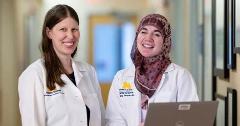
The Eye Plastic, Orbital and Facial Cosmetic Surgery Service at Kellogg provides comprehensive evaluation and treatment of conditions of the eyelids, orbit, and lacrimal (tear drainage) system arising from diseases, malignancies, congenital abnormalities, trauma, or aging. In addition to providing comprehensive, multidisciplinary oculoplastics care, the program is known internationally for excellence in research and education.
The physician team includes Section Lead Vinay Aakalu, M.D., M.P.H., Victor Elner, M.D., Ph.D., Denise Kim, M.D., Christine Nelson, M.D., and ocular oncology specialist Hakan Demirci, M.D. To continue to meet a large and growing clinical volume, two additional oculoplastics surgeons, Brittany Simmons, M.D., and Elaine Downie, M.D., joined the team in 2023.
“
teaching alongside clinical practice. Dr. Downie plans to receive faculty training to develop as an educator, and Dr. Simmons is involved in building a resident education curriculum in oculoplastics.
“Kellogg is now home to one of the largest and most comprehensive oculoplastics programs in the country,” says Dr. Aakalu. “We are the region’s high volume referral hub.”
The expansive program includes specialty care for patients with rare and debilitating conditions such as congenital anophthalmos—a syndrome in which a baby is born without one or both eyes. Dr. Nelson has led groundbreaking research into the condition, and developed a regional referral program, in collaboration with Kellogg’s recently expanded Ocular Prosthetics Service.
“ KELLOGG IS NOW HOME TO ONE OF THE LARGEST AND MOST COMPREHENSIVE OCULOPLASTICS PROGRAMS IN THE COUNTRY.
The Oculoplastics program continues to benefit from consultation with Terry J. Smith, M.D., Professor Emeritus of Ophthalmology and Visual Sciences and Internal Medicine. Dr. Smith’s game-changing translational research led to the development of teprotumumab—the first FDA-approved drug to treat thyroid eye disease.
Vinay Aakalu, M.D., M.P.H.
Faculty members guide trainees of all levels, including medical students, ophthalmology and plastic surgery residents, oculoplastics and ocular oncology fellows, research post-docs, and international research fellows and observers. In 2023,
The team’s newest members are also emphasizing
Another example of the program’s depth is multidisciplinary ocular surface reconstruction, available to patients with severe chemical or thermal eye injuries, or advanced cicatrizing keratoconjunctival disorders such as Stevens Johnson Syndrome and Graft vs. Host Disease. Dr. Kim works closely with other experts at Kellogg, including cornea specialist Nambi Nallasamy, M.D., Steven Archer, M.D., from the Pediatric Ophthalmology and Adult Strabismus Service, Karen DeLoss, O.D., from the Optometry, Corneal, and Contact Management Service, and rheumatologist April Marquardt, M.D., to restore the damaged ocular surface using complex techniques available in very few centers around the country.
“With volume comes experience,” he adds. “Patients and referring physicians can be confident that, no matter how complex or rare the symptom profile, someone on our team has dealt with it before.”
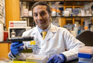
Vinay Aakalu, M.D., M.P.H.
Kellogg is also home to one of the nation’s most robust and diverse oculoplastics research portfolios, including numerous basic, clinical and translational research grants, clinical trials, and patent applications.
Dr. Aakalu in Principal Investigator on two NIH R01 grants. The latest aims to develop a novel treatment approach for the fatal disease Niemann-Pick Type C (N-P). The project leverages his groundbreaking work deriving therapeutic agents from the peptides found in tears.
N-P is a rare, genetic neurodegenerative disorder characterized by the body’s inability to transport cholesterol and other fatty substances (lipids). As a result, lipids build up within brain and other cell types.
In earlier research into lipid and small molecule therapies to treat dry eye and corneal injuries,
“
OUR GROUP IS AT THE FOREFRONT OF THE EMERGING STUDY OF TEAR BIOLOGY, WE HOPE THAT MINING THE MOLECULAR COMPONENTS OF TEARS MAY YIELD NOVEL BIOMARKERS FOR NEW THERAPIES.
“
Vinay Aakalu, M.D., M.P.H.
Dr. Aakalu and his colleagues discovered that the peptide they were studying had something in common with N-P; namely, the protein receptor TMEM97.
“The TMEM97 receptor has been shown to impact the protein that is damaged in N-P,” explains Dr. Aakalu.
“We found that TMEM97 can, in turn, be impacted by the peptide-based formula we developed for dry eye. With that knowledge, we applied those peptides to models of N-P and found that they yielded improvements in some features of the disease.”
The grant will fund future studies to advance a potential peptide-based treatment for N-P.
While N-P is an unusual target in ophthalmology research, the development of peptide-based therapeutics represents an exciting new frontier for treating a range of ocular disorders.
“Our group is at the forefront of the emerging study of tear biology,” Dr. Aakalu says. “We hope that mining the molecular components of tears may yield novel biomarkers for new therapies.”
A U.S. patent has been issued to Yannis M. Paulus, M.D., Xueding Wang, Ph.D., and Xinmai Yang, Ph.D., for Photomediated Ultrasound Therapy, or PUT, a noninvasive method of removing the microvessels whose growth is associated with numerous eye diseases, including wet age-related macular degeneration (AMD) and diabetic retinopathy.
PUT combines synchronized nanosecond laser
pulses with bursts of ultrasound. It delivers laser pulses at more than 10 times lower intensity than conventional laser combined with synchronized ultrasound bursts, making it possible to target microvessels with much greater precision, while posing far less risk to surrounding tissue.
“The ability to deliver more targeted therapy is especially valuable in the treatment of diseases such as AMD, where the growth of new blood vessels tends to cluster around the fovea—the center of the macula,” explains Dr. Paulus. “Unlike conventional laser treatment, which can result in a blind spot in the patient’s center of vision, PUT can remove microvessels while preserving central vision.”
In July 2022, Santa Ono, Ph.D., was named the 15th President of the University of Michigan. At the same time, the Kellogg Eye Center gained a renowned vision researcher.
Dr. Ono brings vast leadership experience to the president’s office, having previously held the position at both the University of Cincinnati and the University of British Columbia. At Michigan, he oversees the activities of more than 65,000 undergraduate and graduate students in nearly 300 degree programs across 19 schools and colleges on three campuses, as well as a research volume in excess of $1.86 billion.
But the chance to lead one of the world’s most influential academic institutions wasn’t the only thing that drew Dr. Ono to Michigan. He is, after all, a researcher at heart.
Dr. Ono began his academic career in immunogenetics research, studying the molecular biology of the immune system and its impact on the development of type 1 diabetes. He was urged to apply his expertise in immunology to eye diseases when recruited from Johns Hopkins University to the Harvard Medical School Schepens Eye Research Institute.
At Schepens and in subsequent positions, including at the University College London’s Moorfields Eye Institute, Dr. Ono led pioneering research into the role of the immune system in ocular surface diseases and agerelated macular degeneration (AMD). Among his many published findings was the identification of antibodies that may contribute to the progression of AMD and the transition from the dry to the wet form of the disease.
After a year fully devoted to his role as president, Dr. Ono is able to engage once again in research. A U-M professor of ophthalmology and visual sciences (as well as microbiology, immunology, and molecular, cellular and developmental biology) he is in the process of setting up his lab at Kellogg.
Dr. Ono credits Paul P. Lee, M.D., J.D., U-M Medical Group Executive Director, Senior Associate Dean of Clinical Affairs and former Kellogg Chair, with recruiting him to U-M. “Michigan has built an international reputation for research excellence, and that was a major draw for me,” he says. “Kellogg is globally respected for making fundamental contributions to our understanding of eye diseases and their treatments.”
“I have been involved in eye research at top institutions in the U.S., the U.K. and Canada, and have followed the work of basic and clinical scientists at Kellogg for decades,” he continues. “Kellogg’s clinical expertise
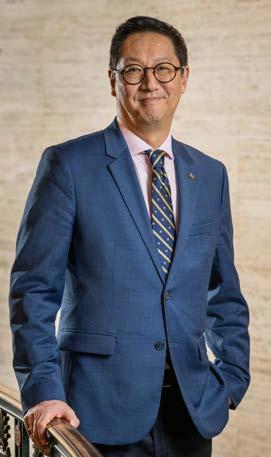
“KELLOGG’S CLINICAL EXPERTISE AND CRITICAL MASS OF RESEARCH TALENT, COMBINED WITH THE ALMOST UNLIMITED POTENTIAL FOR COLLABORATIONS ACROSS THE UNIVERSITY, MAKES IT ONE OF THE MOST ATTRACTIVE PLACES TO DO EYE RESEARCH ANYWHERE. IT’S A THRILL TO BE HERE, AND I CAN’T WAIT TO GET BACK INTO THE LAB.
Santa Ono, Ph.D.
“and critical mass of research talent, combined with the almost unlimited potential for collaborations across the University, makes it one of the most attractive places to do eye research anywhere. It’s a thrill to be here, and I can’t wait to get back into the lab.”
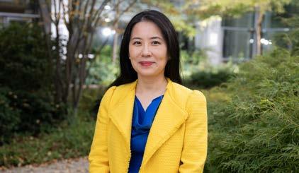
The doctors, researchers and staff at Kellogg put patients’ health and safety first. But as Amy Zhang, M.D., is quick to remind us, it’s just as important to look out for our own well-being.
“To give patients our very best, we have to be at our best,” says Dr. Zhang. A glaucoma and cataract surgeon, she is one of seven Faculty Associates appointed to the Michigan Medicine Wellness Office. The office is dedicated to improving well-being and reducing professional burnout among faculty, staff and learners. Faculty Associates like Dr. Zhang undertake scholarly projects that inform wellness policies across the health system.
Since joining the office in 2022, Dr. Zhang’s projects have included collecting and analyzing data on how physicians manage their in-baskets—the mailbox function within the electronic health records database.
“Keeping current on correspondence and scheduling is one way to lessen daily stress,” she explains. She also analyzed health system-wide data on vacation utilization and presented her findings at the 2023 meeting of the AMA’s American Conference on Physician Health.
Now Dr. Zhang is addressing a specific workplace risk for ophthalmic clinicians: musculoskeletal pain and injury related to poor ergonomic design in equipment and instruments.
“It’s a bigger problem than we tend to acknowledge,” Dr. Zhang says. “It’s well documented that, as a result of our examination and operating room equipment, ophthalmologists are twice as likely as primary care doctors to suffer work-related neck or upper back pain.”
For example, the ophthalmologist’s go-to examination tool, the slit lamp, has remained essentially the
“
“ AS PHYSICIANS, WE NEED TO TAKE THE ADVICE WE GIVE OUR PATIENTS ABOUT STRESS REDUCTION, EXERCISE AND CONDITIONING. IT’S NEVER TOO EARLY OR TOO LATE TO PRIORITIZE HABITS THAT PROMOTE WELLNESS.
Amy Zhang, M.D.
same since its invention in 1911. Its design limits one’s ability to sit as close as needed, requiring overextension of the neck to compensate.
Ophthalmic surgeons, too, risk developing musculoskeletal pain from maintaining static positions and improper ergonomic positioning throughout procedures. And eye clinicians are also prone to repetitive motion injuries to hands and arms, such as carpal tunnel syndrome and cubital tunnel syndrome.
Over time, what starts out as discomfort can lead to serious upper extremity, cervical and spinal issues.
With grant support from Michigan Medicine, input from experts in ergonomics, biomedical engineering and physical therapy, and guidance on logistics and promotion from Kellogg’s cultural committee, Dr. Zhang is designing a pilot intervention for her fellow clinicians targeted for early 2024.
“The plan is to evaluate the effectiveness of offering ‘drop in’ instruction and support on-site to help clinicians learn simple exercises—micro-stretches—to counteract the postures we’re forced to maintain,” Dr. Zhang explains. “The intervention needs to be quick, easy and accessible, not just another item for an already jammed to-do list.”
In addition to the pilot intervention, Dr. Zhang is working with the AUPO to field a national survey of residents to gauge how early musculoskeletal issues arise.
“I didn’t realize how fit you need to be for a career in ophthalmology,” she acknowledges. “As physicians, we need to take the advice we give our patients about stress reduction, exercise and conditioning. It’s never too early or too late to prioritize habits that promote wellness.”
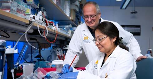
For the retina to do its job to help the brain convert light into vision, it requires a well-regulated metabolic environment. The blood-retinal barrier (BRB) is a structure of tightly joined cells that protect that environment, allowing nutrients in while keeping potentially damaging substances out.
A buildup of vascular endothelial growth factor (VEGF) in the retina damages the BRB, making it more permeable. This leads to fluid buildup, much like a bruise after a hard bump. VEGF-induced retinal injury is linked to blinding eye diseases like diabetic retinopathy (DR).
Understanding the molecular physiology of the BRB is a central focus in the lab of Kellogg investigator David Antonetti, Ph.D. He has received NIH R01 grant support for the following new studies:
The Antonetti lab has worked for many years to understand how VEGF contributes to loss of the BRB. Current therapies blocking VEGF action have proven invaluable in the clinic. Now, Dr. Antonetti will serve as Principal Investigator on a project aimed at exploring the possibility of regenerating the BRB in diabetic retinopathy.
Specifically, he is exploring whether the cytokine norrin may play a role in BRB regeneration.
Norrin has already been shown to play a role in the development of the BRB. Building on that, the Antonetti lab has demonstrated in animal models that norrin can help restore the barrier properties of retinal blood vessels after they are weakened by diabetes.
“Loss of norrin signaling may be as important as increased VEGF action in diabetic retinopathy,” he says. “We have already demonstrated that norrin acts to restore the BRB, helping the diabetes-injured retina recover function. And we have found new pathways that cells use to build the BRB. In all, this research supports prioritizing norrin as a therapeutic target.”
The Role of Heme in Retinal Vascular Development Dr. Antonetti will collaborate on a separate initiative arising from studying the genes expressed in retinal blood vessels—in particular, the gene FLCVR2.
This project will explore a completely novel hypothesis, that FLCVR2 plays a role in brain and retinal blood vessel growth by transporting a critical molecule— heme—into developing cells. Heme is best known as a component of hemoglobin, where it binds with the iron to carry oxygen in the bloodstream. But that’s not heme’s only function.
“In cell biology, heme is a real multitasker,” says Dr. Antonetti. “It’s a co-factor in several key reactions in cells. And remarkably, it appears to act as a signaling molecule in the regulation of blood vessel growth in the BRB.”
As Dr. Antonetti was connecting the dots between heme and blood vessel growth in the BRB at Michigan, Thomas Arnold, M.D., was studying the role of the same transporter in his neonatal brain research at the University of California, San Francisco.
The two laboratories have merged their efforts, with Dr. Arnold and Dr. Antonetti as Co-Principal Investigators on the resulting R01 project grant.
“It’s really a brand new way to think about blood vessel development,” Dr. Antonetti says. “We hope it will lead to new ways to counter the out-of-control blood vessel growth that fuels blinding eye diseases.”
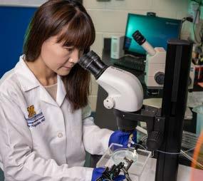
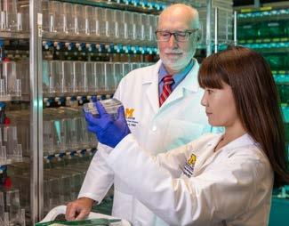
The degeneration and death of photoreceptor cells is a primary cause of blinding diseases like age-related macular degeneration and retinitis pigmentosa.
A fundamental focus in the study of these degenerative retinal diseases, as well as neurodegenerative diseases like Alzheimers, is the role inflammation plays in fueling retinal cell and neuron degeneration, and what can be done about it.
Basic science researchers investigating inflammation and cell death in the brain and retina often study zebrafish models. Unlike humans, when these creatures’ brain and retinal cells die, they have the ability to make new ones.
“tory response, and that this inflammation is required to make new photoreceptors. The inflammatory molecules signal specific cell types to begin to form new, regenerated photoreceptors.
“In zebrafish retinas, inflammation is a required step for photoreceptor regeneration,” explains Dr. Hitchcock. “This is in distinct contrast to inflammation in the human retina, where it is a part of healing, but can lead to degeneration and disease.”
THESE TINY FISH NATURALLY ACCOMPLISH EXACTLY WHAT WE WOULD HOPE TO ACHIEVE IN PATIENTS WITH DEGENERATIVE EYE AND BRAIN DISEASES.
“These tiny fish naturally accomplish exactly what we would hope to achieve in patients with degenerative eye and brain diseases,” explains Peter Hitchcock, Ph.D., Professor of Ophthalmology and Cell and Developmental Biology. “We can learn a lot from them.”
Peter Hitchcock, Ph.D.
“Their game-changing finding will be pursued with a new NIH R21 grant, with Dr. Hitchcock as Principal Investigator and Dr. Nagashima as Co-Investigator. The R21 provides short-term funding to pursue ideas that break new ground.
New insights from their studies of zebrafish retinas have led Dr. Hitchcock and his colleague, Assistant Research Scientist Mikiko Nagashima, Ph.D., to view inflammation in a whole new way.
From their research, Drs. Hitchcock and Nagashima have established that when photoreceptors in the retinas of zebrafish are dying, they trigger an inflamma-
“The subretinal space is where photoreceptors live and die in humans, and where they live, die and regenerate in zebrafish,” explains Dr. Hitchcock. “Our project explores the unique inflammatory environment of that space in zebrafish, and the specific cells within it that aid photoreceptor regeneration.
“The insights we gain may someday be used to combat degenerative eye diseases by inducing regrowth of photoreceptor or other cells in the human retina.”
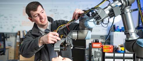
Mark Draelos, M.D., Ph.D., stands at the intersection of ophthalmology and robotics. From that unique vantage point, the view is amazing.
Dr. Draelos began a joint faculty appointment at both Kellogg and Michigan Robotics in January 2023, launching the University’s Image-Guided Medical Robotics (IGMR) Lab with dedicated space at both Kellogg and the Ford Robotics building at U-M.
Applying a deep understanding of clinical medicine, biomedical imaging and robotics, IGMR investigators are in the early stages of developing robotic systems with real-time image feedback to solve a range of healthcare problems.
In ophthalmology, those systems are built around Optical Coherence Tomography (OCT)—a non-invasive technology that uses light waves to image the structural layers and blood vessels of the eye.
“Marrying OCT with medical robotics promises to be a game-changer in ophthalmology,” says Dr. Draelos, “addressing some of our most crucial surgical and diagnostic challenges.”
OCT is particularly well suited to ocular surgery because it provides images of the tiny structures of the eye in real time, without interrupting the procedure. Dr. Draelos is developing a robot-assisted system that helps the surgeon better leverage that capability.
Eye surgeons perform incredibly delicate maneuvers while seated at the patient’s head, looking through a microscope that offers a top-down view. OCT can provide images from any angle, but once it is rotated (for example, to better view the angle and depth at which an instrument is penetrating), surgeons must
also ‘mentally rotate’ to transform their hand motions accordingly.
“Much like driving a vehicle in reverse guided by looking in the mirror, you have to think about which direction to turn the wheel,” he explains.
Dr. Draelos is developing a robotic system that does that mental transformation, better connecting hand and tool movements. At the same time, it employs virtual reality to visually and tactilely ‘separate’ the surgeon from the surgical field, replacing it with a greatly increased scale representation on which to operate.
Taking OCT Where It’s Never Been Before Robotic enhancements to OCT may soon make the technology more accessible and portable too, bringing earlier diagnosis and treatment to more people.
Today’s OCT, which requires an on-site skilled operator, is a stationary device largely relegated to the ophthalmology clinic. But the eyes are impacted by many diseases that might be initially diagnosed and managed elsewhere.
“Consider the potential to save eyesight if diabetic retinopathy could be found before patients are symptomatic, by using OCT technology in the primary care setting,” suggests Dr. Draelos. His team is working on an OCT-imaging robot that automates scanning. “Eventually, patients’ eyes could be scanned while their blood pressure is being checked.”
An automated, mobile system could bring better ophthalmic health to underserved, at-risk communities with limited familiarity with or access to specialty care. It could also help those unable to be scanned with a traditional system, due to mobility or motion disorders.
“The goal of emerging technologies like these is not to move away from traditional ophthalmology clinics,” Dr. Draelos notes. “Rather, it’s about improving surgical practice, and expanding access to early diagnosis and referral for specialized care.”
The length of a normal eye is about 16 mm at birth, growing over the first two years to about 23 mm. In some babies, eye growth stops too soon, and in others, eyes grow too large.
Nanophthalmos is an inherited disorder in which the eye is structurally normal, but stops growing at 16-20 mm. Exceedingly rare, it results in extreme farsightedness (hyperopia), angle-closure glaucoma, and other vision problems. Conversely, myopia is a more common condition, characterized by lengthening of the eye (as large as 30-32mm in some extreme cases). Myopia results in nearsightedness, and in some instances, structural problems.
To support his work understanding the genetic factors that contribute to eye size disorders, Lev Prasov, M.D., Ph.D., has received a grant from the E. Matilda Ziegler Foundation for the Blind. The foundation supports innovative vision research by outstanding early-career investigators.
Dr. Prasov’s current project builds on his previous studies of families with instances of nanophthalmos caused by a variant of the gene MYRF. It aims to describe how the MYRF transcription factor impacts the development of retinal pigment epithelium (RPE) and its interactions with other eye cells. This work is
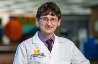
in collaboration with Kellogg medical retina specialist Jason Miller, M.D., Ph.D.
“We know that as the eye grows, RPE cells are critical in eye size determination,” explains Dr. Prasov. “But it’s unclear why defects in the RPE cause eye size disorders.”
A better understanding of the cell-to-cell interactions and genetic pathways that govern eye size could eventually lead to new methods of treating eye size disorders.
“Ultimately, we may be able to design interventions that manipulate those pathways,” he says, “stimulating eye growth in patients with nanophthalmos, and slowing eye growth in patients developing myopia.”
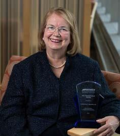
In recognition of a career of dedicated service, Christine Nelson, M.D., was presented with the Women in Ophthalmology (WIO) Humanitarian Award. The annual award recognizes a WIO member’s efforts to improve lives, alleviate suffering and contribute to the basic human dignity of those in need in the U.S. or abroad.
A clinician, researcher and educator, Dr. Nelson plays a pivotal role on the oculoplastics team. At the same time, as a Co-Director of the Jerome Jacobson International Program at Kellogg, she has traveled to locations from Ethiopia to Jamaica, lending her boundless energy and compassion to countless efforts to improve eye health among the underserved.
“No matter where she finds herself, she goes about the work at hand with humility and grace,” says Kellogg Interim Chair Shahzad Mian, M.D. “In her uniquely selfless way, she has represented Kellogg around the world while blazing the trail for many others, from the greenest medical students to the most seasoned faculty and alumni, to step out of their comfort zones and serve. She’s an inspiration to us all.”
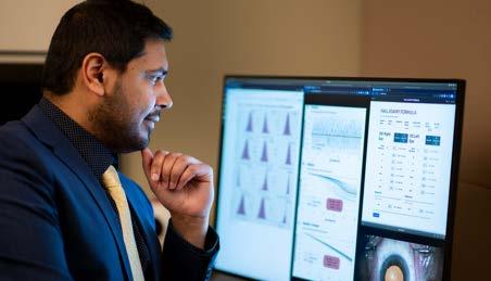
Nambi Nallasamy, M.D.
With more than 23 million procedures performed each year, cataract surgery is the world’s most common surgical procedure. To achieve the best possible visual outcome for the patient, it’s crucial to implant an intraocular lens (IOL) with the optimal power. To select the IOL’s power, the surgeon relies on a formula to calculate which power will achieve a postoperative refraction that best matches the patient’s goals.
A number of such formulas are available, using different methodologies to weigh variables like the eye’s length, corneal shape and lens thickness. While the accuracy of IOL power calculation has risen over the years, so have patient expectations for their refractive outcomes, creating demand for still more powerful formulas.
The resulting Nallasamy Formula was then tested against seven of the most commonly used formulas, using retrospective patient data. It outperformed them all.
Dr. Nallasamy and fellow cataract surgeons at Kellogg now employ it side-by-side with other formulas.
Having demonstrated that ML can improve IOL calculations for his patients, Dr. Nallasamy is now applying it to challenges specific to other populations.
IN ML, COMPUTERS ‘LEARN’ FROM PROCESSING DATA
“ “
First, he hopes ML can help make cataract surgery outcomes around the world more equitable by overcoming data biases inherent in IOL formulas. “Eye size and shape vary throughout the world,” he explains. “But our data sets reflect our local population. We need an ML tool that can easily adjust to different populations and IOL types with minimal additional information—a challenge called domain adaptation.”
SETS FAR LARGER AND MORE MULTIFACETED THAN A HUMAN MIND CAN ABSORB.
Nambi Nallasamy, M.D.
Cornea and cataract surgeon Nambi Nallasamy, M.D., saw this as an ideal application for an artificial intelligence (AI) technology called machine learning (ML).
“In ML, computers ‘learn’ from processing data sets far larger and more multifaceted than a human mind can absorb,” he explains. “In this case, we taught the computer to make IOL power decisions by processing data from nearly 10,000 patients who underwent cataract surgery at Kellogg.” That data set included key patient demographics plus eight different eye measurements.
Dr. Nallasamy is also putting his ML approach to work to help surgeons navigate more complex conditions, including patients requiring cataract surgery along with corneal transplantation, and those with corneal thinning disorders like keratoconus.
Selecting the right lens to meet these patients’ needs is especially challenging, because the cornea continues to change as they recover from surgery.
“Again, ML can generate a more customized lens calculation formula when we fine tune the dataset we use,” he explains, “emphasizing measurements from optical biometry and tomography to better model the postoperative evolution of the cornea.”
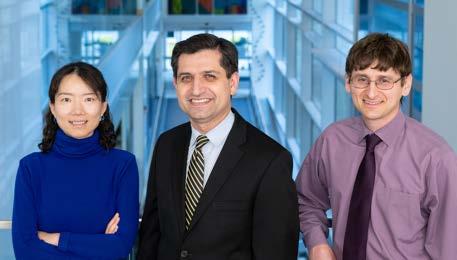
Gene editing therapy—making changes to the DNA of cells to correct mutations—is a promising avenue to treat inherited ocular defects and blinding disorders.
Much research is devoted to applying gene editing to conditions of the retina. Yet, as ophthalmic geneticist Lev Prasov, M.D., Ph.D., and cornea specialist Shahzad Mian, M.D., note, far less attention has been paid to how gene editing might be used to treat corneal dystrophies, which impact about one in 2,000 Americans.
“
“Corneal dystrophies are good therapeutic candidates for gene editing,” says Dr. Prasov. “Specific genetic mutations common to several dystrophies have already been identified. Moreover, those genes have been shown to be amenable to manipulation with editing tools like CRISPR-Cas9 base editing.”
initiative at the U-M Medical School Office of Research, was the perfect opportunity to develop this project. The program empowers a team of U-M researchers (‘scouts’) to identify bold early-stage ideas and spark the needed connections across the medical campus to make them happen.
The idea caught the attention of ‘scout’ Ivo Dinov, Ph.D., who introduced Drs. Prasov and Mian to Yan Zhang, Ph.D., a researcher in the Department of Biological Chemistry. Dr. Zhang is an expert in developing and applying CRISPR-based technologies to tackle diverse biological challenges.
IT’S A TESTAMENT TO WHAT’S POSSIBLE IN AN INSTITUTION THAT FOSTERS COLLABORATION AND PRIORITIZES BOLD SCIENCE.
Shahzad Mian,
M.D.
Dr. Mian sees two promising paths for using gene editing to treat inherited corneal dystrophies. The first is early intervention. “This could prove a sightsaving approach for a patient identified as carrying a mutation whose disease has not yet progressed to a completely opaque cornea.”
The second is treating dystrophies that recur in corneal grafts. “Recurrence is all too common in corneal transplants,” Dr. Mian says. “If we could ‘edit out’ the genetic culprit, it would save many patients from needing multiple transplants.”
The Research Scouts Program, a new funding
“
With funding from a Research Scouts grant, their collaboration will focus on two types of genetically linked corneal dystrophies both linked to disruption of the same protein (TGFBI): lattice and granular.
Mining data from electronic medical records, Drs. Prasov and Mian will identify Kellogg patients with those corneal dystrophies for genetic testing. Separately, they will use donor corneal tissue to test different methods of delivering a treatment into the cornea to modify the gene. Dr. Zhang will apply the latest CRISPR technology to design various gene editing reagents, evaluating them first in corneal cell lines, then in corneal tissue samples to identify the reagent that produces the optimal editing change.
They hope the project will generate enough data to apply for larger NIH and/or foundation funding. “We could not have pursued this idea without Dr. Zhang, and we would likely never have connected with her had it not been for the Scout program,” says Dr. Mian. “It’s a testament to what’s possible in an institution that fosters collaboration and prioritizes bold science.”
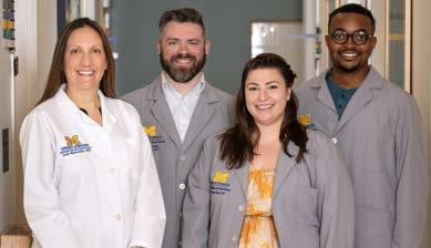
M.S.,
At Kellogg, research involving patients is coordinated through the Kellogg Clinical Research Center (KCRC).
The KCRC provides the staff, infrastructure and resources to see a clinical study from initial Institutional Review Board approval through final data reporting.
Since opening its doors in 2015, clinical research volume at the KCRC has grown to more than 360 active studies, including more than 300 investigator-initiated studies and 50+ sponsored studies.
Along with coordinating the hundreds of patient interactions and protocol details required for those projects, a special team within the KCRC assists researchers from other Michigan Medicine departments whose studies have potential consequences for eye health.
“Many treatments for cancers and other diseases have potential ocular side effects,” explains Julie Rosenthal, M.D., M.S. The Associate Medical Director of the KCRC, Dr. Rosenthal oversees the Collaborative Ophthalmic Research Support (CORS) unit. “The clinical trials that evaluate those medications have specific protocols for monitoring participants’ eyes. Our team coordinates all of those eye-related components.”
Along with Dr. Rosenthal, the CORS team includes KCRC Administrative Director James Green and Clinical Research Coordinators Kevin Haynes and Lianne Shami.
A CORS study protocol—which can exceed 100 pages—is first thoroughly reviewed and its eye-related components consolidated. A plan is developed, detailing the required testing, imaging and measurements, and identifying which Kellogg specialists are needed.
““ WE BELIEVE IN THE TRANSFORMATIVE POWER OF RESEARCH, THAT’S WHY WE DO ALL WE CAN TO SUPPORT EVERY AMAZING PATIENT WHO STEPS UP TO PARTICIPATE IN A CLINICAL STUDY.
Julie Rosenthal, M.D., M.S.
With a plan in place, every patient interaction is managed through the ODS team to streamline the process for everyone involved.
“Without this centralized service, patients might be seen by Kellogg physicians without knowledge of the protocol, which could lead to incorrect or incomplete examinations and testing,” notes Dr. Rosenthal. “This could jeopardize the viability of the studies of our Michigan Medicine research partners.”
“Most appointments begin with a workup at the KCRC, after which Kellogg doctors complete the examinations,” explains James Green.
“It’s quite a scheduling challenge,” adds Kevin Haynes. “We try to work with the patient’s existing study appointments, to minimize trips to the medical campus, and coordinate with the availability of the right Kellogg specialists for exams and imaging.”
Currently, the KCRC is providing support for more than 60 active CORS studies, coordinating with researchers in endocrinology, oncology, neurology, immunology, pediatrics and more.
“In a cancer drug trial, we may be monitoring for side effects like changes in vision or intraocular pressure,” Lianne Shami explains. “In studies of genetic syndromes, we may be watching for visible changes in the cornea, in the case of Wilson’s disease, or the development of tumors in Von Hippel-Lindau disease.”
A comprehensive resource like the KCRC is rare, even in the largest academic eye centers. Extending that same high level of support to researchers in other specialties is a testament to Kellogg’s commitment to cooperation in advancing in research while delivering outstanding patient care.
“We believe in the transformative power of research,” says Dr. Rosenthal, “That’s why we do all we can to support every amazing patient who steps up to participate in a clinical study.”
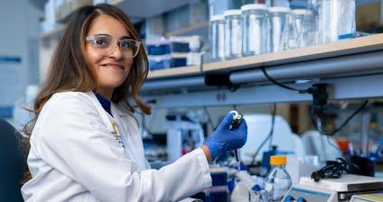
Shilpa Kodati, M.B.B.S.
Uveitis refers to a group of diseases that can inflame and destroy tissue within the eye. Occurring in one or both eyes, suddenly or gradually, it can cause severe pain, redness and light sensitivity, blurred vision, and can lead to complications including glaucoma, cataracts, macular edema and impaired vision.
Uveitis is responsible for up to 30,000 new cases of legal blindness and roughly 10-15 percent of all cases of blindness in the U.S. each year.
Uveitis can occur at any age, but is more common in working age adults. It can result from infections, autoimmune diseases or injuries. Because uveitis can present in conjunction with systemic autoimmune diseases and other conditions that can drive treatment decisions, it requires a nuanced, patient-specific approach to care.
Kellogg has built a multidisciplinary team to deliver that care. The Uveitis Service is a referral hub for not only ophthalmologists, but primary care doctors, rheumatologists and dermatologists around the region to connect their patients with individualized care and the latest treatment options, including clinical trials. The goal is to make Kellogg’s Uveitis Service a center of excellence in both clinical care and translational research.
The Uveitis team is anchored by specialists Therese Sassalos, M.D., Susan Elner, M.D. and Shilpa Kodati, M.B.B.S., who joined the service in 2023. Additional expertise is provided by April Marquardt, M.D., a rheumatologist with an interest in autoimmune eye diseases, cataract and uveitis specialist Zvi Kresch, M.D., who has expertise in complex uveitic cataract
“
WE ALSO NEED PATIENT DATA TO BETTER UNDERSTAND THE MANY FACTORS THAT COULD EXPLAIN VARIATIONS IN TREATMENT RESPONSE, TO HELP US DESIGN NEW THERAPIES.
“
surgery, and inherited eye disease expert Thiran Jayasundera, M.D. Colleagues in pediatric ophthalmology and cornea are also consulted as needed.
The team is supported by a dedicated nurse, Lori Sager, R.N., responsible for monitoring immunomodulatory therapies and providing patient counseling. The multidisciplinary approach at Kellogg allows the uveitis clinic to provide the highest levels of individualized patient care.
Uveitis Research
Spearheading uveitis research is Dr. Kodati, who has been recognized as one of two investigators supported annually by Kellogg’s NIH Mentored Clinician Scientist Training Grant (K12).
To understand the mechanisms of uveitis, Dr. Kodati is studying the molecular and cellular pathways that drive the disease. She is also investigating immunogenetic risk factors, as well as the role of the gut microbiome.
“Using bio-specimens from uveitis patients is a necessary tool, since animal models of the disease, while useful, don’t represent the heterogeneity of uveitis in humans,” she explains. “We also need patient data to better understand the many factors that could explain variations in treatment response, to help us design new therapies.”
With support from the K12, Dr. Kodati is overseeing the development of a uveitis biorepository at Kellogg. Recently granted IRB approval, work is underway to collect clinical data and bio-samples including peripheral blood and intraocular samples. Fecal samples will also be collected for microbiome analysis.
Shilpa Kodati, M.B.B.S. K12 Grant
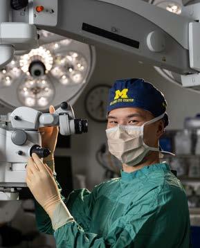
Chances are no clinician reading this report has ever treated a patient with spaceflight associated neuroocular syndrome. Ophthalmology resident Joshua Ong, M.D., helped edit the textbook on it.
Raised in Columbus, Ohio, Dr. Ong’s introduction to space medicine was co-authoring a NASA grant to study the impact of space travel on human health as an undergraduate at The Ohio State University. It was there he conducted tissue engineering research into skeletal muscle loss in astronauts; research that was launched into space in 2019.
He has remained connected to space medicine ever since, through medical school at the University of Pittsburgh, to his PGY1 year in Internal Medicine at Michigan Medicine, and now his residency at Kellogg.
Ophthalmology and space medicine converge in the phenomenon known as spaceflight associated neuroocular syndrome (SANS), a condition observed only in astronauts who have undergone long-duration spaceflight. The clinical characteristics seen in SANS include optic disc edema, hyperopic refractive shift, posterior globe flattening, and choroidal folds. A serious occurrence in space travel, NASA considers SANS a significant barrier to future planetary space missions, such as traveling to Mars. Yet the pathophysiology and mechanisms surrounding SANS are not clearly understood.
In 2022, Dr. Ong served as associate editor of the first textbook on SANS, published by a global team of multidisciplinary experts. Dr. Ong also co-authored two chapters, including “An introduction to space medicine and the physiological effects of spaceflight on the human body.” The book is intended to serve as a reference for space medicine clinicians and researchers studying SANS to protect vision in astronauts.
“A condition unique to space travelers may seem like an niche topic of study,” notes Dr. Ong. “But as the range of science discussed in the book illustrates, SANS research may drive innovations to improve human health on Earth.”
Dr. Ong is collaborating on several of those innovations, including:
Visual assessment technology and machine learning —New low mass, low footprint technologies may help optimize the assessment of astronauts’ vision in real time in the austere environment of space. Those same advances might also help overcome barriers to screening and early intervention in underserved communities.
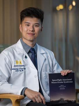
“
IMAGINE A MORE SUPPORTIVE ENVIRONMENT FOR PURSUING ONE’S OWN UNIQUE PATH IN VISION SCIENCE THEN KELLOGG.
“ I CAN’T
Joshua Ong, M.D.
Wearable technology to address central vision distortion —Prolonged periods of microgravity may lead to macular disruption, diminishing central vision. Particularly on extended planetary missions, this could threaten astronaut safety and mission performance. Emerging head-mounted display technology can digitally suppress central visual distortion. This may prove a useful tool for improving vision in individuals on Earth with irreversible central vision loss from age-related macular degeneration.
For his work on the latter, Dr. Ong placed second at the 2022 NASA Human Research Program Investigators’ Workshop. He was also named one of Forbes’ “30 Under 30” in healthcare for his pioneering work. Here at Kellogg, the focus of Dr. Ong’s residency is gaining experience in different ophthalmic subspecialties. But he is staying engaged in space medicine research. “I can’t imagine a more supportive environment for pursuing one’s own unique path in vision science then Kellogg.”
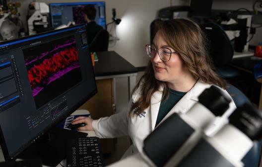
Molly Thorson, Ph.D.
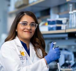
Molly Thorson, Ph.D., a postdoctoral fellow at Kellogg, has received a Ruth L. Kirschstein National Research Service Award (NRSA), or F32, from the National Institutes of Health (NIH)/National Eye Institute (NEI). Dr. Thorson has been part of the lab of investigator Jillian Pearring, Ph.D., since 2020, focusing on retinal cell biology.
This fellowship provides postdoctoral scientists with three years of funding for research and professional development to maximize their potential for success as investigators in their chosen health specialty.
“have much to learn about how it’s built and maintained through the life of the cell.”
THIS AWARD IS REALLY A TESTAMENT TO KELLOGG’S COMMITMENT TO AND LEADERSHIP IN BASIC SCIENCE EYE RESEARCH. IT’S A WIN FOR US ALL.
Molly Thorson, Ph.D.
The F32 is among the most highly sought after government awards in academic medicine. By some estimates, as few as 500 to 700 individuals across all NIH medical specialties receive support from an F32 fellowship each year. The NEI awards only 14 to 16 F32 grants each year.
The projects funded by Dr. Thorson’s F32 involve the study of the outer segment of rod photoreceptors within retinal cells.
“The outer segment is highly organized to maximize light capture,” she explains. “It’s within that tiny architectural marvel that vision originates, yet we still
The outer segment is constructed of stacks of flattened membranous disks. Using a mouse model, Dr. Thorson has identified several proteins involved in that specialized architecture. Her work will provide a more detailed blueprint of how those proteins form the disk architecture in a healthy cell, laying the foundation for studies of how changes in specific proteins might alter the architecture, possibly contributing to retinal degeneration.
“The F32 award recognizes not only the potential impact of Dr. Thorson’s work, but the depth and breadth of resources and support available at Kellogg. “In addition to a rigorous evaluation of my proposed research, the reviewers considered the many ways Kellogg makes projects like mine possible,” Dr. Thorson says. That includes the impressive track record of Dr. Pearring and her lab, and the training, research cores and data analysis capabilities available here.
“This award is really a testament to Kellogg’s commitment to and leadership in basic science eye research,” she adds. “It’s a win for us all.”
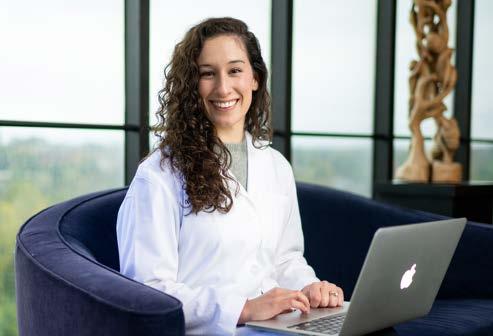
Alejandra Maiz, M.D.
In addition to the unending ‘to do list’ of a resident, PGY4 Alejandra Maiz, M.D., is finding time to ensure the high quality of ophthalmic training across the U.S.
Dr. Maiz was selected to serve a two-year term on the Ophthalmology Residency Program Review Committee of the Accreditation Council for Graduate Medical Education (ACGME). The ACGME sets and monitors compliance with standards for graduate medical training, ensuring that learners are prepared to deliver highquality, patient-focused care.
“
Program Review Committees for each major medical specialty are tasked with setting residency education and training standards that reflect the best practices in their field. They also monitor programs for compliance, fielding surveys, conducting site visits, collecting and analyzing data, and providing actionable feedback.
Association and the American Osteopathic Association, as well as one public member and one resident member. Dr. Maiz was nominated during her post-graduate year two (PGY2) of training by Kellogg Residency Program Director Bradford Tannen, M.D., J.D.
“I was honored to be selected,” says Dr. Maiz, “and it’s so gratifying that, as the sole voice for all residents, the committee listens and values my opinion.”
THE OPPORTUNITY TO INTERACT WITH FACULTY FROM ACROSS THE COUNTRY HAS GIVEN ME AN INVALUABLE PERSPECTIVE ON THE FIELD.
“
Alejandra Maiz, M.D.
The committee is comprised of representatives from the American Board of Ophthalmology, the American Academy of Ophthalmology, the American Medical
“It has proved to be one of the most rewarding aspects of my residency,” she continues.
“The opportunity to interact with faculty from across the country has given me an invaluable perspective on the field. It’s also reinforced to me what exceptional residency training we receive at Kellogg.”
The experience has also shown Dr. Maiz how much work goes into managing a residency program. In spite of that, she wants to remain involved in residency education going forward. “Among my career goals,” she says, “I hope to be a residency program coordinator.”
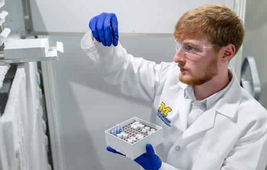
Tyler Carman, B.S.
In May 2023, Tyler Carman earned a B.S. in neuroscience from the University of Michigan. The first college graduate in his family, Tyler plans to pursue an M.D. or M.D./Ph.D. But before taking that next step, he’s making the most of an opportunity to continue learning—and contributing—in the lab of Kellogg physician-scientist Rajesh Rao, M.D.
The Rao translational research lab focuses on the epigenetics of retinal development and disease to identify new biomarkers, therapeutic targets and applications of stem cells for blinding diseases.
“THE THEORY IS THAT THE MEMBERS OF THIS COMPLEX WORK TOGETHER TO CLOSE OFF PARTS OF THE CHROMATIN TO ENSURE CELLS BECOME THEIR SPECIFIED TYPE DURING DEVELOPMENT, WITHIN OUR ORGANOID MODEL, THEY ARE FATED TO BECOME RETINAL CELLS.
Tyler began assisting in the lab as an undergraduate, working with the retinal organoid model under the direction of resident Dan Balikov, M.D., Ph.D., and graduate student Brian Basinski, B.S. His assignments included supporting studies of retinal organoid vascularization with Dr. Balikov and the molecular drivers of North Carolina macular dystrophy, a rare congenital blinding disease, with Mr. Basinski.
Research Supplements to Promote Diversity in HealthRelated Research program, Tyler will complete two years of post-baccalaureate training with the team. His primary focus will be studying a protein-to-protein interaction within the noncanonical Polycomb Repressive Complex 1.6. “The theory is that the members of this complex work together to close off parts of the chromatin to ensure cells become their specified type during development,” he explains. “Within our organoid model, they are fated to become retinal cells.”
“Tyler Carman, B.S.
Tyler credits Dr. Rao with inspiring him to pursue a career as a physician-scientist. “Dr. Rao is incredibly supportive of everyone in his lab, which has been amazing to experience as an undergrad,” he says.
A highlight of his time with the team was observing Dr. Rao perform a human RPE stem cell transplant as part of a first-of-its-kind Phase 1/2a clinical trial for the treatment of dry AMD.
Tyler is already a recipient of NIH funding at this early stage of his career—with support from the NIH
“What an opportunity,” Tyler recalls. “A big milestone for me—observing my first eye surgery—also happened to be a significant scientific milestone.”
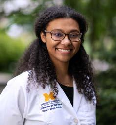
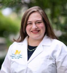
FOUR KELLOGG TRAINEES WERE APPOINTED TO THE PRESTIGIOUS 2023-2024 CLASS OF THE SOCIETY OF HEED FELLOWS, OUT OF A TOTAL OF 24 FELLOWS NATIONWIDE. NEW MEMBERS OF THE SOCIETY RECEIVE A MERIT AWARD FOR POSTGRADUATE STUDIES IN OPHTHALMOLOGY.
Otana Jakpor, M.D., M.Sc., who has also been named a Global Ophthalmology Fellow at Kellogg for the 20232024 academic year, plans to apply her award to an upcoming study of factors associated with the incidence of cataracts in pediatric patients in the African nation of Ghana.
She is developing the research project in collaboration with colleagues at the Korle-Bu Teaching Hospital (KBTH) in Accra, Ghana.
“Pediatric cataracts can result from a number of factors, including genetics, metabolic conditions, infections and trauma,” she explains. “The pediatric ophthalmologists at KBTH want a deeper understanding of which of these causes are most strongly associated with cataract development in their patient population, and the role each may play in determining patient outcomes.”
Dr. Jakpor hopes to augment that analysis by assessing the impact of pediatric cataracts on quality of life, in both patients and their caregivers. “We would like to highlight the importance of addressing pediatric cataracts, not only for children’s physical health, but also for their overall well-being,” she says.
Young children don’t know if they have vision problems. That’s why pediatricians and primary care providers should make vision screening a standard component of every preschooler’s annual check-up.
Yearly vision testing is critical at ages three, four and five, and incrementally after that, flagging any abnormalities for referral to an ophthalmologist. Failure to do so could mean missing the development of conditions like amblyopia which can result in permanent vision loss.
Former Kellogg ophthalmology resident Olivia Killeen, M.D., M.S., A Research Fellow in the National Clinical Scholars Program, dedicates her research to improving vision health and eliminating avoidable blindness by addressing barriers to eye care and strengthening the delivery of ophthalmic services. For a snapshot of the real-world challenges of pediatric vision screening, Dr. Killeen piloted a study within the U-M Health System, combining a medical records review and interviews with providers at 17 clinics.
Screening rates did vary from clinic to clinic. Interviews revealed instances where high screening levels were linked to devoting resources to improved equipment, and where lower screening levels could be connected with Covid-19-associated staffing shortages.
In summer 2023, Dr. Killeen began a one-year pediatric ophthalmology fellowship at Duke University. Before departing Kellogg, she was named a 2023/2024 Heed Fellow.
She plans to apply her Heed award to replicating the U-M study at Duke. “Learning from an additional site will test my theory that the issues we identified are not unique to Michigan,” she says, “but rather, signposts that point to a national public health issue.”
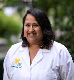
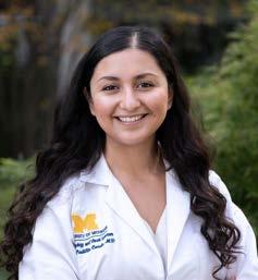
While completing the second, research-focused year of a two-year glaucoma clinical research fellowship, Rithambara Ramachandran, M.D., is pursuing innovative ways to improve detection and outcomes for glaucoma patients, particularly those with socioeconomic barriers to screening and care.
“Unfortunately glaucoma is largely an asymptomatic disease,” she explains. “And nearly seven in ten patients don’t even know they have it. We need to be able to identify individuals before symptoms have developed and the disease has progressed to the point where they face irreversible blindness.”
Dr. Ramachandran’s research, which she is conducting while earning an M.S. in healthcare research at U-M, focuses on how to bring screening and diagnosis to at-risk populations earlier, before they present with late stage disease.
Through community-based participatory research, big data analysis, and implementation science, she is focusing on identifying the best tools, technologies and methodologies to improve access to eyecare screening, address disparities in eye disease screening, and diagnose and overcome eyecare barriers.
“My long term goal is to design, validate and implement a glaucoma screening program for high-risk communities that is clinically sound, scalable, cost effective and equitable.”
Tania Padilla Conde, M.D., a fellow in cornea and external disease, is involved in a number of initiatives to improve access to eye health care—specifically, cornea transplantation—in low resource populations in the U.S. and abroad.
The eldest daughter in a single parent Hispanic household, Dr. Padilla Conde grew up in a community with limited healthcare options. “That firsthand experience fuels my passion to improve vision care and treatment outcomes in underserved communities,” she explains.
Dr. Padilla Conde will apply her Heed Fellow award to an upcoming project in collaboration with colleagues at Eversight, a global nonprofit eye bank. “We are working to improve the delivery of preloaded Descemet Membrane Endothelial Keratoplasty (DMEK) tissue abroad, by validating a novel transport cartridge,” she says. “Our study will improve storage and shipping conditions to minimize endothelial cell loss, a very important marker to ensure these countries receive quality tissue for corneal transplantation.”
She is also conducting a study of refractive outcomes for DMEK triple procedures using a novel approach developed by her mentor, cornea specialist Nambi Nallasamy, M.D. The technique, which employs machine learning technology, aims to improve both preoperative planning and post-operative outcomes.
Dr. Padilla Conde is thankful for the Heed fellowship and hopes to focus her future work on helping advance novel techniques for corneal transplantation, including alternative approaches to endothelial cell replacement.
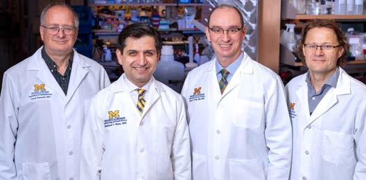
Kellogg researchers are among the most productive, grant-supported, published and cited in all of ophthalmology. How do we attract and retain the best and the brightest? With a structure and culture that fosters innovation and collaboration.
“Our research program is designed to be both a magnet for the most creative minds, and an incubator for their most original ideas,” says Kellogg Interim Chair Shahzad Mian, M.D. “We’re continuously improving the fundamentals—our mix of technologies and resources, training and support—to meet the changing needs of researchers from dozens of specialties, pursuing hundreds of lines of investigation.”
“The P30 center grant funds state-of-the-science instrumentation, services and training for vision researchers to connect and pursue cutting edge ideas. The center has research cores, each supervised by faculty and staffed by technical experts:
Functional Assessment —This core, directed by P30 Principal Investigator David Antonetti, Ph.D., provides training and support to investigators performing nonlethal measures of ocular structure and function, including optical coherence tomography, electroretinography, fundus imaging, and pupillometry.
Instrument and Electronics
OUR RESEARCH PROGRAM IS DESIGNED TO BE BOTH A MAGNET FOR THE MOST CREATIVE MINDS, AND AN INCUBATOR FOR THEIR MOST ORIGINAL IDEAS.
“
Shahzad Mian, M.D.
Kellogg’s research enterprise is anchored by four interconnected institutional grants, supporting basic, clinical and translational investigators at every career stage. All four grants were renewed this fiscal year, and a new core facility was added.
National Institutes of Health (NIH) / National Eye Institute (NEI) University of Michigan Vision Research Center (UMVRC) Grant (P30)
The University of Michigan holds one of the longest standing P30 Center Grants in ophthalmology and visual sciences. Center grants provide resources and facilities for core services shared by researchers across the University that carry out vision research.
Directed by James Weiland, Ph.D., this core provides and maintains specialized instrumentation— both commercially available and custom built—for use by NEIfunded investigators.
Molecular Biology —directed by Steven Abcouwer, Ph.D., this core offers services, equipment and support for projects involving molecular biology, statistical genetics, and bioinformatics.
Morphology and Imaging —this core, directed by Jillian Pearring, Ph.D., processes ocular and brain tissue samples for light microscopy, immunocytochemistry and in situ hybridization, and fixes specimens for electron microscopy. Assistance in analysis and presentation of imaging results is also available, and the core is a hub for training investigators in tissue processing, microtomy, staining and immunohistology techniques.
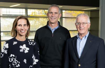
New Data Integrity Core —The renewal of Kellogg’s P30 grant coincides with the establishment of a fifth research core, dedicated to protecting Data Integrity from study design through publishing of findings. The addition of this core reflects the importance of protecting the reliability of the data generated in the course of research studies, as well as the methodologies and strategies used to analyze that data.
Launched by co-directors Maria Woodward, M.D., M.S., and David Musch, Ph.D., M.P.H., the core welcomed co-director Patrice Hicks, Ph.D., M.P.H. in 2024. The core is supported by senior biostatistician Chris Andrews, Ph.D., and biostatistician Amy (Ming-Chen) Lu, M.S.
“Central to our mission is ensuring that Kellogg researchers are trained in and adhere to NIH FAIR principles– findability, accessibility, interoperability and reusability,” explains Dr. Woodward. “Following FAIR standards guarantee that our data is of the highest quality, and that it is gathered and analyzed in ways that can be shared and reproduced by others.”
As more researchers collaborate over open access platforms, the ability to reproduce and publicly share code is key. Experts in the data integrity core advise eye researchers on how to plan and monitor the use of datasets, algorithms, tools and pipelines (DATP), the building blocks of code sharing and duplication.
The team goes beyond reviewing a proposed statistical approach, offering independent, expert input to critique the feasibility of a study’s aims and hypotheses. “There may be more than way to go about answering a question,” says Dr. Woodward. “This additional step ensures that diverse viewpoints have been considered.”
“
FOLLOWING FAIR STANDARDS GUARANTEE THAT OUR DATA IS OF THE HIGHEST QUALITY, AND THAT IT IS GATHERED AND ANALYZED IN WAYS THAT CAN BE SHARED AND REPRODUCED BY OTHERS.
“
Maria Woodward, M.D., M.S.
One of only six U.S. ophthalmology programs to hold a K12, Kellogg was first awarded the grant in 2013. The K12 funds the Michigan Vision Clinician-Scientist Development Program, which provides mentored training for ophthalmologists interested in developing research programs in tandem with their clinical practice.
Directed by Principal Investigator David Zacks, Ph.D., the K12 provides protected research time, core instruction and skill building for up to two scholars at a time, each for up to three years.
Under the supervision of Principal Investigator Patrice Fort, Ph.D., M.S., and Associate Director Sara Aton, Ph.D., this training grant supports graduate students and postdoctoral fellows launching careers in vision science. Primarily focused on basic and translational research tracks, it supports a diverse group of trainees in the completion of their theses or the development of research projects geared to help them progress towards independent positions. By providing extensive visioncentric education, mentoring and professional development, the T32 aims to train the next generation of vision science researchers.
Kellogg has held a T32 grant for more than 20 years.
This institutional grant provides maximum flexibility to develop and expand eye research programs in new and creative ways. Kellogg’s RPB grant, which automatically renewed for five years at the end of 2022, is overseen by Dr. Mian.
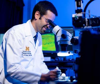
Yannis Paulus, M.D.
“IF WE
CAN SEE MOLECULAR CHANGES WHILE THEY ARE HAPPENING, INSTEAD OF JUST SEEING AN EYE BLEED, WE CAN RESPOND WITH THE RIGHT THERAPY IN TIME TO PRESERVE VISION.
Yannis Paulus,
M.D.
“
The development of new blood vessels underneath the retina—choroidal neovascularization—is the primary cause of vision loss in patients with several diseases including age-related macular degeneration (AMD).
Over time, AMD can progress from its dry form to the more advanced wet form. It is at this point that choroidal neovascularization does most of its damage. All too often, the diagnosis comes late, after a patient notices significant vision loss and has retinal bleeding. By that time, the effectiveness of the current standard treatment—intravitreal injections of anti-vascular endothelial growth factor (anti-VEGF)—can be limited.
With support of an NIH-R01 grant, retinal surgeon and biomedical engineering researcher Yannis Paulus, M.D., is developing a new molecular imaging platform with the potential to revolutionize the early diagnosis and treatment monitoring of AMD.
Dr. Paulus’ new imaging system marries one established modality with two next-gen advances:
Light —Optical Coherence Tomography (OCT), the standard non-invasive retinal imaging technology, creates cross-sectional images of the retina using light waves.
Sound —Low-energy Photoacoustic Microscopy uses nanosecond-duration laser pulses of light and captures the resulting sound from tissue. “Like thunder accompanying lightning,” he says.
Fluorescence —Dr. Paulus is also refining a new nanoparticle contrast agent to detect levels of VEGF in tissue more accurately and safely.
The eventual clinical goal: predicting which patient will benefit from a given treatment and which will not. Another promising benefit: fine-tuning how often patients need treatment.
“Today’s anti-VEGF injections are not easy for patients, so we attempt to extend the time interval between treatments,” Dr. Paulus explains. “But that can be risky. Sometimes a change of just a week can lead to vision loss. If we can see molecular changes while they are happening, instead of just seeing an eye bleed, we can respond with the right therapy in time to preserve vision.”
Dr. Paulus’ new imaging system holds the promise of changing the AMD paradigm—accelerating the development of new treatments, facilitating earlier diagnosis, and making possible more individualized, personalized therapies and treatment planning.
Ron Kurtz, M.D., was part of a team that received the Golden Goose Award from the American Association for the Advancement of Science in September 2022. The award recognizes scientists whose unsung, federally-funded research has led to breakthroughs of significant societal benefit.
Dr. Kurtz, who completed his ophthalmology residency at Kellogg in 1994 and returned as an assistant professor from 1995 to 2000, shares the award with four other investigators: U-M engineering alum Detao Du, Ph.D.; Tibor Juhasz, Ph.D., a former U-M research associate professor in ophthalmology and biomedical engineering; Gérard Mourou, M.Sc., Ph.D., the A.D. Moore Distinguished University Professor Emeritus at U-M; and Donna Strickland, Ph.D., a professor at the University of Waterloo in Ontario, Canada.
“KELLOGG
Optical Science (CUOS) at U-M, working on the femtosecond laser. Removing his safety goggles prematurely, Dr. Du suffered an eye injury. He had the eye examined at Kellogg by Dr. Kurtz, who had just completed his first year of residency.
Curious about a laser that etched such a precise pattern on the retina, Dr. Kurtz visited the CUOS to learn more. A research collaboration developed, beginning with Drs. Kurtz, Mourou and Du (who suffered no lasting effects from the injury), and eventually expanding to include Dr. Juhasz, who was concurrently researching laser vision correction, and Dr. Strickland, co-developer of the femtosecond laser with Dr. Mourou (for which they share the 2018 Nobel Prize in physics).
Dr. Kurtz is reluctant to take credit for a ‘lightbulb moment’ that led to Femtosecond LASIK. “As a resident, one of my first thoughts was that laser safety might make a good topic for a grand rounds presentation,” he recalls. “The CUOS was already exploring multiple uses for the laser, including medical applications. I helped them see the possibilities for eye surgery.”
HAS ALWAYS PROVIDED THE HIGHEST LEVEL OF CLINICAL INSTRUCTION AND EXPERIENCE, INCLUDING PRIORITIZING RESEARCH AS PART OF A WELL-ROUNDED CLINICAL EDUCATION.
“Ron Kurtz, M.D.
Together, this team developed Femtosecond LASIK, a vision correction procedure that employs a femtosecond (quadrillionth of a second) pulsing laser, rather than a scalpel, to help reshape the cornea. Since its launch in 2001, Femtosecond LASIK has revolutionized refractive surgery, improving the vision of more than 30 million people.
Femtosecond LASIK’s origin story is as extraordinary as the technology itself. In the early 1990s, Dr. Du was a graduate student in Dr. Mourou’s lab in the National Science Foundation-funded Center for Ultrafast

The trajectory of Dr. Kurtz’s career has evolved since his time at Kellogg, where his faculty appointment was in vitreoretinal diseases. Today, he is President and C.E.O. of California-based RxSight®, an ophthalmic medical technology corporation that has commercialized the world’s first and only adjustable intraocular lens (IOL) that is customizable after cataract surgery.
“Kellogg has always provided the highest level of clinical instruction and experience, including prioritizing research as part of a well-rounded clinical education,” he says. “I am no longer a practicing clinician, but I continue to draw on what I learned as a resident. In fact, most of the front-of-eye clinical experience so essential to my work today I gained at Kellogg.”
Paul Lichter, M.D., M.S., past Chair and current Professor Emeritus of Ophthalmology and Visual Sciences, was invited to give the Allan and Claire Jensen, M.D., Lecture in Professionalism and Ethics at the 2023 Annual Meeting of the American Academy of Ophthalmology. The lecture was created in 2015 as a platform for a recognized ophthalmologist or member of the medical or bioethics community with demonstrated knowledge of ethical issues and professionalism to address these complex topics in an accessible and relevant format.
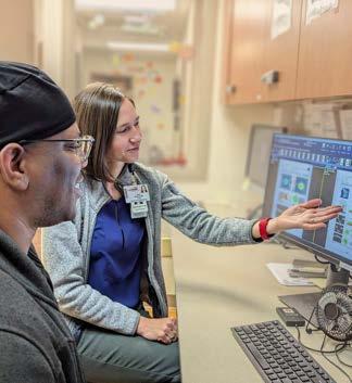
Anna Momont, M.D., Associate Professor in the Department of Ophthalmology and Visual Sciences at the University of Wisconsin School of Medicine and Public Health, became Residency Program Director on July 1, 2023.
Dr. Momont, who previously served as the Associate Program Director, has been working to improve team morale and connectedness as the Resident Wellness Director. She has also led initiatives to increase the ophthalmology resident class from three to four trainees per year, and to recruit candidates that are underrepresented in medicine. In addition, Dr. Momont serves as the faculty lead for a free eye clinic that serves uninsured individuals in her community.
A glaucoma specialist, Dr. Momont completed both her residency and a glaucoma fellowship at Kellogg, including serving as Chief Resident in Ophthalmology.
“At Kellogg, my Residency Program Director was Dr. Mian,” she recalls, “so I learned from the very best. I knew that if I was fortunate enough to work in academic medicine, I wanted to be involved in residency training.”
Also an active member of Women in Ophthalmology, Dr. Momont will chair the group’s national meeting, the Summer Symposium, in Carlsbad, CA in 2024.
For blinding eye diseases like macular degeneration and diabetic retinopathy, regular drug injections into the eye are the current standard of care. Translational investigators like Yannis Paulus, M.D., are developing alternative ways to administer treatments that are more effective, longer lasting, and less distressing for the patient.
His latest collaboration is with Roland Chen, Ph.D., a fellow pioneer in the development of miniaturized platforms for sustained, localized ocular drug delivery. An expert in biomaterials and biotransport engineering, Dr. Chen, an adjunct faculty member at Kellogg, is an Associate Professor at Washington State University.
Separately, Dr. Paulus developed and continues to test a method to deliver treatment via an array of nanoneedles on a dissolvable contact lens. After delivering their medication, they dissolve. “The nanoneedle platform is a very promising option,” says Dr. Paulus, “but because diverse patients and diseases may respond better to different drug concentrations and penetrations, we need a variety of tools in our toolbox.”
Dr. Chen and Dr. Paulus are developing an alternative that utilizes an array of somewhat larger microneedles.
“The advantage of microneedles is that they can carry larger loads of medicine,” Dr. Paulus explains. “However, they can be too large to dissolve, so another method is needed to attach and remove them. Dr. Chen’s novel solution is to use light.”
Dr. Chen has designed a light-responsive microneedle which swells when exposed to light of a certain wavelength, binding and locking it in place. Shining light of a different wavelength on the microneedles reduces the swelling, allowing them to be removed.
They have received a Peer Reviewed Medical Research Program Expansion Award—Funding Level 3 grant from the U.S. Department of Defense to develop and evaluate their new platform. Studies in their laboratories will validate the technology’s effectiveness, reliability, durability, and safety, and compare the treatment outcomes achieved with the platform’s sustained drug delivery to those of regular intravitreal injections.
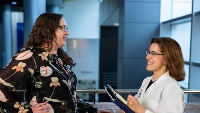
When it comes to ensure patient safety, flagging and addressing even the smallest concern can make a big difference. Making time to celebrate those ‘good catches’ helps build a culture that values respect, communication and teamwork.
In 2023, Kellogg’s Patient Safety Committee, led by Jennifer Weizer, M.D., initiated a High Reliability Award Program to recognize and reward staff members who take action to improve quality and safety.
The goal is to increase awareness and application of two Michigan Medicine human resource initiatives: Universal Relationship Skills and Universal Reliability Tools and Skills.
“Like all Health System employees, Kellogg faculty and staff receive training in constructive, respectful workplace communication and strategies to identify potential safety concerns before they lead to harm,” Dr. Weizer explains. “Our monthly High Reliability
“IT’S ABOUT LEARNING FROM EACH OTHER’S EXPERIENCES, AND CELEBRATING WINS, BIG AND SMALL. IT’S A POSITIVE WAY TO ENCOURAGE EVERYONE TO SPEAK UP FOR SAFETY.
“Jennifer Weizer, M.D.
Awards reinforce that training and encourage everyone to speak up for safety.”
Each time faculty or staff notice a colleague taking a proactive step for safety, they are encouraged to nominate that person for the month’s award. A random drawing each month rewards one nominee and one nominator with a prize for each—a $25 gift card from Zingerman’s, a legendary Ann Arbor culinary destination. A center-wide email announces the winners and showcases all the month’s submissions.
A wide variety of ‘good catches’ and high reliability skills have been highlighted since the program launched. Respectfully questioning a surgical or medication instruction, confirming transcriptions of measurements to ensure there are no errors, and checking the readiness of equipment, materials or work areas have all resulted in recognition and in some cases, timely corrections.
“The contest has been very well-received,” says Dr. Weizer. “It’s about learning from each other’s experiences, and celebrating wins, big and small. It’s a positive way to encourage everyone to speak up for safety.”

Combining the powerful number crunching capabilities of Big Data analytics with the information held in patient electronic health records (EHRs) has revolutionized health research. Investigators can now combine vast amounts of patient information and, using tools like machine learning, analyze the data to answer important questions about diseases, risk factors and treatment outcomes.
At the forefront of applying this approach to the study of ophthalmic diseases is Joshua Stein, M.D., M.S. A glaucoma specialist and health services researcher, Dr. Stein is Principal Investigator and Chief Data Officer of the Sight Outcomes Research Collaborative (SOURCE), a consortium of 14 leading academic ophthalmology departments that pool EHR and other ocular diagnostic data (removing protected health information) into a common research database housed at U-M.
The SOURCE platform is utilized in two of Dr. Stein’s latest initiatives. Both aim to improve the quality of research into the most common vision-threatening diseases, and both are supported by NIH R01 grants.
To leverage the power of exciting tools such as machine learning and artificial intelligence (AI) to come up with new ways to improve the quality of eye care, enhance patient outcomes, and reduce disparities in care, it is essential to train these models using high quality data. When researchers build these sophisticated models, if enough patients are misclassified as affected by a given ocular disease when indeed they do not have that condition, or vice versa, this can negatively affect the performance and usefulness of these models.
One way that a patient may be misclassified as having or not having an ocular disease is when researchers rely on administrative billing codes entered by clinicians during office visits to determine which patients are and are not affected by diseases of interest. Relying solely on billing codes to tell which patients have and do not have particular eye diseases poses a number of problems: certain eye diseases lack unique billing codes; some billing codes are not specific enough to distinguish between different conditions; and occasionally clinicians pick incorrect billing codes.
To address these problems, Dr. Stein and his team have developed a sophisticated approach that goes well beyond sole reliance on administrative billing codes. They incorporate information from multiple areas of the electronic health record to more accurately identify which patients have common sight-threatening
diseases such as glaucoma, diabetic retinopathy, and age-related macular degeneration. For patients who have these conditions, Dr. Stein’s team can accurately determine the type and severity of the condition, which eye or eyes are affected, and whether the condition is stable, improving, or getting worse.
Just because an eye disease is common does not mean every patient experiences it in the same way. Similarly, different eye care professionals in different settings may document the same signs and symptoms of a condition differently in the electronic health record. These factors make it very difficult to use electronic health records to build models that can accurately predict the likelihood of a patient having a particular condition. To overcome some of these challenges, Dr. Stein’s team uses tools such as natural language processing to dive deeper into the electronic health record to locate additional relevant details from the clinic visit.
As Dr. Stein explains, “for each of the four million patients in our SOURCE Ophthalmology Big Data repository, we are able to assign a probability from 0 to 100 percent as to whether they have glaucoma, diabetic retinopathy, or macular degeneration. With accurate classifications for these patients, researchers can integrate this information into the AI models they are building to predict, among other things, which patients are most likely to experience favorable or unfavorable outcomes.” Other applications of this enhanced disease identification approach include incorporating the predictions into studies of the epidemiology of these different eye diseases, using them to determine if disparities in care exist for patients of different sociodemographic characteristics, and helping identify patients who may be eligible for randomized clinical trials of new therapeutic interventions for these conditions.
Health Disparities in Utilization, Quality and Outcomes (HealthDOC)
Dr. Stein and his team, in collaboration with researchers at Northwestern University, have also been leveraging the Sight Outcomes Research Collaborative (SOURCE) Ophthalmology Big Data Repository to study disparities and inequities in the receipt of eye care services and outcomes of eye care among racial and ethnicity minorities and other vulnerable groups. The main objective of this NIH-funded research is to determine the extent to which social determinants of health may affect quality of eye care and outcomes for patients with common sight-threatening eye diseases such as cataract, glaucoma, and diabetic retinopathy.
An innovative aspect of this project is the linking of clinical data from the electronic records of eye care recipients in SOURCE to other large pots of data.
This linking provides Dr. Stein and his team a more holistic view of patients and the array of factors that may affect their care quality and outcomes. For example, by incorporating information on race, ethnicity, level of education, marital status, income, household net worth, type of health insurance, primary language and even the number of family members living in the household, the team can see how these factors may be impacting the eye care patients are receiving.
“The first step in reducing or eliminating long standing inequities in eyecare that contribute to poorer outcomes among vulnerable populations is to identify the underlying factors that are most responsible,” explains Dr. Stein. “Once we can identify such factors, especially those which are modifiable, we can develop strategies to improve care quality and outcomes for these patients.”
Glaucoma and data analytics specialist Joshua Stein, M.D., M.S., and ocular geneticist Lev Prasov, M.D., Ph.D. share a 2023 Dr. David L. Epstein Award from the Association for Research in Vision and Ophthalmology (ARVO) Foundation for Eye Research.
The award recognizes a senior investigator distinguished in both glaucoma research and scientific mentorship (the mentor, Dr. Stein), and an early-career investigator (the mentee, Dr. Prasov), and is designated for a collaborative project between the two.
Drs. Prasov and Stein will partner on a proposal that combines Big Data analytics with familial studies to define novel glaucoma genes.
The origins of the project date back several years. “Dr. Stein and I first became acquainted with a family of patients with glaucoma when I was still a medical student,” Dr. Prasov recalls. “About a dozen family members suffered from normal tension glaucoma that responded poorly to traditional therapies to lower interocular pressure (IOP).” To better characterize their disease and look for a genetic root cause, whole genome and exome sequencing was performed on that group, and about 50 extended family members underwent eye examinations and deep phenotyping.
Those analyses yielded a promising lead on a genetic mutation. But proving a gene/disease association is a high bar; more work is needed to characterize the mutation, and identify the same mutation and disease profile in other patients and families. This award will help fund those crucial next steps.
“More lab studies will validate the mutation and document its functional effects,” explains Dr. Prasov. “Along with data from the initial family, we now have data from a second family.” William Pressley II, a Ph.D.
candidate in genetics and genomics in the Prasov lab, will spearhead this component.
To hunt for ‘the ‘needle in a haystack,’ Dr. Stein and Dr Prasov will use Big Data analytics to sift through highly detailed information from a large cohort of glaucoma patients, including, but not limited to Kellogg patients.
Their haystack: the Sight Outcomes Research Collaborative, or SOURCE, a database of information collected by a consortium of U.S. academic ophthalmology programs for use in members’ research. Dr. Stein is Principal Investigator and Chief Data Officer of SOURCE, which is housed at U-M.
Drs. Stein and Prasov will flag patients with the same type of glaucoma, whose diseases are also unresponsive to IOP-lowering therapy. They will look at disease characteristics that track with those seen in the families, factors like corneal thickness and stiffness, and a particular pattern of visual field loss.
The group identified will provide DNA samples for gene sequencing, looking for the suspected mutation and any risk factors associated with it or other genes.
The goal is to establish whether one or more genetic mutations are the root cause of this type of glaucoma, and whether other factors shared by these patients distinguish them from other people with glaucoma.
Diabetic retinopathy (DR) is a common and sightthreatening disease driven largely by tissue inflammation and deterioration of the nerve cells of the retina. While therapies like anti-VEGF injections can help slow the damage in the later stages of DR, new strategies are needed to intervene earlier in the disease process, protecting the retina before irreparable harm is done.
Biomedical researcher Patrice Fort, Ph.D., M.S., has been at the forefront of investigating targets for retinal neuroprotection. For more than a decade, the Fort lab at Kellogg has pursued the potentially protective role of the protein αA-crystallin.
First described in the lens, Dr. Fort’s team has also demonstrated that αA-crystallin/HspB4 plays a protective role in the retina, guarding neurons from the stressors associated with diabetes. They have also shown that one way this protein protects is by changing the environment that promotes inflammation. However, their studies have also demonstrated that, over time, diabetes weakens those protective effects.
“We believe that a specific modification of the protein—called phosphorylation—is key to αA-crystallin’s ability to protect retinal neurons and modulate inflammation,” explains Dr. Fort. “But the mechanisms regulating this process are unknown.”
Dr. Fort has been awarded an NIH R01 grant to close that knowledge gap.
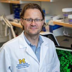
His project has two aims. The first is to identify the key proteins that regulate the phosphorylation of αA-crystallin and describe how they work. The second is to apply the findings from previous studies to demonstrate the potential of using this protective mechanism as a novel treatment option for diabetes complication.
“Using retina cells in culture, we’ve shown that an enhanced form of αA-crystallin is highly protective,” he explains. “The next step is to find out whether it can protect the whole retina from conditions associated with diabetes—in the case of this study, reduced blood flow.”
The long term goal is to design treatments that harness the protective powers of this protein to control or prevent DR.
Mark Johnson, M.D., Professor of Ophthalmology and Visual Sciences and Director of the Retina and Uveitis Service, is the latest Kellogg leader to receive the Heed-Gutman Award from the Society of Heed Fellows of the American Academy of Ophthalmology (AAO).
The award recognizes extraordinary and distinguished clinical, research and/or educational leadership and service to the field. A leading clinician and investigator in the medical and surgical treatment of macular, vitreomacular and vitreoretinal disorders, Dr. Johnson has served as principal investigator on numerous
multicenter clinical trials. A prominent voice on retina topics, he has delivered more than 20 named lectures and 240 invited talks in the U.S. and internationally and authored or co-authored nearly 250 peer-reviewed papers and book chapters.
“I was humbled—and surprised—to learn about the award,” he says. “Over the years I’ve been in the audience to celebrate numerous Heed-Gutman awardees. Hearing about their accomplishments has always inspired me to keep learning and contributing. What an honor to find myself in such esteemed company.”
In addition to the Heed-Gutman Award, Dr. Johnson, a past president of both The Macula Society and The Retina Society, has received a U.S. Presidential Scholar Award and both the Senior Achievement Honor and Life Achievement Honor Awards from the AAO.
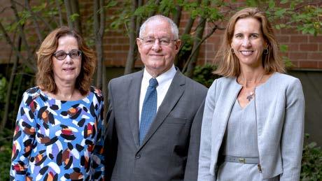
Alan Sugar, M.D., a corneal transplantation specialist, has served on the U-M Department of Ophthalmology and Visual Sciences faculty for more than 40 years, and is known for his compassionate care and determination to help his patients feel and see better.
Ophthalmology is in Dr. Sugar’s DNA. His twin brother, Joel, is also an ophthalmologist. Both were influenced by their ophthalmologist father, who discussed his work at dinner each night. Growing up, Dr. Sugar’s youth is filled with other ties to the field, U-M, and the Kellogg Eye Center. The elder Dr. Sugar was W.K. Kellogg’s physician. He would bring his sons on house calls so they could play with the Kellogg’s dog—a relative of TV’s Rin Tin Tin. Every Christmas, the Sugar family received a case of their favorite Kellogg cereal. Coincidentally, the Sugars lived next door to the uncle of Paul R. Lichter, M.D., foreshadowing a future when Dr. Sugar and the former department chair and founder of the Kellogg Eye Center would be U-M colleagues.
“Ophthalmological Society in 1988. He has received the Eye Bank Association of America Paton Award, the Cornea Society Castroviejo medal, and the American Academy of Ophthalmology Lifetime Achievement Award. He is a medical director of Eversight Eye Bank.
I RECEIVED EXPERT CARE FROM DR. SUGAR FOR MORE THAN 30 YEARS.
I WANTED TO HONOR HIM AND SUPPORT INNOVATIVE SCIENCE.
Susan Lane
When he learned his longtime patient, Susan Lane, established a professorship in his honor, he was surprised and touched. Ms. Lane has long dedicated her time, expertise, and passion to her interests and causes. A retired teacher, she actively volunteers at a local school, tutoring kids, and working in the library. Her appreciation to of Dr. Sugar for his care over the years inspired her to establish the professorship in his name, which was inaugurated on August 25, 2023. Recognizing Ms. Lane’s generosity, Dr. Sugar added his own significant gift to the professorship fund.
“Dr. Sugar joined the U-M faculty in 1979 as an associate professor, rising through the ranks to professor in 1984. He was associate and vice chair of the department from 1993 to 2019. The author of 235 papers, he was associate editor of Ophthalmology and editor-inchief of Cornea. He was president of the Cornea Society from 2004 to 2006 and elected to the American
“I received expert care from Dr. Sugar for more than 30 years. I wanted to honor him and support innovative science,” she said. In Maria Woodward, M.D., M.S., the inaugural Sugar Professor, Ms. Lane sees someone who is pursuing “great ideas” to better treat corneal diseases of all kinds, a meaningful way to honor Dr. Sugar’s many contributions to corneal disease. Ms. Lane’s father also played a role in her gift when he simply suggested she support the people and the place that took such good care of her.
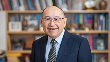
In January of 2023, Paul P. Lee, M.D., J.D., stepped down as chair of the University of Michigan Department of Ophthalmology and Visual Sciences and director of the W.K. Kellogg Eye Center, to serve as executive director of the U-M Medical Group and senior U-M Medical School associate dean for clinical affairs. As chair, Dr. Lee greatly impacted the department and the field through his internationally recognized expertise in glaucoma, patient-centered care, health services research, innovative leadership, and successful recruitment of top health services researchers to U-M.
“
I BELIEVE
He was instrumental in raising the profile of health services research, a field that encompasses many areas. In ophthalmology, health services researchers focus on how eye care is accessed and delivered in local, low- to middle-income communities as well as to impoverished populations around the world. Our faculty are finding ways to help kids and older adults receive regular vision screening to detect problems before they become worse. We are using big data sets to identify trends and determine best practices. In addition to vision care, our work influences all medical care, and it is because of Dr. Lee that our foundation in this vital area is solid.
Dr. Lee’s interest in health services research began when the field was new. His focus on developing best practices and broadening access to care has led to innovations such as better tools, technologies, treatments, and methods enabling providers to reach more people. The benefits of health services research are many, the possibilities endless.
“Dr. Lee was one of the key people in the field of ophthalmology health services research. Early on he saw its importance ahead of the industry. Back in 2005 to 2006, there were less than 10 people in this space. Today, there are dozens more— all thanks to Dr. Lee.” Joshua Stein, M.D.
THE WORK WE ARE SPEARHEADING WILL INFLUENCE NOT JUST OPHTHALMOLOGY, BUT HEALTH CARE MORE BROADLY AT MICHIGAN MEDICINE, IN THE STATE, ACROSS THE NATION, AND AROUND THE GLOBE.
Angela Elam, M.D., M.P.H.
To honor Dr. Lee’s distinguished leadership and impact, we created the Paul P. Lee, M.D., J.D., Chair’s Innovation Research Fund. It is the first step to establishing an endowed professorship in his name which will ensure the innovative research Dr. Lee has produced and championed will continue in perpetuity, allowing future physician-researchers to impact the field.
“
“I believe the work we are spearheading will influence not just ophthalmology, but health care more broadly at Michigan Medicine, in the state, across the nation, and around the globe.” Angela Elam, M.D., M.P.H.
Please join us in honoring Dr. Paul Lee’s many contributions to ophthalmology and health services research while ensuring tomorrow’s leaders can carry on his innovative efforts. To make a gift or get more information, please contact our W.K. Kellogg Eye Center development partners, Lindsay Baden at 734-604-7977 or linmwell@umich.edu, or Amanda Barnett at 734-274-3009 or akbarnet@umich.edu.
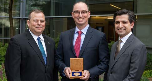
The Edna H. Perkiss Research Professorship in Ophthalmology and Visual Sciences was established in 2018 through an estate gift from Alvin Perkiss. Making the gift was a meaningful way for Mr. Perkiss to honor the memory of his wife, Edna. It was also a way he could provide future support for research aimed at curing blinding vision diseases, like the age-related macular degeneration Edna faced.
Edna Perkiss was born in Detroit in 1918. She was known for her love of reading, travel, and for being a dedicated friend. “Helping others was part of who she was,” said Mr. Perkiss. In addition, Mrs. Perkiss is remembered for her long and distinguished 32-year career at Ford Motor Company. Her work as a secretary was very important to her, so much so that she occasionally returned to work in the evening after dinner. During her tenure at the company, she worked with several vice presidents.
sister and were married for 61 years. Later in life, Mrs. Perkiss lost her vision due to age-related macular degeneration. While forced to give up some of her favorite activities, she never lost her giving nature. She passed away in 2008, and this professorship honors her generosity of spirit through its intent to spare others the pain of losing their sight.
David Zacks, M.D., Ph.D., has long been committed to saving sight and improving vision through his innovative research focusing on methods to prevent retinal cell death and damage before it leads to blindness. “We hope to develop core knowledge to allow for a greater understanding of how disease results in cell death, allowing for the creation of new treatments to improve vision,” he says.
“ “
WE HOPE TO DEVELOP CORE KNOWLEDGE TO ALLOW FOR A GREATER UNDERSTANDING OF HOW DISEASE RESULTS IN CELL DEATH, ALLOWING FOR THE CREATION OF NEW TREATMENTS TO IMPROVE VISION.
David Zacks, M.D., Ph.D.
Alvin Perkiss served in the U.S. Army during World War II. The G.I. Bill enabled him to attend college and build his automotive supplier business in Dearborn, Michigan. Alvin and Edna were introduced by Alvin’s
An internationally renowned retina specialist, Dr. Zacks has identified key molecular pathways that control retinal cell health and therapeutic targets to help keep these cells alive. He has published over 115 peer-reviewed articles,holds numerous patents, has received the Macula Society W. Richard Green and the Terry J. Bergstrom Faculty Teaching awards, and serves on the Executive Committee of the Retina Society. He is also co-founder of ONL Therapeutics, an Ann Arbor based biotech company developing a first-in-class neuroprotective agent for patients with retinal disease. Dr. Zacks joined U-M as an assistant professor in 2002 and was promoted to professor in 2015. He was inaugurated as the Edna H. Perkiss Professor of Ophthalmology and Visual Sciences on July 19, 2023.
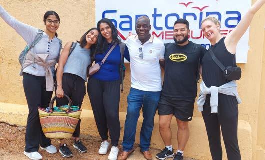
Kellogg has a longstanding partnership with the Kisii Eye Hospital (KEH), located in southwest Kenya. Since 2017, the collaboration has provided opportunities for mutually enriching training and high-impact clinical and epidemiological research.
KEH is one of the only tertiary eye hospitals serving a population of more than 10 million people. Under the leadership of CEO and Medical Director Daniel Kiage, M.D., a glaucoma specialist, KEH has grown rapidly since its founding in 2013, in diagnostic and surgical volumes as well as in the number of ophthalmologists and allied ophthalmic personnel receiving training.
“
tional Program at Kellogg. “Together, we offer a variety of opportunities to get involved in effecting positive change around the world.”
The seeds of the current KEH project were planted when a medical student learned about those opportunities at one of Kellogg’s International Night events. But it really took root when she ventured across campus to take a class at the business school.
THE REAL CHALLENGE FOR KISII IS MAKING THESE AND FUTURE VISION CENTERS FINANCIALLY INDEPENDENT, SUSTAINABLE AND POSITIONED TO DELIVER THE RIGHT CARE FOR THE COMMUNITIES THEY SERVE.
Recently, the hospital opened its first satellite vision clinic, the Keroka Vision Centre. As they contemplated opening another, they looked to their colleagues in the University of Michigan community for guidance.
—Bela Parekh
“Kellogg is one part of a strong, interconnected cohort of U-M faculty and students from a number of departments, schools and institutes, all engaged in international health issues,” says Joshua Ehrlich, M.D., M.P.H., Co-director of the Jerome Jacobson Interna-
“
Bela Parekh first became interested in issues of global health equity while working for a top management consulting firm after college. She brought that same passion for and commitment to reducing health disparities to her training at the U-M Medical School (UMMS).
After the completion of her core clinical year, she enrolled in a course through the William Davidson Institute (WDI) at the U-M Ross School of Business. The WDI leverages business expertise to build lasting economic and social prosperity in lowand middle-income countries. The class, Healthcare Delivery in Emerging Markets, assigned each student a business case. Fate stepped in when Parekh, who was
“KELLOGG IS ONE PART OF A STRONG, INTERCONNECTED COHORT OF U-M FACULTY AND STUDENTS FROM A NUMBER OF DEPARTMENTS, SCHOOLS AND INSTITUTES, ALL ENGAGED IN INTERNATIONAL HEALTH ISSUES.
“leaning toward specializing in ophthalmology, was given the KEH Vision Centre expansion case. She simultaneously traveled to Ghana on a research scholarship through Global REACH, a UMMS program that engages faculty and students in international health collaborations.
Dr. Kiage’s team opened a second clinic in March 2023 while Parekh and the rest of the student team were on the ground in Kenya conducting interviews. Parekh presented the initial plan for the Vision Centres in May 2023 to Dr. Kiage and WDI President Paul Clyde, Ph.D.
“When the project wrapped up, I felt like there was more to do,” says Parekh, now an M4. “The real challenge for Kisii is making these and future vision centers financially independent, sustainable and positioned to deliver the right care for the communities they serve.”
Parekh pitched the idea of developing a comprehensive vision center expansion strategy for Kisii to Drs. Ehrlich and Clyde, who embraced the concept right away.
With input from her Kellogg and WDI mentors, Parekh and others will work with Kisii staff to understand the financials and logistics of the enterprise, including revenues, resource allocation, and referral patterns between clinics and the main hospital. Just as important are clinical considerations like staffing, training and equipment.
“Dr. Kiage has assembled a talented, highly motivated team at KEH,” says Dr. Ehrlich. “Like every initiative we undertake with them, this project will be informed by the epidemiology and culture of the region. Together, we’ll identify the vision health issues of greatest local concern, and determine how best to address their eyecare needs at a Kisii Vision Centre.”
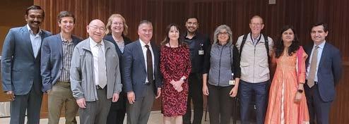

James Ellis, M.D.
B.S. – University of Utah
M.D. – University of Utah School of Medicine

Kristine Nachbor M.D., M.S.
B.A. – Carleton College
M.S. – Michigan Technological University
M.D. – University of Minnesota Medical School

Harry Levine, M.D., M.P.H.
B.A., B.S., M.P.H., – University of Miami
M.D. – University of Miami
LM Miller School of Medicine

JoshuaOng, M.D.
B.S. – The Ohio State University
M.D. – University of Pittsburg School of Medicine

David Mundy, M.D.
B.S. – Stanford University
M.D. – Stanford University School of Medicine

Ahmed Owda, M.D., M.P.H.
B.S. – University of Michigan
M.D. – Columbia University
College of Physicians and Surgeons
M.P.H. – Columbia University

Anvesh Annadanam, M.D.
Cornea Fellow Cleveland Clinic Cleveland, OH

Otana Jakpor, M.D., M.Sc.
Pediatric Ophthalmology Fellow
Kellogg Eye Center
Ann Arbor, MI

Jaqueline Stoutin, M.D.
B.A. – The University of Chicago
M.D. – Texas A&M HSC College of Medicine

Daniel Balikov, M.D., Ph.D.
Vitreoretinal Fellow
Bascom Palmer Miami, FL

Omar Moinuddin, M.D.
Vitreoretinal Fellow
Associated Retinal Consultants
Royal Oak, MI

Nikhil Bommakanti, M.D.
Vitreoretinal Fellow
Wills Eye Hospital Philadelphia, PA

David Skanchy, M.D.
Pediatric Ophthalmology Fellow
Kellogg Eye Center
Ann Arbor, MI

Jeffrey Yu, M.D.
Vitreoretinal Fellow
Kellogg Eye Center
Ann Arbor, MI

Nicholas Carducci, M.D.
Medical Retina
Brighton Vision Center Brighton, MI

Emily Eton, M.D.
Vitreoretinal Surgery
Kellogg Eye Center
Ann Arbor, MI

Olivia Killeen, M.D., M.S.
National Clinical Scholar
Pediatric Ophthalmology Fellowship Duke University Durham, NC

Chelsea Reighard, M.D.
Pediatric Ophthalmology & Adult Strabismus
Asheville Eye Associates Asheville, NC

Rebecca Edwards Mayhew, M.D., Ph.D.
Pediatric Ophthalmology & Adult Strabismus
University of Colorado Aurora, CO

Miles Greenwald, M.D.
Global Ophthalmology
University of Kansas Eye Center Kansas City, KS

Brett Malbin, M.D.
Retinal Dystrophy
Kresge Eye Institute Detroit, MI

Meari Taguchi, M.D.
Neuro-Ophthalmology
Renown Regional Medical Center Reno, NV

Jennifer Enright, M.D., Ph.D.
Cornea, External Disease & Refractive Surgery
Washington University in St. Louis St. Louis, MO

Ahmad Halawa, M.D.
Pediatric Ophthalmology & Adult Strabismus
Truhlsen Eye Institute Omaha, NE

Justin Park, M.D. Glaucoma
Cornerstone Eye Associates Rochester, NY

Alexa Thibodeau, M.D.
Cornea, External Disease & Refractive Surgery
Kaiser Permanente San Diego, CA


Elaine Downie, M.D., is a fellowship trained oculoplastic surgeon. She earned her medical degree from the University of Minnesota. She then went on to complete her ophthalmology residency and ASOPRS fellowship at the University of Wisconsin. She cares for patients at the Ann Arbor and Brighton clinics as well as the VA.
Emily Eton, M.D., earned her medical degree from Harvard Medical School. She then completed an ophthalmology residency followed by a vitreoretinal surgery fellowship at the University of Michigan Kellogg Eye Center. She will provide adult and pediatric retinal care in the Ann Arbor and Brighton offices.
Charles Frank, M.D., returns to Ann Arbor after completing his ophthalmology residency at the University of Illinois at Chicago. He attended medical school at the University of Michigan. He is practicing comprehensive ophthalmology at the Chelsea, Ypsilanti, and Wall St. offices.


Farida Hakim, M.D., earned her medical degree from the University of Chicago Pritzker School of Medicine and stayed at the University of Chicago for her residency training. She then went on the complete a cornea fellowship at the University of Pittsburgh Medical Center. She joins as a Clinical Assistant Professor and provides corneal, refractive and comprehensive care at the VA and at Kellogg’s Brighton, Ann Arbor, and Canton offices.
Shilpa Kodati, M.B.B.S., completed medical school at Imperial College London, United Kingdom, followed by a postdoctoral research fellowship at Schepens Eye Research Institute, ophthalmology residency at the University of Pittsburgh Medical Center (UPMC), a uveitis fellowship at the National Eye Institute (NEI), and a vitreoretinal surgical fellowship at Baylor College of Medicine. She was a staff clinician at the NEI before joining Kellogg.
Brittany Simmons, M.D., received her medical degree at the University of Illinois at Chicago. She completed an ophthalmology residency at the Flaum Eye Institute and ASOPRS fellowship at the University of Iowa. Her research interests span education, surgical outcomes, novel markers of periorbital malignancy, clinical decision-making and comparative outcome studies, and diversity and inclusion in healthcare.
Nita Valikodath, M.D., M.S., attended the University of Michigan Medical School and completed her ophthalmology residency at the University of Illinois at Chicago and surgical retina fellowship at Duke University. She cares for adult and pediatric retina patients in Ann Arbor and Northville. Her research focuses on novel technologies to improve clinical outcomes and quality of life.
Marguerite Weinert, M.D., earned her medical degree from Duke University. She then went on to complete her ophthalmology residency and chief residency year at Massachusetts Eye and Ear, and her fellowship in Pediatric Ophthalmology and Adult Strabismus at Boston Children’s Hospital. She provides pediatric ophthalmology and adult strabismus care in Ann Arbor and Brighton.
Mark Draelos, M.D., Ph.D., ., earned his medical and doctoral degrees at Duke University as well as general surgery internship and postdoctoral training. He holds appointments in the departments of Robotics and Ophthalmology. His lab focuses on image-guided medical robots with an emphasis on clinical translation and ophthalmic applications.
Thanh Hoang, Ph.D., earned his PhD in Developmental Biology at Miami University and completed his postdoctoral training in Neuroscience at Johns Hopkins before joining the Kellogg Eye Center. His research goal is to develop cell-based therapies for neurodegenerative diseases.


Qiang Li, Ph.D., completed his Ph.D. training (Surgical oncology) at Shanghai Jiaotong University School of Medicine in China. He competed his postdoctoral training at the University of Pittsburgh and Case Western Reserve University. His research interests focus on gene regulation and cell fate decisions in pluripotent stem cells.
Elise Savier, Ph.D., earned her doctoral degree from the University of Strasbourg, France in 2016, during which she investigated the development of the visual system. She completed her postdoctoral training at the University of Virginia. As a faculty member of both the Department of Molecular and Integrative Physiology and Ophthalmology, her research focuses on the central processing underlying vision.
Kyung No Son, Ph.D., attended Kyung Hee University, South Korea and completed postdoctoral training at the University of Illinois at Chicago. He previously served as Director of Preclinical Drug Development at the University of Illinois at Chicago. He focuses on ocular surface diseases and Niemann Pick Type C Disease.
Qitao Zhang, Ph.D., his Ph.D. at Peking University in Beijing, China. He works on myriad RPE projects, ranging from optimizing cell culture, developing RPE culture models of AMD, and testing promising therapeutic targets in mouse, rat, and rabbit animal models.
Vinay Aakalu, M.D., M.P.H., F.A.C.S.
• Top Doctor, Castle Connolly
• Editorial Board Member, Orbit
• Editorial Board Member, Translational Vision Science and Technology
• Editorial Board Member, The Ocular Surface
• Reviewer, Study Section: ZEY1 VSN (04) [NEI/NIH Mentored Clinician Scientist Grant Applications (K08, K23) and Conference Grants (R13)]
• Assistant Chair, Written Qualifying Examination Working Group, American Society of Ophthalmic Plastic and Reconstructive Surgery
• Chair, Awards Committee, American Society of Ophthalmic Plastic and Reconstructive Surgery
• Member, Publications Committee, The Association for Research in Vision and Ophthalmology
• Methodologist, Ophthalmic Technology Assessment Committee-Oculoplastics, American Academy of Ophthalmology
Steven Abcouwer, M.D.
• Chief Editor, Journal of Ophthalmology
• Editorial Board Member, Investigative Ophthalmology and Vision Science
• Editorial Board Member, Translational Vision Science & Tecnology
• Editorial Board Member, Frontiers in Ophthalmology, Retina Section
• Editorial Board Member, American Journal of Physiology: Endocrinology and Metabolism
• Editorial Board Member, Frontiers in Immunology
• Editorial Board Member, Frontiers in Neurology
• Scientific Review Editor, Molecular Vision Journal
• Chair, National Institutes of Health Neurotechnology and Vision (NV) Study Section ZRG1 NV-Q 91 (01)
• Chair, National Institutes of Health Neurotechnology and Vision (NV) Study Section ZRG1 NV-Q 91 (02)
• Member, National Institutes of Health Pathophysiology of Eye Disease 1 (PED1) Study Section
• Member, National Institutes of Health Study Section ZRG1 MGG-G (70) R RFA-RM-22-015: Investigational New Drug-enabling Studies of Somatic Genome Editing Therapeutic Leads (U19)
• Member, Department of Defense FY22 Peer Reviewed Medical Research Program for the Congressionally Directed Medical Research Programs Diabetes Treatment Clinical Trials Study Section
• Reviewer (Ad Hoc), Czech Science Foundation
Kari E. Branham, M.S., C.G.C.
• Chair, Genetics Review Committee, Foundation Fighting Blindness Clinical Consortium
• Member, Steering Committee for the National Ophthalmic Disease Genotyping and Phenotyping Network (eyeGENE®), National Eye Institute
JULY 1, 2022 — SEPTEMBER 30, 2023
Kari E. Branham, M.S., C.G.C. (cont.)
• Expert Review Panel Member, Early Onset Retinal Degeneration Variant Curation, ClinGen
• Expert Review Panel Member, Retinal Disease Gene Curation, ClinGen
• Member, Scientific Advisory Board, Foundation Fighting Blindness
• Member, Evidence Based-Guideline for Retinal Dystrophies Workgroup, American College of Medical Genetics
• Editorial Board Member, Investigative Ophthalmology & Visual Science
• Community Engagement Award, National Society of Genetic Counselors
Sherry Day, O.D., FAAO
• Board Member, American Academy of Optometry Foundation
• Vision Rehabilitation Committee Board Member, American Optometric Association
• Co-Chair, Vision Rehabilitation Committee, Michigan Optometric Association
Karen DeLoss, O.D., FAAO
• Secretary, American Optometric Association Cornea and Contact Lens Section
• Vice Chair, Global Specialty Lens Symposium Education and Planning Committee
• Education Chair, Global Ophthalmic Women organization
• Advisory Board, Gas Permeable Lens Institute
Joshua Ehrlich, M.D., M.P.H.
• Reviewer, Special Emphasis Panel, Fogarty International Center
• Co-Chair, International Education Sub-Committee, American Glaucoma Society
• Editorial board, Ophthalmic Epidemiology
• Associate Director, Innovative External Networks Core, P30 Michigan Center on the Demography of Aging
Angela Elam, M.D.
• Secretariat Award, American Academy of Ophthalmology
• National Medical Association Scientific Contribution Award
• Advisory Committee, Center for Population and Vision Health, Prevent Blindness
• Co-Chair, Taskforce on Disparities in Eye Care, American Academy of Ophthalmology
• Scientific Contribution Award, National Medical Association
• Reviewer, Merit Review Panel, Achieve Health Equity Broad Pragmatic Studies, Patient-Centered Outcomes Research Institute
• Editor, Translational Vision Science and Technology Journal, Equity in Eye Care special issue
Peter Hitchcock, Ph.D.
• Fulbright Specialist, United States Department of State; Bureau of Educational and Cultural Affairs
• Invited Symposium speaker, Regulation of Müller glia-dependent neuronal regeneration in the damaged retina, 24th Biennial Meeting, International Congress of Eye Research, Gold Coast, Australia
• Invited speaker, Annual Meeting of the Scientific Advisory Board for the ECaBox Project, Porto, Portugal.
Mark Johnson, M.D.
• William S. and Grace Snyder Lecture, Vanderbilt University
• Chandler Lecture, Mass Eye and Ear Infirmary, Harvard University
• Heed Gutman Award, Society of Heed Fellows
• Chair, Awards Committee, The Macula Society
• Castle Connelly Top Doctor in Ophthalmology
• Best Doctors in America
• Associate Editor, American Journal of Ophthalmology
• Editorial Board Member, Retina: The Journal of Retinal and Vitreous Diseases
Ariane Kaplan, M.D.
• Secretary, AUPO Directors of Medical Student Education Council
• Member, AAO BCSC Update on General Medicine Book Committee
• Member, WIO Academic Outreach Committee
Shilpa Kodati, M.D.
• Editorial Board Member, Scientific Reports
• Member, Continuing Medical Education Committee Association for Research in Vision and Ophthalmology
• Chair, Research Committee, Young Uveitis Society
• Member, Young Uveitis Society Executive Committee
• Invited lecturer, Journées Ophtalmologiques Universitaires de Québec
Paul P. Lee, M.D., J.D.
• Trustee-at-Large, President-Elect, Chair, Data Resource Committee, Association of University Professors of Ophthalmology
• Invited Speaker, John and Suzanne Shore Lectureship, University of Texas
• President, National Alliance for Eye and Vision Research/Alliance of Eye and Vision Research
• Board of Directors, Ophthalmology Foundation
• Invited Speaker, Richard D. Richards Symposium, University of Maryland
• Board of Directors, Society of Heed Fellows
Paul Lichter, M.D., M.S.
• Best Doctors in America
• Top Doctor, Castle Connolly
• Top Doctor, US News and World Report
JULY 1, 2022 — SEPTEMBER 30, 2023
Shahzad I. Mian, M.D.
• Advisory Committee, American Academy of Ophthalmology
• Examiner, Member, Writer, American Board of Ophthalmology
• Member, American Ophthalmological Society
• Best Doctors in America
• Associate Editor, Cornea Open
• Board of Directors, Secretary, Cornea Society
• Board of Directors, Governance Committee, Big Data Task Force Medical Advisory Board, Eye Bank Association of America
• Board of Directors, Eversight Eye Bank
• Board of Directors, Secretary, Michigan Society of Eye Physicians and Surgeons
Jason Miller, M.D., Ph.D.
• David Hinton Scholarship, Ryan Initiative for Macular Research
David Musch, Ph.D., M.P.H.
• Scientific Reviewer: Health and Medical Research Fund, Government of Hong Kong SAR
• Member, Editorial Boards: JAMA Ophthalmology
• Member/Chair: numerous NEI/NIH and company-sponsored Data and Safety Monitoring Committees for ongoing, multicenter, randomized clinical trials
Christine Nelson, M.D., FACS
• Board of Directors, ASOPRS Foundation Committee
• Board of Directors, Member, World Association of Eye Hospitals
• Co-Chair, Fellowship Curriculum Project, International Council of Ophthalmology
• Chair, Wendell Hughes Awards Committee
• Awards Committee, Women in Ophthalmology
• Fellowship Curriculum Project Committee, International Council of Ophthalmology
• NASAOS Membership Committee
• Senior Advisor, International Global Opportunities Committee, ASOPRS
Paula Anne Newman-Casey, M.D., M.S.
• Physician Scientist Award, Research to Prevent Blindness
• Glaucoma Subcommittee Chair, Annual Meeting Program Committee, American Academy of Ophthalmology
• Member, American Academy of Ophthalmology Community Health Center Taskforce
Yannis Paulus, M.D.
• Grant Reviewer, United Kingdom Research and Innovation Medical Research Council
• Grant Reviewer, University of Maryland, Maryland Industrial Partnerships Program
• Grant Reviewer, Food and Health Bureau of the Health and Medical Research Fund for The Government of the Hong Kong Special Administrative Region
• Grant Reviewer, Israeli Ministry of Innovation, Science and Technology
Yannis Paulus, M.D. (cont.)
• Review Committee, Study Section, NIH Council ZEY1 VSN 04 NEI Mentored Clinician Scientist Applications
• Grant Reviewer, Dutch Research Council
• Review Committee, Study Section Ad hoc member, Military Operational Medicine Research Program for the Department of Defense
• Review Committee, Study Section Ad hoc member, National Science Foundation SBIR/STTR Phase II Panel Review Committee Digital Health Medical Devices and Diagnostics panel
• Grant Reviewer, Swiss National Science Foundation
• Grant Reviewer, Croatian Science Foundation
• Massey Foundation TBI Grand Challenge Award
• Editorial Board, Ophthalmic Surgery, Lasers & Imaging Retina
• Editorial Board, Photonics
• Editorial Board, Scientific Reports
• Chair, Continuing Medical Education Committee, Association for Research in Vision and Ophthalmology
Lev Prasov, M.D., Ph.D.
• Epstein Award, Association for Vision Research and Ophthalmology Foundation
• Gene Curation Expert Panel for Glaucoma/Neuro-Ophthalmology, ClinGen
• Co-Chair, Variant Curation Expert Panel Anterior Segment Dysgenesis, ClinGen
• Guest Editor, Frontiers in Neuroscience
• Guest Editor, Genes
• Schaffer Prize, Glaucoma Research Foundation
• Member, International Society of Genetic Eye Disease and Retinoblastoma
Rajesh Rao, M.D.
• Election to The Club Jules Gonin
• Secretariat Award, American Academy of Ophthalmology
• Study Section Reviewer, Neurobiology-F VA Merit Grant Review Program
• Study Section Member, The Aging and Development, Auditory, Vision and Low Vision Small Business Study Section (ET TN 12/ SBIR/STTR), Center for Scientific Review, National Institutes of Health
James Weiland, Ph.D.
• Bioengineering of Neuroscience and Vision Technologies study section, National Institutes of Health
• Past Vice President, Medicine and Biology Society Conferences, Institute of Electrical and Electronics Engineers
Kwoon Wong, Ph.D.
• Associate Editor, Frontiers in Cellular Neuroscience
• Grant reviewer, Hong Kong Research Grants Council
JULY 1, 2022 — SEPTEMBER 30, 2023
Sarah Wood, O.D., M.S., FAAO
• Secretary, Optometric Glaucoma Society
• Awards Committee, Women in Ophthalmology
• Fellowship Curriculum Project Committee, International Council of Ophthalmology
• NASAOS Membership committee
• Membership Committee, Optometric Glaucoma Society
• AAO Glaucoma Diplomate executive committee
• Guest Reviewer, American Academy of Optometry Journal
Maria A. Woodward, M.D., M.S.
• Senior Achievement Award, American Academy of Ophthalmology
• Deputy Section Lead, American Academy of Ophthalmology
• Senior Editor, Cornea
• Board Member, Cornea Society
• Dermatologic and Ophthalmic Drugs Advisory Committee, Food and Drug Administration
• Member, JAMA Ophthalmology Advisory Committee
• Member, World Cornea Congress IX Program Committee
Thomas Wubben, M.D., Ph.D.
• Member, Retina Society
• Assistant Editor, RETINA Round Up
David Zacks, M.D., Ph.D.
• Member, Data and Safety Monitoring NEI-sponsored NAC Attack trial – a Phase 3 multicenter clinical trial of oral treatment with N-acetylcysteine (NAC) in patients with retinitis pigmentosa.
Amy Zhang, M.D.
• Invited Section Editor, Advances in Ophthalmology and Optometry
Jason Zhang, M.D.
• Member, Multimedia Subcommittee, Education and Communication Committee, American Glaucoma Society (AGS)
Each year, Kellogg offers an informative series of continuing medical education (CME) programs designed to share new approaches to the diagnosis and management of eye disease across subspecialties.
For more information, visit: www.umkelloggeye.org
For questions: contact Jennifer Burkheiser, CME Coordinator, at (734) 763-2357 or kelloggCME@umich.edu.


Vinay Aakalu, M.D., M.P.H.
Fernanda Abalem, M.D.
Steven Abcouwer, Ph.D.
Robin Ali, Ph.D.B.Sc.
Sameerah Ali, M.D.
David Antonetti, Ph.D.
Steven Archer, M.D.
Bernadete Ayres, M.D.
Audree Bass, O.D.
Cagri Besirli, M.D., Ph.D.
Amanda Bicket, M.D.
Jill Bixler, M.D.
Brittany Boland, O.D.
Kari Branham, M.S., CGC
Roland Chen, Ph.D.
Grant Comer, M.D., M.S.
Theresa Cooney, M.D.
Wayne Cornblath, M.D.
John Cropsey, M.D.
Julia Dalia, M.D.
Sherry Day, O.D., FAAO
Lindsey De Lott, M.D., M.S.
Karen Deloss, O.D., FAAO
Monte Del Monte, M.D.
Hakan Demirci, M.D.
Tatiana Deveney, M.D.
Courtney Dewey, O.D.
Elaine Downie, M.D.
Mark Draelos, M.D., Ph.D.
Joshua Ehrlich, M.D.,M.P.H.
Angela Elam, M.D.
Susan Elner, M.D.
Victor Elner, M.D., Ph.D.
Emily Eton, M.D.
Abigail Fahim, M.D., Ph.D.
Cherie Farkash, O.D
Charles Frank, M.D.
Patrice Fort, Ph.D.
Carlton Foster, O.D.
Bruce Furr, C.O., Ph.D.
Philip Gage, Ph.D.
Samantha Gagnon, O.D., FAAO
Christopher Gappy, M.D.
Thomas Gardner, M.D., M.S.
Daniel Green, Ph.D.
Paul Grenier, O.D.
Farida Hakim, M.D.
Sean Hansen, M.D.
Peter Hitchcock, Ph.D.
Thanh Hoang, Ph.D.
Christopher Hood, M.D.
Bret Hughes, Ph.D.
Michael Huvard, M.D.
Adam Jacobson, M.D.
K. Thiran Jayasundera, M.D., M.S.
Vanitha Jeyaraj, M.D.
Denise John, M.D., FRCSC
Mark Johnson, M.D.
Ariane Kaplan, M.D.
Harjeet Kaur, M.D.
Naheed Khan, Ph.D.
Sangeeta Khanna, M.D.
Denise Kim, M.D.
Erin Klukas, O.D
Shilpa Kodati, M.D.
Zvi Kresch, M.D.
Amy Lagina, O.D., FAAO
Paul Lee, M.D., J.D.
Helios Leung, O.D., Ph.D
Qiang Li, M.D., Ph.D.
Executive Officers of Michigan Medicine
Marschall S. Runge, M.D., Ph.D.
Executive Vice President for Medical Affairs, Dean, University of Michigan Medical School, C.E.O., Michigan Medicine
David C Miller, M.D., M.P.H
President, University of Michigan Health System, Executive Vice Dean for Clinical Affairs, Medical School
Debra F. Weinstein, M.D.
Executive Vice Dean for Academic Affairs, Medical School
Chief Academic Officer for Michigan Medicine
Steven L. Kunkel, Ph.D.
Executive Vice Dean for Research, Medical School
Chief Scientific Officer, Michigan Medicine
The Regents of the University of Michigan
Jordan B. Acker, Michael J. Behm, Mark J. Bernstein, Paul W. Brown, Sarah Hubbard, Denise Ilitch, Ron Weiser, Katherine E. White, Santa J. Ono (ex officio)
© 2023 Regents of the University of Michigan
A Non-discriminatory, Affirmative Action Employer
Paul Lichter, M.D., M.S.
Nathan Liles, M.D., M.P.H.
Cheng-Mao Lin, Ph.D.
Xuwen Liu, M.D., Ph.D.
April Maa, M.D.
Matthew McKee, M.D.
Shahzad Mian, M.D.
Jason Miller, M.D., Ph.D.
Sayoko Moroi, M.D., Ph.D.
Kevin Mundy, M.D.
David Musch, Ph.D., M.P.H.
Mikiko Nagashima, Ph.D.
Nambi Nallasamy, M.D.
Christine Nelson, M.D.
Paula Anne Newman-Casey, M.D., M.S.
Jacqueline Nguyen, O.D.
Santa Ono, Ph.D.
Gale Oren , M.I.L.S.
Yannis Paulus, M.D.
Jillian Pearring, Ph.D.
Howard Petty, Ph.D.
Colleen Podd, O.D.
Lev Prasov, M.D., Ph.D.
Donald Puro, M.D., Ph.D.
Rajesh Rao, M.D.
Julia Richards, Ph.D.
Alan Robin, M.D.
Julie Rosenthal, M.D., M.S
Maria-Dolors Sans-Gili, Ph.D.
Therese Sassalos, M.D.
Elisa Savier, Ph.D.
Thielen Seng, O.D., FAAO
Anjali Shah, M.D.
Roni Shtein, M.D., M.S.
Brittany Simmons, M.D.
Annual Report Team
Editor: Julie Rosenthal, M.D., M.S.
Terry Smith, M.D.
Kyung No Son, Ph.D.
H. Kaz Soong, M.D.
William Sray, M.D.
Joshua Stein, M.D. M.S.
Alan Sugar, M.D.
Jeffrey Sundstrom, M.D., Ph.D.
Bradford Tannen, M.D., J.D.
Sally Temple, Ph.D.
Debra Thompson, Ph.D.
Jonathan Trobe, M.D.
Meagan Tucker, O.D.
Nita Valikodath, M.D.
Angela Verkade, M.D.
Grace Wang, M.D., Ph.D.
Eric Weh, Ph.D.
Sara Weidmayer, O.D., FAAO
James Weiland, Ph.D.
Marguerite Weinert, M.D.
Jennifer Weizer, M.D.
Sebastian Werneburg, Ph.D.
Adrienne West, M.D.
Donna Wicker, O.D., FAAO
Pamela Williams, M.D.
Kwoon Wong, Ph.D.
Sarah Wood, O.D., M.S., FAAO
Maria Woodward, M.D., M.S.
Annie Wu, M.D.
Rebecca Wu, M.D.
Thomas Wubben, M.D., Ph.D.
Guan Xu, Ph.D.
David Zacks M.D., Ph.D.
Amy Zhang, M.D.
Jason Zhang, M.D.
Qiato Zhang, Ph.D.
Design and Art Direction: David Murrel
Head Writer: Shelley Zalewski
Contributing Writers: Kristy Demas
Editorial Assistant: Jo Kristine Scott, Dani Shordt
Photographers: Michigan Photography: Eric Bronson, Erin Kirkland, Daryl Marshke, Scott Soderberg
FOR PATIENT APPOINTMENTS, PLEASE CALL 734.763.8122
For additional copies, please contact: University of Michigan
Department of Ophthalmology and Visual Sciences
W.K. Kellogg Eye Center 1000 Wall Street
Ann Arbor, Michigan 48105 www.umkelloggeye.org
Department of Ophthalmology and Visual Sciences
1000 Wall Street
Ann Arbor, MI 48105
The University of Michigan Kellogg Eye Center is proud to be ranked in the top 10 in the country by U.S. News & World Report—recognizing our outstanding care for patients with complex eye conditions.
Last year we celebrated 150 years of excellence in ophthalmology. Kellogg has seen extraordinary growth in all aspects of patient care, research and education since the department was established in 1872. Every day, our clinicians, scientists, trainees and staff work together to shape the future of eye care and vision science. We are proud to be part of Michigan Medicine.
~ Our Purpose ~ To improve lives through curing, preventing and treating eye disease