3 minute read
The Role of Digital Pathology in Head and Neck Tumor Diagnosis

Soft Tissue Sarcomas: What Every Pathologist Should Know
Introduction
Soft tissue sarcomas (STSs) represent a diverse and often challenging group of malignant tumors arising from the mesenchymal tissues of the body, including fat, muscle, nerve, fibrous tissue, blood vessels, and deep skin tissues. Although rare—comprising less than 1% of all adult cancers—their wide histological variety and overlapping morphologic features demand a high level of diagnostic precision from pathologists.
For pathologists, early and accurate identification of soft tissue sarcomas is essential—not only for guiding clinical management but also for informing prognosis and ensuring appropriate molecular or genetic testing. This blog aims to provide a comprehensive overview of STSs, offering insights into key definitions, diagnostic criteria, anatomic distribution, and the tools essential to differential diagnosis. Whether you're a trainee or an experienced practitioner, understanding the evolving landscape of soft tissue sarcoma pathology is crucial.
Definition: What Are Soft Tissue Sarcomas?
Soft tissue sarcomas are malignant tumors that arise from non-epithelial, extraskeletal connective tissues, such as adipose, muscle, fibrous tissue, and peripheral nerves. These tumors are distinct from carcinomas (which arise from epithelial cells) and are characterized by their mesenchymal origin, variable histologic appearance, and often aggressive clinical behavior.
They are classified based on histological features and lineage differentiation, such as:
Adipocytic tumors (e.g., liposarcoma)
Fibroblastic/myofibroblastic tumors
Skeletal and smooth muscle tumors (e.g., rhabdomyosarcoma, leiomyosarcoma)
Peripheral nerve sheath tumors
Vascular tumors
Undifferentiated/unclassifiable sarcomas
Focus Areas for Pathologists
Histopathological Identification
Understanding key microscopic patterns
Recognizing hallmark cellular features
Assessing mitotic rate, necrosis, cellularity
Immunohistochemistry (IHC)
Essential for distinguishing between morphologically similar tumors
Common markers: S100, Desmin, Myogenin, SMA, CD34, MDM2, etc.
Molecular Testing
FISH or PCR to detect characteristic translocations (e.g., t(X;18) in synovial sarcoma)
Use of NGS for complex or unclassifiable cases
Tumor Grading and Staging
FNCLCC grading system
Importance in prognosis and treatment planning
Anatomic Distribution: Where Do They Occur?
Soft tissue sarcomas can arise anywhere in the body, but common locations include:
Extremities (especially the thigh) – most frequent site
Retroperitoneum – often large, deep-seated tumors
Trunk and abdominal wall
Head and neck region
Visceral soft tissues and GI tract (e.g., GISTs—gastrointestinal stromal tumors)
Understanding location helps narrow the differential and supports histological findings. For example:
A deep-seated thigh mass in an adult may suggest a high-grade liposarcoma.
A retroperitoneal mass with lipoblasts should prompt testing for MDM2 amplification.
Benefits of Accurate Diagnosis
Treatment Guidance
Sarcomas are managed differently than carcinomas; histologic subtype influences surgery, radiation, and chemotherapy decisions.
Prognostication
Grade and type predict recurrence risk, metastasis, and overall survival.
Targeted Therapy
Molecular characterization (e.g., KIT mutations in GISTs) allows use of therapies like imatinib.
Avoiding Misdiagnosis
Prevents inappropriate treatment of benign mimickers (e.g., nodular fasciitis, lipoma)
Conclusion
Soft tissue sarcomas present one of the most diagnostically complex areas of surgical pathology. Given their rarity, varied presentation, and overlapping histological features, they require a systematic and skilled approach to diagnosis. Pathologists must integrate histologic findings with immunohistochemical stains and, increasingly, molecular diagnostics to deliver accurate and actionable diagnoses.
In an era of precision medicine, your role as a pathologist extends far beyond the microscope. Your expertise shapes clinical decisions, helps avoid diagnostic pitfalls, and ultimately contributes to better outcomes for patients with these rare but serious tumors.
Stay informed, stay curious—and never underestimate the impact of a precise diagnosis in the world of soft tissue sarcomas.
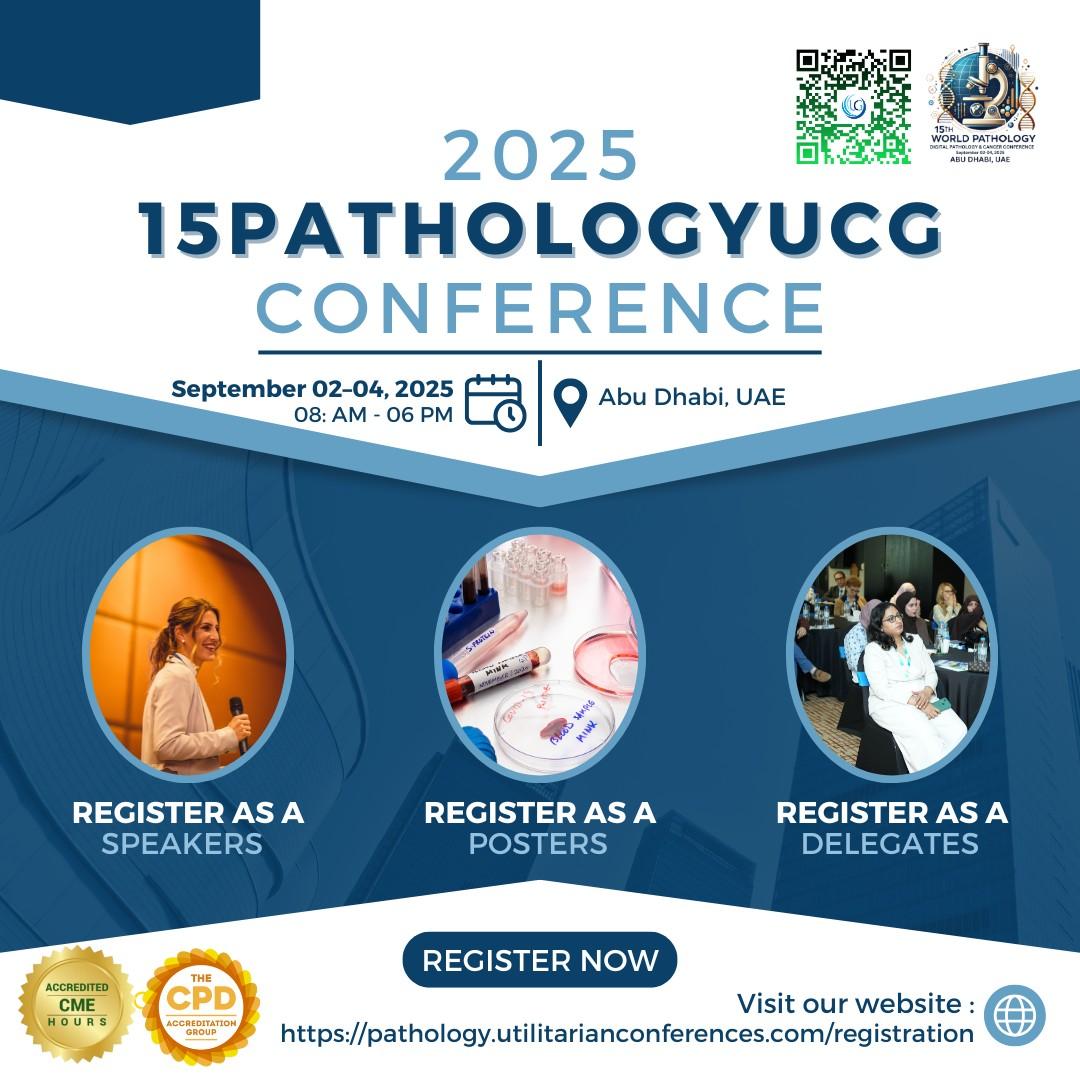
Conference Information:Conference Name: 15th Emirates Pathology, Digital Pathology & Cancer Conference Date: September 02-04, 2025Location: Abu Dhabi, UAE & OnlineWhatsApp No: +971551792927Email: pathology@ucgconferences.comhttps://pathology.utilitarianconferences.com/https://pathology.utilitarianconferences.com/submit-abstracthttps://pathology.utilitarianconferences.com/registrationhttps://pathology.utilitarianconferences.com/virtual-registrationhttps://pathology.utilitarianconferences.com/exhibitor-registrationhttps://pathology.utilitarianconferences.com/sponsor-registration
