2 minute read
Kellogg Invests in Lab for Human Retinal Stem Cells


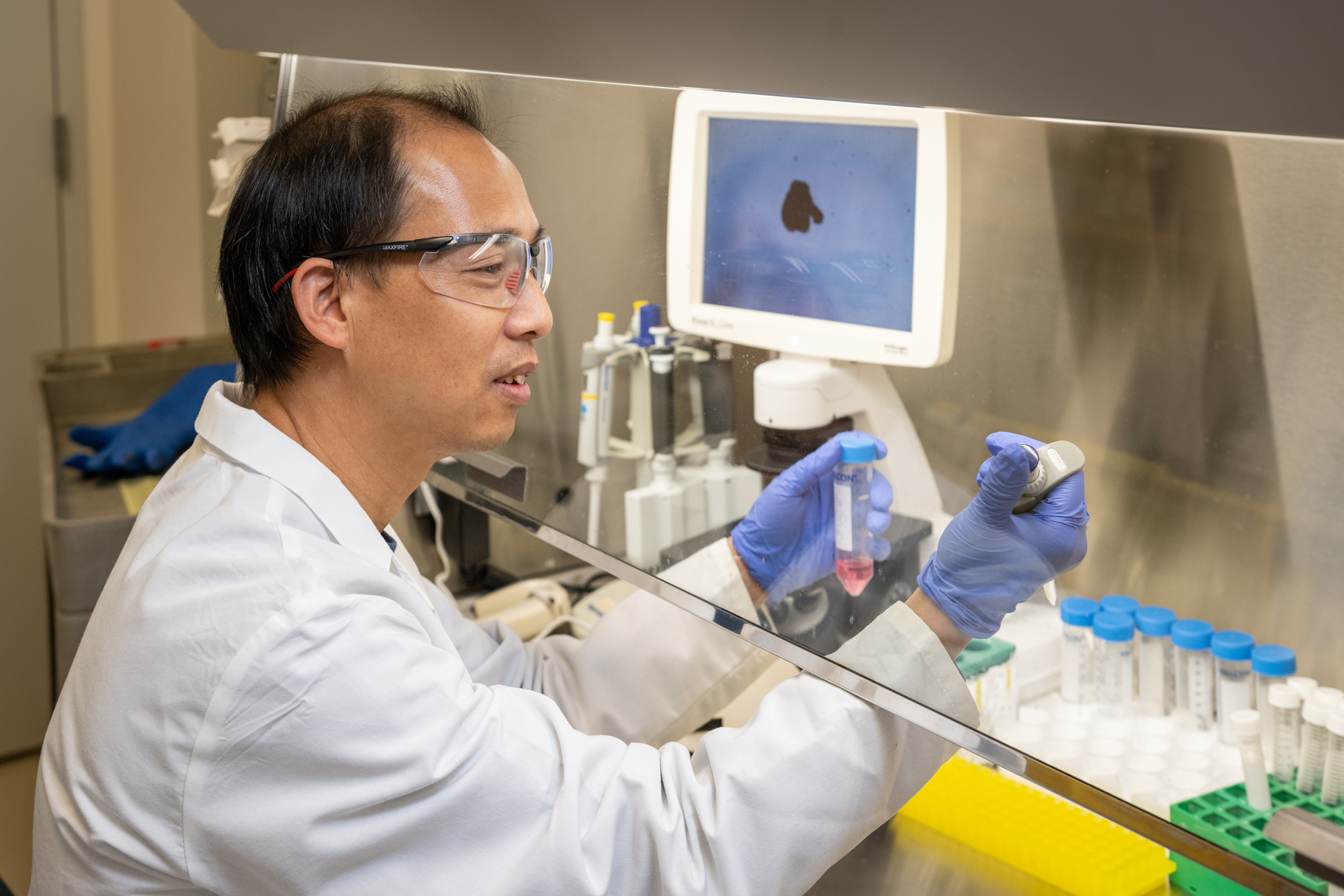
Animal models of disease, while extremely useful in basic science, do not always make the best models for the study of human disease. For example, it is difficult to study macular degeneration using a mouse model because a mouse retina does not have a macula. That’s why Drs. Rajesh Rao and Abby Fahim are spearheading the establishment of a new laboratory dedicated to directing human pluripotent stem cells into various retinal cell types involved in retinal diseases. “This new laboratory will allow us to build and study retinal organoids—three-dimensional ‘mini organs’ that more closely approximate the development, structure and function of the retina,” Dr. Rao explains. Building a human disease model is also far more complicated and time-consuming process, with more potential opportunity for contamination, than developing an animal model. A dedicated space reduces that risk.
Retinal organoid models will also be particularly useful in the study of a range of poorly understood pediatric eye diseases. For example, congenital malformations like microphthalmia, anophthalmia, and coloboma are very well suited to modeling by retinal organoid technology and may share some characteristics with congenital forms of macular degeneration. Dr. Fahim will lead an effort in the new facility to generate retinal pigment epithelial (RPE) cells. RPE cells nourish photoreceptors, the light-sensing cells responsible for one of the first steps of vision. Generating RPE cells may be helpful in modeling genetic forms of retinal dystrophies, and in better understanding RPE cell transplants in clinical trials for patients with dry AMD, such as the current Phase 1/2a trial (page 14) led by Dr. Rao.
Advertisement
“With this new lab, we’ll now have a central point for new collaborations among researchers from across the University. We hope it will be a catalyst for making regenerative medicine and stem cell-based disease modeling central research themes for Kellogg.” says Dr. Rao.