Journal of the Irish Dental Association
Iris Cumainn Déadach na hÉireann

Journal of the Irish Dental Association
Iris Cumainn Déadach na hÉireann
Case report: drug-induced gingival overgrowth associated with the use of a calcium channel blocker (amlodipine)





245 PRACTICE MANAGEMENT: Great expectations
223
224
226 QUIZ
228 DIARY OF EVENTS
229 BUSINESS NEWS: Identex 2015 report
233 INTERVIEW: Dr Frances O'Callaghan
236 CLINICAL FEATURE: Understanding the IOTN
241 New IDA learning management system; calculating annual leave
EDITOR Professor Leo F.A. Stassen
FRCS (I), FDSRCS, MA, FTCD, FFSEM, FFDRCSI, FICD
DEPUTY EDITOR Dr Dermot Canavan BDentSc, MGDS(Edin), MS(UCalif)
EDITORIAL BOARD Dr Iseult Bouarroudj BDS NUI
Dr Michael Crowe BSc BDentSc DPDS (Bristol)
Dr Peter Harrison BDentSc MFD DChDent
Dr Mark Kelly BA BDentSc
Professor Christopher D. Lynch BDS PhD MFDRCSI FDS (RestDent)RCSI FACD FHEA
Ruth Moore RDN, BSc Management & Law, MIA Dip Journalism
Donna Paton RDH
Dr Ioannis Polyzois DMD, PhD, MDentCh, MMedSc
Dr Ciara Scott BDS MFD MDentCh MOrth FFD (RCSI)
Dr Seamus Sharkey BDS NUI FRACDS (Syd) MFDSRCS DChDent (Prosthodontics) FFDRCSI
Dr Simon Wolstencroft BDS FDSRCS MScD MOrth FDSOrth
Irish Dental Association Unit 2 Leopardstown Office Park, Sandyford, Dublin 18.
Tel: +353 1 295 0072 Fax: +353 1 295 0092 www.dentist.ie
Follow us on Facebook (Irish Dental Association) and Twitter (IrishDentists).

248 PEER-REVIEWED
248Case report: drug-induced gingival overgrowth associated with the use of a calcium channel blocker (amlodipine)
O Carty, E Walsh, A Abdelsalem, D MacCarthy
252Fundamentals of occlusion and restorative dentistry. Part II: occlusal contacts, interferences and occlusal considerations in implant patients
A Warreth, K Doody, O Morcos, M Al-Mohsen, N Ibieyou
260 ABSTRACTS
263 CLASSIFIED
266 MY IDA: Dr Andrew Kelly
IDA PRESIDENT Dr Anne Twomey
IDA CHIEF EXECUTIVE Fintan Hourihan
CO-ORDINATOR Fionnuala O’Brien
The Journal of the Irish Dental Association is the official publication of the Irish Dental Association. The opinions expressed in the Journal are, however, those of the authors and cannot be construed as reflecting the Association’s views. The editor reserves the right to edit all copy submitted to the Journal. Publication of an advertisement does not necessarily imply that the IDA agrees with or supports the claims therein.
For advice to authors, please see: www.dentist.ie/resources/jida/authors.jsp
Published on behalf of the IDA by Think Media, 537 NCR, Dublin 1 T: +353 1 856 1166 www.thinkmedia.ie
EDITORIAL Ann-Marie Hardiman, Paul O’Grady
DESIGN/LAYOUT Tony Byrne, Tom Cullen, Ruth O’Sullivan
ADVERTISING Paul O’Grady paul@thinkmedia.ie

and
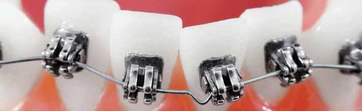
We should listen to what Dr Frances O’Callaghan and other contributors have to say in this edition of the Journal
Dr Frances O’Callaghan is interviewed by Ann-Marie Hardiman in this edition of the Journal and she reveals some of the hard truths about the provision of oral healthcare by the State. Of course, children can be treated at any age, but it’s tough for dentists to be seeing children too late to make a meaningful difference in preventing problems that are easy to prevent if caught in time. It’s taking us out of the positive spiral of prevention that occurs when dental professionals make early interventions. The crucial difference between seeing children for the first time in senior infants class as opposed to second class appears to be lost on our bureaucrats and legislators. Well, let’s work hard at making them aware of the damage the cutbacks continue to cause.
We need to pay attention to Frances and our colleagues who provide the Public Dental Health Service – they know what works and what doesn’t work, and what can be achieved. We trust that when the Minister for Health, Dr Varadkar, attends the Public Dental Surgeons Seminar of the Association this month, he will pay particular attention to sensible, evidence-based arguments that can improve the health of our citizens.
One of the issues that our patients, and especially the parents of our patients, feel very strongly about is the provision of orthodontic treatment. The criteria for qualification for treatment under the public health system in the Republic of Ireland is explained in our clinical feature by Dr Ciara Scott. The article offers a visual guide to simplify the Index of Treatment Need (IOTN) guidelines on eligibility for referral to the HSE. The idea is to help practitioners to offer advice to their patients and refer appropriate cases. This is a practical, helpful guide, which readers may wish to keep for use over the coming months and years. The Journal is grateful to Dr Scott and her colleagues Dr Marielle Blake and Dr Sinéad O’Hanrahan for their work on it.
Drug-induced gingival overgrowth (DIGO) is examined in a case report from Dr Orla Carty and colleagues in Dublin Dental University Hospital and reported in this Journal. In this case, two possible causative factors were identified as amlodipine medication and poor plaque control. The report concludes that it is important that dental professionals are aware that individuals taking calcium channel blockers need to demonstrate excellent plaque control to reduce their risk of developing DIGO and to reduce its severity should it arise.
Dr Abdulhadi Warreth and his colleagues present the second part of their excellent paper on fundamentals of occlusion and restorative dentistry. We know that the outcome of restorative treatment is highly dependent on the occlusion when the treatment is complete.
Three hundred cases of mouth, head and neck cancer are detected in Ireland each year with over 100 deaths. This year, Mouth Cancer Awareness Day had a focus on rural Ireland. Organisers wanted everyone who lives in rural areas to become aware of the disease, to go to their dentist or doctor, and to have a dental check-up on a regular basis. Our collective thanks are due to all the parties involved in raising awareness of mouth cancer.
Congratulations are due to our Editorial Board colleague Dr Chris Lynch on his appointment as Professor of Restorative Dentistry and Dental Education at Cardiff University. We wish him continuing success.
Well done to the Irish Dental Trade Association and the IDA for joining forces in the presentation of Identex and the IDA Autumn Meeting. We read that it proved a fruitful joint venture.
Finally, can I encourage all and any dentists who have been nominated for our Sensodyne Sensitive Dentist of the Year to attend the Awards Ceremony Gala Ball in the RDS in December? It is a fantastic occasion that reminds us of the many good things there are about dentistry.
Dr Anne Twomey IDA President

Identex and MCAD were great, the Minister is coming to the PDS Seminar, a case for a sugar tax and other health matters.
It seems clear that the remainder of the year is going to be very busy for the profession and the Association.
It was a great pleasure to attend the Identex Conference, where huge numbers of dentists attended and I was very struck by the large interest in the innovative workshops we arranged showing dentists how they can comply with the Dental Council’s new guidelines on infection control. We will have similar educational sessions for members in the coming months and they will also be part of next year’s Annual Conference in Galway. What was reassuring is that the profession is taking seriously its continued support for the highest standards of infection control and is seeing how best to ensure that practices meet the standards set out by the Dental Council.
September also saw Mouth Cancer Awareness Day take place on September 16. There were a number of compelling cases highlighted by patients who have successfully come through treatment by medical and dental professionals, and all of whom appreciate better the importance of early detection and early attendance with the dentist. It is so gratifying to see the continued interest in the awareness-raising programme organised by the Association and its partners each September, and to see the huge impact that real-life case studies have in explaining an entirely preventable disease and its treatment. It really was a perfect illustration of the dental family working together to promote the strong connections between oral and general health, and the critical importance of regular attendance with the dentist.
There are further events near on the horizon and these include the HSE Dental Surgeons Seminar, which I am looking forward to. Once again there is an outstanding line-up of speakers, and I am particularly pleased that the Minister for Health, Dr Leo Varadkar TD, has agreed to address the meeting. The Association has engaged extensively with the Minster in the past year and we can only hope that our representations will have elicited a real and meaningful response as regards investment in oral healthcare, treatment and prevention in the 2016 budget, which is due to be announced on the Tuesday prior to the HSE Dental Surgeons Seminar.
Looking further ahead, of course we have already seen huge interest, as illustrated in the number of nominations received, for the Sensitive Dentist Awards, which are organised in conjunction with GSK. Once again, Dr Varadkar has said he is looking forward to attending the Gala Dinner on Saturday, December 5 in the RDS. There is no doubt but that the decision to change the format of the evening last year was a terrific success and we are expecting that there will once again be huge numbers, including many members of the dental practice team, in attendance on the night. It is great to celebrate all the good things we do. It is often when we go that extra mile for someone that we are most satisfied with our career choice as dentists.
Like many of you I saw the recent Jamie Oliver TV programme and recoiled with horror when we saw the distress suffered by small children being treated under general anaesthetic for multiple extractions. Of course, while this seems shocking to the general public, it is something we are aware of as a profession, although it is still shocking when it is presented in such compelling terms. I am very much at a loss to understand why the Department of Health has made no moves to address this issue and the incidence of high sugar intake is directly linked to many of the problems. Equally, more funding is necessary to allow improved access for under fives to dental services if we are to switch off this ticking time bomb. Many people believe that the answer to these difficulties lies with the introduction of a sugar tax. Within the Association we have convened a small group to look at this issue, but also the linked issue of food labelling. I would welcome suggestions and contributions from members of the Association if they wish to share with me their thoughts on the desirability and suitability of sugar tax, and any other comments that you may have in regard to labelling generally.
In the coming days the election for a new Dental Council will take place and it is good to see so many dentists raise their hand and make themselves available for election. I encourage all members to take the time to vote. It is so important that we have a Council which is actively supported by the dental profession and is not left to others to decide and to dictate the future of the profession. I should also like to pay tribute to the outgoing Council members who have undertaken their work with great commitment and diligence and most particularly to the President, Dr Eamonn Croke, who has always been accessible and has always taken a very professional and pragmatic approach to the regulation of dentistry. We can all only hope that the introduction of a new dental bill will give the Dental Council appropriate powers to ensure proper regulation of dentistry and assist all of our members who are committed to practising to the highest standards, mindful of the need to protect the interests of the public at all times.
Finally, in these difficult times I was delighted to see the launch of the Practitioner Health Matters Programme, with which our Chief Executive Fintan Hourihan is associated. This is a really positive development and allows dentists, doctors and pharmacists with mental health or addiction problems access to confidential and expert care and treatment without fear of any form of stigma and with appropriate safeguards agreed with the regulatory authorities. It is important that all members of the profession are aware of the programme and also take time to consider that this is an option for colleagues who may be showing signs of distress in these difficult times.
In a letter published recently in the British Dental Journal, Dr P Neville drew attention to a perceived lack of emphasis on oral health of prisoners in the Irish Prison System. She stated that “no oral epidemiological survey of the Irish prison population has been conducted”, and further stated that “the current prison dental policy … only permits routine dental treatment to those who are sentenced to 16 months or more in prison. There is also no on-site access to dentists; prisoners are referred to a dentist in the nearby community, based on the clinical judgement of a prison medical officer or prison nurse.” The following is a letter from Prof. June Nunn in response to Dr Neville’s letter.
The full text of Dr Neville’s letter is available at http://www.nature.com/bdj/ journal/v219/n4/full/sj.bdj.2015.641.html.
Sir,
I write to respond to the misinformation published in a letter in a recent issue of the BDJ (British Dental Journal 2015; 219: 146-147) by Dr P Neville. In that article, a statement was made, among others, that prisoners had no on-site access to dentists in the Irish prison system.
The Irish Prison Service has a well-developed healthcare service with published standards, including protocols for both emergency and routine dental care. Contrary to what was stated, all the closed prisons in Ireland have at least one dental surgery on site. In the five Dublin prisons, for example, there are 21 dental treatment sessions available to prisoners each week.
No doubt this will be reflected in the review commissioned by the Inspector of Prisons, Judge Michael Reilly, to which Professor Andrew Coyle, Emeritus Professor of Prison Studies, University of London, has contributed, and which is due for publication shortly.
Yours sincerely,
Professor June Nunn
Dental Consultant to the Irish Prison Service
Ballinasloe dentist remembered Dr Pat Costello has let the Journal know of the death of longstanding member Dr Liam Nolan. Dr Nolan practised in Ballinasloe, where he lived with his family (wife Emer and their daughters Geraldine and Hilary). Dr Costello said: "Liam Nolan was honoured by the Association with a specially struck medal for 25 years continuous attendance at the Midland Branch".

What everyone who loses their teeth needs to know
Professor
David Harris
London Books. 2015. ISBN 978-1-907535-10-9

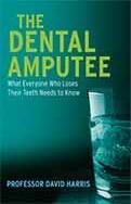
Sometimes when you see a book’s title, it attracts your eye and you wonder what it is about, for example,
The Dental Amputee
It was not until I read the book that I got a real understanding of the need for a book like this for the Irish dental patient. This book of 11 chapters and 167 pages is entertaining, enlightening, stimulating and educating. It is not an easy read, as there is a lot of information. It is a must read for every dental student and should be part of the undergraduate dental curriculum, an important read for those interested in dentistry, especially with an interest in implants, and an essential read for any patient with missing teeth, to help them to decide what they should do.
The chapters all start with an interesting quote – I love that. Each chapter is full of great stories, anecdotes, facts and information presented in a wonderful, articulate and personal manner. David Harris manages to educate without preaching or
undermining the reader.
The history, pioneering work, collaborations and dedicated research of PI Brånemark is beautifully described. He was a true mentor. The explanation on how some of the best research ideas and inventions come from “observation of the unexpected by the visionary” is well illustrated. The failings of a historical, purely mechanical attitude to implants and the harm they did, but the huge advantage the discovery of ‘osseointegration’ of titanium implants gave our patients, makes good of the past and shows where dentistry has come from.
I enjoyed the description of the different techniques available to patients, from the single tooth to ‘teeth in a day’, with all the other developments.
I found the clear, non-biased and honest advice for patients about ‘dental tourism’ an important chapter and non judgemental. This chapter probably deserves publication on its own on the JIDA website.
Overall, this book is an enjoyable and informative read, and I highly commend it to all dentists with an interest in implants to read for themselves, and to give to their patients.
Although understanding David’s reason for the name ‘dental amputee’, I feel it may put the public off picking it up and browsing it. I hope that I am wrong.
We need more books like this, bringing dentistry to the forefront of the public (our patients).
Prof Leo F A Stassen, Professor/Chair of Oral and Maxillofacial Surgery TCD.
Dr Chris Lynch (right) has been appointed Professor of Restorative Dentistry & Dental Education at Cardiff University.
A graduate of University College Cork, Chris holds an InterCollegiate Specialty Fellowship in Restorative Dentistry, a PhD, and ad eundem fellowships from the Faculty of General Dental Practice and the RCSI. Chris has achieved international recognition with Fellowships from the Academy of Dental Materials, the American College of Dentists and the International College of Dentists. He has received the ‘Award for Excellence in Dental Education’ from the Association for Dental Education in Europe.
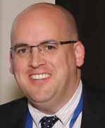
A recognised expert in operative dentistry and dental education, Chris has published 135 papers in peer-reviewed journals, as well as a textbook, Successful Posterior Composites He is Editor-in-Chief of the Journal of Dentistry, the leading international journal in the field of restorative dentistry. Chris is an elected board member of the Faculty of Dentistry at RCSI, Dublin, and is Head of the Learning & Scholarship Theme at Cardiff Dental School.
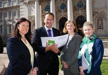
The sixth annual Mouth Cancer Awareness Day was held on September 16 and the focus of this year’s campaign was on highlighting the dangers of the disease to the rural community. Organisers wanted everyone who lives in rural areas to become aware of the disease, to go to their dentist or doctor if they have any concerns and to have a dental check-up on a regular basis. Three hundred cases of mouth, head and neck cancer are detected in Ireland each year with over 100 deaths: that’s almost two people every week. Although not as well known, mouth cancer kills more Irish people than skin melanoma. Farmers are generally viewed as poor attenders at doctors and dentists.
Minister for Health, Dr Leo Varadkar TD, with (from left) Elaine Hughes, Assistant Chief Executive of the Irish Dental Association; Professor Denise MacCarthy of the Dublin Dental University Hospital; and, Etain Kett of the Dental Health Foundation on Mouth Cancer Awareness Day 2015.
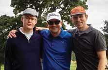
At the Captain’s Prize for members of the IDA Golf Society in Carlow Golf Club in September were (from left): Drs Hugh O’Broin, Emlyn Bratton and Mark Condon.
Sleep apnoea and snoring course: There are still a couple of places left on the full-day course on sleep apnoea and snoring, which takes place on Saturday, November 7. The course will be given by Dr Roy Dookun at the Radisson Hotel, Dublin Airport. Dr Dookun is a GDP in the UK and is President of the British Society of Dental Sleep Medicine. To book, call IDA House.
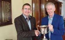
Dr Michael Galvin (winner, right) receiving the Captain’s Prize from the Captain of the IDA Golf Society, Dr Emlyn Bratton.
Submitted by Dr Conor Bowe
Professor Michael Lewis to lecture in Dublin: Professor Michael Lewis, Dean and Professor of Oral Medicine at the University of Cardiff, will give a half day course in Dublin on Friday, January 15. Professor Lewis will present on antimicrobial resistance and the over-prescribing of drugs, and what it means for the dental profession. Further details will be issued shortly.
Dr Cliff Ruddle for Galway: The Annual Conference will return to the City of Tribes in 2016 from April 21-23 at the Radisson Hotel. Worldrenowned endodontist Dr Cliff Ruddle will give two half-day courses on Thursday, April 21. Further details and a booking form will be available later in the year.
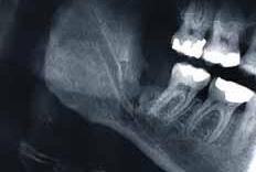
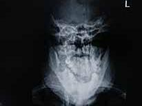
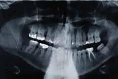
A 49-year-old Caucasian male presents to the emergency department 12 days post removal of his mandibular right third molar. He describes pain, swelling and a distinct cracking noise after eating.
1. What do you see radiographically? 2. What is the initial management? 3. How can this rare complication be avoided?
Answers on page 262
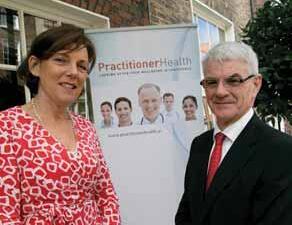
Dr Íde Delargy, Clinical Lead for the new Programme, with Hugh Kane, Chairperson of the PHMP.
A new service, the Practitioner Health Matters Programme (PHMP), which aims to support health professionals, including dentists, who may have a mental health difficulty or an alcohol- or drug-related problem, was launched recently in Dublin. The new Programme, which is open to doctors, dentists and pharmacists, has been designed to reflect international best practice in practitioner health. An independent charitable organisation, the PHMP has the support of the representative and training bodies for the medical, dental and
pharmacy professions, including the Irish Dental Association, as well as the three professional regulatory bodies for those professions.
Dr Íde Delargy, Clinical Lead for the new Programme, said there is clearly a need out there, as health professionals are very slow to come forward to declare that they may have a mental health or alcohol- or drug-related problem. She said health practitioners were often reluctant to come forward because the people they needed to approach were very often their peers and this was a potential barrier to treatment: The key to overcoming this reluctance is to ensure that they will receive a high standard of care in a non-judgmental atmosphere and with complete confidentiality assured. We want people affected by these issues to seek help early and to know they can come forward safely and in confidence to have their health needs met”.
Hugh Kane, Chairperson of the PHMP, said: “This is clearly an important service for practitioners. Often the person experiencing difficulties is the last person to realise they need help. That is why we are raising awareness around these issues and we would encourage colleagues, family and friends to watch out for everything from subtle warning signs to the more obvious ones. Discussing these issues with someone in difficulty can be extremely challenging, but it can also be lifesaving. Ignoring problems, as we often did in the past, benefits no one”. For full details, telephone 01-297 0356, email confidential@practitionerhealth.ie, or log on to www.practitionerhealth.ie.
OCTOBER
22Alexander Hotel, Dublin 2, 7.30pm
Metropolitan Branch IDA – Meeting
Speakers: Drs Naomi Rahman on ‘Bleeding, surgical procedure’, Andrew Norris on ‘Medical emergencies’ and Mary Clarke on ‘Forensic dentistry’.
6-7Gibson Hotel, Dublin City Centre
Irish Dental Hygienists Association
Annual Winter Scientific Conference 2015
Further details on www.idha.ie.
19Alexander Hotel, Dublin 2, 7.30pm
Metropolitan Branch IDA – Meeting
Speakers: Drs Rachel Doody on ‘Dentine hypersensitivity’, Rona Leith on ‘Trauma bites’ and Barry Dace on ‘Diabetes and the dentist’.
JANUARY 2016
28Alexander Hotel, Dublin 2, 7.30pm Metropolitan Branch joint meeting with Irish Endodontic Society Preceded by Metropolitan Branch supper for learning – 6.00pm Email supperforlearning@hotmail.com to book your place.
FEBRUARY
26Alexander Hotel, Dublin 2, 2.00pm
Metropolitan Branch IDA – extended meeting: ‘Wellbeing and Eliminating Stress’
Speakers: Dr Garry Heavey on ‘Consent’, Dr Brid Hendron on ‘Controlling stressful situations’, Eamonn O Muirchearthaigh on ‘Preventing back pain’ and Donal Atkins on ‘Finance’.
10Alexander Hotel, Dublin 2, 7.30pm Metropolitan Branch IDA – meeting and AGM
21-23Radisson Hotel, Galway IDA Annual Scientific Conference





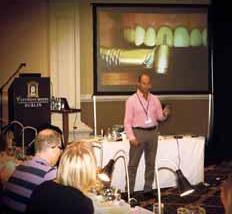




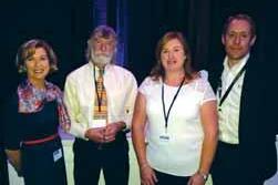
Dr Jane Renehan; Dr Nick Armstrong; HSE Nurse Manager Siobhan Carrigan; and Pete Gibbons of Henry Schein, who presented the demonstration on infection control and decontamination.
This year’s Identex was staged with the IDA Autumn Meeting at Citywest Hotel in September. Already a well-established feature of the Irish dental calendar, the Irish Dental Trade Association (IDTA) has partnered for two years now with the Irish Dental Association in presenting both the trade show and dental education events. The strategy seems to be paying off, as there was a 20% increase in numbers this year – an increase on what was already a strong attendance in 2014. Needless to say, the organisers were pleased with the outcome.
NSK, one of the world’s foremost manufacturers of dental handpieces and small equipment, has appointed Jonathan Singh as its new Product Specialist/Technical Services and Support Engineer for Ireland. Jonathan, who is based in Belfast, has a wealth of experience in the dental and medical industry. He specialises in the maintenance and servicing of autoclaves, and is available to service and validate DX Domina and NSK iClave autoclaves, as well as taking care of handpieces. Jonathan has a comprehensive knowledge of the methods, techniques and processes used in the validation and verification of all decontamination equipment.
Alexander Breitenbach, NSK UK & Ireland Managing Director, commented: “We believe that our many customers in Ireland will benefit from Jonathan’s detailed knowledge of dental handpieces and decontamination equipment”. NSK has recently launched a range of autoclaves, including
To mark 30 years of the IDTA, a Volkswagen UP car was put on offer through a raffle open to anyone who bought ¤250 worth of goods at the show. There was a very strong entry and the draw is due to take place shortly. A photograph of the winner will appear in the next edition of the Journal
In addition to the more than 60 trade stands that exhibited, the IDA put on a mixture of demonstrations (decontamination), lectures (managing patient expectations) and hands-on courses (emergencies and composites), which were booked out in advance.
the NSK iClave, to help meet the needs of busy dental practices and Jonathan comments: “Fast instrument cleaning is vital in a busy practice, so quick cycles, large chamber volume and low power consumption are key. The iClave, previously known as the Domina from Dental X, has sold more than 60,000 units worldwide, demonstrating a proven track record”.
Liz Madden, dental nurse at Abbeyglebe Dental Practice in Donegal, is one happy iClave customer. She said: “Our previous autoclaves kept breaking down and were difficult and expensive to repair. Morris Dental recommended the NSK iClave to us and we now have four in the practice –and they’ve been great. We use the fast cycle a lot, as we are a big practice, and the large chamber allows us to autoclave up to four trays at a time. We would highly recommend the NSK iClave, it’s excellent and very easy to use”.

John O’Connor of Omega Financial Management (left) advises dentists that as your pension contribution is an allowable expense in your tax bill, writing a cheque for it is deemed to be a legitimate business cost. As such, even though you own the pension fund that you are paying into, it is still treated as a business expense for you and reduces your taxable profit in that year.
Does it reduce preliminary tax or just the balance of last year’s tax?
So, in October 2015 you will submit your income tax return for the tax year 2014 and pay the remainder of the tax in excess of the preliminary tax you paid last year. In addition, you will be required to pay your preliminary tax for 2015. This figure is likely to be 90% of your expected 2015 income tax liability or 100% of last year’s figure. Therefore, if you pay ¤20,000 into your pension in 2015 for the tax year 2014, you will, in effect, reduce your preliminary tax obligation for 2015. However, remember that you must make the contribution when the time comes a year later or your balancing payment will go back up again.
Here are the percentages of income you can contribute. Only ¤115,000 of income is allowed, and NRE stands for net relevant earnings.
Cerezen is, according to its makers, a first-of-its-kind medical device that is a safe and effective aid in reducing temporomandibular joint (TMJ) disorder pain and associated symptoms such as grinding of teeth and headaches. It is available in the UK and Ireland, and was on display at Identex. The device consists of two custom-made, hollow ear canal inserts that allow full passage of sound and are practically invisible from the outside. Traditionally, TMJ disorder has been treated with bite splints that are worn at night, and because the Cerezen device is comfortable and discreet, it makes daytime patient compliance easy.
The Cerezen device takes a different approach in treating TMJ disorder. The ear canal is located very close to the temporomandibular joint and the volume of the ear canal increases when the jaw is opened through movements such as chewing, smiling, and speaking. The Cerezen device uses this anatomical change to provide a unique near field treatment.
Patients wearing the device in a three-month clinical study experienced a significant reduction in the pain and dysfunction associated with TMJD. In addition to the pain reduction, 100% of subjects indicated excellent (71%) or good (29%) overall satisfaction with the Cerezen device. During the threemonth clinical study, patients wearing the Cerezen device did not report any unexpected or serious adverse events, and a comparable safety profile to the stabilisation splint was observed. It is not recommended that patients wear the device while bathing, swimming or in a moist environment such as a steam bath and sauna. It is recommended that it be removed when participating in contact sports.
What are my pension entitlements and how do I benefit at age 60?
When you turn 60 you have a variety of options. If you wish to move some or all of your pension funds into ‘post retirement’ funds then you have the option to do so. Most people decide to avail of the cash lump sum, which is there for them if they choose. This means that they can take up to 25% of the fund out in cash, the first ¤200,000 of which is tax free and the remainder, up to ¤500,000 is taxed at 20%, with marginal rate income tax for anything above this level. The maximum fund you can have is ¤2m for those lucky enough to reach that level. It is likely that your remaining 75% will then be invested to your appropriate risk level, to provide you with an ongoing income from retirement funds. You can buy an annuity if you choose but in most cases rates are so low that it doesn’t make financial sense. If you do have a high annuity rate locked into your pension you would probably be wise to avail of it. However, most of us will be investing in what are called ‘approved retirement funds’ (ARFs) and ‘approved minimum retirement funds’ (AMRF). If you do not have a pension of at least ¤12,700 per annum then you must invest your first ¤63,500 in an AMRF. All funds above that are invested in an ARF. The main difference between these two is that you must take an annual income of 4% from an ARF, and with funds in an AMRF you have the option to take up to 4% annually.
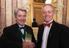
President of the International College of Dentists, Dr Joe Kenneally, with the President of the European Section, Dr Tom Feeney.
As we went to press, the International College of Dentists (ICD) was holding a joint meeting of its International Council and its European Section in Dublin. The European Section is holding its 60th Annual Meeting and its current President is Dr Tom Feeney, a former member of the Editorial Board of the Journal. The world President of the ICD is Dr Joe Kenneally, who was interviewed in our April/May edition.
In addition to the formal meetings, the ICD was also staging a scientific programme and a public forum. The scientific programme under the theme “Learning without limits” was taking place at the Royal College of Surgeons in Ireland on St Stephen’s Green, while the public forum was on the theme of volunteer dental projects and was being held in the Dublin Dental University Hospital. It’s a significant global dental event, which brought 210 dentists to Ireland, with a further 150 accompanying persons, providing a welcome boost to the economy. Sponsors for the event were Henry Schein, Fáilte Ireland and Kildare Village.
As she begins her second term as President of the IDA's HSE Dental Surgeons Group, DR FRANCES O'CALLAGHANdiscusses ongoing industrial relations issues, cutbacks to the Public Dental Service, and the results of a recent survey of HSE members.
On the day of our interview, Frances has just received the results of a survey of HSE dental surgeon members of the Irish Dental Association (IDA). The survey has been a personal project and she is delighted to see it completed: “Last year when I took over as President, I was very conscious that staff morale in the HSE Dental Service was very low, and that many of my colleagues appeared extremely stressed. Work-related stress is hugely significant from a health and safety point of view, and I decided to commission a survey of members to try to quantify it”.
With help first from the IDA's Sarah Gill and then from Roisín Farrelly, the survey was circulated to all HSE members and results have now been collected, with a response rate of over 50%. Levels of stress are indeed high, with 60% of respondents saying they regularly feel stressed at work, and a shocking one in five saying they have missed work due to stress. There is also evidence of unhappiness with management structures, with 70% unhappy with the level of consultation on change, and only 37% feeling they get support from management. What might surprise though is that the highest stressor for respondents was not pay or working conditions: "The lack of access to secondary care services for patients, in particular to general anaesthetic services, was identified as the highest source of stress for respondents”.

within a few weeks. The remaining cases, including young children often requiring four or more extractions, are currently waiting up to a year".
The problem is not unique to Dublin, Wicklow and Kildare. Waiting periods around the country are typically six to nine months, compounded by the fact that dental cases are not included on hospital priority lists, with theatre slots regularly cancelled in favour of other general anaesthetic cases: "If a child is waiting longer than six months to have their tonsils out, the hospital is penalised, so hospitals have cancelled dental theatre slots to facilitate paediatric ENT procedures. We would argue that a child who needs a general anaesthetic in order to have painful, infected teeth extracted is just as entitled to treatment as a child with chronic tonsillitis”. The implications of this are very serious, both for the children and for the dental professionals charged with their treatment: "Anecdotally, we are hearing of children having to be admitted to acute hospitals for IV antibiotics for oral infection. Our concern is that general anaesthetic services for dentistry will not become a priority until a child has a serious outcome from dental infection. The crisis also extends nationally for children and adults with special needs who require general anaesthetic for routine dentistry”.
The HSE Group of the IDU continues to campaign on this issue, and plans to raise it once again with Minister for Health Dr Leo Varadkar when he attends the Group's Annual Seminar in Carlow this month.
With the closure in October 2014 of the dental emergency extraction clinic in St James's Hospital in Dublin, waiting lists for general anaesthetic services in Dublin, Wicklow and Kildare are now up to 12 months for young children with chronic dental infection, many of whom require multiple extractions. With only limited access to some private facilities around the city, dentists are under severe pressure to prioritise the most serious cases, and Frances has strong feelings on this: "All of the children on the waiting list need dental extractions, and all require general anaesthetic to have the extractions completed. Dentists are required to prioritise cases on the basis of urgency. The most acute cases, and very young children suffering trauma, are prioritised as requiring immediate access to general anaesthetic. These are referred to the private sector and will receive treatment
A motion at the Group's AGM will also call for a more radical change to dental healthcare for children: "In recognition of the Minister for Health’s introduction of free GP care for children under six, the HSE Group of the IDU will be calling on the Minister to implement a policy supported by the required funding to put comprehensive oral healthcare programmes in place for young children under six in this country".
Ann-Marie Hardiman Journalist and sub-editor with Think Media, with an interest in further education and CPD.

This comes after years of cutbacks to the Public Dental Service, which have severely curtailed dentists' ability to target children and their families at a young enough age to make a meaningful difference to their oral health: "The earliest any child receives a screening examination now is age seven to eight, either first or second class, which is too late. In some parts of the country children are not examined by a HSE dentist until sixth class. In my area (Frances is a Senior Dental Surgeon in the HSE Dublin South–East Wicklow region) we used to see children in senior infants, but we had to stop three years ago because staffing levels have dropped so much. Our staff were devastated because they had seen significant improvement in the oral health of pupils in second classes when they had seen the same children in senior infants. We were able to highlight concerns at six years of age, before there was any damage to permanent teeth”.
A summary of the HSE guidelines for eligibility for orthodontic treatment so that dentists have a visual guide to which cases should be referred to a specialist.
Throughout our professional lives, we will be asked the question: “will I need braces?” A self-conscious teenager might ask, but what a worried parent may really be wondering is: “will my child be eligible for treatment within the HSE?”
The modified Index of Treatment Need (IOTN) guidelines for eligibility were introduced in the HSE in 2007 as a way to assess orthodontic need objectively rather than subjectively. This article aims to offer a visual guide to simplify the IOTN guidelines on eligibility for referral to the HSE and aid general practitioners to offer advice to their patients and refer appropriately.
A healthy smile starts at home with good habits regarding diet, snacks, drinks and brushing, which are key to a healthy dental future. Only patients with good oral hygiene and dental health are eligible for orthodontic treatment.
Keep the child’s age in mind when assessing dentition or looking at OPGs (see normal eruption dates). Dental age and chronological age are not always the same so be alert to an abnormal sequence or asymmetric eruption patterns. Contralateral teeth should erupt within six months of each other. Refer if interceptive treatment is appropriate.
Check family history for anomalies such as impacted canines. Palpate for canines and monitor dental development. A median diastema can be normal before the canines erupt. Check for severe skeletal problems, an overjet >9mm, impacted teeth, missing teeth and crossbites with displacements, and refer for orthodontic assessment if appropriate.
Check aesthetic component once the canines have erupted. Refer 4d cases with AC 8-10 or other eligible patients.

Dr Ciara Scott
Is a specialist orthodontist with the HSE in Dublin Mid-Leinster.
Ciara wishes to acknowledge the assistance of Dr Marielle Blake of the HSE and Dr Sinéad O’Hanrahan of the OSI in the preparation of this material.


Increased overjet >9mm.Extensive hypodontia with restorative implications (more than one congenitally missing tooth in any quadrant).
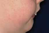
Lip traps and lip incompetence can increase risk of trauma, so encourage gum shield wear.
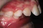
Reinforce need to stop thumb or digit habits and promote oral hygiene and dental health.
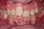
patients
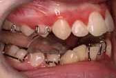
Patients may benefit from functional appliance when 11-14 years old.
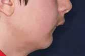
Ideally refer by 12 to allow treatment with functional appliances if appropriate. Later referrals and patients with a marked skeletal discrepancy and poor chin throat angle may need orthognathic surgery.
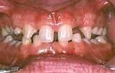
Check family history; missing, impacted, supernumerary and infraoccluded teeth are linked genetically.
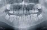
Promote oral hygiene and prevention to preserve primary molars.
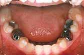
Restore carious primary molars to preserve longevity when permanent successors are missing.
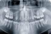
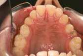
Caries in primary molars may cause tipping or space loss. Some 5h cases may have low orthodontic need.
Active treatment may be best in mid teens leading into restorative care. Joint treatment planning will achieve the best occlusal and aesthetic outcome.

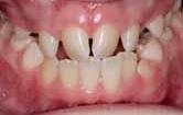
Impeded eruption of teeth (except for third molars) due to crowding, displacement, the presence of supernumerary teeth, retained deciduous teeth and any pathological cause.
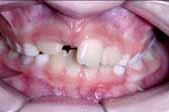
UR1 has failed to erupt and the sequence is disrupted as UR2 has erupted.
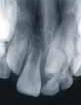
A conical supernumerary may displace or delay eruption of the permanent tooth; tuberculates usually obstruct eruption.
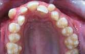
Canines should be palpable buccally. Peg laterals can increase risk of impaction.
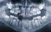
Canines can be palatal or high and buccally impacted. Interceptive treatment can help, so refer.
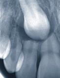

Canines can cause resorption of permanent teeth, so radiographs can help to locate and determine urgency.
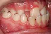
Reverse overjet >3.5mm with reported masticatory and speech difficulties.
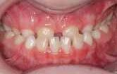
A marked reverse overjet. Always check for a displacement.
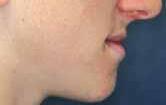
The profile and underlying skeletal pattern can determine if interception is indicated.
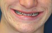
Maxillary deficiency may cause poor upper tooth show and poor upper lip support.
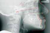
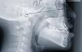
Cephalometrics and clinical examination will determine the treatment plan and timing.
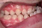
Severe Class III malocclusions with marked Skeletal III base will usually benefit from joint planning and orthognathic surgery as a young adult.
5i5m5p5s
Defects of cleft lip and palate (CLP), and other craniofacial anomalies.
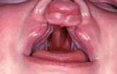
CLP patient’s treatment should be co-ordinated by the national cleft unit.
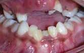
Caries and early tooth loss can further complicate difficult malocclusions, so diet and oral hygiene are key.
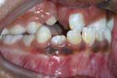
expansion and bone grafting in the mixed dentition help dental development.
anomalies can include enamel hypoplasia, hypodontia and impacted canines.
Patients will have complex dental needs and benefit from joint orthodontic, orthognathic and restorative planning.

Severely submerged deciduous teeth (only two cusps showing above gingiva).
Infraocclusion, such as this LLE, can be severe in young children.
The permanent successor can be displaced. An appliance may be required to provide surgical access to remove the LLE.
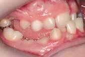
molars
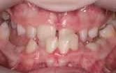
Infraoccluded primary molars can cause increased overbites and lateral open bites.
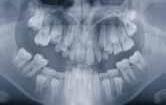
Infraoccluded molars can delay or prevent eruption of permanent teeth and may need to be removed.
Reverse overjet >3.5mm with no masticatory or speech difficulties. See 5m
References 1. Scott, J.H., Symons, N.B.B. Introduction to Dental Anatomy (3rd ed.). Churchill Livingstone, 1990.
Anterior or posterior crossbites with >2mm discrepancy between retruded contact position and intercuspal position.
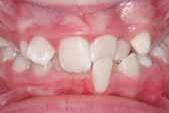
Anterior crossbites can cause damage to the hard and soft tissues such as tooth wear and dehiscence.
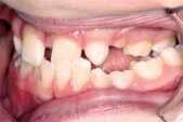
Interception with a URA/RME can relieve the displacement and expand the upper arch.
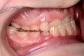
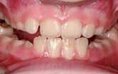
The patient can achieve edge to edge and there is a forward displacement.
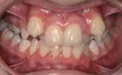
Cases with anterior and posterior crossbites can also have a high aesthetic component.
Severe contact point displacements >4mm (in HSE modification, eligible only if AC is 8, 9 or 10).
Eligibility of 4d cases is based on the appearance of the upper anterior segment only.
The aesthetic component is assessed in the early permanent dentition, AFTER the upper canines have erupted.
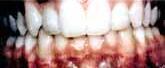
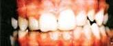
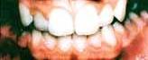
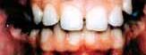
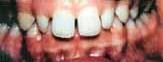
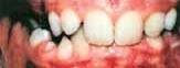
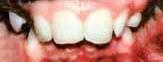
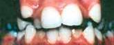
Cases do not need to match the 8-10 pictures, just look as severe.
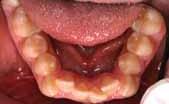
Poor alignment of lower incisors does not meet the eligibility.
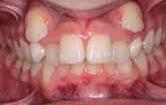
Extracting over-retained primary teeth can improve the position of displaced teeth.
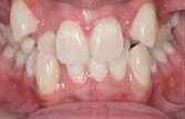
Severe crowding with high aesthetic component is eligible.
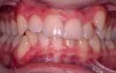
This patient has moderate crowding and IOTN 4d, but is not eligible as the AC <8.
The AC is a series of photographs rating dental attractiveness on a scale of 1-10. It was developed at the University of Manchester using laypersons to grade the photographs. In the HSE, the AC is used for 4d cases. Only cases with an AC of 8-10 are eligible for treatment.
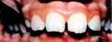
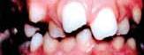
4e4f4m
Extreme lateral or anterior open bites >4mm.
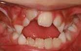
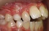
Digit
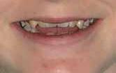
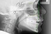
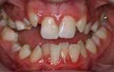
Increased and complete overbite with gingival or palatal trauma.
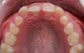
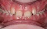
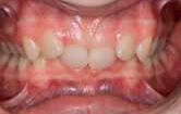
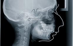
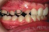
Reverse overjet >1mm but <3.5mm with recorded masticatory and speech difficulties.
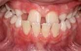
will show the
for
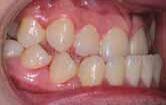
Definitive treatment planning best after puberty to determine scope for camouflage or need for orthognathic surgery.
With dentists commenting that patient expectations appear to be higher than ever before, what are the key issues to consider in record-keeping when providing orthodontic treatment?
Like many elective treatments, one of the keys to successful orthodontic treatment is ascertaining and managing the patient’s expectations from the outset. Some patients aim for a simple cosmetic improvement, whereas others have specifications for key areas, which would be a ‘deal breaker’ if the treatment outcome does not deliver. Some advertising material may lead a patient to believe that orthodontic treatment is much more straightforward than it actually is and patients can misunderstand the significance of their own compliance in particular treatment modes. In reality, a practitioner is only in a position to discuss orthodontic treatment once an appropriate orthodontic assessment has been carried out. Making the appropriate record of this is crucial, as a patient complaint or claim often flows from the initial diagnosis and treatment-planning phase.

Increasingly, we are seeing patients dispute the consent process. In essence, the patient says that they were not presented with all of the appropriate information as part of the consent process, and if they had been aware, they would not have chosen the treatment that was subsequently provided.
A typical example is the patient who says: “If I had known that fixed appliances would have provided a better outcome I would never have had clear aligners”.
It is therefore essential that the
Dr Martin Foster BDS MPH DipHSM
Martin is a Dento-legal Adviser at Dental Protection and spoke recently at the IDA Autumn Meeting staged with Identex.

consent process is appropriately documented in the records, with evidence of the relevant information provided to the patient, including treatment options, information regarding the risks and benefits, and any limitations associated with the treatment options. The record should also detail the discussions regarding the patient’s expectations, and whether or not these are likely to be met by the agreed treatment plan, a copy of the treatment plan, and compliance advice provided to the patient, including appliance wear and attendance at appropriate appointments.
In addition to this, some disputes flow from treatment fees, and it is important to ensure that the patient is aware of both the fees and any agreed payment intervals. A recent dispute arose from a patient misunderstanding the treatment fees, which he was paying at agreed intervals. The treatment was successfully completed ahead of the predicted timescale and two further agreed payments were outstanding. The patient took the view that as the treatment had concluded, any further payments should be written off. The practitioner was able to demonstrate by way of his treatment plan letter that the fees related to the treatment itself, not the duration of the treatment. In any event, in accordance with best practice, the practitioner had estimated the treatment duration but advised the patient that this was only an estimate and that treatment durations vary.
Another issue that is becoming more common in patient complaints and claims is that of retention, with patients attempting to argue that they were either unaware of the need for retention, or unaware of the significance of retention. Issues often arise if a patient is unclear as to the cost of a retainer and whether or not it is included in the initial treatment fee.
We commonly see situations where a patient has gained the impression that the initial cost of orthodontic treatment includes a lifetime of replacement retainers, when this is often not the case. It is helpful to ensure that the patient is aware of who has responsibility for checking retention, and who will provide replacement retainers when they are required.
Good records are the best defence
Many of these elements of orthodontic care can be quite obvious to practitioners; the challenge lies in ensuring that the patient is fully informed from the outset. The overall theme, therefore, is good communication between the dentist and the patient, and evidence of this in the records, so that if the practitioner is challenged at a later date the evidence is there for all to see.
The vast majority of treatments are carried out uneventfully, with the patient being satisfied with the outcome; however ,it pays to be prepared. We encourage orthodontic practitioners to consider audits of their records to see if they would provide robust evidence to any such challenge that could be made in future.
Introduction
Many factors can contribute to the development of gingival overgrowth (GO), including: plaque control; periodontal variables; medications and their relative dose; age; sex; and, genetic factors.3 Medications associated with drug-induced gingival overgrowth (DIGO) are broadly categorised according to their therapeutic actions, namely anticonvulsants, immunosuppressants and calcium channel blockers.4
In 1994, Bokenkamp et al. graded gingival overgrowth:5
0: No sign of enlargement
Grade 1: Enlargement confined to dental papilla
Grade 2: Enlargement involving dental papilla and marginal gingiva
Grade 3: Enlargement covering three-quarters of crown of tooth or more
Calcium channel blockers are regularly prescribed in the treatment of conditions such as hypertension and angina. They may be classified chemically as dihydropyridines (nifedipine, isradipine and amlodipine), phenylalkylamine derivatives (verapamil) and benzothiazepine derivatives (diltiazem).1 Nifedipine is the calcium channel blocker most commonly cited to induce GO,6,7 with approximately 10% of medicated patients presenting with the condition.8
Other risk factors for calcium channel blocker DIGO include: gender; duration of therapy; concomitant medications; plaque; and, oral hygiene.3 Males appear to be at greater risk and tend to present with more severe gingival hyperplasia.9 Combination therapies, in particular in relation to nifedipine and ciclosporin, can produce more GO than if either drug was used independently.10,11,12
There is a lack of data reporting amlodipine specifically as a causative agent.13 The mechanism by which amlodipine may induce these changes remains poorly understood and the literature is sparse, with few case reports outlining its association.9,10,12 Regarding the clinical presentation of DIGO, the mandibular anterior gingiva is the most commonly affected site, but in severe cases the condition may affect multiple areas of the mouth. If oral hygiene is good, the swelling tends to be less apparent, with a reduction in inflammation seen. However, in the presence of plaque the inflammatory changes appear to enhance the activity of the fibroblasts, thus increasing their number and also increasing the production of collagen fibres and proteoglycans, worsening the severity of the hyperplasia. The hyperplasia tends to mainly be constructed of highly vascular fibrous tissue with acanthosis of the epithelial lining in a dense collagenous matrix.8
The care pathway for these patients generally involves an initial non-surgical approach alongside surgical management as required.
Non-surgical management
Selection of a treatment modality depends on the severity of the DIGO. Elimination of local factors, plaque control and regular periodontal maintenance therapy may ameliorate but not prevent DIGO in a susceptible patient.9 Plaque control should always be a first-line measure in an attempt to control the inflammatory oedematous gingivitis.14,15 There is evidence that good oral hygiene and plaque removal decreases the degree of GO and improves periodontal health.16,17 The use of chlorhexidine digluconate mouthwash (0.1% w/v) may reduce the incidence of DIGO recurrence following gingival surgery.18
Surgical management
Surgical treatment is only advocated where GO is severe and should be combined with cause removal where possible and non-surgical management.16 GO may be assessed using the method described by Seymour et al. (1985).19 The
index measures the degree of GO in a labio-lingual and apico-coronal direction. Surgical interventions have been suggested with GO index scores in excess of 30%.20 If drug therapy is likely to be continued for life, psychosocial considerations must be taken into account in an effort to reduce the frequency and extent of surgical intervention.20 Factors to be considered when deciding on appropriate treatment include the extent of the surgical area, the presence of periodontitis, the presence of osseous defects combined with gingival enlargement, and the position of the base of the periodontal pocket in relation to the existing mucogingival junction.15 If the mucogingival junction is too close to the base of the periodontal pocket, a surgical approach such as a gingivectomy would be inappropriate. This is because there would not be enough attached gingiva remaining after the procedure and rapid recession could occur as a result.
The conventional external bevel gingivectomy is a viable treatment option in small areas (up to six teeth), with no evidence of attachment loss.15 The internal bevel periodontal flap is indicated in situations with larger areas of GO, or areas where attachment loss is combined with an osseous defect.15 It was found that no difference exists between flap surgery and conventional gingivectomy with respect to recurrence of GO.21 Provided clinical guidelines based on research are adhered to, evidence supports the biological compatibility of electrosurgery to excise papillary enlargement.22 The carbon dioxide (CO2) laser has been advocated in surgical management of DIGO due to decreased surgical time and rapid postoperative haemostasis.20 A comparison split-mouth crossover study conducted by Mavrogiannis et al. (2006) revealed less recurrence of DIGO within a six-month period with laser excision than with conventional gingivectomy.21 The use of CO2 lasers in combination with conventional gingivectomy has been advocated for dual DIGO.23
Meticulous oral hygiene is required in order to maintain a healthy periodontal condition. Poor plaque control is likely to result in recurrence of the GO and thus these patients should be provided with regular hygiene visits. With adequate plaque control, recurrence is less likely, although the patient remains at risk.24 In a study of induced severe DIGO (nifedipine and ciclosporin), a recurrence rate of 34% was reported over an 18-month review period following periodontal surgery.25 Using multiple regression analysis, it was also reported that age, gingival inflammation and attendance at recall appointments were significant factors in determining the likelihood of recurrence. For this reason, regular follow-up appointments are required to recognise any signs of deterioration early.25
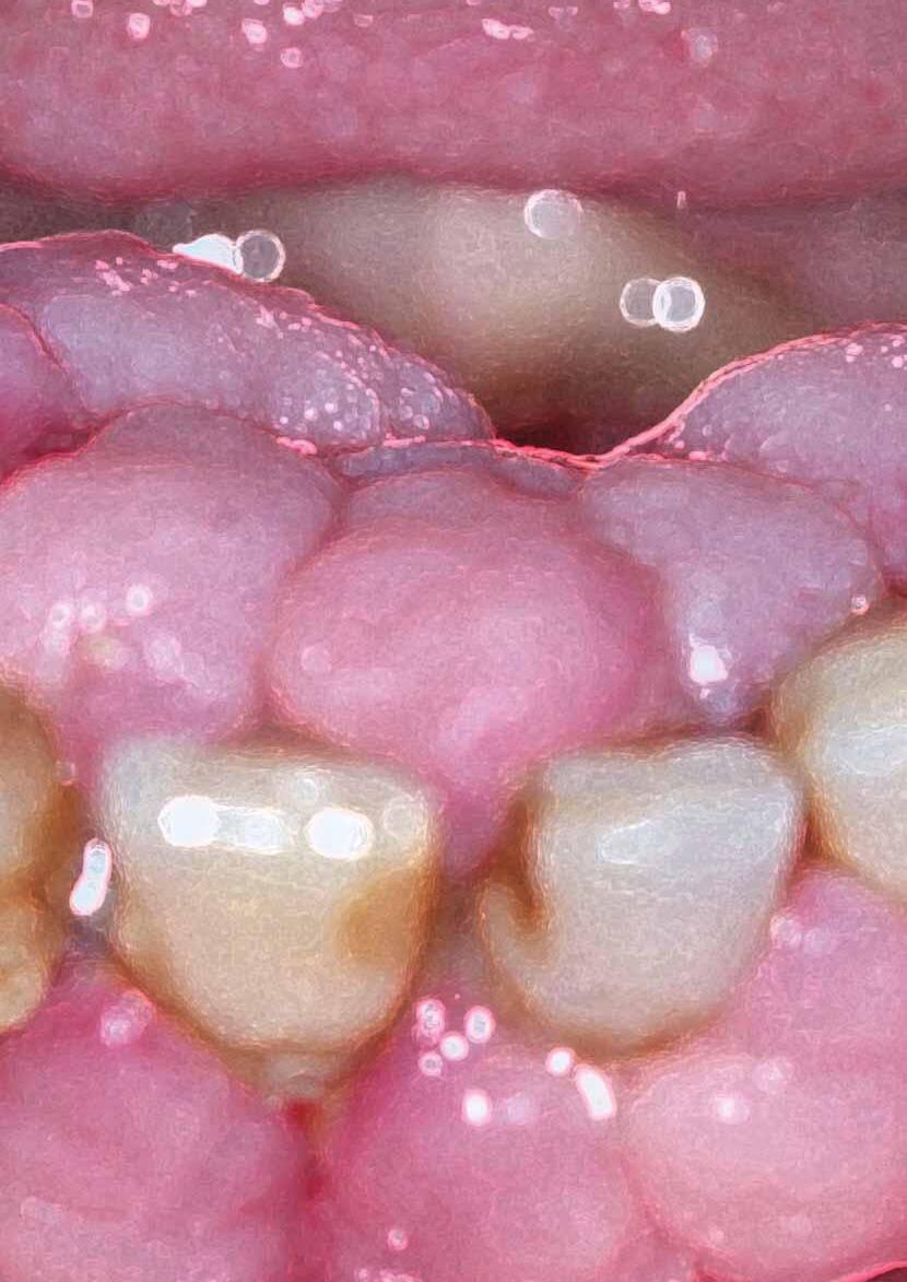
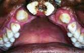
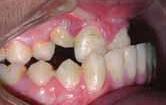
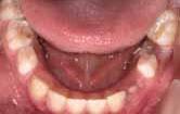
A 63-year-old male was referred to the accident and emergency department at the Dublin Dental University Hospital (DDUH) complaining of swollen gums. The patient reported that they had started to enlarge two years previously, but that he was now concerned as they were increasing in size and had become more firm. Medically, the patient had hypertension, hyperlipidaemia and was taking amlodipine 10mg OD (calcium channel blocker) and aspirin 75mg daily. He was a former smoker, having quit 12 years previously (Figures 1-5). Extra-oral examination revealed no cervical lymphadenopathy, no swelling and cranial nerve responses were normal; however, there was marked halitosis. Intraorally there was moderate gingival hyperplasia affecting the mandible extending from tooth 3-3 to 4-3, affecting buccal and lingual gingiva, and also hyperplasia
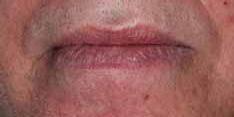
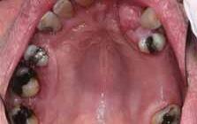
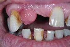
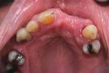
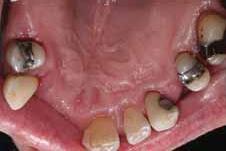
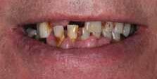
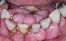
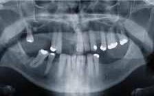
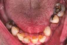
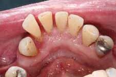
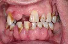
FIGURES 1-5: A 63-year-old male was referred to the accident and emergency department at the Dublin Dental University Hospital (DDUH) complaining of swollen gums. The patient reported that they had started to enlarge two years previously, but that he was now concerned as they were increasing in size and had become more firm.
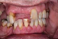
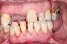
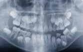
affecting the maxillary gingiva palatal to teeth 2-3 and 2-4. There was a pedunculated lump mesial to tooth 1-3 on the attached gingiva measuring 7x7mm in size. The mucosa overlying the area was intact, mobile and firm to touch (Figure 6). Special investigations included an OPG (Figure 7) and blood tests (full blood count and white cell differential). Diagnosis was grade 2 gingival hyperplasia due to a combination of poor oral hygiene and the use of calcium channel blocking medication.
Initial management was provided in the form of intensive oral hygiene instruction and gross supra-gingival calculus removal. Figure 9 was taken following the initial root surface debridement.
The polyp-like growth mesial to 1-3 was removed with an external bevel gingivectomy. This was completed under local anaesthetic only with buccal and
palatal infiltrations using 2.2ml of Lignospan (2% lidocaine) 1:80,000 epinephrine. The biopsy site was sutured with black silk and a sample was sent for histopathological analysis. The diagnosis was “marked fibroepithelial overgrowth associated with patchy chronic inflammation”.
Following review, oral hygiene was greatly improved, plaque score reduced and gingival inflammation lessened. The hyperplasia remained prominent in areas buccal and lingual to 3-3 to 4-3. The decision was made to complete surgical periodontal treatment with a gingivectomy of the hyperplastic tissue buccal and lingual to the mandibular anterior gingiva. This was also completed under local anaesthetic using infiltrations (4.4ml of Lignospan (2% lidocaine) 1:80,000 epinephrine). A Coe-pac dressing was placed to protect the surgical site, postoperative instructions were explained and the patient was booked for review in one week. Figures 8-10 demonstrate the appearance post biopsy and gingivectomy. Tissue removed was sent for histopathological analysis. The diagnosis was ‘gingival overgrowth’, which was further described as ‘fibroepithelial overgrowth showing moderate chronic inflammation’.
Currently, the gentleman is undergoing regular reviews within the periodontal department and feels a great improvement in his confidence since the gum condition was addressed. The clinical team and the patient remain happy with the results (Figures 11-13). If his plaque scores and bleeding scores remain stable and there is no recurrence noted, he should be able to be discharged back to his GDP for maintenance in the future.
Discussion and conclusions
Two possible causative agents were identified as: (i) poor plaque control; and, (ii) amlodipine medication. Initial treatment commenced with identification of causative factors and conservative management, highlighting the essential role of excellent oral hygiene and providing professional cleaning and support for the patient. The removal of the excess hyperplastic gingival tissues anteriorly in both the maxilla and mandible allowed for definite histopathological analysis, improved access for professional and at home cleaning/maintenance, and facilitated the good aesthetic outcome. If the condition recurs we may contact the patient’s general medical practitioner to discuss the possibility of an alternative antihypertensive to help to reduce the likelihood of such severe GO occurring. However, changing a patient’s medication should only be considered as a last resort, when local measures have had limited success. The dihydropyridine group of calcium channel blockers (amlodipine, nifedipine, felodipine) are more commonly involved with GO.26 Non-dihydropyridine (phenylakylamine) calcium channel blockers such as verapamil have a reduced rate of gingival overgrowth, but do not have the same systemic vasodilating effects.27 Therefore, consultation with the patient’s GP is appropriate, and medication would only be changed once risks and benefits were assessed appropriately. The most important factor is of course recognising those who are at risk of developing the condition and advising the maintenance of a low plaque score, and thus a low bacterial load.
References
1. McCullough, K. (ed.). Dorland’s Pocket Medical Dictionary. W.B. Saunders Company; Sydney, 1982.
2. Marshall, R.I., Bartold, P.M. Medication induced gingival overgrowth. Oral Diseases 1998; 4: 130-151.
3. Seymour, R.A., Ellis, J.S., Thomason, J.M. Risk factors for drug-induced gingival overgrowth. Journal of Clinical Periodontology 2000; 27: 217-223.
4. Marshall, R.I., Bartold, P.M. A clinical review of drug-induced gingival overgrowths. Australian Dental Journal 1999; 44 (4): 219-232.
5. Bokenkamp, A., Bahuharst, B., Beier, C., Albers, N., Offner, G., Brondehl, J. Nifedipine aggravates cyclosporine A-induced gingival hyperplasia. Pediatr Nephrol 1994; 8: 181-185.
6. Lederman, D., Lumerman, H., Reuben, S., Freedman, P. Gingival hyperplasia associated with nifedipine therapy. Oral Surg Oral Med Oral Pathol 1984; 57: 620-622.
7. Barclay, S., Thomason, J.M., Idle, J.R., Seymour, R.A. The incidence and severity of nifedipine-induced gingival overgrowth. J Clin Periodontol 1992; 19: 440-441.
8. Lafzi, A., Farahani, R.M., Shoja, M.A. Amlodipine induced gingival hyperplasia. Med Oral Patol Oral Cir Bucal 2006; 11: E480-482.
9. Ellis, J.S., Seymour, R.A., Steele, J.G., Robertson, P., Butler, T.J., Thomason, J.M. Prevalence of gingival overgrowth induced by calcium channel blockers: a community based study. J Periodontol 1999; 70: 63-67.
10. Thomason, J.M., Seymour, R.A., Ellis, J.S., Kelly, P.J., Parry, G., Dark, J., et al Determinants of gingival overgrowth severity in organ transplant patients. An examination of the role of HLA phenotype. J Clin Periodontol 1996; 23: 628-634.
11. Thomason, J.M., Seymour, R.A., Rice, N. The prevalence and severity of cyclosporine and nifedipine-induced gingival overgrowth. J Clin Periodontol 1993; 20: 37-40.
12. Thomason, J.M., Seymour, R.A., Ellis, J.S., Kelly, P.J., Parry, G., Dark, J., et al Iatrogenic gingival overgrowth in cardiac transplantation. J Periodontol 1995; 66: 742-746.
13. Jorgensen, M.G. Prevalence of amlodipine-related gingival hyperplasia. J Periodontol 1997; 68: 676-678.
14. MacCarthy, D., Claffey, N. Fibrous hyperplasia of the gingivae in organ transplant patients. J Ir Dent Assoc 1991; 37 (1): 3-5.
15. Mavrogiannis, M., Ellis, J.S., Thomason, J.M., Seymour, R.A. The management of druginduced gingival overgrowth. J Clin Periodontol 2006; 33: 434-439.
16. Seymour, R.A., Jacobs, D.J. Cyclosporine and the gingival tissues. J Clin Periodontol 1992; 19: 1-11.
17. Dongari, A., McDonnell, H.T., Langlais, R.P. Drug-induced gingival overgrowth. Oral Surg Oral Med Oral Pathol Oral Radiol Endod 1993; 76: 543-548.
18. O’Neill, T.C.A., Figures, K.H. The effects of chlorhexidine and mechanical methods of plaque control on the recurrence of gingival hyperplasia in young patients taking phenytoin. Br Dent J 1982; 152: 130-133.
19. Seymour, R.A., Smith, D.G., Turnbull, D.N. The effects of phenytoin and sodium valproate on the periodontal health of adult epileptic patients. J Clin Periodontol 1985; 12: 413-419.
20. Thomason, J.M., Seymour, R.A. Phenytoin-induced gingival overgrowth in general medical practice. Journal of Dental Research 1990; 69: 969.
21. Mavrogiannis, M., Ellis, J.S., Seymour, R.A., Thomason, J.M. The efficacy of three different surgical techniques in the management of drug-induced gingival overgrowth. J Clin Periodontol 2006; 33: 677-682.
22. Krejci, R.F., Kalkwarf, K.L., Krause-Hohenstein, U. Electrosurgery – a biological approach. J Clin Periodontol 1987; 14: 557-563.
23. Darbar, U., Hopper, C., Speight, P. Combined treatment approach to gingival overgrowth due to drug therapy. J Clin Periodontol 1997; 23: 941-944.
24. Hallmon, W.W., Rossman, J.A. The role of drugs in the pathogenesis of gingival overgrowth. Periodontology 2000; 21: 176-196.
25. Ilgenli, T., Atilla, G., Baylas, H. Effectiveness of periodontal therapy in patients with druginduced gingival overgrowth. Long-term results. J Periodontol 1999; 70 (9): 967-972.
26. Taylor, BA. Management of drug-induced gingival enlargement. Australian Prescriber 2003; 26 (1): 11-13.
27. Cardiovascular pharmacology concepts. Calcium channel blockers (CCBs). http://cvpharmacology.com/vasodilator/CCB.htm (last accessed 18/02/15).
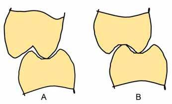
1: A single cusp tip in contact with a flat surface (A); a twin cusp contact when both maxillary and mandibular supporting cusps make contact with opposing fossae (B).
The contact between upper and lower teeth
The area of occlusal contact between unworn opposing teeth is small. The level of friction is low as a consequence, and the space between the occlusal surfaces provides an escape way for the sheared bolus. This enables full use of the overlaps and efficient mastication.7 Increasing wear causes the contacting surfaces to become larger and flattened, and the space for the escape way to be reduced. The whole outer aspect of the supporting cusp may eventually occlude with the inner aspect of the non-supporting cusp.7,8 The occlusal contacts in the MIP and the position of the mandible in the MIP should be stable so that occlusal force does not cause adverse effects. However, the number of occlusal contacts per tooth is low. A lower number of occlusal contacts was found when light occlusal pressure was applied than when a heavier pressure was applied.9 This occlusal area of contact was found to increase with increasing of the occlusal pressure.10
In the MIP, the mandibular teeth make contact with their opposing maxillary teeth and a maximum number of occlusal contacts was found in this position.11
Preferably, the occlusal contacts should lead to stability of the involved teeth and direct the occlusal forces along the long axes of these teeth. The types of occlusal contacts that are found in normal occlusion, or applied in occlusal reconstruction treatments, may be classified as follows:11
A. Supporting cusp against a flat surface
The supporting cusp tip contacts a flat surface of the opposing occlusal table such as the base of the fossa or the occlusal embrasure. This is the ideal relationship as the occlusal forces are directed perpendicular to these surfaces.11 This type of contact is a therapeutic objective recommended for restorative treatment. With this type of contact, there is little potential to develop a horizontal component of force because of the absence of inclined plane effects. Also, the occlusal force is usually directed along and not outside the long axis of the involved teeth (see below). However, this type of occlusal contact may suffer mechanical disadvantages, as will be seen later. The supporting cusp tip occlusal contacts may be further subdivided into a single cusp tip to fossa or a twin cusp contact (Figure 1).
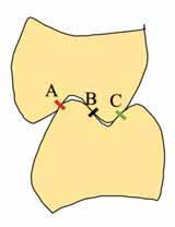
FIGURE 2: Occlusal contacts on an inclined plane surface: A, B and C occlusal contacts.
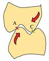
FIGURE 3: Teeth are susceptible to tipping when A and C contacts are present without B contact.
1. Single cusp tip to fossa or marginal ridge contact
This is found when a cusp tip of a supporting cusp of a mandibular or maxillary tooth occludes with a fossa or a marginal ridge of an opposing tooth. It is important to mention that this arrangement, when analysed mechanically, may favour tilting the cusp-contact tooth facially or lingually because the relevant tooth is loaded outside its long axis (Figure 1A).
2. Twin cusp stability
This exists when cusp tips of both mandibular and maxillary supporting cusps occlude with the fossa or the marginal ridge of opposing teeth (Figure 1B). Mechanically, this may be a more favourable type than the single cusp tip contact.
B. Occlusal contacts on inclined planes
Occlusal contacts on inclined planes are found when the supporting cusp makes contact with an opposing inclined surface. In unworn dentitions, a small area of the outer aspect of the supporting cusp makes contact with a small area of the inner aspects of the opposing non-supporting cusp. Hence, the supporting cusp tip will not make contact with the bottom of the fossa or marginal ridge of the opposing tooth. The posterior tooth contacts on the incline plane may be type A, B or C, or a combination of these types of contacts (Figure 2).
A-type occlusal contact indicates the contact between the outer incline of the lower supporting cusp and the inner incline of the upper guiding cusp (Figure 2).
B-type occlusal contact is when the occlusal contact exists between the inner incline of the mandibular and maxillary supporting cusps (Figure 2).
C-type occlusal contact is the contact between the inner incline of the mandibular guiding cusp and the outer incline of the maxillary supporting cusp (Figure 2).
In the theoretical mechanical sense, the most favourable contacts on an inclined plane are either: two contacts on opposing planes such as A-B and BC; or, three-point contact such as A-B-C. For instance, A-B and B-C are more stable occlusal contacts than if only A and C are in contact without B (Figure
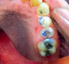
FIGURE 4: A clinical photo of A-B occlusal contacts on the right upper second premolar. This occlusal contact generated a wedging stress, which led to a mesio-distal crack formation. A crack can be seen running mesio-distally along the occlusal table of the right upper second premolar, which was diagnosed as cracked tooth syndrome.

3). In this case, the lower tooth will tip in the lingual direction and the upper tooth will tip in the buccal direction (Figures 3 and 4). This will result in working and non-working interferences (see below).
Occlusal contacts are also described as a tripodal contact, when three contacts (A, B and C) present together on the same pair of teeth, or when the supporting cusp has three points of contact around its tip. This results in a stable occlusion.
Detecting and marking of occlusal contacts
The occlusal contacts can be clinically marked basically by two methods: use of articulating papers; or, waxes and impression materials. Feeler gauges such as shim stock (8-12μm) are used to verify the presence of the occlusal contact when they are detected with the articulating papers, waxes or impression materials. T-scans are also used for recording the time and duration of contacts, but mainly in the field of research.
Articulating papers
The articulating paper does not allow contact between the occlusal surfaces of the opposing teeth as a result of the presence of the strip, such as paper, which is painted with the colouring material. Occlusal pressure results in the coloured material being deposited on the occlusal surface near or at the actual place where tooth surface to tooth surface contact would otherwise occur. The reliability of the marking is related to the thickness and properties of the articulating paper.
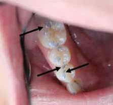
When an articulating paper of about 40μm is used to mark occlusal contacts, only halo marks should be considered as actual occlusal contacts (Figure 5). However, a smudge marking does not represent an actual occlusal contact but indicates that the teeth are close to being in contact.11 The smudge markings indicate that opposing occlusal surfaces are close to a distance that is less than the thickness of the used articulating paper. Therefore, it is important to use a thin articulating paper and the existence of occlusal contacts is verified by the use of shim stock. Thick articulating paper may lead to an inaccurate/false positive and often larger points of occlusal contacts. Furthermore, articulating
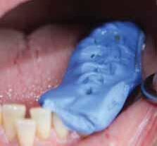


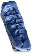

6: Impression materials and waxes can be used for marking occlusal contact. Perforations in the marking media indicate the occlusal contacts.

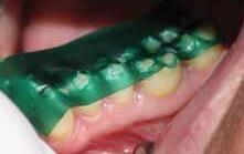
papers cannot easily mark contact on glazed porcelain or polished gold surfaces, or when the tooth surface is wet, which will result in false negative markings and recordings. However, one of the advantages of double-sided articulating paper is that the occlusal contacts in opposing arches can be recorded.
Waxes and impression materials
This method has the characteristic that actual contact between the opposing occlusal surfaces occurs in the process of identifying the contact. The materials are placed, as a covering layer of wax, carbon, coloured chalk, or aerosol, on the mandibular or maxillary occlusal surfaces. Cusps of the opposing occlusal surfaces penetrate the deposited material at the points of occlusal contact. Perforations of the marking media indicate actual tooth contacts. In this method, actual occlusal contacts can be seen when the marking media is displaced and the opposing tooth surfaces touch each other. The major disadvantage is that the contacts are disclosed on only one antagonist at a time. The process must be repeated to find the contacting points on the opposing occlusal table. Hence, the clinical procedure to identify the paired contacts is lengthy and tiresome. Besides indicating the contact spot, i.e., the perforations, some clinicians use trans-illumination of the indented material to identify points of ‘near contact’ as reflected by thinning of the material, which actually indicates close contact but not a real occlusal contact. This is a very subjective and variable procedure that requires a precise and repeatable method of laying the material on the occlusal surface prior to testing for
reducing the occlusal forces on implant prosthetics.24-27 It consists of the following:
■ incisal guidance;
■ bilateral stability and occlusal contacts in MIP;
■ freedom in centric (long centric);
■ evenly distributed occlusal contacts and forces; and,
■ smooth lateral excursive movements without working and non-working side interferences.
Basically, the outcome of this treatment is production of an ideal occlusion.24,28 Therefore, to optimise the occlusion with dental implants several factors should be considered and implemented whenever possible. For instance, a long centric occlusion (in which the MIP and CR do not coincide but are in the same horizontal level) should be used. This requires elimination of any occlusal interferences between MIP and CR. To achieve this target, flat fossae and grooves should be incorporated into the occlusion of the restoration. It is also important to create shallow cusp inclination in order to avoid torque, because with every 10-degree increase in cusp inclination, there is an approximately 30% increase in torque magnitude. Therefore, reduced cuspal inclination leads to less non-axial loading. Furthermore, occlusal table width should be 30-40% smaller in a molar region, as a wider occlusal table than the implant diameter can cause cantilever effects and bending, but a smaller occlusal table than the implant diameter will cause occlusal forces to be axially directed. Cantilevering, which is usually created whenever a force is applied at a distance from the fulcrum, should be avoided when possible. It is recommended that the anterior guidance should be shallow to a degree that allows disclusion of the posterior teeth during lateral and protrusive mandibular movements. A shallow incisal guidance (minimum vertical overlap) generates less loading on the anterior implants than when the guidance is steep. It is also important to protect restoration and implant by occlusal splints whenever possible.
Light force and thin articulating paper (less than 30μm) are used. The occlusal contact on the implant-supported restoration is then relieved. This will place all occlusal forces on the adjacent teeth, while the implant-supported restoration is out of contact with the opposing implant/tooth. Next, a greater occlusal force is applied. If the contact is heavier on the implant, it should be relieved
in order to achieve equal contact on both the implant-supported restoration and the teeth. Hence, the occlusal forces are shared between the implant and the teeth.
One of the main requirements of restorative treatment is to achieve at the end of the treatment dental occlusion that does not deleteriously affect the stomato-gnathic system. Consequently, knowledge of the basic principles of dental occlusion and the masticatory system is vital for every dental student and dentist. Therefore, functional assessment and examination of occlusion should be considered and carried out on a routine basis.
In restorative dentistry, when a patient attends a dental clinic for treatment, the required treatment may involve a small or major alteration in the occlusal surfaces of the teeth. In the former case, the conformative approach can be implemented, as little or no alteration in the occlusion will take place and the original MIP is used as the treatment position. In the second case, as more complex restorative treatment is required, the reorganised approach should be implemented.
When a dental implant is considered, a basic knowledge of how the implants are loaded is of paramount importance, as the way in which the implant is loaded differs from that of the teeth.
References
1. Lobbezoo, F., Ahlberg, J., Manfredini, D., Winocur, E. Are bruxism and the bite causally related? J Oral Rehabil 2012; 39 (7): 489-501.
2. Le Bell, Y., Niemi, P.M., Jämsä, T., Kylmälä, M., Alanen, P. Subjective reactions to intervention with artificial interferences in subjects with and without a history of temporomandibular disorders. Acta Odontol Scand 2006; 64 (1): 59-63.
3. Xie, Q., Li, X., Xu, X. The difficult relationship between occlusal interferences and temporomandibular disorder – insights from animal and human experimental studies J Oral Rehabil 2013; 40 (4): 279-295.
4. Tsukiyama, Y., Baba, K., Clark, G.T. An evidence-based assessment of occlusal adjustment as a treatment for temporomandibular disorders. J Prosthet Dent 2001; 86 (1): 57-66.
5. Riise, C., Sheikholeslam, A. The influence of experimental interfering occlusal contacts on the postural activity of the anterior temporal and masseter muscles in young adults. J Oral Rehabil 1982; 9 (5): 419-425.
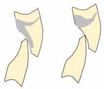
FIGURE 13: Junction between the porcelain and the metal framework should be 2mm away from the occlusal contact in the MIP, which is preferably placed on metal.
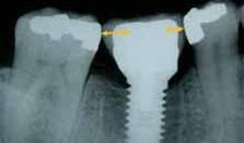
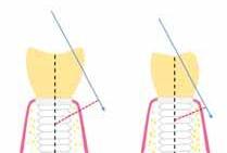
FIGURE 14: Reduction of restoration dimensions is required in order to reduce the leverage arm of these occlusal forces and consequently their negative effects on the implant and the periimplant tissues. Thus, direction of occlusal forces along the long axis of the implant should be considered when possible.
6. Sheikholeslam, A., Riise, C. Influence of experimental interfering occlusal contacts on the activity of the anterior temporal and masseter muscles during submaximal and maximal bite in the intercuspal position. J Oral Rehabil 1983; 10 (3): 207-214.
7. Ash, M.M., Ramfjord, S.P. Occlusion (4th ed.). Philadelphia; Saunders, 1995: 84-85.
8. Beyron, H. Optimal occlusion. Dent Clin North Am 1969; 13 (3): 537-554.
9. Obara, R., Komiyama, O., Iida, T., De Laat, A., Kawara, M. Influence of the thickness of silicone registration material as a means for occlusal contact examination – an explorative study with different tooth clenching intensities. J Oral Rehabil 2013; 40 (11): 834-843.
10. Wang, M., Mehta, N. A possible biomechanical role of occlusal cusp-fossa contact relationships. J Oral Rehabil 2013; 40 (1): 69-79.
11. McDevitt, W.E., Warreth, A.A. Occlusal contacts in maximum intercuspation in normal dentitions. J Oral Rehabil 1997; 24 (10): 725-734.
12. Maness, W.L., Podoloff, R. Distribution of occlusal contacts in maximum intercuspation. J Prosthet Dent 1989; 62 (2): 238-242.
13. Gümüs, H.Ö., Kılınç, H.I., Tuna, S.H., Ozcan, N. Computerized analysis of occlusal contacts in bruxism patients treated with occlusal splint therapy. J Adv Prosthodont 2013; 5 (3): 256-261.
14. Maness, W.L. Laboratory comparison of three occlusal registration methods for identification of induced interceptive contacts. J Prosthet Dent 1991;65 (4): 483-487.
15. Kong, C.V., Yang, Y.L., Maness, W.L. Clinical evaluation of three occlusal registration methods for guided closure contacts. J Prosthet Dent 1991; 66 (1): 15-20.
16. Davies, S.J., Gray, R.M. The examination and recording of the occlusion: why and how. Br Dent J 2001; 191 (6): 291-296, 299-302.
17. Wassell, R.W., Steele, J.G., Welsh, G. Considerations when planning occlusal rehabilitation: a review of the literature. Int Dent J 1998; 48 (6): 571-81.
18. American Academy of Prosthodontics. The Glossary of Prosthodontic Terms. J Prosthet Dent 2005; 94 (1): 10-92.
19. Steele, J.G., Nohl, F.S., Wassell, R.W. Crowns and other extra-coronal restorations:
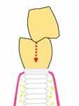
FIGURE 15: Direct the occlusal forces along the long axis of the implant. Only the supporting cusp of the maxillary tooth contacts the implantretained restoration. The cusps of the restoration do not make occlusal contact with the opposing maxillary tooth. In this case, the occlusal load is directed along the long axis of the implant, which is favourable.
occlusal considerations and articulator selection. Br Dent J 2002: 13; 192 (7): 377380, 383-837.
20. Milosevic, A. Occlusion: I. Terms, mandibular movement and the factors of occlusion. Dent Update 2003; 30 (7): 359-361.
21. Wilson, P.H., Banerjee, A. Recording the retruded contact position: a review of clinical techniques. Br Dent J 2004; 196 (7): 395-402.
22. Dawson, P.E. Evaluation, Diagnosis, and Treatment of Occlusal Problems(2nd ed.). St Louis, MO; Mosby, 1989.
23. McDevitt, W.E. An investigation of the clinical reference position of centric maxillomandibular relation in the context of the sagittal envelope of mandibular movement. PhD thesis. Trinity College Dublin, Ireland, 1993.
24. Kim, Y., Oh, T.J., Misch, C.E., Wang, H.L. Occlusal considerations in implant therapy: clinical guidelines with biomechanical rationale. Clin Oral Implants Res 2005; 16 (1): 26-35.
25. Misch, C.E. Density of bone: effect on treatment plans, surgical approach, healing, and progressive loading. Int J Oral Implantol 1999; 6 (2): 22-31.
26. Rilo, B., da Silva, J.L., Mora, M.J., Santana, U. Guidelines for occlusion strategy in implant-borne prostheses. A review. Int Dent J 2008; 58 (3): 139-145.
27. Davies, S.J. Occlusal considerations in implantology: good occlusal practice in implantology. Dent Update 2010; 37 (9): 610-612, 615-616, 619-620.
28. Rosenstiel, S.F., Land, M.F., Fujimoto, J. Contemporary Fixed Prosthodontics(4th ed.). St. Louis, MO; Mosby, 1995.
In part I of this article (Journal of the Irish Dental Association 2015; 61 (4): 201-208), the reference in the caption for Figure 11 was omitted. The reference is: “Adapted from Craddock & Youngson (2004)26”. We apologise for this error.
Effect of mesiodistal angulation of the maxillary central incisors on aesthetic perceptions of the smile in the frontal view
Yang, S., Guo, Y., Yang, X., Zhang, F., Wang, J., Qiu, J., et al
The purpose of this study was to analyse the effect of mesiodistal angulation of the maxillary central incisors on the smiling aesthetics in young adults.
Methods
Frontal smile photographs of a young man and a young woman were digitally modified to produce 28 smile images that were grouped into four series. These images were judged using visual analogue scales by 52 orthodontists and 61 lay people. The effects of the judges’ professions, subjects’ sexes, and photograph framings on the appreciation of smile aesthetics with different mesiodistal angulations of the maxillary central incisors were evaluated. The data were analysed with paired t tests, Dunnett t tests, and independent samples t tests; statistical significance was set at p<0.05.
Results
The images were ranked less attractive as the mesiodistal angulation of the

maxillary central incisors increased during smiling. No relationship was found between the judges’ professions and the aesthetic evaluations of incisal angulation, but the statistical analysis showed that both the subject’s sex and the photograph framing were significant variables (p<0.05).
Conclusions
The mesiodistal angulation of the maxillary central incisors plays an essential role in smile aesthetics in the frontal view. When formulating treatment plans, orthodontists should never underestimate the influence of mesiodistal angulation on smile attractiveness.
American Journal of Orthodontics and Dentofacial Orthopedics 2015; 148 (3): 396-404.
Implants failures related to endodontic treatment. An observational retrospective study
López-Martínez, F., Gómez Moreno, G. Olivares-Ponce, P., Eduardo Jaramillo, D., Eduardo Maté Sánchez de Val, J., Calvo-Guirado, J.
Objective
The aim of the study was to analyse potential aetiological risk factors that constitute a complex problem in the clinical management of peri-implantitis.
Materials and methods
An observational retrospective study was conducted to describe the possible effect of lesions of pulpar origin and/or periapical success or failure of the implant. The sample consisted of review of 800 implants, of which 500 were conducted at the Faculty of Dentistry of the UANL and 300 at the private clinics of maxillofacial surgeons experienced in the placement of implants and residing in Monterrey, Mexico. A total of 580 cases were in female patients, while 220 patients were of male gender. The age of patients at the time of placing the implant ranged from 28 to 81 years.
Results
Of 800 study subjects who underwent dental implant treatments, 200 cases (25%) were detected that presented endodontic failure prior and/or adjacent to the placement of the implant. The 50.41% had peri-implantitis, recording 62 cases in the Faculty and 18 cases (23.38%) in private clinics, finding that there was a statistically significant difference between the presence and absence of peri-implantitis in terms of endodontic failure prior and/or adjacent to the placement of the implant.
Conclusions
Within the limitations of this observational retrospective study, it could be concluded that the development of inflammatory changes mediated by the presence of remnant bacteria surrounding hard tissues adjacent to implants might induce late failures of implants, and potentially trigger pathological features of apical peri-implantitis.
Clin Oral Implants Res 2015; 26 (9): 992-995.
Oral squamous cell carcinoma presenting in a patient receiving adalimumab for rheumatoid arthritis
Beattie, A., Stassen, L.F., Ekanayake, K.
The efficacy of biologic agents in the treatment of inflammatory immunemediated conditions has been clearly shown, but there also are numerous reports of adverse effects. Most reported adverse effects have been associated with tumour necrosis factor-α (TNF-α) inhibitors and include a possible increased risk of malignancy. There have been some reported cases of oral cancer developing in patients treated with TNF-α inhibitors. This case report describes a patient who was taking adalimumab for rheumatoid arthritis and who presented with a squamous cell carcinoma (SCC) in the mandible. Diagnosis was complicated because the clinical appearance was of a nonhealing extraction socket and the patient had a history of bisphosphonate therapy. An initial diagnosis of bisphosphonate-related osteonecrosis of the jaws was made, which delayed the commencement of appropriate treatment. This case highlights the importance of ruling out SCC in patients taking biological agents with unusual symptoms.
J Oral Maxillofac Surg 2015; May 22
The evaluation of the heat generated by the implant osteotomy preparation using a modified method of the measuring temperature
Yamaba, T., Suganami, T., Ikebe, K., Sogo, M., Maeda, Y., Wada, M.
Purpose
To establish a method for measuring the heat generated when preparing an
(questions on page 226)
1.There is an undisplaced fracture of the right angle of the mandible.
2. Manage the patient as per the advanced trauma life support (ATLS) protocol. The patient should be admitted to hospital under the care of the oral and maxillofacial surgery team. The patient should be kept nil by mouth, and broad-spectrum antibiotics commenced. The patient should have analgesia regularly and intravenous fluids to prevent dehydration while awaiting further management.
3.Late fracture of the mandible after third molar surgery is a rare event (0.005%). These fractures predominantly occur in patients older than 25 years, who are fully dentate, and those with deeply impacted third molars. The greatest risk is two to three weeks post removal due to replacement
osteotomy site, and to assess for correlations of rotational speed, proceeding speed, loading value of the drill (contact pressure), motion pattern, and bone density with temperature increases.
Materials and methods
A thermocouple was placed in the internal irrigation hole of a 2mmdiameter twist drill used for measuring osteotomy site temperature. In the artificial blocks, two different densities were used to drill under varying conditions including drill proceeding, rotating speed, and motion pattern. The drilling procedure was repeated five times for each combination, and the data collected were statistically analysed using multiple regression analysis.
Results
Strong positive correlations were found among bone density, drill motion pattern and maximum temperature, and a positive correlation was found in proceeding speed (p<0.001). Rotation speed and maximum temperature were not correlated (p<0.001). Conversely, loading values of the drill increased with the lower rotation speed and higher proceeding speed, which were effective in controlling the temperature rise.
Conclusion
When preparing a simulated bone for an osteotomy with a thermocouple inserted into a twist drill with internal irrigation, the drilling motion pattern, bone density, drill speed and proceeding rate affected bone temperature, in descending order. It was also observed that bone temperature correlated positively with speed and negatively with proceeding speed, independent of density. This indicates that lower drill speed and higher proceeding speed without excessive loading values minimise the bone temperature heat.
International Journal of Oral and Maxillofacial Implants 2015; 30 (4): 820-826.
of the granulation tissue with connective tissue at the extraction site. It is necessary to warn patients that the removal of wisdom teeth may weaken the mandible and that activities that increase the risk of injury, including sports and heavy masticatory forces, should be avoided for four weeks post removal.
It is appropriate, before surgery:
■ to make an appropriate assessment of the wisdom teeth and the amount of bone removal required, and to warn the patient of the possibility of late mandible fracture during chewing.
During surgery:
■ to be conservative with the amount of bone removed, to divide the tooth where appropriate and to avoid excessive force on the mandible.
After surgery:
■ especially if it is a difficult operation with significant bone removal, to advise the patient to continue a soft diet for four weeks and avoid excessive masticatory force.
Continuing our series on IDA membership, we chat to DR ANDREW KELLYwho is in a singlehanded practice in Nenagh, Co. Tipperary, and is a member of the Council of the IDA.
What led you to first get involved in the IDA?
I qualified in 1994, and went away to work in England and Northern Ireland. When I returned to the Republic to set up in practice in 1997, I joined the Association straight away. Initially, I didn’t take an active role – I went to lectures and meetings, but nothing major. Back then I was younger, and the IDA didn’t seem to have a huge amount of relevance to me. But I think that over the years the Association has changed a lot and improved in terms of the level of services that it offers to members.
How did your involvement progress?
I saw that there was no general practice representative from my area on the IDA’s GP Committee, so I decided to give it a go. Until then, for me the IDA was an association in Dublin; I’d never been to the offices, and wasn’t really aware of everything that went on there. In the last few months I was asked to go on the IDA Council as a GP rep.
What has your involvement in the IDA meant to you?
There is a lot to gain from membership. The IDA operates a huge amount of services, which, to my mind, are way ahead of what they were before. You can save money on indemnity, and the Association also offers mentoring, and general assistance in terms of employment law, and best practice for dentists.
The website also contains a lot of information. You can join in on discussions in the discussion forums without leaving your room! I would encourage members to take a more active role, even if it is only logging on to the website, getting a user name and looking at the discussion forums to see what people are thinking.
A lot of people are very passive members, as I was. They pay their sub every year and might go to the odd meeting but they don’t really do anything beyond that. Dentistry is quite a lonely profession. A lot of dentists are like me – single-handed – and wouldn’t have much interaction with their colleagues. I’ve found by being on the GP Committee, and talking to colleagues, that many other dentists have the same problems and the same opinions on things that we don’t ever really vocalise with each other because we’re just working away by ourselves.
What has been the single biggest benefit of IDA membership for you?
Being aware you’re not the only one who thinks in a certain way and that a lot of problems and views are common has been very important. Being on the GP Committee, I see that it’s the same all over the country – our views are very similar.
How would you like to see the Association progress into the future?
We have to be unified. I do feel in the future that as the economy improves, and the Government brings in new dental schemes, unless we speak with a single voice we’re going to be trampled upon. In the past dentists have been very poor at standing up for themselves – we’ve just accepted whatever the Government has given us. I see the IDA having a role in unifying everybody so that our voices are heard. I always thought over the years that the Association was just this distant big body that didn’t have anything to offer me – but there is a lot that people are unaware of.
When not doing ‘a bit of everything’ in his practice in Nenagh, John likes to run and has completed several marathons – he finds it great for mental and physical well being. He also has a busy sideline as ‘Dad’s taxi’ for his three kids.