14 minute read
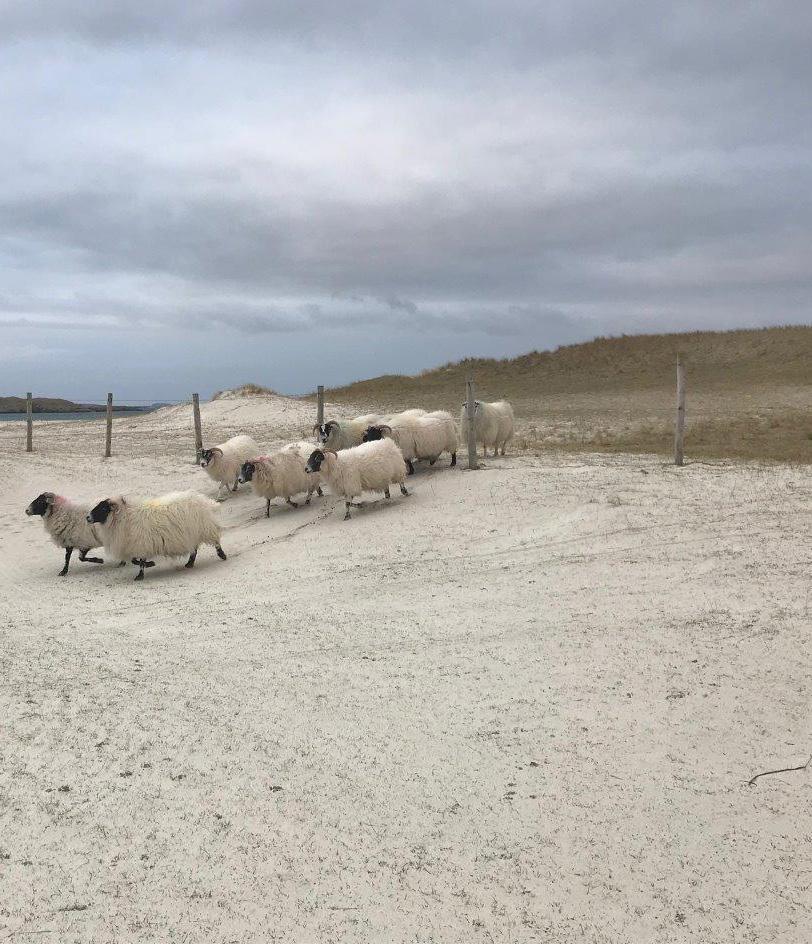
Feb Agritech hub launched. Page 36. Mar Electric sheep: the future of VR

by thedickvet

Interview The future of VR in veterinary education
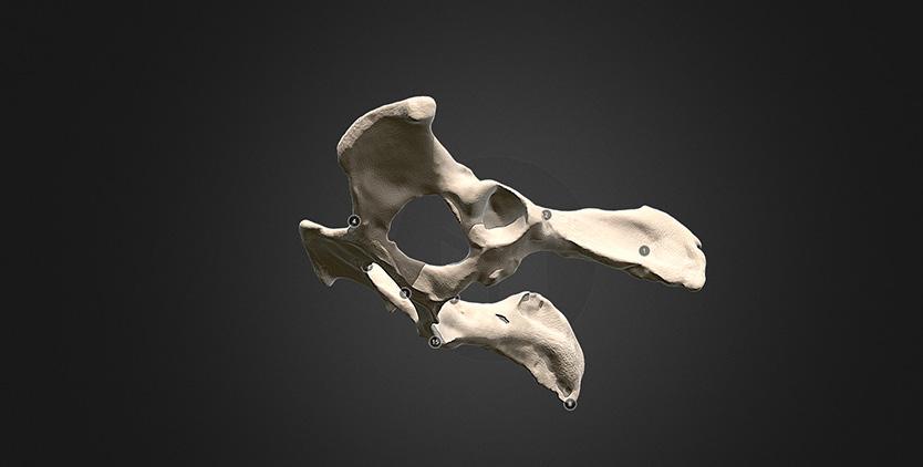
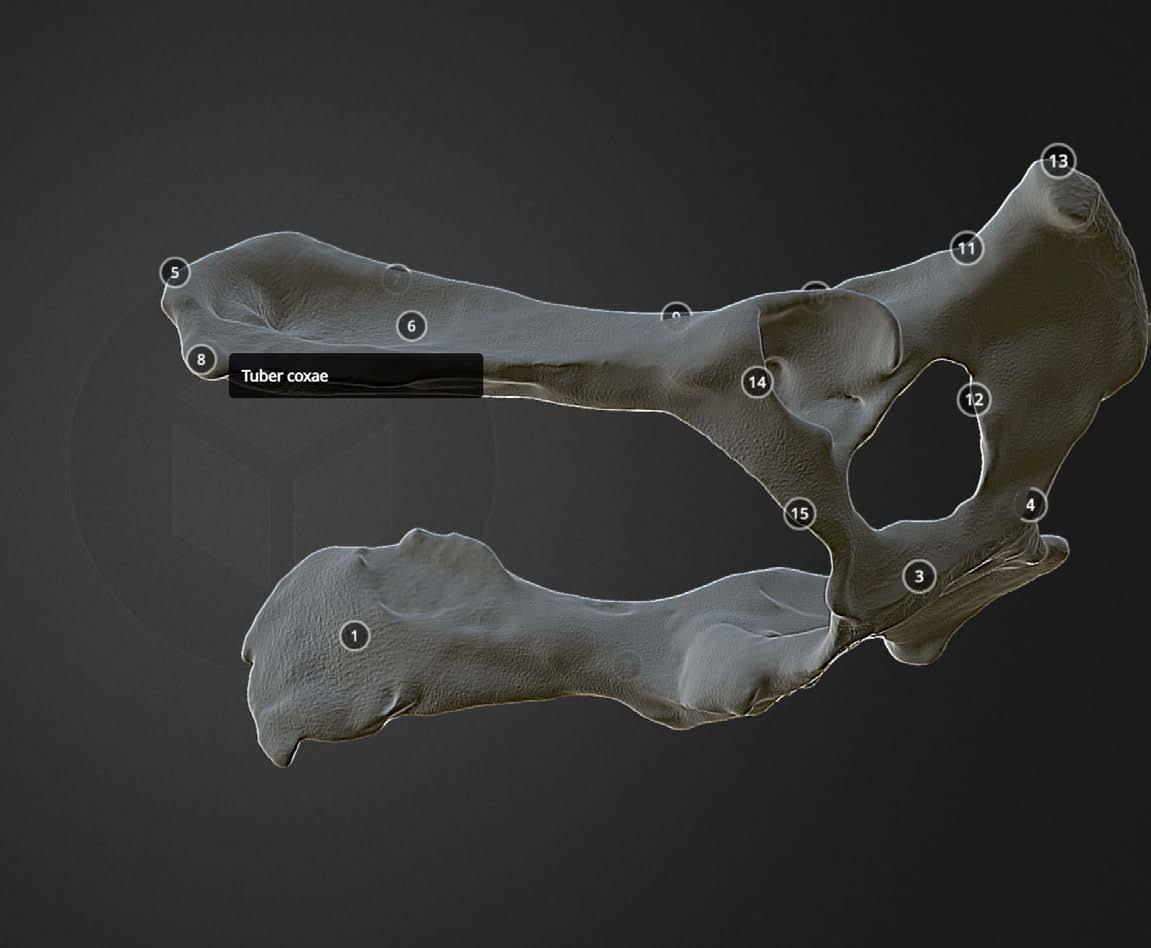
Our Digital Education Unit (DEU) works with lecturers to provide students with immersive learning experiences. Here, Senior E-Learning Developer Brian Mather talks about digital education and the future of Virtual Reality in teaching. “The term ‘Immersive Media’ includes Virtual Reality, Augmented Reality and 360* video – but each is very different,” explains Mr Mather. “Virtual Reality (VR) places you inside a completely digital environment; augmented reality overlays digital content onto the real world, and 360* video plays out a recording that you observe and can manipulate but cannot move independently within.” The School has been using 3D models in teaching since 2015, and it has been possible to view the content in VR since 2018, but in a limited sense. “The dream is to offer students a virtual practical experience, but the reality is still a way off,” says Mr Mather. “However, we use some fantastic technology in our teaching to enhance the student experience.” Explorations into 3D printing at the School formed the beginnings of what is now an extensive 3D modelling project. “Once you’ve done the preparatory work to create a 3D model, you can actually do quite a lot with it beyond print it. You can put it into a virtual space for students to explore, you can label it, you can create layers,” says Mr Mather. “It’s better than a printed model. There’s huge potential.” The School has a bone library for students to borrow from, which is helpful for their anatomical studies. The collection is incredibly valuable, and difficult to replace. However, thanks to 3D modelling techniques, portions of the library have been digitised. The digital bone library can be accessed anytime, anywhere, and provides the added benefit of enabling students to zoom in or out, rotate the images, view overlays or cross-sections and even study at the dinner table without upsetting friends and family! 360* photography and video technology has enabled the recording of high-quality video tutorials for students to review at home or in practice. Students at the Dick Vet can use their phones to access hundreds of video tutorials via an online library or by using QR codes around the School. For example, a student in the Equine Practice can remind themselves of tutorials they previously received on a particular procedure by pointing their phone at a QR code in the relevant area. This technology is also used to ease the transition from practicals in the Clinical Skills lab to practice in our clinics. Students can view videos of techniques they have learned being put into practice by the School’s clinical staff, which enables students to move more confidently into the next stage in their learning. This also eases
Advertisement
anxiety, helps to consolidate learning and enables students to ‘meet’ the team they will work with in the hospital environment before they progress. There is considerable interest from staff, students and the wider University in developing the use of VR as a teaching aid. “The tricky part is developing the teaching material. Without digital content there is nothing to experience in the virtual space. Pedagogical practice still applies with the development of interactive simulation. ‘What am I teaching? Why am I teaching in this way? How will the students learn? How will they benefit from this form of teaching?’”
Future development of VR to enhance clinical education depends in part on the technology. “The exciting part is when you can manipulate and interact with the environment. The development of hand (and, more importantly, finger) tracking will make the experience more valuable. After that, the sense of touch needs to be replicated. Haptic feedback is in development, but it will be some time before it is commercially available.
“Surgical training is all about repetition and feel. When you can put your headset on, repeat your surgery as often as you like (no cadaver required) and receive tactile feedback from your haptic gloves, we’re there.” “Surgical training is all about repetition and feel. When you can put your headset on, repeat your surgery as often as you like (no cadaver required) and receive tactile feedback from your haptic gloves, we’re there.”
- Brian Mather, Senior E-Learning Developer
Clinical Services Imaging team lends expertise to racehorse studies
Specialists in equine health at the Dick Vet have joined forces with imaging teams in the University’s Medical College to advance understanding of heart health and leg fractures in horses. The team sought to evaluate the patterns of small fibres in the left atrium, or chamber, of the equine heart, which is useful knowledge in developing techniques to identify electrical activity in the heart. Equine cardiology experts and teams from Edinburgh Imaging used scanning technologies, known as diffusion tensor MR and micro CT, to create detailed images of post-mortem specimens from horses. Both techniques led to excellent visualisation of the three-dimensional organisation of fibre tracts within the heart chamber. “Applying non-destructive imaging techniques to better understand the anatomy of equine hearts has shown to be a valuable approach, which we hope can inform efforts to develop methods of studying electrical activity in the heart – an important indicator of heart health,” says Dr John Keen, Senior Lecturer.
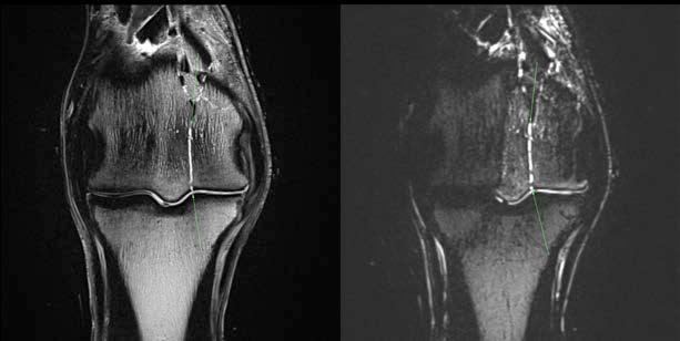
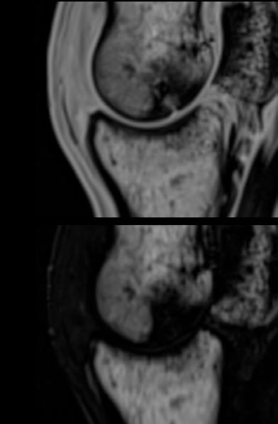
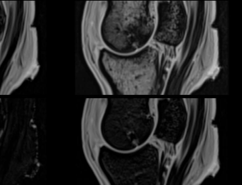
In a separate project, experts from Edinburgh Imaging collaborated with equine orthopaedic specialists at the Dick Vet on imaging of the Thoroughbred fetlock – the high motion joint between a horse’s knee and hoof.
The team used a newly installed 3T Siemens Skyra MR scanner to capture images of 17 limbs recovered from horse cadavers. The scanner forms part of the advanced imaging services available at the Dick Vet’s Large Animal Research Imaging Facility (LARIF).
Our researchers hope to use MR look for biological indications to help predict fracture occurrence in athletic horses, whose bones becomes more dense as they train.
-Dr Sarah Taylor, Equine Orthopaedic Surgeon
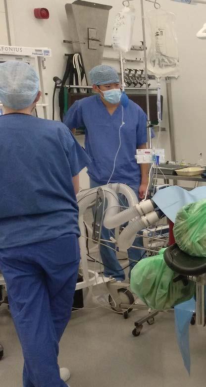
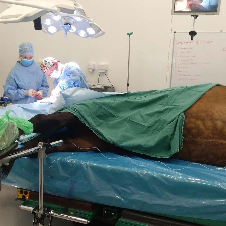
CEPEF4 equine anaesthesia mortality study gathers data
Veterinary Anaesthesia and Analgesia service staff are leading the worldwide milestone Confidential Enquiry into Perioperative Equine Fatalities (CEPEF4) study, investigating equine mortality associated with general anaesthesia and standing sedation.
Thousands of anaesthetists and equine clinicians are being invited to participate in a study collecting data on horses undergoing general anaesthesia and standing sedation, to help lower the associated risks of general anaesthesia in horses.
Results from the multicentre study will reveal trends in equine anaesthetic practice and the outcome, in terms of disease and mortality. The study uses an online questionnaire.
Equine anaesthetic fatalities have been reported in previous studies as 10 times more likely than for other animals such as dogs and cats. The team behind the CEPEF4 initiative hopes to help reduce this risk of death by throwing light on practices associated with higher fatality. Researchers behind the global project will update the findings of a similar initiative carried out almost two decades ago, known as CEPEF2. Use of new technologies allows interactive data collection with virtually simultaneous analysis, allowing continuous updating of the information.
The project is led by the R(D)SVS in collaboration with specialists from CEU Cardenal Herrera University, Spain and the University of Zürich, Switzerland. The team also is integrated with two authors of the previous CEPEF studies and is supported by the Association of Veterinary anaesthetists (AVA). “We hope to generate a large dataset from different clinics around the world, to assess current trends and practices, and point to potential improvements in anaesthesia for horses and other equine animals.” - Miguel Gozalo-Marcilla, Senior Lecturer in Veterinary Anaesthesia and Analgesia.
Veterinary practices in remote and rural areas of Scotland face challenges in recruiting and retaining vets to work in their area; issues that are shared with other professions such as medicine and dentistry.
Veterinary provision in rural areas of Canada,
America and Australia have similar challenges.
Given the international nature of the veterinary profession and our student cohort, we are developing closer links with colleagues in rural mixed practices to assist where needed, offering R(D)SVS students the chance to experience life in this part of the world and to showcase career opportunities in the rural sector outside of standard veterinary practice.
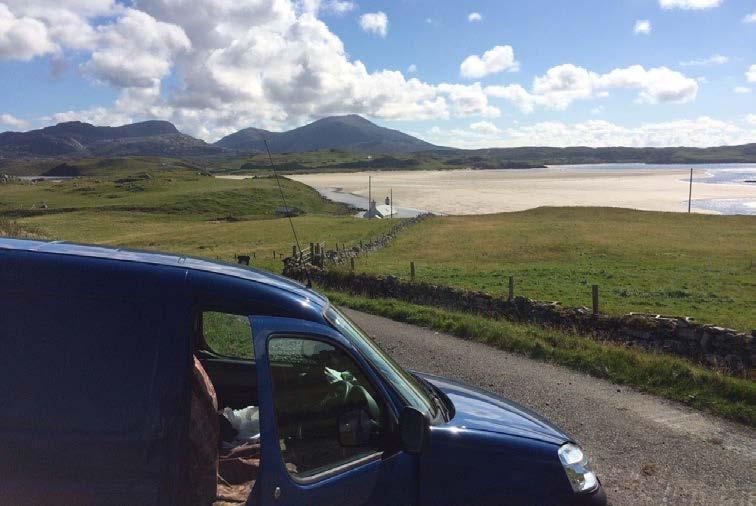
The Highlands and Islands of Scotland covers a vast area of remote communities which are home to a wide variety of different animals. To ensure veterinary provision to the crofting communities in these areas, the Scottish
Government provides support to these remote rural practices through the Highlands and Islands
Veterinary Services Scheme (HIVSS), which includes over 20 practices.
Dr Patrick Pollock from our Equine team and
Dr Fraser Murdoch from our Farm Animal team have been working together with the HIVSS practices to deliver virtual CPD evenings, as well as targeted CPD on topics such as large animal rescue. We have also been awarded a grant from the University of Edinburgh Principal’s Teaching Award Scheme to develop resources for schools and practices to use with students in the Highlands and Islands interested in pursuing a career in veterinary medicine.
Working in collaboration with the HIVSS network, Scotland’s Rural College (SRUC) and Glasgow University Veterinary School (GUVS), we have established a selected rotation offering for our Final Year students in the Highlands and Islands. This involves spending time at one of the mixed veterinary practices within the HIVSS network, and a subsequent week based at the SRUC Inverness Campus focusing on other career options within the rural sector including aquaculture, applied epidemiology, veterinary diagnostic laboratory work and supporting field diagnostics services.
Feedback from students on this selective rotation in spring 2021 was excellent, even resulting in job opportunities for one of our students.
We are actively working to develop this partnership further, so that we can respond to changes in the profession, and meet the specific needs of colleagues working in remote and rural veterinary practice.
Pet recovering well after treatment for rare cancer
A pet cat is recovering from a rare type of cancer following care from a range of expert teams at the Hospital for Small Animals.
Misha, a 14-year-old shorthair, was found to have cancer affecting several of his organs, despite showing no signs of ill-health. Following surgery to remove tumours, and ongoing chemotherapy, Misha’s condition is now in remission.
Misha’s illness was discovered when blood checks in preparation for dental work flagged a change in his liver health. Levels of key enzymes in his liver, which had been slightly high in a routine check six months earlier, had worsened to become markedly elevated. Further tests revealed Misha had multicentric plasma cell neoplasia, also known as myeloma related disorder, which had caused tumours in his spleen, liver and on a lymph node. This type of tumour, previously known as multiple myeloma, occurs in stem cells of the bone marrow or, as in Misha’s case, in other organs.
Teams from surgery, anaesthesia, feline medicine, intensive care, oncology, pathology and dentistry at the Dick Vet collaborated on Misha’s treatment and recuperation.
Surgery was carried out to remove Misha’s spleen and lesions from his lymph node, as well as resolving dental issues. He was prescribed chemotherapy to treat the lesions on his liver. ”I’m very happy that Misha is now doing so well. His illness highlights the importance of regular health checks to help spot issues that may otherwise be undiagnosed, and the benefit of treating animals early on their illness, to improve their chances of recovery”.
- Tobias Schwarz, Radiologist and Misha’s owner
Collie home with barely a scratch after sticky situation
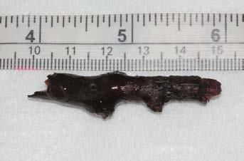
A twig was removed from a dog’s eye after the animal had an unusual accident while playing outdoors.
Mia the collie had the 5-centimetre twig removed by veterinary ophthalmologists thanks to specialist scans that allowed them to see the stick’s placement.
Remarkably, the stick was carefully removed, leaving Mia with only a small scratch.
Mia’s owners feared their six-year-old pet had lost her eye after she was playing in a hedge and emerged with a stick poking from her eye socket.
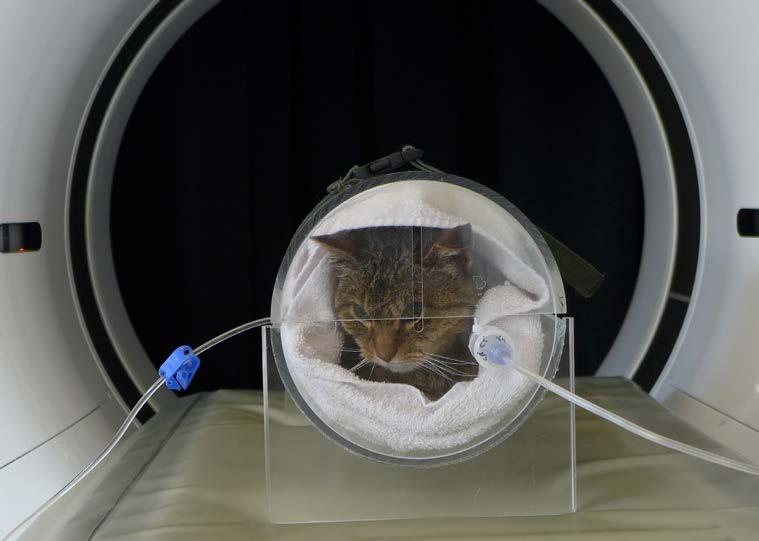
She was referred by her local vet to the specialist ophthalmology service at the Hospital for Small Animals. Once there, our vets performed a CT scan. Images revealed that the stick was still in one piece and lodged just above the eye.
Pictures from the CT scan allowed the vets to carefully remove the object without the need for an invasive operation, leaving Mia with only a superficial scratch on her eye. After flushing the area with fluids to make sure there were no remaining fragments, Mia was able to walk out of the clinic unaided and has suffered no long-term damage to her sight.
“This was an unusual situation for us, and we are really pleased to see Mia back to full health and enjoying life. We are lucky to have such an amazing group of people here, including Specialists in anaesthesia and radiology, and a highly skilled and compassionate nursing team.” - Ben Blacklock, Vet Specialist Ophthalmologist
Walking aid helps dogs recover after spinal injuries
Generosity between dog owners has given injured pets from five different families the chance to learn to walk again.
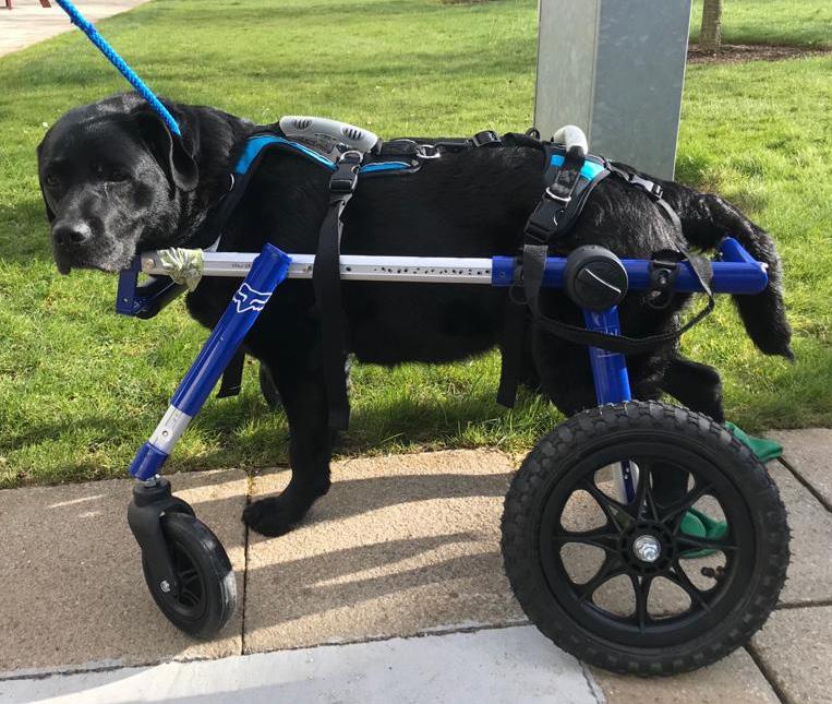
Polar, a nine-year-old husky, underwent emergency neurosurgery at the Hospital for Small Animals after a sudden, severe neck injury – a slipped disc, also called an intervertebral disc herniation.
After the successful surgery, at the recommendation of the Dick Vet, Polar’s owners invested in a wheelchair to aid his recovery, from a ruptured disc compressing his spinal cord.
An intensive programme of physiotherapy and hydrotherapy, started by Royal (Dick) Edinburgh Physiotherapy Assessment and Intensive Rehabilitation Centre (REPAIR) centre and continued at home, helped Polar recover.
With use of the wheelchair and a support harness, Polar was able to run unaided just three months after the procedure.
Following Polar’s recovery, his owners lent the wheelchair to four other dogs treated at the Dick Vet, each of whom had suffered spinal problems.
In each case the custom-made wheelchair was professionally adjusted to fit each dog. All were treated by experts in neurology, neurosurgery and physiotherapy. Ripley, a six-year-old Labrador, became paralysed while playing with a ball. Within a few hours she was evaluated by the Neurology and Neurosurgery team and underwent an MRI.
Ripley was found to have experienced a slipped disc. A fragment of disc had hit the spinal cord at speed, causing bruising. Injuries of this type respond best to medical treatment with pain relief and physiotherapy, rather than surgery.
When Ripley was showing good progress, Dick Vet staff contacted Polar’s family, who kindly offered to lend her their wheelchair. With continued physiotherapy and enthusiastic use of the wheelchair, Ripley was able to walk again unaided. Susie, a 10-year-old Labrador, became unable to walk due to a degenerative condition in her neck commonly referred to as ‘wobbler syndrome’.
An MRI scan identified that her condition had progressed too far for surgery. Susie was prescribed a palliative plan of pain relief and physiotherapy, to ease her final days.
Polar’s owners once again offered the use of the wheelchair, enabling Susie to spend the final weeks of her life happy at home.
Shadow, a six-year-old spaniel, became weak and wobbly in all four limbs after a walk, and quickly became unable to move.
Shadow was diagnosed by the Dick Vet with an intervertebral disc problem, which had caused bruising to the spinal cord. Similarly to Ripley’s paralysis, injuries of this type respond best to medical treatment, rather than surgery.
The wheelchair was again adapted to suit Shadow’s needs, and together with intensive physiotherapy and hydrotherapy, enabled Shadow to walk with minimal support, just two months after his injury. Diesel, an 11-year-old Dalmatian, suffered a herniated disc in his neck and needed surgery.
Diesel also had a degenerative condition in his spine, which over time had affected his spinal cord function. Owing to this, Diesel was at risk of not being able to walk again.
However, the surgery was successful and followed by intense physiotherapy and hydrotherapy with the REPAIR clinic.
With the help of the wheelchair Diesel was back on his feet within a month and able to play with his four other Dalmatian family members.
- Dr Zohra Khan, Veterinary Neurologist and Neurosurgeon


