2 minute read
Oral and Maxillofacial Pathology Case of the Month


ORALand maxillofacial pathology case of the month
Clinical History
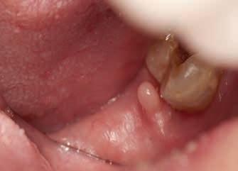
A 49-year-old man visited a new dentist after moving from another city. During his first oral examination, the dentist noticed that there were bilateral masses on his retromolar pads. The one on the left was only a sessile, slightly elevated mass, but the one on the right was a “finger-like” tubular projection 5 mm high (Figure 1). When asked if any previous dentist had mentioned it, he said, “yes, they all did.” He said the masses had been present since his grade school days. The lesions were moderately soft to palpation and were asymptomatic, with no other oral masses present. The underlying bone was unremarkable on periapical radiographs. His dentist elected to biopsy the right side “column” because the patient had complained about occasional food debris getting caught between the tissue column and the adjacent tooth.
AUTHORS
Safia Durab, DDS, MS
Chief Resident in Oral & Maxillofacial Pathology, Department of Diagnostic & Biomedical Sciences, School of Dentistry, University of Texas, Houston, Texas
Carolyn Huynh, DDS, MEd, EdD
Associate Professor, School of Dentistry, Department of Diagnostic & Biomedical Sciences, University of Texas, Houston, Texas
Jerry E. Bouquot, DDS, MSD, DABOMP, DABOM (hon), FAAOMP, FICD, FACD, FRCM (UK)
Emeritus Professor & Past Chair, Department of Diagnostic & Biomedical Sciences, School of Dentistry, University of Texas, Houston, Texas
1A 1B
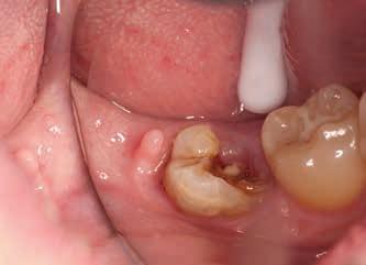
Figure 1. Right retromolar pad area. A) Soft tissue “column” (arrow) just distal to the carious third molar was uniformly circular, soft and 5 mm. high; B) Close up of the column.
Microscopic Appearance
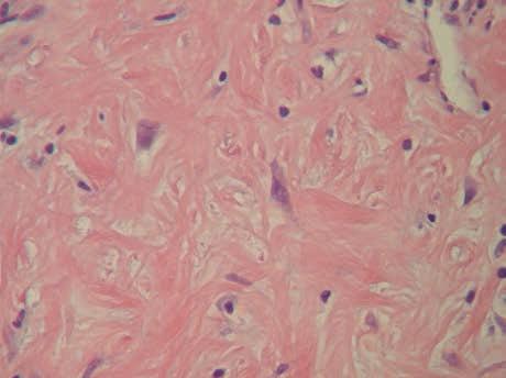
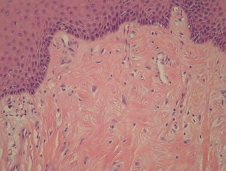
The columnar mass consisted entirely of dense, mature collagenous tissue with few blood vessels, although some of the vessels were dilated (Figure 2). The surface stratified squamous epithelium was unremarkable, except for a slightly thickened layer of orthokeratin. It showed no elongated, thin rete processes and there were no papillary or verruciform surface projections. A few lymphocytes were found within the stroma directly beneath the epithelium, along with occasional fibroblast-like cells which were much larger than normal, were angular (even some nuclei) and showed cytoplasm that was faintly purple. The giant cells were positive for vimentin (indicating mesenchymal origin) and Factor VIIIa (indicating a dendrocyte origin) by immunohistochemical staining.
What is your diagnosis?
See page 203 for the answer and discussion.
2A
Figure 2. Histopathology of the columnar mass. A) The mass consisted almost entirely of dense, somewhat avascular fibrous stroma covered by unremarkable stratified squamous epithelium, with a few scattered lymphocytes beneath the epithelium; B) The fibrous stroma contained several large, faintly stained, angular fibroblasts (arrows).
2B


