THERAPY USING THE TAP-PAP INTERFACE FOR SEVERE CPAPRESISTANT OBSTRUCTIVE SLEEP APNEA
MARTIN A. DENBAR, DDS, DABDSM
116 ASK THE POWERS CENTER
KATIE JULIEN, DDS, MS
JORGE AGUIRRE, DDS, MSC
RYAN MCGOVERN, DDS, MSD
GUEST EDITOR RADE D. PARAVINA, DDS, MS, PHD
118
FBI: FOUND BUT NOT IDENTIFIED (YET)
KATHLEEN A. KASPER, DDS, D-ABFO
In-network dentists
benefit from a dedicated point of contact
Den ve their best health. Joining Humana’s dental netw actice be more efficien ofitable. In-netw oviders with Humana receive a dedicat esentative who can ou solve issues, answer questions and get you and your staff back to what matters most—y ts.
A dedicated representative
Get answers to all your questions from your single t of contact.
Resources at your fingertips
Answers and solutions are just a call or click away for in-netw oviders.
Streamlined processes
P t care and st ts quickly with an average credentialing time of just 2.2 days.*
Scan the QR code or visit Humana.com/ memberfinder to find Humana members in your ar e ways to grow your business.
Martin A. Denbar, DDS, DABDSM
HIGHLIGHTS
Oral and Maxillofacial Pathology: Case of the Month
Oral and Maxillofacial Pathology: Case of the Month Diagnosis and Management
Editorial Staff
Jacqueline M. Plemons, DDS, MS, Editor
Juliana Robledo, DDS, Associate Editor
Nicole Scott, Managing Editor
Barbara Donovan, Art Director
Lee Ann Johnson, CAE, Executive Director
Editorial Advisory Board
Ronald C. Auvenshine, DDS, PhD
Barry K. Bartee, DDS, MD
Patricia L. Blanton, DDS, PhD
William C. Bone, DDS
Phillip M. Campbell, DDS, MSD
Michaell A. Huber, DDS
Arthur H. Jeske, DMD, PhD
Larry D. Jones, DDS
Paul A. Kennedy, Jr., DDS, MS
Scott R. Makins, DDS, MS
Daniel Perez, DDS
William F. Wathen, DMD
Value for Your Profession: Growth Strategies: Don’t Let Uncollected Production Sit Idle on Your Books
Katie Julien, DDS, MS Jorge Aguirre, DDS, MSc Ryan McGovern, DDS, MSD Guest Editor Rade D. Paravina, DDS, MS, PhD
Kathleen A. Kasper, DDS, D-ABFO
Index to Advertisers
ABOUT THE COVER:
The cover features an undated photo of Jacob’s Well, an artesian spring in Wimberley, Texas. A beloved Texas swimming hole, it has been closed since 2022 due to ongoing drought conditions, population growth, and groundwater demand.
Robert C. White, DDS
Leighton A. Wier, DDS
Douglas B. Willingham, DDS
The Texas Dental Journal is a peer-reviewed publication. Established February 1883 • Vol 143 | No. 3
Texas Dental Journal (ISSN 0040-4284) is published monthly, except January-February, March-April, July-August, and November-December, which are combined issues, by the Texas Dental Association, 8701 W Hwy 71, Ste 201-M Austin, TX 78735, 512-443-3675. Periodicals Postage Paid at Austin, Texas, and at additional mailing offices. POSTMASTER: Send address changes to TEXAS DENTAL JOURNAL, 8701 W Hwy 71, Ste 201-M, Austin, TX 78735. Copyright 2026 Texas Dental Association. All rights reserved. Annual subscriptions: Texas Dental Association members $17. In-state ADA Affiliated $49.50 + tax, Out-of-state ADA Affiliated $49.50. In-state Non-ADA Affiliated $82.50 + tax, Out-of-state Non-ADA Affiliated $82.50. Single issue price: $6 ADA Affiliated, $17 Non-ADA Affiliated. For in-state orders, add 8.25% sales tax. Contributions: Manuscripts and news items of interest to the membership of the society are solicited. Electronic submissions are required. Manuscripts should be typewritten, double spaced, and the original copy should be submitted. For more information, please refer to the Instructions for Contributors statement at tda.org. All statements of opinion and of supposed facts are published on authority of the writer under whose name they appear and are not to be regarded as the views of the Texas Dental Association, unless such statements have been adopted by the Association. Articles are accepted with the understanding that they have not been published previously. Authors must disclose any financial or other interests they may have in products or services described in their articles.
The staff of the Texas Dental Journal acknowledges erroneously listing Dr Rene Rosas as deceased in a photo caption in the March 2026 issue of the publication. We sincerely apologize to Dr Rosas and for any confusion this may have caused and thank our readers for their feedback.
Advertisements: Publication of advertisements in this journal does not constitute a guarantee or endorsement by the Association of the quality of value of such product or of the claims made.
Anesthesia Education & Safety Foundation
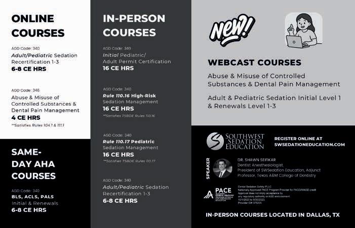
Two ways to register: Call us at 214-384-0796 or e-mail us at sedationce@aol.com Visit us on the web: www.sedationce.com
NOW Available: In-Office ACLS & PALS renewals; In-Office Emergency Program Live Programs Available Throughout Texas
Two ways to Register for our Continuing Education Programs: e-mail us at sedationce@aol.com or call us at 214-384-0796
OUR GOAL: To teach safe and effective anesthesia techniques and management of medical emergencies in an understandable manner. WHO WE ARE: We are licensed and practicing dentists in Texas who understand your needs, having provided anesthesia continuing education courses for 34 years. The new anesthesia guidelines were recently approved by the Texas State Board of Dental Examiners. As practicing dental anesthesiologists and educators, we have established continuing education programs to meet these needs.
New TSBDE Requirement of Pain Management
Two programs available (satisfies rules 104.1 and 111.1)
Live Webcast (counts as in-class CE) or Online (at your convenience)
All programs can be taken individually or with a special discount pricing (ask Dr. Canfield) for a bundle of 2 programs:
Principles of Pain Management
Fulfills rule 104.1 for all practitioners
Use and Abuse of Prescription M edications and Provider Prescription Program Fulfills rules 104.1 and 111.1
SEDATION & EMERGENCY PROGRAMS:
Nitrous Oxide/Oxygen Conscious Sedation Course for Dentists:
Credit: 18 hours lecture/participation (you must complete the online portion prior to the clinical part)
Level 1 Initial Minimal Sedation Permit Courses:
*Hybrid program consisting of Live Lecture and online combination
Credit: 20 hours lecture with 20 clinical experiences
SEDATION REPERMIT PROGRAMS: LEVELS 1 and 2
(ONLINE, LIVE WEBCAST AND IN CLASS)
ONLINE LEVEL 3 AND 4 SEDATION REPERMIT AVAILABLE! (Parenteral Review) Level 3 or Level 4 Anesthesia Programs (In Class, Webcast and Online available):
American Heart Association Advanced Cardiac Life Support (ACLS) and Pediatric Advanced Life Support (PALS) Initial and Renewal Programs
NOTE: ACLS or PALS Renewal can be completed by itself at any combined program Combined ACLS-PALS-BLS and Level 2, 3 and 4
Program
WEBCASTING and ONLINE RENEWALS AVAILABLE! Live and archived webcasting to your computer in the comfort of your home. Here are the distinct advantages of the webcast (contact us at 214-384-0796 to see which courses are available for webcast):
1. You can receive continuing education credit for simultaneous live lecture CE hours.
2. There is no need to travel to the program location. You can stay at home or in your office to view and listen to the course.
3. There may be a post-test after the online course concludes, so you will receive immediate CE credit for attendance
4. With the webcast, you can enjoy real-time interaction with the course instructor, utilizing a question and answer format
OUR MISSION STATEMENT: To provide affordable, quality anesthesia education with knowledgeable and experienced instructors, both in a clinical and academic manner while being a valuable resource to the practitioner after the programs. Courses are designed to meet the needs of the dental profession at all levels.
Our continuing education programs fulfill the TSBDE Rule 110 practitioner requirement in the process to obtain selected Sedation permits. AGD Codes for all programs: 341 Anesthesia & Pain Control; 342 Conscious Sedation; 343 Oral Sedation This is only a partial listing of sedation courses. Please consult our www.sedationce.com for updates and new programs. Two ways to Register: e-mail us at sedationce@aol.com or call us at 214-384-0796
JKJ Pathology
Oral Pathology Laboratory
John E Kacher, DDS
¥ Available for consultation by phone or email
¥ Color histology images on all reports
¥ Expedited specimen shipping with tracking numbers
¥ Reports available online through secure web interface
Professional, reliable service with hightechnology solutions so that you can better serve your patients. Call or email for free kits or consultation.
SPEAKER OF THE HOUSE* Gregory W. Rashall, DDS 936-336-5171, rashdent@sbcglobal.net
PARLIAMENTARIAN** Rob Neal, DDS 972-377-7333, robertnealdds@gmail.com
EDITOR** Jacqueline M. Plemons, DDS, MS 214-369-8585, drplemons@yahoo.com
LEGAL COUNSEL** Carl R. Galant
EXECUTIVE DIRECTOR** Lee Ann Johnson, CAE 512-443-3675, ext 134, ljohnson@tda.org *Non-voting member **Non-voting Board of Directors Texas Dental Association
Malpractice insurance
As a dentist, you face unique challenges every day. That’s why at MedPro Group, we created an industry-leading malpractice policy that keeps you safe. Join the 45,000+ dental professionals who trust MedPro.
Get affordable coverage. Practice more safely.
Your premium costs may be discounted for claims-free history, risk management CEs, part-time practice, and more. Plus, get stronger coverage with options like Occurrence policies and a pure consent clause — all designed to give you peace of mind at a fair price.
With 24/7 access to our free risk resources and on-staff experts, you and your practice will be better prepared for every day challenges. We don’t just defend claims, we help you avoid them.
Protect your good name.
The average dentist is sued at least once in their career, which is why we’re in your corner when it matters most. We lead the industry with a 95% dental trial win rate (plus 8 out of 10 claims close without payment).
Combination Therapy Using the TAP-PAP Interface for Severe CPAP-Resistant Obstructive Sleep Apnea
A Clinical White Paper Presenting Three Case Outcomes in Patients with Severe OSA Unresponsive to Conventional Positive Airway Pressure Therapy
Martin
A. Denbar, DDS, DABDSM
Diplomate, American Board of Dental Sleep Medicine
Adjunct Assistant Professor, Department of Internal Medicine, Texas A&M School of Medicine Co-Founder, Texas Dental Sleep Services
Published by Texas Dental Sleep Services Clinical Research Division, March 2026
Abstract
Obstructive sleep apnea (OSA) affects an estimated 25% of adults nationwide and carries well-documented cardiovascular consequences including increased risk of hypertension, arrhythmia, heart failure, stroke, and coronary artery disease.12 Despite continuous positive airway pressure (CPAP) therapy remaining the most commonly prescribed treatment for moderate to severe OSA, real-world adherence is poor: only 30% of patients achieve high adherence after 24 months of therapy, with overall adherence rates ranging from 30 to 60%.3,4 This adherence deficit substantially undermines the clinical effectiveness of CPAP, particularly for patients with severe disease.
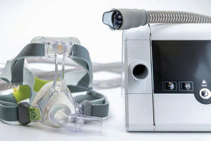
This white paper presents the clinical rationale and three documented case outcomes for interfaced combination therapy using the TAP-PAP (CS) Interface (Airway Management, Inc.) coupled with the TAP 3 Triple Laminate oral appliance. The approach simultaneously delivers mandibular advancement and nasal positive airway pressure through a single, headgear-free interface. Across three patients with baseline apnea-hypopnea indices (AHI) ranging from 72.3 to 84.4 events per hour, interfaced combination therapy achieved a mean AHI reduction of 95%, with all patients reaching therapeutic AHI levels (defined as fewer than 5 events per hour) and demonstrating adherence substantially exceeding published CPAP benchmarks. Average therapeutic pressure requirements were modest (mean 11.4 cmH5O), substantially below pressures typically required for severe OSA with CPAP monotherapy. These findings, grounded in more than 30 years of clinical experience with combination therapy, support recognition of interfaced combination therapy as an evidence-informed, patientcentered treatment option for CPAP-intolerant patients with moderate to severe OSA.
Martin A. Denbar, DDS, DABDSM is a Diplomate of the American Board of Dental Sleep Medicine (obtained 1999) and co-founder of Texas Dental Sleep Services. He earned his Doctor of Dental Surgery from the University of Texas Dental School in Houston and served as a Lieutenant in the United States Navy Dental Corps. Dr Denbar holds an appointment as Adjunct Assistant Professor (non-principled faculty) in the Department of Internal Medicine at Texas A&M School of Medicine, where he teaches dental sleep medicine.
Dr Denbar served on the Board of Directors of the American Academy of Dental Sleep Medicine as Director-at-Large from 1999 to 2002. He is a certified myofunctional therapist and an External Fellow of Buteyko Professionals International.
Dr Denbar has delivered lectures and presentations at universities, dental schools, and professional conferences throughout the United States, including guest appointments at the University of Texas at Austin School of Nursing, Texas State University Graduate Polysomnography Program, and the University of Texas Southwestern Medical School Sleep Fellowship Program.
Introduction
1.1 The CPAP Adherence Problem
Obstructive sleep apnea is one of the most prevalent chronic respiratory conditions in adults, estimated to affect 25% of the adult population nationwide 1 Cardiovascular consequences of untreated OSA are well established, including a 140% increased risk of heart failure, 60% increased risk of stroke, and 30% increased risk of coronary heart disease. 2 The repetitive cycles of hypoxemia and reoxygenation that characterize the condition trigger oxidative stress, sympathetic nervous system activation, systemic inflammation, and endothelial dysfunction, cumulatively accelerating cardiovascular aging and elevating mortality risk.6,7
Continuous positive airway pressure (CPAP) therapy is the most commonly prescribed treatment for moderate to severe OSA and demonstrates superior apnea reduction under controlled conditions. However, sustained clinical benefit depends on patient adherence, and adherence to CPAP therapy remains chronically poor in the real-world setting. Only 30% of patients achieve high long-term adherence after 24 months, with overall adherence ranging from 30 to 60% across published studies.3,4 This gap between device efficacy and populationlevel effectiveness creates a clinical imperative to identify alternative treatment pathways for patients who cannot or will not sustain CPAP use.
1.2 The Limitations of Conventional Combination Approaches
Oral appliance therapy (OAT) emerged as an established modality for mild to moderate OSA in patients who demonstrate CPAP intolerance. The traditional approach to severe, CPAP-resistant OSA has involved adding an oral appliance to a conventional CPAP mask. This approach fundamentally fails to address the core problem: patients referred to dental sleep medicine for CPAP intolerance were originally unable to tolerate the mask interface itself. Combining an oral appliance with the same mask that drove the referral does not resolve the source of intolerance and may compound it.
A more principled solution is the interfaced combination approach, in which an oral appliance is integrated directly with a purpose-built nasal interface that eliminates conventional headgear, chin straps, and full-face mask components entirely. This paper describes the clinical rationale and outcomes for this approach as implemented using the TAP-PAP (CS) Interface and the TAP 3 Triple Laminate oral appliance.
Clinical Background and Rationale
2.1 Comparative Efficacy of CPAP and Oral Appliance Monotherapy
Systematic reviews and meta-analyses consistently demonstrate that CPAP produces greater reductions in AHI compared to oral appliance monotherapy, with a reported mean difference of approximately 6.24 events per hour.8 This efficacy advantage, however, is substantially attenuated when real-world adherence is incorporated into the analysis. The concept of Effective Treatment AHI (ET-AHI) accounts for the proportion of time therapy is actually used and the residual AHI during periods of non-use.9
ET-AHI: A patient with severe OSA (baseline AHI 60) using CPAP at 30% adherence achieves an effective AHI of approximately 42. The same patient using combination therapy at 80% adherence achieves an effective AHI of approximately 15.
Under this framework, a device that achieves excellent AHI reduction under ideal conditions but is tolerated by fewer than a third of patients over the long term may deliver inferior population-level outcomes compared to a device with moderately lower efficacy but substantially higher acceptance. For the subpopulation of CPAP-intolerant patients with severe OSA, interfaced combination therapy may represent the superior clinical option on an effectiveness basis.
2.2 Mechanisms of Interfaced Combination Therapy
Interfaced combination therapy leverages two complementary mechanisms to maintain upper airway patency during sleep:
Mandibular Advancement: The TAP 3 Triple Laminate oral appliance advances the mandible anteriorly, increasing pharyngeal airway cross-sectional dimensions and mechanically stabilizing the tongue base to resist retroglossal collapse.10
Positive Airway Pressure: The TAPPAP (CS) Interface delivers positive airway pressure through nasal pillows integrated directly with the oral appliance, pneumatically splinting the airway against collapsibility during inspiration without the requirement for conventional CPAP headgear, chin straps, or full-face mask components.11
The synergistic relationship between these two mechanisms allows for reduced mandibular advancement relative to OAT monotherapy, minimizing temporomandibular joint stress and dental side effects, while simultaneously permitting lower therapeutic pressure requirements compared to CPAP monotherapy. Research on combination approaches
using mandibular advancement splints with positive airway pressure demonstrates that combined therapy produces greater AHI reductions than either modality in isolation.12
2.3 Patient Selection Criteria
Interfaced combination therapy is indicated for specific patient populations in which conventional CPAP has failed or is clinically contraindicated. Optimal candidates include: patients with moderate to severe OSA and documented CPAP intolerance; patients requiring high therapeutic pressures (greater than 12 cmH₂O) who experience pressure-related discomfort; individuals with significant nocturnal hypoxemia (SpO₂ below 90%); patients with claustrophobia or anxiety related to conventional mask interfaces; those experiencing persistent air leak with full-face or nasal mask configurations; patients requiring positional flexibility during sleep; and individuals with comorbid conditions placing them at elevated cardiovascular risk and for whom untreated or undertreated OSA carries the highest clinical stakes.
The availability of objective compliance data through PAP device downloads provides clinicians and referring physicians with quantifiable treatment adherence and efficacy metrics,
supporting evidence-based clinical communication and documentation for insurance and medical oversight purposes.
Clinical Case Series
The following three case presentations document patients with severe OSA successfully managed using interfaced combination therapy. Each case represents a clinical scenario in which conventional CPAP therapy had failed or was judged unlikely to succeed, and in which untreated disease carried substantial cardiovascular and metabolic risk. Cases are drawn from the clinical practice of the author, with data derived from home sleep testing, PAP device downloads, and oximetry verification studies.
3.1
Case 1: Severe OSA with Metabolic Comorbidities
Patient and Clinical Presentation. The patient was a 53-year-old male with a clinical history significant for Type 2 Diabetes Mellitus. He presented with newly diagnosed obstructive sleep apnea following home sleep testing performed in August 2023. Baseline polysomnographic results are summarized in Table 1.
Diagnosis
Severe OSA with significant nocturnal hypoxemia
Table 1. Baseline home sleep testing results, Case 1 (August 2023).
Treatment Protocol. The treatment plan initiated interfaced combination therapy at the time of oral appliance delivery. Device configuration consisted of the TAP 3 Triple Laminate oral appliance with an initial mandibular advancement of 3 full turns from maximum retrusion, paired with the TAP-PAP (CS) Interface delivering auto-adjusting positive airway pressure (APAP) at a pressure range of 8 to 20 cmH₂O, ramp time 15 minutes beginning at 4 cmH₂O, humidification level 3, and expiratory pressure relief (EPR) at 3 cmH₂O.
Treatment outcomes at serial follow-up timepoints are summarized in Table 2.
4 Months (Mar 7, 2024)
Eight-month oximetry verification (June 2024) demonstrated a CT90 of 91.7% and a 3% Oxygen Desaturation Index (ODI) of 15 events per hour, reflecting substantial oxygenation improvement relative to baseline, with residual mild desaturations suggesting persistent low-level respiratory events warranting monitoring.
Clinical Discussion. This patient achieved a 95% reduction in AHI from baseline severe disease (80.1 events per hour) to therapeutic levels (4.2 events per hour) within four months of initiating interfaced combination therapy. The average therapeutic pressure of 11.4 cmH₂O represents a modest requirement, substantially below pressures typically titrated for severe OSA with CPAP monotherapy. The progressive improvement in nightly adherence from 5 hours 14 minutes at Day 5 to 7 hours 13 minutes at four months suggests growing patient acceptance over the early treatment period. The patient continued therapy with reported comfort and satisfaction, representing a clinically significant outcome given the severity of baseline disease and the high probability of CPAP intolerance in this patient profile.
3.2 Case 2: Super-Obesity with Complex Comorbidities
Patient and Clinical Presentation. The patient was a female with super-obesity (BMI 63.87) and an extensive comorbidity burden including gastroesophageal reflux disease, asthma, hypertension, chronic fatigue syndrome, chronic pain syndrome, major depressive disorder, insomnia, restless legs syndrome, thyroid disorder, and recurrent sinus infections. She presented with newly diagnosed obstructive sleep apnea following home sleep testing in October 2025.
Baseline polysomnographic results are summarized in Table 3.
Table 2. Serial PAP download outcomes, Case 1.
Table 3. Baseline home sleep testing results, Case 2 (October 2025).
Diagnosis
Severe OSA with super-obesity and multiple comorbidities
Treatment Protocol: Staged Approach. The patient elected to trial oral appliance monotherapy before progressing to combination therapy. The TAP 3 Triple Laminate oral appliance was delivered December 2, 2025. Oximetry verification performed January 5, 2026, with the appliance at maximum therapeutic protrusion, revealed a CT90 of only 66.9% and a 3% ODI of 42 events per hour, consistent with an inadequate response to OAT monotherapy.
The treatment plan was revised to convert the patient to interfaced combination therapy. The TAPPAP (CS) Interface was added January 14, 2026, with mandibular advancement at 4 full turns from maximum retrusion and APAP settings of 8 to 20 cmH₂O, ramp time 15 minutes beginning at 4 cmH₂O, humidification level 3, and EPR disabled (0 cmH₂O).
Treatment outcomes following interface addition are summarized in Table 4.
Table 4. Serial PAP download outcomes following interface addition, Case 2.
Clinical Discussion. This case illustrates several important principles. First, the staged approach allowed the patient to develop familiarity with the oral appliance before the interface was added, with oximetry verification providing objective evidence that OAT monotherapy was insufficient, thereby justifying and motivating the transition to combination therapy. Second, the immediate therapeutic response on Day 1 post-interface (95% AHI reduction, from 84.4 to 4.1 events per hour) demonstrates the capacity of interfaced combination therapy to achieve rapid and substantial airway control even in the context of super-obesity and an extreme comorbidity burden. Third, progressive improvement in nightly adherence from 4 hours 26 minutes on Day 1 to 6 hours 38 minutes at 2 weeks reflects growing patient acceptance over the early post-interface period.
The patient reported complete resolution of chronic fatigue symptoms, a clinically significant improvement attributable to restoration of sleep quality and elimination of severe nocturnal hypoxemia. All treatment data were forwarded to the physician of record for ongoing medical management and monitoring.13
3.3 Case 3: Severe OSA in an Elderly Patient with Low BMI
Patient and Clinical Presentation. The patient was a 73-year-old female with Type 1 Diabetes Mellitus (managed via continuous subcutaneous insulin infusion) and osteoporosis. She presented with newly diagnosed obstructive sleep apnea following home sleep testing in September 2025.
Baseline polysomnographic results are summarized in Table 5.
Table 5. Baseline home sleep testing results, Case 3 (September 2025).
Diagnosis
Severe OSA in elderly patient with normal body mass index
Treatment Protocol. Interfaced combination therapy was initiated at the time of oral appliance delivery (February 25, 2026). Given the patient’s age and osteoporosis, the treatment approach prioritized comfort and minimization of temporomandibular stress, with mandibular advancement set at 0 turns (minimal advancement from maximum retrusion), relying primarily on positive airway pressure for airway patency. APAP settings were 6 to 20 cmH₂O, ramp time 15 minutes beginning at 4 cmH₂O, humidification level 3, and EPR at 3 cmH₂O.
PAP download results from the first night of therapy are summarized in Table 6.
Table 6. PAP download outcomes, Day 1 post-delivery, Case 3.
Clinical Discussion. This case presents several clinically significant observations. First, a 93% AHI reduction from 72.3 to 4.7 events per hour was achieved within 24 hours of initiating therapy, accomplished with minimal mandibular advancement. This outcome demonstrates that positive airway pressure alone, delivered through the nasal pillow interface, may be sufficient to achieve therapeutic AHI levels in normal-weight patients without the obesity-related upper airway loading that typically necessitates both higher pressures and greater mandibular advancement. Second, first-night adherence of 9 hours 39 minutes represents exceptional acceptance, particularly notable in an elderly patient initiating sleep apnea therapy for the first time. Third, the average therapeutic pressure of 9.7 cmH₂O was modest despite severe baseline disease, supporting the hypothesis that body habitus is a significant determinant of pressure requirements independent of AHI severity. This case also challenges the common clinical heuristic that severe OSA primarily affects obese, middle-aged males. Anatomical factors, including craniofacial structure and airway collapsibility, drive severe OSA across the BMI and age spectrum.
Synthesis of Case Outcomes
4.1 Aggregate Treatment Results
Across all three cases, interfaced combination therapy achieved therapeutic AHI levels (defined as fewer than 5 events per hour) in patients presenting with a mean baseline AHI of 78.9 events per hour, representing a mean AHI reduction of 95%. Aggregate outcomes are summarized in Table 7.
Table 7. Summary of aggregate treatment outcomes across all three cases.
The average therapeutic pressure of 11.4 cmH₂O across cases is substantially below the 15 to 20 cmH₂O typically required for severe OSA treated with CPAP monotherapy.14 Lower pressure requirements directly reduce the most common mechanisms of CPAP intolerance: aerophagia, pressurerelated discomfort, and oral and nasal air leak. The nightly adherence profiles, ranging from 6 hours 38 minutes to 9 hours 39 minutes, substantially exceed the published long-term CPAP adherence benchmark of 4 to 5 hours per night that is typically applied as a reimbursement threshold.
4.2 Cardiovascular Risk Reduction
The documented reduction in AHI from severe levels (mean 78.9 events per hour) to therapeutic levels (mean 3.7 events per hour) carries significant cardiovascular implications. Each untreated apneic event triggers a sympathetic surge, transient blood pressure elevation, and inflammatory activation.2 By substantially eliminating these repetitive physiologic insults, interfaced combination therapy interrupts the pathophysiologic cascade linking OSA to hypertension, cardiac arrhythmias, heart failure, myocardial infarction, and stroke. Recent research has demonstrated that untreated severe OSA creates a cumulative burden on the cardiovascular system that accelerates biological aging and significantly increases mortality risk.6,7 Successful treatment at the level demonstrated in these cases represents clinically meaningful cardiovascular risk reduction.
4.3 The Alternative Treatment Landscape
Published literature emphasizes the importance of individualized treatment approaches for CPAP-intolerant patients with OSA.15 Alternative modalities
include positional therapy, expiratory positive airway pressure devices, mandibular advancement devices in monotherapy, upper airway surgery, and hypoglossal nerve stimulation. Each of these approaches carries limitations for severe OSA: OAT monotherapy demonstrates inferior AHI reduction compared to CPAP (mean difference 6.24 events per hour) and is frequently insufficient at baseline AHI levels exceeding 40 to 50 events per hour; positional therapy is effective only for positional OSA; surgical approaches carry procedural risk and variable efficacy; and neurostimulation, while effective, is expensive and involves an implanted device.8
Interfaced combination therapy occupies a distinctive position in the treatment algorithm: it is non-invasive, immediately reversible, non-implanted, objectively monitored through PAP device compliance data, and capable of managing severe disease while maintaining patient acceptance rates that conventional CPAP cannot reliably achieve in this population. For appropriately selected CPAP-intolerant patients with moderate to severe OSA, it may represent the most clinically complete available option.
Reconsidering the Gold Standard
The clinical outcomes documented across these three cases raise a broader conceptual question: does the designation of CPAP as the universal gold standard for severe OSA remain clinically appropriate?
The gold standard designation reflects CPAP’s superior AHI reduction under controlled conditions. However, treatment efficacy in the real
world depends not only on device performance under ideal conditions but on the probability that the patient will use the treatment consistently over years or decades. A more nuanced clinical framework might recognize CPAP as the preferred first-line therapy based on robust efficacy data while explicitly acknowledging that individualized treatment selection, incorporating patient preferences, comorbidities, anatomical characteristics, lifestyle factors, and adherence probability, is the clinically meaningful determinant of long-term outcomes.15,16
The author has previously argued in published work that the continued insistence on CPAP as the singular gold standard for severe OSA may, paradoxically, worsen populationlevel outcomes by directing CPAPintolerant patients toward therapy they will not sustain, rather than toward high-adherence alternatives that achieve therapeutic AHI levels in clinical practice.17 Without access to interfaced combination therapy, the patients described in this paper would likely have remained untreated or inadequately managed, facing substantial ongoing cardiovascular and metabolic risk.
Limitations and Evidence Gaps
Several limitations of the present report warrant acknowledgment. First, the evidence base consists of three clinical cases from a single clinical practice. While the cases are clinically illustrative and representative of the patient populations most likely to benefit from interfaced combination therapy, they do not constitute a controlled trial, and no comparison group was included. The outcomes reported cannot be attributed exclusively to the
combination therapy approach in the absence of randomization and control.
Second, follow-up duration varies across cases, ranging from a single overnight PAP download to eight-month oximetry verification. Long-term adherence and efficacy beyond the first year of therapy are not documented in this series. Third, the case series does not include formal quality-of-life assessments, polysomnographic confirmation of outcomes beyond home-based oximetry, or objective measurement of cardiovascular endpoints. Fourth, all cases involve the TAP 3 Triple Laminate oral appliance and TAP-PAP (CS) Interface; generalizability to other device combinations has not been assessed.
Future research should examine long-term outcomes with interfaced combination therapy through prospective, controlled study designs, including cardiovascular event rates, validated quality-of-life instruments, long-term adherence patterns, and head-to-head comparison with CPAP monotherapy in CPAP-naive patients with severe OSA. Cost-effectiveness analysis and assessment of outcomes across diverse patient populations and device configurations would also advance the evidence base.
Conclusions
Interfaced combination therapy utilizing the TAP-PAP (CS) Interface with the TAP 3 Triple Laminate oral appliance provides an effective treatment solution for patients with moderate to severe obstructive sleep apnea who have failed or are unlikely to achieve sustained benefit from conventional CPAP therapy. The three cases presented demonstrate consistent achievement of therapeutic AHI levels, with a mean AHI reduction of 95% and nightly adherence profiles
substantially exceeding published CPAP benchmarks. Therapeutic pressure requirements were modest across all cases, averaging 11.4 cmH₂O, supporting the tolerability profile of this approach relative to CPAP monotherapy.
The clinical experience underlying this paper spans more than 30 years and encompasses hundreds of successfully managed patients across a broad range of baseline disease severity and comorbidity profiles. Medical and dental sleep professionals are encouraged to consider interfaced combination therapy as a viable, evidence-informed treatment option for appropriate patients, recognizing that the therapeutic goal extends beyond device assignment to the achievement of durable, objectively confirmed airway control in the context of each patient’s individual clinical circumstances.
Oral appliance therapy and combination therapy should be prescribed by the patient’s physician of record while being instituted and monitored by a qualified dental sleep medicine provider.
References
1. Gabryelska A, Sochal M, et al. Cardiovascular Complications of Obstructive Sleep Apnea in Adults. PMC. 2022 Oct 2. Worldwide prevalence estimated at 25% of the adult population, with documented independent increases in cardiovascular morbidity and mortality.
2. Drager LF, et al. Obstructive Sleep Apnea and Cardiovascular Disease: Role of the Metabolic Syndrome. PMC. 2008 Jun 14. OSA increases heart failure risk by 140%, stroke risk by 60%, and coronary heart disease risk by 30%.
3. Torsvik M, et al. Patterns and stability of long-term adherence in continuous positive airway pressure therapy. PMC. 2025 Jul 14. Study of 1,907 OSA patients found only 30% achieved high adherence after 24 months; 39% remained nonadherent.
4. de Castro CM, et al. Factors Affecting CPAP Adherence in an OSA Population during the COVID-19 Pandemic. PMC. 2024 Sep 4. CPAP adherence typically ranges from 30% to 60%, with potential overestimation due to data-masking behaviors.
5. Gabryelska A, Sochal M, Wasik B, et al. Factors Affecting Long-Term Compliance of CPAP Treatment: A Single Centre Experience. J Clin Med. 2021 Dec 27;11(1):139.
6. Badran M, Gozal D, et al. New study links
untreated sleep apnea to premature cardiovascular aging and increased mortality. npj Aging. 2025 Dec 10. Prolonged intermittent hypoxia associated with increased cardiovascular biological aging markers.
7. Badran M, et al. Cardiovascular Consequences of Obstructive Sleep Apnea. PubMed. 2019 Nov 8. Studies link untreated moderate to severe OSA to hypertension, cardiac arrhythmias, coronary artery disease, and elevated allcause mortality.
8. Labarca G, et al. Oral Appliances in Obstructive Sleep Apnea. PMC. 2019 Nov 7. AASM/AADSM task force meta-analysis concluded CPAP is more effective than oral appliances with a mean AHI difference of 6.24 events per hour.
9. Camacho M, et al. Effectiveness of Treatment Apnea-Hypopnea Index (ETAHI). PMC. 2012 Jul 5. ET-AHI accounts for effectiveness during use, effectiveness when not in use, and the proportion of time therapy is actually used.
10. Sutherland K, et al. Oral Appliance Therapy for Obstructive Sleep Apnea. Sleep Med Res. 2023 Dec 25. Oral appliances reduce apnea and hypopnea by expanding the pharyngeal airway through mandibular and tongue advancement.
11. TAP Into Sleep. Combination Therapy: Using Both Oral Appliance Therapy and PAP to Treat Obstructive Sleep Apnea. 2024 May 21. Overview of the TAP-PAP interface mechanism and clinical application.
12. Edwards BA, et al. Combination therapy with mandibular advancement and expiratory positive airway pressure valves reduces obstructive sleep apnea severity. PubMed. 2019 Jul 31. Combination therapy produced greater AHI reductions than either modality alone in a controlled study.
13. Essick G, DDS, PhD, DABDSM. Combination Therapies with an Oral Appliance in the Management of Obstructive Sleep Apnea. Dental Clinics, Volume 68, Issue 3. 2024.
14. American Academy of Sleep Medicine. Clinical guidelines for CPAP pressure titration typically recommend starting pressures of 4 to 6 cmH₂O with titration to 15 to 20 cmH₂O for severe OSA based on apnea resolution.
15. Vonk PE, et al. Personalized Treatment for Obstructive Sleep Apnea: Beyond CPAP. PubMed. 2024 Aug 12. Emphasizes systematic documentation of treatment paths in CPAP-intolerant patients to define optimal individualized care.
16. Prehn RS, Swick T. Descriptive Report of Combination Therapy (Custom Face Mask for CPAP Integrated with a Mandibular Advancement Splint) for Long-Term Treatment of OSA. J Dent Sleep Med. 2017;4(2).
17. Denbar M. Continuous Positive Airway Pressure: Is It Still the Gold Standard? Texas Dental Journal, Vol. 141, No. 3.
18. Thornton K. Combined CPAP-oral appliance therapy: a case report. Sleep Review January Issue. 2002;1–8.
19. Denbar M, Essick GK, Schramm P. Hybrid Therapy: A case study using hybrid therapy to treat a soon-to-be-deployed soldier with obstructive and central sleep apnea. Sleep Review Magazine. June 2012.
20. Skalna M, Novak V, Buzga M, et al. Oral Appliance Effectiveness and Patient Satisfaction with Obstructive Sleep Apnea Treatment in Adults. Med Sci Monit. 2019 Jan 18;25:516–524.
ORAL and maxillofacial pathology
case of the month
Case Presentation
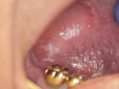
An 84-year-old male presented to an oral and maxillofacial surgery clinic with an incidental finding of a white corrugated plaque measuring approximately 7 × 3 mm on the right lateral border of the tongue (Figure 1). The patient denied associated pain, bleeding, or paresthesia. His medical history was significant for prostate cancer treated surgically, hypertension, pacemaker placement, recurrent bronchitis, basal cell carcinoma, and shingles. A provisional diagnosis of traumatic hyperkeratosis or leukoplakia was initially considered.
The patient was on multiple medications, including allopurinol 100 mg, amlodipine 5 mg, losartan potassium 50 mg, omeprazole 20 mg, pravastatin sodium 40 mg, valacyclovir 1 g, albuterol inhaler 8.5 g, calcipotriene cream 0.005%, and clobetasol propionate cream 0.05%. An excisional biopsy was performed, and the specimen was submitted to the Oral Pathology Biopsy Service at Texas A&M University College of Dentistry for histopathologic evaluation.
What is your clinical diagnosis?
AUTHORS
Bushra Javed, BDS
MS student, Department of Diagnostic Sciences, Texas A&M University College of Dentistry, Dallas, Texas
Ashim Raj Shrestha, BDS, MPH, MS
Resident, Oral & Maxillofacial Pathology, Department of Diagnostic Sciences, Texas A&M University College of Dentistry, Dallas, Texas
Deepthi Bellippady, BDS, MDS
MS student, Department of Diagnostic Sciences, Texas
A&M University College of Dentistry, Dallas, Texas
Madhu Shrestha, BDS, MDS, MS, PhD Faculty, Department of Diagnostic Sciences, Texas A&M University College of Dentistry, Dallas, Texas
Figure 1. White corrugated plaque-like lesion (arrow), measuring approximately 7 × 3 mm, located on the right ventrolateral tongue.
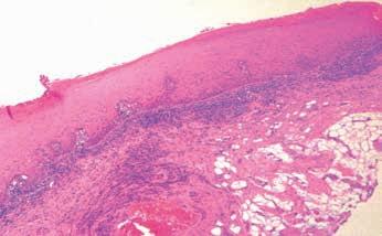
Figure 2A. Low-power view (4×), H&E stain, showing hyperparakeratosis and hyperplastic stratified squamous epithelium with atypical features and a moderate inflammatory infiltrate in the lamina propria.
Histopathological Features
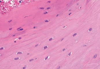
Low-power magnification (4×) of the hematoxylin and eosin–stained sections revealed a hyperparakeratotic and hyperplastic stratified squamous epithelium with mild architectural and cytologic atypia, largely confined to the basal and parabasal layers. A moderate inflammatory infiltrate was also noted within the underlying lamina propria (Figure 2A). High-power magnification (40×) demonstrated numerous candidal hyphae within the superficial keratin layer (Figure 2B). In addition, higher magnification (40×) revealed an interesting finding in multiple areas of the superficial epithelium, where keratinocytes showed a homogeneous nuclear pattern with perinuclear clearing, chromatin condensation, and nuclear beading. These features are strongly suggestive of an Epstein–Barr virus–associated viral cytopathic effect within the epithelium (Figure 2C).
See page 112 for the answer and discussion
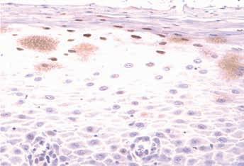
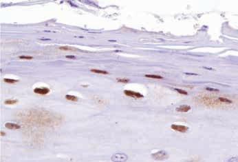
Figure 3A. Medium-power magnification (20×) showing EBER (EBVencoded RNA) in situ hybridization positivity, confirming EBV infection in multiple superficial keratinocytes.
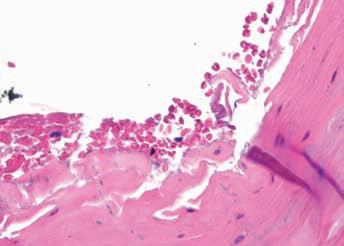
Figure 2B. High-power view (40×), H&E stain, demonstrating Candidalike fungal hyphae in the superficial epithelium, with organisms seen penetrating at right angles to the superficial keratin layer.
Figure 2C. High-power view (40×), H&E stain, showing keratinocytes with a homogeneous nuclear pattern, perinuclear clearing, chromatin condensation, and nuclear beading, features suggestive of EBVassociated cytopathic changes.
Figure 3B. High-power magnification (40×) of the same section demonstrating confirmed EBER (EBV-encoded RNA) positivity in multiple superficial keratinocytes.
Oral hairy leukoplakia (OHL) is a benign condition characterized by a painless, white corrugated plaque, typically involving the lateral border of the tongue and most commonly triggered by reactivation of Epstein–Barr virus (EBV). It occurs predominantly in individuals with significant immunosuppression, such as patients with HIV/AIDS or organ transplant recipients. EBV, also known as human herpesvirus 4 (HHV-4), is a doublestranded DNA virus of the Herpesviridae family that infects more than 90% of the adult population worldwide.
OHL was initially considered pathognomonic for HIV infection; however, it has also been reported in HIV-negative patients with other immunocompromised states, and rarely even in immunocompetent elderly individuals (Alramadhan et al., 2021). Clinically, it presents as a white, non-scrapable plaque with a rough or corrugated surface, most commonly on the lateral tongue, though it may occasionally involve the buccal or labial mucosa. Early lesions may appear relatively smooth before developing characteristic vertical corrugations. The true incidence is uncertain, as OHL is often misdiagnosed as oral candidiasis (Resnick et al., 1990).
A key consideration is EBV-associated OHL in the setting of iatrogenic immunosuppression. Long-term use of systemic, topical, or inhaled
corticosteroids may induce local immunosuppression. In one study of 35 non-HIV-associated OHL cases, 28 were linked to prolonged inhaled steroid use and 4 to combined systemic and inhaled therapy. In the present case, the patient’s history of inhaled and long-term topical steroid use may have contributed to local immunosuppression. Age-related immune decline may have also played an additional role (Chambers, 2015). Histologically, OHL shows a parakeratotic surface with corrugation and epithelial hyperplasia. Characteristic “balloon cells” are present in the upper spinous layer, showing nuclear chromatin changes with peripheral condensation along the nuclear membrane. A central amphophilic homogeneous area is often seen, representing viral aggregates consistent with EBV infection (Sciubba et al., 1995).
Differential Diagnosis
1. Leukoplakia:
Leukoplakia is a clinical term describing a white plaque that cannot be characterized as any other definable lesion. It presents as a patch or plaque that does not wipe off. Histologically, it shows hyperkeratosis with or without acanthosis, and approximately 5–25% demonstrate varying degrees of epithelial dysplasia. The presence of a white keratotic lesion with no identifiable cause raises concern for dysplastic change. While earlier reports (Schwimmer) suggested up to 80% malignant potential, later studies clarified that not
all leukoplakias are premalignant. In mild dysplasia, changes are limited to the basal and parabasal layers. The terms “mild,” “moderate,” and “severe” reflect the extent of dysplasia (Shirani et al., 2014).
2. Candidiasis:
Pseudomembranous candidiasis is the most common form, presenting as white plaques that can be wiped off, leaving an erythematous surface. The pseudomembranes consists of desquamated epithelial cells, fibrin, and fungal hyphae. It may involve multiple oral sites and is often asymptomatic. In contrast, hyperplastic candidiasis appears as a persistent, non-wipeable white plaque, most commonly on the buccal mucosa. Diagnosis is based on clinical features, risk factors, histopathology, and response to antifungal therapy, with cytologic or histologic confirmation when needed (Taylor et al., 2025).
3. Oral Lichen Planus (OLP):
OLP is a chronic immune-mediated inflammatory condition of the oral mucosa with variable clinical presentation, including reticular, erythematous, or erosive forms. Patients may experience mucosal roughness, sensitivity to spicy foods, white or red patches, or ulceration, and occasionally esophageal involvement. Diagnosis is based on clinical and histopathologic correlation, although classic bilateral reticular lesions may be diagnosed clinically. Histopathology shows liquefaction degeneration of basal cells, a dense band-like lymphocytic infiltrate in the superficial lamina propria, normal epithelial maturation, saw-tooth rete ridges, and hyperkeratosis. Erosive forms may show surface ulceration (Alrashdan et al., 2016). OLP is uncommon on the lateral tongue.
4. Traumatic keratosis
Traumatic keratosis is a localized white plaque caused by chronic mechanical irritation (e.g., sharp tooth, dental appliance) and should be considered alongside leukoplakia, oral lichen planus,
and early squamous cell carcinoma in the differential diagnosis of white oral lesions. In this case as the lesion was in close approximation to a metal crown, it could be included in the differential diagnosis. Unlike a true leukoplakia, it has an identifiable irritant and may regress after removal of the trauma. In contrast to oral lichen planus, it is usually unilateral and lacks a bilateral reticular pattern. It typically resolves once the source of irritation is eliminated, and it is a bit uncommon to get traumatic keratosis in the posterior lateroventral tongue.
Management and Follow-up
Unlike conventional leukoplakia with high-grade dysplasia, EBV-associated oral hairy leukoplakia (OHL) does not require aggressive surgical excision. Management is primarily directed toward identifying and addressing the underlying cause of immunosuppression. In HIV-positive patients, initiation or optimization of antiretroviral therapy (ART) often results in restoration of immune function and spontaneous resolution of the lesion. Antiviral agents such as acyclovir, valacyclovir, and famciclovir have also been reported to suppress EBV-related lesions. In addition, topical application of 25% podophyllum resin has been described as a therapeutic option (Brasileiro et al., 2014).
Given the atypical epithelial features, the high-risk location on the lateral tongue, and the potential overlap with early epithelial dysplasia, long-term clinical follow-up was recommended to monitor for recurrence or progression. The patient was also referred for medical evaluation to investigate possible systemic or iatrogenic causes of immunosuppression.
Challenge Questions for the Reader
1. What clinicopathologic features help distinguish EBV-associated oral hairy leukoplakia from conventional leukoplakia with epithelial dysplasia?
2. How do the histopathologic findings in this case differ from those typically observed in classic oral hairy leukoplakia?
3. Why is a history of long-term steroid use clinically significant in patients presenting with atypical oral leukoplakic lesions?
Answer & Discussion
Clinically, oral hairy leukoplakia (OHL) may be indistinguishable from conventional leukoplakia, particularly when presenting as a non-scrapable white plaque on a high-risk site such as the lateral tongue. Therefore, diagnosis depends largely on careful histopathologic evaluation and recognition of subtle epithelial changes, which can be easily overlooked in uncommon or atypical cases.
In this case, the diagnosis was EBVassociated OHL with limited cytologic atypia. The atypia was interpreted as virally induced reactive change rather than true preneoplastic dysplasia. Because the lesion clinically resembled conventional leukoplakia, it could easily have been misdiagnosed as mild epithelial dysplasia or hyperparakeratosis with focal atypia.
Histologically, the findings were subtle. The presence of keratinocytes with chromatin condensation, beading, perinuclear clearing in the superficial epithelium, along with strong EBV positivity by EBER in situ hybridization, established the diagnosis. Unlike classic OHL, which often shows marked hyperparakeratosis and corrugated architecture, this case demonstrated only minimal and atypical histologic alterations, highlighting the broader morphologic spectrum of EBVassociated lesions.
This case underscores an important diagnostic challenge: EBV-driven epithelial lesions can closely mimic conventional leukoplakia while showing only limited atypia. Failure to recognize these lesions may underestimate the true spectrum and biologic significance of EBV-associated
leukoplakias, whereas diagnosing them solely as “mild dysplasia” without EBV testing may lead to overdiagnosis. Careful clinicopathologic correlation and ancillary testing are therefore essential, especially for lesions in high-risk locations.
An additional important consideration is the history of long-term steroid use. Chronic corticosteroid therapy, whether systemic, inhalational, or topical, may contribute to local or systemic immunosuppression and should raise suspicion for EBV-associated OHL, even in HIV-negative patients, especially in elderly patients. Thus, recognizing this is important to consider potential immunosuppressed lesions of the oral cavity such as EBV-OHL.
Clinical Significance (Take home message):
Oral hairy leukoplakia may occur in the setting of iatrogenic immunosuppression, including non-HIV patients.
• Steroid inhalers (such as albuterol) and topical corticosteroids (such as clobetasol) have been implicated as potential causes of local immunosuppression associated with EBV-related OHL.
• OHL may exhibit subtle, focal, or atypical histopathologic features that deviate from classic descriptions.
• EBV in situ hybridization (EBER) is invaluable in confirming the diagnosis.
• Recognition and documentation of EBVassociated atypical epithelial changes are important for understanding their biologic behavior and possible malignant potential.
References
1. Alramadhan SA, Bhattacharyya I, Cohen DM, Islam MN. Oral Hairy Leukoplakia in Immunocompetent Patients Revisited with Literature Review. Head Neck Pathol. 2021 Sep;15(3):989–993. doi:10.1007/s12105021-01287-8. PMID: 33428065; PMCID: PMC8384929.
4. Shirani S, Kargahi N, Razavi SM, Homayoni S. Epithelial dysplasia in oral cavity. Iran J Med Sci. 2014 Sep;39(5):406–417. PMID: 25242838; PMCID: PMC4164887.
5. Taylor M, Brizuela M, Raja A. Oral Candidiasis. Updated July 4, 2023. In: StatPearls [Internet]. Treasure Island (FL): StatPearls Publishing; 2026. Available from: https://www.ncbi.nlm.nih.gov/books/NBK545282/ 6. Alrashdan MS, Cirillo N, McCullough M. Oral lichen planus: a literature review and update. Arch Dermatol Res. 2016;308:539–551. https://doi. org/10.1007/s00403-016-1667-2
7. Brasileiro CB, Abreu MH, Mesquita RA. Critical review of topical management of oral hairy leukoplakia. World J Clin Cases. 2014 Jul 16;2(7):253–256. doi:10.12998/wjcc.v2.i7.253. PMID: 25032199; PMCID: PMC4097151.
8. Chambers A, Conn B, Pemberton M, et al. Twenty-first-century oral hairy leukoplakia—a non–HIV-associated entity. Oral Surg Oral Med Oral Pathol Oral Radiol. 2015;119:326–332.
Those in the dental community who have recently passed
Jack Edwin Beeler
Victoria 2/10/48–3/29/26
Good Fellow: 1999
Life: 2013
Fifty Year: 2023
Richard C Blackwell II
Leander
2/14/54–4/20/26
Good Fellow: 2018
Life: 2021
Thomas B Carter
Victoria 1/23/55–4/13/26
Good Fellow: 2019
Life: 2022
Benge Robert Daniel Jr
Fort Worth
1/1/45–4/7/26
Life: 2010
Fifty Year: 2024
Frank Feuille V
El Paso
10/5/69–4/7/26
Good Fellow: 2023
Ronald E Groba
Friendswood
3/27/46–3/29/26
Good Fellow: 2001
Life: 2011
Fifty Year: 2025
John E Kidwell
Dallas
8/31/26–4/2/26
Good Fellow: 1979
Life: 1991
Fifty Year: 2004
William C Madsen
McAllen
2/11/43–4/2/26
Good Fellow: 1998
Life: 2008
Fifty Year: 2022
Don K Maggard
Plano
4/19/50–4/13/26
Good Fellow: 2003
Life: 2015
Joe Kenneth Smith
Lockhart
10/11/37–4/30/26
Good Fellow: 1989
Life: 2002
Fifty Year: 2014
Richard Thorp
Weber Jr
Austin
7/21/44–3/25/26
Good Fellow: 1996
Life: 2009
Fifty Year: 2018
The PACE Center proudly presents our Dental Hygiene Local Infiltration Anesthesia Certification Course, meticulously crafted for dental hygienist to meet TSBDE’s Rule 115 10 in the administration of local infiltration anesthesia. This course includes 20 hours of didactic education and 12 hours of clinical hands-on training
To Extract or Not to Extract: Similar Malocclusions, Different Treatment Paths in Twin Brothers: Part 1. The Extraction Approach
Part 2. The Non-extraction Approach & Part 3. Discussion and Comparison will appear in future TDJ editions
Katie Julien, DDS, MS, Jorge Aguirre, DDS, MSc, Ryan McGovern, DDS, MSD Department of Orthodontics
UTHealth Houston, School of Dentistry
ABSTRACT
Objective
To present the diagnosis, treatment rationale, and clinical outcome of an adolescent patient with a skeletal Class II, division 2 malocclusion, convex profile, deep overbite, and significant arch length deficiency treated with an extraction-based orthodontic approach.
Clinical
Considerations
A 15-year-old hypodivergent patient presented with a skeletal Class II, division 2 pattern characterized by SNA 80.9, SNB 73.5, ANB 7.4, Wits +4.8 mm, bilateral Class II molar and canine relationships, convex profile, deep mentolabial sulcus, and a 7-mm overbite. Arch analysis showed constricted but coordinated arches
and clinically relevant arch length deficiency, greater in the mandibular arch. The treatment objectives were to improve incisor angulation/incisor display, resolve crowding, improve the sagittal relationship, and improve facial/ lip balance. The treatment plan followed an extraction-based strategy combined with fixed appliances, staged space management, and sagittal correction mechanics. Treatment progress records showed persistent space-closure and Class II correction demands that required additional biomechanical refinement, including fixed functional and elastic mechanics. Final retention consisted of a mandibular fixed 3-3 retainer and maxillary and mandibular clear retainers.
Conclusions
This case illustrates how an extractionbased orthodontic approach can be used to manage a skeletal Class II division 2 adolescent patient with crowding, deep bite, and soft-tissue convexity when treatment goals include space resolution, dental compensation to a skeletal issue, occlusal improvement, and controlled esthetic change.
Clinical Significance
Extraction decisions in Class II patients should not be based on crowding alone. Facial profile, growth, incisor position, overbite, arch coordination, and individualized treatment objectives must be integrated to produce a predictable and esthetically balanced result.
Guest Editor Rade D. Paravina, DDS, MS, PhD
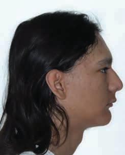
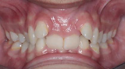
Figure 1. Pretreatment profile and intraoral frontal views of a 15-year-old patient with a skeletal Class II division 2 pattern, convex profile, deep overbite, and clinically significant anterior malalignment.
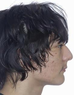
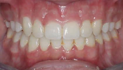
Figure 2. Posttreatment profile and intraoral frontal views demonstrate improved alignment, overbite control, occlusal relationships, and facial balance after comprehensive extraction-based orthodontic treatment.
FBI: Found But not Identified (yet)
A series of articles intended to assist Texas Medical Examiners in giving a name to “unidentified” individuals using postmortem dental evidence. Do you recognize the dental work/conditions presented?
Kathleen A Kasper, DDS, D-ABFO
The Extent of the Problem
Over 600,000 individuals go missing in the United States every year. Fortunately, many missing children and adults are quickly found, alive and well. However, tens of thousands of individuals remain missing for more than 1 year—what many agencies consider “cold cases”.1
It is estimated that 4,400 unidentified bodies are recovered each year, with approximately 1,000 of those bodies remaining unidentified after 1 year.2
Medical examiner and coroner offices reported 11,380 unidentified remains on record as of 2018.3
“Estimates suggest there are as many as 40,000 unidentified human remains nationwide at any given point in time. The cumulative effect of so many missing and unidentified persons cases has created a significant societal burden as families and communities are left to deal with the ambiguous loss of their missing neighbors, friends, and loved ones.”4
The challenge in giving a name to these unidentified decedents is that the research and time invested can be extensive, expensive, and after normal protocols have been exhausted, many individuals still remain unidentified. The hope in writing this series of articles on long term unidentified in Texas is that one of the many Texas dentists who read the Texas Dental Journal will recognize the dental work presented as theirs or possibly remember a dental condition or feature that can give a name to the unidentified described and help bring closure to the family.
The Medical Examiner’s Protocol to Legally Identify Individuals
Medical examiners follow a specific protocol to identify individuals who are found without any presumptive identity. This protocol is described below and, in this order, depending on the condition of the body.
Latent Prints (finger, palm, sole) are the fastest and least expensive way to make a positive identification. This method of identification is not always possible if remains are severely decomposed, incinerated, or skeletonized.
Medically Implanted Devices (hip/joint replacements, pacemakers) require knowing what hospital/physician placed the device to get the matching serial number, which can be difficult.
Dental Records are also an easy, fast, and inexpensive way to identify a decedent; however, success depends on a presumptive identity and availability/existence/ quality of those antemortem (before death) dental records. If all leads for a presumptive identity are exhausted, then a postmortem (after death) dental charting/ profile is created and entered in the NamUs (National Missing and Unidentified Persons System) and NCIC (National Crime Information Center) databases.
Anthropologic Methods will use skeletal features to make a positive identification.
DNA samples are collected. This takes the longest period of time and is most expensive. Depending upon the individual, it may involve obtaining DNA samples not only from the decedent but also from genealogically appropriate family members or select personal hygiene products of the decedents, if available. If no matches are obtained, then:
DNA Samples are submitted to Combine DNA Index System (CODIS), which is a computer program that operates local, state, and national databases of DNA profiles from convicted offenders, unsolved crime scene evidence, unidentified remains, and missing persons. This is maintained by the Federal Bureau of Investigation. If this search fails then the State of Texas requires medical examiners to submit a DNA sample from long-term unidentified decedents to the University of North Texas Center for Human Identification for additional genetic testing and comparison. This comparison may take up to a year to process, and a decedent may still remain unidentified once completed. This is the proverbial end of the road; therefore, all Texas dentists, dental auxiliaries, and staff; Can you help the Dallas County Medical Examiner’s Office give a name to the unidentified decedent described on the next page?
Author
Kathleen A. Kasper, DDS, D-ABFO
Dr Kasper has been practicing general dentistry in Carrollton, Texas, since 1994. She is a graduate of the University of Iowa College of Dentistry.
She provides 3 counties in the Dallas-Ft. Worth Area with forensic dental services. These include Collin, Dallas, and Tarrant Counties. She is currently the only active board certified forensic dentist in north Texas.
She received her formal forensic odontology training at the University of Texas Health Science Center San Antonio, Texas, Center for Education and Research in Forensics (CERF) in 2003-2004.
Dr Kasper obtained Board Certification from the American Board of Forensic Odontology in 2010.
She is a fellow of the American Academy of Forensic Sciences, a member of the American Board of Forensic Odontology, and a member of the American Society of Forensic Odontology.
She is the current American Board of Forensic Odontology Dental Age Assessment Committee Chair.
Dr Kasper is also a published author in the Journal of Forensic Sciences and coauthor of a chapter in each of 3 forensic textbooks.
Perhaps one of her greatest undertakings was chairing a working group for the American Dental Association that has published the “Technical Report” for Forensic Dental Age Estimation which went on to become a “standard” for the Organization of Scientific Area Committees (OSAC) for Forensic Science.
On 11/23/2025, at approximately 4:43 PM, a passerby reportedly located a human cranium and multiple post-cranial skeletal elements believed to be human while walking in a wooded area near 376 County Road 4813 in Copperas Cove, Texas. The bystander reported the location of the remains to local law enforcement on 11/26/2026. The Department of Public Safety | Texas Rangers responded and did not locate any clothing or identifying documents within proximity of the remains. The local Justice of the Peace pronounced the death on 11/26/2025 at 3:55 PM.
The decedent’s remains were sent to the Southwestern Institute of Forensic Sciences (SWIFS) for further examination. An anthropological consult was completed, and the decedent was found to be consistent with an adult male of European descent. A forensic odontology consult was completed, and the decedent was subsequently sent to The University of North Texas Center for Human Identification for efforts to obtain a CODIS eligible DNA profile. This case has been entered into NamUs as #UP155800.
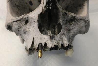
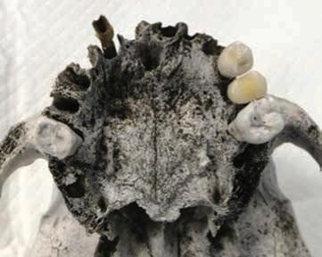
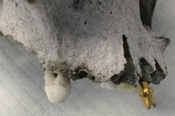
Dental Evidence Recovered: Maxilla Only
Dental Postmortem Photographs DCME Case # 25-19096:
Right Left
Maxilla Occlusal View
Right Lateral View
Left Lateral View
Frontal View
Right Left
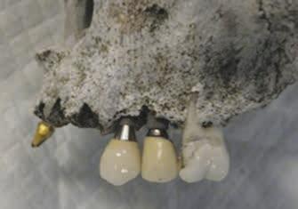
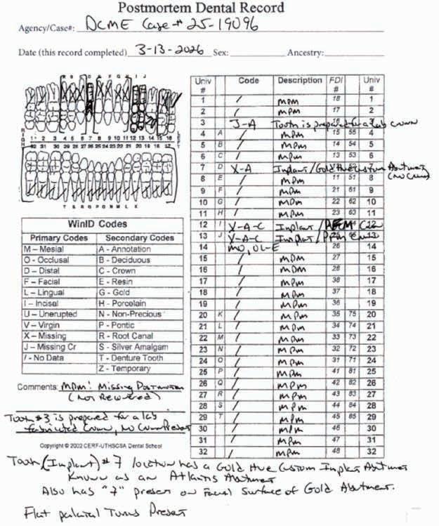
#3
#7
#12 #13 #14
Implant
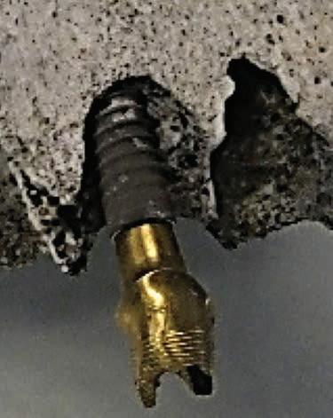
#7 Has A
“7”
Etched in Facial Surface of Abutment
DENTAL NOTES: IT HAS BEEN DETERMINED THAT ALL IMPLANTS IN THIS CASE ARE “STRAUMANN” AND PLACED BY A SKILLED SURGEON DUE TO THEIR IMMENSE LENGTH. THOSE PRESENT INCLUDE THE #7, #12 AND #13 LOCATIONS.
FURTHURMORE, THE IMPLANT IN THE #7 LOCATION HAS BEEN CONFIRMED BY THE STRUAMANN MANUFACTURER TO BE A “STRAUMANN BL”, WHICH CAME OUT FOR DISTRIBUTION IN THE YEAR 2007.
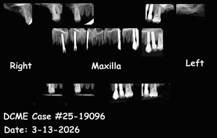
Dental Postmortem Radiographs DCME Case #25-19096:
#6
#7
#8
Dental Existing Conditions DCME Case #25-19096:
Closing
If you believe you have any dental records, dental radiographs, intraoral/extraoral dental photographs, dental scans or other dental information regarding the unidentified decedent described above as DCME Case #25-19096 , please contact the Dallas County Medical Examiner’s Office, (214) 920-5900, ask for Steven Kurtz, Chief Medicolegal Death Investigator or Keara St Louis, Deputy Chief Medicolegal Death Investigator.
Let’s give this individual a name!
References
1. Between 2007 and 2020, an average of 664,776 missing persons records annually were entered into the National Crime Information Center. See https://www.fbi.gov/ services/cjis/ncic
2. Medical Examiners And Coroners’ Offices, 2004. Matthew J. Hickman, Ph.D., Kristen A. Hughes, M.P.A., Bureau of Justice Statistics, Kevin J. Strom, Ph.D., Jeri D. RoperoMiller, Ph.D., DABFT, RTI International
3. Medical Examiner and Coroner Offices, 2018. Connor Brooks, Bureau of Justice Statistics, November 2021
Growth Strategies: Don’t Let Uncollected Production Sit Idle on Your Books
By Lois Banta for CareCredit
Bottomline: It’s not really production or revenue until the money is collected and is working to help you meet your practice and personal goals. Here are 3 tips to help you minimize A/R and maximize cash flow.
Start With a Plan
It’s important—especially when you are growing or have new team members—that you have a detailed and effective collections and financing arrangement guideline for the office. This is an internal document designed to drive consistency in patient communication and payment behavior.
The policy should include:
• When payment is collected, which ideally would be as the appointment is set or at check in.
• All payment options available to patients.
• Your guidelines for patients who pay in full prior to the appointment.
• Your check policies. Consider setting a maximum amount you will accept using this tender type.
• Financing options available through your third-party financing partner.
• Which patients are credit worthy and have an impeccable payment history.
• Your past-due account collection strategy.
If you choose to extend credit, it’s important to review the Fair Debt Collection Practices Act report with the team and keep it for future reference.
Discuss Financial Arrangements
When the Dentistry Is Scheduled
Once the value of care has been communicated and the patient is interested in moving forward with treatment, it’s important they understand their financial investment and payment options before they reserve time on the schedule. No one likes surprises when it comes to their mouth or money. If the patient out-of-pocket cost is more than $300, here’s how the conversation might go:
After your insurance benefits contribute to the cost of care, your portion is $850. We can collect this now or when you arrive for your appointment. We accept cash and credit cards, including the CareCredit credit card with flexible financing for your convenience. Which will work best for you?
If the patient expresses cost concerns, it’s important to be ready with a solution. It is rarely in the practice’s best interests to extend credit to patients. This courtesy should be reserved for long-term patients with a stellar payment history. If patients ask to make payments, don’t dead end the conversation with a “no;” give them options.
Yes. We accept the CareCredit credit card. If approved, you can use CareCredit to pay over time with promotional financing available on purchases of $200 or more. Would you like to know more about this option?
Know Your Last Resort Payment Options
Even though you have the financial conversation when the appointment is scheduled, it’s inevitable that a few patients will be unprepared to pay when they arrive at the practice.
Having financing available if the patient is unable to pay cash or use their general-purpose credit card is one way to help them move forward with care and improve cash flow. But what if the patient is not approved for credit? You can choose to extend credit and bill patients, but this should truly be your last resort because it not only impacts cash flow but may impact the patient relationship.
If you do choose to extend credit, be sure to provide patients with a written agreement that details the payment requirements.
When you start with a plan, have team alignment and have payment options available that enable you to collect at time of treatment. Your revenue will be able to work for you instead of your team having to work to collect accounts.
CareCredit is endorsed by TDA Perks Program. If you don’t yet accept CareCredit and you’re interested in doing so, call 800-300-3046 or visit tdaperks.com (Insurance & HR).
This content is subject to change without notice and offered for informational use only. You are urged to consult with your individual advisors with respect to any information presented. Synchrony and any of its affiliates, including CareCredit, (collectively, “Synchrony”) makes no representations or warranties regarding this content and accepts no liability for any loss or harm arising from the use of the information provided. All statements and opinions are the sole opinions of the author. Your receipt of this material constitutes your acceptance of these terms and conditions.
Once the value of care has been communicated and the patient is interested in moving forward with treatment, it’s important they understand their financial investment and payment options before they reserve time on the schedule.
classifieds
Opportunities Online at TDA.org and Printed in the Texas Dental Journal
CLASSIFIEDS INFORMATION
DEADLINE
Copy text is due the 20th of the month, 2 months prior to publication (ie, January issue has a due date of November 20.)
MONTHLY RATES
PRINT: First 30 words—$60 for ADA/TDA members and $100 for non-members. $0.10 each additional word.
ONLINE: $40 per month (no word limit). Online ads are circulated on the 1st business day of each month, however an ad can be placed within 24 business hours for an additional fee of $60.
SUBMISSION
Ads must be submitted, and are only accepted, via www.tda.org/MemberResources/TDA-Classified-Ads-Terms. By official TDA resolution, ads may not quote specific incomes or revenues and must be stated in generic terms (ie “$315,000” should be “low-to-mid-6 figures”). Journal editors reserve the right to edit and/or deny copy.
PRACTICE OPPORTUNITIES
ALL TEXAS LISTINGS FOR MCLERRAN & ASSOCIATES. AUSTIN (ID #745): Established, two-location GD practice in the Austin area. The practice serves a large, diverse, FFS/ PPO patient base with excellent new patient flow, a combined 14 equipped operatories across both locations (with substantial room for expansion at one of the locations). The practice comes fully equipped with digital radiography, digital sensors, digital scanners, digital pano, CBCT, and paperless charts.
AUSTIN (ID #749): Established GD office located in the heart of central Austin. The practice has an impeccable online reputation in the community and features a strong hygiene recall program with multiple avenues for future growth. 3,000+ sq ft office, 4 active operatories, ample room for expansion, equipped with a digital scanner, intra oral cameras, digital x-ray sensors, and paperless charts. AUSTIN (ID #762): Established GD office in the heart of central Austin with a majority PPO patient base, 1000+ active patients, a robust hygiene recall program, and a strong reputation online and in the community they serve. The 1,500 sq ft office space contains 3 total operatories, computers throughout, a digital x-ray scanner, and intra-oral cameras. The practice has
consistently generated revenue of mid-6 figures with abundant upside opportunities to expand production/cash flow through launching strategic marketing and advertising initiatives, driving new patient flow, and offering additional advanced procedures in-house.
AUSTIN (ID #787): Large, GD practice with a great reputation in north Austin. Large facility in a free-standing building with great visibility featuring 8 fully equipped operatories with computers in each, digital radiography, an iTero digital scanner, and a 3D CBCT unit. FFS/ PPO patient base, 25+ new patients per month, and a stellar online reputation. AUSTIN (ID #790): Established periodontic practice in highly sought after central Austin with abundant upside potential. The well-appointed 1,840 sq ft space has 5 fully equipped operatories with room for future expansion, computers throughout, digital x-ray sensors, and paperless charts. The 100% FFS practice is built on an impeccable foundation with a diverse referral network, solid hygiene recall, and virtually no investment in marketing and advertising.
AUSTIN (ID #795): Established, 100% FFS, high-end, boutique cosmetic dental office with an impeccable reputation in the heart of downtown Austin. The modern 2,200 sq ft condo has incredible cityscape views, 3 fully equipped operatories with room for a 4th,
computers throughout, digital x-ray sensors, iTero digital scanner, dental laser, etc. you are seeking a turnkey office in a prime location with abundant upside potential and strong, consistent cash flow, please reach out for details! AUSTIN (ID #797): Established GD office in south Austin. PPO/FFS practice, 1,800+ active patients, strong hygiene recall program with 30%+ of annual production coming from the hygiene department. Four total operatories (3 equipped) and comes furnished with a digital pano, digital scanner/x-ray sensors,
Premier Sell-Side Advisor for Dental Practice Owners
classifieds
intra-oral cameras, and is fully paperless. This is a profitable, turn-key practice with multiple avenues for future growth.
AUSTIN-NORTH (ID
#798): Established, highly profitable GD office located less than an hour north of Austin. Large PPO/FFS patient base, approximately 40 new patients per month. Modern, well-equipped facility located in a high traffic retail location. 5 operatories, furnished with computers throughout, digital x-ray sensors, panoramic x-ray unit, 3D CBCT, digital scanner, intra oral cameras, and is fully digital. AUSTIN (ID #801): Rare opportunity to acquire a high-upside, modern general dentistry practice in central Austin. Large FFS/PPO active patient base. Spacious facility off a high-traffic roadway. 5 total operatories, all digital equipment including a pano, and paperless charts. The practice has immense upside potential and we expect this listing to move quickly.
AUSTIN-SOUTH (ID
#803): GD practice located south of Austin in a high-traffic retail center. Five fully equipped operatories with an additional ortho bay set up, digital radiography, intra oral cameras, paperless charts, a digital pano, and a digital scanner. Large PPO/FFS/Medicaid patient base with over 1,900 active patients and see 45+ new patients per month. If you are looking for a practice that is primed for continued growth in
a growing suburb of Austin with great visibility, reach out to learn more! DALLAS-WEST (ID #810): Rare opportunity to acquire a wellestablished GD practice in a prime location west of the DFW metroplex. The 3,800 sq ft facility contains 13 fully equipped operatories with digital x-ray sensors, CBCT, digital scanner, dental laser, intraoral cameras, and room for future growth. The practice supports a large PPO/FFS patient base, has strong cash flow and robust hygiene recall. This office provides a strong clinician and operator the ability to take advantage of the extraordinary foundation built and future upside potential. HOUSTONSOUTHWEST (ID #782): Large, modern GD practice located in a high-visibility retail center on the southwest side of Houston. The spacious 3,900 sq ft practice features 6 fully equipped operatories (with several additional ops built out and ready to equip), is fully digital with CBCT, digital scanner, digital radiography, intra oral cameras, and computers throughout. The practice serves a mix of PPO/FFS patients, has realized revenue of approximately 7 figures over the last several years, and has a fantastic reputation within the community and online. HOUSTON-NORTHWEST (ID #802): Premier GD practice in northwest Houston. State-of-the-art office, fully equipped with
6 operatories, digital radiography, intraoral cameras, digital pano, and a digital scanner. Large PPO/FFS patient base, over 4,000 active patients, and a robust hygiene recall program accounting for 20%+ of annual production.
HOUSTON-SOUTH (ID #809): GD practice plus real estate in a highly desirable suburb south of downtown Houston. The office features modern build-out, occupies just over 2,800 sq ft, and includes 5 fully equipped operatories with digital radiography, intraoral cameras, paperless charts, and CBCT. Majority PPO/FFS patient base with over 1,300 active patients, strong hygiene recall program with just over 20% of production coming by way of hygiene procedures.
HOUSTON-SOUTHWEST (ID #811): GD practice in a highly desirable suburb of southwest Houston. The practice has revenue of 7 figures and has robust profitability margins over 50% annually and strong net cash flow. PPO/FFS patient mix, a strong hygiene recall program, and tremendous upside by way of bringing nearly all specialty procedures in-house. The office occupies 1,800+ sq ft and has 4 fully equipped operatories with digital radiography, computers throughout, intra oral cameras, and a digital scanner.
HOUSTONPROSTHODONTIC (ID #805): High-end Implant focused practice in the heart of Houston. State-
of-the-art office, 6 fully equipped operatories with computers in operatories, digital sensors, CBCT and a digital scanner. The practice also features a full in-house lab with lab technicians on staff. The practice has maintained strong revenue, primarily through implants, with the vast majority of those patients coming via internal referrals and advertising. The seller is open to remaining on for 6-12 months to assist with the transition.
SOUTH
TEXAS COAST (ID #783): Established
GD office in a quaint town near the Texas Gulf Coast. 100% FFS office, multi-generational active patient base (1,000+). 1,600+ sq ft facility containing 3 operatories with computers in each, digital x-ray sensors, a pano, and fully digital charts. The practice has impressive net cash flow margins (75%+) and there are several opportunities for post-sale growth.
WEST TEXAS (ID #796): Large pediatric dentistry and orthodontic practice located in a large metro area in west Texas. Fantastic reputation in the local community. Large, predominantly FFS/PPO patient base, strong growth potential, and a massive, turn-key facility with 15+ operatories. The owner doctor would love to continue working part-time for up to 3 years following closing before eventually
classifieds
transitioning into retirement. TO REQUEST MORE INFORMATION ON MCLERRAN & ASSOCIATES’ LISTINGS: Please register at www.dentaltransitions.com or contact us at 512-900-7989 or info@dentaltransitions. com.
CENTRAL TEXAS HILL COUNTRY: For sale by owner, thriving fee-for-service general dental practice. Established rural 4 operatory dental practice for sale in the heart of the beautiful Texas Hill Country. This all-digital practice with a new pano produces mid-6 figure income with just 2 days per week of operation, offering great room for expansion and growth. The recently renovated real estate which offers a modern, welcoming environment for patients is also for sale. This is a perfect opportunity for a dentist looking to step into a successful, modern and established dental practice with plenty of room to expand. Contact dentalofficesale2024@gmail. com for more information and showings.
KERRVILLE: Well established general practice and real estate located in a scenic Hill Country town. This 3-operatory practice was completely remodeled in 2016. There is a steady new patient flow with a mid-6 figure yearly production on a 4-day week. This is a great opportunity for someone wanting to escape a big city practice or begin and grow. Contact Dental Practice Sales 713-504-7117.
WATSON BROWN PRACTICES FOR SALE: Practices for sale in Texas and surrounding states, For more information and current listings please visit our website at www. adstexas.com or call us at 469-222-3200 to speak with Frank or Jeremy.
INTERIM SERVICES
HAVE MIRROR AND EXPLORER, WILL TRAVEL: Sick leave, maternity leave, vacation, or death, I will cover your general or pediatric practice. Call Robert Zoch, DDS, MAGD, at 512-517-2826 or drzoch@yahoo.com.
Designed for dentists, with dentists, the new ADA Member App is here and ready to put the resources you need in the palm of your hand.
• Chat 1:1 or with your network
• Newsfeed customized to your interests
• Digital wallet to store your important documents