14 minute read
Airflow vibration of diaphragmatic breathing: model and demonstration using optical biosensor

Toto Saktioto1, Defrianto1, Nurfi Hikma1, Yan Soerbakti1, Syamsudhuha2, Dedi Irawan3, Okfalisa4 , Bambang Widiyatmoko5, Dwi Hanto5
1Department of Physics, Mathematics and Natural Sciences, Universitas Riau, Pekanbaru, Indonesia
Advertisement
2Department of Mathematics, Mathematics and Natural Sciences, Universitas Riau, Pekanbaru, Indonesia
3Department of Physics Education, Teacher Training and Education, Universitas Riau, Pekanbaru, Indonesia
4Department of Informatics Engineering, Science and Technology, Universitas Islam Negeri Sultan Syarif Kasim, Pekanbaru, Indonesia
5Research Center for Photonics, National Research and Innovation Agency, KST BJ HABIBIE, Serpong, South Tangerang, Indonesia
Article Info
Article history:
Received Mar 19, 2022
Revised Aug 20, 2022
Accepted Oct 26, 2022
Keywords:
Airflow
Diaphragm
Fiber optic sensor
Navier-Stokes
Sinusoidal bending
ABSTRACT
Optical fiber is increasingly popular and appreciated as a modern sensor technology in various sectors, one of which is for medical functions. This study was conducted to detect human diaphragmatic breathing flow using theoretical and experimental approaches. Initially, the lung model was formed using the finite element method and the Navier-Stokes equation by applying the principles of momentum and continuity. Furthermore, fiber Bragg grating (FBG) and single mode fiber (SMF) were experimentally designed with sinusoidal patterned macro-scale bending as a stretch sensor in a breathing belt applied to the diaphragm. The simulation model shows the airflow velocity increases up to 4 m/s when it flows into smaller branches. While the experimental results show that the largest power loss occurs at a buffer diameter of 0.8 cm. The power loss detected in SMF is a maximum of -0.18 dBm during inhalation and a minimum of -0.28 dBm during expiration. However, FBG bending is superior with high sensitivity.
This is an open access article under the CC BY-SA license.
Corresponding Author:
Toto Saktioto
Department of Physics, Mathematics and Natural Sciences, Universitas Riau
Pekanbaru, Indonesia
Email: saktioto@lecturer.unri.ac.id
1. INTRODUCTION
The development of science and technology in this century has had a positive impact on the modern industrial revolution, especially fiber optic sensor (FOS) technology in the medical field. FOS has characteristics such as high bandwidth and transfer speed in transmitting signals [1]. FOS also has physical advantages, namely immune to electromagnetic (EM) wave interference, high sensitivity, and low-cost fabrication [2]-[4]. Therefore, FOS technology has potential for detection applications in the medical field other than in the communication field which is already commonly applied. FOS technology in medical applications can be used to detect several vital human organs such as blood pressure, heart rate, body temperature, and respiratory circulation [5]. The current need for medical detection by the FOS is monitoring the human respiratory circulation. The advantage of this respiratory monitoring is that it can identify early symptoms experienced by patients who are indicated to have lung disease, including kidney failure, stroke, and apnea [6]-[8]. Currently, conventional electronic breathing sensors are not very sensitive in describing respiratory circulation. In addition, there is also a risk of damaging and disturbing comfort when in direct contact with the skin [9]. So, FOS technology offers practical and more sensitive detection with the principle of strain at the smallest scale.
Journal homepage: http://telkomnika.uad.ac.id
Detectors based on the strain principle can use single mode fiber (SMF) and fiber Bragg grating (FBG) by showing high sensitivity qualities and great potential for advances in FOS technology as respiratory circulation detection [10]-[12]. SMF and FBG have characteristics that are superior to other fibers such as micro-sized [13], resistant to EM interference [14], easy to modify [15], and high precision [16]. The principle of strain in FOS usually uses a bending method with various patterns such as circular [17], straight [18], and sinusoidal [19]. However, sinusoidal patterns proved to be more effective than other forms in detecting changes in sensor parameters in the human body [20]. Therefore, the FOS technology developed in this study uses a sinusoidal configuration of SMF and FBG arranged in an elastic belt which is then attached to the diaphragm. The sensitivity value of FOS in the form of an elastic belt can be obtained by calculating the power losses from variations in the diameter of the sinusoidal pattern buffer and changes in the abdominal circumference of the experimental sample. Although the demonstration of FOS on diaphragmatic breathing circulation shows optimal results, a theoretical model with a simulation approach is needed in the form of new information in the form of the distribution of airflow dynamics in lung breathing. Therefore, this paper discusses the phenomenon of airflow vibration for diaphragmatic breathing with a theoretical approach of simulation and experimentation.
2. SIMULATION AND EXPERIMENT MODEL
This study discusses circulating airflow through lung modeling and the demonstration of an experimental approach to diaphragmatic breathing. Initially, the lung model was formed theoretically using the Navier-Stokes equation and the finite element method. In addition, this model also pays attention to the principles of continuity and momentum for incompressible fluids. The equation formed from these provisions is [21]:
Where ��, ��, and �� are the velocity of the airflow over the ��, ��, and �� coordinates. The value of �� is fluid density, �� is pressure, and �� is viscosity
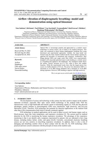
The respiratory circulation model was built using the finite element method with several triangular elements forming the lung tissue as shown in Figure 1. The lung tissue model consists of a trachea that connects the left and right bronchi, then there are bronchioles that form small branches that connect with the bronchi. The dimensions of the lung tissue model can be seen in Table 1.
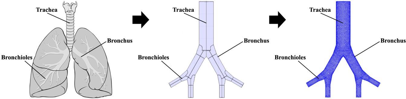
The experimental design and operation of the diaphragmatic breathing circulation detector were carried out using a FOS in the form of an elastic belt attached to the diaphragm as shown in Figure 2. Inside the belt, the SMF and FBG (1310 nm) were bent sinusoidally with a buffer diameter of 0.8 cm and 1.2 cm. The power source in the form of a laser diode is connected to one end of the optical fiber, then at the other end is connected to an optical power meter (OPM) to measure the input and output power of the optical fiber. The sample as the object of the experiment consisted of seven people with variations in abdominal circumference (near the diaphragm) and different ages as shown in Table 2. The sample’s position was standing with normal breathing conditions without any previous physical activity. Data collection was carried out for 60 seconds every 5 trials. The resulting data is in the form of power loss and the number of respiratory frequencies in each unit of time. The sensitivity level is then determined based on the change in power loss from the input and output measurements.
3. RESULTS
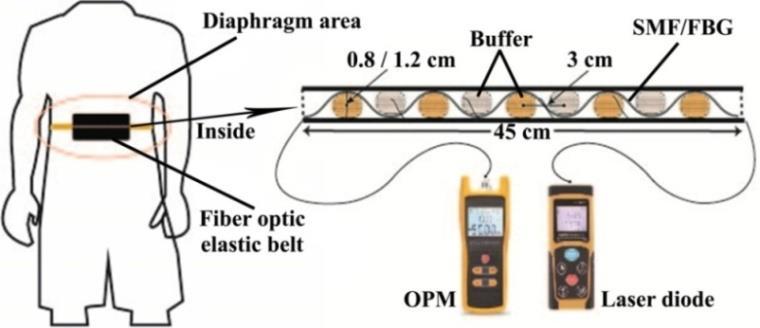
The simulation results of the lung airflow modeling can be seen in Figure 3. The initial parametric settings were carried out by entering the air density value of 1.225 kg/m3 , viscosity 1.7894×10⁻⁵ Pa.s, and velocity of 1 m/s. The airflow velocity in the trachea increases to 1.5 m/s, then in the first branch (bronchus) of the trachea the airflow increases to 2 m/s. The airflow then increases from 3.5 m/s to 4 m/s in the second branch (bronchioles) of the bronchus. This explains that the velocity of airflow is influenced by the geometry of the lungs. In addition, airflow velocity is also influenced by density rather than viscosity [22].
The situation that occurs on the inside of the belt during exhalation is shown in Figure 4. The sinusoidal pattern design of the SMF and FBG optical fibers undergoes a change in position in the diaphragm area. The fiber optic belt changes position during the inhalation state from the initial exhalation state. This change will affect the value that is read on the OPM according to the bending concept in the power loss category [23], [24]. The curved fiber will be pulled back to its original position known as the vibrating belt.
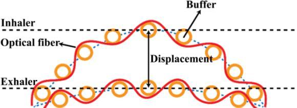
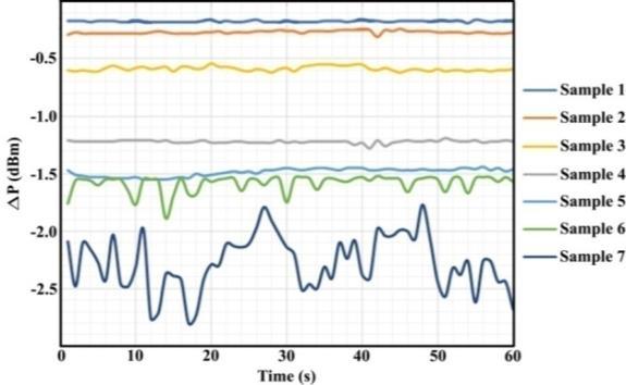
Figure 5 shows a comparison of optical fiber power changes for each experimental sample. The figure illustrates the change in the power of SMF with a buffer diameter of 0.8 cm which has a higher power loss than its type for a diameter of 1.2 cm. An increase in power loss indicates that inhalation is in progress, while power loss decreases during expiration [25]. Inhalation also applies in the seconds before and after although at a peak that is not too high. This shows that there are differences in the breathing carried out by the sample. In addition, the difference in peak power loss in sample 7 which is farther from the other samples is because the fiber optic belt responds more to the large abdominal circumference during inhalation. The further the belt moves during inhalation, the higher the power loss value [26]-[28]. This fiber optic belt actually relies on the principle of strain and stress from the buffer that forms the SMF and FBG in a sinusoidal pattern.
The distribution of respiratory frequency for the five experiments is shown in Figure 6. The median value for the change in power is taken from sample 2 because it has the smallest output power for FBG with a buffer diameter of 1.2 cm. The power changes occurred in the range of -0.40 dBm to -0.55 dBm, while the median area was in the range of -0.45 dBm to -0.50 dBm. These results explain that the sensitivity level of the sample is able to respond to the smallest power changes. Meanwhile, mixed experimental results are reported in the normal radian range.
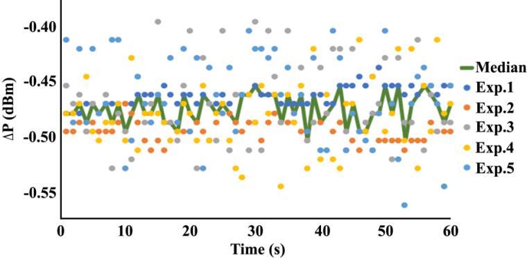
Figure 7(a) to Figure 7(d) shows the sensitivity level of SMF and FBG optical fiber for each different buffer diameter. According to the outline of the measurement results, the sinusoidal pattern formed by a buffer diameter of 0.8 cm as shown in Figure 7(b) and Figure 7(d) gives better results than 1.2 cm as shown in Figure 7(a) and Figure 7(c). This is because the highest and smallest power changes have been successfully detected by the SMF and FBG fiber optic belts. In addition, a critical power threshold factor in optical fiber causes a change in the detected power to depend on the amount of bending applied [29], [30]. In the experimental results on all samples, SMF optical fiber with a smaller buffer diameter has an average sensitivity of 0.26 compared to FBG of 0.23. However, on the other hand, FBG is superior to the measured power loss parameter with the highest value of -1.30 dB compared to SMF of -1.16 dB.
y=0.016x+0.189 R²=0.945
4. CONCLUSION
Detection of human respiratory airflow circulation has been successfully modeled and tested with FOS. Based on the simulation model, the airflow velocity increases from 1.5 m/s to 4 m/s when switching to a smaller branch geometry. In addition, the airflow velocity is also influenced by the density factor rather than the viscosity. Experimentally, the strain variation with the diameter buffer resulted in different diaphragmatic breathing vibration patterns. The power loss tends to be higher with changes in the position of the optical fiber during the inhalation process and is also influenced by a larger abdominal circumference. The sensitivity produced by SMF and FBG for a buffer diameter of 0.8 cm gives better results than 1.2 cm.
Acknowledgements
The author would like to thank the Ministry of Education, Research and Technology of the Republic of Indonesia for the support of the 2023 research grant. Furthermore, the author also thanks the Institute for Research and Community Service, University of Riau Pekanbaru Indonesia for the support and research facilities that have been provided.
References
[1] G. Allwood, G. Wild, and S. Hinckley, “Fiber Bragg grating sensors for mainstream industrial processes,” Electronics, vol. 6, no. 4, 2017, doi: 10.3390/electronics6040092.
[2] H. Purwanto, U. R. Fitriani, A. Dwijosutomo, and A. Marzuki, “Fiber pad for pressure mapping,” JournalofPhysics:Conference Series, 2016, vol. 776, doi: 10.1088/1742-6596/776/1/012106.
[3] T. Saktioto, D. Irawan, P. P. Yupapin, and P. Phatharacorn, “A single eye 3D image perception device using vertical double ring resonator construction,” Microwave and Optical Technology Letters, vol. 57, no. 8, pp. 1802
1805, 2015, doi: 10.1002/mop.29193.
[4] T. Saktioto, R. F. Syahputra, S. Punthawanunt, J. Ali, and P. Yupapin, “GHz frequency filtering source using hexagonal metamaterial splitting ring resonators,” Microwave and Optical Technology Letters, vol. 59, no. 6, pp. 1337–1340, 2017, doi: 10.1002/mop.30531.
[5] M. Fajkus, J. Nedoma, R. Martinek, V. Vasinek, H. Nazeran, and P. Siska, “A non-invasive multichannel hybrid fiber-optic sensor system for vital sign monitoring,” Sensors, vol. 17, no. 1, 2017, doi: 10.3390/s17010111.
[6] C. Y. Chen and K. M. Liao, “Chronic obstructive pulmonary disease is associated with risk of chronic kidney disease: a nationwide case-cohort study,” ScientificReports, vol. 6, 2016, doi: 10.1038/srep25855
[7] M. T. V. Chan et al., “Association of unrecognized obstructive sleep apnea with postoperative cardiovascular events in patients undergoing major noncardiac surgery,” JAMA, vol. 321, no. 18, pp. 1788–1798, 2019, doi: 10.1001/jama.2019.4783.
Airflow vibration of diaphragmatic breathing: model and demonstration using … (Toto Saktioto)
ISSN: 1693-6930
[8] R. Amelia, J. Harahap, A. Lelo, H. Wijaya, N. S. Harahap, and Z. Yamamoto, “Risk analysis for cardiovascular complication based on the artherogenic index of plasma of Type 2 diabetes mellitus patients in Medan, Indonesia,” FamilyMedicine&Primary CareReview, vol. 22, no. 3, pp. 197–201, 2020, doi: 10.5114/fmpcr.2020.98242.
[9] H. Jin, Y. S. A ‐Raya, and H. Haick, “Advanced materials for health monitoring with skin‐based wearable devices,” Advanced HealthcareMaterials, vol. 6, no. 11, 2017, doi: 10.1002/adhm.201700024.
[10] M. A. Jalil, A. Abdolkarim, T. Saktioto, C. T. Ong, and P. P. Yupapin, “Generation of THz frequency using PANDA ring resonator for THz imaging,” InternationalJournalofNanomedicine, vol. 7, pp. 773–779, 2012, doi: 10.2147/IJN.S27625
[11] T. Saktioto, F. D. Fadilla, Y. Soerbakti, D. Irawan, and Okfalisa, “Application of Fiber Bragg Grating Sensor System for Simulation Detection of the Heart Rate,” Journal of Physics: Conference Series, 2021, vol. 2049, doi: 10.1088/17426596/2049/1/012002
[12] Z. S. Lie etal., “H-D analysis employing low-pressure microjoule picosecond laser-induced breakdown spectroscopy,” Analytical Chemistry, vol. 89, no. 9, pp. 4951–4957, 2017, doi: 10.1021/acs.analchem.7b00245.
[13] Y. Tian, G. Xiao, Y. Luo, J. Zhang, and L. Yuan, “Microhole fiber-optic sensors for nanoliter liquid measurement,” OpticalFiber Technology, vol. 72, 2022, doi: 10.1016/j.yofte.2022.102981.
[14] P. P. Yupapin, T. Saktioto, and J. Ali, “Photon trapping model within a fiber Bragg grating for dynamic optical tweezers use,” MicrowaveandOpticalTechnologyLetters, vol. 52, no. 4, pp. 959–961, 2010, doi: 10.1002/mop.25043.
[15] A. Marzuki, N. W. Sari, and Riatun, “Design of reflective optical fiber sensor for determining refractive index and sugar concentration of aqueous solutions,” IOP Conference Series: Materials Science and Engineering, 2016, vol. 107, doi: 10.1088/1757-899X/107/1/012032
[16] Y. Zhao, J. Zhao, and Q. Zhao, “High sensitivity seawater temperature sensor based on no-core optical fiber,” Optical Fiber Technology, vol. 54, 2020, doi: 10.1016/j.yofte.2019.102115.
[17] P. Nagulapally, M. Shamsuddoha, G. Rajan, L. Djukic, and G. B. Prusty, “Distributed fibre optic sensor-based continuous strain measurement along semicircular paths using strain transformation approach,” Sensors, vol. 21, no. 3, 2021, doi: 10.3390/s21030782.
[18] N. Nazeer, and R. M. Groves, “Load monitoring of a cantilever plate by a novel multimodal fibre optic sensing configuration,” SN AppliedSciences, vol. 3, no. 667, 2021, doi: 10.1007/s42452-021-04663-9
[19] W. Hao, J. Xie, F. Wang, Z. Liu, and Z. Wang, “Analytical model of thin-walled corrugated tubes with sinusoidal patterns under axial impacting,” International Journal of Mechanical Sciences, vol. 128-129, pp. 1–16, 2017, doi: 10.1016/j.ijmecsci.2017.03.033
[20] M. Yang, Q. Liu, H. S. Naqawe, and M. P. Fok, “Movement detection in soft robotic gripper using sinusoidally embedded fiber optic sensor,” Sensors, vol. 20, no. 5, 2020, doi: 10.3390/s20051312.
[21] S. V. Ershkov, “On existence of general solution of the Navier-Stokes equations for 3D non-stationary incompressible flow,” International Journal of Fluid Mechanics Research, vol. 42, no. 3, pp. 206–213, 2015, doi: 10.1615/InterJFluidMechRes.v42.i3.20
[22] M. Sarkar, I. Madabhavi, N. Niranjan, and M. Dogra, “Auscultation of the respiratory system,” Annals of Thoracic Medicine, vol. 10, no. 3, 158–168, 2015, doi: 10.4103/1817-1737.160831
[23] S. F. Gao, Y. Y. Wang, X. L. Liu, W. Ding, and P. Wang, “Bending loss characterization in nodeless hollow-core anti-resonant fiber,” OpticsExpress, vol. 24, no. 13, pp. 14801–14811, 2016, doi: 10.1364/OE.24.014801
[24] A. Issatayeva, A. Beisenova, D. Tosi, and C. Molardi, “Fiber-optic based smart textiles for real-time monitoring of breathing rate,” Sensors, vol. 20, no. 12, 2020, doi: 10.3390/s20123408.
[25] D. Wang, B. Sheng, L. Peng, Y. Huang, and Z. Ni, “Flexible and optical fiber sensors composited by graphene and PDMS for motion detection,” Polymers, vol. 11, no. 9, 2019, doi: 10.3390/polym11091433
[26] Y. Koyama, M. Nishiyama, and K. Watanabe, “Smart Textile Using Hetero-Core Optical Fiber for Heartbeat and Respiration Monitoring,” inIEEESensorsJournal, vol. 18, no. 15, pp. 6175-6180, 2018, doi: 10.1109/JSEN.2018.2847333.
[27] J. Xu etal., “Development and Evaluation of a Respiratory Monitoring Smart Garment Based on Notched Optical Fiber Sensing Fabric,” IEEESensorsJournal, vol. 22, no. 15, pp. 14892–14902, 2022, doi: 10.1109/JSEN.2022.3186022.
[28] K. P. Lee, J. Yip, K. L. Yick, C. Lu, and C. K. Lo, “Textile-based fiber optic sensors for health monitoring: A systematic and citation network analysis review,” Textile Research Journal, vol. 92, no. 15-16, pp. 2922–2934, 2022, doi: 10.1177/00405175211036206.
[29] M. Li, N. Nyayapathi, H. I. Kilian, J. Xia, J. F. Lovell, and J. Yao, “Sound out the deep colors: Photoacousticmolecular imaging at new depths,” MolecularImaging, vol. 19, 2020, doi: 10.1177/1536012120981518.
[30] B. Hou et al., “An interactive mouthguard based on mechanoluminescence-powered optical fibre sensors for bite-controlled device operation,” NatureElectronics, vol. 5, pp. 682–693, 2022, doi: 10.1038/s41928-022-00841-8
Biographies Of Authors
Toto Saktioto is a Senior lecturer at Physics Dept., Faculty of Mathematics and Natural Sciences, Universitas Riau, Pekanbaru, Indonesia. He works on Plasma and Photonics Physics. He has published many articles and supervised Master’s and Doctoral degrees since 2009. His current research interests include the field of photonics, plasma, and its applications such as fiber optics and microwave plasma as communication, medical, and industrial technologies. He has published more than 200 journal papers in the fields of photonics and its applications. He can be contacted at email: saktioto@lecturer.unri.ac.id.
Defrianto is a Senior Lecturer in the Department of Physics, Faculty of Mathematics and Natural Sciences, Riau University, Pekanbaru, Indonesia. He works in IT and Computational Physics. He has published numerous articles and oversees a Master’s degree since 1999 at the Universite de Technologie de Compiegne, France. His current research interests include IT, computing, and applications such as communications, medical, and industrial technologies. He has published more than 30 journal papers in the field of computational physics and its applications. He can be contacted at email: defrianto@lecturer.unri.ac.id.
Nurfi Hikma is a alumni Student at Physics Dept., Faculty of Mathematics and Natural Sciences, Universitas Riau, Pekanbaru, Indonesia. He works on in Photonics Physics. He has published many articles and supervised Bachelor degrees since 2021. His current research interests include the field of photonicsapplications such as fiber optics and microwave plasma as communication, medical, and industrial technologies. She can be contacted at email: nurfihikma9998@gmail.com.
Yan Soerbakti is a Postgraduate student at Physics Dept., Faculty of Mathematics and Natural Sciences, Universitas Riau, Pekanbaru, Indonesia. He works on Plasma and Photonics Physics. He has published many articles and supervised Bachelor degrees since 2020. His current research interests include the field of metamaterial, photonics, plasma, and its applications such as fiber optics and microwave plasma as communication, medical, and industrial technologies. He has published more than 16 journal papers in the fields of applied physics and its applications. He can be contacted at email: yansoerbakti2@gmail.com.
Syamsudhuha is a Senior lecturer at Mathematics Dept., Faculty of Mathematics and Natural Sciences, Universitas Riau, Pekanbaru, Indonesia. He works on Mathematics and Statistics. He has published many articles and supervised Master’s and Doctoral degrees since 2009. His current research interests include the field of mathematics, and its applications such as mathematics as statistics, communication, and industrial technologies. He has published more than 100 journal papers in the fields of mathematics and its applications. He can be contacted at email: syamsudhuha@lecturer.unri.ac.id.
Dedi Irawan is a Senior lecturer at Physics Education Dept., Universitas Riau, Pekanbaru, Indonesia. He works on basic and advanced Photonics. He has published many articles and supervised Master’s and Doctoral degrees since 2019. His current research interests include the field of photonics, plasma, and its applications such as fiber optics and microwave plasma as communication. He has published more than 150 journal papers in the fields of basic and advanced Photonics. He can be contacted at email: dedi.dawan@yahoo.com.