1 minute read
know? DID YOU

by TBI Times

FOLLOWING A BRAIN INJURY, CLINICIANS MAY USE IMAGING TESTS TO SEE INSIDE
THE BRAIN AND LEARN ABOUT THE BRAIN’S ACTIVITY.
Advertisement
MRI
MRI scans produce detailed images of the brain using radio waves and a strong magnetic field. There are several MRI techniques:
Diffusion tensor imaging (DTI) is an MRI technique which measures the rate of water diffusion between cells.
Susceptibility weighted imaging (SWI) is an MRI technique which detects small amounts of blood products or calcium.
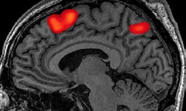
Functional magnetic resonance imaging (fMRI) is an MRI technique that detects changes in blood flow.

CAT/CT
CAT or CT scans produce 3-D images of the brain that will show any abnormalities.
Cta
CTA scans show blood vessels in the brain and can detect blockages or abnormalities.
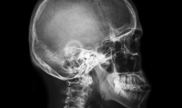
X-RAY
X-ray imaging shows skull fractures or other broken bones.
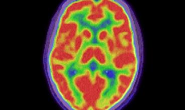
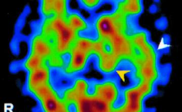
Pet
PET scans use radioactive substances to measure changes in the metabolic processes of the brain.
Spect
SPECT scans use gamma rays to create 3D pictures of the brain.
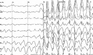
EEG
EEG scans measure electrical activity in the brain and can show any seizure activity.