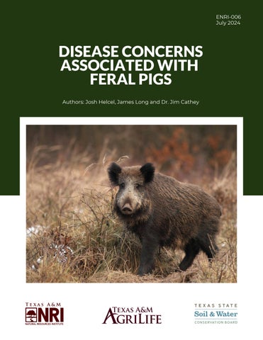
ENRI-006 July 2024
DISEASE CONCERNS ASSOCIATED WITH FERAL PIGS Authors: Josh Helcel, James Long and Dr. Jim Cathey
INTRODUCTION Negative impacts associated with feral pigs (Sus scrofa) extend to agricultural production, water quality, habitat, native species, the human-wildland interface and beyond. Their populations continue to expand across North America and occur today on every continent except Antarctica. Further they transmit diseases to livestock, native and exotic wildlife species, companion animals and even humans. Feral pigs harbor at least 65 bacterial, viral or parasitic infectious agents (Meng et al. 2009, Cooper et al. 2010, Haydett 2018, Preena et al. 2020), and threaten agricultural production, agricultural supply chains, and public health (Peper et al. 2021). In this publication, we address disease concerns endemic to feral populations and domestic pig herds throughout North America and abroad. We also provide management implications and safety precautions that can be enacted to help minimize the risk of disease transmission when handling and processing feral pigs or using outdoor areas where feral pigs may be present.
HISTORY OF FERAL PIGS Pigs originated in Southeast Asia around 5 million years ago, eventually expanding throughout Europe and North Africa, until they were ultimately domesticated and transported by people to every continent except Antarctica over the last 500 years (Wehr 2021). Domestication of Eurasian boar first occurred around 4900 B.C. in Asia, and by as early as 1500 B.C. they were raised for food production throughout Europe (Moeller et al. 2009). They were selectively bred for desirable traits that made them ideal production animals and gained marked increase in reproductive and body growth rates observed today (Wehr 2021). Domestic pigs were transported around the world as a valuable food source, allowing for the population expansion of the species (Figure 1). In North America, those pigs outside of commercial production fall into three groups: “feral pigs” (originating from domestic swine), Eurasian boar (the wild type), and their hybrids. All are all non-native to North America and can collectively be referred to as feral swine, wild swine or feral pigs. Eurasian wild boar are typically found in hunting preserves, rather than free-ranging.
1
Figure 1. Charles Darwin’s depiction of a Eurasian boar (top) and a Yorkshire Large Breed (bottom) in his “The Variation of Animal and Plants and Under Domestication” is a good example of the dramatic transformation that occurred during the domestication process.
DISEASE CONCERNS ASSOCIATED WITH FERAL PIGS
BIOLOGICAL INFLUENCES OF FERAL PIGS Spanish explorer Hernando de Soto is noted for introducing the first domestic pigs from Europe to North America in 1539, with later introductions to Mexico by Hernando Cortez in 1600 (Moeller et al. 2009). Initially brought as a reliable food source by early European settlers, free ranging domestic swine became feral over time, thus establishing populations on the continent. Eurasian boar were then released onto hunting preserves in both the northeastern U.S. and Texas from the 1890s to the 1930s (Mapston 2004). Escaped or intentionally released Eurasian boar then interbred and hybridized with established populations of feral pigs where the two occurred together (DelgadoAcevedo et al. 2010). The food production value of swine and commercial industries that emerged as a result are important factors that facilitated the spread of these animals across the globe. Genetic improvements in domestic pork production yielded traits including high animal gain, maximized growth rates, disease resistance and others (Merks et al. 2011). Selective breeding in the last 100 years resulted in genetic improvements including a 75% decrease in back fat and a 100% increase in growth rate (Merks 2000). Over time, domestic pigs interbred with wild populations, which now benefit from years of animal husbandry, giving us an enduring invasive species that challenges control and disease mitigation efforts worldwide.
Because of their intelligence, adaptability, fecundity, and social structure, feral pigs are highly successful at colonizing new areas and causing problems for humans and livestock. For example, because pigs do not have sweat glands, they regulate their body temperature through wallowing in wet and muddy places. This behavior generally concentrates the species near permanent water sources, particularly during times of drought and low resource availability. The risk for both intraspecific and interspecific disease transmission on the landscape generally increases during such times, as periods of drought and low resource availability can restrict movement patterns and lead to more wildlife concentration close to water sources. Conversely, disease risk associated with feral pigs can decrease during periods of high resource availability and abundant water, as feral pigs generally disperse further from permanent water sources and average sounder sizes decrease (Gabor et al. 1999). Streams, rivers and manmade water sources are examples of places they seek out to cool themselves. Wallowing loosens soil and leads to increased sedimentation, and defecation in these wetland systems introduces E. coli to surface water systems, causing bacterial impairment. The negative impacts feral pigs have on water quality carries implications for habitat, livestock production and human health that must be addressed through aggressive feral pig population reduction efforts (Figure 2).
DISEASE CONCERNS ASSOCIATED WITH FERAL PIGS
2
According to the 2022 Texas State Soil and Water Conservation Board (TSSWCB) 303d report, the majority of Texas surface water systems remain classified as bacterially impaired. Feral pigs are currently distributed in nearly all Texas counties, and have been implicated as significant contributors of bacteria, among other sources. The presence of E. coli is commonly viewed as an indicator of potential water borne pathogens that could harm humans. Agricultural production can also be impacted by E. coli spread by feral pigs, including crops such as spinach, when contaminated prior to consumption or use by livestock, humans or wildlife. Feral pigs are omnivores, and they must forage often due to the relative inefficiency of their simple stomachs (Seward et al. 2004). While most of a wild pig’s diet consists of vegetation, their ability to use a variety of resources contributes to their success across different ecotypes. Social structure and high fecundity also contribute to disease transmissions to livestock and humans, as concentrated animal populations have been shown to increase the potential for pathogen exposure (Guo et al 2021). Worldwide, feral pigs have more young than other mammals, where sows can conceive early (e.g., ~6 months) and mature females can have 4-6 offspring per litter, having 1 or 2 litters per year throughout their reproductive lifespan (Johnson et al. 1982; Mapston 2007). Feral pig densities in Texas ranged from 8.9-16.4 hogs/square mile, resulting in a median estimate of 2.6 million wild pigs for the state based on suitable habitat modeling (Timmons et al. 2012). As their populations continue to expand (Figure 3), their negative impacts compound. For example, annual agricultural losses in Texas alone exceeded $500M in 2022 (McCorkle 2022), a nearly tenfold increase from the 2012 estimate of $52M (Adams et al. 2005; Higginbotham 2013).
3
Figure 2. Feral pigs have been implicated a significant contributors of E. coli bacteria to surface water systems, resulting in widespread bacterial impair-ments in Texas that can lead to disease transmissions and negatively impact wildlife and livestock, as well as limit human use.
Figure 3. USDA-APHIS national distribution map by county of wild pig populations in the United States.
DISEASE CONCERNS ASSOCIATED WITH FERAL PIGS
AN OVERVIEW OF COMMON SWINE DISEASES These animals associate in large, primarily female based groups known as sounders, with juvenile or satellite males remaining nearby until the age of sexual maturity. Sexually mature males, or boars, generally remain solitary unless actively checking for receptive females. As resource availability increases, sounder size generally decreases as they disperse in search of relatively more abundant resources. The opposite is true in times of low resource availability or drought, when wild pig sounder sizes generally increase as they congregate in larger numbers near water and food sources including agricultural fields and supplemental feed stations meant for livestock or wildlife (Figure 4; Allwin et al. 2016). Given that feral pigs foul surface water systems, harm agricultural production, and congregate near livestock and supplemental wildlife feed stations, the potential for disease is a growing concern. Feral pigs can serve as a disease vector themselves or as a host to other organisms that transfer disease, especially given their wide distribution and relatively large home ranges (Helcel et al. 2016). For example, wild pig home ranges in excess of 30 square miles (Garza et al. 2018) have been documented in western Texas as a correlation for potentially spreading disease to new areas quickly.
Contrary to popular belief, consumption of wild pig meat is not the leading cause of disease transmission to humans. It is true that consuming raw or undercooked pork can be a means of disease transmission (e.g., trichinosis), but it can be consistently and effectively mitigated. According to the Centers for Disease Control (CDC), provided wild pig meat is cooked to a minimum internal temperature of 160 degrees, there are no known communicable diseases that cause concern for humans. Rather, most diseases are transmitted through direct or indirect contact among feral pigs, domestic livestock, wild animals and humans. Direct contact transmissions occur when a susceptible animal comes into physical contact with an infected animal, including its body fluids such as blood, saliva, urine, or feces. Among livestock and wildlife, this can occur through nose-to-nose contact, mating, and direct communal feeding/watering interactions with others.
Figure 4. The social structure, reproductive capacity and high numbers of feral pigs contribute to their increased potential for disease transmissions.
DISEASE CONCERNS ASSOCIATED WITH FERAL PIGS
4
In humans, direct contact transmissions generally occur through handling or processing infected animals, but it can also include consumption of raw or undercooked meat. Indirect contact transmissions primarily occur in livestock and wildlife through aerosol transmission (e.g., coughing and sneezing), consumption of contaminated food or water sources, and vector species transfer (ticks, mosquitos, biting flies, etc.). In humans, zoonotic indirect contact transmissions occur primarily through vector species transfer. In rare cases, aerosol transmission and consumption of untreated water sources can also result in indirect transfer of disease from feral pigs to humans.
SWINE BRUCELLOSIS Swine brucellosis is considered a classical zoonotic disease, meaning it can be transferred from an animal reservoir such as feral pigs to each other and to humans. Research in Texas and Oklahoma documented an overall 12.5% prevalence of brucellosis in swine from 553 samples tested (Peper et al. 2021). The documented frequency in south-central Oklahoma was 15.1% (65 of 553 samples), occurring 43 times higher than previously published results in 2016 which documented only a 0.35% prevalence from 282 samples taken in the same area (Gaskamp et al. 2016; Peper et al. 2021). The study also observed brucellosis antibodies present in feral pigs at each sample location across multiple years (Peper et al. 2021). This disease can also occur in domestic swine and may significantly impact the livestock industry or public food supply in the event of widespread infection.
Blood testing remains the only reliable means of diagnosis for swine brucellosis among domestic pigs, and there is not a practical way to diagnose individuals in feral populations. Removal and destruction of infected animals remains the primary best management practice in domestic pig husbandry. The same is true of feral pigs, and with the threat to domestic pork production, this exotic invader should be aggressively removed from the landscape. Zoonosis resulting from Brucella spp. is reported more than any other disease worldwide (Seleem et al. 2010). Out of nine recognized species, four (B. abortus, B. suis, B. melitensis, and B. canis) are known to be transmissible to people (Seleem et al. 2010). Of particular concern regarding transmission from infected swine to humans is the handling and processing of infected animals. For example, if the reproductive tract (especially in sows) is punctured, the bacteria can potentially be aerosolized and/or contact with the fluid can result in transmission (Table 1). The reproductive tissues of swine are perhaps the highest risk of transmission to humans; however, bacteria levels in the blood can also be infectious. While the mortality rate is low, physical symptoms in infected humans can be severe (Table 1), so limiting direct contact with infected animals is wise. Because transmission to humans often occurs through handling or processing meat it is recommended to take precautions like using personal protective equipment (PPE) when handling or processing swine (Table 1).
Symptoms may or may not be present in infected swine, and can therefore be difficult, or impossible to identify (Megid et al. 2010, Olsen et al. 2012; Table 1). Some infected animals will have chronic illness and will become long-term carriers. 5
DISEASE CONCERNS ASSOCIATED WITH FERAL PIGS
Table 1. Summary of swine brucellosis modes of transmission (livestock, wildlife and humans), symptoms, treatment and best management practices.
TULAREMIA Tularemia is caused by the highly infectious bacterium Francisella tularensis and is found in more than 250 species worldwide (Santic et al. 2010). Like brucellosis, tularemia is a zoonotic (transmissible between animals to humans) disease of global distribution and concern. It is stable in certain environments and may remain viable for months in water and sediment. While the primary reservoirs of the disease are mammals of the order Rodentia (rodents) and Lagomorpha (rabbits/hares), tularemia also occurs in swine (CDC, 2019). Seroprevalence research of feral pigs in Texas and southcentral Oklahoma over 4 years (2010-2012 and 2015-2017) documented antibody presence frequency at 14.8%, indicating a relatively high level of exposure for these populations (Peper et al. 2021). Known arthropod vectors in the U.S. include the lone star tick (Amblystoma americanum), the wood tick (Dermacentor andersoni), the dog tick (D. variabilis), and the deer fly (Chrysops discalis).
The two most common forms of tularemia in humans are ulceroglandular and typhoidal. Other more uncommon forms include glandular (without ulcer formation), oculoglandular (F. tularensis contracted through the eye), oropharyngeal (contracted through ingesting undercooked infected meat), and the pneumonic (contracted through aerosol transmission). While rare, severe cases can result in complications including meningitis, osteomyelitis and pericarditis that can be fatal. Tularemia infections in swine, similar to brucellosis, can often present with few or no discernable symptoms compared to those that do present (Table 2). Tularemia is considered a select agent by the CDC, and there are 2 serovars. Type A is far more virulent and is specific to North America, while Type B is less virulent and is more often waterborne than Type A.
DISEASE CONCERNS ASSOCIATED WITH FERAL PIGS
6
Table 2. Summary of tularemia modes of transmission (livestock, wildlife and humans), symptoms, treatment and best management practices.
Both are vectored by deerflies and biting flies. Due to the high potential for transmissibility of tularemia through multiple modes among domestic and feral pig populations, ongoing domestic monitoring and feral population control efforts are necessary to mitigate the potential impacts of this disease among swine populations worldwide.
PSEUDORABIES Also known as Aujeszky’s disease, the pseudorabies virus (PRV) is caused by a neurotrophic α-herpesvirus and represents another zoonotic disease endemic to swine in the U.S. (Pederson et al. 2018). While humans, horses and birds cannot contract the virus from swine, most mammals including livestock, wildlife and companion animals are at risk (USDA-APHIS 2020). PRV can be highly lethal in swine, particularly among piglets, with potential mortality rates up to 100% (USDA-APHIS 2020). Modes of transmission are similar to those mentioned above (Table 3).
7
Symptoms generally affect the central nervous system (CNS) and/or respiratory tract and can also include reproductive complications (Table 3). Reproductive symptoms can vary depending on the trimester of pregnancy in which infection occurs, but can include fetal abortion, stillbirth and severe CNS complications that often prove fatal in piglets and young swine. Once weaned, fatal CNS complications in swine are rare, and the respiratory disease component of PRV becomes more prevalent (USDA-APHIS 2020). Respiratory disease associated with PRV is common in adult swine, however, infected swine of all age classes can exhibit anorexia, weight loss and fevered response (USDA-APHIS 2020). PRV can establish latency in swine after exposure, with reactivation occurring due to stress or in response to natural stimuli (Pederson et al. 2018). Research conducted in Alabama and Arkansas found that PRV exposure levels in feral pigs significantly exceeded the previous national estimate of approximately 18% in
DISEASE CONCERNS ASSOCIATED WITH FERAL PIGS
many counties (Pederson et al. 2013, Pederson et al. 2018). It also documented several counties with local PRV antibody prevalence up to 50% (Figure 5), and further observed mortality and/or required euthanasia in hunting dogs with previous direct/indirect contact with infected swine (Pederson et al. 2018). Swine and raccoons remain the only natural communicable hosts of PRV (Liu et al. 2022). Given the potential for significant localized PRV exposure in feral pigs, multiple modes of zoonotic transfer, and biosecurity concerns, control efforts and continued PRV monitoring protocols remain important management tools in protecting domestic swine production, livestock, native species and companion animals.
Figure 5. Multiple counties in Alabama and Arkansas documented PRV seroprevalence >20% and up to 50% in sampled feral pigs. Exposure to swine borne PRV resulted in mortality and required euthanasia in hunting dogs in both states (Pederson et al. 2018).
Table 3. Summary of pseudorabies modes of transmission (livestock, wildlife and humans), symptoms, treatment and best management practices.
DISEASE CONCERNS ASSOCIATED WITH FERAL PIGS
8
AFRICAN SWINE FEVER African swine fever (ASF) is a highly contagious, double-stranded DNA virus in the Asfarviridae family which can cause substantial mortality and morbidity events in swine (Brown and Bevins 2018). First detected in East Africa in the early 1900s, ASF spread to both domestic and feral pig populations by the late 1950s. More recently, outbreaks in Asian countries including North Korea, South Korea, Indonesia, Vietnam and China resulted in the mortality or forced culling of ~225 million domestic pigs in China alone, from 2018-2019. China remains both the largest producer and consumer of pork in the world, and it is estimated that nearly 25% of the global domestic population perished from 2018-2019. The presentation of ASF in domestic swine can vary depending on the immune system of the infected animal, as well as the virulence of the infecting strain (Table 4). Highly virulent strains can cause death within 4-20 days post infection, with mortality rates of 95-100% and in less virulent strains range from 30-70%. Given the demonstrated potential of this disease to rapidly decimate global pork production, ASF is perhaps the most significant disease of concern affecting swine in the world today. In feral pigs of East and Southern Africa, ASF exists in sylvatic cycle between warthogs (Phacochoerus africanus) and argasid ticks (Ornithodoros moubata) and therefore cannot be fully eradicated (Penrith et al. 2013). Another African suid, the bush pig (Potamochoerus larvatus), is also known to contract ASF from the longstanding sylvatic cycle with African soft ticks (O. spp.). In addition to tick borne transmission, ASF can transfer through direct or indirect contact with infected pigs (Table 5 on page 12). Common modes of transmission among swine include contact with bodily fluids, nose-to-nose contact, fecal contact, and consumption of infected carcasses (Figure 6). 9
Another potential route of transmission is through viral shedding in swine that survive or present asymptomatic ASF infection. The incubation period for the virus is 4-19 days, and shedding can occur in swine surviving ASF for up to 70 days post infection (Carvalho Ferreira et al. 2013).
Figure 6. Common routes of ASF transmission among swine independent of tick-borne conveyance (Sanchez-Cordon et al. 2018). Source: https://pubmed.ncbi.nlm.nih.gov/29486878/
Fortunately, ASF cannot be transmitted to humans (Enjuanes et al. 1977). The mechanisms driving the apparent inability of ASF to infect humans are incompletely understood, and research is needed to determine exactly why this is the case. No vaccines are currently available for swine. Additionally, no outbreaks have occurred in North American domestic or feral swine to date. However, several outbreaks occurred in the Dominican Republic as recently as 2021. During these outbreaks, at least 2,200 pigs succumbed to the disease or were culled during containment protocol (USDA 2021; Ruiz-Saenz et al. 2022). The proximity of these outbreaks to North America raises concern for the United States.
DISEASE CONCERNS ASSOCIATED WITH FERAL PIGS
Table 4. Summary of African swine fever modes of transmission (domestic and wild swine), symptoms, treatment and best management practices.
Surveillance and rapid test ASF diagnosis, followed by immediate quarantine and destruction of infected swine, remains the primary global strategy against ASF outbreaks. However, in January 2020 researchers developed an experimental gene deletion vaccine which showed 100% efficacy against a particular strain of ASF responsible for an outbreak that occurred in the European country of Georgia in 2007 (Borca et al. 2020). In African wild swine, including warthogs and bush pigs, research indicated asymptomatic infection was most common (Penrith et al. 2013), compounding the difficulty of early recognition and mitigation efforts in the event of an outbreak among North American feral pig populations.
CHAGAS DISEASE Another emerging disease of concern, particularly in Texas, is Chagas disease. Here, enzootic transmission cycles of the vector borne protozoan parasite Trypanosoma cruzi, the causative agent of Chagas disease, have occurred (Comeaux et al. 2015). Chagas disease has long been endemic to Latin America, and in these countries feral pigs have been implicated as reservoirs for the triatomines, commonly referred to as “kissing bugs” (Figure 7), which harbor the causative parasite and transmit it during blood meals from their hosts (Comeaux et al. 2015). The estimated worldwide disease burden exceeds 6 million infections, with approximately 30% of human cases presenting clinical signs of infection (Malik and Singh 2015, Molina-Garza et al. 2014, Sanchez-Guillen et al. 2006; Table 8). Chagas disease is considered a chronic and progressive illness, and results in chronic enlargement of organs over time in 30% of infected humans (Nolan et al. 2018). Additional complications can include end stage heart failure (Nolan et al. 2018).
DISEASE CONCERNS ASSOCIATED WITH FERAL PIGS
10
The disease is suggested to be widely underdiagnosed and under reported, however Arizona, California, Louisiana, Mississippi, Tennessee, and Texas have documented vector-borne transmissions to humans (Nolan et al. 2018; Bern et al. 2011; Gunter et al. 2017; Harris et al. 2017). Texas disproportionately had the highest number of human cases (Nolan et al. 2018; Bern et al. 2011; Gunter et al. 2017; Harris et al. 2017). Sylvatic transmission cycles with woodrats, opossums, raccoons and dogs have also been reported in 17 southern U.S. states (Gunter et al. 2016). Due to the relatively inefficient mode of disease transfer from triatomines (kissing bugs) to their host, wildlife species, livestock and companion animals are potentially the most at risk compared to humans (Figure 8). For conventional disease transmission to occur, infected triatomines first obtain a blood meal from their host. Defecation from the kissing bug that occurs during this process can then contact broken skin and potentially result in infection. Often, this process must occur numerous times from a fairly high number of bites to receive a high enough dose for viable infection. Research documented Chagas infection in 6% of Texas feral pigs sampled (Comeaux et al. 2016). More recent research suggested Texas hunters as a potential high-risk population for Chagas exposure (Gunter et al. 2020). With growing feral pig populations in the U.S. and overlapping distribution with triatomines, there is concern over their role in facilitating the spread of Chagas disease.
11
Figure 7. A triatomine or kissing bug compared in size to a U.S. penny. (kissingbug.tamu.edu)
Figure 8. Geographical distribution map of Texas showing humans, triatomines and animals testing positive for Chagas disease from 2013-2020. Source: Texas Health and Human Services
DISEASE CONCERNS ASSOCIATED WITH FERAL PIGS
Table 5. Summary of Chagas disease modes of transmission (livestock, wildlife and humans), symptoms, treatment and best management practices.
TICK-BORNE ILLNESSES Ticks are second only to mosquitos as the most significant vector of disease transmission to humans and animals (Brites-Neto et al. 2015). Feral pigs are routinely infested with ticks capable of transmitting a variety of bacterial, viral, protozoan and rickettsial agents to swine, livestock, wildlife, companion animals and humans (Helcel et al. 2016). A wide variety of hard- and soft-shelled tick species commonly utilize both domestic and wild swine to obtain blood meals. Examples of tick-borne diseases include ASF, anaplasmosis, babesiosis, theileriosis, ehrlichiosis, cytauxzoonosis, hepatozoonosis, spotted fevers, typhus, Lyme borreliosis, tularemia, bartonellosis, and viral encephalitis (Boulanger et al. 2019, Dantas-Torres et al. 2012, Inci et al. 2016, Table 6). These and other tickborne illnesses threaten global pork supply chains, livestock production, native wildlife, and human health. An issue inherent to many vector-borne illnesses, particularly among those contracted by humans, is that diagnosis is often unreliable, unreported and/or misidentified (Dupouy-Camet and Bruschi 2007, Kugeler et al. 2021).
This is generally attributed to factors including disease pathology, unavailable, inaccurate, or inadequate testing, as well as the tendency of many tick-borne diseases to present as flulike, cardiac-related, neurological or as other illnesses more commonly understood by medical science. For example, approximately 30,000-40,000 human cases of Lyme disease are passively reported through surveillance data each year (Mead 2015). However, according to the CDC, approximately 476,000 cases of Lyme disease were contracted by humans annually from 2010-2018 in the U.S. alone (Kugeler et al. 2021). The mechanism of how researchers arrived at these data is found below (Figure 9). The threat of tick-borne illness wherever feral pigs occur is worrisome, and precautions are necessary when processing, handling or even using outdoor areas used by feral pigs. In humans, flu-like symptoms typically predominate early in infection, with later symptoms progressing into a complex variety of presentations that also mimic those of more common illnesses. As a result, people often either fail to associate their condition
DISEASE CONCERNS ASSOCIATED WITH FERAL PIGS
12
Table 6. General summary of various tick-borne illness modes of transmission (livestock, wildlife and humans), symptoms, treatment and best management practices.
with a previous tick bite due or are misdiagnosed by medical professionals. The relatively long incubation periods associated with tick-borne illness in humans further compounds this issue. The Texas A&M Natural Resources Institute (NRI) has previously published an educational resource on the potential for tick-borne illness associated with feral pigs, including implications for livestock and agricultural production. It is recommended that the reader review that resource which provides more analysis of feral pigs and tick-borne illnesses on AgriLife Learn. Figure 9. CDC estimate for actual annual human Lyme disease cases in the US from 2010-2018 when corrected for underreporting, misdiagnosis and factors including Lyme disease-specific codes in patient records (Kugeler et al. 2021).
13
DISEASE CONCERNS ASSOCIATED WITH FERAL PIGS
TRICHINELLOSIS Trichinella spp. are parasites that can be transferred from swine to humans through the consumption of raw or undercooked meat (Kirjusina et al. 2015). While nematodes of the genus Trichinella occur in numerous omnivorous and carnivorous animals, the primary sources of human infection are from wild and domestic swine (Murrell and Pozio 2011, Gottstein et al. 2009, Table 7). Due to the potential for meat-borne zoonosis from swine, veterinary diagnostics and stringent meat inspection protocols remain necessary to ensure food safety (Campbell 1983). However, the annual domestic and international economics costs of countermeasures against diseases including trichinellosis have long remained substantial (Pyburn et al. 2005). For example, the budgeted cost for veterinary diagnostics for fiscal year 2022 was $61,414,000 alone in the U.S., with cumulative federal funding for the U.S. pork industry reaching nearly $6 billion (USDA-APHIS 2022). The relatively high efficacy of surveillance, diagnostic and inspection protocols in the U.S. is widely credited for the trend leading to relatively low levels of human trichinellosis infection today (Figure 10).
In humans, trichinellosis presents with symptoms including myalgia, diarrhea, fever, facial edema and headaches (Murrell and Pozio 2011). Analogous to many tick-borne illnesses associated with swine, overlapping clinical manifestations with common diseases including influenza and chronic fatigue syndrome can result in misdiagnosis (DupouyCamet and Bruschi 2007). Other symptoms in humans include gastroenteritis, malaise, subungual or conjunctival hemorrhages, and increased eosinophils, leukocytes, and muscle enzymes (Dupouy-Camet and Bruschi 2007). While early diagnosis of trichinellosis is generally difficult to obtain, most patients become asymptomatic within 2-6 months of treatment with a combination of antiparasitics, anthelmintics, glucorticosteriods, chemotherapy and others (Gottstein et al. 2009). The disease can become chronic in cases that go extended periods undiagnosed, and research showed that mortality occurred in up to 5% of such high infection intensity cases (Gottstein et al. 2009).
Figure 10. CDC data for the number of reported confirmed cases of trichinellosis, by year — National Notifiable Disease Surveillance System, U.S., 1947– 2012 (Wilson et al. 2015).
DISEASE CONCERNS ASSOCIATED WITH FERAL PIGS
14
In addition to humans and swine, trichinellosis can also be contracted by horses, dogs, foxes, walruses and bears (Gottstein et al. 2009; Figure 11). Infected swine rarely develop visible lesions and most cases present as asymptomatic (Iowa State University 2022). The Food and Drug Administration (FDA) has approved meat irradiation protocols to kill trichinellosis and other bacterium, further contributing to widespread trichinella-free certification in domestically produced swine (Iowa State University 2022). In Figure 11, you will see (A) the main sources of Trichinella sp. infections for humans (including pigs, horses, wild boars, dogs, walruses, foxes, and bears) and (B) the Trichinella sp. cycle in the host body. In the enteral phase, muscle tissues are digested in the stomach, and larvae are released (1); larvae penetrate the intestinal mucosa of the small intestine and reach the adult stage within 48 h p.i., and male and female mate (2); female worm releases newborn larvae in the lymphatic vessels (from the fifth day p.i. onwards; the length of newborn production, from 1 week to several weeks, is under the influence of host immunity) (3); In the parenteral phase, the newborn larvae reach the striated muscle and actively penetrate in the muscle cell (4); the larva grow to the infective stage in the nurse cell (the former muscle cell) (5); and, after a period of time (weeks, months, or years), a calcification process occurs (6) (Gottstein et al. 2009).
Figure 11. Trichinella sp. life cycle.
Table 7. Summary of trichinellosis modes of transmission (livestock, wildlife and humans), symptoms, treatment and best management practices.
15
DISEASE CONCERNS ASSOCIATED WITH FERAL PIGS
PRECAUTIONS AGAINST SWINE-BORNE DISEASES Direct contact with feral pigs generally occurs when handling animals in the field, loading them for transport to an approved holding facility and during processing. Use of personal protective equipment (PPE) as well as basic hygienic practices can help minimize risk while processing feral pigs (Figure 12). Additionally, it is important to implement proper hygienic and sanitary practices for all surfaces (including any direct skin contact), tools, clothing and equipment both prior to and after contact with feral pigs. Examples of sanitary practices include making sure to: Wash hands and any clothing that came into contact with feral pigs after handling or processing; Cover all scratches, skin abrasions and wounds prior to any handling or processing activities; Disinfect any surfaces, tools and equipment with an ammonia or bleach based cleaner; and Conduct self-inspections for ectoparasites such as fleas or ticks after interactions with feral pigs, or frequenting outdoor areas where feral pigs are present.
Figure 12. Wearing PPE is a good precaution to reduce direct contact with feral pigs when handling or processing carcasses.
For those who choose to consume feral pigs, precautions should be taken to minimize disease transmission risk while handling, field dressing and processing the carcass. Some people prefer to have a professional butcher process the carcass, but keep in mind that not all processors will butcher feral pigs, so it is a good idea to contact them first to verify that they provide this service.
DISEASE CONCERNS ASSOCIATED WITH FERAL PIGS
16
FIELD TO FRIDGE Remember to use PPE when field dressing feral pigs, and make sure to keep the carcass free of dirt, debris and stomach contents (Figure 13). It is generally a good idea process in a well-ventilated area and not puncture the intestines or stomach when field dressing feral pigs, as well as to remain on the upwind side of the animal when opening and removing the contents of the body cavity. As soon as possible, the carcass should be cooled to below 40 °F to minimize the potential for food borne illness. Outdoor temperatures <40 °F are fine for safely skinning, quartering and storing meat on ice for several days. When outdoor temperatures are warm, it will be necessary to transfer quarters or field dressed animals to an iced cooler, walk-in cooler or processor as soon as possible. Once properly cooled, packaging and freezing meat (0 °F) is recommended to slow the growth of microorganisms and preserve the meat. Prior to cooking wild pig meat intended for human consumption, it is recommended to take these steps:
Figure 13. Prior to field dressing feral pigs, it is a good idea to employ a skinning rack or other means of hanging the animal to keep it free of dirt and debris.
Before preparing or cooking wild pig meat, always remember to wash your hands for at least 20 seconds with warm, soapy water. Preventing cross-contamination is critical. Keep the raw meat from touching multiple surfaces and designate a cutting board and set of knives for processing raw meats. Keep foods out of the “temperature danger zone” of 40-140 °F. USDA recommends cooking wild pig products to a minimum internal temperature of 160 °F. Internal temperature can be verified by placing a food thermometer in the thickest part of the meat. With a meat thermometer, maintain cooked foods at proper temperatures, where cold foods should be held at 40 °F or below, and hot food should be kept at 140 °F or above.
17
DISEASE CONCERNS ASSOCIATED WITH FERAL PIGS
DISPOSAL OF FERAL PIGS Consumption of feral pigs is often neither feasible nor desired, especially following management efforts such as aerial gunning or corral trapping, which can result in having many dead pigs at one time. Other factors such as location, challenging terrain, and hotter temperatures and times influence decisions to consume or dispose of feral pigs. In these situations, landowners and wildlife managers often choose to employ a “boneyard” in which to bury or otherwise dispose of feral pigs at a centralized location. Here they should follow guidance by the Texas Animal Health Commission and prepare the site to their specifications. When disposing of feral pigs, ensure that proper PPE is worn, appropriate sanitary measures are practiced, and that disposal sites are a selected in relatively flat locations at least 50 yards or more from any creek, stream, riparian zone or other surface water system. Following the same instructions to protect water sources, scattering carcasses, and allowing time for decomposition returns nutrients back to the landscape.
Once sold to an approved holding facility, feral pigs must be federally inspected both pre and post-mortem prior to food sales in both domestic and international markets. When loading feral pigs for transport, ensure that proper PPE is worn and appropriate sanitary measures are practiced to limit direct and indirect contact with feral pigs. With proper permitting, wild pig boars may also be sold to approved hunting preserves in Texas. Transporting feral pigs should be done in a stock trailer dedicated to this purpose. To help reduce the risk of disease, transporting domestic livestock should be done in a separate trailer.
TRANSPORT AND SALE OF LIVE FERAL PIGS Another option for wild pig removal in Texas is the transport and sale of live animals to approved holding facilities, which is regulated by the Texas Animal Health Commission (TAHC). As of 2023, there were over 110 approved holding facilities statewide where live feral pigs may be legally transported and sold. Income generated from the sale can help offset costs associated with control efforts. Many buying stations place minimum size restrictions on the feral pigs they purchase, so it is a good idea to contact holding facilities to learn of any restrictions and the price they are paying per pound.
DISEASE CONCERNS ASSOCIATED WITH FERAL PIGS
18
WILD PIG CONTROL AS A DISEASE MITIGATION STRATEGY Due to the significant threat that feral pigs pose to water quality, agriculture, livestock production, habitat, global supply chains, native species and human health, lethal control measures are necessary to reduce their numbers, damages and potential for disease transmissions. While available techniques for lethal control vary worldwide, legal methods for reducing feral pigs in Texas currently include trapping, aerial gunning, shooting, snaring and the use of trained dogs. Some techniques such as shooting and the use of trained dogs can alter the movements of feral pigs and force them to abandon areas where they are causing damage. Effective abatement often involves a combined approach (McCann and Garcelon 2008), and landowners should consider working cooperatively with neighbors to improve reduction per acres managed. This can be accomplished by sharing traps, head gates, snaring sites, aerial gunning costs, property access for the use of trained dogs and other collaborative efforts. The Texas A&M Natural Resources Institute has previously published many free educational control measure resources that will be helpful in the fight against feral pigs. Consider the following:
CONCLUSION Feral pigs are known reservoirs for many viral, parasitic and bacterial pathogens capable of infecting humans, livestock, wildlife species and companion animals. The potential for widespread disease transmissions by wild pig represents a threat to global supply chains, water quality, agriculture, livestock production, and humans worldwide. Swine transmit diseases through direct transmissions including noseto-nose contact, communal feeding areas, communal watering sources, fecal contact, consumption of infected carcasses, sexual contact, consumption of undercooked/raw meat, viral shedding, processing, handling, and others. Indirect transmissions can occur through aerosol transmission, shared habitat, viral shedding, vector species transfer and other indirect means. The nearly global distribution and growing populations of feral pigs, coupled with their natural biological factors and selection for specific traits imparted by man over time, compound control and disease mitigation efforts worldwide. Precautions such as wearing proper PPE, enacting sanitary practices, proper carcass disposal, and proper meat preparation remain necessary to minimize risk for disease transmission.
https://wildpigs.nri.tamu.edu/ https://www.youtube.com/c/TexasAMNaturalResourcesInstitute
19
DISEASE CONCERNS ASSOCIATED WITH FERAL PIGS
LITERATURE CITED Adams, C.E., B. Higginbotham, D. Rollins, R. B. Taylor, R. Skiles, M. Mapston, and S. Turman. 2005. Regional perspectives and opportunities for feral hog management. Wildlife Society Bulletin 33(4):1312-1320. Allwin, B., N.S. Gokarn and S. Vedamanickam. 2016. The wild pig (Sus scrofa) behavior- a retrospective study. Journal of Dairy, Veterinary and Animal Research. 3(3):115-125. Beard, C.B., G. Pye, F.J. Steurer, R. Rodriguez, R. Campman, A.T. Peterson, J. Ramsey, R.A. Wirtz and L.E. Robinson. 2003. Chagas disease in a domestic transmission cycle, southern Texas, USA. Emerging Infectious Diseases 9(1):103-5. Bern, C. S. Kjos, M.J. Yabsley and S.P. Montgomery. 2011. Trypanosoma cruzi and Chagas' disease in the United States. Clinical Microbiology Reviews 24(4):655–81. Borca, M. V., E. Ramirez-Medina, E. Silva, E. Vuono, A. Rai, S. Pruitt, L. G. Holinka, L. VelazquezSalinas, J. Zhu, and D. P. Gladue. 2020. Development of a highly effective African swine fever virus vaccine by deletion of the I177L gene results in sterile immunity against the current epidemic Eurasia strain. Journal of Virology 94. Boulanger, N., P. Boyer, E. Talagrand-Reboul and Y. Hansmann. 2019. Ticks and tick-borne diseases Tiques et maladies vectorielles à tiques. Médecine et Maladies Infectieuse 49:87–97. Brites-Neto, J., K.M. Duarte and T.F. Martins. 2015. Tick-borne infections in human and animal population worldwide. Vet World 8:301–15. Brown, V. R., and S. N. Bevins. 2018. A review of African swine fever and the potential for introduction into the United States and the possibility of subsequent establishment in feral swine and native ticks. Frontiers in Veterinary Science 5. Campbell, W.C. 1983. Trichinella and trichinosis. New York: Plenum Press p. 1–30. Comeaux, J.M., R. Curtis-Robles, B.C. Lewis, K.J. Cummings, B.T. Mesenbrink, B.R Leland, M.J. Bodenchuk and S.A. Hamer. 2016. Survey of feral swine (Sus scrofa) infection with the agent of Chagas disease ( Trypanosoma cruzi ) in Texas, 2013-14. Journal of Wildlife Diseases 52(3):627-30. de Carvalho Ferreira, H. C., J. A. Backer, E. Weesendorp, D. Klinkenberg, J. A. Stegeman, and W. L. A. Loeffen. 2013. Transmission rate of African swine fever virus under experimental conditions. Veterinary Microbiology 165:296–304. Dantas-Torres, F., B.B. Chomel and D. Otranto. 2012. Ticks and tick-borne diseases: a one health perspective. Trends Parasitol. 28:37–46. Darwin, C. 1868. The variation of animals and plants under domestication. The American Naturalist 2, no. 4, pp. 208-209.
DISEASE CONCERNS ASSOCIATED WITH FERAL PIGS
20
LITERATURE CITED Delgado-Acevedo, J., A. Zamorano A, R.W. DeYoung, T.A. Campbell, D.G. Hewitt and D.B. Long. 2010. Promiscuous mating in feral pigs (Sus scrofa) from Texas, USA. Journal of Wildlife Research 37:539–546. Dupouy-Camet, J. and F. Bruschi. 2007. Management and diagnosis of human trichinellosis. In: Dupouy-Camet, J., Murrell, K.D., editors. FAO/WHO/OIE guidelines for the surveillance, management, prevention and control of trichinellosis. Paris: World Organisation for Animal Health p. 37–68. Enjuanes, L., I. Cubero, and E. Vinuela. 1977. Sensitivity of macrophages from different species to African Swine Fever (ASF) virus. Journal of General Virology 34:455–463. Gabor T.M., E. Hellgren, R.A. Van Den Bussche and N.J. Silvy. 1999. Demography, sociospatial behaviour and genetics of feral pigs (Sus scrofa) in a semi-arid environment. Journal of Zoology 247(3):311-322. Gaskamp, J. A., K. L. Gee, T. A. Campell, N. J. Silvy, and S. L. Webb. 2016. Pseudorabies virus and Bru-cella abortus from an expanding wild pig (Sus scrofa) population in southern Oklahoma, USA. Journal of Wildlife Diseases 52:383–386. Garza, S.J., M.A. Tabak, R.S. Miller, M.L. Farnsworth and C.L. Burdett. 2018. Abiotic and biotic influences on home-range size of wild pigs (Sus scrofa). Journal of Mammalogy 99, 97–107. Gottstein, B., E. Pozio and K. Nöckler. 2009. Epidemiology, diagnosis, treatment, and control of trichinellosis. Clinical Microbiology Reviews 22:127–45. Gunter, S.M., E.O. Brown, K.O. Murray and M.N. Garcia. 2016. Historical perspectives on sylvatic transmission among wildlife reservoirs in Texas. Zoonoses and Public Health 64:313-327 Gunter, S.M., K.O. Murray, R. Gorchakov, R. Beddard, S.N. Rossmann and S.P. Montgomery. 2017. Likely autochthonous transmission of Trypanosoma cruzi to humans, south central Texas, USA. Emerging Infectious Diseases 23(3):500–3. Gunter, S.M., S.E. Ronca, M. Sandoval, K. Coffman, L. Leining, R. Gorchakov, K.O. Murray and M.S. Nolan. 2020. Chagas Disease infection prevalence and vector exposure in a high-risk population of Texas hunters. American Journal of Tropical Medicine and Hygiene 102(2):294297. Guo, Y., U. Ryan, Y. Feng and L. Xiao. 2022. Association of common zoonotic pathogens with concentrated animal feeding operations. Frontiers in Microbiology. 10;12:810142. Harris, N., L. Woc-Colburn, S.M. Gunter, R. Gorchakov, K.O. Murray and S. Rossmann. 2017. Autochthonous Chagas disease in the southern United States: A case report of suspected residential and military exposures. Zoonoses and Public Health 64(6):491–3. 21
DISEASE CONCERNS ASSOCIATED WITH FERAL PIGS
LITERATURE CITED Helcel, J., P. Teel, M. Tyson, J. Cash, T. Hensley and J.C. Cathey. 2016. Wild pigs and ticks: Implications for livestock production, human and animal health. AgriLife Extension Service. Higginbotham, B. 2013. Wild pig damage abatement education and applied research activities. Texas A&M AgriLife Research and Extension Center-Overton. Inci, A., A. Yildirim, O. Duzlu, M. Doganay and S. Aksoy. 2016. Tick-borne diseases in Turkey: a review based on One Health perspective. PLOS Neglected Tropical Diseases 10:e0005021. Johnson, K.G., R.W. Duncan and M.R. Pelton. 1982. Reproductive biology of European wild hogs in the Great Smokey Mountains National Park. Proceedings of the Annual Conference of the Southeastern Fish and Wildlife Agencies 36:552-564. Khanna, K. 2022. African swine fever virus: A global concern. ASM.org. American Society for Microbiology. <https://asm.org/Articles/2022/March/African-Swine-Fever-Virus-Is-A-GlobalConcern>. Kirjušina, M., G. Deksne and G. Marucci. 2015. A 38-year study on Trichinella spp. in wild boar (Sus scrofa) of Latvia shows a stable incidence with an increased parasite biomass in the last decade. Parasites Vectors 8, 137. Kugeler, K. J., A. M. Schwartz, M. J. Delorey, P. S. Mead, and A. F. Hinckley. 2021. Estimating the frequency of lyme disease diagnoses, United States, 2010–2018. Emerging Infectious Diseases 27:616–619. Liu, A., T. Xue, X. Zhao, J. Zou, H. Pu, X. Hu and Z. Tian. 2022. Pseudorabies virus associations in wild animals: review of potential reservoirs for cross-host transmission. Viruses 14(10):2254. Malik, L.H., G.D. Singh and E.A. Amsterdam. 2015. Chagas heart disease: an update. The American Journal of Medicine 128(11):1251.e7–9. Mapston, M.E. 2004. Feral hogs in Texas. College Station, TX: Texas Cooperative Extension. Mapston, M.E. 2007. Feral Hogs in Texas. Texas Wildlife Services. McCann, B. E. and D. K. Garcelon. 2008. Eradication of feral pigs from Pinnacles National Monument. The Journal of Wildlife Management 72:1287-1295. Mead, P.S. 2015. Epidemiology of lyme disease. Infectious Disease Clinics of North America 29:187– 210. Merks, J. W. M., P. K. Mathur, and E. F. Knol. 2012. New phenotypes for new breeding goals in pigs. Animal 6:535–543.
DISEASE CONCERNS ASSOCIATED WITH FERAL PIGS
22
LITERATURE CITED Moeller, S. J. and F. L. Crespo. 2009. Overview of world swine and pork production. Agricultural Sciences. <https://www.eolss.net/Sample-Chapters/C10/E5-24-03-04.pdf>. Molina-Garza, Z.J., J.L. Rosales-Encina, R. Mercado-Hernandez, D.P. Molina-Garza, R. GomezFlores and L. Galaviz-Silva. 2014. Association of Trypanosoma cruzi infection with risk factors and electrocardiographic abnormalities in northeast Mexico. BMC Infectious Diseases 14:117 Murrell, K.D. and E. Pozio. 2011. Worldwide occurrence and impact of human Trichinellosis, 1986– 2009. Emerging Infectious Diseases 17(12):2194-2202. Pedersen, K., S.N. Bevins, J.A. Baroch, J.C. Cumbee Jr., S.C. Chandler, B.S. Woodruff, T.T. Bigelow, T.J. DeLiberto. 2013. Pseudorabies in feral swine in the United States, 2009–2012. Journal of Wildlife Diseases 49(3):709–13. Pedersen, K., C. T. Turnage, W. D. Gaston, P. Arruda, S. A. Alls, and T. Gidlewski. 2018. Pseudorabies detected in hunting dogs in Alabama and Arkansas after close contact with feral swine (Sus scrofa). BMC Veterinary Research 14. Penrith, M.-L., W. Vosloo, F. Jori, and A. D. S. Bastos. 2013. African swine fever virus eradication in Africa. Virus Research 173:228–246. Peper, S. T., A. Hoffarth, K. Athanasiou, S. L. Hawkins, A. N. Wilson‐Fallon, A. Gibson, C. Reinoso Webb, J. A. Gaskamp, S. L. Webb, K. M. Haydett, H. S. Tiffin, and S. M. Presley. 2021. Brucella spp. and francisella tularensis from an invasive alien species (Sus scrofa) in the southcentral USA. Ecosphere 12. Pseudorabies (PRV). 2020. USDA APHIS. Pseudorabies (PRV). USDA. <https://www.aphis.usda.gov/aphis/ourfocus/animalhealth/nvap/NVAP-ReferenceGuide/Control-and-Eradication/Pseudorabies>. Pyburn, D.G., H.R. Gamble, E.A. Wagstrom, L.A. Anderson and L.E. Miller. 2005. Trichinae certification in the United States pork industry. Vet Parasitol. 132:179–83. Ruiz-Saenz, J., A. Diaz, D.K. Bonilla-Aldana, A.J. Rodríguez-Morales, M. Martinez-Gutierrez and P.V. Aguilar. 2022. African swine fever virus: A re-emerging threat to the swine industry and food security in the Americas. Frontiers in Microbiology 13: 1011891. Sánchez-Cordón, P.J., M. Montoya, A.L. Reis and L.K. Dixon. 2018 Mar. African swine fever: A reemerging viral disease threatening the global pig industry. The Veterinary Journal. 233:41-48. Sanchez-Guillen, M.C., A. Lopez-Colombo, G. Ordonez-Toquero, I. Gomez-Albino, J. RamosJimenez and E. Torres-Rasgado. 2006. Clinical forms of Trypanosoma cruzi infected individuals in the chronic phase of Chagas disease in Puebla, Mexico. Memorias do Instituto Oswaldo Cruz 101(7):733–40. 23
DISEASE CONCERNS ASSOCIATED WITH FERAL PIGS
LITERATURE CITED Seward, N.W., K.C. VerCauteren, G.W. Witmer and R.M. Engeman. 2004. Feral swine impacts on agriculture and the environment. Sheep & Goat Research Journal. Tularemia. n.d. Tularemia - an overview | ScienceDirect Topics. ScienceDirect. <https://www.sciencedirect.com/topics/veterinary-science-and-veterinarymedicine/tularemia>. USDA statement on confirmation of African swine fever in the Dominican Republic. 2021. USDA APHIS | USDA Statement on Confirmation of African Swine Fever in the Dominican Republic. USDA. <https://www.aphis.usda.gov/aphis/newsroom/news/sa_by_date/sa2021/asf-confirm>. Wehr, N.H. 2021. Historical range expansion and biological changes of Sus scrofa corresponding to domestication and feralization. Mammal Research 66, 1–12. Wildlife-Damage-Management. 2019. Brucellosis in feral hogs. Wildlife Damage Management. <https://wildlife-damage-management.extension.org/brucellosis-in-feral-hogs/>. Wilson N.O., R. Hall, S. Montgomery and J. Jones. 2015. Trichinellosis surveillance – United States, 2008-2012. Division of Parasitic Diseases and Malaria, Center for Global Health 64(SS01); 1-8. Zimmerman, W. and Zinter, D. 1971. H.S.M.H.A. Health Reports 86: 937-945.
Thank you to our project sponsors and supporters. Providing education to landowners about effective removal and management strategies is crucial to successfully reducing wild pig populations and improving the water quality of Texas streams. Learn more about our programming and resources using the QR code. Funding for this project is provided through a Clean Water Act §319(h) non-point source grant from the Texas State Soil and Water Conservation Board and the U.S. Environmental Protection Agency. This publication was also funded in part by the Renewable Resources Extension Act (RREA).
DISEASE CONCERNS ASSOCIATED WITH FERAL PIGS
24