iPad This publication is also available for the iPad (English, German, Spanish, French and Italian). Visit the official App Store and download „STARGET for iPad“.
Legal Notice Exclusion of liability for articles by external authors: articles by external authors published in STARGET have been systematically assessed and carefully selected by the publisher of STARGET (Institut Straumann AG, Basel). Such articles in every case reflect the opinion of the author(s) concerned and therefore do not necessarily coincide with the publisher’s opinion. Nor does the publisher guarantee the completeness or accuracy and correctness of articles by external authors published in STARGET. The information given in clinical case descriptions, in particular, cannot replace a dental assessment by an appropriately qualified dental specialist in an individual case. Any orientation to articles published in STARGET is therefore on the dentist’s responsibility. Articles published in STARGET are protected by copyright and may not be reused, in full or in part, without the express consent of the publisher and the author(s) concerned. Straumann® and all other trademarks and logos are registered trademarks of Straumann Holding AG and/or of its affiliates. Third party corporate names and brand names that may be mentioned may be registered or otherwise protected marks even if this is not specially indicated. The absence of such an indication shall not therefore be interpreted as allowing such a name to be freely used. This version of STARGET is not intended for distribution or use in the United States. If you are a U.S. citizen, please contact our U.S. subsidiary, Straumann USA LLC, for a complimentary copy of the U.S. version of STARGET or visit www.straumann.us/starget for a PDF version.
Product availability Certain products and services mentioned in this edition of STARGET may not be available or not yet available in all countries. In case of doubt please contact your local Straumann distributor for information on product availability (addresses of Straumann branches can be found on the last page).
Straumann – a name to live up to
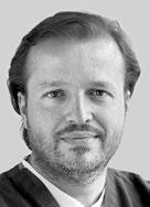
Marco Gadola CEO
Dear readers
With the introduction of the world‘s first single-stage implant in 1974, our company was right at the forefront in the pioneering era of implant dentistry. Thanks to consistent evidence-based research, an uncompromising quality standard, along with continuous development, optimization and simplification of products, instruments and protocols, we have made a crucial contribution to establishing this dental discipline worldwide with the Straumann® Dental Implant System.
Over 11 million Straumann dental implants have since been placed. All over the world, referrers and implantologists, who wish to make no compromises on behalf of their patients in terms of quality and safety, decide for our products. These customers put their patients‘ quality of life first. And they are personally convinced that Straumann delivers just the right products and solutions to achieve this.
For this issue of STARGET we surveyed an international selection of implantologists on their decision criteria for their implant system of choice. In a nutshell: our customers know that dependable values are associated with the Straumann name. This instills a confidence that is also conveyed to patients. Moreover, there is the assurance of dealing with an industrial partner steeped in tradition, who will also be there in the future as an innovative pacemaker to support dental professionals the throughout the world in their daily work and therefore to also continue advancing implant dentistry in science and practice.
I trust that it will make interesting reading.
Best regards,
Marco Gadola
Quality in implant dentistry
What values are important for quality-oriented implantologists when selecting suppliers of implants and materials, which features of a system make the difference, and what do successful implantologists expect from a premium manufacturer and the corresponding product portfolio? An interview with Dr. med. Martina Hartstock, Dr. Jörg Kälber and Friedrich Eiche.
Instruments and protocols
Feedback and insights of our customers have always played a major role in product development at Straumann. For this reason, we conducted a series of interviews with dental implantology experts from all over the world to learn more about their experience with Straumann instruments and surgical protocols in daily practice.
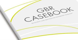
MembraGel® Case Book
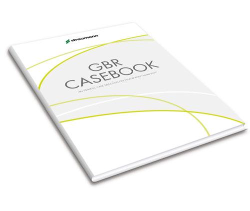
In 2010, Straumann introduced MembraGel®, which ranks as one of the most important innovations in guided bone regeneration . The new MembraGel® Case Book includes a summary of current knowledge on the successful application of Straumann® MembraGel® in GBR procedures. Our specialists have compiled specific step-by-step instructions and clinical report cases by renowned clinicians for each indication.
The fundamentals of quality-oriented implantology
Instruments and surgical protocols from Straumann Clinical case reports
Steffen A. Wolf (NNC)
Mario Kirste (MembraGel®/BoneCeramic®)
Mario Roccuzzo (MembraGel®)
Guido Rhemrev (Emdogain®)
1st international Symposium CCDE in Bern, Switzerland
The fundamentals of quality-oriented implantology
What is important to quality-oriented implantologists when selecting implant suppliers and materials? What system characteristics make the difference?
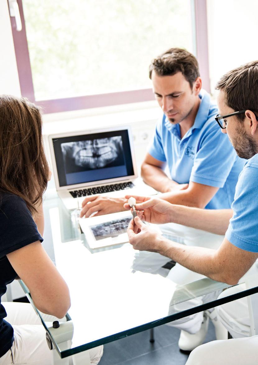
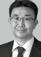
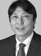
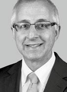
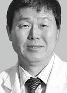
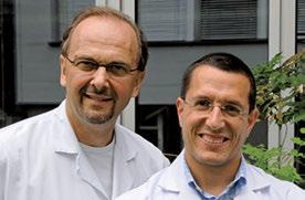
Three surgeons explain what successful implantologists expect from a premium manufacturer and the corresponding product portfolio in an interview with dentist and specialized journalist Dr. Aneta Pecanov-Schröder: prospective oral and maxillofacial surgeon Dr. Martina Hartstock (practice clinic of Dr. Achim Herrmann & Colleagues, Starnberg/Germany) and oral dental surgeons Dr. Jörg Kälber and Friedrich Eiche (practice of Dr. Eiche & Colleagues, Stuttgart/Germany).
Quality, a scientific approach and validated data
“It has always been extremely important to me to use an implant system with validated study data on longterm outcomes, which should also cover further developments,” says Jörg Kälber, who is closely involved with research and teaching by the International Team for Implantology (ITI) as a Fellow of the ITI. “This ensures that the risk to patients with regard to implant failure is as low as possible in this respect,” adds Kälber, who has gained experience of many different implant systems, including as senior physician at the clinic for oral and maxillofacial surgery at the Katharinen Hospital in Stuttgart (Medical Director: Prof. Weingart). Quality, a scientific approach and the availability of validated data are relevant selection criteria for collaboration with a premium supplier in implantology.
Permanency must be ensured
A total of six different implant systems are used at the practice of Dr. Eiche & Colleagues “in order to cater to the needs of referring colleagues.” According to Friedrich Eiche, “as a matter of principle, it’s important to us in the team to use an implant system for which replacement parts are also likely to be available in 30 or 40 years’ time,
even for implants that are no longer manufactured.” This ensures that mechanical complications, such as loosening of the abutment, can be dealt with.
“In the future, we will increasingly meet patients in whom the dental restoration has to be renewed on older boneintegrated implants. We need a certain degree of flexibility and longevity here to find the right solutions.” Martina Hartstock
Prospective oral and maxillofacial surgeon Dr. Martina Hartstock also values the longevity of a supplier, including in terms of ensuring credible support for discontinued products: “In principle, implant suppliers should make prosthetic components and related instruments available for older implants that are in situ at all times. Ultimately, explantation of bone-integrated implants just because further prosthetic maintenance is no longer possible is not a solution.” The surgeon from Starnberg predicts that “in the future, we will increasingly meet patients in whom the dental restoration has to be renewed on older bone-integrated implants. We need a certain degree of flexibility and longevity here to find the right solutions.”
Customer care with competent service and support
In these types of situation, a practice notices in particular how important it is for an implant supplier to provide targeted customer care with competent service and support. Both Kälber and Eiche firmly believe that “a sales rep is an integral part of the sales structure of a quality-oriented implant supplier.” But implantologists don’t only value a confident sales rep in technically demanding situations: “We also take advantage of the rep to support our training events.” Hartstock adds: “Just recently, we organized training for our referring colleagues. Straumann provided us with competent support at all times in this.”
Colleagues in Stuttgart/Germany)
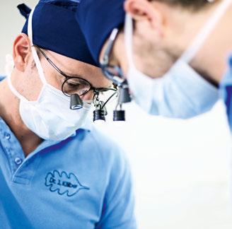
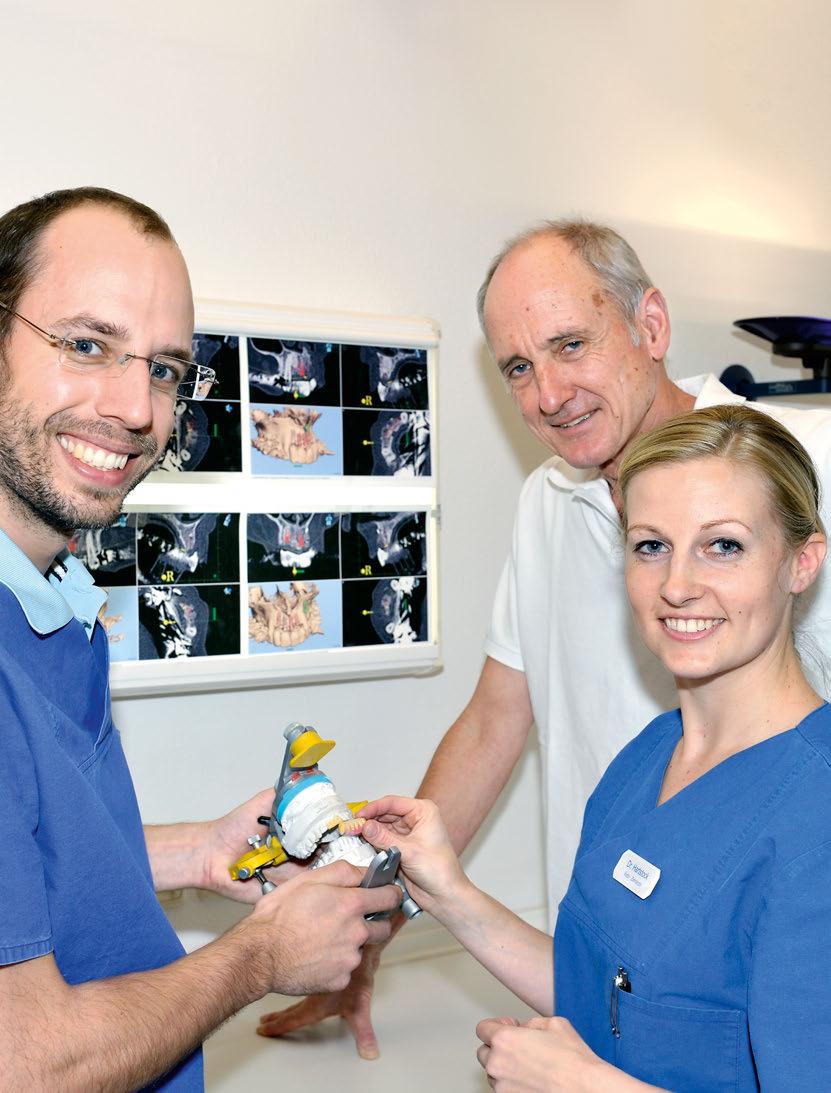
7 Photo opposite (right to left): Friedrich Eiche and Dr. Jörg Kälber with colleague (Dental Practice Dr. Dr. Eiche &
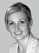
Following completion of studies in medicine and dentistry, training as an oral and maxillofacial surgeon in the practice clinic of Dr. Achim Herrmann in Starnberg/Germany. Areas of interest include surgical and prosthetic strategies for practice, complex situations in implantology and innovative soft tissue management techniques.
The reason given above is one of the motivations as to “why we largely only use premium implants in our practice clinic now,” says Hartstock. Like her colleagues in Stuttgart, she also believes it to be important that the supplier both integrates scientific advances in the further development of the implant systems and also “makes these readily available to us as treating dentists, for example within various expert meetings.” Hartstock underpins her high expectations of herself and of the treatment outcome by, among other things, being active in training alongside her work at the practice. In this way, she passes on scientific knowledge and allows an expert audience to question her practiceoriented theories. “We also do this regularly within the dentists’ team, to ensure we stay up to date with the latest knowledge,” she adds.
The constant exchange within the team promotes treatment quality
One of the benefits of working in a quality-oriented team is always being able to discuss patient cases “in order to develop the best treatment plans for our patients” (Kälber, Eiche). As Hartstock says, “constant exchange promotes critical scrutiny of individual treatment options and thus improves the level of treatment.” It also enables many team members to react in a far more flexible way to needs and requirements. Eiche says, “This is demonstrated by the follow-up, for example. In addition to normal follow-up appointments, in the post-operative phase, we can be reached by patients 24/7.” Practice duties are allocated based on strengths and specialization. “Our patients benefit from this overall situation,” Eiche adds.
Focusing on the patient
The fundamental principle is to implement the patient’s wishes as far as possible, and to inform him or her about possible treatment concepts and implantological rehabilitation. This practice concept can be brought to life and successfully integrated into daily routine when the (dentists’) team functions and adapts the implantological tools to a quality-oriented treatment approach that targets lasting success. Treatment safety, as demonstrated by documented and predictable outcomes, is extremely important to patients. “Patients often stress how important it is for them that a ‘cheap implant’ isn’t used,” Eiche notes. From the point of view of patients, costs play a minor role with regard to the implant system. Kälber remarks that “aspects such as quality and longevity of the materials used are important.” In the practice, to ensure the long-term success of implant prosthetic rehabilitation, it is important to use the original parts
Dr. Martina Hartstock
Photo opposite (right to left): Dr. med. Martina Hartstock, Dr. Dr. Achim Herrmann and Dr. Johannes von der Gathen (Practice clinic Dr. Dr. Achim Herrmann & Colleagues in Starnberg/Germany) 3
Dr. Jörg Kälber
After studying dentistry, he trained as an oral surgeon at the Clinic for Oral and Maxillofacial Surgery at the Katharinen Hospital in Stuttgart/ Germany, where he was a senior physician for several years before joining the practice of Dr. Eiche & Colleagues. Fellow of the International Team for Implantology (ITI).
from the supplier. This is where, for example, the Pro-Original Initiative comes in, through which Straumann campaigns for the use of original components. Hartstock says that “everyone involves benefits from this in the long term.”
“In our opinion, a sales rep is an integral part of the sales structure of a qualityoriented supplier.” Jörg Kälber and Friedrich Eiche
The benefits of the Straumann® Dental Implant System
What is it that the surgeons Hartstock, Eiche and Kälber find so convincing about the Straumann® Dental Implant System (SDIS), comprising various soft tissue and bone-level implant types, which they use regularly for implants? Kälber says “In addition to the quality, I’m persuaded by the solid scientific background offered by the company and the implants.” One current example of this is the publication by the company of new research findings from a largescale, long-term study carried out by the University of Berne on the survival and success rates of implants. The study headed by Prof. Daniel Buser assessed the ten-year outcomes of 511 Straumann® Soft Tissue Level implants with SLA® surfaces in 303 patients .
The study is one of the first major ten-year clinical studies on the outcome of treating patients with a dental implant of this type which is still on the market. According to the company, the Straumann® Soft Tissue Level implant with the SLA® surface is one of the best documented dental implants. The design of the Soft Tissue Level implant makes soft tissue management easier and means it can be placed in a single procedure, avoiding the need for a second surgical intervention. The typical ‘tulip’ design of the implant collar offers built-in soft tissue management.
These benefits, as proven in long-term studies, are reflected in current practice. As Hartstock says, “such a simple and feasible concept is important to many referring colleagues as well as many patients. The prosthetic implant interface is also easily accessible.” In addition to soft tissue-level implants, Hartstock and her colleagues Dr. Achim Herrmann and Dr. Johannes von Gathen also use bone-level implants. In the case of bone-level implants, the operator has to create an emergence profile, which can be perceived as ‘more difficult’ to some
extent. “But I’ve personally always been fascinated by soft tissue management,” says Hartstock.
A single set of instruments for all implants
The desire for a perfect solution in all indications with a single implant type is unrealistic. “For example, with the current level of knowledge and taking into account esthetic possibilities, I would favor a two-stage bone-level implant with platform switching for upper incisors, and would use, for example, the Straumann® Bone Level implant,” explains Kälber. Irrespective of which of the four implant lines within SDIS is chosen, all implants can be placed using a single set of instruments and very similar surgical procedures. Simplification of the logistics together with easier handling of an implant system are relevant criteria when making a selection, says Kälber. This limits potential failures and thus also lowers the technical complication rate.
“From a surgical point of view, the advantage of still being able to decide which implant to use after preparing the implant site using the same drill bit should not be underestimated.” Jörg Kälber
In addition, “from a surgical point of view, the advantage of still being able to decide which implant to use after preparing the implant site using the same drill bit should not be underestimated.” say the Stuttgart-based oral surgeons, “Whether it’s a classic Straumann® Soft Tissue Level implant, a Bone Level implant, a titanium implant, one made from the innovative material Roxolid® – or, even in the future, a ceramic implant: only the diameter of the implant used has to be specified beforehand.” This provides a sense of security in daily practice. “And this sense of security is conveyed from the operator to the patients,” adds Eiche, “which is conducive to the most comfortable and calming overall atmosphere possible, particularly in the case of surgical interventions.”
The choice of implant is then not difficult: “I placed a premolar implant for my mother in her upper jaw around nine years ago,” says Kälber. “It was a Straumann® Soft Tissue Level implant, 10 mm long and with a 4.1 mm diameter. Both my mother and I were very happy with it then – and still are today.”
Martina Hartstock, Jörg Kälber and Friedrich Eiche were interviewed by Dr. Aneta Pecanov-Schröder, Dentinform agency in Bonn/Germany. The article first appeared in the professional journal DENTALE IMPLANTOLOGIE 17, 2 (2013).
Friedrich Eiche
Studied dentistry; training at a practice for oral and maxillofacial surgery in Ingolstadt/Germany and at the practice of Dr. Hans Thomas Eiche (his father and senior colleague), an oral and maxillofacial surgeon. Oral surgeon since October 2012.
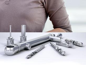
Straumann offers straightforward surgical protocols together with a comprehensive portfolio of highly functional and easy to use instruments to help our customers tackle even the most challenging clinical cases effectively and meet rising patient expectations.
Straumann instruments and surgical protocols
Focusing on the patient
Simplicity, reliability and quality
These are the cornerstones of the product philosophy we apply to developing surgical protocols and instruments. And this is why, at the heart of the Straumann® Dental Implant System, there is one single instrument kit for the entire range of implant types and indications. We want our customers to be able to focus instinctively on those aspects of surgery that really matter. In this respect, we are convinced that every clinician can master the procedural steps of the Straumann® Dental Implant System in a minimum of time and for most of the indications they may meet in daily practice.
Reducing complexity
One of our main objectives is to reduce complexity and allow practitioners to focus on the most important aspect of treatment – the patient. This enables clinicians to offer advanced and effective treatments and, at the same time, keep confidence regarding the outcomes. Since feedback and insights from our customers have always been important drivers behind Straumann’s product development, we conducted a series of interviews with dental implantology professionals from all over the world to learn more about their daily experience with Straumann instruments and surgical protocols. Read more about it on the following pages.
“I have been able to observe numerous postgraduates at the university trying to use the system for the first time. It usually takes about 2 – 3 procedures before feeling confident with the kit and the protocol.” Jordi Gargallo
When it comes to surgical protocols and instruments, what is important to you and why?
Gargallo: For me, the patient is center stage. Therefore I always favor treatment protocols and instruments that allow me to obtain excellent results while focusing on the well-being of the patient. I always aim to shorten the total procedure time and minimize operational trauma. Because it is coherent, the Straumann® Dental Implant System gives me peace of mind and the confidence that the planned outcome will be achieved.
Lascoe: Simplicity and ease of use. If a protocol is straightforward and entails only few steps, there is less room for complications and the whole procedure is more efficient. It’s important to me that I am able to focus on the patient
Jordi Gargallo Albiol, Spain PhD, MSc, DDS
Director of the Master program for Oral Surgery and Implantology, Faculty of Odontology, at the International University of Catalonia/Spain. Private practice in Tarragona and Torredembarra, Spain, dedicated entirely to implant dentistry, periodontology, and esthetic restorations. ITI Fellow, speaker and director of the ITI Study Club’s Tarragona section.
Questioning of the interviewees in alphabetical order.
during surgery, rather than on the protocol. For the same reason, I do not need a wide selection of instruments – as long as they cover the system and I can deal with all bone types, I am satisfied.
“We noticed the excellent cutting performance of the drills straightaway. This gives the feeling of controlled progression without extra pressure during the operation, which is very mindful of bone integrity.” Masaaki Hojo/Hideaki Katsuyama
Lee: I have a very busy practice and place around 400 implants a month, so for me and my team excellent organization, efficiency and predictable treatment outcomes are the most important factors. Our aim is to make most of the processes in our clinic as efficient as possible, which means that we only use products with proven functionality and a high clinical success rate. Every member of my team is well trained and has a clearly defined role or task during the surgical procedure. This allows me to focus entirely on the patient and the specifics of the operation. Manageable instruments and accessories, together with straightforward protocols, help us reduce surgery times, minimize patient trauma and ensure consistent treatment success.
Katsuyama/Hojo: Nowadays there is a wealth of scientific evidence in support of dental implant therapy. As clinicians we apply this knowledge and try to assure the best possible treatment results. From a practical point of view we often have to operate with submillimeter accuracy to achieve the desired outcome. Here, high quality and precise instruments help us handle even the very demanding clinical cases effectively.
Zarrine: I cannot view the surgical procedure without the implant; for me, these should work as a coherent, integrated system. I therefore consider straightforward sur-
gical protocols combined with well-researched implants backed by solid long-term data to be the crucial factors for excellent clinical outcomes.
What were the main benefits you noticed during your first surgery with the Straumann® Surgical Kit?
Gargallo: Ease of use and good organization, which make learning more effective. From the surgical to the prosthodontic stage, everything is designed to function perfectly: the planning aids, the implants, the healing abutments, as well as the temporary and final abutments. During surgery I always have all the required instruments on hand. While I have been working with the Straumann system for more than 13 years, I have also been able to observe numerous postgraduates at the university trying to use the system for the first time. It usually takes about 2 – 3 procedures before feeling confident with the kit and the protocol.
“When it comes to surgical procedures and instruments, I demand high precision and cutting efficiency, as well as consistent high-performance standards.” Ronald Lascoe
Lascoe: I liked the fact that it was just one easy-to-manage kit with a very lean range of instruments for all implant types. The sharp drills provided fast but balanced vertical progression. I also appreciate the rounded tip of the final twist drills, which resembles the implant tip. The V-shaped extension of the tip is very small which is helpful when I need to work close to the nerves and blood vessels.
Katsuyama/Hojo: We noticed the excellent cutting performance of the drills straightaway. This gives the feeling of controlled progression without extra pressure during the operation, which is very mindful of bone integrity. The cassette layout with its color-coded workflows makes it
easy to follow the protocol. We are, of course, aware that the ability to perform complex surgical treatments depends, above all, on the technical skills and experience of the practitioner. But the use of such top quality instruments also enables even experienced practitioners to attain new technical heights.
“I sometimes have to deal with quite difficult cases and clinical situations like insufficient bone quantity, immediate placement or early loading. Innovative implant surfaces and materials supplied by Straumann help me meet these challenges, extend my professional horizons and provide an excellent treatment to my patients.” Sepehr Zarrine
Zarrine: The excellent cutting power of the drills, together with perfect drill centering, makes osteotomy preparation extremely precise and efficient. The clearly visible markings on the instruments are very helpful when working under spray. And what surprised me most was the fact there was only one surgical cassette. When you have become used to separate kits it is a pleasant surprise to see how much easier your operations become when there is just one. Surgery sometimes requires last minute changes to be made to the plans. What happens if the correct instruments are not available? It also improved my organization if I had to perform several surgical procedures on one day. If you are using a system with different kits, you need two kits of each type to make sure treatment is not compromised because the required kit is in the autoclave!
As a practitioner with many years of hands-on experience in implant dentistry, where do you see Straumann surgical procedures making a difference in your practice?
Gargallo: I like the consistency and high level of standardization of Straumann’s surgical procedures. All implant types require similar steps, which gives me flexibility in implant placement. The drills provide a precise cut and a good view of the drilling depth. When using the Straumann® Dental Implant System I really feel that all the instruments have been designed to work as a system and make surgery easier.
Lascoe: When it comes to surgical procedures and instruments, I demand high precision and cutting efficiency, as well as consistent high-performance standards. And this is where Straumann meets my needs. They supply an
Masaaki Hojo, Japan DDS
Private practice in Yokohama, Japan. ITI Fellow and speaker. Director of the ITI Study Club MM.
Prof. Hideaki Katsuyama, Japan DDS, PhD
Professor at the Center for Oral Diseases, Fukuoka Dental College, Japan. Member of the ITI Board of Directors. Education Delegate of the ITI Section Japan.
instrumentation that is simple to use, works across different implant types and allows a high degree of efficiency, since the same instruments are used for almost all procedures.
Lee: From an operational point of view, I appreciate the fact that Straumann’s surgical protocols are well standardized and can be used intuitively by a trained practitioner. The surgical workflow is straightforward because the concept for preparing the implant bed is similar for all implant types within the system. Yet the protocol is flexible enough to cover all bone classes and I do not need dedicated instruments for hard or soft bone. Also, the instruments come in a single cassette – this means I have full flexibility during the operation to change from one type of implant to another within the same setup. Ultimately, the straightforward protocol has allowed me to optimize my operating times continuously, which has helped me successfully complete around 60’000 implant surgeries during 20 years of implant dentistry.
Katsuyama/Hojo: In the past we used our skills and experience to overcome specific deficiencies in surgical instruments. Now, with Straumann instruments and procedures, we can efficiently tackle even complex surgical cases. The entire system is well thought out; high precision instruments complement quite simple surgical protocols. This approach allows us to perform challenging surgical treatments accurately and safely, while taking our professional experience to a higher level.
“Today Straumann and its cooperation partner Dental Wings supply a complete solution that includes gonyX®, coDiagnostiX® and instruments specially designed for Guided Surgery. We are now actively integrating these procedures in our practice and exploring the new possibilities that Guided Surgery offers for challenging protocols.” Masaaki Hojo/Hideaki Katsuyama
Zarrine: What I appreciate is that the Straumann protocol is comprehensible, with very similar steps for all implant types – this operational ease of use saves valuable time in the practice. The logic of the procedure and the colorcoded drilling sequences are easy to follow, which gives me peace of mind during complex surgical cases.
What is your particular interest in surgical procedures?
Gargallo: My interest is to move towards instruments and materials that scientific evidence has shown provide consistent and predictable clinical outcomes. This includes, for instance, a preference for the Straumann® SLActive surface, with the aim of offering my patients excellent treatment results, even in challenging cases.
Lascoe: I would like to find out if there is any further room for protocol optimization. For me, the ideal solution would be to decrease the variety of drills, shorten the drilling sequence even further and make surgical steps as similar to each other as possible, irrespective of the implant type.
“Simplicity and ease of use is important to me when it comes to surgical protocols and instruments. If a protocol is straightforward and entails only few steps, there is less room for complications and the whole procedure is more efficient. It’s important to me that I am able to focus on the patient during surgery, rather than on the protocol.” Ronald Lascoe
Katsuyama/Hojo: Modern technologies such as Guided Surgery allow the precise digital planning of surgical procedures. In the past, however, difficulties arose because of the lack of a dedicated instrumentation when these plans were put into practice. Today, Straumann and its cooperation partner Dental Wings supply a complete solution that includes gonyX®, coDiagnostiX®and instruments specially designed for Guided Surgery. We are now actively integrating these procedures in our practice and exploring the new possibilities that Guided Surgery offers for challenging protocols.
Lee: I think the design of the drill is very important, particularly in cases where I have to work very close to alveolar nerves, blood vessels and sinuses or proceed with extreme caution and accuracy when every half-millimeter counts. As the tip of the Straumann final twist drill closely resembles the profile of the implant tip, the drill has a very small V-factor. This “flat tip” design and high precision of the drill cutting geometry give me confidence when working close to vital anatomical structures. At the same time, the drills provide good cut and stability, which allows me to progress effectively, even in hard bone.
Ronald J. Lascoe, United States
DMD
Graduate of the University of Pennsylvania School of Dental Medicine, Philadelphia. Chief dental resident at the Veteran’s Administration Hospital in West Los Angeles, California from 1984 – 1985. Taught at the UCLA School of Dentistry 1983 – 1985 and was Head of Periodontics at Rancho Los Amigos Hospital in Downey from 1986 – 1990. Private practice in Burbank, California.
Jae-Yoon Lee, Republic of Korea DDS
Graduated in dentistry at the University of Seoul National Dental School in 1982. Private practice in Dae-Gu City/ South Korea since 1988. His main field of interest is implantology in single to edentulous cases. Author of numerous books on implant dentistry.
Zarrine: I have a strong experience in immediate implantation and the holding key is a true benefit of the Straumann® Dental Implant System in cases of very soft bone and low primary stability since it allows me to keep the implant securely in place, while safely removing the transfer piece.
Would you recommend Straumann’s surgical workflow and instrumentation to your colleagues, and if so, what features would you highlight?
Gargallo: Essential arguments are that the Straumann® Dental Implant System is simple to handle both surgically and prosthetically, and is also highly predictable in terms of the long-term stability of the soft and hard tissues surrounding the implant. It is also a system that can be mastered with a steep learning curve; straightforward protocols complemented by the color-coded navigation on the cassette make even the very first surgery easily manageable.
Lascoe: I would highlight the operational ease of use, the right number of instruments, the very handy planning aids and the excellent quality of the drills. I believe that all these advantages are significant factors for every practice when it comes to increasing operational efficiency.
“The straightforward protocol has allowed me to optimize my operating times continuously, which has helped me successfully complete around 60’000 implant surgeries during 20 years of implant dentistry.” Jae-Yoon Lee
Lee: Having performed thousands of implant surgeries in the past two decades, I had the opportunity to try different systems. I eventually chose the Straumann® Dental Implant System – and have not looked back since. The system works well operationally and has provided my clinic with high longterm success rates. If someone were now to ask me why I only use Straumann, my answer would be: the continued bone stability even in old cases that I completed several years ago. On the technical side, as the surgical protocols are flexible, I am able to meet the intra-operative challenges as they arise. It means I am free to modify my plans during an operation. For instance, I
can make a basic implant bed preparation for tissue level, but then, if necessary, I can change to bone level by performing the corresponding profiling and taping. I appreciate Straumann’s commitment to the “One surgical kit” concept because it simplifies clinical complexity in respect of instrument use and reprocessing, as well as giving me peace of mind knowing that the next generation of products and workflows will still be compatible with the same concept.
“I consider straightforward surgical protocols combined with well-researched implants backed by solid long-term data to be the crucial factors for excellent clinical outcomes.” Sepehr Zarrine
Katsuyama/Hojo: We would emphasize the flexibility of the system, because it provides consistently standardized instruments for both guided and standard implant placements. Furthermore, the Straumann® Guided Surgery portfolio provides advanced solutions for the more experienced surgeons wishing to tackle clinically complex or challenging cases. This makes the Straumann® Dental Implant System a fully comprehensive system that offers practitioners of different levels of experience an excellent portfolio.
Zarrine: The whole system is very well conceived: it is advanced, yet simple and efficient to use. You plan for surgery, decide on the implant to be placed and then simply follow the color-coded roadmap of the protocol and concentrate on your treatment – all in all, it is ergonomic, logical, and straightforward. I sometimes have to deal with quite difficult cases and clinical situations like insufficient bone quantity, immediate placement or early loading. Innovative implant surfaces and materials supplied by Straumann help me meet these challenges, extend my professional horizons and provide an excellent treatment to my patients. As technologies get more sophisticated, I would also appreciate the company’s continuous commitment to providing progressive and easy-to-use solutions, which will allow me to address all kinds of clinical situations efficiently.
Private implant dentistry practice in Saint Dié, France. European Master’s Degree in Implant Dentistry and Bone Grafting. University Diploma in Maxillofacial Surgical. Course trainer at the French ITI section. Author of scientific papers on implant dentistry.
Sepehr Zarrine, France DDS
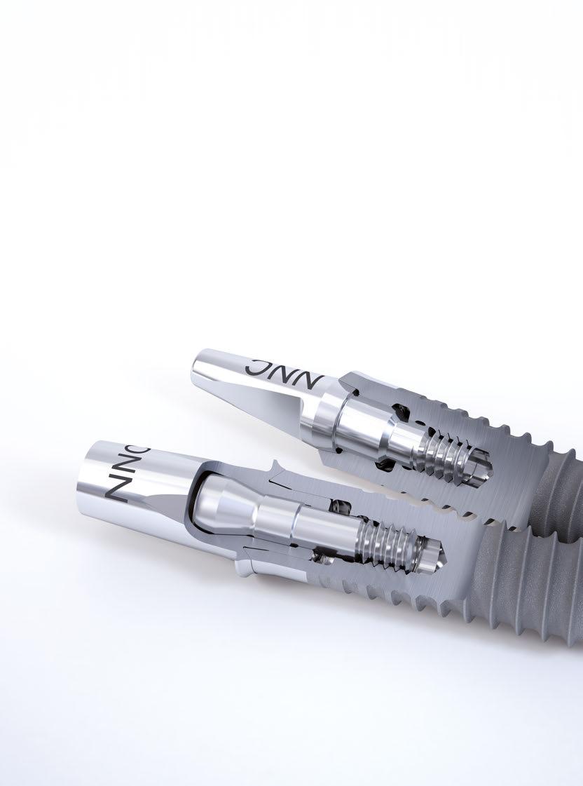
Straumann® Standard Plus Narrow Neck CrossFit® Implant (NNC)
Bridge construction in the anterior tooth area of the maxilla
Steffen Wolf, Germany
Initial situation
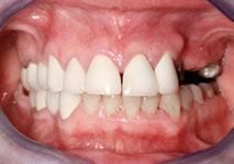
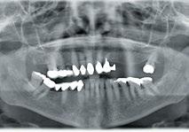
A 67-year-old patient presented in the dental practice for implant consultation. The anamnesis revealed some specific conditions, particularly an allergy for dental metals. At this time, prosthetic restoration in the area to be reviewed consisted of an insufficient crown block in the anterior tooth area corresponding with an attachmentmonoreducer-combination denture. Significant loosening of the abutment teeth in the anterior tooth area was found, posts and cores that had already loosened several times were found in the insufficiently filled root canals, probably due to monoreducer leverage (Fig. 1). The prognosis for conservative restoration was thought to be extremely poor. During the consultation the patient expressed preference for an implant solution. The patient also specified a cost limit.
Procedure
Treatment planning. For optimum assessment of the initial situation and subsequent treatment planning, after assessing the clinical situation, a DPR diagnosis with intraoperative assessment of the implant site was favored as method of choice (Fig. 2). This would take into account a minimally invasive therapeutic concept of surgical augmentation. Operation planning involved the extraction of nonconservable teeth and immediate restoration of a Straumann® Bone Level Implant in the region. Two implants were to be inserted in the premolar region. We planned to expand bone with the bone spreading procedure and to use two Straumann® Standard Plus Narrow Neck CrossFit implants (NNC) made from the implant material Roxolid® if the transversal bone at the site was compromised. Prosthetic restoration must fulfil the re-
Fig. 1
Fig. 4
Fig. 2
Fig. 5
Fig. 3
Fig. 6
quirements of an allergy free dental prosthesis. The prosthetic construction was to be manufactured with the Straumann Cares System in the in-house dental master laboratory.
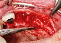
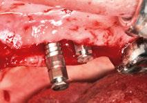
Surgical procedure. Due to the impaired vasoconstriction, anaesthetization was adrenaline-free with local anaesthetic and one subsequent injection during the operation. Extraction of the middle and left lateral incisors was without complication. A central crestal incision was made with little crestal bone denudation and no relief incision. The anticipated reduction of the transverse bone then became clearly visible and, as method of choice, the bone spreading procedure and two NNC implants were performed (Fig. 3). The insertion site in the region of both left premolars was prepared by manually shaving the bone until an even bone plateau had been created. The
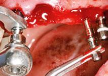
autologous bone chips gained here were later used for implant augmentation in the central left incisor area. Once the implant site had been carefully prepared by means of bone spreading (Fig. 4) and the final implant cavities drilled, the prepared bone was meticulously examined with a bulbous probe and gauges from the Straumann surgery set. Two NNC implants were then inserted in the controlled, intact bony structures (Fig. 5). The NNC implant 3.3/14/SLActive® was inserted in the region of the first premolars, the 3-mm reduced height NNC healing cap was used for both the implant seal, as well as for primary soft tissue conditioning. We decided to use NNC implant 3.3/12 NNC/SLActive® and the identical 3-mm closure screw for the region of the second premolars. Once this stage of the operation was complete, alveolar implant restoration in the central anterior tooth area was performed. The immediate implantation of a Straumann bone level
Fig. 7
Fig. 10
Fig. 8
Fig. 11
Fig. 9
Fig. 12
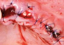
implant with the dimensions 4.1/10 which is fitted with the 0.5-mm RC closure screw was then performed. The alveolar walls were undamaged, primary implant stability was given. As a sufficient amount of autologous bone chips had been gained from maxillary crest leveling in the premolar area, this was used as volume filler for alveolar augmentation. The distance between the body of the implant and the alveolar wall that required augmentation was 1 – 2 m m. Augmentation was vertical with slight overlap by means of a platform switch at the implant shoulder. Alveolar restoration of the lateral incisor was performed using collagen matrix. Suture closure in the area of the anterior tooth implant resulted in complete coverage of the augmentation area, the closure screw lay only minimally exposed approx. 3 m m below the mucogingival soft tissue. Soft tissue closure at the NNC closure screw supported transgingival healing of the
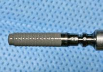
implant (Fig. 6). Intraoperative haptic assessment of the various fixations of the implant insertion aids was easily possible (Fig. 7). To assess postoperative treatment success, in particular with regard to adequate peri-implant bone coverage, a control DTV was made on which the correct implant-bone relation could be verified. This meant additional augmentation measures could be safely dispensed with (Fig. 8). Perioperative medication included antibiotic endocarditis prophylaxis; the patient was also given postoperative pain medication for one day.
Prosthetic restoration. Following integration of a provisional denture and a complication free healing time, individual gingival architecture then was performed in the anterior tooth area. To facilitate continued wearing of the provisional denture during the gradual process of soft tissue conditioning, our dental laboratory prepared and short-
Fig. 13
Fig. 16
Fig. 14
Fig. 17
Fig. 15
Fig. 18
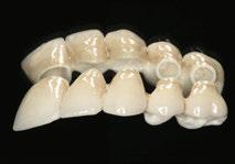
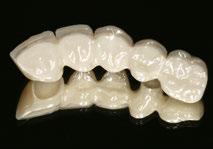
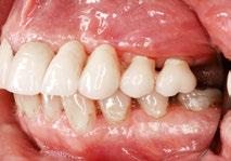
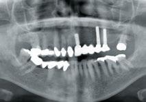
ened an RC temporary abutment with hard polymer plastic, individualized to the area of the soft tissue profile (Figs. 9 – 11). The impression for the individual incisor abutment was made with a gingiva-former on the basis of an RC impression post to match the individual impression post. The NNC implants were incorporated into the impression (Fig. 12) with the ready-made NNC impression posts. On account of the patient’s allergy and in consideration of the esthetic aspect, in addition to titanium abutments (Fig. 13) it was also decided to use a zirconium-based bridge framework with ceramic veneering (Figs. 14, 15). The titanium abutments and zirconium bridge were constructed virtually in CADCAM procedure with the Straumann CS2 scanner in our own dental laboratory and the framework was made at the Straumann Milling Center in Leipzig. Because of the interocclusal distance, the decision was to use an anatomically formed zirconium morsal surface, which could be optimally prepared with the Straumann® CARES® system processing software during the construction phase. In consideration of the esthetic aspect, the individual veneer was mostly in the vestibular region (Figs. 16, 17). Postoperative x-ray control confirmed correct positioning of the prosthetic components (Fig. 18)
Conclusion
The patient is extremely satisfied with both the result and the cost-effect relation. Appropriate design of the emergence profile, the titanium abutment and the zirconium bridge entirely fulfil the esthetic requirements of the visible areas. In the event of later loss of the second molars the patients wishes to undertake prosthetic restoration of the ensuing end gap situation. As shown here, in cases of compromised bone and in consideration of the esthetic zone and CADCAM-made elements of different materials, the use of NNC implants can lead to very positive results.
Steffen A. Wolf Dr. med. dent., M.Sc. (DGI)
Attainment of the degree of Doctor of Dentistry in 2000 from the Clinic for Oral and Maxillofacial Surgery at the Free University of Berlin headed by Prof. B. Hoffmeister. Since 2000 working in own private practice in Halberstadt/Germany. Received the degree of Master of Science in Oral Implantology in 2010.
Treatment of a dehiscence-type defect around a dental implant
Mario Kirste, Germany
Case description
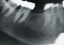
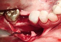
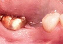
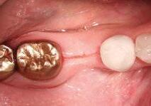
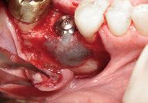
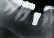
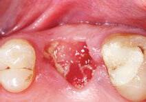
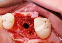
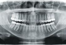
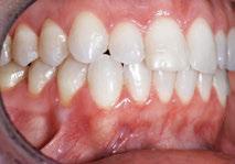
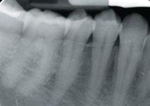
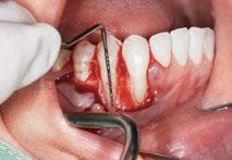
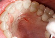
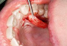
The patient (female, 59 years, non-smoker, general good health conditions) presented purulent pulpitis at tooth # 46. As endodontical treatment did not succeed in improving her symptoms it was decided to extract tooth # 46 and place an implant in combination with a guided bone regeneration procedure in a submerged healing approach after the site has healed. The bone defect after tooth removal at site # 46 was obvious in the radiograph as well as during visual inspection (Figs. 1 – 3)
Surgical procedure
Treatment planning. Preoperatively, antibiotics were given (Penicillin, 1.5 m g). The surgery was performed under local anesthesia (3.4 m L Articain + Epinephrin).
In addition, an intraossary anesthesia was performed (Anesto, W&H).
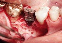
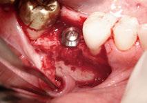
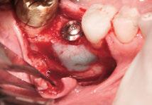
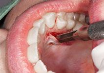
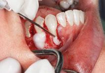
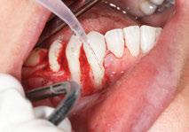
The horizontal incision was performed mid-crestally, with one vertical releasing incision 1 tooth distant in the mesial aspect of region # 46; care was taken that the flap was elevated sufficiently in all directions. To improve access to the implant site, the lingual flap was slightly elevated, approx. 3 mm (Fig. 4). The implant (Straumann® Standard Plus ∅ 4,8 WN, 10 m m) was inserted according to the standard protocol. After the placement of the implant, a small dehiscence-type alveolar bone defect of approx. 4 m m was present. Small bleeding points were created in the cortical layer of the augmentation site to improve bone healing capability (Fig. 5)
Fig. 1
Fig. 4
Fig. 2
Fig. 5
Fig. 3
Fig. 6
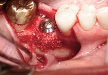
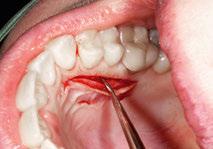
Using the sandwich technique, the implant surface was augmented first with a layer of autogenous bone which was collected during careful implant site preparation. Straumann® BoneCeramic™, proven to be a slowly resorbable scaffold for bone regrowth, was added to the outer layer of the lateral defect. Prior to its application, Straumann BoneCeramic was slightly rehydrated in blood collected at the defect site. Overbuilding of the defect was avoided during augmentation procedure (Figs. 6, 7). In order to optimize the attachment of Straumann® MembraGel to the recipient site, the host bone was carefully dried with a sterile gauze immediately before application.
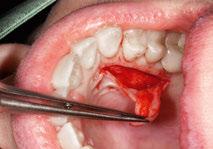
Straumann® MembraGelwas applied to the defect site in a thin layer, outlining the bone substitute material with
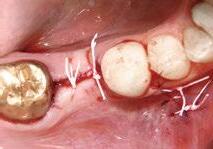
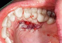
1 – 1.5 m m in all dimensions. To avoid full coverage of the implant head with Straumann® MembraGel in the crestal aspect, the membrane surface was reduced carefully with a sharp scalpel (Figs. 8, 9). Flap release for tension-free wound closure was achieved by means of periosteal releasing incision. Healing was attempted with the implant in a submerged position after tension-free closure of the released mucoperiosteal flap. The wound was closed with interrupted sutures (Gore-Tex® 0 – 5 RT16, Fig. 10).
Postoperative treatment and result. The patient was instructed to rinse 3 – 5 t imes daily with CHX (0.1 %) and, for post-surgical pain, to use analgesics according to her individual need. Furthermore, antibiotics were prescribed for 5 days (Penicillin 1.5 Mega, 3⊗daily). The sutures were
Fig. 7
Fig. 10
Fig. 8
Fig. 11
Fig. 9
Fig. 12
Private practice for referrals in oral surgery and implantology in Frankfurt (Oder)/Germany. Speaker/course instructor for implantology, regenerative surgical treatment and bone management). Clinical and scientific investigations for augmentative treatment methods, piezosurgical devices and intraossary anesthesical devices. Author of clinical articles in national magazines. Author of a handbook about intraossary injection. Memberships: DGI, ITI (Fellow).
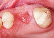
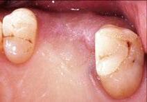
removed 14 days after the implantation procedure. The overall healing period was uneventful. At time of healing cap insertion 6 months post-surgically clinical and radiological evaluation showed stable hard and soft tissue conditions (Figs. 11 – 14). The final restoration by means of a full ceramic crown was completed 6 months later (Figs. 15, 16). The patient presented a fully restored buccal profile.
Findings
It could be demonstrated that the employment of Straumann® MembraGel and Straumann® BoneCeramic can be very useful for lateral recontouring of this dehiscence type defect and, in this regard, this case report confirms the data* on the osteoconductive properties of Straumann® BoneCeramic.
3 Scientific reference: see www.straumann.com/stargetref
Fig. 13
Fig. 14
Fig. 15
Dr. Mario Kirste DDS, M.SC.
Fig. 16
Straumann® MembraGel
Socket preservation of a compromised extraction site
Mario Roccuzzo, Italy
Case description
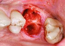
The patient (m, 45) presented to my practice with severe periodontitis apparent from a deep periodontal pocket of 8mm with 3mm recession and pus visible through the mucosa on the buccal side with involvement of the furcations (Fig. 1). He was generally in good health. The recorded OPG shows the hopeless situation for tooth # 26 (Fig. 2). The proposed treatment plan was careful extraction of # 26 followed by simultaneous socket preservation in a flapless approach with staged implant placement into the regenerated socket and a fixed metal ceramic prosthesis at # 26 as a final restorative solution. The aim of this patient-friendly treatment approach was to preserve the soft tissue conditions and to prevent bone resorption in the posterior area in order to avoid the need for any sinus augmentation procedure at the time of implant placement.
Surgical procedure
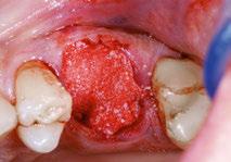
Treatment planning. Preoperatively, antibiotics (Augmentin 1 g) and analgesics (Ibuprofen 600 mg) were given. The surgery was performed under local anesthesia. The tooth was extracted carefully; removal of granulation tissue was achieved by thorough curettage (Fig. 3). The buccal plate was partially absent and significant bone dehiscence could be identified. Before the bone grafting procedure, the epithelium around the border of the socket was eliminated by means of a 15c blade and the extraction socket was rinsed thoroughly with sterile saline solution. The defect was homogenously filled with bone graft substitute material up to the mesial and distal bone margins (Fig. 4) and then moistened with sterile saline. Before membrane application, the excessive saline was removed by means of a sterile gauze. Straumann® MembraGel was
Fig. 1
Fig. 4
Fig. 2
Fig. 5
Fig. 3
Fig. 6
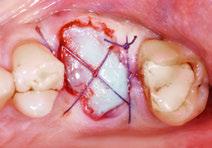
applied to completely cover the bone substitute material in all dimensions. Care was taken not to overfill the socket and stay slightly underneath the crestal soft tissue margin to facilitate soft tissue granulation afterwards. After setting, Straumann® MembraGel was kept in place by placing a modified external criss-cross-suture (Vicryl, 5 – 0). Care was taken not to disrupt the membrane during the suturing procedure (Fig. 5)
Postoperative treatment. The patient was instructed to rinse three times daily with a 0.2 % CHX solution (1 minute) for a period of 3 weeks. For post-surgical pain, analgesics were prescribed (Ibuprofen 600 mg, as needed). Furthermore, antibiotics were prescribed for the next 6 days (Augmentin 1 g in the evening immediately postop, twice daily for the next 5 days). The patient was seen
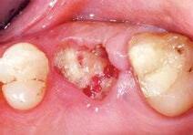
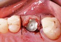
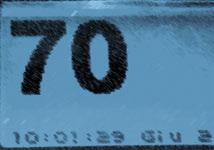
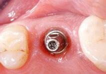
after 10 days for suture removal (Fig. 6) and then weekly for the first month to monitor the healing process (Fig. 7). Complete wound closure was detected approx. 5 – 6 weeks post-op (Fig. 8). The overall healing process was uneventful. At the time of re-entry and implant placement 4.4 months after extraction socket treatment, the patient presented healthy gingival conditions (Fig. 9). After flap preparation, a nicely regenerated alveolar bone was found with sufficiently preserved volume (Fig. 10). Therefore, the implant (Straumann® Standard Plus ∅ 4,8 WN) could be placed without any further augmentative procedure (Fig. 11). The bone quality present at the time of implant insertion allowed stable implant placement, as indicated by an Osstel value of 70 ISQ (Fig. 12). Therefore the abutment could already be placed 4 weeks later (Figs. 13, 14). The final restoration by means of a single fixed metal ceramic
Fig. 7
Fig. 10
Fig. 8
Fig. 11
Fig. 9
Fig. 12
prosthesis in area # 26 was installed 12 months after socket preservation surgery (Fig. 15). The clinical and radiological evaluation at this time revealed stable bone and soft tissue conditions with a fully restored emergence profile (Fig. 16)
Findings
Thanks to its liquid application, Straumann® MembraGel presents a userfriendly method to seal a grafted extraction socket in a flapless procedure. In the presented case, Straumann® MembraGel led to uneventful post-surgical soft tissue healing, while at the same time sufficiently protecting the bone graft. Therefore, at time of implant placement, the patient presented sufficiently preserved bone and soft tissue conditions, allowing implant placement without any need for additional soft or hard tissue augmentation procedures.
ISQ is a trademark of Osstell AB, Sweden.
Lecturer in Periodontology at the University of Siena/Italy. Private practice limited to Periodontology and Implantology in Torino/Italy. Extensive research in the field of mucogingival surgery, bone regeneration, implant loading protocols and implants in periodontally compromised patients. Active member of the Italian Society of Periodontology and ITI Fellow.
My thanks goes to Franceso Cataldi for the fabrication of the crown.
Fig. 13
Fig. 14
Fig. 15
Fig. 16
Osstell
Mario Roccuzzo D.M.D.
Straumann® Emdogain
Buccal bone dehiscencies in single periodontal infections
Guido Rhemrev, Netherlands
Initial situation
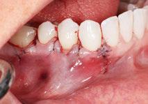
A 41-year-old healthy (ASA I) non-smoking female was referred for periodontal treatment of a localized severe periodontal infection at the first mandibular premolar (Figs. 1, 2). A regular oral hygiene treatment prior to the referral did not improve the clinical situation. During the periodontal intake (Figs. 3 – 4) a severely deepened pocket (10 m m) with inflammation and suppuration buccally at # 4 4 was confirmed.
General periodontal examination showed some minor inflammation with few shallow pockets among other teeth. A frenulum near # 4 4 and some brushing trauma were noticed. No abnormalities were affirmed during the radiographic examination. The oral hygiene status was
on an acceptable level. No previous injury was reported. The patient complained about some minor pain sensation during the last year before intake.
Treatment plan
Treatment planning. All teeth in the fourth quadrant reacted positive to cold test. A thoroughly initial periodontal therapy with supra- and subgingival debridement was avoided in order not to aggravate further loss of periodontal attachment and gingival recessions. Therefor it was decided to perform a diagnostic flap by means of periodontal plastic (micro-) surgery with the preservation of the papillae in order to evaluate the clinical condition in the fourth
Fig. 1
Fig. 4
Fig. 2
Fig. 5
Fig. 3
Fig. 6
quadrant. A regenerative procedure would be possible as a second stage during surgery. Oral hygiene instructions were given prior to surgery and an extended postoperative care program was scheduled during the first 6 months after. Oral hygienists were instructed to refrain from any subgingival curettage or probing during this period.
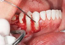
Surgical procedure and clinical findings. A diagnostic flap with two releasing incisions was prepared (Fig. 5). Intrasulcular incisions by using a micro blade were made in the area of # 43, 44 and 45. The gingival tissue of the interproximal papillae was carefully thinned and separated from the proximal gingiva. The buccal flap was fully raised to make clinical examination possible of the bone
dehiscence and the surrounding tissues. By coincidence a second severe bone dehiscence buccally of # 43 was exposed. It seemed clear that # 43 and # 4 4 were positioned outside the genetically determined envelope and thereby had lost most of their buccal alveolar wall. The rootsurface in the apical area of # 4 4 showed minor areas of deterioration explaining possible episodes of rootresorption. A secondary diagnosis could be found in an iatrogenic trauma after subgingival instrumentation. No clear remnants of calculus were visible. A free connective tissue graft of 1,5 to 2 m m in thickness was harvested using a modification of the Single-Incision-Technique described by Weng and Hurzeler* (Figs. 6 – 11). The rootsurface in the apical area of # 4 4 was thoroughly debrided by using an
Fig. 7
Fig. 10
Fig. 8
Fig. 11
Fig. 9
Fig. 12
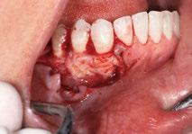
ultrasonic device and small diamond burs. No mechanical debridement was carried out at the surface of # 43. Both root surfaces were conditioned with 24 % EDTA gel for 2 minutes, copiously rinsed with sterile saline and dried with air by using the multi-unit syringe. Straumann® Emdogain was applied on the rootsurfaces when the surgical area was completely free of blood starting from the most apical bone level and covering the entire surface (Figs. 12 – 14). The free connective tissue graft was carefully adapted to the wound area and partially covered by the soft tissue in the area of # 42 (Fig. 15). A horizontal releasing incision was made in the periosteum at the base of the flap to allow tension-free coronal flap replacement. Subsequently the flap was coronally repositioned on top
of the grafted area and immobilised by using modified vertical matrass sutures (Seralene 6-0) interdentally and single sutures (Seralene 7-0) in the proximities. Both flap and connective tissue graft were thereby firmly secured (Fig. 16)
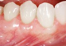
Clinical results. 6 months after surgery a periodontal evaluation was performed (Fig. 17). The general periodontal conditions further improved during the post-operative care program. No periodontal pockets > 3 mm were detectable. Deep pocketing near # 4 4 or 43 was completely absent and the buccal gingival recession at # 4 4 improved from 3 to 0. Patient was then placed in a supportive periodontal treatment program every 3 months during the first year.
Fig. 13
Fig. 16
Fig. 14
Fig. 17
Fig. 15
Findings
In this particular case initial periodontal treatment was not performed on basis of the localized periodontal defect. From several studies published in the eighties about the results of initial periodontal therapy it is known that subgingival debridement in shallow pockets causes attachment loss and gingival recession. In areas with deep inflamed pockets initial therapy will result in some clinical gain of periodontal attachment and gingival recession. However the formation of a new periodontal attachment with periodontal fibres and new cementum is strongly questioned. Therefore attachment gain after initial therapy should be considered as a consequence of a long junctional epithelium. Periodontal regeneration of a localised defect becomes more challenging after initial therapy due to loss of periodontal tissues.
A proper diagnosis of a localised periodontal defect is of utmost importance in decision making during treatment planning. A non- surgical approach is not suitable in any case. For specific cases a diagnostic flap clarifies better a pathologic condition and gives the opportunity to change a treatment strategy. In our case it was decided to perform a diagnostic flap in order to evaluate the injured area. The flap design enabled us to act directly to the clinical situation.
Guido Rhemrev Periodontist MSc.
One of 5 partners in a specialized clinic for dentistry (Kliniek voor Parodontologie, Amsterdam/Netherlands) and clinical instructor in implant dentistry at the department of periodontology at the Acedamic Centre for Dentistry in Amsterdam (ACTA). Author of several publications in the field of periodontology and implant dentistry. Main field of interest is periodontal plastic (micro-) surgery and he is frequently requested as a lecturer in this field.
guidorhemrev@kvpa.nl
3 Scientific reference: see www.straumann.com/stargetref
Straumann® MembraGel casebook
A summary of practice-oriented state-of-the-art knowledge
One of the most significant innovations in GBR
In 2010, Straumann introduced Straumann® MembraGel®, an advanced-technology membrane which can be regarded as one of the most significant innovations in guided bone regeneration. It is designed to achieve undisturbed bone regeneration, a prerequisite for an optimal clinical and esthetic outcome. Based on innovative PEG (polyethylene glycol) hydrogel technology, Straumann® MembraGel® can be applied in liquid form and moulds to the defect. Due to its gel-like consistency, the application procedure is facilitated, enabling a very precise defect contouring. After solidification of the liquid components, the bone graft is stabilized, allowing for undisturbed bone healing. Straumann® MembraGel® provides an effective barrier to tissue infiltration and subsequently biodegrades over time.
The three key factors considered important for achieving clinical success
1. The new way of applying Straumann® MembraGel® demands users to change some of their surgical habits to adapt to the new technology. New users should exercise this new technology in a step-by-step-approach and adhere to new practices in some aspects of their surgical work.
2. As the surgical procedure with Straumann® MembraGel® differs in some modes of treatment from the conventional protocol for a collagen membrane, it is necessary to follow special surgical guidelines that have been evaluated by the Straumann® MembraGel® educators in order to optimize the practical outcome when using this new product.
3. Users have reported a learning curve with this product and thus recommend starting with some straightforward cases. This will help in adapting to the new requirements. An example of a procedure that aids getting familiar with
this new technology quickly is using Straumann® MembraGel® for socket preservation to seal an augmented extraction site. This methodology presents a user-friendly option since MembraGel® can be applied in a flapless procedure without the need for any shaping or extensive microsutures to seal the site.
Optimized state-of-the-art treatment protocols for all GBR indications by expert clinicians
To facilitate the learning curve, the Straumann® MembraGel® educators who have gained clinical experience with this new product were asked to provide specific guidance for new customers using Straumann® MembraGel® for the first time. With the aid of 15 clinicians from Europe and the US, all with a wide experience of Straumann® MembraGel® in all standard GBR indications, a specific step-by-step protocol for all standard GBR indications has been created. This protocol provides guidance for individual modalities needed to treat a specific surgical situation with Straumann® MembraGel®.
Socket preservation to seal an augmented extraction site is an adequate indication to get used to this new technology
First-hand experience by expert clinicians
As a result, Straumann presents the Straumann® MembraGel® GBR casebook, which contains a summary of practice-oriented state-of-the-art knowledge about usage of Straumann® MembraGel® in GBR procedures. By means of specific step-by-step instructions and one or two clinical case studies for each indication provided by our expert clinicians, the reader gains access to relevant information for successful application of Straumann® MembraGel® in standard GBR procedures:
p Dehiscence-type defects around dental implants with a submerged healing approach
p Dehiscence-type defects around dental implants with a transgingival healing approach
p Sinus augmentation
p Lateral ridge augmentation
p Socket preservation
Availability M ay 2013
Formats PDF, printed (limited number)
Languages English, German, Italian
Please contact your local Straumann sales representative for further information.
3 Scientific reference: see www.straumann.com/stargetref
“With
the DWOS we are able to cover a broader spectrum”
DWOS – the open standard software platform from Dental Wings
DWOS is a collaboration initiative by 3M ESPE, Straumann and Dental Wings. It increases flexibility and simplifies processes for dental labs and practices. Being an open system, it provides a solution to the growing problem of incompatible systems that lock users into the technology of individual companies. The scope, quality, and functionality of DWOS make it a solution of choice for communication, design and collaboration within dentistry. DWOS is commercially available as an open system and offers dental labs the flexibility of designing prosthetics using data from multiple systems and sources. Restorations can be manufactured in-house or outsourced to milling centers offering high precision and additional material options. www.dwos.com
Mr Fehmer, what made you decide in favor of a CADCAM system in combination with the DWOS platform?
Next to research with conventional manufacturing techniques, our dental laboratory at the University of Zurich has over the past years focused heavily on computer-guided techniques in dentistry and dental technology. At the moment we are concentrating on laboratory applications.
Presently we see four major players on the market for dental CAD design software – and as the various systems are purchased and amortized by the user, we see it as our task at the university to compare these and to provide general dentists or dental technicians with the differences between the functions and indications of the CADCAM systems. The fact that the DWOS platform allows open access – i.e. one is free to choose the milling center for fabrication of the restoration – technicians are today no longer tied to a specific CAM process. Instead, a number of different variants can be combined. This is the major benefit of the DWOS platform in cooperation with Straumann products. The user of CADCAM systems operating with DWOS software has the option of resorting to all the resources of an industrial partner like Straumann - for example, in materials research. At the same time the technician also has a number of other options available, for example, if specific materials are desired or if more attractive offers are available.
How has your work changed by using the DWOS platform?
Using the DWOS platform today allows us to link computer-guided processes available in our dental laboratory and the clinic. This lets us cover a far broader spectrum of possibilities - both in terms of materials selection as well as improving workflow efficiency.
Which aspects impress you most, where do you still see room for optimization?
The principle on which the DWOS platform is based, is the right one. For example, I appreciate being able to link the coDiagnostix® implant planning software with the Straumann® CARES® Visual Software. This combination allows our laboratory to plan the prosthesis prior to implant placement. In addition, we perceive the simple recording and linking of intraoral digital data using intraoral scanners and further processing with the CARES® as an
advantage. I also believe it is important that I have the option to rely on the validated workflow of the CARES® system and can thus depend on the product safety offered by the Straumann company. On the other hand, I can work together with the fabrication center of my choice.
Thinking ahead, the integration of virtual articulation into the DWOS platform would be important in my opinion. This will become increasingly important in the area of monolithic reconstruction. Restorations made of monolithic materials such as glass ceramic, full zirconium dioxide or reconstructions made of synthetic materials are increasing considerably. Using a virtual articulator, adaptations to occlusion and function can be performed digitally, which will result in considerably more efficient treatment processes.
How did your colleagues feel about the introduction of the DWOS platform? It is highly interesting to compare the differences and preferences during the training of colleagues on the various systems. In my opinion the DWOS platform holds the middle ground between the two extremes “complexity” and “intuitiveness”. I do, however, believe that the cooperation between 3M, Straumann and Dental Wings will result in additional synergies in the further development of the DWOS platform, leading to improved userfriendliness.
What is your overall conclusion with regard to the DWOS platform?
The open interfaces and resulting flexibility are of major importance to me. At the same time I appreciate the option of using the fully validated workflow in cooperation with the Straumann CARES® fabrication center. The fit of restorations produced with this workflow are excellent in my opinion. A stronger link to the implant planning software will be crucial. There is considerable potential here, especially with regard to cooperation between dentist and dental technician. The validated workflow offers security and gives the technician backing for fabricating a product which complies with the laws on medical devices, scientific requirements as well as customer demands – this is how I envisage optimal working.
Mr Fehmer, many thanks for the interview.
Vincent Fehmer
Master Dental Technician. Clinic for crown and bridge prostheses, partial prostheses and dental materials science, Center for Dentistry, University Zurich, Switzerland. Fields of experience: fixed and removable reconstructions, choice of reconstructive material (implant and tooth borne), CADCAM digital workflow, templates for computer guided surgery, minimal-invasive reconstructions.
Selected literature from recently published journals
Straumann® Dental Implant System
Buser D, Chappuis V, Bornstein MM, Wittneben J-G, Frei M, Belser UC. Long-term stability of contour augmentation with early implant placement following single tooth extraction in the esthetic zone a prospective, cross-sectional study in 41 patients with a 5- to 9-year follow-up.
J Periodontol 2013; (Epub ahead of print).
This prospective study in 41 patients documented the long-term results of early implant placement with simultaneous contour augmentation. All patients had a tooth extracted in anterior maxilla and a Straumann implant (SLA Standard Plus or Tapered Effect) placed 4 – 8 weeks later. All implants, with implant-borne single crowns, demonstrated ankylotic stability with no signs of periimplant infection. Stable peri-implant hard and soft tissues were demonstrated for all implants, and esthetic outcomes were satisfactory. The 5-9-year follow-up confirmed that the risk of mucosal recession is low with early implant placement. In addition, contour augmentation with guided bone regeneration (GBR) was able to establish and maintain a facial bone wall in 95 % of patients. The concept of early implant placement with simultaneous GBR 8 weeks after tooth extraction resulted in positive esthetic outcomes with a low risk of recession of the facial mucosa.
Harris D, Höfer S, O’Boyle CA, Sheridan S, Marley J, Benington IC, Clifford T, Houston F, O’Connell B. A comparison of implant-retained mandibular overdentures and conventional dentures on quality of life in edentulous patients: a randomized, prospective, within-subject controlled clinical trial. Clin Oral Implants Res 2013;24(1):96 – 103.
Denture satisfaction and quality of life were assessed in 122 edentulous patients using the Oral Health Impact
Profile-49 (OHIP-49) and a Denture Satisfaction Questionnaire. The patients were assessed after wearing conventional dentures for 3 months, after which time they either continued with these or received overdentures retained on Straumann SLA implants, and were re-assessed after a further 3 months. After the first 3 months, significant improvements in satisfaction and quality of life were found. After a further 3 months, no further improvements were observed for conventional dentures, while significant additional improvements were observed for implant-retained overdentures (IODs). Implant-retained overdentures significantly increase patient satisfaction and quality of life above the benefits obtained with conventional dentures.
Kokovic V, Jung R, Feloutzis A, Vladimir T, Jurisic M, Hämmerle CH. Immediate vs. early loading of SLA implants in the posterior mandible: 5-year results of randomized controlled clinical trial. Clin Oral Implants Res 2012; (Epub ahead of print).
In 12 patients with edentulous posterior mandibles, 72 Straumann TE SLA implants were placed and loaded either early (control; 36 implants) or immediately (test; 36 implants). Implant stability was measured at implant placement and after 6, 12 and 52 weeks, and clinical parameters were recorded after 1 and 5 years. Implant stability significantly increased in both groups in the first 6 weeks after placement, with no significant differences between the groups. Implant survival was 100 % i n both groups, and there were no significant differences between the groups for bone loss, bleeding index or plaque index. Straumann TE implants provide good primary stability for immediate and early loading.
Lai HC, Si MS, Zhuang LF, Shen H, Liu YL, Wismeijer D. Longterm outcomes of short dental implants supporting single
crowns in posterior region: a clinical retrospective study of 5 – 10 years. Clin Oral Implants Res. 2013;24(2):230 – 7.
A retrospective study to evaluate the long-term clinical and radiographic outcomes of short Straumann SLA implants supporting single crowns in the posterior regions. The clinical and radiographic data of 231 short implants in 168 patients, were collected after 5 – 10 (mean 7.22) years’ follow-up. 29 of the implants had length of 6 mm long 139 had length of 8 m m. In total 4 implants and 11 prostheses failed. 1 of the failed implants was 6 m m long. The 10-year (5-year) cumulative survival rate was 98.3 % (98.7 %) for implant-based analysis and 97.6 % (98.2 %) for patient-based analysis. The short implants placed in type IV bone yielded more failures than in type I-III and presented a survival rate of 94.0 %. The 10-year survival rate of the prostheses was 95.2 %. The mean marginal bone loss between implant installation and the 10 years’ follow-up visit was 0.63 ± 0.68 m m. The marginal bone loss between the first and 5th year was minimal (0.05 ± 0.10 m m and not statistically significant). 18 (7.8 %) implants were exposed to biological complications, whereas 29 (12.6 %) implants were involved in technical complications. High survival rates for both the implants and the prostheses could be achieved after 5 – 10 years for short implants supporting single crowns, without severe marginal bone loss and complications. One may conclude that a single crown supported by a short implant is a predictable treatment modality. However, short implants in type IV bone sites should be applied with caution.
Schlegel KA, Prechtl C, Möst T, Seidl C, Lutz R, von Wilmowsky C. Osseointegration of SLActive implants in diabetic pigs. Clinical Oral Implants Res 2013;24(2):128 – 134.
This was a study to investigate peri-implant bone formation in a diabetic animal model in comparison to healthy
animals and to evaluate the differences between conventional (SLA) and modified (SLActive) titanium implant surfaces on osseointegration. Six implants each were placed in the calvaria of 11 diabetic and 4 healthy domestic pigs. At 30 and 90 days after implant placement, the bone-to-implant contact (BIC) and bone density (BD) were evaluated. Additionally, the expression of the bone-matrix proteins collagen type I and osteocalcin was evaluated at both points in time by using immunohistochemical staining methods. Overall, BIC was reduced in the diabetic group at 30 and 90 days. After 90 days, the SLActive implants showed significantly higher BIC compared with the SLA implants in diabetic animals. Peri-implant BD was higher in the SLActive group at 30 and 90 days in healthy and diabetic animals. Collagen type I protein expression was higher using SLA implants in diabetic pigs at 30 days. Values for osteocalcin expression were not consistent. The results indicate the negative effect of untreated diabetes mellitus on early osseointegration of dental implants. The modified SLA surface (SLActive) elicited an accelerated osseointegration of dental implants, suggesting that a better prognosis for implant treatment of diabetic patients is possible. The use of the hydrophilic SLActive surface resulted in positive effects in healthy and diabetic animals. Therefore, this surface seems especially suitable for compromised patients
Srinivasan M, Vazquez L, Rieder P, Moraguez O, Bernard JP, Belser UC. Survival rates of short (6 m m) micro-rough surface implants: a review of literature and meta-analysis. Clin Oral Implants Res 2013; (Epub ahead of print).
Short dental implants may be as a suitable alternative to more complex procedures in cases of reduced alveolar bone height, since they can help to avoid anatomical structures, increase patient comfort and reduce treatment time. A literature search was performed for studies with
6 m m rough-surfaced Straumann implants with a minimum follow-up time of 1 year. A total of 842 publications were initially screened, of which 12 were selected for review and statistical evaluation. The studies included a total of 690 Straumann 6 m m implants. A meta-analysis of the results indicated that the overall survival rates were 94.7 % a nd 98.6 % i n the maxilla and mandible, respectively, and that the vast majority (76 %) of failures were early failures. The results indicate that Straumann 6 mm implants are a predictable treatment option. Survival rates are very good, with slightly greater survival in the mandible, and failures are predominantly at an early stage.
Tolentino L, Sukekava F, Seabra M, Lima LA, Garcez-Filho J, Araújo MG. Success and survival rates of narrow diameter implants made of titanium-zirconium alloy in the posterior region of the jaws – results from a 1-year follow-up. Clin Oral Implants Res 2013; (Epub ahead of print).
In 42 patients, either Straumann Roxolid (test; 21 implants) or Straumann titanium SLActive (control; 21 implants) narrow diameter implants were placed in the posterior jaw region to support metal-ceramic single crowns, which were adapted to the implants after 8 weeks. Implant survival, success and clinical parameters were evaluated after 6 weeks and 1 year. In both groups the implant survival rate was 95.2 % (one implant in each group had to be removed at 6 weeks due to implant mobility and peri-implant radiolucency) and the prosthesis survival rate was 100 %. Probing depth was significantly less for Roxolid implants at 6 weeks, but there was no significant difference after 1 year. Similarly, no significant differences were found between the groups for mobility, suppuration or bleeding on probing. The result suggested that Roxolid or titanium narrow-diameter implants may be suitable to support single-tooth restorations in the posterior region of the jaw.
Verket A, Lyngstadaas SP, Rønold HJ, Wohlfahrt JC. Osseointegration of dental implants in extraction sockets preserved with porous titanium granules – an experimental study. Clin Oral Implants Res 2012; (Epub ahead of print).
Osseointegration of dental implants into healed extraction sockets preserved with porous titanium granules (PTG) was investigated in this preclinical trial. Upon extraction of the mandibular teeth 2, 3 and 4 from 3 minipigs, sockets were preserved with metallic PTG (n=12), heat oxidized white porous titanium granules (WPTG, Tigran, n=12) or left empty (n=6). Bio-Gide membranes were used and after 11 weeks of healing, Straumann Bone Level implants (diameter: 3.3mm, length: 10mm, surface not mentioned) were inserted. The mean bone volume measured by microCT after 6 weeks was 61.7 % for the PTG group, 50.3 % for the WPTG group and 57.1 % for the control group. Bone to implant contact measured by histology after 6 weeks was 68.2 % for the PTG group compared to 36.6 % for the WPTG group and 60.9 % for the control group. The differences between the PTG group and the WPTG group were statistically significant; however the differences between the PTG group and the control group were not significant. PTG preserved extraction sockets similarly as untreated extraction sockets. WPTG showed a less favorable effect on the preservation of extraction sockets.
Wen B, Zhu F, Li Z, Zhang P, Lin X, Dard M. The osseointegration behavior of titanium-zirconium implants in ovariectomized rabbits. Clin Oral Implants Res 2013; (Epub ahead of print).
Roxolid or titanium implants, both with the SLActive surface, were placed in the tibiae and femurs of 12 ovariectomized and 12 sham-aged rabbits. Removal torque and histomorphometric analyses were performed after 3 and 6 weeks. In both the sham and ovariectomized
groups sham group, removal torque was significantly higher for Roxolid versus titanium implants; peak torque after 6 weeks was 46.0 Ncm and 50.8 Ncm for titanium implants in the ovariectomized and sham groups, respectively, while the corresponding values for Roxolid implants were 60.7 Ncm and 76.2 Ncm, respectively. Boneto-implant contact (BIC) and bone density increased from weeks 3 to 6 in the sham group; the difference was not significantly different between the groups. BIC and bone density appear to behave in similar manner with both implant types in this experimental model, but the removal torque was significantly higher for Roxolid in both groups, which may reflect an increase in bone quality around Roxolid implants.
Straumann® Regenerative System
Döri F, Arweiler N, Húszár T, Gera I, Miron R, Sculean A.
Five year results evaluating the effects of platelet-rich plasma on the healing of intrabony defects treated with an enamel matrix derivative and a natural bone mineral. J Periodontol 2013; (Epub ahead of print).
This was a randomized clinical study to evaluate the long-term outcomes following regenerative surgery of deep intrabony defects with an enamel matrix protein derivative (EMD) and a natural bone mineral (NBM) with and without addition of platelet-rich plasma (PRP). The primary outcome variable was clinical attachment level (CAL). Twenty-four patients were included in this study. Compared to baseline, in both groups at 5 years, a CAL gain of ≥ 4 m m was measured in 75 % (i.e. in 9 out of 12) of the defects. . No statistically significant differences in any of the investigated parameters were observed between the two groups. Within their limits, the present results indicate that i) the clinical outcomes obtained with both treatments can be maintained up to a period of five years,
and ii) the use of PRP did not appear to improve the results obtained with EMD + NBM.
Jepsen K, Jepsen S, Zucchelli G, Stefanini M, de Sanctis M, Baldini N, Greven B, Heinz B, Wennström J, Cassel B, Vignoletti F, Sanz M. Treatment of gingival recession defects with a coronally advanced flap and a xenogeneic collagen matrix: a multicenter randomized clinical trial. J Clin Periodontol 2012; (Epub ahead of print).
This study evaluated either coronally advanced flap (CAF) alone (control) or in combination with a xenogeneic collagen matrix (test; Mucograft; Geistlich Pharma AG, Wolhusen, Switzerland) for the treatment of 90 Miller Class I or II recessions in 45 patients. After 6 months, root coverage was 75.29 % and 72.66 % for test and control, respectively, while complete coverage was 36 % a nd 31 % for test and control, respectively. Test sites also showed a greater increase in mean width of keratinized tissue and had more gain in gingival thickness. Significantly higher root coverage was observed in the test group in larger recession defects (72.03 % versus 66.16 %). Root coverage was not superior with the addition of collagen matrix to CAF, but increased gingival thickness and width of keratinized tissue was observed.
McGuire MK, Scheyer ET, Nunn M. Evaluation of human recession defects treated with coronally advanced flaps and either enamel matrix derivative or connective tissue: comparison of clinical parameters at 10 years. J Periodontol 2012;83(11):1353 – 62.
Of 17 patients with Miller class I or II recession defects originally treated with either Emdogain or connective tissue graft (CTG), both in combination with coronally advanced flap (CAF), nine were available for follow-up evaluation after 10 years. With the exception of PD, all quantitative clinical parameters showed significant im-
provement from baseline after 10 years, including width of keratinized tissue (wKT), which had not significantly improved at 1 year in the Emdogain group; indeed, wKT at 1 year was the only measured parameter that was significantly different between the groups. Similar results were observed with qualitative parameters except for marginal tissue contour, which was greater than adjacent tissues at most of the CTG sites. Six of the nine patients indicated that they would choose EMD over CTG to avoid a secondary harvesting procedure. The results showed that both CTG and Emdogain treatment were clinically effective, stable and similar after 10 years. Long-term follow-up data are crucial to demonstrate the stability of a treatment or intervention.
Mueller VT, Welch K, Bratu DC, Wang HL. Early and late studies of EMD use in periodontal intrabony defects. J Periodontal Res 2013;48(1):117 – 125.
Since some recent studies have questioned the validity of early findings, a literature search was performed for early (1997 – 2003) and late (2004 – 2010) studies on Emdogain in the treatment of intrabony defects. Clinical attachment level, probing pocket depth and bone gain were assessed. The results showed no significant difference in these parameters between early and late studies, but the beneficial effects of Emdogain treatment were seen in studies from both periods. The clinical effectiveness of Emdogain in the treatment of periodontal intrabony defects was confirmed, with no significant differences between the results from early and late studies.
Parashis AO, Polychronopoulou A, Tsiklakis K, Tatakis DN. Enamel matrix derivative in intrabony defects: prognostic parameters of clinical and radiographic treatment outcomes. J Periodontol 2012;83(11):1346 – 52.
This was a retrospective case series that included 61 patients with 2- or 3-wall intrabony defects treated with Emdogain. Clinical and radiographic parameters were evaluated after 12 months and compared to the values recorded at baseline. Baseline probing depth (PD) and distance from the cemento-enamel junction to the base of the defect (CEJ-BD) were significant predictors of gain in clinical attachment level (CAL), and the probability of a post-operative PD > 4 m m increased 1.6-fold with each 1 mm increase in baseline PD. Supracrestal soft tissue (SST) and smoking were found to be significantly associated with defect resolution. Baseline PD appeared to be a predictor of post-operative PD and CAL gain in Emdogain-treated intrabony defects, while smoking and SST were predictors of defect resolution. The study also confirmed that Emdogain treatment results in significant improvements in CAL gain, PD reduction and defect resolution.
Peres MF, Ribeiro ED, Casarin RC, Ruiz KG, Junior FH, Sallum EA, Casati MZ. Hydroxyapatite/β-tricalcium phosphate and enamel matrix derivative for treatment of proximal class II furcation defects: a randomized clinical trial. J Clin Periodontol 2013;40(3):252 – 9
Proximal class II furcation defects in 30 patients were randomized to receive open-flap debridement (OFD) plus Straumann BoneCeramic (SBC) or OFD + SBC + Emdogain (EMD). Clinical soft and hard tissue parameters were evaluated at baseline and after 6 months. Significant improvements were seen in both groups. Gains in attachment level and vertical bone level were slightly greater in the OFD + SBC + EMD group, but the differences were not significant. Furcations were completely closed in 7/15 cases in the OFD + SBC + EMD group compared to 4/15 cases in the OFD + SBC group. Clinical improvements were
slightly greater for SBC + EMD than for SBC, although the differences were not significant. The closure of proximal class II furcation defects still seems to be an unpredictable process.
Shirakata Y, Yoshimoto T, Takeuchi N, Taniyama K, Noguchi K. Effects of EMD in combination with bone swaging and calcium phosphate bone cement on periodontal regeneration in one-wall intrabony defects in dogs. J Periodontal Res 2013;48(1):37 – 43.
One-wall intrabony defects were created at the mesial and distal aspects of mandibular premolars in dogs and treated with Emdogain, bone swaging (BS) only, Emdogain + BS, or Emdogain + BS + calcium phosphate cement (CPC). After 8 weeks, a significantly greater height of newly formed bone was found with Emdogain+BS+CPC compared to Em-
dogain + BS or BS only. The area of newly formed bone was also significantly greater with Emdogain + BS + CPC compared to the Emdogain, BS only or Emdogain+BS groups. The BS only group showed significantly lower cementum formation than the other groups. Emdogain in combination with bone swaging and calcium phosphate cement may be a good combination for periodontal healing in one-wall intrabony defects.
STARGET FOR iPAD
IADR/Straumann Award in Regenerative Periodontal Medicine Awardee 2013: Andreas Stavropoulos
Andreas Stavropoulos DDS, CDT, PhD, Dr Odont.
An internationally renowned researcher in periodontology. After studies in Athens, Heidelberg and Thessalonica, he pursued his career in research at the School of Dentistry at Aarhus University, Denmark. In 2011, he won First Prize in the Basic Research Competition of the European Association of Osseointegration. In 2012, Andreas Stavropoulos was appointed Professor of Periodontology at Malmö University School of Dentistry, Sweden. He has authored and co-authored a large number of publications and contributed to specialist textbooks in the field of periodontology.
At the General Session of the International Association for Dental Research (IADR) in Seattle, USA, the 2013 IADR/Straumann Award in Regenerative Periodontal Medicine was presented to Professor Andreas Stavropoulos of Malmö University, Sweden, in recognition of his remarkable achievements in periodontal research. Worth USD 5000, the Award was created to honor significant contributions in basic and/or clinical research in regenerative periodontal or peri-implant medicine. The 2013 Award was presented by Professor Philip Preshaw, President of the IADR Periodontal Research Group.
“Professor Andreas Stavropoulos has made a sustained and high quality contribution to research in periodontal regeneration over the past 15 years. His excellent track record of research publications in the areas of wound healing, bone regeneration, implant therapy and osseointegration reflects the consistent quality and broad range of his research” Prof. Philip Preshaw
Initiated in 2010, the IADR/Straumann Award in Regenerative Periodontal Medicine is sponsored by Straumann and administered by the Periodontal Research Group of the IADR. Straumann is a leading contributor to R&D in implant and regenerative dentistry and this award is a further example of the Group’s commitment to fostering and recognizing excellence in dental research.
The International Association for Dental Research (IADR) is based in Alexandria, Virginia/USA and a nonprofit organization with more than 12’000 individual members worldwide, dedicated to: (1) advancing research and increasing knowledge to improve oral health, (2) supporting the oral health research community, and (3) facilitating the communication and application of research findings for the improvement of oral health worldwide. Within the IADR, the Periodontal Research Group is the forum for members who are active in periodontology. Its aim is to improve periodontal health through encouraging research activities. For more information www.iadr.org
Confidence in limited space
Straumann® Narrow Neck CrossFit®
The Straumann Soft Tissue Level solution to address space limitations
Confidence when placing small diameter implants
Wide range of treatment options Simplicity in daily use
FUSIONOF OSSEOINTEGRAT
Straumann® Roxolid® ANDMATERIAL STRENGTH!
“Preparing for the Future of Implant Dentistry”
October 17 – 19 2013 in Dublin, Ireland at the Convention Centre.
The EAO’s Scientific Committee has prepared a programme that will address many practical and highly relevant issues of concern to both clinicians and patients and also highlight the real and emerging issues that arise in a population that is ageing and which has increasingly complex needs. Dublin is a very historic and exciting capital city. The medieval, Georgian and modern architecture provides an intriguing backdrop to this cosmopolitan city,
famous for its musical, theatrical and literary traditions. You will have an opportunity to experience the many cultural activities and historic landmarks of the city and enjoy the extensive and varied opportunities for entertainment, good food and shopping. The congress will take place at the newly opened and internationally acclaimed Convention Centre Dublin situated on the river Liffey in the heart of the city with spectacular views across Dublin and its surroundings.
“Treatment flexibility supported by technology”
Reducing case complexity, increasing treatment predictability, and optimizing workflows are key topics for technology advancements and less invasive procedures.
As an example, the development of intelligent synthetic materials and the opportunity for an improved control of the bone remodeling process and the subsequent substitution by newly formed bone may increase overall flexibility in everyday patient treatment. Bone substitutes evolve from being a purely incorporated filling and space maintaining material towards scaffolds effectively facilitating pristine bone regeneration. Early findings on a new bone substitute targeted to guide natural bone regrowth leaving no remnants are an illustration for high-performance innovation by Straumann®.
Clinical data on pioneering implant technologies such as the Roxolid® material and SLActive® hydrophilic surface clearly documents the higher treatment predictability, increasing confidence in using small-diameter implants. Recent data further underlines the benefits, particularly
Treatment flexibility supported by technology
Date Thursday, 17 October,
Time 16.45 – 18.45 h
Room L iffey B
Moderator Prof. David Cochran, San Antonio, USA
2.
A new bone grafting material bridging existing gaps in clinical bone regeneration
the potential for reducing treatment steps and greater treatment flexibility in limited bone volume. This allows for interesting considerations in regards to reducing implant diameter and length, preserving more vital structures around the implant. There may be new ways of implant treatment, which may help to increase patient acceptance with less invasive procedures and enhanced prosthetic solutions.
What is the role of technology advancements to support new ways of implant treatment? Which are the possibilities for increased treatment flexibility and patient acceptance, especially to increase the quality of live for elderly patients, arising from the availability of technology innovations? Where are the considerations and boundaries in every day practice? These questions will be addressed in the following topics from a bone regeneration, implantation and prosthetic point of view.
Please check our website for the speakers’ abstracts, CV’s and program updates: www.straumann.com/eao2013
Evolution of dental implants due to technology innovations
3. Prof. Frauke Mueller (Geneva, Switzerland): Prosthetically driven aspects to smaller diameter and shorter implants
1. Dr. Isabella Rocchietta (London, UK)
Prof. David Cochran (San Antonio, USA)
1st International Symposium
Regeneration and Esthetics in Periodontology and Implant Dentistry
Chairmen: Prof. Daniel Buser and Prof. Anton Sculean November 8/9, 2013 • Congress Center, Kursaal Bern
Dear colleagues
We encourage you to come to Bern in early November 2013 to attend the 1st International Symposium on Regeneration and Esthetics in Periodontology and Implant Dentistry offered by the University of Bern. Both areas are fi elds of high interest in Dental Medicine, and the University of Bern is known to have an excellent international reputation in these surgical disciplines.
Friday, November 8, 2013
9.00–12.30 h
Session 1: Regeneration in Periodontology
13.45–17.45 h
Session 2: Bone Augmentation in Implant Dentistry
Saturday, November 9, 2013
8.45–12.15 h
Session 3: Esthetics in Periodontology
13.30–16.15 h
Session 4: Esthetics in Implant Dentistry
Faculty
S. Aroca, SUI/FRA
Z. Artzi, ISR
D. Bosshardt, SUI
D. Buser, SUI
V. Chappuis, SUI
S. Chen, AUS
D. Cochran, USA
N. Donos, GBR
R. Gruber, SUI
U. Grunder, SUI
R. Jung, SUI
C. Nemcovsky, ISR
G. Rasperini, ITA
G. Salvi, SUI
A. Sculean, SUI
M. Simion, ITA
A. Stavropoulos, SWE
L. Trombelli, ITA
I. Urban, HUN
F. Vailati, SUI
O. Zuhr, GER
Pre Symposium workshops and video sessions
Thursday, November 7, 2013
13.00–15.00 h
Workshop 1* Implant placement with simultaneous contour augmentation using GBR to achieve predictable esthetic outcomes
Speakers: D. Buser / V. Chappuis
Video session 1 Flap designs and surgical techniques to improve the outcomes in regenerative and plastic esthetic surgery
Presentation: A. Sculean / L. Trombelli
15.30–17.30 h
Workshop 2 Flap designs to optimize the outcomes in intrabony and furcation defects
Speakers: A. Sculean / L. Trombelli
Workshop 3 Surgical techniques using connective tissue replacement material for coverage of single and multiple gingival recessions
Speakers: S. Aroca / G. Salvi
Video session 2 Surgical techniques with GBR in various indications
Presentation: D. Buser
* if more than 30 participants sign up, a second course will be offered
Registration
Organization
Organizing Committee
• Prof. Daniel Buser and Prof. Anton Sculean, University of Bern
• Caroline Chételat, CCDE, Bern
Administrative Secretariat
Center for Continuing Dental Education, CCDE Marktgasse 7, 3011 Bern
Phone: +41 31 312 43 12
Fax: +41 31 312 43 14 info@ccde.ch, www.ccde.ch
Congress Venue
Congress Center, Kursaal Bern Kornhausstrasse 3, 3000 Bern 25 www.kursaal-bern.ch
Supported by our founding partners
Publishing Partner
1st International Symposium, Congress Center, Kursaal Bern, November 8/9, 2013
Congress language is English. Simultaneous translation will be offered in French and German. Payment can be done either by netbanking or by credit card (Visa, Mastercard, only online registration).
Registration form: on-line: www.ccde.ch, e-mail: info@ccde.ch or by fax: +41 31 312 43 14
Name First name
Title Sex female male
Address Postal Code/City
Country E-Mail
Phone Fax
Tuition fees
Pre Symposium: Thursday, November 7, 2013 – Workshops and video sessions
Workshop 1 CHF 250.– Video session 1 CHF 150.–Workshop 2 CHF 250.– Video session 2 CHF 150.–Workshop 3 CHF 250.– Both video sessions CHF 250.–
1 workshop and CHF 350.–
1 video session
(please indicate the desired workshop and video session beside)
1st International Symposium: Friday & Saturday, November 8/9, 2013 before August 31 from September 1
Dentist
Post-doc student (only with confirmation of the University)
Dental technicians
Headphones for the translation
On-site registration additional fee of CHF 80.–
Date Signature
Every
CHF 550.– CHF 700.–
CHF 250.– CHF 350.–
CHF 400.– CHF 500.–
André Schroeder Research Prize
Jung-Chul Park wins 2013 André Schroeder Research Prize
Jung-Chul
Park DDS, MSD, PhD
After studying biochemistry, Dr. Park graduated from Yonsei University in Seoul with a degree in dentistry in 2005 and was awarded his PhD from the Dental College at Yonsei University in 2012. He is currently in London, England at the UCL Eastman Dental Institute taking part in the ITI Scholarship program.
Periodontist Dr. Jung-Chul Park from South Korea has been named the winner of the 18th André Schroeder Research Prize. This annual prize awarded by the International Team for Implantology (ITI) carries a cash endowment of 20’000 Swiss francs and was presented on April 6 during the ITI Congress North America in Chicago. Dr. Park received the award for his study on the “acquisition of human alveolar bone-derived stromal cells using minimally irrigated implant osteotomy: in vitro and in vivo evaluation”. Together with his co-authors, he aimed to evaluate the osteogenic differentiation capacity of human alveolar bone-derived stromal cells obtained from the bone chips trapped within drill flutes during implant osteotomies.
“Stem cells are regarded as the future of medical and dental treatment. We all know that this area still needs lots of research before we can actually utilize the stem cells obtained from the patient, however, it is very important to know that these cells are relatively easy to access. Now we will have to see what clinicians and researchers do with this. I expect very interesting ideas to follow our study.”
Dr. Jung-Chul Park
Established more than 20 years ago, the André Schroeder Research Prize is an annual award presented by the ITI in honor of the late Professor André Schroeder (1918 – 2004), the founding ITI President, who pioneered implant dentistry and whose lifework contributed significantly to modern dentistry. Worth 20’000 Swiss Francs in cash, the André Schroeder Research Prize is today one of the most highly sought-after awards in implant dentistry. It is awarded to independent researchers for the advancement of dental research and development. The aim is to promote new scientific findings in implant dentistry, oral tissue regeneration and related fields. More information on www.iti.org
Announcements
New membership heights/ 2013 Brånemark Award
The ITI welcomes its 15’000th member during this year’s AO Annual Meeting
The International Team for Implantology (ITI) signed up its 15’000th member during this year’s AO Annual Meeting in Tampa, Florida, USA. This achievement marks another milestone in the 33-year history of the ITI and is also a worthy tribute to the efforts of ITI President Prof. Dr. Daniel Buser, during whose four-year term of office membership figures more than doubled.
The 15’000th member of the ITI is Dr. Michael Jaffin, a board certified periodontist from Hackensack, New Jersey, USA. He was welcomed personally by Prof. Buser as well as members of the ITI Board of Directors and the US ITI Section Leadership Team.
“The ITI’s extraordinary rate of growth clearly demonstrates that our broad educational offering is very appealing to colleagues. I am strongly convinced that it directly relates to the fact that, in terms of treatment recommendations, the ITI represents a safe haven to practitioners all over the world,” said Prof. Buser.“ Prof. Dr. Daniel Buser
ITI President Daniel Buser presented with 2013 Brånemark Osseointegration Award
Prof. Dr. Daniel Buser is the sixth recipient of the highly respected Brånemark Osseointegration Award. It is presented annually by the Osseointegration Foundation and honors an individual whose contribution to implant dentistry has had a significant impact on the field. The award recognizes Prof. Buser’s long-term achievements as a leader, teacher and researcher. It was presented at the Academy of Osseointegration’s annual business meeting in Tampa, Florida, USA on March 9, 2013.
“I am very honored to receive this prestigious award,” said Prof. Buser. “I see it as recognition of the achievements of my team at the University of Bern, School of Dental Medicine, and also, more broadly, of the treatment philosophy championed by the ITI since 1980 that has contributed to the standards and norms that guide the field today.”
Do your patients want more?
Implant dentistry has earned its place as a standard treatment option and patients‘ expectations of oral health and esthetics have grown in parallel. ITI Education Weeks are the ideal place to freshen up and extend new skills associated with implant dentistry.
ITI Education Weeks offer:
• Up to the minute evidence-based teaching
• Top lecturers at s tate-of-the-art facilities
• Continuing education credits
• Par ticipation in treatment planning
• Surgical and prosthetic sessions: live and hands-on
The courses are delivered by leading academic institutions around the world in partnership with the ITI – an independent academic organization dedicated to all aspects of implant dentistry.
ITI Education Weeks 2013
AUGUST 26–30
University of Bern, School of Dental Medicine, Bern, Switzerland “Evidence-Based Clinical Concepts in Implant Dentistry”
SEPTEMBER 8–14
University of Hong Kong, Prince Philip Dental Hospital, Hong Kong SAR, PR China
“Comprehensive Care with Dental Implants”
OCTOBER 23–26
Holland Bloorview Kids Rehabilitation Hospital/University of Toronto, Toronto, Canada
“Best Practices in Implant Dentistry”
For more information on ITI Education Weeks and our academic partners go to: ww w.iti.org/educationweek. ITI International Team for Implantogy I Peter Merian-Strasse 88 I 4052 Basel I Switzerland I www.iti.org
IT I Nationa l C ongr ess es
Jun e-Dec ember 20 13
A great year for knowledge and networking
ITI Na tion al Con gre sses repres ent the be st wa y to com bi ne networ king with cat chi ng up on the la te st in implan t de nt ist ry. Wit h pl en ty o f ITI National Con gre ss es in t he coming m onth s, th ere are m a ny oppor tunit ies to conso lid a te your knowl e dg e, earn credit p oi nt s and meet th e top speakers in you r re gion or count ry.
Check o ut al l our upcoming congresse s at w ww.it icon gress.org.
ITI Congress Argentina & Uruguay June 14–15, 2013Buenos Aires, Argentina
ITI Congress Austria June 21–22, 2013Salzburg, Austria
ITI Congress Australasia July 26–27, 2013 Sydney, Australia
ITI Congress Southern Africa July 26–27, 2013 Johannesburg, South Africa
ITI Congress China August 16–17, 2013Shanghai, China
ITI Congress Italy September 26–28, 2013Venice, Italy
ITI Congress Iran Decemb er 5–7, 2013Kish Island, Iran
ITI World Symposium coming up in Geneva, Switzerland: April 24–26, 2014.
ITI International Team for Implantology | Peter Merian-Strasse 88 | 4052 Basel | Switzerland | www.iti.org
Venice combines art, architecture and romantic charm. Take this opportunity to sample some of its attractions by visiting us at the ITI Congress Italy.
Distinguished international speakers: Daniel Buser (Switzerland), Michael Gahlert (Germany), Dean Morton (USA), Frauke Müller (Switzerland), Irena Sailer (Switzerland), Tom Taylor (USA) will share their knowledge and skills, complemented by the cream of the region‘s speakers.
www.iti.org/congressitaly City of Beauty, City of Learning
Straumann South Korea (formerly: B.I. Trading Co. Ltd.) 1467-75, Seocho3 -Dong, Seocho-Gu, Seoul
Tel. +82/72 265 8777
Fax +82/72 265 8797
Spain/Portugal
Straumann S.A.
Edificio Arroyo - A Avda. de Bruselas, 38 Planta 1
28108 Alcobendas (Madrid)
Tel. +34/902 400 979
Fax +34/913 449 517
Sweden
Straumann AB Fabriksgatan 13 41250 Göteborg
Tel. +46/31 708 75 00
Fax +46/31 708 75 29
USA
Straumann USA, LLC
60 Minuteman Road
Andover, MA 01810
Tel. +1/800 448 8168 +1/978 747 2500
Fax +1/978 747 2490
STARGET DIGITAL
STARGET for iPad
With video functionality and context-relevant extra material. Available free in the App Store in German, English, Spanish, French and Italian language.
STARGET on the screen
Browse STARGET comfortably on the screen like a printed version: www.straumann.co.uk/starget, select option “interactive version“ there.
STARGET as PDF
STARGET can also be downloaded as PDF version from our website: www.straumann.com/starget