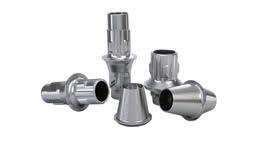
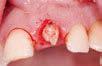
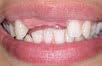
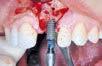
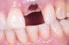
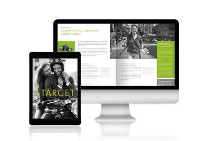
iPad This publication is also available for the iPad (English, German, Spanish, French and Italian). Visit the official App Store and download “STARGET for iPad”.
Legal Notice Exclusion of liability for articles by external authors: articles by external authors published in STARGET have been systematically assessed and carefully selected by the publisher of STARGET (Institut Straumann AG, Basel). Such articles in every case reflect the opinion of the author(s) concerned and therefore do not necessarily coincide with the publisher’s opinion. Nor does the publisher guarantee the completeness or accuracy and correctness of articles by external authors published in STARGET. The information given in clinical case descriptions, in particular, cannot replace a dental assessment by an appropriately qualified dental specialist in an individual case. Any orientation to articles published in STARGET is therefore on the dentist’s responsibility. Articles published in STARGET are protected by copyright and may not be reused, in full or in part, without the express consent of the publisher and the author(s) concerned. Straumann® and all other trademarks and logos are registered trademarks of Straumann Holding AG and/or of its affiliates. Third party corporate names and brand names that may be mentioned may be registered or otherwise protected marks even if this is not specially indicated. The absence of such an indication shall not therefore be interpreted as allowing such a name to be freely used. This version of STARGET is not intended for distribution or use in the United States. If you are a U.S. citizen, please contact our U.S. subsidiary, Straumann USA LLC, for a complimentary copy of the U.S. version of STARGET or visit www.straumann.us/starget for a PDF version.
Product availability Certain products and services mentioned in this edition of STARGET may not be available or not yet available in all countries. In case of doubt please contact your local Straumann distributor for information on product availability (addresses of Straumann branches can be found on the last page).
Plenty of reasons to smile
DEAR READERS
This year we are once again launching several innovative products and services that will make your day-to-day work easier and more efficient and further improve your patients’ quality of life.
It is clear that our industry is experiencing highly dynamic development, and the environment for dental laboratories in particular is becoming increasingly challenging. In spring 2015 we launched a large number of new products and services in the area of implant prosthetics, in order to offer you active, steadfast support in making your dental laboratory even more efficient and competitive. Our clear mission is to be your first choice when it comes to the future of your laboratory.
The new Straumann® Bone Level Tapered Implant is a unique, impressive combination of clinically proven features and the specific advantages in terms of primary stability that the new design offers. Together with the Straumann® Pro Arch concept, which is also new, you now have the opportunity to offer immediate screw-retained complete restorations, even with low bone level.
There have been signs of a paradigm shift in the way in which patients obtain information for some time now. Whereas doctors were previously the only official authorities on medical issues, patients now very often obtain information themselves. And it is the modern online media that mainly influence patients in their decision-making processes. In order that you can actively support your patients and gain new patients for yourself and your implantology practice, we have therefore launched the comprehensive marketing package Straumann® Patient Pro
We have also received some revealing information from the scientific world. Researchers at the Swedish University of Gothenburg have analyzed the short-term and longterm success of dental implants and the underlying factors in a large-scale study. One major finding of the study is that the brand of implant used makes a difference. In this analysis, Straumann implants achieved excellent success rates compared with many other manufacturers.
So as you can see, there are lots of reasons to smile. You can find out more about all these interesting topics in this issue. I hope you find this an enjoyable read.
Sincerely
Frank Hemm
FRANK HEMM HEAD CUSTOMER SOLUTIONS & EDUCATION
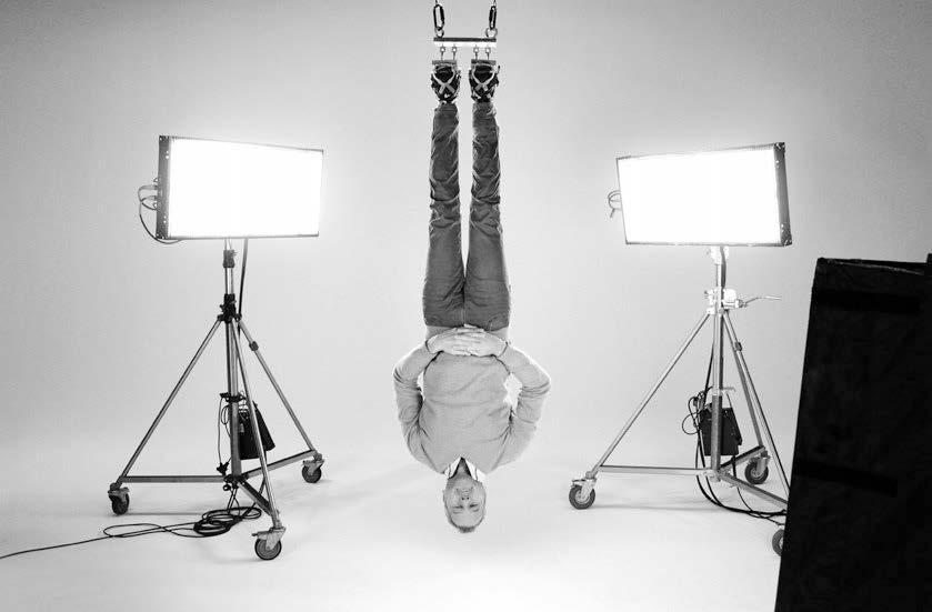
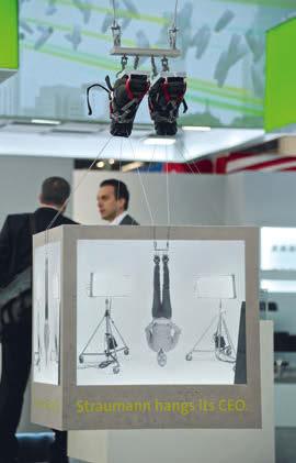
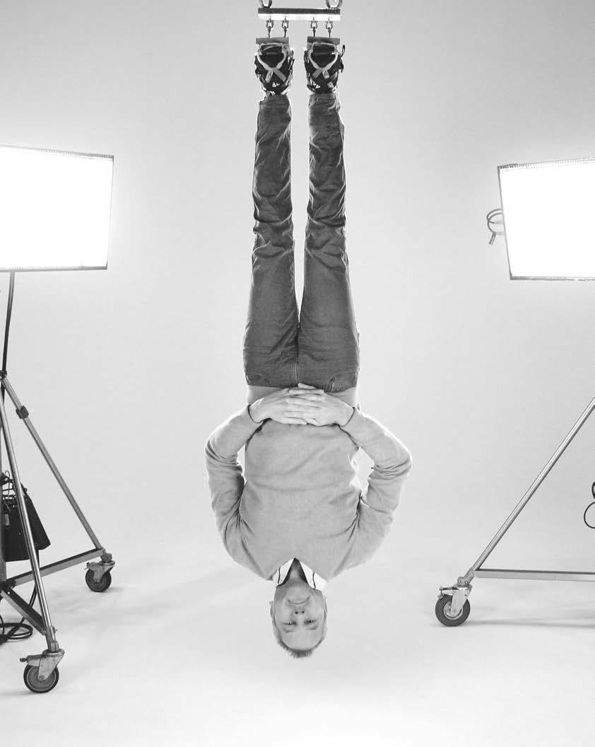
WHY IS OUR CEO HANGING UPSIDE DOWN FROM THE CEILING?
Uncompromising quality and precision have always been cornerstones of our company philosophy. We know that our products work and we keep our promises. Take a look here to see Straumann CEO Marco Gadola demonstrate how he isn’t into just paying lip service.
→ PAGES 10 – 11
Content
MORE THAN A LAB PARTNER
04
Due to today’s dynamic changes in the dental market, it is very challenging for dental labs to make the right decisions that ensure the long-term success of their business. Catching up with new technological trends and increasing your competitiveness are now more important than ever. We want to be more than a lab partner for you. Our aspiration is to be your provider of choice when it comes to efficiency, innovation, and quality.
THE
STRAUMANN® BONE LEVEL TAPERED IMPLANT
26
The Straumann® Bone Level Tapered Implant is a significant step forward in setting a new standard within the field of tapered implants. It is a unique and powerful symbiosis between the established and clinically proven features of the Straumann® Bone Level Implant and the benefits of a tapered implant design regarding surgical flexibility and primary stability.
STRAUMANN® PATIENT PRO
60
Your patients are already gathering information about tooth restoration, especially on the internet. Are you already there to meet and guide them through the decision-making process? This new Marketing toolbox was released to reflect the paradigm shift in the patient’s information behavior. It supports you by providing patient information materials and marketing tools you can use on the internet and social media channels as well as in your dental practice.
STRAUMANN® PROSTHETICS
New products and solutions 04 More than a lab partner. True ambition to increase your efficiency
Original-on-Original campaign 10 Why does the Straumann CEO hang upside down from the ceiling?
Straumann® CARES® Advanced Fixed Bar 12 Clinical case report by F. Ruggia and R. Wöhrle
Straumann® Pro Arch 16 More than a full-arch rehabilitation. A reason to smile.
Straumann® Bone Level Tapered Implant 26 The new tapered standard
28 Clinical case report by S. Zarrine
32 Interview with S. Piano
Straumann® PURE Ceramic Implant 34 The 3.3 mm diameter-reduced PURE implant – “purely amazing”!
36 Clinical case report by F. Thieringer
38 Clinical review
Guided Surgery 40 coDiagnostiX™ meets Straumann® CARES X-stream™
Dental Wings 44 Straumann and DWOS: fully committed
Straumann® Roxolid® 46 Long-term clinical data shows: Roxolid® works!
ORAL TISSUE REGENERATION
Straumann® Emdogain® 48 Twenty years of success: It’s just the beginning.
50 Clinical case report by C. Nemcovsky botiss 52 Review: 1st botiss “bone & tissue days” world congress
56 Clinical case report by A. Puišys
SIMPLY DOING MORE
Straumann® Patient Pro 60 The plus for your business
EVENTS
EAO 66 Preview: 25th Annual Scientific Meeting
More than a lab partner.
True ambition to increase your efficiency
Due to today’s dynamic changes in the dental market, it is very challenging for dental labs to make the right decisions that ensure the long-term success of their business. Catching up with new technological trends and increasing your competitiveness are now more important than ever.
Under these circumstances, close collaboration with a strong and reliable partner is now becoming paramount. At Straumann, we are fully committed to taking care of you and the success of your business. We stand for the highest quality, and our passion is to continuously shape our portfolio offering with innovative products and services in order to grow your lab business and make your life easier. With the introduction of our newest products and services, we intend to further drive efficient workflows. Additionally, we are addressing your need for complex, highly precise, and esthetic CAD/CAM restorations. In short, we want to be more than a lab partner for you. Our aspiration is to be your provider of choice when it comes to efficiency, innovation, and quality.
Read more about our new products and services on the next pages.
True ambition to increase your efficiency.
Enduring passion for life-changing innovation.
Lifelong commitment to uncompromising quality.
STRAUMANN® CARES® CENTRALIZED MILLING
Straumann® CARES® X-stream™
THE ONE-STEP PROSTHETIC SOLUTION: ONE SCAN – ONE DESIGN – ONE DELIVERY
This is an innovative example of an efficient digital workflow streamlining clinical steps. Straumann® CARES® X-Stream™ provides a full prosthetic solution to restore Straumann implants in a flexible way. With only one scan and one design procedure, all required prosthetic components are manufactured by Straumann and delivered together, with an excellent fit. Such an optimization of the processing steps significantly reduces turnaround time and related costs.
What’s new?
The indication spectrum for Straumann® CARES® X-stream has now been extended to multi-unit restorations. From now on, your bar and bridge reconstructions can also benefit from the above-mentioned advantages.
3M™ ESPE™ Lava™ Plus Zirconia
OUTSTANDING ESTHETICS WITH TRUE COLORS AND A PLUS IN TRANSLUCENCY
With the addition of 3M™ ESPE™ Lava™ Plus Zirconia, we are expanding our Straumann® CARES® portfolio with a leading brand for high translucency zirconia.
Lava™ Plus Zirconia is the only zirconia system that matches the 16 VITA® Classical A1-D4 shades, including the two bleached shades. It has been smartly engineered for excellent translucency and provides significantly higher shade values compared to other shaded zirconia materials – without compromising strength. Therefore, Lava™ Plus Zirconia can be used anywhere and for any indication – from full-contour to traditionally layered restorations. Together with Straumann® CARES®, you can confidently rely on the quality and precision of the delivered prosthetics.
Straumann® CARES® Variobase ALL THE FLEXIBILITY AND VERSATILITY YOU NEED
Straumann® Variobase brings efficiency to your lab by providing all the flexibility and versatility you need for an excellent prosthetic outcome. It can be used with a coping or crown, and you can select your material and workflow of choice – be it traditional pressing, casting, or in-lab milling. The individualized coping or crown is then simply bonded to the base before being delivered to the dentist.
What’s new?
1. STRAUMANN® VARIOBASE® FOR BRIDGES/BARS
Enjoy the excellent Straumann product performance, now also for your multi-unit restorations of straight and tilted* Straumann implants.
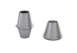
1. Non-engaging conical shape designed for cost-effective and versatile bridge and bar restorative options.
2. Helix threads for strong retention of the prosthetic restoration.
3. Reference edge for long-term stability and passive fit, specially required for implant-based bridge or over-denture constructions.
2. STRAUMANN® VARIOBASE® WITH A SECOND BODY HEIGHT
For more flexibility with longer crowns, we now offer you the well-established Straumann® Variobase® for crowns with a higher, customizable abutment height of 5.5 mm for all platforms (NNC, RN, WN, NC, RC). You can tailor** the body height down to 3.5 mm in order to optimally match the individual patient situation.
3. STRAUMANN® VARIOBASE® FOR CEREC®
Make use of all the benefits offered by Straumann® Variobase® – now also for your chair-side implant-borne workflow. The new Straumann® Variobase® for Cerec® is compatible with the pre-fabricated screw hole for available CAD/CAM material blocks. It provides you not only the Straumann® original connection, but also an optimized abutment design for your bone level implant platforms.
* Variobase® can be used to compensate up to 30° divergence between two implants; Variobase® copings can be used in combination with Straumann® Screw-retained Abutments to compensate larger divergences.
** For the CAD/CAM workflow the body height cannot be reduced.
CEREC® is a registered trademark of Sirona Dental Systems GmbH, Germany – 3M™ ESPE™Lava™ Plus Zirconia is a registered trademark of 3M.
IN-LAB FABRICATION
CHAIR-SIDE FABRICATION
Straumann® Pre-milled Abutment Blanks MORE THAN IN-HOUSE MILLING –ADD VALUE TO YOUR LAB
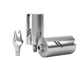
The new Straumann® Titanium Abutment Blanks with pre-fabricated implant connection geometry enable you to fabricate one-piece customized titanium abutments in-house. By allowing you to keep the entire value chain within your dental laboratory, you can significantly shorten your turnaround time (compared to outsourced fabrication). With the Straumann® Pre-milled Abutment Blanks, you also benefit from the original Straumann® implant-abutment connection. The blanks are compatible with a wide range of milling equipment.*
* Compatibility: various preface abutment holders (Datron, imes-icore, Wissner, Röders, etc.) and AG blank holders. For more detailed information, please contact your local sales representative.
Screw-retained Bridges and Bars A COMPREHENSIVE CAD/CAM OFFERING
The Straumann CARES® Screw-retained Bridge and Bar portfolio is continuously expanding and offers a broad choice of CAD/ CAM prosthetics for fixed and removable restorations.
What’s new?
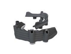
The newly introduced screw-retained abutment connection allows more treatment flexibility for your CAD/CAM restorations. With the new Straumann® CARES® Fixed Bars, the need for cost-effective fixed prosthetic solutions is addressed. Together with Createch Medical, Straumann now offers you a comprehensive portfolio of screw-retained implant-borne restorations, including a full-fledged scan and design service.
Createch specializes in the research, development, and manufacture of high quality, innovative, implant-borne prosthetics. Createch Medical performs the measurement, design, and milling of frameworks for implant-supported prostheses on Straumann implants and abutments. These processes are controlled and executed entirely by Createch Medical, allowing for products that meet the highest quality demands.
Straumann® CARES® Scanner NEW GENERATIONS
The launch of our new Straumann® CARES® scanners is the next milestone for our CAD/CAM product portfolio. The new scanner generation combines proven and established scanning processes with the latest computer technology in an elegant, functional design. Straumann partners with Dental Wings to develop, produce, and support software and hardware of a reliable and high quality.
MAXIMUM FLEXIBILITY
Straumann: CARES® Advanced Fixed Bar with polished intaglio surface for direct acrylic application.
Createch: double-frame titanium prosthesis, fixed with screws or distal pins. A solution to esthetic issues of vestibular screw emergence.
Why is the Straumann CEO hanging upside down from the ceiling?
COMPATIBLE IS NOT ORIGINAL!
Certain third-party manufacturers have copied Straumann prosthetic components and claim that their products are “compatible” with Straumann® originals. However, it is a fact that copycat manufacturers do not have access to the exact manufacturing specifications, which are proprietary and strictly confidential to Straumann. “Compatible” is not “original” and if the connection between implant and abutment is imprecise, the risk of complications may be increased. In contrast, the long-term scientific evidence1 available underlines and confirms the excellent clinical performance of original prosthetic components from Straumann. Non-original abutments are used to reduce initial treatment costs – often without the patient being adequately informed of this proceeding and the possible consequences that may result from combining original components with lookalikes. In some cases, even the treating dentist is not informed by the laboratory that non-original products have been processed.
With “Straumann hangs its CEO”, the company has launched an unconventional social media campaign to illustrate the importance of using original components, causing a stir in the dental markets worldwide.
Uncompromising quality and precision “Made in Switzerland” have always been cornerstones of Straumann’s philosophy. We stand by our products and we are personally convinced that they work because they are made-to-last and reliable.
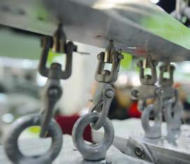
Thus, to demonstrate the performance and reliability of original Straumann® components and his absolute confidence, Straumann CEO Marco Gadola, allowed himself to be suspended upside down from the ceiling – from just four Straumann dental implants and abutment blanks. The video clip, which can be watched on Youtube, is the cornerstone of an awareness campaign to underline the importance of original Straumann prosthetic components for long-term implant treatment. Marco Gadola is carried entirely by four standard Straumann connecting screws (measuring 2 mm in diameter) that are held by three tiny thread pitches within the implants. This is shown clearly in the “Making of” video, which explains how the suspension was performed.
The campaign addresses dentists and dental technicians and provides specific guidelines to the target groups on how to “become a Straumann original”.
BECOME AN #ORIGINALSTRAUMANN!
Don’t worry. You don’t have to hang yourself upside down from four implants to become a true Straumann original. Actually it is quite easy. Just follow these three steps:
DENTISTS
1 Insist on original Straumann® components.
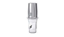
2 Verify the authenticity of the components by checking if the two stripes of the Straumann logo are on the abutment connection or use our online verification tool: www.straumann. com/verification
3 Document the use of original Straumann® components in the patient’s implant passport.
DENTAL TECHNICIANS
1 Learn about the benefits of original Straumann® components.
2 Use and recommend original Straumann® components.
3 Explain the value of original Straumann® components to your dentist customers.
MORE THAN 70’000 VIEWS WITHIN 15 DAYS
Straumann launched the campaign in March prior to the IDS. Within 15 days, more than 70.000 people had watched the video clips and made the #OriginalStraumann campaign a great success, generating many positive feedbacks on Facebook and Twitter.
• 70’000+ video views on Youtube
• 120 retweets on Twitter
• 20’000+ likes on Facebook
• 97 #OriginalStraumann adoptions
Straumann’s presence at the IDS in Cologne has contributed significantly to this great success. At the world’s leading dental fair the company exhibited the original suspension attachment used in the stunt and aroused curiosity and interest among thousands of visitors from all over the world.
Watch how Straumann CEO Marco Gadola was hung upside down from the ceiling and check out the “Making of” video.
It is worth it. Visit www.straumann.com/original
MORE THAN A PROMISE
Our ultimate goal at Straumann is to instill complete confidence – in our products, our services and our people. By taking part in this campaign personally, I wanted to share my complete confidence in the precision and reliability of our implant system with customers – in words and action. I also wanted to highlight my confidence in our specialists. I did not need test results or demonstrations; their word that this would work was enough. The commercial also reflects the innovative and creative power of our team. I wouldn’t have done this with any other product on the market.
Marco Gadola
Marco Gadola is carried entirely by four standard Straumann connecting screws (measuring 2 mm in diameter) that are held by three tiny thread pitches within the implants.
At the IDS 2015 in Cologne.
Restoring chewing function, comfort, and self-confidence
FIDEL RUGGIA
DR. MED. DENT SWITZERLAND
Founder and owner of the Dentalclinic Lugano in Lugano, Switzerland. Teaching position at the University of Zurich, Switzerland. Secretary of the Dentists’ Association of the Canton of Ticino. Director of the ITI Study Club Ticino. www.dentalcliniclugano.ch
STARTING SITUATION
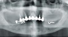
Our patient was a 55-year-old female who wanted her impaired chewing function to be restored. Four years ago, the teeth in her mandible were extracted and replaced with a full prosthesis due to advancing periodontitis (Fig. 1). The teeth in the maxilla also fell victim (Fig. 2) and had to be removed. The patient was otherwise in good health. In addition to restoring chewing function, she wanted in particular to overcome the social handicap associated with the removable prosthesis.
APPROACH
Treatment planning and surgical procedure
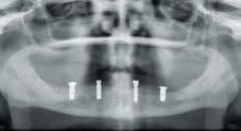
The patient’s desire to replace the teeth with permanent reconstructions was to be met by implants in the maxilla and mandible as well as fixed restorations. The bone volume and quality in the mandible were sufficient for successful implant therapy (Fig. 3). Four implants (Straumann® Bone Level ∅ 4.1 mm, length 8 mm, position 36 and 46; Straumann® Bone Level ∅ 3.3 mm, length 12 mm, position 33 and 43) (Fig. 4, 5) were placed.
Prosthetic approach
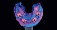
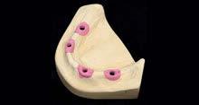
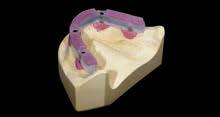
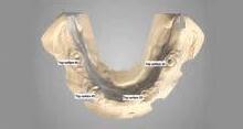
The dental impression (Fig. 6) was mounted with implant analogues and prepared with a gingival mask made of scannable material (Fig. 7). The master cast was then made from class IV plaster (Fig. 8). For the purpose of bite registration and verification of the implant position, temporary abutments were blocked with composite on the cast (Fig. 9). This ensured that the position of the implants and the jaw-to-jaw relation could be checked in one work step. Care was taken to ensure that the composite did not hamper checking of the position – particularly for the emergence profile. After
mounting the cast, the initial tooth set-up was carried out, using the composite bar as a framework. An artificial gingiva was not used in the mandible in favor of better oral hygiene.
The initial esthetic intraoral try-on was performed. Tooth position, emergence profile, relation of tooth length and occlusal plane, color, and of course the esthetics were checked. After checking all of these factors and the patient’s feedback, the process moved to CAD software for planning of the bar.
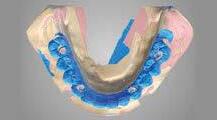
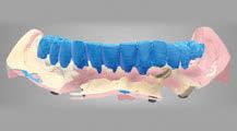
The work was digitized using the Straumann cs2 scanner (Figs. 10 – 12). The restoration was then designed in the Straumann® CARES® 9.0 software. We opted for the Straumann® CARES® Advanced Fixed Bar with basal polished metal surface. This has lower plaque accumulation compared with acrylic veneering.
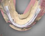
It was possible to edit the bar (Fig. 13), individual segments, and bar copings directly in edit mode. The different tools were very easy and intuitive to use, e.g. an eye was kept on the necessary cross-section thickness using the 2D cross-section window (Fig. 14) Using the mock-up scan (Fig. 14), the full shape of the bar was adapted to the set-up. The bar geometry was simple to edit in the segment editing and the interdental spaces were adjusted to the mock-up. The shape and height of the individual bar copings were defined to avoid space problems.
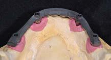
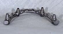
After designing, the bar data were sent directly to the Straumann milling center in Leipzig, Germany. The cast was forwarded by courier to ensure a perfect fit. The sandblasted bar was returned after three to five days, ready for further processing (Figs. 15 – 17)
RENÉ WÖHRLE
DENTAL TECHNICIAN SWITZERLAND
Swiss Certified Dental Technician. Owner of the 3Digital Vision SA dental laboratory in Lugano, Switzerland. Advisor and Project Manager for Digital Solutions. Specialties: CAD/CAM, 3D printing, laser melting. www.3dvlab.com
The bar was then treated with silane (for adhesion). The opaque coating was then applied and set. The veneers were degreased, sandblasted, and repositioned in the previously made transparent index. The index was secured to the cast over the bar; the luting composite was injected and cured with light. crea.lign, a light-curing composite which achieves a homogeneous, dense surface and thus inhibits plaque formation, was used to finish the CARES® Advanced Fixed Bar (Fig. 18). From experience, use of this material reduces the risk of the veneers flaking in the long term. After curing, the occlusion was ground and the bridge was finished and highly polished as usual.
CONCLUSION
After completion of treatment, the patient confirmed that her wishes had been met in full. Chewing function, comfort, and
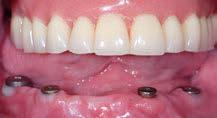
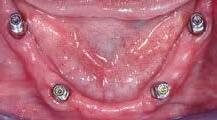
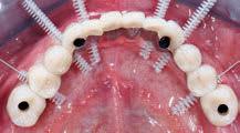
self-confidence had been fully restored. From a financial point of view, this reconstruction did not cost more than manufacturing a conventional removable prosthesis with bar construction. By contrast, the follow-up costs were reduced to a minimum. A fixed reconstruction has had a psychological benefit for this patient, which would not be possible with a removable solution. We therefore firmly believe that this solution is a valid therapeutic option for the treatment of edentulous jaws.Nowadays, patients want solutions for an unrestricted quality of life, combining lasting functionality with attractive smiles. Implant-borne restorations (Fig. 19 – 21) can offer patients precisely this, as this case demonstrates.
We would like to thank the ESTETIKER laboratory in Lugano, Switzerland, for its collaboration. crea.lign is a trademark of Bredent GmbH & Co. KG, Senden, Germany.
www.straumann.com/dentallab
More than a full-arch rehabilitation. A reason to smile.
THE ORIGINS OF THE “IMMEDIATE FULL-ARCH RESTORATION” APPROACH
To address the requirements and expectations of patients seeking fast, convenient, and reliable solutions for full dental replacement, Dr. Paulo Malo from the Malo Clinic® developed a special treatment concept in the early 1990s called “The Malo Clinic® Protocol.” It offered immediate restorations for edentulous patients despite limited bone availability. Since then, the protocol has become a popular procedure in a large number of clinics worldwide and influenced further developments in shortening time-toteeth. Straumann® Pro Arch now offers the components to provide this kind of treatment to your patients: immediate full-arch fixed restorations on either straight or tilted implants – together with the proven advantages of Roxolid® and SLActive®, Straumann’s cutting-edge material and surface technologies.
One of the possible treatment options with the new Straumann components: Straumann® Bone Level Tapered Implant, angled screw-retained abutment, and a Straumann® CARES® Basic Fixed Bar.
Patients who have suffered from ill-fitting removable dentures and, as a consequence, decreasing self-confidence are interested in reliable alternatives in order to regain their lost quality of life. Removable dentures are not an option for them anymore
NEW TREATMENT OPTIONS WITH NEW PRODUCTS AND TOOLS
Until fairly recently, people suffering from the debilitating handicap of a severely damaged, “hopeless” dentition had little or no alternative to having their remaining, compromised teeth removed and being treated with plastic dentures held in place by suction or adhesive. Yet the inconveniences that are associated with these types of unanchored dentures – functional limitations and the loss of self-confidence – are well-known. Straumann® Pro Arch a is the comprehensive combination of proven implant technology, sleek-design abutments, CADCAM frameworks, and auxiliary components, enabling clinicians and dental labs to provide fixed full-arch replacements in reduced treatment time.
Straumann® Bone Level Tapered Implant. This new member of the Straumann® Dental Implant System provides the proven features of the Straumann® Bone Level Implant and a new design feature: the tapered tip. It offers high stability for immediate or early loading procedures and provides excellent primary stability for a reliable anchorage in the bone.
Roxolid®, Straumann’s unique high-strength material from titanium alloy, allows use to be made of the existing bone volume, leveraging of the individual situation, and performance of an overall less complex treatment.
Straumann® SLActive, the cutting-edge and truly hydrophilic surface, enhances osseointegration, resulting in a significantly reduced implant healing time. This way, secondary stability can be achieved faster than with conventional surfaces.
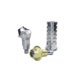
Straumann® Screw-retained Prosthetic portfolio. The new components of this product range were designed to offer a higher level of prosthetic flexibility. The abutment design allows for a reliable prosthetic fixation – even if the implants have to be tilted before being placed in anatomically challenging indications.
Straumann® CARES® Visual 9.5. With the new version of Straumann’s CADCAM software solution, clinicians are now able to provide custom-milled frameworks for screw-retained abutments from Straumann – not only at implant-level but also at abutment level. Furthermore, a wide range of materials for this type of restoration is offered: titanium grade 4, coron®, as well as zirconia frameworks (later this year*).
THE PROSTHETIC RESTORATION MUST BE AS SIMPLE AS POSSIBLE AND THE SURGERY MUST BE DESIGNED TO MAKE THE PROSTHETICS SIMPLE. THE NEW PRODUCTS MEET THIS CRITERION IN FULL.
Dr. Jean-Louis Zadikian, France
LESS COMPLEX AND AT THE SAME TIME MORE INDIVIDUAL TREATMENTS FOR EDENTULOUS PATIENTS
The combination of surgical and prosthetic components with aligned design properties helps to provide a less complex treatment that takes into account the individual patients’ needs and personal expectations. After thorough planning and preparation, the implant and abutment placement can take place in only one surgical session.
As soon as the abutments are in place, the clinical situation is transferred to the dental lab with the help of direct (with titanium copings) or indirect pick-up (with impression components). In the dental lab, the temporary fixed bridge is prepared and adapted to the individual situation. Once the temporary fixed bridge is finished, it can be placed into the patient’s mouth and, if there is no contraindication, the patient can leave the dental office with their new temporary prosthesis in place. Thanks to the complementary components, this is all possible within a very short time. After four to six months, the patient receives the final restoration.
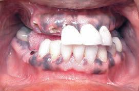
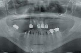
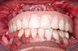
Figs. 1 – 3 show the exemplary workflow for a patient with severely damaged remaining dentition (initial situation and temporary prosthesis). Please note that this procedure is just one of several different possible approaches to providing a fixed full-arch prosthesis.
Courtesy of Dr. William Runyon, Fort Worth, TX, USA.
Locator® on 4 implants
Locator® on 2 implants Bar with pre-fabricated / Individualized parts on > 3 implants
Fixed screw-retained restoration on 4 implants, posteriortilted avoiding sinus
Fixed screw-retained restoration on 4 implants, posteriortilted avoiding mandibular nerve
Fixed screw-retained restoration on 6 implants
Fixed screw-retained restoration on > 6 implants
Fig. 4: A selection of different treatment options (other approaches are possible) within the existing Straumann portfolio – from straightforward to complex restorations.
POSITIONING WITHIN THE SAC CLASSIFICATION
About the SAC classification: In order to identify the degree of complexity and risk involved in individual procedures, the ITI has formalized a system of classification for dental implant procedures to support clinicians at every level of expertise and experience. The classification is based on the debate and findings of an ITI Consensus Conference attended by a multidisciplinary group of 28 clinicians that was held in Mallorca in March 2007. It provides guidelines for a broad variety of implant situations for both restorative and surgical cases, which are classified according to three categories: straightforward (S), advanced (A), and complex (C).
For more information see: www.bit.ly/SACclassification
THE EVOLUTIONARY STEPS OF STRAUMANN® PRO ARCH
The Straumann® Screw-retained Abutment portfolio was developed and continuously improved in a systematic collaboration with experts from all over the globe. Based on their experience with the Straumann® Multibase portfolio, the design was adapted to allow fullarch screw-retained restorations, even in cases where implants have to be tilted. Additional product improvements resulting from the preliminary clinical phase, such as the transfer and alignment pins, and the lab polishing aid, will be made available in 2015. In parallel, Straumann released a variety of new custom-milled bar designs in Straumann® CARES® Visual: the Basic Fixed Bar and the Advanced Fixed Bar, made from grade 4 titanium and coron®, for the manufacturing of custom-milled frameworks for final, full-arch, screw-retained restorations at abutment level. Last but not least, the long-awaited Straumann® Bone Level Tapered Implant was launched in a preliminary phase. A limited number of customers in different countries had access to this new tapered-design member of the Straumann® Dental Implant system (see p. 26) and the feedback was overwhelming: within four weeks, more than 2’000 implants were placed. One of these first clinicians to use the new implant was Dr. Jean-Louis Zadikian from Sarcelles, Paris, France. Within three days he treated 14 edentulous patients with immediate bimaxillary fullarch restorations from Straumann (see a detailed feature in STARGET 02/2014, p. 54 – 63).
Maxilla
Mandible
Implant-supported
Abutment selection and long-term success
JULIA-GABRIELA WITTNEBEN
D.M.D., DR. MED. DENT., MMSC. SWITZERLAND
Senior Lecturer at the Department for Reconstructive Dentistry and Gerodontology, School of Dental Medicine, University of Bern, Bern, Switzerland. Lecturer at the Department of Restorative Dentistry and Biomaterials Sciences, Harvard School of Dental Medicine, Boston, USA julia.wittneben@zmk.unibe.ch
The presented case was treated in a multidisciplinary approach:
• Prof. Dr. med. dent. Daniel Buser (Surgical)
• Dr. med. dent. Julia-Gabriela Wittneben, mmSc (Prosthetics)
• Thomas Furter, CDT (Lab).
The selection of the implant abutment for each individual patient case is an important part of the implant-prosthetic treatment phase. Long-term clinical studies on fixed implant-supported reconstructions show low technical complication rates regarding the abutment itself 1. In this article, different implant abutment types, the various abutment materials, and their clinical indications are discussed. A clinical case presenting step-by-step the treatment of a single edentulous gap with an all-ceramic screw-retained implant crown is shown on page 23
1. IMPLANT ABUTMENT TYPES
Implant abutments can be either standard or customized (Fig. 2). The use of a standard abutment is indicated if the implant is placed in an almost ideal prosthetic position. The advantages of standard abutments are time efficiency in the overall treatment and therefore shortening of the technical manufacturing time. Divergences between implants supporting multi-unit prostheses can be corrected with angled standard abutments. In the esthetic zone, it is important that the collar height of a prefabricated abutment is not a uniform 360 degrees, as the interproximal position of the crown margin would be placed too far submucosally.
Therefore, the ideal design of a standard abutment should be similar to a tooth preparation, following the contour of the gingival margin2 (Fig. 1). Clinical limitations exist regarding the position of the implant in a vertical dimension. If the implant is placed too apically, standard abutments are not indicated, especially for screw-retained reconstructions, as they do not provide enough support for the veneering ceramic.
Fig. 1: Standard abutments made of zirconium dioxide
Customizing an abutment gives the clinician the freedom to individualize its position and angulation. In the case of a bone level implant, it is also possible to individualize the emergence profile and future crown margin position of the final restoration. It allows abutments to be designed to provide optimal support for the veneering ceramic material, especially for screw-retained reconstructions. Individualization may be achieved using CAD/ CAM technology, gold abutments produced with traditional lost-wax casting methods, or titanium base abutments (Fig. 2)
Customized abutments manufactured via CAD/CAM can be made of titanium or zirconium dioxide for bone- and tissue-level implants. They can be used for cement- or screw-retained single crowns or cement-retained bridges. The benefits of the CAD/ CAM abutment include the possibility of using a high-performance ceramic material, which again offers many advantages, especially in esthetic sites. In patients with a thin tissue biotype, no grayish shining-through will be visible with a white-colored abutment. However, it is also possible to choose titanium as a material. Another advantage is individualization regarding the angulation and design of the abutment to support the veneering ceramic.
Traditional cast gold abutments can be used for screw- and cement-retained single crowns and bridges, and are available for implants placed at soft tissue or bone level. Their advantages consist in the facilitation of the screw retention with
bridges. Disadvantages, however, are that gold abutments are technique-sensitive, require more time, and generate higher manufacturing costs. An in vivo histological study in dogs has demonstrated that gold alloys also have disadvantages in terms of soft tissue integration. Histologically, an apical shift of the barrier epithelium and the marginal bone around gold alloy abutments has been shown3
The third group of customized abutments on implants are the titanium base abutments. They are two-piece abutments with a titanium base. Clinicians are sometimes concerned about the handling of complications with a full ceramic abutment regarding the retrieval of broken-off ceramic fragments in the implant, which can be difficult. The main advantage of this abutment type is that there is no ceramic material inside the titanium implant connection. However, the disadvantage lies in the lack of evidence in published clinical data to date.
In particular, the soft-tissue reaction regarding the bonding gap, especially in bone-level implant cases in the esthetic zone, remains unknown. In consequence, this type of abutment should be used with this current limitation in mind4. However, use with soft tissue-level implants with a microgap above bone level might be less of a concern. An example of a soft tissue-level implant case is presented step-by-step on the following pages (Figs. 3 – 15)
Different biomaterials are available for implant abutments. PMMA (polymethyl methacrylate), titanium, and PEEK (polyether ether ketone) are indicated for abutments supporting provisionals – especially for bone-level-type implants – to customize the emergence profile and individualize the peri-implant mucosa with soft tissue conditioning5. The materials of choice for abutments for final restorations are titanium, gold, zirconium dioxide, and aluminum oxide-based ceramic.
Titanium and zirconium dioxide will be discussed in this article regarding clinical and histological performance. Titanium is the biomaterial of choice regarding long-lasting and well documented behavior under functional loading for both soft and hard tissues. It has excellent biocompatibility, mechanical strength, and is resistant to corrosion. Therefore, it is the abutment material of choice for posterior sites. However, the expectations of patients in the anterior zone are increasing. In esthetic sites, mucosal thickness plays an important role. An animal study comparing different dental materials under different mucosal thicknesses showed that titanium induced the most prominent color change. Zirconium dioxide did not induce visible color changes in 2 and 3 mm thick mucosa.6
With the background of the available clinical evidence and systematic reviews, no differences were found between zirconium dioxide and metal abutments in clinical performance based upon esthetic, technical, or biological outcomes7,8,9,10. In vitro studies have shown statistically significant greater wear of zirconium dioxide than of titanium abutments inside the titanium implant11. However, the clinical relevance remains unclear. In our clinic, we have been using Straumann® CARES® CAD/CAM fabricated zirconium dioxide abutments since 2009 on a daily basis in esthetic cases with bone level implants, and have had
no issues with abutment fractures so far. The correct CAD/CAM design of a zirconium dioxide abutment and the quality and precision of the connecting part into the implant play a crucial role in long-term success. Focusing on the outcome of histological studies, an in vivo study shows that there were no visible differences in soft tissue health in peri-implant mucosa adjacent to zirconium dioxide and titanium abutment surfaces12. Another study found that soft tissue around zirconium dioxide heals faster than when in contact with titanium13.
A systematic review14 evaluating the existing literature on zirconium dioxide abutments concludes based on evidence from animal and human histological studies that zirconium dioxide is as suitable a material for dental implant abutments as titanium. Regarding plaque accumulation, zirconium dioxide appears to have a lower tendency for surface-bound bacterial plaque in early stages, which is advantageous.
3. CONCLUSION AND CLINICAL RECOMMENDATION
Abutment selection in esthetic sites. Implant abutments are located in a transition zone where they are in contact with the implant and the surrounding peri-implant tissues. Therefore, the choice of abutment is of major importance, especially in a sensitive region like the esthetic zone.
For single-unit reconstructions, zirconium dioxide abutments are indicated, which can be either standard or customized depending on the prosthetic position of the implant. For multi-unit reconstructions, zirconium dioxide abutments are recommended for cement-retained bridges, and gold titanium abutments for screw retained bridges.
Abutment selection in posterior sites. Clinical indication of each implant abutment type depends primarily on the prosthetic position of the implant and whether single or multiple units need to be replaced. Standard and Straumann® Variobase abutments are the abutment of choice in posterior sites if the prosthetic position of the implant is ideal. Angled standard abutments, individualized CAD/CAM abutments made of titanium, or cast abutments in gold are indicated in cases where the implant is not placed in an ideal prosthetic position. In multi-unit reconstructions, standard titanium or individualized gold abutments are recommended.
CLINICAL CASE REPORT
Restoration of a single edentulous gap with an all-ceramic screw-retained implant crown in a posterior site using the Straumann® Variobase Abutment
Patient: non-smoking, healthy female, 43 years. Situation: a single edentulous tooth gap, region 46 (FDI). A Straumann® Soft Tissue Level Regular Neck Implant with Straumann® SLActive® surface was placed in a correct three-dimensional position (Fig. 3). Open-tray impression and bite registration followed eight weeks later. Peri-apical radiograph for evaluation of the
impression coping position (Fig. 4). Fabrication and articulation of the master casts. Insertion of the scanbody. The cast was centralized in the scanning machine (Fig. 5)
Bite registration with the scanbody in place (Fig. 6). Verification of digital image and manual modification, matching occlusion of the opposing dentition (Figs. 7, 8). A Straumann® Variobase Abutment was used (Fig. 9). An IPS e.max CAD crown made of lithium disilicate glass ceramic was ordered and delivered to the dental laboratory in a bluish color (Fig. 10). The crown was cut back with a diamond bur and crystallized in a furnace. Characterization and finalization of the crown followed by the manual addition of veneering ceramic (IPS e.max. Ceram) and the use of stain and glaze paste (IPS e.max Ceram Essences and FLUO). Different firing cycles. Cementation of the crown on the Straumann® Variobase Abutment with adhesive cement (Multilink Hybrid Abutment Cement). The excess cement was removed and polished (Figs. 11, 12). The final crown was tried intraorally and inserted with 35 Ncm inside the implant (Figs. 13, 14). Evaluation of the crown position (Fig. 15). The occlusion was adjusted and oral hygiene instructions given to the patient.
IPS e.max CAD, IPS e.max. Ceram, Essences and FLUO are registered trademarks of Ivoclar Vivadent, Schaan/ Liechtenstein. List of scientific references: www.straumann.com/stargetref
Independent landmark study shows: brand matters!
Researchers at Gothenburg University in Sweden have published a landmark study1 analyzing the short- and long-term effectiveness of dental implants in a large number of randomly selected patients. The basic finding is that implant success is influenced by brand, with Straumann being ahead.
Using the national data register of the Swedish Social Insurance Agency, the investigators gained unique access to the records of 2’765 patients treated in 2003 with a total of 11’311 implants. This is exceptional in that most studies on treatment outcomes in implant dentistry have assessed survival rates only in small, selected groups of patients treated in university clinics or by specialists. Information on the patients, treatment procedures, and outcomes was obtained from the records, which had been collected by more than 800 clinicians. In addition, the study included a clinical evaluation of 596 patients approximately nine years after their treatment had been completed.
Study design
KEY OBSERVATION: IMPLANT BRAND INFLUENCES IMPLANT LOSS
10 different implant brands were included, the most popular of which were Astra Tech Implant System, Nobel Biocare and Straumann. In addition to reporting the percentages of early and late implant losses, the study presented the relative probability of implant loss as shown by “odds ratios”. Compared with Straumann, the odds ratios for early implant failure were approximately two times higher (statistically significant) with Nobel Biocare and Astra Tech Implant System, and more than five times higher (not statistically significant) for late failure. The ratios for early failure with the other implant brands collectively were nearly eight times higher, and almost sixty times higher for late failure (both statistically significant).
Results
Early implant loss
implant loss
154 implants lost (1.4
121 patients affected (4.4
Details of the study are published in a Clinical Research Supplement of the Journal of Dental Research under the title ’Effectiveness of Implant Therapy Analyzed in a Swedish Population: Early and Late Implant Loss’ by J. Derks, J. Håkansson, J.L. Wennström, C. Tomasi, M. Larsson, and T. Berglundh. Link to full text: www.bit.ly/Straumann-JDR-DerksEffectiveness
List of scientific references: www.straumann.com/ stargetref
Astra Tech Implant System is a registered trademark of Dentsply IH AB
Nobel Biocare is a registered trademark of Nobel Biocare AB, SE
Straumann® Bone Level Tapered Implant
Case review: the new tapered standard
STANDARD WITHIN THE FIELD OF TAPERED IMPLANTS
The Straumann® Bone Level Tapered Implant is a significant step forward in setting a new standard within the field of tapered implants regarding convenience and reliability.
It features the outstanding mechanical and biological properties of the Straumann® Bone Level Implant line: the unique Roxolid® material, the SLActive® surface and the Bone Control Design™ concept for excellent osseointegration and optimized crestal bone preservation. Thus, it is a unique and powerful symbiosis between the established and clinically proven features of the Straumann® Bone Level Implant and the benefits of a tapered implant design regarding surgical flexibility and primary stability.
CASE REVIEW
On 22nd January 2015, Straumann conducted an all-day case review with three experts and a collection of 20 clinical cases from Central and Western Europe. The aim was to identify the best case that represented the strength and benefit of the new implant.
Evaluation
committee
Dr. Andreas Stavropoulos from Malmö University (Sweden)
Dr. Thomas Ziebart from Mainz University (Germany)
Dr. Michel Dard (Switzerland) from New York University (USA).
ELECTED CASES
THE NEW STRAUMANN® BONE LEVEL TAPERED IMPLANT IS THE ANSWER TO TODAY’S CLINICAL AND ANATOMICAL CHALLENGES.
Selection criteria
1. Immediate implantation
2. Quality of documentation and pictures
3. Adequate level of information on both surgical and prosthetic procedures.
4. Highly diagnostic pre-operative and post-operative X-rays
The BLT’s contribution to osseointegration in damaged esthetic areas by Dr. Sepehr Zarrine, France
This winning case demonstrates impressively what can be achieved with the Straumann® Bone Level Tapered Implant under the following circumstances:
1. A complex situation requiring bone augmentation (in the esthetic area)
2. Immediate placement and loading with provisional Soft tissue management
3. Good primary stability with both implants reaching more than 50 Ncm insertion torque
Read the whole documentation on the following pages.
Further winning cases
The evaluation committee selected further winning cases covering different placement and loading protocols. They will be made available for peer-to-peer communication among the Straumann network.
AUTHOR
Massimo Ciocco Switzerland Early Late
Jochen K. Alius Germany Early Immediate
Arndt Happe Germany Immediate Immediate
Airoldi Giulio Switzerland Immediate Late
Straumann would like to thank all participants of the Straumann® Bone Level Tapered Implant Case Review for the efforts they have put into their contributions.
Michel Dard, Andreas Stavropoulos and Thomas Ziebart.
The BLT’s contribution to primary stability and osseointegration in damaged esthetic areas
SEPEHR ZARRINE
DDS FRANCE
Oral surgeon. Exclusive private implantology practice (Saint Dié, France). Speaker ITI France. European Master in Dental Implantology. Surgery, prosthetics, bone grafts (Frankfurt, Germany). University diploma in surgical maxillofacial rehabilitation (Medicine, Paris VII).
Surgitechstudies.com dr.zarrine@gmail.com
Thanks to Pierre Chapuis and his team at Laboratoire Oral Beauty (Bruyeres, France).
oralbeauty.com
BACKGROUND
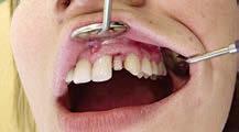
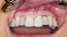
The patient is a 56-year-old, active and healthy man. He does not smoke, does not take any medications and has no allergies. He was referred to us, presenting a loose anterior bridge from 11 to 22. Since his profession requires speaking in the public, his clinical situation in the esthetic zone has a negative impact on his self-confidence. The clinical examination (Figs. 1, 2) revealed a slightly inflamed gum with no abscess. Clinical probing indicated that there was vestibular bone loss at tooth 11 and a decayed root, but no bone loss at tooth 22.
TREATMENT PLAN
Two treatment options were considered:
1. Extraction of both roots, healing, re-entry to place two non-submerged implants with simultaneous GBR, healing, gingivoplasty and placement of the final prosthesis.
2. Extraction of both roots, immediate implantation with simultaneous bone reconstruction and gingival reinforcement, dental crown placement on the same day, i.e. immediate placement and immediate provisionalization.
In order to restore the patient with a reduced overall treatment time (including the time needed for surgery), we opted for the second solution.
We have adapted an osteogingival tissue graft technique that has been employed for several years with excellent results. Pre-surgical assessment indicated potential primary stability issues at the site of tooth 11, where only a few millimeters of bone remained for implant anchorage.
Therefore, we used this novel bone level tapered implant, designed to achieve good primary stability even in clinical indications where this would be difficult to achieve. Furthermore, the Straumann® SLActive® surface is known to provide security for the critical osseointegration period. Pre-surgical impressions were taken in order to prepare a surgical guide and a perforated impression tray.
SURGERY
Once the bridge was removed, a vertical radicular fracture was confirmed in tooth 11 and an extensive decay in tooth 22. Atraumatic root extractions were performed, on tooth 22 by applying the Benex® Extraction System (Fig. 3) and on 11 with a very fine elevator (Fig. 4). A gingival flap was elevated to access the bone defect at tooth 11 (Fig. 5)
Since the buccal side at tooth 22 was still intact, the gingiva was also left attached to the bone (i.e. flapless procedure) in order not to disturb the periostium attachment to the bone. At tooth 21, a flap was rolled to thicken the buccal alveolar ridge aspect.
The two tooth sockets were meticulously scraped and cleaned with a round bur. The surgical guide helped identify the ideal axis for the implant as well as the best emergence level (Figs. 6, 7). Once the landmarks were identified, the drilling was performed and then the guide was repositioned to verify the axis of the alignment pins (Fig. 8). To achieve a sufficient primary stability, the BLT drill with a diameter of 2.8 mm was used as the final preparation step.
The insertion torque for both Roxolid® Bone Level Tapered Implantat, ∅ 4.1 mm RC, SLActive® 14 mm was greater than 50 N/cm. The implant at site 22 was placed towards the palate to maintain a narrow buccal gap between the implant and the socket wall.
The implant at site 11 had 8 visible threads after insertion. It was also inserted towards the lingual aspect of the extraction
socket such as that a gap remained between the implant and the buccal bone wall (Fig. 9). The anchoring for both implants was achieved with the last apical millimeters, which is why the 14 mm length and underpreparation were necessary.
The majority of the drilled bone fragments were recovered thanks to the design of the BLT drills and a portion of this bone was used to fill the gap around the implant on position 22. The remaining material was used to cover the surface of the implant. To simultaneously regenerate the bone deficit and reinforce the gingiva, we used a special technique that we have been optimized during several years.
We took an osteogingival graft form the maxillary tuberosity of the wisdom tooth. A prior 3D examination had confirmed that the maxillary tuberosity presented good volume. This combined graft for osseointegration was de-epithelialized (Fig. 10) and then impacted into the vestibular gap of the central incisor. To prevent any mobility, an osteosynthesis screw fixed the bone core on the residual alveolar ridge by engaging the cortical bone of the palate (Fig. 11)
Straight Straumann® Screw-retained Abutments (SRA) with a height of 2.5 mm were placed on the implants. Suturing was performed using resorbable 5.0 monofilament.
Then, abutment-level open tray impression posts were screwed on and a sectoral impression was taken using the perforated impression tray (Fig. 12). Once the impression was taken, two protective caps were placed to prevent the gingiva from covering the SRAs during the day. The prosthetic planning process was performed with the dental laboratory, which will also perform the final work. Six hours after surgery the gingiva was stable enough (Fig. 13) to remove the protective caps without anesthesia and the temporary bridge was screwed onto the SRAs (Figs. 14 – 17)
Visual and radiological examinations were performed one week post-surgery (Figs. 18, 19) which demonstrated that the initial healing phase was successful. The gingiva looked healthy and the patient was without clinical complications. At a re-entry after two months clinical observation determined that the implants, bone and gingiva had healed perfectly (Fig. 20)
CONCLUSION
This challenging case demonstrates the possibility to compress a multi-step clinical procedure into a manageable and cost efficient time-frame. Furthermore, given this implant’s location in the esthetic zone, the technique could be of particular interest to both clinicians and patients alike. The restoration was successful due to the combination of several state-of-the-art technologies and techniques:
1. The use of Straumann® Bone Level Tapered Implants, which, thanks to their design and a flexible drilling protocol, allow for good primary stability in compromised recipient bone conditions
2. The retrieval and reuse of bone fragments using the Straumann® Bone Level Tapered implant drills
3. The use of the maxillary tuberosity osteogingival tissue graft technique.
With this, we were able to provide our patient fixed teeth in a single day. The temporary bridge had no occlusal contact and only served to enable the patient to speak and smile. Once osseointegration is completed and the graft has been consolidated, the final bridge can be planned.
Benex® is a registered trademark by BENEX, Lucerne/Switzerland
Straumann® Bone Level Tapered Implant (BLT)
“Thanks to the BLT, I can now provide ideal conditions for immediate loading in most patients”
SERGIO PIANO DDS ITALY
Private practitioner in Genoa, Italy. Graduated DDS cum laude at the University of Genoa in 1988. Visiting Assistant in 1991 – 1992 at the University of Geneva, Departments of Fixed Prosthodontics (Prof. Belser), Periodontology (Prof. Cimasoni) and Oral Surgery (Prof. Fiore-Donno, Dr. Bernard). Active Member of SIO (Italian Society of Osseo-integration) and IAED (Italian Academy of Esthetic Dentistry). Fellow of the ITI (International Team for Implantology). Regular national and international lecturer on surgical and prosthetic topics in Implant Dentistry. serg.piano@tiscali.it
Dr. Piano, what are your favorites in the Straumann® Dental Implant System?
Sergio Piano: Today, I use predominantly SLActive® Roxolid® implants. They are my first choice. I prefer them because I think that there are a lot of advantages, and I have been able to simplify my portfolio. Roxolid® is a very interesting option because I can place implants with a narrow diameter and still trust that I can obtain the same result as with a standard diameter implant. It’s a good option and gives patients a low or less invasive treatment that is well received by patients.
Do you find that you can treat patients that you couldn’t otherwise?
Sergio Piano: Yes, because, as I have observed in my practice – and would I think be confirmed by other clinicians around the worldpatients always opt for the simplest and least invasive solution. Patients are always happy when I am able to provide them with a treatment without having to perform a bone augmentation. In certain cases, with a reduced diameter implant made from Roxolid®, we are able to propose exactly what is requested.
You’ve been looking at the new Straumann® Bone Level Tapered Implant.
Sergio Piano: Yes, and in my opinion the new Straumann® Bone Level Tapered Implant is a very interesting product. When I have to perform an immediate loading procedure with very low-density bone, the standard bone level implant is not always able to provide the primary stability that is needed. In such cases, I think the possibility of using an implant that has a better “grip” is very useful. At times, I have not been able to perform an immediate loading procedure due to the low torque of the bone level implant. So, I think that this new implant can improve this particular clinical situation. In fact, recently, thanks to this new implant, I have been able to provide ideal conditions for immediate loading in most patients. The overall feeling has been very good when I have inserted the first of these implants. It was very comfortable, and the fact that they are self-tapering and that we can under-prepare the site makes them a very good option from a mechanical point of view. On the other hand, we have the same quality in terms of the surface and the material - I like this! In fact, I can easily achieve primary stability when needed for immediate loading or when I have to perform a bone regeneration procedure.
Tilted implants: why are they useful and how have you used them?
Sergio Piano: When I perform immediate loading, sometimes it’s very useful to place a tilted implant. In this way, we are able to reduce the number of implants and, at the same time, to reduce the impact of surgery. By tilting the implant, you can avoid an anatomical structure that might be involved in a more invasive surgical procedure. By reducing the number of implants with the tilting of the posterior implant, we are able to produce a simple prosthesis that is , affordable for the patient and at the same time gives us – in selected cases, obviously – the conditions for delivering a satisfying prosthesis.
According to Steven Chens presentation* at the EAO 2014, patients are beginning to expect that implant solutions last at least 40 years, possibly even 60. Do you think that we’re at that stage? Do you think that the products you’re putting into patients’ mouths today are going to last that long?
Sergio Piano: This is an important issue because 40 years is a long timeframe. We are used to thinking about a shorter period when we speak about implants – 10 years, 15 years, even 20 years; but not 40 or 60 years. I think we have to push for the quality of implants, of abutments and of the materials that are involved in implant dentistry, because it is a goal that we can obtain, to extend the duration of our treatment. I don’t know if
my crowns or my implants will last 40 or 60 years. Who knows? So the first step is to start from a level of excellence in quality on the materials side. In fact, I think that if we have to save money, we should do so, not on the implant or the abutment, but on other components of the treatment that we offer to the patient. For instance, reducing the amount of time spent in the chair, or the type of veneering materials used, and so on. But the basis must be of the highest quality possible.
Now, having worked with the Straumann® Dental Implant System for 25 years, what would you say to those who might feel a little hesitant about freely using a tapered bone-level implant?
Sergio Piano: When the Straumann® Bone Level Implant was launched on the market, I was also quite reluctant to use it because I love the tissue level implant line, so I said “No, bone level is not for me.” Then I started to use it and I began to understand that in certain clinical cases, it is better than a tissue level implant. I think that the same is true of the new Straumann® Bone Level Tapered Implant because it provides some clear advantages.
Dear Dr. Piano, thank you for this interview.
* Stephen Chen: Scientific evidence and clinical benefits of titanium-zirconium implants. A presentation held on 25 September at the EAO Symposium 2014 in Rome.
Straumann® PURE Ceramic Implant 3.3 mm
PUREly amazing!
The 3.3 mm diameter-reduced Straumann® PURE Ceramic Implant was launched at the end of 2014 and completes the portfolio of Straumann monotype ceramic implants.
OVERCOMING SPECIFIC REQUIREMENTS OF CLINICAL SITUATIONS
Specific clinical situations, especially in the anterior region, require reduced-diameter implants. In addition, some patients require special treatment, ask expressly for metal-free solutions or demand a highly aesthetic restoration. After nine year of scientific research and development, the 4.1 mm Straumann® PURE Ceramic Implant was officially launched at the ITI World Symposium in April 2014. The diameter-reduced Straumann® PURE Ceramic Implant 3.3 mm was fully introduced to the market at the end of 2014.
RIGOROUSLY TESTED AND BACKED BY SCIENCE
The manufacturing process of Straumann® PURE implants involves some innovative steps, including a rigorous test procedure in order to assure the stability of every single implant. Data from mechanical tests also show that the Straumann material is capable of meeting the requirements imposed on the strength of diameter-reduced implants from ceramic. Static fracture strength tests according to ISO 14801 show that the Straumann® PURE Ceramic Implant has a significantly higher resistance to forced rupture than competing products in both regular and reduced diameters.2
THE MOST COMPELLING FEATURES OF THE STRAUMANN® PURE CERAMIC IMPLANT INCLUDE:
• Design features that combine the known advantages of the Straumann® Bone Level and the Soft Tissue Level implant design
• The high-performance, yittria-stabilized zirconia that provides an even higher fatigue strength than grade 4 titanium implants
• The original Straumann® ZLA® surface, designed to enhance and shorten the healing process and to provide a highly predictable osseointegration3,4,5
The first 3.3 mm Straumann® PURE Ceramic Implants were placed between July and October 2014 in a controlled market acceptance test with selected dentists. The very first implant was placed by Dr. Florian Thieringer from the Clinic of Oral- and Maxillo-Facial Surgery at the University Hospital of Basel in Switzerland. Dr. Thieringer and Dr. Röhlings’ feedback was very promising (see pg. 36 – 37).
100% PROOF-TESTED
A HIGH-END ESTHETIC SOLUTION THAT MAKES THE DIFFERENCE
The Straumann® PURE Ceramic Implant makes available a different esthetic solution to treat patients who have specific needs and demands. While some patients have a thin gingival biotype, which requires a different treatment approach, others express an explicit wish for a metal-free alternative. With this innovative metal-free solution, you will be able to expand your patient base and business: a patient survey conducted in Europe by Straumann, including more than 250 participants has shown that, given the choice, considerably more people would prefer a ceramic to a titanium implant. The Straumann® PURE Ceramic Implant is backed by Straumann’s decades of experience in research and product development. This means that it comes to the market with the supporting evidence and highest quality manufacturing standards that you can expect from a member of the Straumann® Dental Implant System.
Restoration of a lateral maxillar incisor, missing due to aplasia
FLORIAN THIERINGER
DR. MED. ET MED. DENT., MHBA SWITZERLAND
Specialist in Oral and Cranio-Maxillofacial Surgery, Consultant at the Department of Oral and Maxillofacial Surgery, University Hospital Basel, Switzerland. Medical Degree of the Munich Technical University, Dental Degree of the Ludwig-Maximilians-University Munich, Master of Health Business Administration of the FA University Erlangen-Nuremberg, Germany. Member of the International Team of Implantology (ITI).
INITIAL SITUATION
This 22-year old female patient suffered aplasia of both lateral maxillar incisors. After orthodontic treatment, the local dentist filled the intermediate spaces in regions 12 and 22 with two Implants. The patient came to our clinic with advanced peri-implantitis on the implant in region 22 (Figs. 1, 2). An impression was taken of the maxilla and mandible. Then a removable single tooth clamp temporary prosthesis was fabricated pre-operatively, supported occlusally on the neighbouring teeth. This was to fill the space that arose after explantation of the implant.
SURGICAL PROCEDURE
The implant in region 22 was explanted and the tissue sections modified by inflammation were carefully curetted. As suggested by the X-rays, the operation found a significant multiwalled bone defect (Figs. 2, 3). Eight weeks after explantation, the defect region was augmented with an autologous bone block from the mandibular angle as well as bone substitute material (botiss cerabone®) and a collagen membrane (botiss Jason® membrane).
Six months after bone augmentation, a one-piece diameter-reduced ceramic implant was implanted (diameter: 3.3/length: 10 mm). Because of the narrowness of the mesio-distal spaces in region 22, only a diameter-reduced implant could be used here.
On the buccal side, autologous bone chips from the zygmomatic bone region were harvested (Figs. 4, 5). The healing process was normal (Fig. 6). The implant was exposed to the oral cavity and healed without inflammation under the protection of the preoperatively fabricated temporary clamp prosthesis.
TAKE A LOOK
Scientific literature available on the Straumann® PURE Ceramic Implant → PAGES 30 – 31
PROSTHETIC RESTORATION
To form the gingival soft tissue, a single temporary tooth restoration was fabricated from composite on a Straumann temporary coping. Optimal soft tissue formation was then ensured using a CAD long-term temporary restoration (Fig. 7). After approx. 16 weeks, the final impression was taken for a full ceramic crown (Fig. 8). The postoperative X-ray shows the correction positioning of the implant in the maxilla bone (Fig. 9)
CONCLUSION
It was possible to treat the highly esthetically challenging situation with limited space and significant bone deficit well using a gradual treatment concept. The choice of a reduced-diameter 3.3 mm Straumann one-piece ceramic implant with macro- and micro-rough surface and optimal biocompatibility ensured not only treatment success but also met the desire of the patient for a tooth-colored, completely metal-free solution, with a dark titanium implant showing through the gingiva on the opposite side. The measures for GBR were consistent with the usual approach for titanium implants.
A TOOTH-COLORED IMPLANT FOR RELIABLE AND ESTHETIC CARE
This one-piece 3.3 mm diameter-reduced Straumann® PURE Ceramic Implant is what we have been waiting for. The 3.3 mm implant extends the therapeutic possibilities, particularly in situations where the diameter of the 4.1 mm ceramic implant is too wide or in the esthetically demanding region of the anterior teeth. This exhibits the same outstanding properties as the 4.1 mm Straumann® PURE Ceramic Implant with respect to surface and biocompatibility. The micro- and macro-rough surface of the ZLA® has been subjected to experimental and clinical tests and guarantees reliable osseous ingrowth, as with the Straumann Titanium implants with SLA® surface. In addition, the new 3.3 mm ceramic implant exhibits outstanding fracture strength even at the lower diameter. Thus it is an ideal tooth-coloured implant for reliable and esthetic care when space is limited.
Florian Thieringer
The treatment described in this article was performed with substantial contributions from Dr. Michael Gahlert, Munich (Germany) and Dr. Stefan Röhling, Basel (Switzerland).
Straumann® PURE Ceramic Implant
Clinical review
Z-Systems Z-Look3 Evo
3.6mm/4.0mm
Fig. 1: Static fracture strength tests according to ISO14801 show that Straumann® PURE Ceramic Implants have significantly higher resistance to forced rupture than competitor implants in reduced and regular diameter (data on file).
In this 29-year-old female patient, a vertical fracture of tooth 21 led to marginal inflammation, which was particularly noticeable due to the high smile line (A). Situation after implant at loading at 1 year (B). Courtesy of Dr. Michael
Nowadays, patients are more esthetic and health conscious than ever before (Montero et al., 2014). Healthy-looking oral soft tissues and bright teeth are considered a prerequisite for a beautiful smile and self-esteem, adding directly to health-related quality of life (Bennadi and Reddy, 2013; Klages et al., 2004; Pithon et al., 2014). The Straumann® PURE Ceramic Implant is ivory-colored like a natural tooth root and provides a highly esthetic and metal-free alternative to implants made out of titanium.
STRONG AND RELIABLE
Ceramic components have been used successfully in orthopedic surgery for over 35 years (Bhandari et al., 2011) and are also valued by the aerospace industry for their enhanced toughness and dimensional stability even in high temperatures. However, the stability of ceramic dental implants has long been questioned. To overcome these objections, Straumann® has established an innovative manufacturing process followed by a rigorous 100 % proof test in which every single Straumann® PURE Ceramic Implant is tested mechanically before leaving the production site. Here, forces that exceed the maximum human bite capability are applied, and only implants that pass the test are delivered to the dentist. The outstanding quality becomes evident when comparing the Straumann® PURE Ceramic Implants with other commercially available ceramic implants. The Straumann® PURE Ceramic Implant (∅ 4.1 mm and ∅ 3.3 mm) shows significantly higher resistance to forced rupture (Fig. 1). Forced rupture is the most frequent cause of ceramic implant failure. The reliability of the Straumann® PURE Ceramic Implant has been clinically verified in a multicenter study, where zero implant fractures were reported during a follow-up period of 24 months (Gahlert et al., 2015).
ESTHETIC
Most patients perceive a treatment as successful when they are satisfied with the overall dentofacial appearance after treatment. Unlike other white ceramics, Straumann® PURE Ceramic Implants are ivory-colored, which most closely resembles natural tooth roots – an advantage in patients with a thinner mucosal biotype or a high lip line smile (Bidra and Rungruanganunt, 2013; Gahlert et al., 2015; Jung et al., 2008).
FAVORABLE SOFT TISSUE FORMATION
Zirconia shows a favorable formation of the epithelial attachments, as well as lower bacterial accumulation compared to titanium surfaces (Degidi et al., 2006; Institut Straumann AG, 2014b; Welander et al., 2008). This is an important observation since bacterial adhesion to implant surfaces can lead to bone loss in the tissues surrounding the implants (Lindquist et al., 1996). Studies were able to show lesser gingival recession after placement of zirconia implants (Tete et al., 2009), as well as excellent esthetic outcomes and papilla formation around the implant after one year follow-up (Fig. 2) (Gahlert et al., 2015; Kniha, 2014).
Fig. 2:
Gahlert and Dr. Heinz Kniha.
CLINICALLY PROVEN
Surface modifications play an important role in the osseointegration process and thereby influence implant strength as well as aging resistance (Buser et al., 1991; Shalabi et al., 2006). The surface of the Straumann® PURE ceramic implant, Straumann® ZLA®, features a topography characterized by macro- and micro-roughness similar to the proven Straumann® SLA® surface (Fig. 3) (Bormann et al., 2012; Gahlert et al., 2012; Institut Straumann AG, 2011). With over 20 years of experience and more than 100 clinical and preclinical studies, the Straumann SLA® surface is one of the most successful and best clinically documented surfaces in dental implantology, with proven osseointegration properties (Buser et al., 2012; Fischer and Stenberg, 2011; Roccuzzo et al., 2008). In preclinical studies, the ZLA® surface demonstrated similar healing patterns, healing times and osseointegration in terms of peri-implant bone density and boneto-implant contact (BIC) as seen for the SLA® surface (Gahlert et al., 2012; Gahlert et al., 2010). Other studies observed even higher BIC with ceramic implants compared to titanium (Dubruille et al., 1999; Schultze-Mosgau et al., 2000). A recent multicenter clinical trial reported survival and success rates of 97.6 % for the Straumann® PURE Ceramic Implant after one year (Gahlert et al., 2015), which is a value within the range of reported one-year survival and success rates for titanium or titanium alloy implants (den Hartog L. et al., 2008).
METAL-FREE
The prevalence of allergic diseases has increased worldwide in recent years (Lotvall et al., 2012), with a growing number of patients suffering from multiple allergies (Simpson et al., 2008). Although hypersensitization to titanium is quite uncommon (Sicilia et al., 2008), many people are generally aware of allergic reactions to metals such as nickel and cobalt (Thyssen and Menne, 2010). In this light, health-conscious patients or patients with susceptibility to allergic reactions may request a metal-free alternative to titanium implants. Straumann® PURE Ceramic Implants are made out of zirconia (yttria-stabilized tetragonal zirconia polycrystal, Y-TZP), which is biocompatible and guaranteed 100 % metal-free.
List of scientific references: www.straumann.com/stargetref
Fig. 3: The ZLA® surface (A) combines the micro- and macro-roughness of the SLA® surface (B) with reliable osseointegrative properties. The torque-out value of the Straumann® PURE Ceramic Implant is equivalent to SLA® implants from titanium.
coDiagnostiX™ meets Straumann® CARES X-stream™
A new world of treatment possibilities
The dental implant market is rapidly evolving, with prosthetically-driven implant placement becoming the key factor for a successful dental implant therapy. Digitalization is the main driver of this progress. With the new versions of coDiagnostiX™ and Straumann® CARES® Visual, and their implementation of the Straumann® CARES® X-stream™ workflow, software integration has become a reality. This opens a new world of treatment possibilities and an efficient access to a true prosthetically-driven digital implant treatment.
CODIAGNOSTIX™ + STRAUMANN® CARES® X-STREAM™ = IMMEDIATE DIGITAL TOOTH REPLACEMENT
The continuous progress in both computer technology and dental manufacturing creates new opportunities in the clinical workflow. Straumann, in association with Dental Wings, offers a powerful combination in a fully digital pathway and model-free approach.
In this context, the Straumann® integrated workflow offers an additional instrument for treatment planning, surgical placement and prosthetic rehabilitation, using an interdisciplinary team approach:
• All components for guided surgical procedure, guided implant placement and immediate customized temporization are available in a single surgical visit.
• The CARES® X-stream™ solution is delivered in one step before the surgery and provides perfect conditions for immediate customized esthetics.
WORKFLOW
The benefit for patients is obvious: the implant procedure and the temporary restoration can be provided in a single step, with perfect conditions for immediate individualized esthetics.
2. Surgical and prosthetic planning in 3D → Printing of Surgical guide and fabrication of immediate CADCAM temporization
CLINICAL EXAMPLE
3. Guided surgery > second intraoral scan and immediate
4. Conception of CADCAM final prosthetic rehabilitation
5. CADCAM final prosthetic rehabilitation
The benefit for patients is obvious: the implant procedure and the temporary restoration can be provided in a single step, with perfect conditions for immediate individualized esthetics.
Fig. 1: Prosthetic project design in Straumann® CARES® Visual.
Fig. 2: Prosthetically-driven implant planning in coDiagnostiX™, integration with the digital wax-up from Straumann CARES® Visual.
Fig. 3: Design of the digital drill guide in coDiagnostiX™.
Fig. 4: Printed digital drill guide, with the Straumann® CARES® X-stream™ solution (Straumann® Variobase abutment and temporary crown).
Fig. 5: Guided surgical procedure and guided implant insertion
Fig. 6: Treatment planning transferred to the patient’s mouth in a single session. Initial CARES® X-stream™ Variobase abutment and temporary crown are in place.
1. CBCT without Rx template → First intraoral scan
CADCAM temporization
15 YEARS 100’000+ ofclinicalsuccess guidedcases 1 2 3
THE NEW CODIAGNOSTIX™
A sophisticated and user-friendly implant planning software
coDiagnostiX™ is the digital implantology solution that covers dental implant planning and design of surgical drill guides. It helps dental professionals to provide safer and predictable results even in challenging protocols while increasing efficiency and productivity.
1. DWOS Synergy™: moving from connected to integrated software
DWOS Synergy™ opens up a completely integrated, seamless and time-saving workflow between coDiagnostiX™ and Straumann® CARES® Visual. This allows you transfer of your coDiagnosiX™ implant planning to DWOS and return of the restoration planning in real time.
You can collaborate within the local network with just one click – or over the internet with a free DWOS Synergy™ account.
2. Digital drill guide: the next level of guidance
With the new coDiagnostiX™, you can create bone-supported drill guides. It is also possible to design drill guides with a combination of teeth and bone support. For this purpose, a new optional wizard page has been added to the digital drill guide wizard. Additionally, you can have the rotation markers engraved into the drill guide, which will indicate the implant connection position to enable precise placement of a previously planned restoration.
3. Virtual planning export: model-free workflow as an option
The virtual planning export has been completely redesigned and comes with the following new features:
• Virtual model builder
• Export of virtual plaster model with implant analogs to create a 3D print of the model
• Export of segmentation to create a 3D print as physical mock-up
• Multi STL file export
• Many more functions …
Knowledge Network Credit points
ITI Congress Finland September 11–12, 2015Helsinki, Finland
ITI Congress Greece & Cyprus October 9–10, 2015Athens, Greece
ITI Congress Russia October 10–11, 2015Moscow, Russia
ITI Congress Middle East October 15–16, 2015Dead Sea, Jordan
ITI Congress Chile October 23, 2015 Santiago de Chile, Chile
ITI Congress Benelux April 15–16, 2016 Maastricht, The Netherlands
ITI Congress Norway & Sweden April 15–16, 2016 Malmø, Sweden
ITI Congress North America April 28–30, 2016 Chicago, USA
ITI Congress Denmark May 27, 2016 Nyborg, Denmark
ITI Congress Austria June 10–11, 2016 Vienna, Austria
ITI Congress Southern Africa July 15–16, 2016 Pretoria, South Africa
Fully committed to becoming an even stronger player in the field of digital dentistry
THE OPEN STANDARD SOFTWARE PLATFORM FROM DENTAL WINGS
DWOS is a collaboration initiative by 3M ESPE, Straumann, and Dental Wings. It increases flexibility and simplifies processes for dental labs and practices. Being an open system, it provides a solution to the growing problem of incompatible systems that lock users into the technology of individual companies. The scope, quality, and functionality of DWOS make it a solution of choice for communication, design, and collaboration within dentistry. DWOS is commercially available as an open system and offers dental labs the flexibility of designing prosthetics using data from multiple systems and sources. Restorations can be manufactured inhouse or outsourced to milling centers offering high precision and additional material options.
www.dwos.com
With the launch of coDiagnostiX™ 9.5 and Straumann® CARES® Visual 9 in October 2014, implant surgery and prosthetic planning were raised to the next level in terms of software convergence and workflow integration. At the IDS 2015 in Cologne/Germany, Straumann presented a new software update (Version 10) with more prosthetic options for Straumann® CARES® X-stream and improvements in usability. In this interview, Straumann CEO Marco Gadola outlines Straumann’s strategy, commitment, and vision in the field of digital dentistry.
Two years ago, Straumann consolidated software development resources in order to strengthen Dental Wings as a software powerhouse in digital dentistry. Why?
Marco Gadola: The idea of amalgamating our own software development into the Dental Wings platform or organization clearly came out of the conviction that our core strength at Straumann is not in developing scanners, equipment, or software. What we can contribute is obviously input in terms of what functionality has to be reflected in the software. That’s actually our responsibility, specifically when it comes to implant-borne prosthetics and solutions. However, the programming and handling of the software are not our but Dental Wings’ core strength. That’s why we decided in 2013 that Dental Wings should take over this responsibility on our behalf.
Where do we stand today?
Marco Gadola: As you have seen, we recently launched a new version of the coDiagnostiX™ software, version 9.5, as well as a new version of Straumann® CARES®, version 9.0. It is already very close to the Dental Wings system. We have tried over the last couple of versions to bring these two platforms as close together as possible and this objective will be achieved with release 10.0.
The spin-off in software development had an impact on our guided surgery solutions customers. Straumann’s strategy, on the other hand, is to be a full solution provider in tooth replacement. What can customers expect in terms of our further commitment to digital dentistry?
Marco Gadola: Straumann’s clear ambition is to become a full solution provider when it comes to replacing teeth. Not to be only the number one dental implant company but to be also the leader when it comes to providing solutions for replacing teeth. Obviously, to achieve that, we have to make sure that our customers are able to work with our solutions and products independently, whether they work completely digitally, semi-digitally, or are still using conventional workflows. This results in our ambition to have a fully digital solution available for our customers, ranging from the planning software coDiagnostiX™ to CAD/CAM software, and eventually being able to pro-
Marco Gadola
vide the equipment, materials and the services to supply CAD/ CAM elements to our customers. So we are fully committed to continuing to put resources and effort into becoming an even stronger player in the field of digital dentistry.
Could you describe the collaboration with Dental Wings in the specific field of Guided Surgery and its key success factors?
Marco Gadola: As I have already pointed out, Dental Wings’ core competence is developing planning, CAD/CAM, and intraoral scanning software, and hardware, chair-side, and lab-side. Obviously, what we as Straumann have to contribute is input regarding in which direction this software should be developed. We can also specifically push the planning software with our customers, with our dentists, making them aware that we have a great product at hand, generating leads on behalf of coDiagnostiX™, and encouraging them to actually use this software because it helps them to become more efficient in their practice, and through that, making sure that the partnership between Dental Wings and our software in the field of treatment planning is actually a successful one.
“The cooperation with Dental Wings allows our customers to become very efficient in handling CAD/CAM in their dental practice.”
How can our customers benefit and profit from the synergies between Straumann and Dental Wings?
Marco Gadola: A valid question. In the end, what we will be able to provide together with Dental Wings is an integrated digital workflow, starting with radiography through to the planning software/treatment planning, then taking the same information and data and putting it into CAD software, and finally, milling the elements either chair-side or sending the file to a lab to get it milled through a lab solution or sending it to one of our milling centers. The cooperation with Dental Wings allows our customers to become very efficient in handling CAD/CAM in their dental practice. Again, as pointed out before, through this partnership, we are combining the strengths of Dental Wings,
which are clearly in software development, and the strengths of Straumann, which are access to dentists, being close to the needs of these customers, and then giving input to Dental Wings in terms of which features are needed in the software, and in which direction this software should actually be developed.
“We have to make sure that we get connected to all these platforms that play a major role in the market.”
How do you see the role of Straumann in digital dentistry and what are the challenges in the near future?
Marco Gadola: Our greatest challenge is making sure that we are connected to all these digital platforms. In my view, we are on the right path when it comes to getting connected more to the lab-side of the CAD/CAM business, such as the 3shape scanners. We are also working on getting access to the ExoCaD-based scanners, and we have connectivity to the Dental Wings lab scanners through our plugins. The trend, however, is clearly moving toward intraoral scanning, where the dentist takes a digital impression of the patient in his practice and then uses this to either mill an element or a crown or a simple bridge chair-side – or sends this file to a lab or to a centralized milling set-up as we have in Europe and the US. Another challenge is also to make sure that we are actually present on these intraoral scanning platforms. We all know that there is one key player there, Sirona, with more than 35’000 installed CEREC systems worldwide, or Planmeca. We have to make sure that we get connected to all these platforms that play a major role in the market. This, in my view, is the key strategic challenge for the foreseeable future.
“In the end, what we will be able to provide together with Dental Wings is an integrated digital workflow, from radiography to the milling of the elements, be it either chair-side or through a lab solution or a Straumann milling center.”
Long-term clinical data shows: Roxolid® works!
THE DENTAL IMPLANT MATERIAL FOR SUPERIOR STRENGTH AND MORE INDICATIONS
Roxolid® from Straumann (introduced 2009) has been specifically designed for the use in dental implantology. It is a unique implant material – a metal alloy composed of ~15 % zirconium and 0° 85 % titanium – combining both excellent osseointegrative properties and high mechanical strength. Roxolid® leads to increased mechanical resistance1 and up to 30 % higher fatigue strength compared with titanium implants2. This allows dental professionals to use reduced diameter implants to preserve bone and reduce the amount of invasive grafting procedures3. In combination with Straumann’s unique SLActive® surface, Roxolid® implants offer increased predictability even in challenging protocols4-10, broader treatment possibilities even for patients with compromised health11-17 as well as safer and reduced treatment times (from 6 – 8 weeks down to 3 – 4 weeks) in all indications18-26
Scientific references: see www.straumann.com/stargetref
Could you treat more patients with implants if you could avoid bone augmentation procedures, and thus meet your patient’s needs better? Would you be more interested in implant treatment, if the procedure was less invasive?
In 2012, the first 1-year data showed promising results, confirming the benefits of having the option of small-diameter implants made of Roxolid®. Now, at the end of 2014, we are happy to present you 3-years clinical on Roxolid® implants. These could be a good basis for your decision-making. Why? Because this promising data over a long-term observation period now shows the fact, that Roxolid® works and has kept its promises.
IT BUILDS CONFIDENCE
Roxolid® splinted with Titanium implants
Study goal: Evaluation of long-term results with 22 patients treated with a Straumann® Standard Plus RN 3.3 mm Roxolid® SLActive® implant splinted to at least one supporting 4.1 or 4.8 mm Titanium SL Active® implant in partially edentulous patients.l,ll
Findings: 20 out of 22 patients could be successfully treated (1 did not complete assessment, 1 underwent early loss caused by infection of adjacent tooth).
l 1 Barter, S., P. Stone, and U. Bragger A pilot study to evaluate the success and survival rate of titanium-zirconium implants in partially edentulous patients: results after 24 months of follow-up Clin. Oral Implants Res. (2012)
ll Barter et al. A pilot study to evaluate the success and survival rate of titanium-zirconium implants in partially edentulous patients 3 year follow-up poster presentation EAO, Copenhagen (2013)
IT
IS AS GOOD AS TITANIUM
performance of small-diameter implants made of Roxolid®
Study goal: A randomized, controlled and double-blind multi-center study to evaluate the long-term results of small diameter implants made from Roxolid®, a headsto-heads comparison to titanium opposites.
Findings: Small diameter implants made of Roxolid® showed high survival (98.7 % af ter one year, 97.3 % in 3 years) and success ratesl. The 12-month results could be maintained over time. In terms of crestal bone change, bleeding and plaque, small diameter implants made of Roxolid® showed no difference to titanium implantsll
l Storelli S, A Randomized, Controlled, Double Blind, Clinical Trial Comparing Two Different Implant Alloys (TiGrIV vs. TiZr): Year Report. SIO 2013 Milan; Poster 81 - Clinical Research
ll Quirynen M. et al. Small-diameter titanium Grade IV & titanium-zirconium implants in edentulous mandibles: 3-year results from a double-blind, randomized controlled trial. Clin Oral Implants Res. 2014 Apr 9. [Epub ahead of print]
NARROW IS THE PREFERENCE
Roxolid®/narrow vs. Titanium/regular
Study goal: Comparison of Straumann® implants made of Roxolid® with narrow diameter versus titanium implants with regular diameter for anterior and premolar single crowns.
Findings: In terms of success rate (100 %) and crestal bone change (no difference), implants from Roxolid® with 3.3 mm diameter are as successful as implants with 4.1 mml,ll, with reduced chair time and a clear surgeon preference2 .
l Benic G. et al: Titanium-zirconium narrowdiameter versus titanium regular-diameter implants for anterior and premolar single crowns, 1-year results of a randomized controlled clinical study J Clin Periodontol. 2013 ll Benic GI, Gallucci GO, Mokti M, Hammerle CHF, Weber H-P, Jung RE, 3-year results, EAO presentation, Rome, September 2014
IT IS SUCCESSFUL IN A VARIETY OF INDICATIONS
Roxolid® implants put to the test of daily practice
Study goal: A non-interventional study to evaluate the success and survival rates of implants made from Roxolid® in different indications and health conditions under daily practice conditions.
Findings: Roxolid® implants provide high success and survival rates in various indications and under daily practice conditions. In 54,2 % of the cases, an augmentation could be avoided due to the availability of a Roxolid® implant with a small diameter of 3,3 mm
Freiberger I., Al-Nawas B. Non-Interventional Study on Success and Survival of Titanium-zirconium Implants. J Oral Implantol. 2014 Mar 25. [Epub ahead of print]
SUMMARY: ROXOLID® SETS NEW STANDARDS IN LESS INVASIVE PLACEMENT
Different 3-years data from clinical studies confirm that Roxolid® implants are a reliable option for more treatment options and less invasive procedures, setting new standards in less invasive implant placement.
• In terms of material properties, Roxolid® behaves like Titanium, but offers key advantages during the surgical procedure.
• Roxolid® is a safe option in load-bearing indications, where implants of reduced diameter are needed due to limited bone availability.
• The now clinically proven advantages offered by Roxolid® can lead to an increased acceptance of implant therapy from clinicians, referrers and patients.
Outlook: In Q4 of 2015, we will present 5-years data on the same subject – stay tuned!
ROXOLID® IS ONE OF THE MOST SIGNIFICANT ADVANCES IN THE LAST 20 YEARS.
Paul Fugazzotto DDS, USA
It’s just the beginning.
TWENTY YEARS OF SUCCESSFUL PERIODONTAL REGENERATION
Straumann® Emdogain® is a proteinbased gel which is designed to promote predictable regeneration of lost periodontal hard and soft tissues caused by periodontitis, helping to save and preserve the tooth1. Easy to apply with prefilled syringes, it is convenient to use and easily integrated into periodontal surgery. More than 200 clinical publications demonstrate predictable and natural-looking results, including up to ten years, as well as safe and effective stimulation of new periodontal attachment formation (periodontal ligament, cementum and alveolar bone)2,3. To date, more than 1.2 million patients have been treated worldwide*. * based on the number of syringes sold
STRAUMANN® EMDOGAIN® STIMULATES
THE REGENERATION OF BOTH THE HARD AND SOFT TISSUES OF THE PERIODONTIUM AT THE SAME TIME.
Dr. David Cochran
ONCE UPON A TIME IN SWEDEN …
Twenty years after the initial introduction of Emdogain® on the market, it is difficult to appreciate how avant-garde its developers – Prof. Hammarström and his team – were at the time. In the late 1980s, when the best outcome of surgical periodontal treatment was merely to halt the progression of periodontal disease by means of open flap debridement, this research team from Sweden had an ingenious alternative idea. They set out to use enamel matrix derivative (EMD), a protein extract from unerupted porcine tooth buds, to mimic the processes that occur during tooth development in a periodontitis model in monkeys and showed that they were able to recreate a fully functional periodontium4,5
THE KEY TO SUCCESS: ACELLULAR CEMENTUM FORMATION
The success of this approach is due to the amelogenins in EMD. This family of proteins enables the formation of acellular cementum, which is a key step for the subsequent recreation of a functional periodontal ligament and alveolar bone, a process which occurs during normal tooth development. In contrast, the conventional methods practiced until then led to the generation of cellular cementum, which instead of yielding a functional peridontium gives rise to scar-like tissue between teeth and their surrounding bone4. This understanding was a true revolution in the field of periodontology and quickly led to clinical (human) trials and the commercial product Emdogain®. Emdogain® was first marketed in Sweden in 1995 and major markets such as the United States and Japan followed in 1996 and 1998 respectively. In 2003, Straumann acquired Biora, the manufacturer of Emdogain®, in order to build a product portfolio that also includes the best solution for tooth preservation.
STILL UNRIVALED AFTER 20 YEARS
Over 20 years have passed since the first patient was treated with Emdogain® and still no other technology can rival this product. A wide range of barrier membranes for guided tissue regeneration have been developed in the meantime and are widely popular. While these work on a mechanical principle based on space provision which allows for periodontal regeneration, Emdogain® is based on a rational biological mechanism of action which biologically induces periodontal regeneration. From a physiological point of view, membranes and the GTR concept work well. Nonetheless, they remain artificial and foreign to the body. On the other hand, Emdogain® is a natural product which reinitiates in adults processes that occur naturally in the developing human body.
EMDOGAIN® AND IMPROVED PATIENT EXPERIENCE
Improving patient comfort and treatment safety have become priorities in periodontology. This is reflected in the current trend in this field toward minimally invasive procedures. Interestingly, one of the main – and priceless – advantages of using Emdogain® as opposed to membranes is the improved quality of life of patients after the surgery. Indeed, it has been shown that patients treated with Emdogain® report significantly less pain and swelling6. Moreover, patients treated with Emdogain® are less likely to have complications7 due to the fact that amelogenins are known to the human body and are therefore exceptionally well tolerated.
AMELOGENINS – TODAY AND TOMORROW
At 20 years of age, Emdogain® has an impressive record with over 840 peer-reviewed publications (including over 200 clinical studies) dedicated to gaining a better understanding of its composition, properties, mechanism of action and potential. Behind this solid long-term data in all of its indications are millions of teeth saved in over 50 countries. In spite of all these achieve-
Emdogain is the biological way to establish a new attachment by making the tooth believe it’s a child again.
ments to date, we believe that the most exciting chapter of the story of the clinical use of EMD is yet to be written. At Straumann, we are currently working on unlocking the full potential of this active component.
The recent finding that Emdogain® enhances bone healing and bone maturation when combined with bone graft materials has inspired us to step into the shoes of the Swedish inventor to make further developments with EMD and bring it once more to the forefront of regenerative dentistry. Straumann® Osteogain®, a liquid formulation which has been optimized for applications with bone substitutes, is scheduled to be launched at the end of 2015. By taking advantage of the osteopromotive potential of EMD, Straumann intends to revolutionize current implant therapy associated with GBR procedures in the very near future.
To sum up: Emdogain® is 20 years old but fit for the future. It will keep embracing the therapeutic trends and evolutionary scientific developments to come – stay tuned!
List of scientific references: www.straumann.com/stargetref
20-year follow-up of a bony defect
CARLOS E. NEMCOVSKY PROFESSOR IN PERIODONTOLOGY ISRAEL
Private practice limited to Periodontics and Dental Implants. Professor at the Department of Periodontology and Implant Dentistry, The Maurice and Gabriela Goldschleger School of Dental Medicine, TelAviv University/Israel. Author, co-author and reviewer of numerous extensively quoted scientific publications in leading international scientific journals and scientific publications. Basic and clinical research, with main interest in tissue regeneration. Lecturer on an international level. carlos@post.tau.ac.il www.professor-nemcovsky.com
AFTER 20 YEARS, WE REALIZE THAT THE CLINICAL POTENTIAL OF EMDOGAIN® IS STILL TO BE EXPLORED.
Carlos E. Nemcovsky
Being one of the first users of Emdogain®, Prof. Nemcovsky comments on the product introduction and underlines the long-term success the product is able to provide in regenerative therapies.
Prof. Nemcovsky, Emdogain® was introduced in 1995 to the European market and is celebrating its 20th birthday this year. How did Emdogain change periodontology and how has it changed your work?
Nemcovsky: Real breakthroughs in clinical practice are those that provide a real treatment planning alternative. Accordingly, when Emdogain® was first introduced for clinical application in 1995 by Lars Hammarstrom, Lars Heijl and Stina Gestrelius, we were extremely doubtful that it would fulfill the high expectations and be such a breakthrough in periodontal regenerative treatment.
Have you been positively surprised?
Nemcovsky: Yes. Because, in the meantime, basic, pre-clinical and clinical research has clearly confirmed the enormous value of Emdogain® for periodontal treatment. And after 20 years, we realize that the clinical potential of Emdogain® is still to be explored. At this point, there is no alternative supported by a comparable level of scientific evidence. It is histologically and scientifically proven that the application of Emdogain® on the exposed root surface is able to achieve a biologically-induced periodontal regeneration. Cases with distinctive periodontal destruction may be successfully treated and maintained in health for long periods of time by providing a more biologically-oriented treatment compared to tooth extraction and implant placement.
How do you see Emdogain®’s role in future periodontology?
Nemcovsky: It is the never-ending quest of Periodontology to explore new, target-directed periodontal treatment alternatives, and it is difficult to envisage possible next-generation compounds that will be able to further improve treatment outcomes. But it can be stated that Emdogain® was and continues to be a real breakthrough in periodontal treatment.
INITIAL SITUATION
A systemically healthy 17-year old patient was diagnosed with a localized severe aggressive periodontitis. A pre-operative X-ray revealed an intra-bony defect in the mesial aspect of the first lower right molar (Fig. 1). Following the initial preparation, a remaining 10mm-periodontal pocket was evident (Fig. 2)
PROCEDURE
Treatment planning: A regenerative periodontal surgery with Emdogain® and bone graft was scheduled.
SURGICAL PROCEDURE
Following intrasulcular incisions and a full thickness flap elevation, thorough debridement was performed. An intrabony
lesion, which could be classified as a 1-wall defect in the coronal area, while in the apical area, a 2- or 3-wall defect became evident (Fig. 3), 10 mm CAL was confirmed. Root conditioning with PrefGel® was performed. After rinsing and slightly drying the area with gauze pads, Emdogain® was applied on the exposed root surface and into the defect (Figs. 4 and 5). Bone grafting was performed and the area sutured to achieve primary soft tissue closure (Fig. 6).
TREATMENT OUTCOME
An immediate post-operative radiograph captured the bone graft in place (Fig. 7). The next sequence of radiographs shows the gradual bone fill of the defect at six months (Fig. 8), 3 years (Fig. 9), seven years (Fig. 10) and twenty years (Fig. 11)
A young congress with substantial interest from around the world
18 – 20/SEP/2014
BERLIN, GERMANY
bone & tissue days – A high flyer among medical congresses
The botiss bone & tissue days were first held in 2010 and have since become a leading event of international importance in the area of dental bone and soft tissue regeneration. Top-class speakers and clinicians from around the world ensure an extensive and varied program of presentations and workshops over three days. While only 80 participants registered for the first congress in 2010, the 2014 event in Berlin welcomed more than 850 people.
The first world congress focused on various established treatment and therapy concepts in bone and soft tissue regeneration, as well as presentations and discussions on new, innovative technologies such as:
Recession coverage in periodontal surgery – proven concepts and innovative approaches, with particular emphasis on Straumann® Emdogain® and botiss Mucoderm® (by Anton Sculean, Raúl Caffesse, Adrian Kasaj and Stefan Hägewald).
Augmentation techniques, for example in larger three-dimensional bone defects using the bonering® technique, impressively demonstrated in a live OP (by Bernhard Giesenhagen and Orcan Yüksel), as well as planning and approach in augmentation procedures in the esthetic zone (by Krzysztof Chmielewski).
The benefits of narrow diameter implants in everyday practice, using clinical examples, with particular emphasis on Straumann® Roxolid® implants (by Vincenzo Mirisola).
There were also opportunities in numerous workshops to experience practical application of the Straumann® Dental Implant System in dental regeneration together with the botiss product range. There was an extremely positive response to Emdogain® in the workshops and presentations. The high regenerative potential of this product has after all been demonstrated in numerous scientific studies.
AN UNPARALLELED RANGE OF REGENERATIVE SOLUTIONS
“MORE
THAN A PARTNERSHIP – A SYNERGY OF STRENGTHS”
The bone & tissue days 2014 also marked the official start of the cooperation between Straumann and botiss that was announced in April 2014. The common goal of the companies is to provide complete solutions for oral tissue regeneration in connection with dental implantology. As the exclusive distributor of the botiss product range in most countries and co-distributor in Germany, Straumann was able to present its implant system at the congress for the first time. At the press conference held in parallel to the congress, both companies highlighted the commercial and scientific potential of this partnership.
The next “bone & tissue days” world congress will be held once again in Berlin in 2016, with Straumann as a gold sponsor.
botiss will enable us to offer an unparalleled range of regenerative solutions to support implant and periodontal procedures. Their quality, effectiveness, handling characteristics and clinical track record will have great appeal to our customers – as will the possibility to obtain every component for a complete solution from one company.
Marco Gadola , CEO Straumann
Dr. Raúl Caffesse in his workshop for soft tissue management in periodontal lesions with Emdogain® and Mucoderm®.
Oliver Bielenstein (Managing Partner botiss), Marco Gadola (CEO Straumann) and Dr. Drazen Tadic (Managing Partner botiss)
COMPANY PROFILE
Based in Berlin, botiss biomaterials is Europe’s second-largest supplier of oral tissue regeneration products. Its full range of clinically proven, high-quality solutions includes membranes for guided tissue and bone regeneration, a full range of bovine, allogenic and synthetic bone graft materials, as well as soft tissue graft products. botiss is a fast-growing entrepreneurial company, whose products are used in leading clinics across Europe and are backed by many years of clinical experience. The company prides itself on innovations, such as its maxgraft® bonering for simultaneous implant placement and bone augmentation, as well as the maxgraft® bonebuilder, a bone block customized for individual patients.
NEW REGENERATIVE AWARD TO PROMOTE YOUNG PROFESSIONALS WORKING IN THE FIELD OF PERIODONTOLOGY
It was also at the botiss bone & tissue days in Berlin that Straumann and botiss together announced the creation of an annual award to foster and encourage the development of young dental professionals and researchers in the field of periodontal dentistry and care.
Entitled “The Straumann/botiss Young Periodontal Professional of the Year”, the award will comprise a 5000 Euro cash prize plus travel to and participation in the botiss bone & tissue days event.
Dental professionals (including practitioners, hygienists, students, researchers and others) under the age of 35 may apply for the prize. Applications should be in English, presenting original work that contributes to the advancement of periodontal treatment and care. Dissertations, projects or even practical work experience may form the basis of the application. The prize will be adjudicated by a panel of experts in the field. Further details regarding applications, conditions and rules will be published in due course.
The first award will be presented at the botiss bone & tissue days in Salzburg/Austria on 4 – 5 December 2015.
Presentation of the “botiss buddy bear” as a present for the speakers.
Today, almost every second implant treatment requires GBR procedures. We as a global leader in implant and restorative dentistry are driving this trend by partnering with botiss, a leading manufacturer of high-quality dental biomaterials.
ѹ Dental biomaterials for every indication and preference to complement implant therapy
ѹImplants, biomaterials and prosthetics out of one hand
Learn more about our products at www.straumann.com/regen
Mucosal tissue thickening around bone-level implants
ALGIRDAS PUIŠYS LITHUANIA
Periodontologist at the Medical University in Kaunas/ Lithuania. Engaged by the Vilnius Implantology Center and Vilnius Research Group in Lithuania. Course instructor and lecturer on a national and international level. Member of the Baltic Osseointegration Association, Fellow of the Lithuanian Association of Periodontology , the International Team of Implantology (ITI) and the European Association for Osseointegration (EAO). algirdas@vicklinika.lt
Coauthors: Tomas Linkevičius PhD, assoc. prof. Asta Rakauskaitė DDS
INITIAL SITUATION
A 54-year old patient with a missing tooth 3.6 came to our clinic and asked for a restoration of an implant-borne crown. He demonstrated good oral and systemic health; no significant health problems that might influence the treatment were marked. Intraorally, the tooth gap in region 3.6 was noted. The alveolar ridge was slightly flattened and a width of the fixed, keratinized mucosa of approximately 7 mm was observed (Figs. 1, 2)
PROCEDURE
Treatment planning: The treatment plan included the placement of a Straumann® Bone Level Implant and, if necessary, a simultaneous thickening of the peri-implant mucosa. Mucosal thickness has been shown to be an important factor in the etiology of early crestal bone loss around dental implants. A clinical study demonstrated that a mucosal thickness of 2 mm or less increased the risk of crestal bone loss within the first year after implantation . Consequently, in cases of thin biotypes, we perform a
thickening of the soft tissue by application of the acellular porcine collagen matrix botiss mucoderm®. The matrix is applied instead of an autogenous transplant to spare the patient the harvesting from the palate.
Surgical procedure: After local anesthesia, a crestal incision in region 3.6 and a sulcular incision from 3.5 to 3.7 were performed and a full-thickness flap was raised. A vertical thickness of the mucosal tissue of 2 mm was measured with a periodontal probe (Fig. 3), indicating the augmentation of the peri-implant mucosa. The bone was prepared and a Straumann® Bone Level Implant (Regular Neck , ∅ 4.8 10 mm, SLActive®) was placed (Figs. 4, 5)
After implant placement, the crestal bone around the implant was contoured using a straight hand piece with round bur, and the healing cap was screwed down. The botiss mucoderm® collagen matrix was rehydrated in sterile saline for about 10
minutes to ensure a sufficient flexibility of the graft, and then perforated. After that, the matrix was pulled over the healing cap and placed in direct contact with the bone (Fig. 6). Finally, the margins of the flap were adapted and sutured with Assucryl 4/0, leaving the healing abutment open (Fig. 7)
Prosthetic procedure: The sutures were removed one week after the surgery (Fig. 8). At the same time, a perpendicular dental radiograph was taken, showing the implant in correct prosthetic position (Fig. 9). After four months, a wider standard healing abutment was screwed in to create optimal soft tissue conditions for the forthcoming prosthetic restoration (Fig. 10). The healing abutment was removed two weeks later. The mucosal appearance indicated stable and healthy soft tissue around the implant, satisfying biological width and a smooth emergence profile (Fig. 11). An impression was taken. One month later, the ceramic crown was integrated and cemented (Figs. 12 – 14)
FINAL RESULT
The final prosthetic restoration shows an optimal emergence profile supporting good hygiene. The patient is very satisfied with the esthetic result and the increased width of the peri-implant mucosa will be helpful to maintain the stability of the crestal bone.
FINDINGS
From a prospective controlled clinical study , the following observation was reported: in cases with a tissue thickness at the crest of 2 mm or less, all implants – irrespective of their position to the bone level – developed a crestal bone loss within one year. In cases of a low vertical soft tissue thickness of 2 mm or less, the placement of an acellular collagen matrix derived from porcine dermis (botiss mucoderm®) resulted in increase of peri-implant soft tissue volume and therefore helped to maintain crestal bone stability. In comparison with thin biotypes, significantly less bone loss is being observed around bone-level implants placed in naturally thick mucosal tissues. Therefore, the augmentation of thin soft tissues with a xenogenic collagen matrix (like botiss mucoderm®) during implant placement may be a way to reduce crestal bone loss .
List of scientific references: www.straumann.com/stargetref
FETCH YOUR PATIENTS FROM WHERE THEY INFORM THEMSELVES ABOUT DIFFERENT OPTIONS FOR DENTAL CARE.
The plus for your business
A SHIFT IN PARADIGM: THE PATIENT WHO INFORMS HIMSELF PRO-ACTIVELY
If patients require additional information on a medical issue that affects them, they search for sources of information that they consider to be plausible and trustworthy. Doctors used to enjoy a quasi-monopoly of information. As a person with university training, the doctor was regarded as an authority on medical matters and his opinions were rarely if ever called into question. Nowadays the paradigm has shifted for how patients acquire information. They independently search out information from a variety of different sources and may fetch a second or third opinion.
THE INTERNET AND SOCIAL MEDIA ARE INCREASINGLY IMPORTANT SOURCES OF INFORMATION WHEN THE PATIENT MAKES A DECISION
Nowadays, information about dental restoration options is just a few clicks away. Though the dentist is still the most important and credible information source for patients, it is important to consider that the internet and social media can have considerable influence on the patient’s decision-making process. As an upward trend, 48% of patients are using the Internet and social media as some of their most important “information sources with a high degree of credibility” . These patients are constantly gathering knowledge – before, during and after the consultation. Based on this information, they will choose a treatment that is tailored to their needs. And, quite importantly, they will choose the dentist who makes the best “first impression”, embodies high-class competence and radiates trustworthiness.
ADVISE YOUR PATIENTS, CONVINCE THEM AND WIN THEM OVER
Our new patient marketing concept aims to provide you with active support when you address, inform and advise existing and potential patients with a modern marketing mixture and using popular channels of information. One of the ideas behind this is that totally satisfied patients will automatically express favorable opinions about implants and your services as a dentist. This can happen in a wide variety of ways – perhaps in personal conversations with relatives, friends or colleagues, or in the Internet (e.g. through social media such as Facebook or Twitter). And this is how you gain the attention of potential new patients.
48 % OF YOUR POTENTIAL PATIENTS ARE ALREADY ON THE INTERNET. ARE YOU?
Fig. 1: Ranking list of patient information sources, their frequency of use and perceived credibility.
“DIGITAL PRO”
CONNECT & INFORM
Your patients are already gathering information online. Are you ready to meet them there? “Digital Pro” is the first of four tool sets of the Straumann® Patient Pro program. It aims to support dentists in being present and create awareness in the digital world.
Content template material
• Professional multimedia content (images, texts, illustrations, videos) for your website, social media profiles and eMail newsletters
• Approx. 50 episodes on implant dentistry topics for your online marketing activities
Guidance & Support
• Online dentist finder
• Straumann eBook “Online Marketing Manual”
• Straumann Playbook “Social Media Episodes”
• Training for online marketing (website, social media, email marketing)
• Planning, realization and maintenance of your digital presence with professional online marketing agencies at special Straumann partner rates.
WHEN
PATIENTS
BECOME
AWARE THAT THEY REALLY NEED TO DO SOMETHING IN ORDER TO RESTORE THEIR TEETH – THAT IS THE MOMENT OF TRUTH AND THE FIRST STEP OF THEIR PATIENT JOURNEY.
THE 4 KEY STAGES OF A PATIENT’S JOURNEY
Accompany your patients through their journey towards new teeth
For Straumann® Patient Pro, we have evaluated all the stages a patient goes through, with the first milestone being the “Moment of Truth”. This is when patients become aware that they need and truly want to take measures and do something in order to restore their teeth. The next milestone is the “Moment of Choice” for the best kind of treatment (= an implant-borne solution) and even for the choice of dentist to provide this treatment. In the final phase of an “ideal” loop, the patient is very happy with the solution provided and with the dentist’s services, leading to an overall feeling of having made the right decision. That’s when they become loyal advocates and start to recommend implant therapy and their dentist to others – relatives, friends, colleagues, or other patients through a variety of channels (especially via the internet/social media).
Stage 1: Consideration
Finding credible sources is a prerequisite of making a good decision. Before they decide on the dental therapy that suits them most, patients take their time to consider the options available and to find the treatment provider of their own choice. They will look up dentists online and may spend considerable time on e-health and social media platforms, watch movies on YouTube, and talk to relatives and friends who may be able to provide first-hand experience on the topic.
Stage 2: Evaluation
Making the right decision takes time. After the consideration stage is over and the patient is (back) in the dentist’s practice, they are provided with educational information about the details and advantages of implant therapy as well as on the differences in quality and price. Here, recommendations can have a substantial impact on the patient’s decision.
Stage 3: Experience
What is the difference when treated with a premium solution from Straumann? The patient will now experience first-hand what it feels like to undergo a dental implant treatment and to have new, implant-borne teeth. During this stage, it is crucial for the patient to be carefully assisted and receive good aftercare and reassurance.
Stage 4: Advocacy
Truly satisfied patients will recommend implant therapy and their dentist. You have provided your patients with a solution that has restored their quality of life. Patients who have an overall positive experience trust their dentist and are satisfied with the result and will likely recommend your practice and Straumann solutions to others – be it by word of mouth or through electronic channels.
“PRACTICE PRO” CONSULT AND CONVERT
This module for in-depth consultation aims to support you in leading your patient to the right choice.
The “Awareness & Image” set contains art print posters, basic patient information literature, informative video clips and certificates for your waiting room, as well as a support package for open-house information events in your practice.
The “Information & Consulting” set provides you with in-depth patient information brochures, the Straumann® Patient Information app, 3D patient education software, 3D animation clips and a handson illustration model (3:1).
Nowadays, patients use a variety of information sources – printed, electronic and social – to get further information and guidance.
Individual consulting is key in the decision-making process.
Happy patients are more likely to become advocates for their practice –and for the applied Straumann implant-borne solution.
A positive patient experience is fundamental.
INTERESTED?
GET IN TOUCH WITH YOUR OFFICIAL STRAUMANN SALES REPRESENTATIVE TO LEARN MORE ABOUT THIS OUTSTANDING SERVICE AND LET IT BECOME THE PLUS FOR YOUR BUSINESS.
Availability and services offered The services described here will become progressively available in different countries between January 2015 and the third quarter of 2015. The composition of the service portfolio will also differ from country to country. Your Straumann sales representative can provide you with precise information on this.
A HOLISTIC SOLUTION – FROM DECISION-MAKING TO IMPLANT THERAPY/ PRACTICE ADVOCACY
Taking into account these four stages of a patient’s journey through implant therapy, Straumann has put together all the relevant information material you will need for each stage. This results in four tool sets (see below) that will help you to guide patients and prospects through the entire process – from decision-making to implant aftercare and finally, to advocacy of implant therapy and your practice.
This solution reflects the paradigm shift in the patient’s information behavior. It supports you by providing patient information materials and marketing tools you can use on the Internet and social media channels as well as in your dental practice.
Consideration
Digital Pro
Connect & inform by creating awareness and provide information on your website and through social media channels
Content
• Professional content for your website, social media profiles or email newsletters (images, texts, illustrations, videos)
• Online marketing concept with approx. 50 social media episodes
Guidance & Support
• Online dentist finder eBook “online marketing manual”
• Playbook “social media episodes”
• Training for online marketing (website, social media, email marketing)
• Planning, realization and maintenance of your digital presence with professional online marketing agencies at special Straumann partner rates.
Practice Pro
Consult & convert for in-depth consultation that leads your patient to the right choice
Awareness & Image set
• Art print posters
• Patient information literature for first information
• Movies for waiting room
• Open-house information events in your practice (support package)
• Certificate Information & Consulting set Patient information literature for in-depth education
• Straumann Patient Education App
• 3D patient education software
• 3D animations for treatment education
• Illustration model (3:1 model)
Patient Relation Pro
Care & assure for a positive experience and reassurance
Aftercare set
• Cool pad
• Aftercare flyer
• Online patient aftercare information on Straumann’s patient website
Follow up set
• Implant passport with reassurance information Appointment cards
Engagement Pro
Build business & leverage your patients to make them advocates of your practice
Recommendation set
• Recommendation cards to motivate word-ofmouth referrals
• Online marketing guidance for your practice (e.g. how to get online reviews)
• Courses on how to motivate patients to provide positive feedback and reviews on social media channels
Reach patients and guide them in their decision making It will enable you to play an active role when it comes to informing your patients and arousing interest to boost your business. The ready made content is rich in variety and easy to implement in your online marketing activities as well as in your practice information workflow. The “Digital Pro” tool set, for instance, has been designed to meet prospects and patients through online and social media channels. By using all tool sets, you will attract new patients to your practice and be able to guide them carefully through each stage. This will ensure their loyalty so that you can leverage them as advocates for you and the premium implant-borne solutions you are offering.
24 – 26/SEP/2015
STOCKHOLM, SWEDEN IN THE STOCKHOLMSMÄSSAN
Stockholm is the capital of Sweden and the most populous city in the Nordic region, with a total population of close to 2.2 million in the metropolitan area. It is the cultural, media, political, and economic center of Sweden, spreading out over 14 islands in Lake Mälaren and looking out to the Baltic Sea to the east. The city’s grand public buildings, palaces, rich cultural history, and museums tell its 700-year-old history. The oldest part of Stockholm is Gamla Stan (The Old Town) and the tiny adjacent island of Riddarholmen. A place filled with stunning views, picturesque streets, and historical sites.
www.visitstockholm.com
REFLECTING THE PAST AND LOOKING TO THE FUTURE
For this issue of the EAO’s Annual Meeting, the scientific committee has put together a program that, in its own words, intends to “be both thought-provoking and highly relevant” to the daily practice of dental professionals. Björn Klinge, Chair of the Scientific Committee, states in his official announcement: “Our clinical work today is based on half a century’s experience of placing osseointegrated implants. The program for the meeting in Stockholm reflects the progress we have made over the last 50 years, while at the same time focusing on current and emerging techniques. There is a strong emphasis on practical, clinical messages that you can use in your daily practice. We hope this combination of historical perspective and cutting-edge techniques will ensure there is something of relevance for everyone.”
STRAUMANN AT THE EAO 2015
As always, the annual EAO meeting will offer a broad variety of parallel sessions, workshops, and presentations, accompanied by basic and clinical research competitions and pre-congress step-by-step courses. As one of the “Founding Diamond Sponsors,” Straumann will be hosting a Corporate Forum on Thursday.
“Innovation, Experience, and Predictability in Implant Dentistry”
Date: 24 September 2015, Time: 4:45 – 6:45 p.m.
Venue: K1/K2
Moderator: Dr. Chatchai Kunavisarut, Thailand
Speakers: ▹ Prof. Irena Sailer, Switzerland: “Surgical and prosthetic solutions for single tooth implant reconstructions”
▹ Prof. Daniel Wismeijer, Netherlands: Surgical and prosthetic solutions for the edentulous patient
▹ Prof. Hom-Lay Wang, USA: Indication based solutions for hard and soft tissue regeneration
Language: English
More information on www.straumann.com/eao2015
Founded in 1991, the European Association for Osseointegration (EAO) is a non-profit organization following the recommendations made by an international group of clinicians and research workers. The EAO was formed as an international, interdisciplinary, and independent science-based forum for all professionals interested in the art and science of osseointegration. Bridging the gap between science and clinical practice, EAO is improving the quality of patient care through its role as the leading voice and resource center in the field of implant dentistry in Europe. www.eao.org
Straumann South Korea (formerly: B.I. Trading Co. Ltd.) 1467-75, Seocho3 -Dong, Seocho-Gu, Seoul
Tel. +82/72 265 8777
Fax +82/72 265 8797
Spain/Portugal
Straumann S.A.
Edificio Arroyo - A Avda. de Bruselas, 38 Planta 1
28108 Alcobendas (Madrid)
Tel. +34/902 400 979
Fax +34/913 449 517
Sweden
Straumann AB Fabriksgatan 13 41250 Göteborg
Tel. +46/31 708 75 00
Fax +46/31 708 75 29
USA
Straumann USA, LLC 60 Minuteman Road Andover, MA 01810
Tel. +1/800 448 8168 +1/978 747 2500
Fax +1/978 747 2490
STARGET DIGITAL
STARGET for iPad
With video functionality and context-relevant extra material. Available free in the App Store in German, English, Spanish, French and Italian language.
STARGET on the screen
Browse STARGET comfortably on the screen like a printed version: www.straumann.co.uk/starget, select option “interactive version” there.
STARGET as PDF
STARGET can also be downloaded as PDF version from our website: www.straumann.com/starget