
Straumann® Bone Level Tapered Implant (BLT)
Replacement of Congenitally Missing Lateral Incisors with Straumann Bone Level Tapered Roxolid® SLActive® Implants
Edward GottEsman, dds
Dr. Edward Gottesman is a diplomate of the American Board of Periodontology and a fellow of the International Congress of Oral Implantologists. He is a clinical assistant professor at Stony Brook University School of Dental Medicine, State University of New York, Stony Brook, N.Y., where he received his DDS degree and completed his periodontal residency training. Dr. Gottesman maintains a private periodontal and dental implant practice in Manhattan, NY.
robErt w. bErG, dmd
Dr. Robert W. Berg received his DMD degree from Tufts University School of Dental Medicine and a Certificate in Prosthodontics from Albert Einstein College of Medicine/Montefiore Medical Center. He currently maintains a full-time private practice limited to prosthodontics in Manhattan, NY. He is a clinical instructor in Surgery (Dentistry, Oral & Maxillofacial Surgery) at New York Presbyterian Hospital – Weill Cornell Medical College.
david H. sEliGman, dmd
Dr. David H. Seligman received his DMD from University of Connecticut School of Dental Medicine and a Specialty Certificate in Orthodontics and Dentofacial Orthopedics from Vanderbilt University Medical Center. Dr. Seligman is currently in full-time private practice in Manhattan, NY.
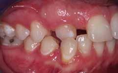
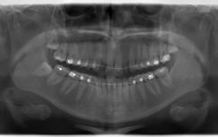
A thirty-one-year-old female in good health presented for the replacement of her congenitally missing lateral incisors. She was wearing an upper transitional removable partial denture (flipper) which was no longer satisfactory to her as it retained disproportionally small, unesthetic lateral incisors. Her initial clinical presentation (Fig. 1, Fig. 2) and radiographic exam (Fig. 3, Fig. 4) revealed that her permanent maxillary canines were tipped palatally in crossbite and mesially into the lateral incisor spaces.
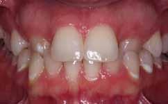
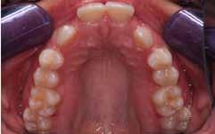
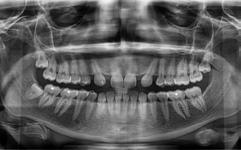
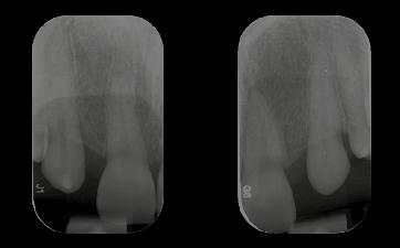
Fig. 1 Pre-operative facial view of 31 year old female patient with congenitally missing lateral incisors
Fig. 3 Pre-operative Panorex radiograph confirming congenitally missing lateral incisors
Fig. 2 Pre-operative occlusal view of 31 year old female patient with congenitally missing lateral incisors
Fig. 4 Pre-operative periapical radiographs confirming congenitally missing lateral incisors with inadequate inter-radicular space to
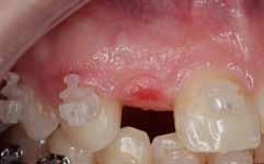
She had inadequate restorative space to replace her absent adult lateral incisors (Fig. 5, Fig. 6). Orthodontic therapy was necessary to improve the esthetics and occlusion and to develop space between the permanent canines and central incisors (Fig. 7, Fig. 8, and Fig. 9) to accommodate two dental implants in the lateral incisor positions which would replace her adult lateral incisor teeth. During the final weeks of her orthodontic therapy, a CBCT scan (Fig. 10) was taken to ensure adequate ridge dimension and proper root alignment (Fig. 11) for two narrow diameter (3.3 mm) dental implants. To best maximize the esthetic outcome, Straumann® Bone Level Tapered (BLT) Roxolid® SLActive® implants were selected (Fig. 12 and Fig. 13). A tapered implant, specifically, was selected to avoid damaging the neighboring
teeth during osteotomy preparation and placement. Its taper also facilitated initial stability due to bone compression at the apical extent of the osteotomy when the implant is fully seated. The bone level implant is designed to provide the restorative dentist with many restorative options including custom milled ceramic abutments which avert metal collar exposure or the potential for a dark hue at the gingival margin in patients with a thin tissue biotype. The hydrophilic nature of the BLT implant’s SLActive surface promotes faster osseointegration and reduces healing time* for the patient. This was a real benefit as the implant permitted the patient to achieve their desired esthetic outcome earlier.
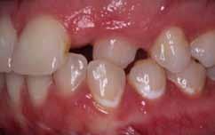
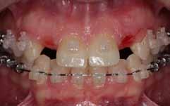
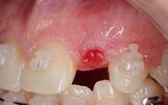
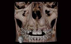
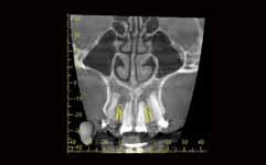
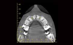
Fig. 5 Right lateral view showing inadequate restorative space for right lateral incisor due to mesially and palatally tipped upper right canine
Fig. 8 Orthodontics completed with ideal restorative space for right lateral incisor
Fig. 11 Panorex confirming appropriate root spacing and adequate bone height for dental implants in the edentulous sites of the permanent maxillary lateral incisors
Fig. 6 Left lateral view showing inadequate restorative space for left lateral incisor due to mesially and palatally tipped upper left canine
Fig. 9 Orthodontics completed with ideal restorative space for left lateral incisor
Fig. 12 CBCT reconstructed Panorex confirming appropriate root spacing and adequate bone height for dental implants in the edentulous sites of the permanent maxillary lateral incisors
Fig. 7 Orthodontics completed with ideal restorative space for lateral incisors
Fig. 10 CBCT scan volume rendering confirming appropriate root spacing and adequate bone volume in for dental implants in the edentulous sites of the permanent maxillary lateral incisors
Fig. 13 CBCT axial view confirming appropriate root spacing apically for narrow diameter dental implants in the edentulous sites of the permanent maxillary lateral incisors
After crestal incisions were made in the edentulous sites corresponding to teeth #7 and #10, full thickness buccal and palatal mucoperiosteal flaps were elevated to visualize the osseous crest. Two 3.3x10 mm Straumann® BLT Roxolid® SLActive® implants were placed with an electric hand-piece and seated fully in the osteotomy with a hand ratchet at high torque (>45Ncm). Initial stability was objectively measured with an Ostell® revealing an ISQ value of 69 for implant #7, and an ISQ value of 67 for implant #10. Straumann 3.3x3 mm healing abutments were attached to the implants in a stage one fashion avoiding a second stage uncovering surgery owing to the primary stability achieved (Fig. 14-Fig. 19).
After a healing period of twelve weeks to allow for osseointegration, the orthodontic wire was removed and PVS impressions were made for provisional restorations. Screw retained provisional crowns were fabricated and inserted. A new orthodontic retainer was fabricated and inserted.
Five weeks after the implants were provisionalized, a crown lengthening procedure was performed to correct for Type 2B altered passive eruption (Fig. 20) defined as a normal amount of gingiva and normal crest-to-CEJ relationship. By repositioning the attached gingiva and more apically utilizing a full thickness apically positioned flap (Fig. 21 and Fig. 22), the complete clinical crown was exposed, reducing the pseudo pockets (Fig. 23) and displaying the appropriate crown proportions.
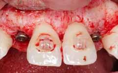
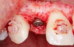
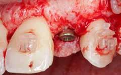
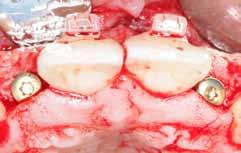
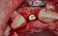
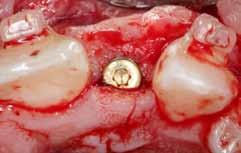
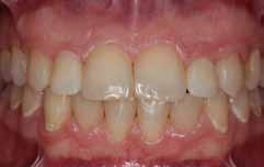
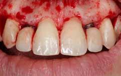
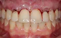
Fig. 14 After full thickness flap elevation, two 3.3 x10 mm Straumann® SLActive®, Bone Level Tapered implants were placed along with NC 3.6 x 2 mm healing abutments
Fig. 17 Occlusal view after full thickness flap elevation; two 3.3 x 10 mm Straumann SLActive, Bone Level Tapered implants were placed along with an NC 3.6 x 2mm healing abutments in sites #7 and #10
Fig. 15 Right lateral view after full thickness flap elevation; a 3.3 x 10 mm Straumann SLActive, Bone
Tapered implant was placed along with an NC 3.6 x 2 mm healing abutment in site #7
Fig. 18 Occlusal right view after full thickness flap elevation; a 3.3 x 10 mm Straumann SLActive, Bone Level Tapered implants was placed along with an NC 3.6 x 2 mm healing abutment in site #7
Fig. 16 Left lateral view after full thickness flap elevation, a 3.3 x 10 mm Straumann SLActive, Bone Level Tapered implant was placed along with an NC 3.6 x 2 mm healing abutment in site #10
Fig. 19 Occlusal left view after full thickness flap elevation; a 3.3 x 10 mm Straumann SLActive, Bone Level Tapered implants was placed along with an NC 3.6 x 2 mm healing abutment in site #10
Fig. 20 Altered Passive Eruption (normal amount of gingiva and normal crest-to-CEJ relationship)
Fig. 21 Buccal full thickness flap elevated from teeth #3-14 beyond the mucogingival junction
Fig. 22 Flap sutured apically with 5-0 Chromic gut sutures exposing complete clinical crown
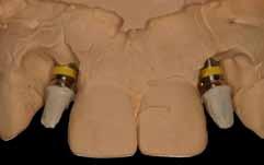
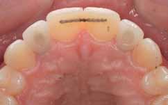
After a healing period of 7 weeks to allow for the soft tissue to fully mature and remodel, new impressions were made for final restorations (Fig. 24). Two cast custom abutments were fabricated and opaqued (Fig. 25 and Fig. 26). Two e.max® crowns were fabricated with occlusal accesses (Fig. 27). The crowns were etched, silinated and luted to the custom abutments with resin cement (Panavia™ f) and torqued into place. Mild blanching of the soft tissue was evident immediately after this insertion visit (Fig. 28). PTFE tape and composite was placed in the occlusal accesses (Fig. 29).
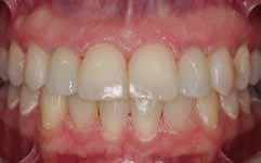
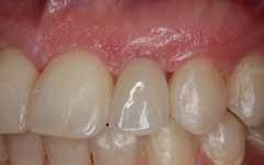
Two weeks after final delivery, the soft tissue surrounding the final crowns in sites #7 and #10 have molded and replicate the emergence profile of a natural tooth. (Fig. 30, Fig. 31 and Fig. 32). The final smile demonstrates a natural esthetic result for the patient who was very pleased with the outcome (Fig. 33).
*Compared to Straumann SLA surface
Credits:
Prosthodontist: Dr. Robert W. Berg, D.M.D.
Orthodontist: Dr. David H Seligman, D.M.D.
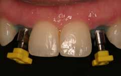
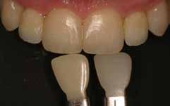
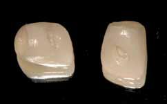
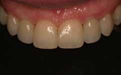
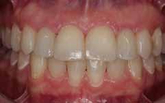
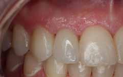
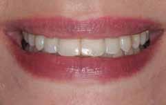
Fig. 26 Opaqued custom abutments
Fig. 32 Close up view, final screw retained crown in site #10
Fig. 29 Occlusal view of final screw retained crowns inserted in sites #7 and #10
Fig. 27 Pressed e.max® crowns with occlusal access holes that will be cemented to custom abutments
Fig. 33 Final smile
Fig. 30 Final screw retained crowns inserted in sites #7 and #10
Fig. 28 Crowns definitively torqued immediately after insertion
Fig. 31 Close up view, final screw retained crown in site #7
Fig. 23 Clinical crowns are completely exposed, reducing pseudo pockets and displaying appropriate crown proportions
Fig. 24 Impression copings placed and verified with radiograph
Fig. 25 Shade determination for e.max® crowns