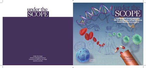
Undergraduate Research Magazine with Proceedings of the Biological Sciences Student Research Showcase 2016
Under the Scope
Division of Biological Sciences University of California, San Diego sqonline.ucsd.edu
Volume 7 2016-2017
Division of Biological Sciences
LETTER from the editor A blueprint lays the foundation for any masterpiece. Da Vinci’s Vitruvian Man best exemplifies this appreciation for the blending of mathematics and art—a balance of scientific proportion with the grace of the physical human form. But this painting also represents just how far we have come. Modern science has continually presented us with an evolving vision of truth and an appreciation for our own biological beauty. Some of the greatest discoveries include Rosalind Franklin’s X-ray crystallography images of the double-helix structure of DNA, which laid the foundation for the Human Genome Project completed just at the turn of the 21st century. Each revelation has taught us more about ourselves. About not only our perfections, but also our flaws. Although there have been missteps along the way, each finding has guided us further along in our search for understanding and growth. The Under the Scope magazine aims to highlight these advances and make this knowledge open and accessible to all. Our writing staff has worked tirelessly throughout the year to weave a tale from the data and communicate the groundbreaking findings of young and eager scientists on our campus. This constantly evolving process began
with weeks of research and interviews as our writers sought out the most exciting work being done by their peers. Then followed several months of writing and editing to distill the information down to its core ideas, while illustrators and the design staff worked hand-in-hand with writers to craft something truly beautiful. Through thoughtful reflection and resilience, our staff, like the scientists before them, created something bold and new. Change, the theme of Under the Scope Volume 7, embodies the progress of our writing staff, production team, and organization as a whole. It is a physical representation of the hours of writing and revision done by our writers. It is the culmination of countless iterations of illustrations and design styles completed by the production team. Change is the essence of the new energy and vision that the organization came to realize this year. We deliver content ranging from the neurobiology of stress to the perils of antibiotic resistance and even the possibility of rewriting the human genome. This year’s magazine not only tells the stories of undergraduate researchers and our staff, but it fulfills our longstanding desire to blend art and science into a singular passion. It is with great honor that I present to you, Under the Scope Volume 7.
Sid Ambulkar Executive Editor, Under the Scope
EDITORIAL BOARD
HEAD ADVISORS
FACULTY ADVISORY BOARD
WRITERS
ILLUSTRATORS
Executive Editor Sid Ambulkar
Steven Wasserman, Ph.D. Professor of Cell & Developmental Biology
Eric Allen, Ph.D. Timothy Baker, Ph.D. James Cooke, Ph.D. James Golden, Ph.D. David Holway, Ph.D. Suckjoon Jun, Ph.D. Jill Leutgeb, Ph.D. Elvira Tour, Ph.D. Steven Wasserman, Ph.D. James Wilhelm, Ph.D. Martin Yanofsky, Ph.D.
Christian Bey Samreen Haque Belle Hilton Emma Huie Victoria Leung Daniel Rebibo Grace Sahyouni Ashni Vora Aishwarya Vuppula Tomomi Yoshida
Victoria Hoznek Myra Julom Lulu Li Clara Oh
Features Editor Rithvik Shankar Production Editor Tushara Govind
James Cooke Ph.D., Assistant Teaching Professor of Neurobiology
Features Design Editor Nicki Guivatchian
Hermila Torres Manager, do/Bio Center
Head Technical Editor Shuen Sun Technical Editors Jaidev Bapat Alicia Ho Rebecca Hu Anokhi Saklecha
COVER ILLUSTRATION Clara Oh
TABLE OF CONTENTS ILLUSTRATION Myra Julom
TABLE OF CONTENTS 10
22 Stem Cells: A New Frontier in Regenerative Treatment
The Superheroes in Our Body and Their Difficult Fight With Cancer TOMOMI YOSHIDA & DANIEL REBIBO
CHRISTIAN BEY & GRACE SAHYOUNI
16 4
Deconstructing the Clockwork of the Brain
The Future of Antibiotics VICTORIA LEUNG & EMMA HUIE
34
Biological Science Student Research Showcase 2016
28
CRISPR/Cas9 Reconstructing Our Understanding of the Genome BELLE HILTON & AISHWARYA VUPPALA
SAMREEN HAQUE & ASHNI VORA
2
Under the Scope • Vol 6
sqonline.ucsd.edu
3
DECONSTRUCTING OF THE BRAIN
By Samreen Haque and Ashni Vora Cover and Illustrations by Stephanie Hadimulia
f
reshman year of college can be a nerve-wracking time. Apart from the academic rigor, students are faced with an entirely new social setting. Attending classes of hundreds of students, sharing a room with other people, and finding oneself amidst a crowd of almost 30,000 other students can be both intimidating and exciting. Students will often see that some of their friends are motivated by feelings of intense stress whereas some are discouraged. Some are comfortable in social situations while others would rather avoid them entirely. While people can be affected by similar stressors, their responses can vary widely. The American Psychiatric Association defines social anxiety as “a persistent fear of one or more social or performance situations in which the person is exposed to unfamiliar people or to possible scrutiny by others.” By this definition, one can see why the transition to a college may foster social anxiety in many students. Neurobiology has helped scientists uncover the complex physiological mechanisms that underlie the above-mentioned behaviors. A person’s behavior is the result of the interactions between different levels of brain functions. These include the molecular level of neurotransmitter expression and release, the widely branched circuits, and the larger structures of the brain. Much like a fine-tuned clock, our brain’s activation, circuitry, and neurotransmitters function in concert to produce behaviors. A minute change in one of the gears can affect the function of all the others and produce a new outcome.
THE SECOND HAND: NEUROTRASMITTER PLASTICITY At the molecular level of brain function is neurotransmitters, which are the chemical signals used by neurons (the most basic units of brain function) to communicate with one another. A study conducted by undergraduate Christiana Joan Stark under the supervision of Dr. Davide Dulcis in the Department of Psychiatry at the UC San Diego School of Medicine examined the variability of neurotransmitter secretion upon exposure to stress. Dr. Dulcis first contributed to the discovery of the phenomena of neurotransmitter respecification. His research showed that exposure to a stressor would cause a neuron to synthesize and secrete a different neurotransmitter than the one specified during development. As a student in Dr. Dulcis’ lab, Stark worked to understand neurotransmitter plasticity in the face of chronic social defeat. Plasticity refers to the nervous system’s ability to adapt and change in response to environmental stimuli. In the following study, neurons were found to be plastic, or capable of altering their function, secreting alternate neurotransmitters in response to social stress. As part of Stark’s research, rats were exposed to situations that would cause feelings of defeat (to replicate chronic social defeat), the effect of the stressors was measured by staining cells and observing neurotransmitter levels. An alteration of neurotransmitters in a subregion of the dorsal raphe, a region of the brain primarily responsible for serotonin release, was noted in mice susceptible to
sqonline.ucsd.edu
5
depressive behaviors. The levels of coexpressing cells that secrete both tryptophan hydroxylase isoform and nitric oxide synthase, which are associated with depression, were significantly higher in the susceptible rats compared to the control and resilient groups. This may indicate that the increased coexpressing cells may be recruited from a group of reserve cells, ready to undergo neurotransmitter respecification given the right stimuli, specifically social defeat. Stark’s findings were suggestive of a physiological marker linking this subregion of the dorsal raphae with the ability to cope with stress. The study produced compelling evidence in support of both nature and nurture on the causes of depressive behavior—that resilience is a naturally identifiable trait that may be found as a marker in certain cells, and that environmental factors such as social defeat and exposure to stress may alter neurotransmitter secretion thus leading to such behaviors. The classic argument of whether behavior is caused by either hereditary “natural” causes or environmental factors relating to “nurture” is thus confronted with a third alternative of a cause consisting of elements of both nature and nurture.
THE MINUTE HAND: NEURAL CIRCUITRY The brain’s neural circuitry is the network of neurons that are connected to each other. Individual parts of circuits are involved in specific functions and are activated only when that function needs to be carried out. Delicate structures comprised of only a few neurons, the brain’s circuitries
6
Under the Scope • Vol 7
cumulatively result in a more large scale activation of the brain, just as movements of the minute hand eventually cause the hour hand to tick forward on a clock. By the activation of various circuits, different parts of the brain are activated and engaged in their functions. In the Lab of Functional Anatomy of Neural Circuitry at UC San Diego, undergraduate senior Christopher Pham Pacia worked with Dr. Byungkook Lim to better understand the mechanisms mediating depressive behavior. They hoped to map the neural circuitry of the different symptoms of major depressive disorder in a region previously implicated in the disorder. By finding the specific circuits that underlie symptoms such as social withdrawal, diminished pleasure, and anxiety, Pacia hopes to be able to better treat them. To find the neural circuits involved in depression, Lim and Pacia induced depressive symptoms in mice using the social defeat stress model. The test mice were subjected to a series of bouts with more aggressive mice that caused social interaction deficits amongst them, which resulted in increased psychological stress simulating the experience of depression. Different techniques were then used to further characterize the circuits within the ventral pallidal, a region of the brain associated with reward and motivation behavior. Virus-mediated circuit-tracing, a process in which a virus identifiable by a fluorescent protein is injected into a circuit, was used to map the circuits originating from within the ventral pallidal circuit and their outputs. Immunohistochemistry was then used to stain synaptic proteins to identify whether a signal was
excitatory or inhibitory. Lastly, behavioral assays were used to observe pre- and post-stress behaviors. Upon using a virus expressing tetanus toxin to disrupt the neurons in the above circuit, the researchers discovered that the mice were more resilient to social defeat and displayed fewer symptoms of depression. This finding suggests that certain depressive symptoms are housed within a particular circuit, which can be used as a target for future medication. By correlating depressive behavior to this particular circuit, the study also characterized depression as a disorder that involves both internal and environmental factors. This prompts more research into the other functional effects of this region, as the ventral pallidal is dense with connections to many other parts of the brain.
THE HOUR HAND: BEHAVIOR & SOCIAL FUNCTION Undergraduate student Christina Liu Cui strove to understand how neural activation changes during After exposure to the stress, the susceptible rat will undergo neurotransmitter switching, acquiring more of one neurotransa computerized attention modification program mitter than the resilient rat, with receptors similarly matching this for social anxiety. Under advisor Dr. Charles change. Taylor, she analyzed data from a computerized Attention Modification Program (AMP) divert their attention away from stressful stimuli. In previfor social anxiety, which utilized probe detection task to ous research. AMPs have been shown to be as effective as alleviate anxiety. The computerized AMP can be utilized pharmacological and psychological interventions, but the in the treatment of social anxiety; it teaches patients to
sqonline.ucsd.edu
7
lyzed was time — more specifically, differences in response times and variables. For her results, Cui looked at two key regions of the brain. One of them was the left anterior cingulate cortex (LACC), which is involved in allocating attention. LACC activity implied that socially anxious people tended to have a more difficult time withdrawing attention from the threatening stimuli, but with more trials, participants were able to learn to redirect their focus. The second region of the brain she examined was the left dorsal lateral pre-frontal cortex, which is involved in inhibitory control. This region experienced a similar reduction in activity with increasing trials, indicating that less cognitive control was required for the task of redirecting control. Overall, Cui was able to more thoroughly understand this alternative approach to addressing anxiety and depression, which she feels is “an important and more accessible addition to the pharmaceutical aspect of treatment.”
TELLING TIME: IMPLICATIONS neural mechanisms that make this possible have yet to be studied. In order to better understand this alternative method of addressing anxiety and depression, Cui studied 14 participants who underwent an AMP during a functional MRI (fMRI) scan. fMRI provides activity coded brain activation feedback, so Cui was able to better understand the neural
8
Under the Scope • Vol 7
A portion of the basal ganglia of the brain, the ventral pallidal region may contain a circuit that houses certain depressive symptoms.
mechanisms that make AMPs effective. Participants were presented a threat in the form of an intimidating face. A probe would lead attention away from said threat, while participant responses were recorded. The main factor ana-
students are constantly faced with stressful situations, it is important to understand the effects certain stressful situations can have on the morphology of one’s brain. Cui also mentions how alternative treatment methods could help avoid stigma around medication and address disorder at the fundamental level while looking at causes rather than just symptoms. When asked what he takes away from his research experience, Pacia says, “There is still a lot to learn, but all it takes is a couple hours a day at this lab, or any of the many hard-working labs at UC San Diego, to make a difference for the neuroscience community and the general health of people.”
WRITTEN BY SAMREEM HAQUE AND ASHNI VORA. Samreen is a Physiology and Neuroscience major graduating in 2019. Ashni is a Biochemistry and Cell Biology major graduating in 2019.
Assessing the various physiological factors involved in social behavior, responses to stressors, and relevant disorders helps highlight the extent of social stress that many college students experience. The brain’s circuitry is delicate and extremely susceptible to stimuli, as shown in the three studies above. The distinct levels of brain processing are all affected by forms of social stress, often in ways that lead to short-term changes in brain function. Since college
sqonline.ucsd.edu
9
A NEW FRONTIER IN REGENERATIVE TREATMENT By Christian Bey and Grace Sahyouni Cover and Illustrations by Clara Oh
f
or most people, the words “stem cell” may elicit a variety of reactions—enthusiasm, disapproval, or simple indifference. Regardless of one’s pre-existing notions, stem cells continue to advance countless medical discoveries by demonstrating how a microscopic medium within the body can be capable of cellular manipulation. Just a few decades ago, individuals afflicted with serious illnesses, such as Parkinson’s disease, severe hearing loss, or neurological damage, had to live harder and shorter lives due to the lack of medical technology. Although the 20th century saw an expansion of the arsenal of medical tools, most notably vaccines, none of these advancements were able to point to a cure for many of the diseases that plagued millions. However, as we now possess a better understanding of the tools that we have, we can no longer accept the fact that many illnesses remain untreatable. Stem cells have long been a tool in the arsenal of medical scientists, but these cells were not understood to the degree that they are today and, as a result, were not fully utilized. Therefore, many scientists were searching for external cures for disease while a prospective cure was lying dormant within the human body itself. Current research explores the three fundamental states of the stem cell’s existence—genesis, differentiation, and proliferation—and seeks to better understand how each of these processes occurs in order to enhance the effectiveness of the stem cell as a tool in medical technology. Stem cells, in the most general sense, are groups of undifferentiated cells that have the unique ability to take on the function of nearly any type of cell in the body. Stem cell research has historically focused on two types of cells:
embryonic cells, which originate in the womb, and somatic animal cells, which are bodily cells. However, a new type of stem cell, called the induced pluripotent stem cell (iPS), has recently become the new focus of research. These are already differentiated cells that, when subjected to the right environmental stresses, undergo changes in their function. After a stem cell undergoes genesis or is converted from another cell, it divides into distinct human cell lines. This ability to differentiate gives it numerous applications in the development of treatments. Once a cell finishes differentiation, it will proliferate and ultimately be used in the body where it is needed. Along with traditional sources of stem cells, newer sources will likely play an essential role in the sustainable development of treatments in the future.
GENESIS OF SPERMATOGONIAL STEM CELLS In the Wilkinson Lab, Dr. Miles Wilkinson and undergraduate Vaishnavi Sridhar worked on understanding the establishment of spermatogonial stem cells (SSC) in a mouse model. SSCs are undifferentiated cells that have not yet been converted by the body into germ cells for reproductive use. The lab studied these cells by researching a homeobox transcription factor designated Rhox10. Homeobox genes are responsible for the establishment of bodily structure when an organism is still in its embryonic stages of development. These genes contain coding for a 60-amino-acid-long chain called the homeodomain, which allows the protein to function as a transcription factor. As a transcription factor, that
sqonline.ucsd.edu
11
protein will have the ability to control the activity of other genes by binding to their regulatory regions. Homeobox-containing genes are mostly associated with limb and organ formation, but are also crucial in the maturation of undifferentiated cells.
The Wilkinson lab studied the formation of SCCs in a mouse model using the transcription factor Rhox10. By comparing mice with the transcription factor and those that had it removed, the lab determined that it up-regulated genes that promote the movement of pro-spermatogonia to seminiferous tubes, thereby indirectly affecting SCC production.
12
Under the Scope • Vol 7
The lab chose the Rhox10 transcription factor hoping that it would elucidate the mechanism by which SSCs function. It is primarily responsible for the movement of gametes to a region where meiosis occurs, which is an essential step in the establishment of SSCs. There, it promotes pro-spermatogonia to develop into SSCs. Pro-spermatogonia are reproductive cells that undergo spermatogenesis, making the cells ready for reproduction when combined with an unfertilized egg. The Wilkinson lab proposed that the Rhox10 transcription factor facilitates the physical movement of pro-spermatogonia into a portion of seminiferous tubes. In order to determine the importance of the Rhox10 factor, they were compared to Rhox10KO (knock-out), which were observed to produce considerably fewer SSCs than the unaltered version. This outcome led to the conclusion that
Rhox10 has an effect on the up-regulation of genes that promote the movement of pro-spermatogonia and, by extension, the production of SSCs. To check their theory, the lab used immunostaining, a technique used to detect the presence of a certain type of protein, on the mouse vasa homolog. Further research may show the extent of Rhox10-damaged cells and provide insight into other transcription factors that may also facilitate movement to produce SSCs. Although the lasting impacts of this discovery are not yet apparent,Vaishnavi Sridhar commented that by studying the Rhox10 transcription factor, researchers can elucidate “the mechanisms of certain diseases that are yet unknown.” Learning how SSCs develop may provide insight on how to approach diseases that are not currently well-understood.
DIFFERENTIATION OF TROPHOBLAST STEM CELLS Once stem cells specialize, they begin to divide and multiply. One factor that affects their ability to proliferate and differentiate is the environment in which they are cultured. Dr. David Natale and undergraduate student Priscilla Vu cultured trophoblast stem cells (TSCs) that function in placental development. During her time at the lab,Vu and her colleagues worked with both TSCs and with labyrinth trophoblast progenitor cells (LPCs), which are a differentiating subset of TSCs. These LPCs are found in the labyrinth layer of the placenta where nutrients in the mother’s blood
pass to the fetus’ blood. Through a better understanding of how these cells interact with their environment, conditions such as placental insufficiency—where the placenta cannot supply adequate nutrients to the fetus causing it to become under-developed—can be prevented during pregnancy. The lab used oxygen-deprived hypoxic chambers and measured the cells’ subsequent rates of proliferation and differentiation using quantitative reverse transcription polymerase chain reaction (qRT-PCR). This process measures the amount of genes expressed using a byproduct produced by RNA, thereby giving an idea of how well the TSCs are doing in their environment. Hypoxia is a natural part of the production of TSCs during placental development, which regulates the condition in which the fetus is raised. If the conditions are not within a certain range, the cells’ ability to proliferate or differentiate may be compromised, leading to conditions that can harm fetal development. To test the degree to which the TSCs and LPCs would be affected, the lab hypothesized that hypoxia does affect proliferation and that the TSC and LPC groups would have unique outcomes. Two populations of mice were used based on the type of progenitor cells that they had. The Sca1+ and EpCam+ populations isolated from a TSC culture were both exposed to hypoxic (1% oxygen) and normoxic conditions (normal oxygen levels). Using qRT-PCR, the lab determined that in hypoxic environments proliferation of both TSCs and EpCam+ LPCs was greatly increased. Besides this result, a far more interesting phenomenon was observed in just the EpCam+ LPCs: their
sqonline.ucsd.edu
13
increased gene expression showed the potential to differentiate into cells outside of the labyrinth zone. This observation implies that hypoxic environments may have the ability to change the behaviour or identity of certain LPCs. Further testing is required to solidify the means and feasibility of this process, but this discovery marks an important point where medical researchers can hypothesize ways of utilizing differentiating cells in the repair of placental-related ailments.
PROLIFERATION OF EMBRYONIC-STEM-CELLDERIVED NEURAL CELLS Another type of cell, the embryonicstem-cell-derived neural cell (ESC-NSC), is derived from embryonic stem cells and is being investigated for its potential to differentiate into neural cells in order to repair spinal cord injury in rats. Spinal cord injuries typically result in paralysis, which is the permanent loss of motor functions and feeling in certain parts of the body. This is predominately a result of the neural cells’ inability to regenerate once they are damaged, unlike many other cell types in the body. Besides re-programming existing cells into neural derivatives, the other major option is to use embryonic stem
14
Under the Scope • Vol 7
Rats with damaged spinal cords were treated with neural stem cells derived from embryonic stem cells in an attempt to restore motor functions. Since the body cannot naturally regenerate neural cells at a significant rate, the lab aimed to replace the damaged neural cells with ones from an external source. Currently, only in vitro tests have successfully regenerated new neural cells. The embryonic stem cells in the in vivo trials did not fully differentiate into functional neural cells.
cells to replace the damaged cells altogether. Research on embryonic-derived neural cells was conducted by Arthi Rao, under Dr. Ephron Rosenzweig and Dr. Paul Lu, using differentiated embryonic rat stem cells to restore motor movements in injured rats. The Rosenzweig lab tested ESC-NSCs in vitro (in a culture dish) and in vivo (in a living rat) in order to gauge their abilities to differentiate into both neurons and glial cells, a type of cell that assists in cell repair. To determine whether or not the cells had been successfully differentiated, the lab analyzed intermediate cells that did not retain the properties of the two original cells in both trials. Specifically, the researchers checked to see if the embryonic stem cells began to produce neural stem cells by looking for neural stem cell markers and testing for pluripotency markers. Since there were no pluripotency markers, the embryonic stem cells were in fact successfully differentiated. The in vitro cells were successful in differentiation and expressed markers for both stem cells and glial cells. However, in vivo tests showed that although certain markers of neural stem cells were present, there was no final progression into neural and glial cells. Without fully differentiating into neurons and glia, there is no possibility of spinal cord repair. Although unsuccessful in the living organism, the in vitro success marks an important stage in which embryonic stem cells may actually be used in regenerative treatment for cells that were thought to be permanently damaged. Regardless of where one stands on the ethics or policy of harvesting and use of embryonic stem cells, their regenerative abilities provide important insight into how stem cells
can provide a solution to paralysis and other nervous-system-related ailments.
A PROMISING FUTURE IN REGENERATIVE TREATMENT Research on the three states of a stem cell’s existence can explain the diverse nature of these cells and further improve their effectiveness as medical tools. Stem cells are no longer restricted to creation from certain sources, nor are they limited in the kind of treatment that they are capable of providing. Being able to understand the development of these cells and utilize the processes unique to each state will assist medical researchers in developing treatments for infertility issues, blood-borne diseases, paralysis, and many more ailments. The research that is being done at UC San Diego is leading the way in making stem cells a sustainable and viable treatment option in regenerative medicine. With many uncertainties ahead, persistent research into the mechanisms of these unique cells may cause many to recognize their potential as a sustainable, regenerative treatment in the future. WRITTEN BY CHRISTIAN BEY AND GRACE SAHYOUNI. Christian is a Human Biology major graduating in 2019. Grace is also a Human Biology major graduating in 2018.
sqonline.ucsd.edu
15
THE SUPERHEROES DIFFICULT FIGHT WITH By Tomomi Yoshida and Daniel Rebibo Cover & Illustrations by Vicky Hoznek
i
n everyday life, humans are surrounded by countless disease-causing factors ranging from pathogenic bacteria and viruses to carcinogens. These harmful agents can take advantage of our susceptibility to infections and diseases. When these pathogens see a chance to invade the body, they seize the moment and attack viciously. In order to prevent these attacks from critically damaging the body, the body sends out its own set of superheroes to fight against the evil invaders. These superheroes that fight the villainous pathogens are collectively known as the immune system. Within the immune system, there are countless molecules that interact with each other in order to detect, fight, and take down pathogens. This complex web of molecules immunizes the body from the attacking pathogens, defeating the invading villains. The web can be broken into two different systems, each system having different kinds of superheroes. One such system is the innate immune system. This system is composed of specialized cells that target specific pathogens entering the body. These include certain white blood cells, such as B and T cells, which are created for the purpose of killing specified foreign cells. The B and T cells have special receptors that activate a chemical reaction when in contact with specific substances that are only present on foreign cells. Through these chemical reactions, cells with these receptors “sense” the pathogen and activate an immune response. The cells act like the Avengers, each hero using his or her power in collaboration with the other heroes, to bring the enemy down. In the world of biology though, when so many chemical and biological
signals are sent and activated, there are many unanticipated consequences, one of which helps fight cancer. Even before the superhero cells such as B and T cells are activated, there are more players in this system that work to protect our body. They are the innate system’s first line of defense: the physical and biochemical barriers that block pathogens from entering the body and traveling around it. If the immune system is to be thought of as a main base for the superheroes, this part of the system can be thought of as the police or FBI of the cell. Instead of having specific biological agents for the purpose of fighting a virus, this system is made up of general defenses for the purpose of protection. Most instances are handled by the police, and superheroes only come in when they are really needed. One example of the police in our immune system is our skin, the first line of defense. This thick membrane of millions of living and dead cells shields the insides of our bodies from the outside environment, selectively choosing what should travel in and out of the body. The FBI aspect of this system is a little less general, and is known to have significant degrees of molecular recognition based defenses and specific responses. However, in some organisms, like humans, the adaptive immune system is more specific. While most problems can be prevented by the police and FBI in our body, sometimes this line of defense fails, and the innate immune system resorts to its more specific friend, the adaptive immune system. The adaptive immune system deals with more specific villains, such as retroviruses and harmful bacteria, that are harder to destroy using just the power of the public police and FBI.
sqonline.ucsd.edu
17
sible for fighting diseases and healing tissues. It is important to understand that white blood cells are clumped together so all the unique cells end up functioning together, each acting as a little piece of a big puzzle that will eventually “fix” an area being invaded or damaged. Thus, the pieces of the innate immune system work together to neutralize, effectively weaken, and even destroy the invader, while reducing damage to the body. The police and FBI of our body system are collectively as powerful as the Avengers of the human body in terms of fighting off and protecting the body from harmful pathogens.
Nanoshells are spherical capsules used to deliver drugs and agonists to the target site. In Calderon’s experiment, pairing these 100nm sillica nanoshells with the TLR7 agonist proved most effective in stimulating the immune system.
HOW HEROES HALT CANCER Many researchers are currently looking into the possibility of fighting cancer by either inducing or repressing specific immune responses. Cancer immunotherapy is this collective field of research working to combat cancer by understanding and manipulating immune functions. There are currently many undergraduates working on cancer immunotherapy research at UC San Diego, including Stephen Calderon, a fifth-year Biochemistry/Cell Biology major. Through the California Louis Stokes Alliance for Minority Participation (CAMP) in Science, Engineering and Mathematics program at UC San Diego, Calderon began research in the Andrew Kummel Lab. Ever since, he has been investigating how to suppress cancerous tumor formation by researching the most efficient way to deliver toll-like receptor (TLR7) agonists to the cancer site using nanoshells, or nanoparticle capsules, that de-
18
Under the Scope • Vol 7
liver drugs. The significance of TLR7 is its function within the innate immune system. TLR7 recognizes pathogen-associated molecular patterns, which are often expressed in infectious pathogens. Once the TLR7 agonist is dispatched, it leads to a subsequent immune system chain reaction involving cytokines, white blood cells, and inflammation, which is part of the police and FBI or the innate immune system. This immune response proves powerful. Cytokines are a family of proteins that play a fundamental role in cell signaling and inflammation. Proinflammatory cytokines signal areas of the body to start inflammation, an immune system response that increases the amount of white blood cells. White blood cell functions range from inducing bodily repair to attacking foreign agents. A notable white blood cell is the phagocyte, which indefinitely circulates in the blood stream and “eats” foreign substances it comes across. Mast cells are another type of white blood cell that are respon-
Calderon hopes to develop the most efficient nanoshell to deliver the TLR7 agonist to the cancer site, as the TLR7 agonist is an immunoparticle that assists in recognizing tumors and boosting anti-tumor immunity of the cells in general. Essentially, Calderon is developing a way to strengthen the police officers in our bodies to fight against pathogens and diseases like cancer. The experiment was conducted in live cells so that the researcher could see the effects of the TLR7 agonist directly in a fully functional organism. Initial data from the experiments indicate that the TLR7 agonist successfully stimulates the immune system and is most efficient when conjugated to 100nm silica nanoshells. With further experimentation, Calderon hopes to optimize the conjugation synthesis to find out the appropriate dose of TLR7 to stimulate different degrees of immune responses. Calderon believes that his research will contribute as a useful adjuvant to vaccines and drugs in cancer treatment in the future. Combining different treatments with TLR7 in 100nm silica nanoshells could potentially become a cancer treatment
available for patients. Although Calderon faced a challenge in integrating a variety of science fields within his research project, including nanotechnology, engineering, physics, and biology, he was excited to develop a new method of approaching cancer immunotherapy. To this point, Calderon exclaimed, “I think it is a great time to get involved with immunotherapy research!” demonstrating his passion doing research in this field.
IMPROVING TLR7 WITH TECHNOLOGY Melody Sayrany, an undergraduate researcher working in the Dennis Carson Lab, is also conducting research in the field of cancer immunotherapy. Sayrany is investigating methods to halt cancer metastasis, which is the spreading of cancer within the body. Similar to Calderon, Sayrany is focusing on TLR7 and its efficacy in inhibiting metastasis to improve cancer treatment. The TLR7 ligand 1V270 is a molecule created by the lab to induce immune responses to stop the spread of cancer cells. This occurs by stimulating certain immune molecules in the adaptive immune system that work together to inhibit cancer metastasis. In other words, Sayrany is researching how to strengthen the powers of a specific superhero perfectly matched to fight against a certain type of invader in the cell. Since the first line of defense, the FBI and the police, were not powerful enough to prevent the cancer cells from spreading, she resorted to training a specific superhero to fight off the metastatic cancer cells. Sayrany conducted her experiment on a mouse model that was more prone to cancer tumors and metastasis in order to
sqonline.ucsd.edu
19
effectively see the effects of the TLR7 ligand. Through the results of this experiment, Sayrany found that the immunoactivator molecule is successful in halting breast cancer metastasis in the lungs, as shown through the increase in innate cell response at the cancer site. This demonstrates that 1V270 may assist in the anti-metastatic effects of the innate immune cells specifically in the lungs. In future studies, Sayrany hopes to conduct additional experiments to develop a treatment or therapy that enhances the antitumor effect by 1IV270. Furthermore, Sayrany aims to identify the cells that were responsible for inhibiting lung metastasis by 1IV270.
MINIMIZING THE DAMAGE HEROES SOMETIMES CREATE While Calderon and Sayrany are studying how to halt the spread of cancer, Ayse Sahan, another undergraduate researcher at UC San Diego, is studying how to detect cancer at an earlier state. Sahan is a fourth year, Biochemistry/Cell Biology major hoping to pursue a Ph.D. after graduating. She first got involved in her current lab, led by Dr. Soumita Das, during her sophomore year. Ever since, she has researched different species of bacteria known to inflict damage to the human body. In particular, Sahan took interest in the Helicobacter pylori bacteria; its infection is known to increase the likelihood of gastric cancer. Although cancer is often thought to be an age-related disease caused by mutations in DNA, inflammation caused by bacteria is also a well-known cause for gast cancer. Sahan is investigating the mechanisms of gastric and colon cancer, two inflammation-linked cancers. Inflammation, the innate immune reaction that happens when fighting
20
Under the Scope • Vol 7
invading bacteria, happens to activate one of the unseen consequences resulting from the complex web that is the immune system. Bacterial-inflammation-related cancer arises from chemicals called reactive oxygen species (ROS) that are released by the immune system when the innate immune system attempts to kill the invading bacteria. The ROS not only kills the invading bacteria, but it also inflicts oxidative damage to the host’s own DNA, leading to mutations and eventually cancer. ROS act as superheroes when they destroy their surroundings while fighting the devastating cancer cells. Although the superheroes and immune system have good intentions of destroying the invading pathogen, they inadvertently end up destroying their own healthy DNA and cells. This is analogous to the Avengers accidentally destroying the cities in which they fight. In order to understand the underlying mechanism of this bacterial-inflammation-related cancer, Sahan investigated methods for combatting two of the most common bacterial-infection associated cancers in the gastrointestinal system. Sahan specifically focused on NEIL2, a key enzyme that assists in repairing DNA damaged by reactive oxygen species. After studying the effect of NEIL2 in gastric and colonic cell lines infected by the specific bacteria, Sahan discovered that NEIL2 levels decreased in the infected gastric cells in a dose- and time-dependent manner. This discovery suggests that NEIL2 is a key enzyme that increases the risk of cancer following infection from elevated damage in the gastric surface cells. She was intrigued to discover that H. Pylori infections hinder DNA repair within damaged cells, which later leads to a higher chance of gastric cancer. Sahan hopes that further studies on NEIL2 will help researchers understand how to recognize and prevent bacteria associated cancers early
on so that patients can receive better cancer therapies and treatment earlier.
UC SAN DIEGO’S HEROES: STUDENTS & PROFESSORS FIGHTING DISEASE THROUGH THEIR RESEARCH Cancer immunotherapy and inflammation-related cancer are thriving topics of research for both immunologists and cancer biologists. Many researchers, including the undergraduate researchers mentioned above, are conducting experiments targeting specific components in the immune system to understand and fight cancer. Calderon and Sayrany are investigating ways to fight cancer by creating different immunological molecules to enhance cancer-fighting immune responses. While Calderon is workings to suppress tumor formation itself, Sayrany is working to prevent cancer metastasis. Lastly, Sahan is focusing on combating a specific type of cancer associated with bacteria by looking into how to prevent cancer caused by bacterial inflammation from arising. Sahan is hoping to discover different methods to control the immune response of inflammation for earlier detection and complete prevention of inflammation-related cancers in the human body. In this way, each investigator is focusing on a different aspect of the immune system to combat a specific property of cancer. It is interesting to see the different approaches to both the immune system and cancer, and how these scientists are using the body’s superheroes to fight these
The NEIL2 enzyme assists in repairing DNA damage made by the Reactive Oxygen Species. Sahan discovered that this enzyme plays an important role in increasing the risk of cancer following infection and damage in the gastric surface cells.
disorders. Their efforts to combat cancer through the immune system make them true heroes who are fighting against a difficult disease that affects multitudes of people from all over the world.
WRITTEN BY TOMOMI YOSHIDA AND DANIEL REBIBO. Tomomi is a Molecular Biology major graduating in 2019. Daniel is a Biochemistry and Cell Biology major graduating in 2020.
sqonline.ucsd.edu
21
THE FUTURE OF By Victoria Leung and Emma Huie Cover and Illustrations by Myra Julom
i
magine fighting a battle against something you can’t see.You might have advanced technology and scientific innovations, but the enemy always seems to outpace you. While you are able to combat your attacker once, it quickly develops new ways to outsmart you.You try to arm your forces with new weapons, but these weapons can be misused, allowing the enemy to live to see another day. While both you and the opponent scramble to outsmart each other, the tension in this arms race escalates. The worst part is that as your opponent grows stronger, it can inflict a lot of damage on people, making them sick and even killing them.
THE BATTLE SO FAR The arms race described above is antibiotic resistance. To understand the battle against antibiotic resistance, we need to understand how our antibiotic weapons work. First discovered by Alexander Fleming in 1928, antibiotics are a powerful class of medicines that kill bacteria. One way of classifying these drugs is by their spectrum of activity. Broad spectrum antibiotics can combat a wide range of bacteria while narrow spectrum antibiotics are specific to a few genetically similar bacteria. Another way of classifying antibiotics is by their mechanism of action, or how they harm bacteria; this could be through one of three classifications: bactericidal, bacteriostatic, or a combination of the two. Bactericidal antibiotics, such as penicillin, destroy bacteria by weakening their cell walls, thereby causing the bacteria to lyse, or break open. In contrast, bacteriostatic antibiotics like tetracycline do not kill bacteria directly, but prevent growth and replication of bacteria. They do this by binding to ribosomes — pro-
tein producing parts of cells — thereby blocking the synthesis of critical proteins that bacteria need to sustain growth. By preventing the synthesis of new proteins, bacteriostatic antibiotics reduce the number of bacteria over time, making it easier for the immune system to combat the infection. Antibiotics are only one half of the war. An important part of battle is understanding the enemy. Bacteria, like all life, reproduce to create new generations of their species. When genes are replicated during reproduction, certain parts of the DNA might have little mistakes, or mutations. These mutations can either be detrimental to the new bacteria, or they can be evolutionarily advantageous. Because bacteria reproduce so quickly, extremely diverse generations arise much faster than, for instance, a new generation of humans. The increase of generations in a short time span results in an increase of genetic mutations, resulting in a diverse pool of different bacteria with genes that could be beneficial or harmful. When bacteria are exposed to antibiotics, most are killed, yet some with select mutations survive. These survivor bacteria are evolutionarily superior and are able to reproduce and pass on the genes responsible for keeping them alive. Their offspring carry that gene and survive future antibiotic exposure, and the cycle continues until they collectively become resistant to antibiotics.
SCIENTISTS FIND ANTIBIOTICS TO FIGHT BACK At UC San Diego, under the instruction of Drs. Kit and Joe Pogliano, dozens of undergraduate freshmen collabo-
sqonline.ucsd.edu
23
rated on a project involving the research on novel antibiotics. In fall 2015, these students collected soil samples mainly from around the UC San Diego campus in search of bacteriophages, a type of virus, and Streptomyces, a genus of bacteria that produces antibiotics to kill and outcompete other bacteria strains. From trying to find new antibiotics in bacteria to finding viruses that can be used as vectors to treat infections, the students’ work can give us insight into the battle against antibiotic resistance. Danielle Freeman and her group focused on identifying new strains of Streptomyces and determining the mechanisms of action of the Streptomyces-produced antibiotics. Using 36 types of bacteria from soil samples in San Diego County, the group found that 14 strains were genetically categorized as a type of Streptomyces. The team tested to see if each Streptomyces strain could kill Bacillus subtilis, wildtype Escherichia coli, and an E. coli tolC mutant, and if applicable, what the mechanisms of action were. Undergraduate students use a type of virus called a bacteriophage to inject DNA into Streptomyces, causing the bacteria to translate that DNA to create antibiotics. Bacteriophages have a protein container which contains DNA, and tails which have special receptors that bind to bacteria cell wall and allow passage of the DNA. This technique helps create novel antibiotics by injecting DNA which would not be in the Streptomyces otherwise.
24
Under the Scope • Vol 7
The E. coli tolC mutant was most susceptible to antibiotics because its efflux pumps, the pumps on the bacterial membrane that expel antibiotics from the cell, were defective. When the team found that a few strains of Streptomyces produced antibiotics that killed the E. coli tolC mutant but not the wild-type E. coli, they deduced that those strains of Streptomyces might have yielded antibiotics which engaged with the efflux pumps. One surprise was that there were some Streptomyces strains that were able to kill all three of the bacteria, specifically a strain nicknamed QF, suggesting that it produced a broad-spectrum antibiotic. One team, led by undergraduate Minda Lee, decided to further study strain QF. Using Bacterial Cytological Profiling (BCP), an imaging technique developed by Drs. Kit and Joe Pogliano used to show how bacteria react to antibiotics and depict mechanisms of actions, the team witnessed translational inhibition on the E. coli and B. subtilis after they were exposed to QF. Translational inhibition occurs when antibiotics interfere with protein synthesis. Once the team narrowed down the mechanism of action, they could predict which genes might be responsible for killing the bacteria. Lee’s group reported that two Streptomyces genes, specifically gene 6 and gene 18, encoded for a similar but not identical genetic makeup to common genes that are responsible for the production of known antibiotics pristinamycin and viomycin, implying that these genes code for new antibiotics. To analyze their findings further, both Freeman and Lee’s group took DNA from the strains and analyzed the sequences utilizing antiSMASH, a genome sequencing
computer program. When organized in a phylogenetic tree, their findings revealed that although some strains were closely related, they displayed different mechanisms of action; some targeted the efflux pumps while others utilized translational inhibition. These differences can help better classify different antibiotic-producing genes.
FINDING NEW WEAPONS WITH BACTERIOPHAGES While the aforementioned groups focused on discovering different strains of Streptomyces that could produce novel antibiotics, other teams worked on how to use bacteriophages to destroy bacteria cells in phage therapy. Bacteriophages are viruses that invade bacteria host cells by inserting their DNA into the bacteria. The phage DNA will either be replicated when the bacteria reproduce through the lysogenic cycle, or it will go straight to the lytic cycle, where phage DNA is used in protein synthesis to create more bacteriophage virions. Ultimately, the phage virions assemble and lyse the bacterial cell. Scientists have been using bacteriophages to study phage therapy, but certain drawbacks still remain. One particular drawback is the limited bacteriophage host range, or the number and type of bacteria a phage can invade, which makes them less effective at killing bacteria. In the lab, Tran and her research team sought to understand the biological functions and host ranges of different strains of bacteriophages. After isolating the genome of select bacteriophages, the students used antiSMASH to sequence the DNA of the phages nicknamed IceWar-
sqonline.ucsd.edu
25
very similar genetic sequences. However, while BSD and TS are more closely related than the other two bacteriophages, TS had a larger host range. The team thinks that this is because TS has an extra enzyme called methyltransferase, which can introduce a methyl group to phage DNA and protect it from the host’s restriction enzymes. Also, TS has two more tail proteins than BSD, which could affect the host range. In addition, Tran and her team conjectured that TS and BSD could have different amounts of peptidoglycan binding domains, which determine how well the phage binds to the host cell wall, and thus how large the host range is. Another finding was that IW had the broadest host range; it could infect four strains of Streptomyces. The larger the host range, the more versatile the phage is, and thus the more useful it could be as bacterial therapy.
THE FUTURE IS HOPEFUL
rior (IW), BartholomewSD (BSD), TrvxScott (TS),
AntiSMASH is a computer program that decodes and analyzes and Shawty (SH). Using the the same phylogethe genetic makeup of any living organism in order to produce a netic tree generated by Freeman and Lee, Tran phylogenetic tree. It can determine the similarity between two or added the bacteriophages that could infect more genes, allowing Freeman’s group to compare the genes of various Streptomyces and Tran’s group to conclude that bacteriophages different Streptomyces strains. Tran and her BSD and TS were most closely related. Lee’s group utilized antiSMASH to team hypothesized that BSD and TS would ascertain that genes 6 and 18 were responsible for producing antibiotics.
have similar host ranges since the two had
26
Under the Scope • Vol 7
fight back against antibiotic resistance. By studying the bacteriophages’ host ranges and the Streptomyces QF genes, these scientists have contributed to the phage therapy and the development of new antibiotics. From developing new antibiotics to researching phage therapy, these young researchers offer insight into the battle against bacteria. The fight against antibiotic resistance is a long and hard one, but with contributions from researchers like these undergraduate students, the bacteria are less likely to win.
WRITTEN BY VICTORIA LEUNG AND EMMA HUIE. Victoria is a Human Biology major graduating in 2019. Emma is also a Human Biology major graduating in 2020.
The work of these student researchers does not end here, and the fight against antibiotic resistance continues to be an uphill battle. With the incidents of antibiotic misuse increasing, the availability of effective antibiotics has drastically diminished. Antibiotic resistant bacterial infections have caused 99,000 deaths of two million hospital-acquired infections nationwide per year, according to the Infectious Diseases Society of America. Their findings also determined that the cost of antibiotic-resistant infections is estimated to be $21 billion to $34 billion, in addition to eight million additional hospital days. As alarming as these statistics sound, the work of scientists and researchers has made it possible for humans to
sqonline.ucsd.edu
27
RECONSTRUCTING OUR UNDERSTANDING OF THE GENOME By Belle Hilton and Aishwarya Vuppala Cover and Illustrations by Lulu Li
D
NA is the manual for building all life on Earth; however, this manual is full of complex mechanisms and unknown components. Understanding which set of instructions and genes correspond with which functions and structures of the body is difficult. For a deceptively small molecule, DNA has incredibly large implications; any change to one gene can lead to a ripple effect, potentially causing system failure. Therefore, manipulating DNA may seem a bit far-fetched without the reassurance of a clean ‘cut.’ However, in the past few years, scientists have developed effective genome-editing tools that target specific sequences of DNA. One such tool, originally discovered by scientists at UC Berkeley, is known as CRISPR/Cas9. This tool can give us the opportunity to cure many genetic diseases while providing a clearer understanding of DNA. It is essentially the “whiteout” for our manual, allowing us to cut out or rewrite the instructions. CRISPR/Cas9 was discovered in 2012, so the complete extent of its capabilities has not yet been revealed. Nevertheless, research has demonstrated the potential value of CRISPR/Cas9 in learning about DNA. Fittingly, Dr. Karl Willert, a researcher from the UC San Diego Institute of Engineering in Medicine, applauds the system’s diversity as a tool: “CRISPR/Cas9 is really one of these enabling technologies. Everyone in biomedical research is using it because it has—just such broad implications.” To truly understand this tool’s importance, one must understand how it works. CRISPR stands for “Clustered Regularly Interspaced Short Palindromic Repeats,” or
short sequences of repeated DNA randomly spread across the genome. It was discovered when researchers noticed bacteria using CRISPR as a defense mechanism against viruses. CRISPR enabled the bacteria to target and eliminate invading genetic material. Cas is an acronym for “CRISPR associated genes,” and Cas9, specifically, is an endonuclease, a protein that is able to cut DNA. Cas9 binds to two small ribonucleic acids, or RNAs. The first, called a trans-activating CRISPR RNA, triggers the enzymatic activity of Cas9, while the second, a CRISPR RNA, guides the Cas9 enzyme to a particular gene sequence so the DNA can be cleaved. Although this cutting action may seem like a simple process, CRISPR’s precise ability to target genes has stimulated new research at UC San Diego.
DISCOVERIES IN DNA Under Dr. Amy Pasquinelli, undergraduate student Jessica Huang researched the relationship between lin-41 and let-7 found in Caenorhabditis elegans (microscopic worms that can self-reproduce). lin-41 is a gene that regulates the development of cells while let-7 is a microRNA (miRNA), which is a short noncoding RNA that helps regulate lin-41 by essentially flipping parts of lin-41’s affiliated RNA on or off. In the case of C. elegans, lin-41 disrupts cells involved in the formation of the vulva, or the reproductive tract. When the gene is not regulated properly, the worm’s reproductive tract will burst, causing death. In order to study the relationship between let-7 and lin-41, CRISPR/ Cas9 modified the binding sites on the lin-41 RNA so that let-7 could no longer bind to it. She also modified
sqonline.ucsd.edu
29
the gene so that more miRNAs could potentially bind to and regulate the gene. Although there are other miRNAs similar to let-7, Huang observed that let-7 was a necessary regulatory factor for lin-41. Even though studying C. elegans may seem irrelevant to humans, the let-7 miRNA is a tumor suppressor in human DNA. Learning more about the interactions of miRNAs with the help of CRISPR/Cas9 potentially brings scientists one step closer to developing treatments for cancer. Scientists who understand the DNA manual in a simple, model organism are more efficient at making changes to that manual. Arabidopsis thaliana is a small flowering plant belonging to the mustard family, which has DNA that has been extensively researched, like that of C. elegans. Vince Anthony Corro Garin, a UC San Diego undergraduate student in Dr. Julian Schroeder’s Here, Cas9 looks as if it is engulfing the DNA. A CRISPR RNA, guides the Cas9 enzyme to the gene sequence of interest so the DNA can be cleaved. At the same time, a trans-activating CRISPR RNA triggers Cas9 to cut the DNA.
30
Under the Scope • Vol 7
lab, used CRISPR as a means to explore the implications of tandem genes VICTR and VICTL1 within the DNA of A. thaliana. Tandem genes are copies of the same gene that lie next to each other on a chromosome. VICTR and VICTL1 are immune receptor genes that help regulate water conservation within plants, which in turn affects crop yield. Making mutations in tandem genes through traditional genetics is nearly impossible. To circumvent this problem, Garin used a CRISPR/Cas9 construct targeting the VICTR gene to create double mutants in both VICTR and VICTL1. According to Garin, researchers
mutate genes when determining their functions because, “If you want to find how a gene works, you screw it up and see what happens.” The effectiveness of CRISPR/ Cas9 in inducing mutations demonstrates that CRISPR has several practical applications in places where the economy relies heavily on crops. For example, resilient plants, which result from gene editing, yield a greater harvest and can be incredibly beneficial, especially in light of future climate changes. In the end, by knocking out the tandem genes using CRISPR/Cas9, Garin was able to characterize VICTR and VICTL1 in A. thaliana plants. Garin has contributed to a worldwide database on this model plant that can help scientists understand a broader spectrum of plants. Jaidev Bapat, an undergraduate researcher at UC San Diego in Dr. Dwayne Stupack’s lab, used CRISPR/Cas9 to generate heterozygous deletions of the autophagy genes BECN1 and MAP1LC3B. These deletions are present in a majority of severe ovarian cancer cases. His goal was to study how the deletion of these genes affect cell death due to compromised autophagy, or systematic cell destruction. During the replication process, gene deletions generally occur if a chromosome is cut too short. In a heterozygous deletion, an individual has one normal allele, while the other one is deleted. In Bapat’s research, these deletions were regenerated with CRISPR/Cas9’s ability to target and manipulate specific sequences of DNA. However, verifying whether these cells had these heterozygous deletions proved to be very difficult because DNA sequencing failed to provide
valid data. Thus, Bapat was unable to definitively identify the relationship between apoptosis and these deletions. Instead, he redirected his project and started looking at homozygous deletions, or deletions of both alleles, of autophagy gene LC3B. Bapat used a Western blot to determine if both alleles of autophagy gene LC3B were completely knocked out after using CRISPR/Cas9. A Western blot separates proteins by size on a gel surface, and then the proteins are transferred onto a special type of blot paper and added to a liquid solution. In Bapat’s experiment, labeled antibodies specific to these proteins were added to the solution. When the antibodies bound to proteins on the blot paper, the proteins that were present formed lines on a film. He was then able to conclude that both alleles were deleted as the blot showed no evidence of the protein produced by LC3B. After acquiring cells with these deletions, Bapat can combine his results with the findings of others to gain a better understanding of exactly how these homozygous deletions affect ovarian cancer. Being able to generate these deletions outside of the body gives researchers like Bapat an understanding of how certain changes to the genome can affect the DNA manual instructions that lead to diseases like cancer.
CONTINUALLY EXPANDING HORIZONS Although CRISPR/Cas9 has been able to target and modify specific sequences of DNA, it still fails to be precise, resulting in so-called “off-target effects,” which sqonline.ucsd.edu
31
knowing if it effectively targeted the specific gene. They would have to test the effectiveness of a sgRNA in cell lines or in whole organisms, both of which are timeand resource-demanding approaches. Lonquich’s more accessible lab system paired with the computer model optimizes predictions about whether the sgRNA will effectively target the gene of interest; this reduces the time and money invested into experimentally growing mutated organisms. This method will help in making tools like CRISPR/Cas9 applicable in any locus of DNA and to all threads of genetic research. This method is also cost efficient, as less time in the lab spent setting up equipment results in less money spent. As more and more experiments use CRISPR in a wide variety of fields, this technique could potentially be the new standard of predicting the efficiency of CRISPR systems. At UC San Diego alone, the CRISPR/Cas9 system is being used in various model organisms within a multitude of biological disciplines, and impacts global research.
are unintended DNA cleavage events in the genome. Small RNA molecules, called single-guide RNAs (sgRNAs), guide the Cas9 enzyme to desired locations within the genome. Designing good sgRNAs is critical to specifically and effectively editing the genome using the CRISPR/Cas9 system. Brianna Lonquich, together with post-doctoral fellow Stephanie Grainger, developed a method that allows the screening of
32
Under the Scope • Vol 7
sgRNAs directly in the lab of Dr. Karl Willert and Dr. David Traver. Previously, potential sgRNAs were computationally predicted, but these predicted sgRNAs did not always work as desired, either failing to produce the desired cuts in the genome or generating unwanted off-target effects. She developed a method that tests the sgRNAs in test tubes to determine which ones would effectively target and match the gene of interest within the DNA. Before this screening method, researchers experimented using sgRNAs as part of the CRISPR system without
ovarian cancer are slowly becoming understood with a novel method of testing these particular genetic mutations. Similarly, the effects of genes that have been manipulated in model organisms can be applied to many other organisms. Mutations can be “recreated” using CRISPR/Cas9 to target and manipulate DNA. With emerging CRISPR/Cas9 genome-editing technologies, scientists are preparing for a future of easily rewriting the manual.
WRITTEN BY BELLE HILTON AND AISHWARYA VUPPALA. Belle is a Biochemistry and Cell Biology major graduating in 2020. Aishwarya is also a Biochemistry and Cell Biology major graduating in 2019.
CRISPR/Cas9 has allowed DNA editing to be taken to an entirely new level, especially by increasing scientists’ understanding of the genome. However, CRISPR/Cas9 is a relatively new tool for editing the genome. Garin spent a few years gathering data before technology caught up with his research. Bapat had to change the trajectory of his research because there was no accurate method of measuring heterozygous deletions. Researchers are continuously discovering new ways that CRISPR/Cas9 can be implemented into editing the genome. For instance, the underlying mechanisms of
sqonline.ucsd.edu
33
BIOLOGICAL SCIENCES STUDENT RESEARCH SHOWCASE 2016 POSTER WINNERS IMMUNOLOGY, VIROLOGY AND CANCER BIOLOGY BRINDA RYALI
ECOLOGY, BEHAVIOR AND EVOLUTION ERIN NAVARRO
NEUROBIOLOGY JWAN SALAM & TAYLOR HOWELL CHRISTINA STARK MASTER’S RESEARCH TIFFANY LOCKE
SIGNAL TRANSDUCTION Effects of Aldose Reductase Inhibition on Markers of Diabetic Neuropathy in Mice HANNAH ELIZABETH ALDAY DR. NIGEL CALCUTT
The Effect of PHLPP2 Removal on Cell Survival and Signaling in the Brain and in Astrocytes SARA HAKIM DR. NICOLE H. PURCELL
The Role of mLST8 in mTOR Signal Transduction MATTHEW HO-LUM CHAN DR. DARREN CASTEEL
Protection of Leptin Resistance from Fertility-Altering Neuronal PPARγ SUN KIM DR. NICHOLAS WEBSTER
34
Under the Scope • Vol 7
Structural Motifs of Ly6h that Facilitate Regulation of Nicotinic Acetylcholine Receptors CLIFFORD ZHUO LIU DR. WILLIAM JOINER
Homology Modelling and Molecular Dynamic Simulation of Plasmodium falciparum GGPPS AISHWARYA VENKATRAMANI DR. J ANDREW MCCAMMON
The Role of TRPV Ion Channels in Regulating Scrunching Behavior in Dugesia japonica CAMBRIA JANELL NEAL DR. EVA-MARIA SCHOETZ COLLINS
CELL AND DEVELOPMENTAL BIOLOGY
CELL AND DEVELOPMENTAL BIOLOGY HUMPHREY LIN
GENETICS AND MOLECULAR BIOLOGY RASHINI JAYARATNE & EILEEN LIM CHRISTINE PETERS
BIOCHEMISTRY AND BIOPHYSICS
Enhanced Wound Repair and Regeneration in Heparan Sulfate Biosynthetic Mutant Mice LU LI DR. JEFFREY ESKO
NF-kB and Sp3 Interactions Regulate Key Developmental Genes in the Lung SHANNON KIMBERLY PIANSAY SORIANO DR. LAWRENCE PRINCE
The Role of Rab35 and Endosomal Trafficking in T-tubule Remodeling TZU-HAN LIN DR. AMY KIGER
Transcription Factor Rhox10 Involvement in Establishment of Spermatogonial Stem Cells in Early Germ Cell Development VAISHNAVI SRIDHAR DR. MILES WILKINSON
The Synergistic Effect of Sustained Release of Daunorubicin and Ketorolac in the Treatment of Proliferative Vitreoretinopathy Utilizing a Porous Silicon Particle Delivery System ANNA NILSSON DR. LINGYUN CHENG
The Effects of Vitamin C on Senescence and Proliferation in Human Fibroblasts JONATHAN SUN DR. KANG ZHANG
The Exportin Crm1 Not Only Mediates Nuclear Export, But Also Pore Assembly YVONNE ROCA DR. DOUGLASS J. FORBES The Role of Transmembrane Protein Tmem2 in Heart Development PARTHIV VIPUL SHETH DR. DEBORAH YELON
The Effects of Hypoxia on the Proliferation and Differentiation of Trophoblast Stem Cells PRISCILLA DIEM-HANH VU DR. DAVID NATALE
sqonline.ucsd.edu
35
NEUROBIOLOGY Circular RNA in Neurodevelopment: Investigation into the Role of Rhabdomyosarcoma 2-Associated Transcript KELSEY ROSE BARON DR. SHAUNA YUAN
Nicotine-Mediated Neuroprotection of Dopamine Neurons and Neurotransmitter Switching I-CHI LAI DR. DARWIN BERG AND DR. DAVIDE DULCIS
Development of a High-Content Axon Regeneration Screening Assay PHILIP WILHELM SYBICO CANETE DR. MARK TUSZYNSKI Neural Activation Changes During a Computerized Attention Modification Program for Social Anxiety CHRISTINA LIU CUI DR. CHARLES TAYLOR
Using GCaMP6S to Detect Changes in Activity of Beta Amyloid in Cell Cultures GENEVA QUANG LE DR. ROBERTO MALINOW ZACHARY CARRICO Role of Myelin Basic Protein in the Autoimmune Basis of Neuropathic Pain JAKE BENJAMIN LEUNG DR. VERONICA SHUBAYEV
Experience-Dependent Changes in NPAS4 Driven BDNF Subcellular Localization NATHALIE ASHLEY DJAJA DR. BRENDA BLOODGOOD
Neuromodulation of Drosophila Mushroom Body Circuits by Hunger and Satiety CHAU DAN NGO WILSON TAN JOSEPH AYOUB DR. JING WANG
Brain-Derived Neurotrophic Factor (BDNF)-Elevating Therapy for Huntington’s Disease TAYLOR LEIGH HOWELL DR. ELIZABETH THOMAS JWAN NADHEM Blockage in autophagic induction as a mechanism for ALS4 Disease Pathogenesis SARAH E JORDAN DR. ALBERT LA SPADA
Caveolin-1 Regulation of DISC1 and Synaptic Proteins as a Potential Mechanistic Target for Schizophrenia QUYNHMY NGUYEN DR. BRIAN HEAD The Effects OF LPA-Mediated Signaling in Post-Hemorrhagic Hydrocephalus JENNY NHAN DR. JEROLD CHUN
The Role of the Auditory Cortex in Associative Fear Learning MARISSA ASHLEY JUSTEN DR. JEFFRY ISAACSON MANDY LAI
Elucidating the Brainstem Circuitry Responsible for Mediating Motor Control of the Distal Forelimb ERIC OVRUCHESKY DR. MARK TUSZYNSKI
Understanding Roles of Microtubule Cytoskeleton in Synapse Formation and Maintenance KARINA JEAN KONO DR. YISHI JIN
Mapping the Neural Circuitry of Ventral Pallidal Neurons in Different Subtypes of Depressive Behaviors CHRISTOPHER PHAM PACIA DANIEL KNOWLAND DR. BYUNGKOOK LIM
36
Under the Scope • Vol 7
Effects of Nerve Stretch on Axonal Structure RUSHIL ANIL PATEL DR. SAMEER SHAH Neural Differentiation of Rat Embryonic Stem Cells ARTHI SURESH RAO DR. EPHRON S. ROSENZWEIG Brain-Derived Neurotrophic factor (BDNF)-Elevating Therapy for Huntington’s Disease JWAN SALAM DR. JODY COREY-BLOOM TAYLOR HOWELL
Neurotransmitter Plasticity After Chronic Social Defeat CHRISTIANA JOAN STARK DR. DAVIDE DULCIS Tracing Neural Circuits: Making a Version of the CaLexA Reporter System with a Higher Sensitivity to Calcium VAISHALI TALWAR DR. JING WANG In vivo 2-photon Imaging of Adult Born Dentate Granule Cells THANH DUY TRAN DR. FRED GAGE
ECOLOGY, BEHAVIOR, AND EVOLUTION Spider Diversity at Scripps Coastal Reserve JESSICA AUDREY DAVIDS BLAINE NOVAK- PILCH DUSTIN JAMES DR. HEATHER HENTER Foraging Preferences of the Invasive Argentine Ant, Linepithema Humile ANNA DOMINIQUE DIPAOLA DR. DAVID HOLWAY Compost Tea as an Organic Fertilizer: Investigating the Effect of Compost Tea on Plant Growth BRIANNA JUNG EGAN FRANK JOYCE A Longitudinal Study of the Biodiversity of the Soil Microbiomes of California Sagebrush Versus Crystalline Iceplant KSHITIJ GAUR DR. STANLEY LO KENNETH COLLADO DR. STEPHANIE MEL Passive Acoustic Monitoring of Cuvier’s Beaked Whale Density in the Southern California Bight ELLEN ROSE JACOBS DR. SIMONE BAUMANN-PICKERING
Behavioral Responses of Honey Bees to Waggle and Tremble Dancing CALVIN KAM LAM DR. JAMES NIEH The Persistence of Community: A Comparison of Microbial Community Composition to Elucidate Stability of Dysideidae Sponge Holobionts ERIN LOUISE NAVARRO DR. ERIC E. ALLEN Immune Priming of Adult Apis mellifera Against Nosemaceranae ANDREY RUBANOV DR. JAMES NIEH Effects of Restoration on Arthropod Diversity JILLIAN KRISTINE SCHAT DR. FRANK JOYCE Effect of Stable and Fluctuating Low pH on Growth and Feeding Behavior in Tectura paleacea YAAMINI RANJANI VENKATARAMAN DR. JENNIFER TAYLOR
sqonline.ucsd.edu
37
GENETICS AND MOLECULAR BIOLOGY
Quick and Cost Effective CRISPR Guide RNA Validation in vitro BRIANNA PAIGE LONQUICH DR. KARL WILLERT
M13 Phage Display of Thermus aquaticus Variable Protein ADRIAN JAEMIN BAHN DR. PARTHO GHOSH
The Role of Kisspeptin Signaling in Brown Adipose Tissue in Mice NUHA MAROOKI DR. ALEXANDER S. KAUFFMAN
Analysis of the IceWarrior Phage LAUREN BRUMAGE CAROLINE REN JENNIFER DOAN AISHWARYA VUPPALA ALICIA HO DR. KIT POGLIANO SHIVANI LAKKARAJU DR. JOE POGLIANO Generation of Transgenic Mice for the Investigation of the Role of Protein Kinase G II in Bone FRANCINE TUNGPALAN CASTILLO DR. GERRY BOSS Identification of Plasmodium falciparum Gametocytogenesis Gene ALAN YICONG DU DR. ELIZABETH WINZELER Isolating Antibiotic-Producing Strains of Streptomyces DANIELLE NICOLE FREEMAN VIPULA RACHA ELENA ESTRADA CARLEEN VILLARREAL QUINN FUJII DR. KIT POGLIANO ELAINE LUTERSTEIN DR. JOE POGLIANO SRIYA PANCHAGNULA CRISPR/Cas9 Mediated Double Mutant Generation of Tandem Genes VICTR and VICTL1 in Arabidopsis thaliana VINCE ANTHONY CORRO GARIN DR. JULIAN SCHROEDER New Connections Between Epigenetics and Cell Cycle Regulation: Chromatin Colors the Cell Cycle MICHAEL HAYES DR. LORRAINE PILLUS
38
Under the Scope • Vol 7
Senataxin, a Potential Modulator of Mutant C9ORF72 Mediated Cytotoxicity in an in-vitro Model Study of ALS Pathogenesis JERRY F HSU DR. ALBERT LA SPADA Sequences Complementary to the miRNA 3’ End are Necessary for Specific miRNA Targeting JESSICA L HUANG DR. AMY PASQUINELLI The Usefulness of Novel Enteroid-Derived Monolayers in Understanding Diarrhea caused by Salmonella Infection RASHINI RANDIKA JAYARATNE DR. SOUMITA DAS EILEEN KIM Rare Variants of Vinculin and Tropomyosin 1 in Dilated Cardiomyopathy SOWON KIM DR. ROBERT ROSS Emricasan Improves Survival, Portal Hypertension, and Fibrosis in a Bile Duct Ligation Model of Liver Disease DAVID BOULOS KNEIBER DR. ARIEL FELDSTEIN The Role of Heparan Sulfate in Brown Adipose Tissue Activation CAROLINE HANY ANTOUN LABIB DR. PHILIP L. GORDTS Genome Mining Soil Streptomyces for Antibiotic-Producing Genes MINDA MELINDA LEE JOHN PAUL DAVIS THOMAS DAWSON JEM SORIANO SEAN KIM DR. KIT POGLIANO DANIELLE LE DR. JOE POGLIANO
Elucidating the Mechanism of Circadian Clock Protein KaiC Localization in Synechococcus Elongatus PCC 7942 BRIANA MARIE MCKNIGHT DR. SUSAN GOLDEN Mark of Malady: Use of a MAT-A Yeast Growth Model to Possibly Unveil Cancer’s Cellular Contact Deception MANUEL HOMERO MICHACA MICHAEL HERRON DR. TREY IDEKER Visualization of the Interaction Between Wnt9a and Its Specific Fzd Receptors NICOLE QUYNH-THU NGUYEN DR. KARL WILLERT
A Novel Approach for Identifying Molecules with Activity Against New Targets in Bacteria CHRISTINE ELISE PETERS DR. JOE POGLIANO Factor Analysis of Temperament and Personality Traits in Bipolar Patients: Correlates with Comorbidity and Disorder Severity FRANK YUCHEN QIU DR. TIFFANY GREENWOOD Talin is Essential for the Structural Integrity of Costameres and Membrane Stability of Cardiomyocytes FRANCESCA MIKHAL SAKAMOTO DR. ROBERT ROSS Identification and Characterization of Phage Infecting Streptomyces platensis CASSIDY MAN TRAN YUYA FUJITA PRANATHI GUTALA SHARON TRAN PRIANKA KUMAR DR. KIT POGLIANO SAMUEL SHING DR. JOE POGLIANO
Six3: Role in Circadian Rhythms, Fertility, and Metabolism HALEY JOY OOSTERHOUSE DR. PAMELA MELLON
PLANT MOLECULAR BIOLOGY Visible Root Growth Phenotype in Known Cadmium Signaling and Response Arabidopsis thaliana Mutants KA MAN WONG DR. JULIAN SCHROEDER
SYSTEMS BIOLOGY Noninvasive Diagnosis of Non-Alcoholic Steatohepatitis DARLENE MY CHI DIEP DR. CLAUDE SIRLIN
sqonline.ucsd.edu
39
IMMUNOLOGY, VIROLOGY, AND CANCER BIOLOGY
MASTER’S RESEARCH
Exploring the Effects of Heterozygous Deletions of the BECN1 and MAP1LC3B Genes in Ovarian Cancer Cells JAIDEV PUSHPAK BAPAT DR. DWAYNE STUPACK
TMZ-Induced microRNA Degradation Upregulates DNA Repair in Glioblastoma THIEN PHUC NGUYEN DR. CLARK CHEN
Changes In Microbial Pathogenicity Caused by Cigarette Smoke Exposure SAE JEONG AHN DR. LAURA CROTTY ALEXANDER
Driving Forces of Cell Sorting During Hydra Regeneration From Aggregates TIFFANY TIAN LOCKE DR. EVA-MARIA SCHOETZ COLLINS
The Efficacy of a TLR7 Agonist Conjugated to Silica Nanoshells as an Immune Adjuvant in Innate Immune Cells STEPHEN CALDERON DR. ANDREW KUMMEL
The Non-Apoptotic Roles of caspase-8 in Tumor Cell Malignancy WILLIAM THIEN-TRI QUACH DR. DWAYNE STUPACK
Time Course of Brainstem Glial Activation in Rat Respiratory Centers Following Exposure to Chronic Sustained Hypoxia TARA ELIZABETH ARBOGAST DR. FRANK POWELL JENNIFER STOKES
Investigating Transgressive Segregation by Hybridization in the Intertidal Copepod Species Tigriopus californicus SUMMER RAE MAGA DR. RONALD BURTON
Host-Pathogen Interactions of Candida Albicans with Human Neutrophils BRINDA KRISHNA-GAYATHRI RYALI DR. VICTOR NIZET The Potential Role of DNA Damage and Repair by NEIL2 in Bacteria Associated Cancer AYSE ZISAN SAHAN DR. SOUMITA DAS
Projection-,Cell-Type, and Stage-Specific Effects of Cocaine on Dendritic Spine Densities of Excitatory Inputs to the Nucleus Accumbens CINDY BARRIENTOS DR. BYUNGKOOK LIM DANIEL KNOWLAND Functional Diversity of Human ATF6 Mutations Associated With Achromatopsia PRISCILLA CHAN DR. JONATHAN LIN
Modulation of Antiviral Signaling in Hepatitis B Virus Infection ZUHAYR U HAQ DR. ALEEM SIDDIQUI
The Efficacy of 1V270 Immunoactivator Molecule in Inhibiting Breast Cancer Metastasis MELODY SAYRANY DR. DENNIS CARSON
Functional Characterization of Proteins Essential for Peroxisome Biogenesis SCOTT NEIL FASSAS DR. SURESH SUBRAMANI
Quantitative Method For Monitoring Murine Arthritis Development MICHAEL ALLEN KENNER DR. MARIPAT CORR
Sex Differences in Toll-like Receptor Regulation of Arthritic Pain in Mice STEPHANIE YASUE WONG DR. MARIPAT CORR
Determining the Role of UPF3B in Proper Neurodevelopment KENDALL TODD HIGGINS DR. MILES WILKINSON
The Role of Id2 in Transcriptional Regulation of Memory CD8+ T cells MARIJA SINISA NADJSOMBATI DR. ANANDA GOLDRATH
The Effect of Murine Schlafen Proteins on Retroviruses Such as HIV QINGYUE ZHOU DR. MICHAEL DAVID
Sex Differences in Toll-Like Receptor Regulation of Ankle Swelling in the K/BxN Mouse Model HELEN WENYI CAO DR. MARIPAT CORR Use of GD Doped Hollow Silica Shells for Mechanical Tumor Ablation and Magnetic Resonance Imaging ADRIAN GARCIA BADARACCO DR. ANDREW KUMMEL
40
Under the Scope • Vol 7
How Important Are Honeybees As Pollinators to Natural Plants? JENNIFER MARIE KINGSTON DR. JOSHUA R KOHN Circadian Rhythm Amplitude and Neuronal Survival MELISSA JAYNE LE ROUX DR. DAVID WELSH AND DR. MICHAEL MCCARTHY Multiple Loci Contribute to Synthetic Lethality in the Zfp423 Model of Joubert Syndrome ZHENG LIU DR. BRUCE HAMILTON
Rove Beetle (Staphylinidae) Diversity, Distribution, and Ecology in the Sonoran Desert Necrotic Cactus Niche DIONNE MEJIA DR. THERESE MARKOW Endosomal Abnormalities in Rodent Primary Neurons after Treatment by Soluble Aβ Oligomers AMIN NAJMABADI DR. WILLIAM MOBLEY Investigation and Comparison of Unnatural Nucleotide Incorporation During in vitro Transcription JI HYUN SHIN DR. DONG WANG CREB Regulated Transcriptional Co-activator 2 (CRTC2) in Improving Heart Function in Myocardial Infarction (MI) MAY LING TAY DR. WILBUR LEW Microbial Sensing by Host ELMO1 Linked to Inflammatory Diseases KEVIN VEGA DR. SOUMITA DAS The Effect of PHLPP2 Removal on Cardiac Hypertrophy SZU-TSEN YEH DR. NICOLE PURCELL
sqonline.ucsd.edu
41