
Undergraduate Research Magazine With Proceedings of the Biological Sciences Student Research Showcase 2014


Undergraduate Research Magazine With Proceedings of the Biological Sciences Student Research Showcase 2014

With every year, UC San Diego’s Saltman Quarterly program expands to include more of the community into the world of biological sciences. Not only does the SQ staff wish to share its love of biology with other biology majors, but it also seeks to peak the interest of art and humanities majors, engineering majors, high-schoolers, parents, and faculty that happen upon our print publications and online community. We hope that you, independent of your background, can enjoy this edition of Under the Scope as well.
In only its fifth year, Under the Scope has served to fulfill the program’s mission of bringing excitement to the sciences by featuring the research that UC San Diego students have presented at the Biological Sciences Student Research Showcase. The proceedings you hold in your hands combine each lab’s unique story into an article that provides an overview of the most recent findings in
a biological field, from neuroscience to phage genomics, to cancer and the heart.
Just as all fields of biology are different yet interconnected, the diversity of the people behind the articles bring varied perspectives to the research projects but are joined by the common desire to share scientific discovery. In the world of research, interdisciplinary collaboration is essential, and this holds true for science communication: the scientists behind the research, the writers behind the words, and the artists behind the illustrations come together in what is a true demonstration of teamwork. Several brainstorming sessions, production and design meetings, and rounds of edits went into each article. In this way, science, creativity, and the cooperation of idea were united at both the lab bench and on paper.
I am honored to present to you the fifth volume of Under the Scope: Proceedings of the 2014 Biological Sciences Student Research Showcase, published in conjunction with the twelfth volume of the Saltman Quarterly Science Journal. I hope that each page can inspire you and the greater community to take interest in the sciences and value them as we do.
Florence Lambert-Fliszar Executive Editor, Under the Scope
Editorial Board Head Advisors Faculty Advisory Board Writers Illustrators
Executive Editor
Florence Lambert-Fliszar
Features Editor
Edgar Villaruel
Production Editor
Sejal Patel
Features Design Editor
Justine Liang
Technical Editors
Hanna Tran
Maxwell Ruckstuhl
Ivonnie Shih
Faculty Director of Learning & Commons
Gabriele Wienhausen, Ph.D.
Manager, do/bio Center
Hermila Torres
Staff Advisors
ACMS Web and Graphic Designer
Elaine Fleming
Director, Writing Center
Madeleine Picciotto, Ph.D.

Eric Allen, Ph.D.
Timothy Baker, Ph.D.
James Golden, Ph.D.
Suckjoon Jun, Ph.D.
Andrew Huberman, Ph.D.
Jill Leutgeb, Ph.D.
Steven Wasserman, Ph.D.
James Wilhelm, Ph.D.
Martin Yanofsky, Ph.D.
Elvira Tour, Ph.D.
Darcy Wooten, M.D.

Lawrence Liu
Aneesa Chowdhury
Chandni Patel
Karen Tolentino
Julia Brown
Alexa Sison
Christina Cui
Arushi Atluri
Briana Kusuma
Magen Anthony
Sarah George
Isaac Daviet
Mandy Wong
Andrea Capcap
Rachan Narala
Elizabeth Cai
Vivian Lin
Alexandra Slowik

Arushi Atluri, Julia Brown, and Sarah George
Isaac Daviet, Alexa Sison, and Karen Tolentino The Complex Human Puzzle
Christina Cui, Briana Kusuma, and Rachan Narala
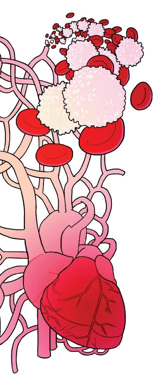
Chandni Patel, Aneesa Chowdhury, and Magen Anthony

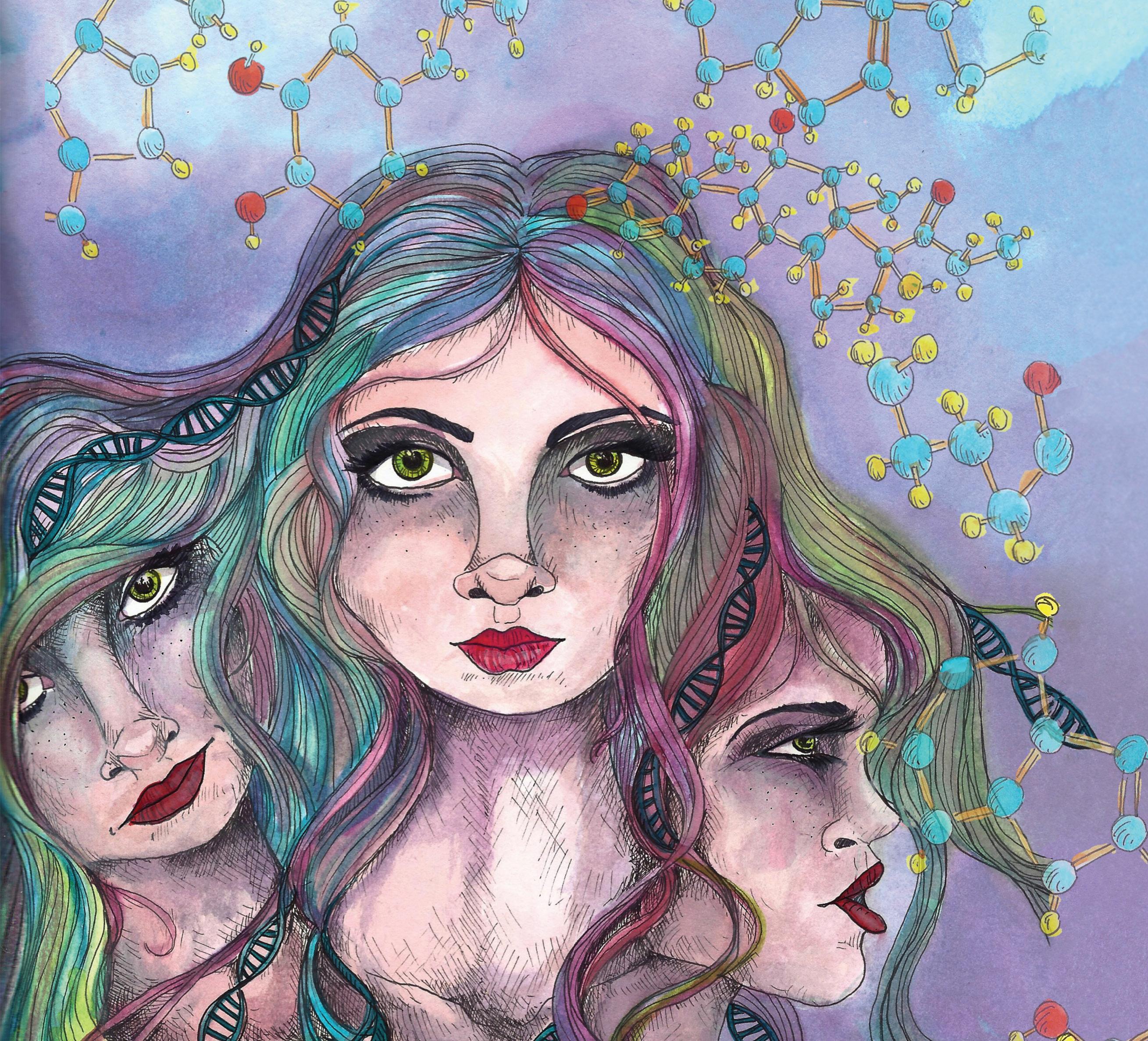

People often think human behavior is determined by choice, that the main difference between a martyr like Gandhi and a dictator like Hitler is that one chose a peaceful path while the other did not. However, new research is beginning to suggest that a large part of our behavior is influenced by biology and our genes. New bridges connect the body’s microorganisms to behavioral patterns and decisions, while biopsychology researchers continue to study the biological mechanisms behind psychiatric diseases that affect behavior, such as bipolar disorder and schizophrenia, in pursuit of the underlying genetic causes. Based on today’s information base, researchers have deduced that behavior is strongly influenced by environmental factors but can be — and often is — influenced by innate biological factors.
Many have heard and quoted the saying “trust your gut;” however, as research in gastroenterology has shown, a more accurate phrasing is “trust your healthy intestinal microbiota.” Gastroenterology is the branch of medicine that deals with the digestive system and its disorders. The major organs of the digestive system include the mouth, esophagus, stomach, liver, gallbladder, pancreas, and intestines. Digestive diseases may come about if any of these organs malfunction or are damaged, and can include symptoms such as heartburn, ulcers, bloating, stomach cramps, and diarrhea. Reaearchers are beginning to learn that digestive disorders are related to the digestive system’s
microbiota. The microbiota includes the diverse bacteria and other microbes living in the digestive system.
Here at UC San Diego, Professor of Medicine and Dean of the Graduate Division Dr. Kim Barrett and her lab are at the forefront of digestive disease research. Dr. Barrett and her colleague Dr. Melanie Gareau, an assistant professor at UC Davis, study how bacteria communicate with the brain through signals from the gut. “It is an interesting idea that what you eat can influence your behavior, [and this is] an emerging field in which there has been quite a bit of work very recently,” says Dr. Gareau. Dr. Barrett adds, “I think that we know very little about how specific intestinal diseases have behavioral manifestations. It has a huge impact on the quality of life, and it’s important to understand those links. Unless you understand them, you don’t have any hope to intervene.”
With Dr. Barrett and Dr. Gareau’s support, BS/MS student Jacob Emge investigated the relationship between certain inflammatory bowel diseases (IBDs) and cognitive performance. Emge studied the way mouse behavior and mood changed in response to colitis, a type of IBD, in response to changes in the gut microbiota. To do this, mice were given a chemical to induce colitis. The mice were then analyzed for differences in anxiety by putting them in a large cage with areas of light and areas of darkness. The mice with a greater tendency to move toward the dark areas were characterized as having higher levels of anxiety. Mice with colitis spent more time in the dark compared to controls,
sqonline.ucsd.edu
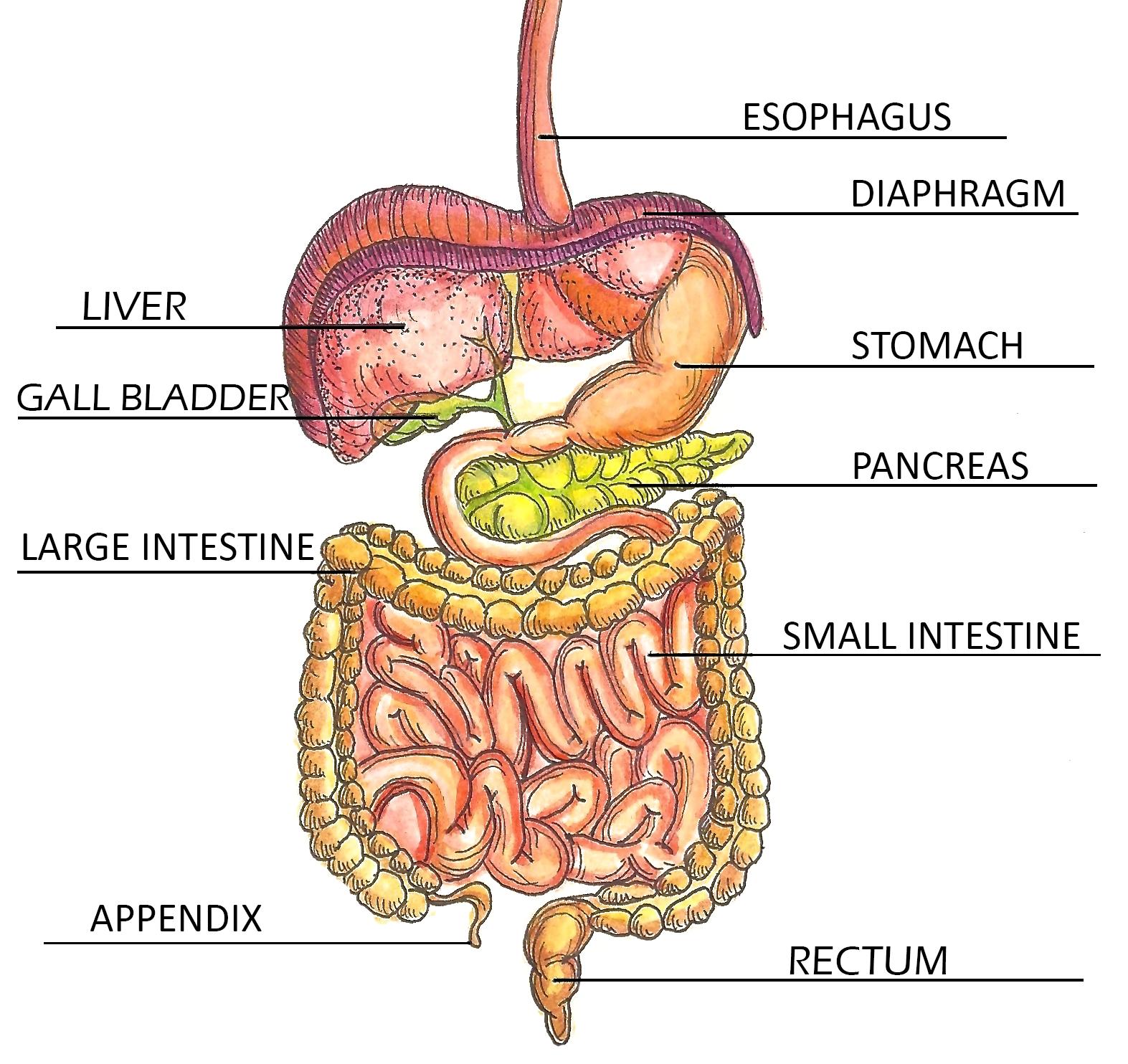
Depicted are the major organs of the digestive tract, in which the diverse gut microbiota reside. Inflammatory Bowel Disease and Colitis can cause inflammation and sores to form on the inner lining of the colon, or large intestine, as well as within the small intestine and rectum.
suggesting that mice with colitis had higher levels of anxiety. Emge also tested whether colitis impacted memory. To test this, mice were put into different cages with two objects and were allowed to explore. Later, they were placed into different cages with one of the same objects as before and a new object. The number of times the mice investigated the new object was recorded. Mice that explored the new object less were characterized as having memory impairment, as they did not remember the old object they had been shown before. Compared to control mice, those with colitis were more likely to exhibit memory impairment. Finally, Emge
assessed the effect of colitis on the diversity and quantity of bacteria comprising the microbiota and on the level of the stress hormone corticosterone. He found that mice with colitis had a different make-up of their microbiota compared to control mice.
Compared to the control group (without inflamed bowels), the mice with inflamed bowels spent more time in the dark parts of the cage, did not explore the new items as much, and had different bacteria in their colons, suggesting that colitis is associated with increased anxiety, memory loss, and changes in microbiota. In these ways, IBD in a mouse model had a significant impact on mood and behavior. At first glance, it seems unusual that a bowel disorder has an effect on cognitive state. Emge assures us that there is much more than meets the eye when considering the bowel. “It’s simple in the sense that it’s one large tube, but it’s so complex,” remarks Emge. “There’s symbiosis with bacteria but also tie-ins with neurological and physiological aspects. It’s very multifaceted.” These results demonstrate the serious impact that gastrointestinal diseases (and associated changes in microbiota) can have on psychology and behavior in mice, and lays the groundwork for possible explorations of this same phenomenon in humans.
Gastrointestinal disease is not the only example of biology affecting behavior. Looking at an even smaller scale, researchers have found that psychiatric diseases associated
with behavioral symptoms, such as bipolar disorder, are often correlated with a person’s genes. Genes are essentially the blueprints for all molecular mechanisms that take place in the body. These blueprints may be suspect in not only affecting, but also triggering bipolar disorder, a psychiatric condition characterized by periods of depression interspersed with episodes of severe mania, or “highs.”
People diagnosed with bipolar disorder are more prone to poor performance in their job or at school, damaged relationships, and/or depressive and suicidal tendencies. Although there is no cure for bipolar disorder, it can be effectively treated so that people with the illness can lead full and productive lives.
Dr. John Kelsoe is a Professor of Psychiatry at UCSD and studies the genetics behind psychiatric illnesses, particularly bipolar disorder. Under his supervision, BS/MS students Brian Chin and Donel Purcella started their own projects to study gene regulation and novel mutations regarding the onset of and responses in bipolar disorder. Chin focused on the NTRK1 gene, which he suspects might be a gene that predisposes people to bipolar disorder. Previous studies have shown that specific mutations in the gene cause the body to produce proteins that may interfere in certain pathways of the nervous system. Chin wanted to explore whether these proteins might make someone more likely to develop bipolar disorder due to their effects on these neural pathways. “I’ve always found the brain and its complexity fascinating. It’s kind of like this bridge where psychology meets physiology. And molecular biology has been one of
my fields of interest, too ... so I wanted to combine the two [in my research],” Chin states.
Chin plans to further characterize the function of this protein by cloning the mutated NTRK1 gene and inserting it into certain cells which would then produce the mutated protein. By studying the phosphate groups on the mutated proteins as well as interactions with other molecules that assist in the function of these proteins, Chin hopes to get a better sense of what this protein does and how it might be involved in bipolar disorder.
Purcella’s study involved the gene known as HTR7, which influences the activity of serotonin receptors. Serotonin is a neurotransmitter commonly associated with happiness. HTR7 is also thought to affect the body’s response to a certain family of antidepressants that prevent too much serotonin from being absorbed by neurons. Purcella’s work focused on a specific set of serotonin receptors in the cell membrane that seem to be correlated with different levels of responsiveness to these medications. The goal of the experiments is to see whether there are fewer serotonin receptors in a group with a mutated HTR7 gene.
Thus far, the study has shown that in the group with the mutated gene, there was a significantly higher rate of response to these medications than in the group without the mutant gene. This implies that the HTR7 gene could be a significant factor in the onset of and diagnosis of bipolar disorder, though further studies and analysis are needed
7 sq.ucsd.edu
to confirm these suspicions. “I myself am disabled and in fact was diagnosed with bipolar disorder when I was in college,” says Purcella, detailing her personal connection to the topic. “[This was] my whole purpose in becoming a Physiology and Neuroscience major.”
“I’ve always found the brain and its complexity fascinating. It’s kind of like this bridge where psychology meets physiology.” - Brian Chin
Demystifying the genetic bases of psychiatric disorders will contribute to the growing school of thought that such diseases are not derived from one’s personality or “character,” but rather are biologically-based diseases similar to diabetes and heart disease and can no longer be subjectively diagnosed. Understanding the molecular pathways that lead to disease manifestations can allow us to develop specific, targeted, and more effective therapy in the future. and can be effectively treated with more knowledge and preparedness.
While the gut microbiota and point mutations in very specific genes are lower-profile topics on their own, the researchers have pinpointed issues that deserve to be more visible to the public. For their part, colleagues Dr. Gareau and Dr. Barrett had often discussed how pediatric diarrheal diseases have a considerable impact on developing countries. “Diarrhea doesn’t exactly make for good cocktail
party conversation,” Dr. Barrett says. “But diarrhea in particular is the second leading cause of child mortality worldwide. So I think from a [perspective of] wanting to work on something with a big impact, it’s a great place to be.”
Since the findings from Dr. Barrett and Dr. Gareau’s research identify a link between cognitive decline and early diarrheal disease, they may have stumbled upon the unseen instigator of a prevalent global issue. “If you have a population that has cognitive deficits, they are less able to be productive and they are less able to move up the social ladder. You could actually say that diarrhea has this economic burden on developing countries as well as the human burden of illness and mortality,” Dr. Barrett states.
Now that a mouse model has been successfully established for the cognitive effects associated with IBD and colitis, researchers can develop a better understanding of the effects in humans. “Ideally, if other models emerge and the scientific and medical communities embrace that changes in the microbiota do indeed cause behavioral defects, we could see if probiotic therapy would become a go-to treatment,” says Emge. While universal acceptance of probiotic therapy as effective treatment by the scientific community is still a long ways away, it is possible that implementing these therapies in developing nations could
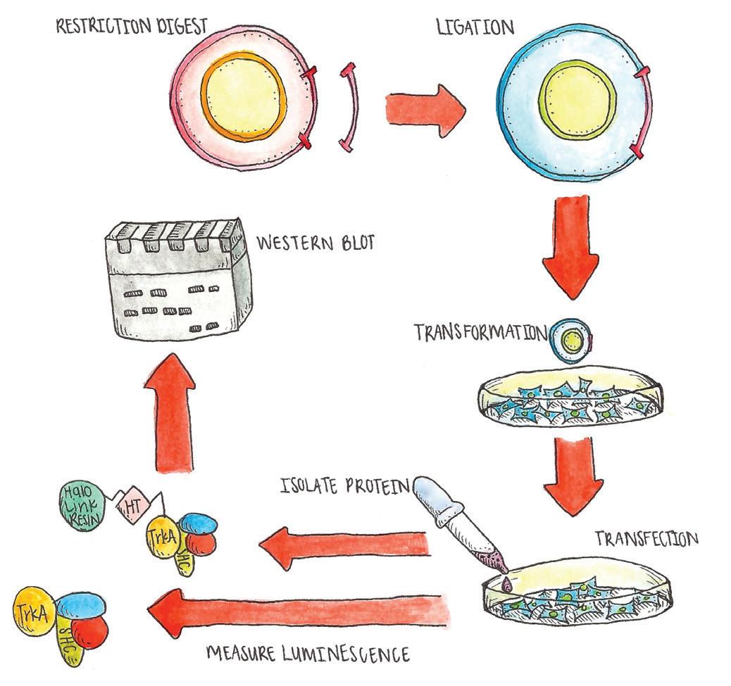
The gene of interest is isolated and inserted into the chosen plasmid with ligation, then transformed into bacteria, and then transfected into cells. The presence of the proteins that the gene codes for can then be analyzed by isolating the proteins and performing a Western blot and tagging the protein with a luminescent marker then measuring the luminescence.
facilitate social mobility, a healthier population and, in turn, progress.
Scientists are only beginning to understand the relationship between intestinal disorders and cognitive function, but it is without question that mental illness has long been identified with behavioral problems. Despite this, not all aspects of mental illness have been completely demystified. The genetic foundations of these diseases are still murky and, even though there is evidence to show that diseases
like schizophrenia and bipolar disorder can run in families, there is much work to be done. Chin’s project targets one very specific gene’s mutation that may influence bipolar disorder. Understanding the genetic reason certain individuals have a propensity to become affected can open up new screening methods to detect the onset of a mental illness much earlier.
If researchers will one day have the ability to develop genetic screening tests for mental illness, then the logical next step is to develop more tests to determine the best path people should take to treat their disorders. Purcella’s project serves as a starting point for the latter. “The ultimate goal would be to make it so that people could go in and get a blood test that tells you what drug to take,” says Purcella. Taking the guesswork out of knowing whether a patient is, for example, simply depressed or actually displaying symptoms of the depressive cycle of bipolar disorder would save lives. An increased public knowledge and awareness regarding the biological basis for these conditions may also decrease the social stigma associated with them and would be instrumental in building an incontrovertible bridge between psychology, biology, and behavior.
WRITTEN BY ARUSHI ATLURI, JULIA BROWN, AND SARAH
GEORGE . Arushi is a General Biology major graduating in 2017. Julia is a Microbiology major graduating in 2017. Sarah is a Physiology and Neuroscience major graduating in 2018.
sqonline.ucsd.edu
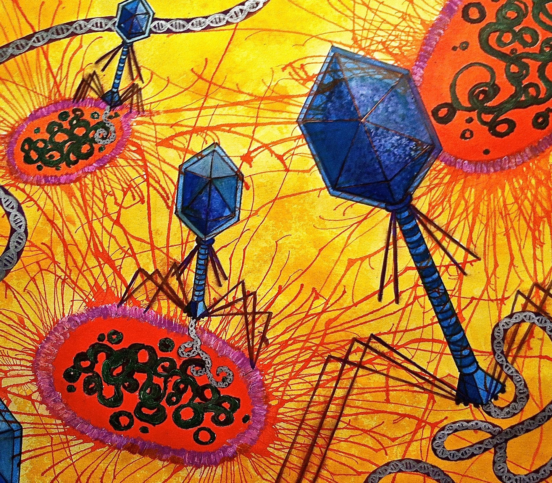

Our knowledge of medicine has dramatically increased in the past century and a half with the discovery of many new vaccines and antibiotics. Today, antibiotics seem to be a failsafe method for treating bacterial diseases such as tuberculosis and gonorrhea. However, we aren’t nearly as safe as we would like to believe. We are starting to notice the effects of overusing our antibiotics. Our abuse of and dependence on antibiotics has slowly given rise to new strains of bacteria that are antibiotic-resistant. Diseases that were once on the verge of being eradicated are coming back with a vengeance. What will happen when microorganisms are completely immune to all of our antibiotics? We can search for new ones, but that would only be a temporary fix; eventually, we would be right back where we started. To address this potentially dangerous situation, we need a brand-new approach.
One promising solution is phage therapy. Phages, short for bacteriophages, are viruses that specifically infect bacteria. Phage therapy holds the promise of effective anti-bacterial treatment with few side effects, such as bacterial resistance and adverse immune reactions. The idea behind phage therapy is to change the genetic code of these viruses so that they attack and destroy invading bacteria in the body. While it may sound simple in theory, this process in fact very complicated. It requires more than just introducing the correct phage to the body. Phage therapy offers multiple advantages
over antibiotics. Since antibiotics attack a wide range of cells, they can cause liver damage, repress the beneficial bacteria in our bodies (similar to those that play crucial roles in the digestive system), and cause opportunistic infections by disrupting the immune system. Phages, on the other hand, selectively target bacteria using specific receptors on bacterial cell walls and therefore eliminate many of the side effects of antibiotics.
Although creating a working phage therapy is not easy, progress is being made. One particularly promising direction of research involves mycobacteriophages, which specifically infect mycobacteria. Mycobacteria are among the most robust organisms in the world; their thick cell membranes allow them to cause widespread damage in the human body while also making them difficult to eradicate. Leprosy and tuberculosis, two of the world’s most devastating diseases, are notably caused by several types of mycobacteria. Finding a way to control the phages that specifically destroy these bacteria could be a medical breakthrough rivaling the discovery of penicillin.
The Phage Genomics Research Initiative (PGRI) may be on the verge of a major breakthrough. This initiative, now in its seventh year, is a undergraduate program that has only twelve participating universities in the country. It focuses specifically on the genetic aspect and function
sq.ucsd.edu

Pictured is the overall structure of a bacteriophage, a virus that specifically infects bacteria. The protein capsid head contains the virus’s genetic material and is attached to the neck, tail, and tail fibers.
of phages. Each year, a handful of incoming freshman students are invited to participate in a yearlong project of sequencing and analyzing the genome of a selected phage. Goals of the program include teaching students how to think in innovative ways and teaching practical research skills and techniques. The PGRI also gives students the chance to publish as coauthors in professional research journals. According to Mili Parikh, a student involved in the program, “It is a unique program tailored towards students that have an interest in research or the medical field.” The program is headed by Dr. Joe Pogliano, a molecular biologist, whose research focuses on microbial systems and structures, and Dr. Madeline Butler, a prominent undergraduate administrator in the biology department at UCSD. Both serve as mentors and take these freshmen under their wings to help them succeed in both the program and their academic careers.
The first step in understanding an organism’s genetic code is reading its DNA, which is composed of four nucleotide subunits: adenine (A), guanine (G), thymine (T), and cytosine (C). Every organism has a different order of these nucleotides. DNA sequencing methods can be used to determine the exact order of these nucleotides in an organism’s genome. For example, polymerase chain reaction (PCR) is a technique used to make copies of specific DNA sequences. After enough copies of DNA are produced, each of the four nucleotides is tagged with
a different radioactive dye. The tagged DNA is placed in a gel and subjected to an electric field, separating the fragments based on size. Analyzing the separation of fragments gives researchers the exact sequence.
Understanding the genetic makeup of mycobacteriophages and the relationships among various clusters of phages is an essential step toward repurposing them for use as antibacterial agents in phage therapy. Recently, students and teachers of the PGRI successfully sequenced the genome of the Weiss13 mycobacteriophage. The students started by collecting soil samples from around La Jolla that contained phages capable of infecting Mycobacterium smegmatis, a wellcharacterized bacterial host. The samples were purified and enriched, and the phages were allowed to multiply. Through a complex process of streaking, serial dilution, and spot titer, different phage strains were isolated on agar plates. The various strains were then analyzed through electron microscopy to determine which sample was best suited for sequencing. Weiss13 was chosen and sent to a lab for evaluation.
After sequencing, it was necessary to determine which genes coded for certain characteristics of the mycobacteriophage. Since mycobacteriophages invade and reproduce in mycobacteria, they could potentially
be used to combat mycobacterial infections such as tuberculosis and leprosy. But to understand how to manipulate phages to fight infections, researchers need to understand the functions of specific gene sequences. Different gene sequences are responsible for an organism’s specific characteristics and traits, from the way it looks to its behavioral patterns. Thus, to manipulate the behavior of the phage, researchers need to understand which gene sequences are responsible for specific behaviors.
It is important to make distinctions between different phages, as not all phages replicate using the same mechanism. Grouping phages into clusters allows generalizations to be made about the phage replication process. Weiss13 was classified as a Cluster R phage in the BLAST catalog based on its genomic sequence. Cluster R is a small group of phages with only four members, including Weiss13. Currently, the only other known members of Cluster R are Send513, Nilo, and Papyrus. These phages are relatively uncommon compared to phages in groups such as Cluster A, which has 249 members. The genome of Weiss13 was also compared to those of other mycobacteriophages using a mycobacteriophage database. Weiss13 was determined to have a mosaic structure, meaning it inherited genes from multiple strains of mycobacteria. Dr. Butler describes the process as a genetic swap between phages eventually resulting in new types of organisms. She states, “It’s as if these phages have borrowed pieces of each other and switched them around.” Due to this history of genetic
transfer between different strains, some of Weiss13’s genes occur commonly in the phage genome database, while others are quite rare.
After the purification and sequencing of Weiss13, the PGRI students analyzed the sequence to determine the purposes of certain genes. To accomplish this, the students used BLAST (Basic Local Alignment Search Tool), a program that compares unknown DNA to DNA from known sources to find how much an organism has in common genetically with another organism. The students used this program to compare the sequence of Weiss13 to known sequences from DNA Master, a database that catalogues known phage genetic sequences. The 95 genes were classified into the following categories: DNA synthesis, lysis, defense, structure, assembly, and unknown. Using this program, the students discovered several genes of unknown function in Weiss13. They then isolated the peptide chains (the chains of amino acids that eventually fold to create proteins) of the virus and separated them by mass and charge.
BLAST was used to produce a list of gene-coding regions in Weiss13. Thanks to these sequences, the researchers were able to associate proteins with their respective genes and develop a basic idea of the gene’s function in the virus. Students then compared the gene sequence of
Weiss13 to the sequences of other phages to determine genetic similarities and possible functions of unknown proteins in the Weiss13 genome. Afterward, this data was stored in DNA Master’s database along with the genetic sequences of other mycobacteriophages sequenced by different labs. Dr. Butler gave a short statement on the implications of the database, saying:
“Having this database of all these phages that infect the same host and may have exchanged information with one another during evolution is teaching us things about how viruses and other organisms can share DNA sequences, can be mixed and matched, forming new organisms.”
By compiling gene sequences of mycobacteriophages and comparing how they replicate, scientists are learning more about viruses. Hopefully this increase in our understanding will lead us to make new discoveries that will improve our current vaccines and help with the development of phage therapies.
Building the BLAST catalog is a major step toward understanding the evolutionary process of mycobacteriophages. Viruses can mutate at an astonishing rate. Unfortunately, there is still we do not know about how viruses evolve so quickly or how. This is a huge issue, as rapid viral mutation and the inherent unpredictable nature
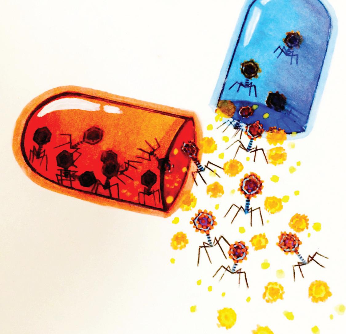
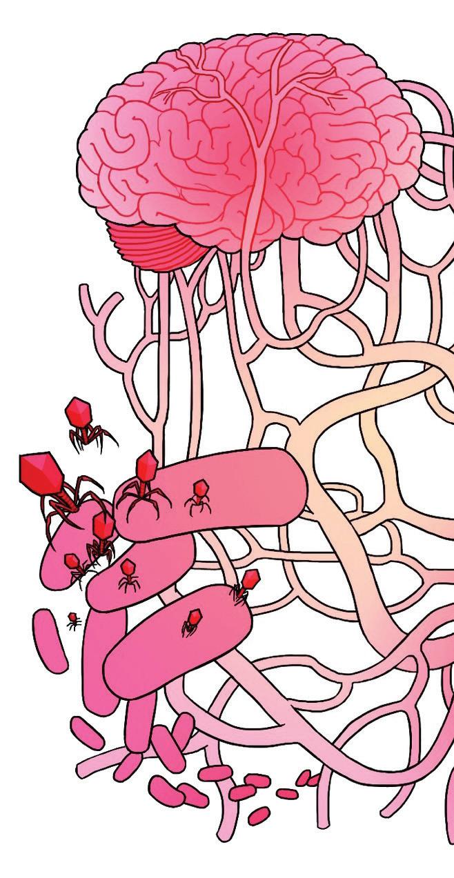
An artistic depiction of the idea behind phage therapy, using viruses as medicine.
of these mutations causes significant issues in the creation of effective vaccines. The seasonal flu vaccine, for example, is actually a combination of several vaccines that provide immunity against whichever influenza strains scientists predict will be most prominent during the upcoming season. However, due to the unpredictability of viral mutations, sometimes the vaccine is not completely effective.
But overall, this is why PGRI so valuable. The program has sequenced the same species of mycobacterium dozens of times. By comparing these sequences among each other we can understand more about viral genetics, specifically the processes through which viruses evolve. For each strain that scientists sequence, we get a clearer picture of how phages have changed over time. For each new genome that is added to this database, we can see how it compares to others in the same group in terms of the specific gene sequences and gain a deeper understanding in the genetics of viruses and improving treatments. Gaining more information about viral genomes can also have other benefits. By increasing our knowledge of the genetic and physical processes in viruses, we get closer to perfecting a viable method of phage therapy. Using phages as therapeutic agents could be a more holistic method for combating infections and other diseases. The sequencing of the Weiss13 mycobacteriophage is a small, yet important, contribution to this genetic catalog and the future of phage therapy and viral genetics as a whole.
WRITTEN BY
ISAAC DAVIET, ALEXA SISON, AND KAREN TOLENTINO.
I saac is graduating in 2018. Alexa is a graduating in 2017. Karen is a Biochemistry and Cell Biology major graduating in 2017.
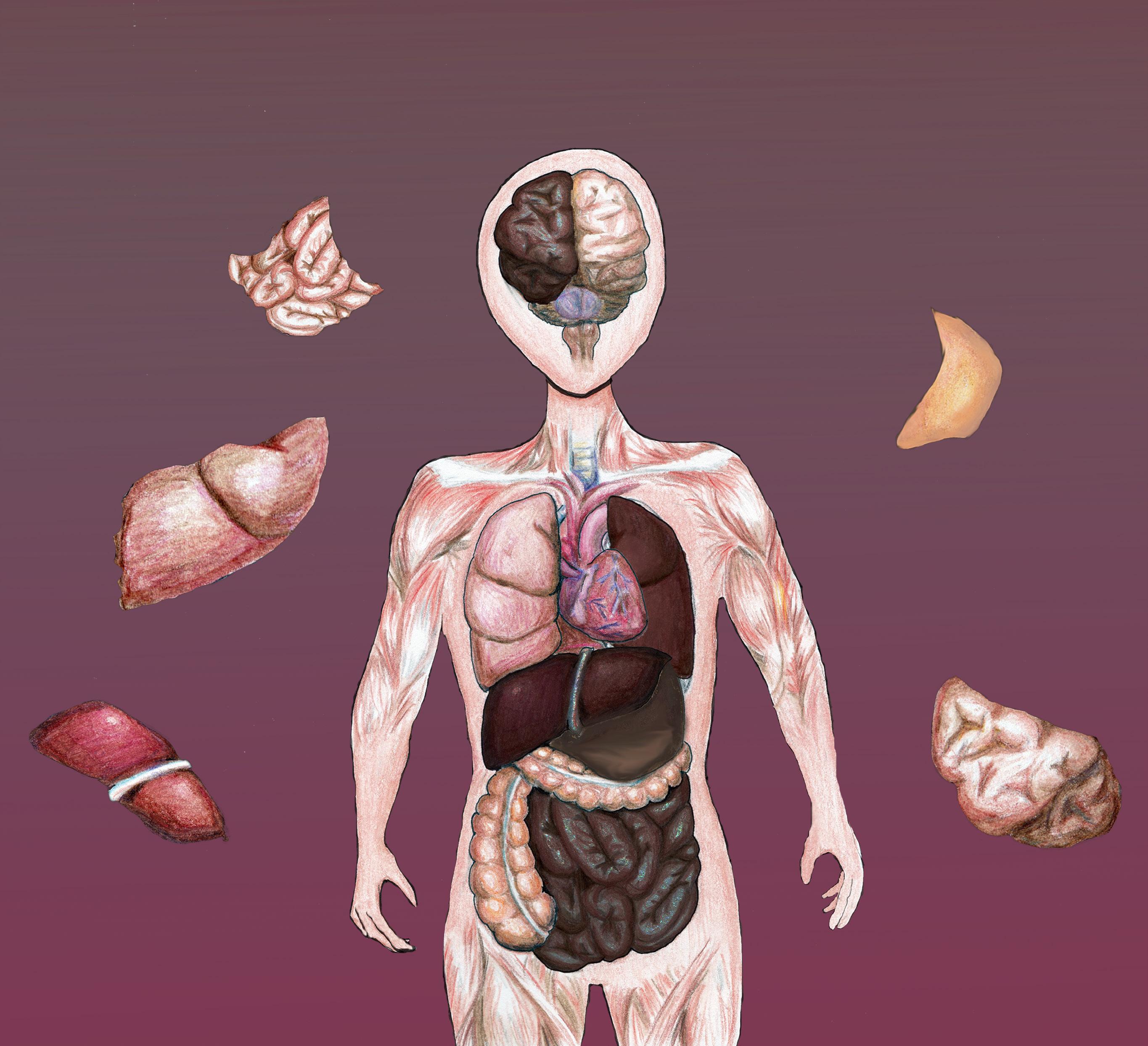
Puzzles are made up of hundreds of tiny pieces that look insignificant on their own, but when placed together in the correct formation, can contribute to a larger and well-defined picture. However, if even one of the individual pieces is changed in some way, then the entire picture will have a hole in it, making it incomplete and defective. The human body works on a similar structure: if proteins and organelles in every cell of the human body perform their functions properly, then the human body can realize its basic needs daily. However, if a protein is significantly mutated in some way, whether through the structure of the protein or the amount of protein that is there, then the functions that the protein mediates will significantly change too, and the body’s systems will be damaged as a result.
The key to starting large puzzles are corner and boundary pieces. These give the indications for where later pieces in the puzzle with fit. Caveolin proteins play an equivalent role in the cell by helping to direct cellular processes.
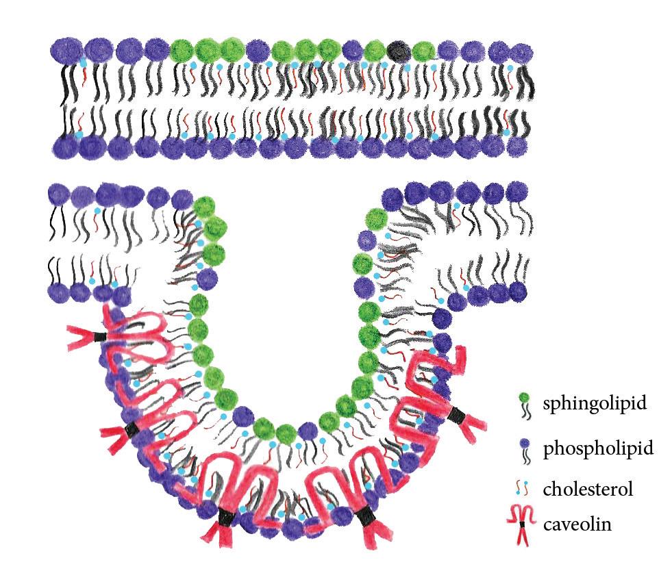
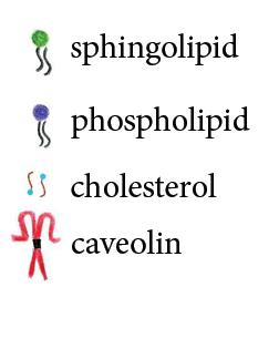
Caveolins are a family of membrane proteins involved in diverse functions ranging from endocytosis to signal transduction to tumor suppression. They are tiny agglomerations of lipids that help compartmentalize signaling and other aspects of the cell. That is, they help to sort the countless molecules bombarding a cell into signals to ignore or signals to initiate downstream pathways. With age, however, caveolin protein amounts
begin to decrease, causing the cells to lose a crucial tool that allows them to, as Jan Schilling, the post-doc working in the lab says, “create efficiency out of chaos.” Similar to the formation of a puzzle, it is as if some of the corner and boundary pieces were lost.
Dr. Hemal Patel’s laboratory at UCSD’s Department of Anesthesia tackles the question of whether increasing caveolin, or cav, protein amounts will counteract the effects of aging and other diseases. Earlier studies confirmed that mice genetically altered to produce higher amounts of Caveolin protein in the heart indeed experience fewer pathologies than in elderly, non-genetically modified mice. But, it is difficult to determine the long-term effects or define the specific mechanism since mice have a relatively long lifespan making studies expensive and time consuming. Dr. Patel’s laboratory was able to overcome this obstacle by choosing a different model organism: Caenorhabditis elegans. These “worms”, as the lab affectionately calls their C. elegans, live only about twenty days which allows researchers to better study survival trends and response to a myriad of experimental conditions. It turns out that C. elegans engineered to express higher levels of caveolin proteins live up to three days longer than the normal worm—in fact, these proteins were later found to be actively responding to external stressors in a variety of species. In subsequent studies conducted by the Patel Laboratory, it was found that caveolin proteins move from the plasma membrane to the mitochondria during high stress situations, such as in sqonline.ucsd.edu
low oxygen environments. Without caveolin, cells sustain mitochondrial damage. These studies demonstrate that the interplay between caveolin, caveolae, and mitochondria is an evolutionary trait designed for stress adaptation.
The success of designing a model organism for efficient survival studies opens opportunities to test the response of caveolin proteins to different stressors. However, Dr. Patel’s laboratory found that these possibilities were limited by the small number of worms that could be grown on the standard culture dishes. Only 30 worms per plate are used in life span studies with multiple plates for each group or condition, leading to a great deal of effort for simply maintaining robust population sizes needed for metabolic and biochemical profiling. Instead, Dr. Patel’s laboratory, based on published studies, utilizes a liquid culture model as a supplement to plating worms in order to allow for the growth of much larger colonies. With these larger populations, new stressors, such as increased glucose and lipids to mimic diabetes in humans, can be
tested to confirm the effectiveness of caveolin proteins in more specific pathologies beyond aging. These findings carry profound implications, particularly in the health sciences. Given the crucial role that caveolin serves in aging, maintaining the levels of theses proteins in humans may help to elucidate the process of aging in human and potential therapeutic targets. For example, caveolin protein overexpression is only effective in highly specialized cells that no longer divide frequently, such as neurons or heart cells. In fact, caveolin may cause certain cells to proliferate more quickly and has been correlated to cancers. Unpublished studies in collaboration with the laboratory of Dr. Brian Head at UCSD have discovered that localized injections of viral vectors in the brain enhances memory and learning in older mice. Would it be possible that caveolin proteins could help preserve our own minds or help our heart function more efficiently as well? The words of Grace Grogman, the undergraduate senior whose honor thesis focused on this project, “the ability to translate these findings across species would be an extremely valuable development.”
A puzzle is only considered finished if all of its pieces are present. What happens when pieces start going missing or getting bent so out of shape that they cannot be used? Missing a few pieces can be called a mere inconvenience, but the more pieces that cannot be used or found, the more incomplete the puzzle becomes. When enough
pieces cannot be used, the puzzle fails to fulfill its purpose: showcasing a complete and faithful image. This is essentially what happens in Alzheimer’s disease, a condition in which the person’s brain slowly decreases in size due to dying neurons. Currently, untreated individuals suffer from worsening symptoms that include increased memory loss and difficulty doing daily tasks such as reading and judging distance. This deterioration has been attributed to beta-amyloid peptide buildup produced by the amyloid precursor protein (APP). This buildup, also known as amyloid plaques, interferes with normal brain function. An alternate explanation states that a protein called tau folds and links together to form tau tangles in the cell’s outer structure, eventually leading to the cell’s death. With these two potential sources of blockage, neurons may become unable to function or signal to one another and die.
In Dr. Edward Koo’s lab at UCSD, a team of researchers is investigating the origin of Alzheimer’s disease. One member was undergraduate Kaivon Sobhani, a graduating general biology major whose interest in Alzheimer’s disease was sparked by the growing number of Alzheimer’s cases in the United States. He and his advisor Dr. Karen Chiang were interested in whether APP affected the number of new neurons produced in mouse brains.
To generate the mouse model, experimental mice were genetically engineered to express three genes: APP, tetracycline transactivator (tTA), and Cre recombinase.
The last two genes were included to express APP in the hippocampus, a portion of the brain responsible for generating neurons. However, the tTA had an unexpected neurotoxic effect and shrank the mice hippocampi, which clouded the experiments results. This unexpected result was a setback that required Sobhani and Chiang to distinguish between the effects of tTA and APP, leading them to modify their protocol and repeat their experiment. Using an antibiotic treatment, they masked the neurotoxic effects of tTA, while allowing the mice to continue to express APP.
The antibiotic treatment was integrated into the diet of pregnant mice. Their pups were administered antibiotics for the first forty to forty-one days after birth, blocking the expression of tTA. This regimen proved successful, as the neurotoxic effect was virtually eliminated, and the mouse brains were able to be removed, sectioned, and used to count the forming neurons.
Though the tTA neurotoxic effect had been eradicated, the results are still inconclusive. It was originally believed that neuron formation would decline. The data currently shows that there are slight decreases in neuron formation, but the results are not yet significant enough to confirm their predictions. In the coming months, they will conclude whether tTA expression or APP overexpression contributes to the brain’s degeneration. Ultimately, the results of Sobhani’s experiments may have strong implications for future Alzheimer’s research.
sq.ucsd.edu
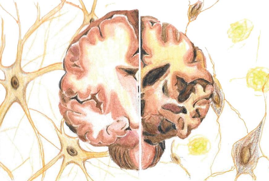
On the left is the brain of a healthy patient and on the right is an example of a patient with severe Alzheimer’s. The brain on the right is noticeably smaller than on the left. The brain on the right also surrounded by neurons with neurofibrillary tangles and beta-amyloid plaques while the brain on the left is surrounded by healthy neurons.
“In Alzheimer’s, there is a decrease in learning and memory. Similarly, there is a decrease in neurons and neuronal connections. The discovery of neurogenesis in adults raises the possibility of preventing or reversing neuronal degradation,” Sobhani explains. “If neuronal degradation can be prevented or reversed, then maybe Alzheimer’s can, too.”
Imagine that you’re building a puzzle of a shark in the ocean, and you start by constructing the outer edge which consists solely of ocean parts. When you come across a piece of the shark that is supposed to fit in the center of the puzzle, you are unsure of where it should be placed. When creating a puzzle, sometimes you confront a piece that you cannot place until you understand more of the picture. In the same way, sometimes specific proteins seem to play a role in certain functions, but you might not understand the specific role until more research is conducted. One such example of this, researched by Kyle Mauricio Yuquimpo, was the role of Interleukin-37 in Inflammatory Bowel Disease.
Interleukin-37 (hIL-37) is a cytokine, or signaling molecule, that is produced by white blood cells and is a novel regulator of the immune system. Though a synonymous version of this cytokine has not been discovered in mice, genetic techniques have allowed for the production of human IL-37 in mice, commonly denoted as “hIL-37tg”. It is also thought to balance the amount of inflammatory and noninflammatory cells in the brain, reducing the number of inflammatory response cells and thereby regulating inflammation due to Crohn’s disease in the gastrointestinal tract.
Kyle Yuquimpo, a senior undergraduate honors researcher at UCSD under the laboratory instruction of gastroenterologist Dr. Jesus Rivera-Nieves, M.D.,
investigated how the hIL-37 cytokine may play protective a role Crohn’s Disease, an adaptive immune-mediated inflammatory response (inflammation of regular pathways due to dysregulation of immune system). He goes on to describe how ilea (sing. ileum - part of the small intestine in the gastrointestinal tract) were collected from mice for histology to determine the severity of ileitis (inflammation of the ileum). This is known to be caused by a genetic manipulation that increases the Tumor Necrosis Factor, a protein responsible for inflammation. Surprisingly, it appeared that interleukin-37 did not protect the mice from ileitis. He purports that a substantial “trigger” of the immune response was necessary to drive a greater increase of hIL-37 to perhaps observe protection from inflammation. Kyle treated mice with a Dextran Sodium Sulfate, a chemical known to cause weight loss and inflammation, and weighed them daily for 7 days before collecting their ilea. Mice with hIL-37 were in fact protected from weight loss, but there was no observed protection from ileitis. Results suggested the opposite, that hIL-37 did not affect ileitis in interleukin-37 induced mice under the observed conditions.
Though primary literature provided evidence for its anti-inflammatory effects, Kyle’s data suggested that Interleukin-37 did not in fact protect mice from ileitis. Kyle agrees that their negative results may have derived from a relatively small sample size, because a trend of weakened ileitis was in fact observed. Though the analysis of the efficacy of Interleukin-37 did not yield significant
results, it is well known that a variety of factors both genetic and environmental play a role in intestinal inflammation. hIL-37 seemed to be a jigsaw piece that did not yet fit into the puzzle of Crohn’s disease. However, teams like Kyle’s research group are one step closer to fitting the entire picture together.
As time goes on and new technology and information make further research possible, it becomes more imperative to understand the molecular and cellular mechanisms that give rise to diseases. The more that is known about diseases, the more progress can be made developing cures, vaccines, or therapies. Almost all diseases have a seemingly insignificant origin—one protein holds a signaling system together, a small change in a protein causes a condition like Alzheimer’s, and one molecule seems to hinder inflammation in Crohn’s disease. Just as fixing an imperfect puzzle piece can allow students to complete a puzzle, figuring out the pieces of a disease can allow researchers to see the bigger picture.
WRITTEN BY CHRISTINA CUI, BRIANA KUSUMA, AND RACHAN NARALA. C hristina is a Physiology and Neuroscience major graduating in 2017. Briana is a Human Biology major graduating in 2016. Rachan is a Human Biology major graduating in 2018. sqonline.ucsd.edu
By
While scrolling through Facebook, a picture pops up in your newsfeed of a four-year-old boy smiling widely. You have never seen this boy before but his picture has over one million likes and has been shared over forty thousand times. You click the picture and learn the reason behind his big smile: he is now leukemia-free! His survival is indeed miraculous and can be attributed to recent discoveries in leukemia research. However, despite being one of the better-known and betterfunded cancers, leukemia still claims the lives of about half of the patients diagnosed with it.
While most have heard of leukemia, many do not know what a diagnosis of leukemia entails. Leukemia is a cancer that originates in the bone marrow and causes an overproduction of white blood cells. Normally, white blood cells (WBCs) and red blood cells (RBCs) begin to develop in the bone marrow from hematopoietic stem cells (HSCs). These stem cells can differentiate into red or white blood cells as well as platelets. Each of these cells plays an important role in maintaining the health of the human body. RBCs deliver oxygen throughout the body, WBCs protect the body from infection, and platelets are utilized in blood clotting.
WBCs usually have short lifespans, and they are constantly being replaced in the bone marrow. Leukemia occurs when the genetic material of the white blood cells mutates, causing the cells to grow uncontrollably and differentiate abnormally. The buildup of nonfunctional white blood cells
in the bone marrow will eventually displace the healthy red and white blood cells and platelets.
As the level of RBCs drops, the level of oxygen circulating through the body drops as well. Additionally, the lack of white blood cells causes infections to become more serious, and the decrease in platelets prevents open wounds from closing properly. Furthermore, since leukemia affects the main cells of the circulatory system, it is highly probable for the cancer to spread to other areas of the body.
Like other cancers, leukemia is a major public health concern. Fortunately, here at the UC San Diego, leukemia is a prominent subject of research. Researchers at UCSD are taking a closer look at different approaches to treat this disease. These researchers are focusing on how certain types of leukemia progress differently as well as looking at regenerative medicine as a possible treatment method.
A diagnosis of chronic myelogenous leukemia (CML) was once a death sentence for patients. However, by identifying the cause of this cancer, scientists have developed a treatment that improves CML patients’ life expectancies such that they are potentially comparable to those of healthy individuals. Unfortunately, the treatment fails to eradicate all of the leukemia stem cells, also referred to as blast cells, residing sq.ucsd.edu
in patients’ bone marrow. A majority of the leukemic blasts are killed by the treatment, which thus artificially selects for more resistant blasts. The accumulation of these treatmentunresponsive blast cells, causes the disease to progress to blast crisis, an acute stage in which blast cells make up more than 20 percent of cells in the bone marrow. Once the disease progresses to blast crisis, the patient’s chance of survival decreases significantly.
“The standard of care for treating CML is no magic bullet. While it has turned CML into a manageable disease, curative effects have yet to be produced,” says Patrick Chang, a UCSD graduate student. Chang and his principal investigator, Catriona Jamieson, M.D., Ph.D., have been looking for a second target on leukemia stem cells for potential treatments. Their goal is to develop combination therapies for CML. Current treatments wipe out most leukemia stem cells in the system, and a second treatment could be used to sensitize and eliminate the leukemia stem cells that resisted the first treatment.
Chang and Dr. Jamieson are investigating how the dysregulated connective tissue growth factor (CTGF) expression could affect the survival of leukemia stem cells. Previous research has linked CTGF to poor patient prognosis in acute myelogenous leukemia. To test their hypothesis, Chang transformed cells in the CML cell line so that the cells could produce CTGF on their own. He then grew the transformed cells and normal CML cells in media without any CTGF. By comparing cell growth, Chang was
able to determine that there is a link between CTGF and survival in CML stem cells.
This discovery could lead to a parallel treatment for patients suffering from CML. The next step for this project is to study the signaling pathways in which CTGF affects programmed cell death or resistance to chemotherapy. As CTGF is essential for many other functions in the human body, it may or may not be an ideal candidate for inhibitor development. Ultimately, the only way to find out is to reveal its signaling pathways and to identify a secondary target for developing a more effective inhibitor or therapy.
By understanding how leukemia develops, scientists are able to identify cancer development early on. Changes in proteins and biological pathways can combine to result in the development of leukemia. Dr. Dong-Er Zhang’s lab at UCSD is currently researching the development of acute myeloid leukemia (AML), particularly the role of AML1ETO, a fusion protein, which is created through the fusion of two genes by the translocation of chromosome 8 onto chromosome 21. Treatment-wise, learning the mechanisms of the development of a disease can unveil therapeutic targets that can stop or slow down a particular disease.
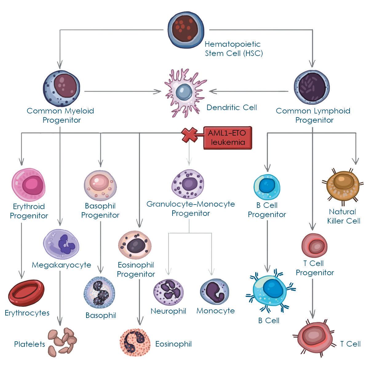
In the bone marrow, hematopoietic stem cells have the potential to differentiate down various lineages and become the cells that make up the blood and lymph. In AML1-ETO leukemia, cell differentiation into the granulocyte-monocyte progenitor is blocked and the bone marrow becomes congested with undifferentiated myeloid progenitor cells. By understanding the complex process of HSC generation and differentiation, researchers may be able to discover novel, individualized treatment for a variety of diseases originating in these cells.
Sheila Tran, a master’s student of Dr. Zhang’s lab, studied the role of an enzyme named UbE1L in the development of leukemia with AML1-ETO. In this project, Tran observed the role of UbE1L in helping proteins bind together. In most cases of AML, there is an upregulation of this enzyme, causing more proteins to associate with each other. UbE1L is being researched to see if there is some correlation between the increased activity of this enzyme and leukemia development. If this enhanced protein interaction is linked to AML development, research can be shifted toward blocking UbE1L. Hopefully, this will prolong or stop the development of AML. Currently, Dr. Zhang’s lab is studying cells that do not utilize this enzyme to see if leukemia progression is hindered.
Cody Mowery of the same lab focused more on slowing the development of AML through GM-CSF, a growth factor needed for granulocyte/monocyte, different stages of white blood cells, development. GM-CSF is known to promote cell survival as well as differentiation (development into different type of cells with different functions). In normal circumstances, stem cells are able to develop into monocytes. However, in AML, the development process is hindered and cells are stuck between the phases of myeloid stem cell and monocyte or granulocyte development. These cells are rendered incapable of differentiation. However, GM-CSF is known to encourage development through differentiation. The hope of Mowery’s project was to see if by adding GM-CSF into the media, cells are able to push towards differentiation despite being affected by AML1-ETO.
sqonline.ucsd.edu
This is significant because leukemia develops due to the t(8;21) translocation as mentioned above. Among patients with AML1-ETO, 35 percent of females are missing an X chromosome, while 56 percent of male patients are missing a Y chromosome. The GM-CSF receptor gene is found on the X and Y genes that these patients are missing. This shows a direct correlation in the necessity of the removed section with leukemia development. In fact, 20 genes are affected when these sections are translocated: genes that regulate cell proliferation and differentiation. Currently, research is being conducted to see whether GM-CSF signaling has a correlation with leukemia. This pathway could involve leukemia development. Hopefully, the addition of GM-CSF may delay leukemia development. However, it is unclear whether addition of GM-CSF will affect other genes. Of the 120 genes that are removed, research is being done to see what functions are displaced and whether manipulation of these genes can slow down the development of leukemia.
While signaling pathways can illuminate potential targets for the treatment of leukemia, they can also provide insight into the development of alternative cancer treatments. Current treatment of leukemia involves removing the cancerous bone marrow and transplanting a matching donor’s bone marrow into the patient. Not only is this a painful and draining process for the patient, but the transplant also runs the risk of being rejected by the host’s body and requires the
patient to remain on immunosuppressant medications for the rest of his or her life. In light of these issues, researchers are taking a closer look at regenerative medicine as a potential treatment for diseases that require transplants. Many researchers are looking into induced pluripotent stem cells (iPSCs), cells that have been programmed to revert to an immature, undifferentiated state. The hope is that eventually, we can use iPSCs from a patient’s own cells and “teach” them to differentiate into tissue-specific stem cells. In the case of leukemia, the objective is to get these iPSCs to differentiate into the HSCs from which all blood cells are derived, a process recent UCSD graduate Chase Melick and his mentor, Dr. David Traver, are aiming to elucidate.
“The challenge is to replicate development,” Dr. Traver elaborated. “The only way to get lifelong reconstitution of cells in the liver [or] cells in the blood forming system is to make tissue specific stem cells. You can’t just take iPSCs and transplant them. They are too immature. They don’t know what to do.”
Fortunately, Dr. Traver’s lab is working to identify the pathways involved in HSC generation. Melick investigated the role platelet-derived growth factor receptor (Pdgfr) signaling plays in HSC formation, using embryonic tissue from zebrafish. Not only were the researchers able to narrow down at what point in HSC development Pdgfr signaling is required, but they also observed that Pdgfr signaling played a role in regulating another signaling pathway required for HSC formation. This discovery opens up new questions
about how all of these pathways are connected and regulated. Being able to pinpoint which factors are needed and how they are integrated into the HSC formation process is the objective of this research. According to Melick, the clinical implications of Pdgfr signaling have already been demonstrated in leukemia treatments in which inhibitors are used to block factors involved in leukemia development. While this discovery is a step in the right direction, both Melick and Dr. Traver caution that there is still more research to be done before a better treatment plan becomes viable. Unraveling the complete molecular mechanisms of the pathways involved, applying them to a human model, and solving immunocompatibility issues are just some of the hurdles that researchers will need to overcome. However, Dr. Traver is optimistic about the progress and application of this research in the clinical setting.
“If we can figure out how to make these stem cells and make it reproducible, reliable, and robust protocol-wise, we can have something in the clinic within a decade or shorter.”
While we understand why leukemia develops, there is still more research that needs to be done to fully understand how to prevent and treat leukemia. With each new discovery of the complex mechanisms that lead to the development of leukemia, we can target steps that prognosticate leukemia at earlier stages, allowing us to treat patients before their cancer reaches a critical state. The hope is that one day we
will be able to treat patients specifically, according to what stage of leukemia they are in.
With a simple click of the mouse, you show your support to an individual who has overcome leukemia, an additional “like” representing your support, along with millions of other supporters. What we would “like” to see, however, is a change in the content of these posts. Hopefully in the near future, the severity of leukemia will be comparable to that of the common cold. With world-class researchers approaching the leukemia problem in various ways, we believe we can look toward a leukemia-free future.
WRITTEN BY MANDY WONG, LAWRENCE LIU, AND ANDREA CAPCAP. Mandy is a Biochemistry and Cell Biology & Computer Science double major graduating in 2016. Lawerence is a Biochemistry and Cell Biology major graduating in 2016. Andrea is a Physiology and Neuroscience major graduating in 2015. sq.ucsd.edu
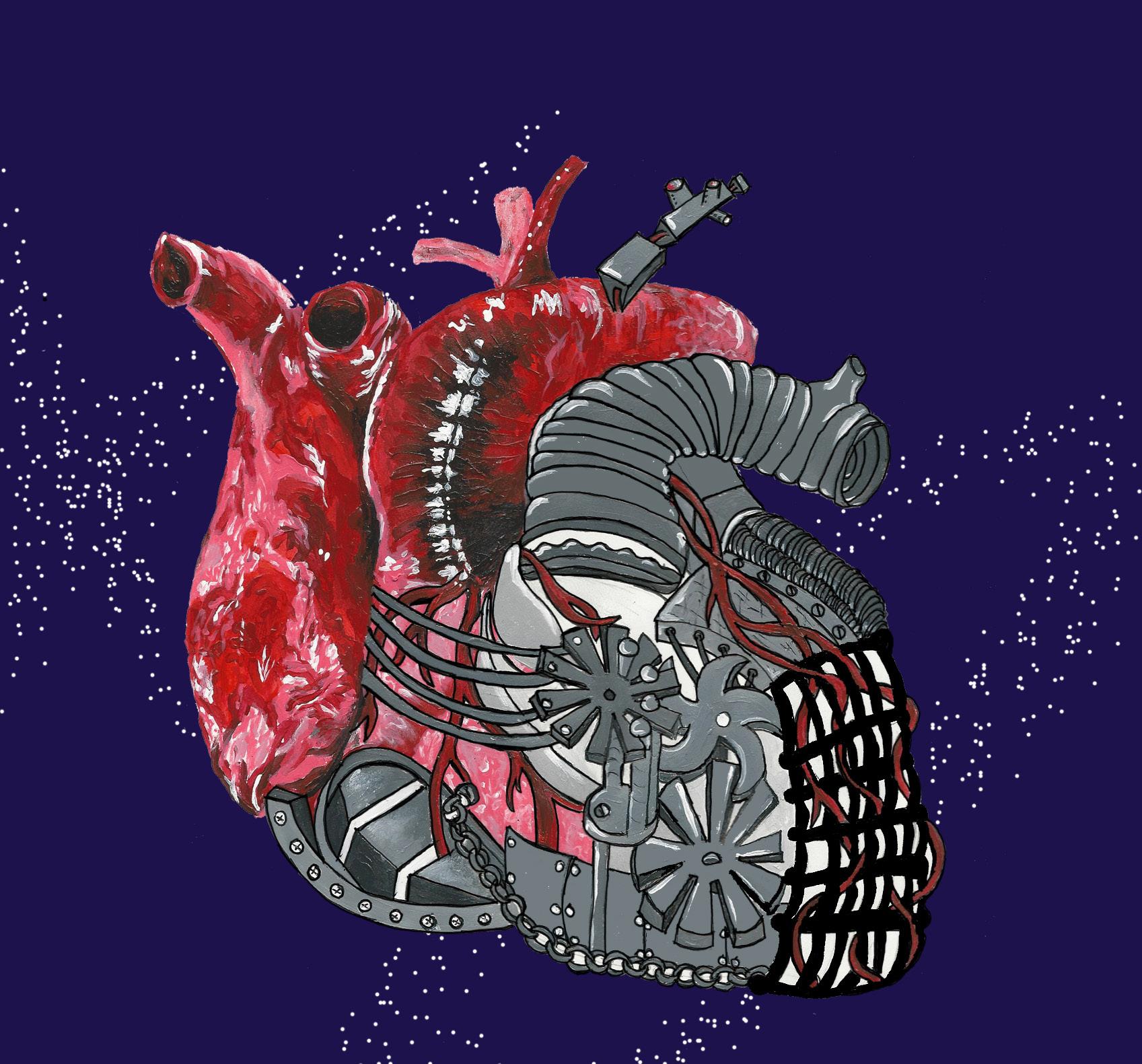


Traveling at twice the speed of sound 47,000 feet above ground, the NASA Space Shuttle Challenger was destroyed just after 73 seconds of flight on January 28, 1986. Although remembered as a tragedy, this accident can be used as a valuable lesson. This fateful day emphasized the importance of each component making up a system. The O-ring, a device used to separate the sections of the rocket booster and keep hot combustion gases from leaking, had failed to completely seal, causing the untimely deaths of the seven crew members aboard the shuttle. The Challenger disaster confirms that a functioning system must have each of its necessary constituents working together effectively. With the failure of only one small, seemingly insignificant element, an entire system can fall apart. Like the Challenger, the heart is a system that is composed of an amalgam of pieces. The human heart is the product of millions of cardiac cells and proteins, and the integrity and function of the heart can be threatened with even the slightest change in these components. Similar to the O-ring in the Challenger, various proteins, signaling pathways, and metabolic enzymes play pivotal roles in the function of our hearts.
One of the ways the heart’s vitality can be tested is by examining the roles of various structural proteins in the heart. Stephanie Myers, a third-year student working in the Lange Lab, studied the function of Obscurin proteins.
Obscurins act as structural components of the sarcoplasmic reticulum, an organelle involved in the release of calcium ions for muscle contractions. Stephanie hypothesized that removing Obscurins from cardiac muscle cells would affect calcium release from the sarcoplasmic reticulum, and subsequently, alter normal heart contractions.
Her experiments used knockout mice, which that were genetically modified to lack Obscurin proteins, to observe calcium handling. She measured calcium release in heart cells of newborn mice, and found that the levels of calcium sparks, caused by the spontaneous release of calcium between regular muscle contractions, varied between the mutant and the control mice. Calcium sparks are involved in the contraction and signaling of cardiac muscle cells, and are also believed to play a role in development of heart diseases, such as arrhythmias, which are irregularities in the heartbeat. More calcium tended to be released during sparks in the knockout mice, and the duration of those sparks were longer than the sparks observed in control mice.
Cellular contractions were also analyzed in mutant and wild- type adult heart cells. She found that the mutant mice had slower and shorter calcium release and uptake cycles. Interestingly, the reduced calcium transients observed in the Obscurin knockout mice actually produced larger contractions than those in the control group of mice. This finding contradicts the assumption that higher levels of calcium increase heart muscle cell contractions.
sqonline.ucsd.edu
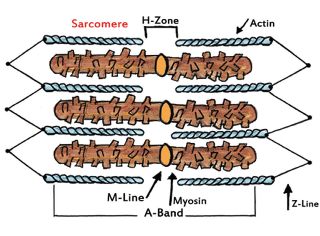
Thick and thin fibers are organized into contractile units named sarcomeres. A sarcomere stretches from one Z line to the next and is the basic unit of muscle contraction.
In the end, the overall life expectancy of the knockout mice was severely reduced compared to the wild-type mice. Wild-type mice had an 80 percent survival rate after two years, whereas mutant mice only had a 10 percent survival rate. These findings highlight the significant effects Obscurin proteins have on heart durability. After removing one seemingly small class of proteins, cardiac activity was dramatically altered, and ultimately had lifethreatening outcomes. Studying these types of proteins helps researchers understand the critical mechanisms of cardiac function that may help prevent certain heart abnormalities, such as the arrhythmias observed in Stephanie’s experiment.
Under the Scope • Vol 5
Another component of the human heart being studied is the signaling pathways involved in remodeling the cells that make up the heart. Tik-Chee (Jenny) Cheng, a fourth-year Warren student majoring in bioengineering, studied the heart’s function in the human body. Jenny noticed that cardiac cells remodel themselves by adding sarcomeres after they have been stretched a certain amount and for enough time. This process was seen as the hearts of mice were extracted and the cardio myositis was isolated. Jenny extracted the hearts of mice and isolated the cardiomyocytes for study. She used the stretching apparatus model, in which cells are plated on top of a deformable substrate and then laminated. This allows the substrate and the cells to stretch simultaneously. After the cells were stretched for different periods of time, they were then stained with a fluorescent dye and analyzed under a microscope to examine the addition of structures such as sarcomeres.
The research then focused on the structure of the sarcomeres to determine what happens to the myosin actin filaments (one of the contractile components in skeletal, cardiac, and smooth muscle fibers), and the sarcomeres after they are stretched a certain percentage longitudinally and transversely. Although this question has not entirely been answered, this research will contribute to the understanding of certain heart diseases.
“This research opportunity was my first chance to ask a question that had never been asked before. I found motivation in my curiosity to stick with my project to find an answer. I remember the night that I analyzed my results and discovered something that was worth writing about!”
-Stephanie Tsoi

For instance, Jenny uses athletes and nonathletes to illustrate specific changes in the human heart. Some athletes have enlarged hearts due to the high pumping of blood, but when non-athletes have enlarged hearts (due to genetic mutation or some other problem), their hearts undergo cardiac hypertrophy, a thickening of the heart muscle (myocardium). The enlargement of the heart is a result of the addition of sarcomeres as the heart is stretched, making it difficult to return to its original state. Examining how long it takes the cells to return to their original state can contribute to the search for a cure to certain heart diseases.
While studies have shown the significance of physical changes on cardiac cells, experiments delving into the biochemical effects on the heart are beginning to rise. Enzymes, or proteins that act as catalysts in biochemical reactions, are one of the many components that play crucial roles in the function of a healthy heart. Stephanie Tsoi, a recent graduate from UC San Diego and researcher in Dr. Rolf Bodmer’s lab, closely studied a particular metabolic enzyme and its role in obesity. A growing health issue and cause of death in the United States, obesity currently
impacts about 33 percent of the American population. Obesity is found to cause heart disease, stroke, and high blood pressure, and it dramatically deteriorates the quality of life of many people around the world. This disorder is a particularly troublesome public health issue in the United States due to the prevalence of high-fat and high-calorie diets along with diminishing physical activity.
Tsoi, a current student at the David Geffen School of Medicine at UCLA, acknowledges her curiosity and determination as responsible for her successes in research, one of which was the ability to publish her work. “This research opportunity was my first chance to ask a question that had never been asked before. I found motivation in my curiosity to stick with my project to find an answer. I remember the night that I analyzed my results and discovered something that was worth writing about!”
In her study, Tsoi explored the relationship between highfat induced obesity and heart disease. She used Drosophila melanogaster (fruit fly) as a model because they share anatomical and genetic similarities with humans. Fly and human hearts have very similar genetic pathways that initiate the differentiation and specification of heart dysfunction when they are under the influence of a highfat diet. Flies were placed in vials of NF (combined yeast,
sq.ucsd.edu
molasses, and corn starch) to mature for five days and then were separated into either new vials of NF or 30 percent coconut -oil concentrated high-fat diet (HFD). Using this model, Tsoi studied the genetics of HFD induced obesity and heart dysfunction that is attributed to this type of obesity. Specifically, HFDs deregulate the TOR (Target of Rapamycin) –- PGC-1 pathway. The TOR pathway is a key regulator in cellular growth and metabolism, and PGC-1 is a coactivator that plays a vital role in regulating energy metabolism. The deregulation of this important metabolic pathway leads to obesity and cardiac lipotoxicity, which is a disease that results in excess fatty acids in non-adipose tissue, or any tissue that does not contain fat cells.
In her experiment, Tsoi researched the role of pyruvate carboxylase, one of many enzymes involved in the glycolytic pathway, which is used to obtain energy from glucose. This enzyme is a downstream regulator of PGC1. In her control group, Tsoi found that the flies that fed on a HFD had increased heart rates and decreased heart periods, or time between heartbeats, compared to flies that fed on regular diets. However, flies without the gene that produces pyruvate carboxylase inhibited the flies’ increased heart rate and decreased heart period. These results suggest that the knockdown of the pyruvate carboxylase gene could potentially protect the heart from dysfunction that is caused by a HFD via downstream of the PGC-1 coactivator pathway. The goal was to find the most downstream target in this pathway. By developing a drug
Under the Scope • Vol 5
that targets this enzyme, there is potential of protecting patients with HFDs from developing cardiac dysfunction.
Tsoi is currently putting what she learned in her undergraduate research studies to good use. “Being able to explore the topics of my project (metabolic pathways, obesity, and cardiac dysfunction) on a more advance level has increased my appreciation for asking the question and seeking an answer. I hope to continue to do research in this field and correlate some of my findings along with other groundbreaking work being done at UCLA.”
While additional research on this topic has yet to be conducted, the power and importance that a single enzyme holds further accentuates the value of each component making up a system.
Rockets, cars, houses, factories —- all of these systems are only able to function with the concurrent viability of the parts that compose them. With the failure of just one component, the entire system can crash down and cease to function. Similarly, the heart is is composed of a plethora of small parts that contribute to its function. The projects that UCSD student researchers are working on have shown the importance of individual proteins and metabolic pathways. Even a seemingly minute detail found in the heart’s natural mechanics can alter its overall activity. Proteins, signaling pathways, and enzymes are
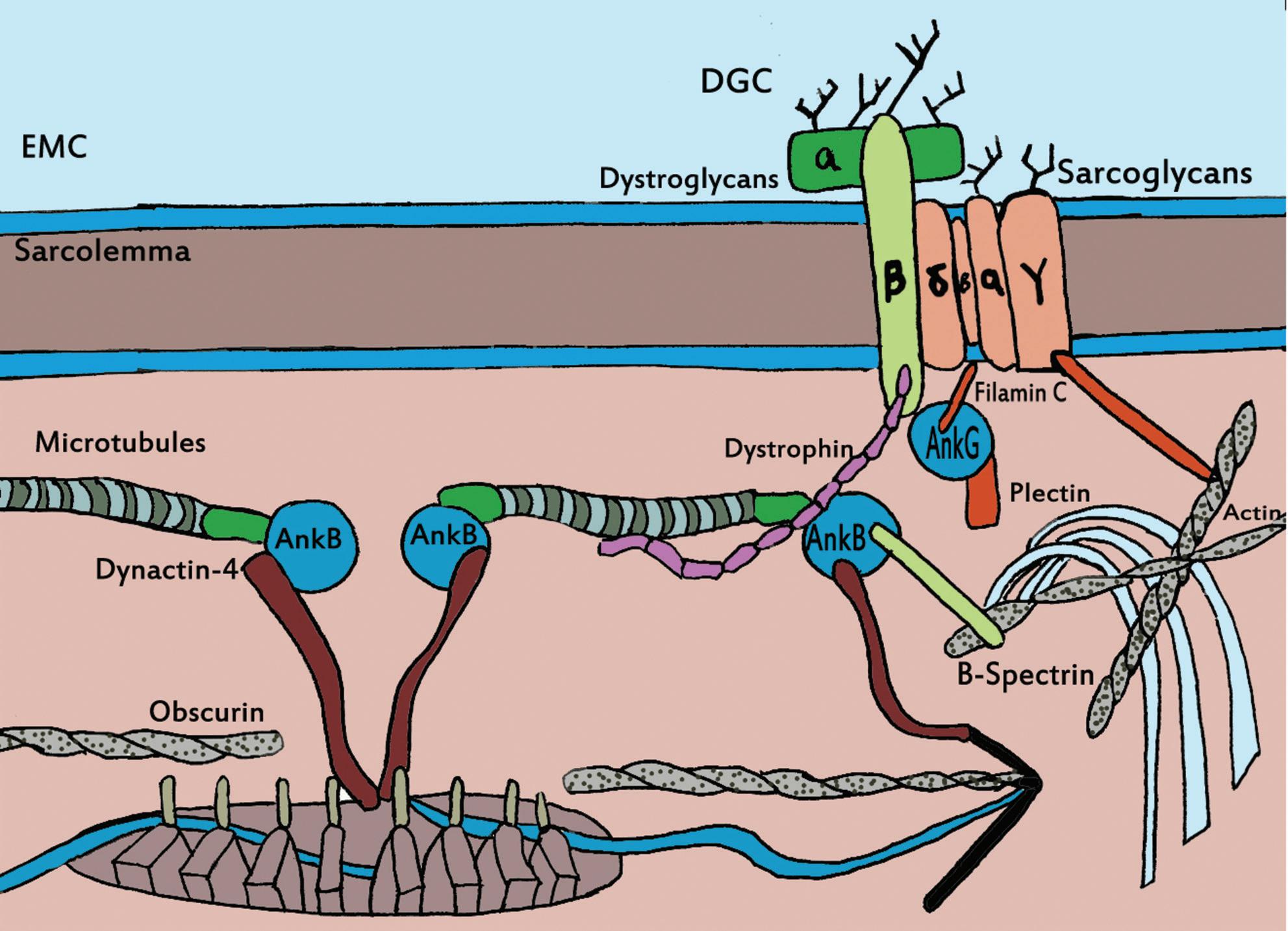
This figure show the interior of the sarcoplasmic reticulum. Obscurin is seen connecting the myofilaments within the sarcoplasmic reticulum to allow for proper muscular contractions.
examples of these components that play vital roles in the function of the heart. Each element studied contributes to the larger understanding of heart vitality, and with every new finding, science grows one step closer to curing more heart diseases and discovering the basis for a strong and functional heart.
WRITTEN BY ANEESA CHOWDHURY, CHANDNI PATEL, AND MAGEN ANTHONY. Aneesa is a Biochemistry and Cell Biology major graduating in 2016. Chandni is a Biochemistry and Cell Biology major graduating in 2016. Magen is a Physiology and Neuroscience major graduating in 2018.
Biochemistry and Biophysics
Trevor Ali Zandi
Cell and Developmental Biology
Wilson Huang
Genetics and Molecular Biology
Myanna Olsen
Immunology, Virology and Cancer Biology
Cody Mowery
Neurobiology
Phoenix Ying
Master’s Research
Patrick Chang
Functional Identification of Mycobacteriophage Weiss13
Erica Birkholz; Samantha Nadeau, Mili Parikh, Natalie
Popescu, Lauren Waller
Dr. Madeline Butler
Meiotic Chromosomal Crossover in Mammals is Regulated by HORMA-Domain Containing Proteins and Specific Binding Partners
Vitaliy Davydov
Dr. Kevin D. Corbett
Validating Gene Expression through Proteomics of Weiss13
Nelish Ardeshna; Joshua Kendall, Christine Elise Peters, Rani Shiao, Dhruva Udani, Bruce Zhang
Dr. Madeline Butler
Computational Design of Crystallizable, Chimeric G-Protein Coupled Receptors
Trevor Ali Zandi
Dr. Ruben Abagyan and Dr. Irina Kufareva
Investigating the Molecular Regulation of CD100-Induced Dendritic Epidermal γδ T Cell Rounding
William Kuo
Dr. Wendy Havran
Characterizing the interaction of STAT5A and HP1α in the noncanonical JAK/STAT pathway
Jesus Villanueva
Dr. Willis X. Li
Evidence for consumption of krill pre-breeding, in Antarctic fur seals (Arctocephalus gazella)
Marcus Bobar
Dr. Carolyn Kurle
Human-created rural development reduces genus richness and changes community composition of freshwater macroinvertebrates in Costa Rican cloud forest waterfalls
Annika Nabors
Dr. David Holway
Group A Streptococcus M2 Protein Interaction with C4BP and Interference by Hyaluronic Acid Capsule
Garrett Brooks
Dr. Victor Nizet
Role of TRAIL in NK Cell-Mediated Control of Murine Cytomegalovirus in C57BL/6 Mice
Bryan Mcdonald
Dr. Mick Croft
AML1-ETO Expression in Hematopoietic Stem/Progenitor Cells (HSPCs) Confers a Hypersensitivity to GM-CSF that Reduces Leukemogenic Potential
Cody Mowery
Dr. Dong-Er Zhang
Reverse phase protein microarray identifies IL6 receptor ligation induced PI3K/AKT, insulin receptor, and JAK/STAT signaling pathways in the CARD11 mutated lymphoma cell line OCI-Ly3
Shruti Shrivastav
Dr. Eva-Maria S. Collins
Role of Intelukin-37 in an adaptive-immune mediated model of Inflammatory Bowel Disease
Kyle Mauricio Yuquimpo
Dr. Jesus Rivera-Nieves
Measures for quantifying the stability of spatial firing patterns in rodent hippocampal place cells
Christopher Cannova
Dr. Stefan Leutgeb
Does HDGF-related protein 3 Regulate Oligodendrocyte
Myelination in the Central Nervous System?
Shereen Georges
Dr. Fred H. Gage
Reorganization of the Dentate Gyrus Neural Network in Medial Temporal Lobe Epilepsy and its effect on Learning and Memory
Tatyana Goldring
Dr. Jill Leutgeb
The Consequences of Voluntary Intermittent Ethanol Exposure During Adolescence on Adult Drinking
Diana Kneiber
Dr. Cindy Ehlers
The Anatomical Changes Associated with Impaired Behavioral Pattern Separation in Chronic Model Temporal Lobe Epilepsy
Raafat Kuk
Dr. Jill Leutgeb
The Role of HTR7 Gene Regulation in Influencing SSRI Response in Bipolar Disorder
Donel Purcella
Dr. John R. Kelsoe Jr.
Characterization of RNA binding protein with multiple splicing (RBPMS) as a marker for retinal ganglion cells
Lior Schenk
Dr. Jeffrey L. Goldberg
The effect of dentate gyrus-specific APP expression on neurogenesis
Kaivon Sobhani
Dr. Edward Koo
Neural Mechanism of Imprinting in Drosophila Melanogaster via Dopamine-ecdyson Receptors and 20-Hydroxy-Ecdyson
Ethan Sy
Dr. Jing Wang
Role of Atypical Protein Kinases in Axon Regeneration
Natalie Warsinger-Pepe
Dr. Daniel Gibbs
MEF2 in Neurons
Huwate Yeerna
Dr. Trey Ideker
Dissecting the nuclear import mechanisms of a bZip protein CEBP-1 and the function of importin proteins in the nervous system
Phoenix Ying
Dr. Yishi Jin and Dr. Hollis Cline
Neural Mechanisms Underlying the Regulation of Food Search Behavior by Social Cues
Christopher Pham
Dr. Jing Wang
Sarcomere Length and Lattice Spacing of Myosin-Actin Filaments in Cardiac Models
Tik-Chee Cheng
Dr. Jeff Omens
Localization and trafficking of ATP-binding cassette transporters in sea urchin embryos
Rose Hill
Dr. Amro Hamdoun
Identification of Rab GTPase roles in muscle cell Transverse
Tubule formation and remodeling
Wilson Huang
Dr. Amy Kiger
Neonatal Enteropathogenic Escherichia coli Infection Causes Long Term Behavioral Defects And Long Term Alterations In The Microbiota
Kevin Huynh
Dr. Melanie Gareau and Dr. Kim Barrett
Androgen Receptor as a Novel Co-regulator of Autophagy
Amy Le
Dr. Constanza Cortes
Pdgfr signaling is required for specification of hematopoietic stem cells
Chase Melick
Dr. David Traver
Role of Obscurin for Cardiac Calcium Handling
Stephanie Myers
Dr. Matthew Kloss, Dr. Stephan Lange, and Dr. Indroneal Banerjee
Homolog Avoidance (Tiling) Requires the Calcium-Activated Protease Calpain for Process Retraction
Luis Solano
Dr. Eduardo Macagno
Circadian regulation of metabolism under diurnal cycles in cyanobacteria
Chase Barber
Dr. Susan Golden
Identification of Gene Effect on Seed Size Phenotype by Screening tDNA Knockouts
Joaquin Reyna
Dr. Joseph Ecker
Phage Genomics Initiative at UCSD
Allen Chu; Charles Farmer, Gabriel Fischer, Miranda
Morgan, Chaelin Song, Andy Wang
Dr. Mandy Butler
Isolation and Characterization of a Bacteriophage 201phi21 Protein
Joanna Coker
Dr. Joseph Pogliano
Cytochrome P450 2B4: A HDX Approach to Plasticity Analysis
Liane De Guzman; David Lee, Danny Tran
Dr. Sheng Li and Dr. James Halpert
An Automated Approach Toward Identifying Transpiration Phenotypes of Mutant Arabidopsis thaliana Lines
Tony Zhang
Dr. Julian I. Schroeder
The Effects of Caveolin on Aging in C. elegans and Establishment of a Liquid Culture Medium Model
Grace Grogman
Dr. Hemal Patel
Effect of diet and adiponectin on fat expansion during gestation
Zhuyu (Julia) Guo
Dr. Jianhua Shao
Role and Regulation of FOXA2 in Early Pancreas Induction
Dieter Lam
Dr. Nigel Crawford and Dr. Maike Sande
Characterization of the interaction between the translational repressor complex GIGYF2-4EHP and the ARE-binding protein TTP
Myanna Olsen
Dr. Jens Lykke-Andersen
Drugs and You: A DXMS study of protein-drug interaction
Kang Choi and Kurt Sakurada
Dr. Sheng Li and Dr. James Halpert
Interaction between Enteric Bacteria and Host Engulfment
Machinery that Regulate Inflammation
Rama Fluensita Pranadinata
Dr. Soumita Das
Application of proteomic mass spectrometry to the characterization of Mycobacterium smegmatis bacteriophage
Chiara Ricci-Tam
Dr. Joseph Pogliano
Towards the Three Dimensional Structure and Function of Hop2/Mnd1
Udayan Saha
Dr. Kevin Corbett
Utilizing Tn-seq to create a mutant library of Syneochococus elongatus to elucidate circadian and diurnal response genes
Anish Pal
Dr. Susan Golden
Comparative Genomics of Mycobacteriophage Weiss13
Identifies Mosaic Structures
Lance Hsieh; Kyle Jackson, Day Kagy, Jasmine Ming Jing
Ting, Cynthia Wu, Alan Yang
Dr. Mandy Butler
Analysis of Pyruvate Carboxylase function in Diet Induced Obesity and Cardiac Lipotoxicity
Stephanie Tsoi
Dr. Rolf Bodmer
The LysE Superfamily of Transport Proteins Involved in Cell Physiology and Pathogenesis
Brian Vay Tsu
Dr. Milton Saier
How do seasonal changes in food web dynamics and climate affect carbon dioxide flux from San Diego reservoirs?
Emily Adamczyk
Dr. Jonathan Shurin
5alpha-Androstane-3alpha,17beta-diol (3alpha-diol) regulates amino PTHrP in human non-small cell lung carcinomas
Thomas Aubuchon
Dr. Randolph Hastings
The modulation of IL-27 signaling during antiviral immunity
Phuc Bao
Dr. Elina Zuniga
The Role of Extracellular Matrix Protein CTGF in Disease
Progression of Blast Crisis Chronic Myelogenous Leukemia
Patrick Chang
Dr. Catriona Jamieson
Functional Studies of a Novel Mutation in NTRK1
Brian Chin
Dr. John Kelsoe
Precipitation of Behavioral Defects Due to Inflammation in the DSS Mouse Model of Colitis
Jacob Emge
Dr. Kim E. Barrett
Invariants of center-surround interactions
Sunwoo Kwon
Dr. Thomas Albright
Depletion of Ovarian Cancer Initiating Cells by a Monoclonal Antibody Against ROR1
Hsien (Sam) Lai
Dr. Thomas J. Kipps
Screen for heterochromatin promoting drugs in drosophila melanogaster
Andre Loyola
Dr. Willis Li
Type 2 innate lymphoid cells are activated by cysteinyl leukotrienes: A novel mechanism of lipid mediated allergic inflammation
Sean Lund
Dr. Taylor Doherty
Sub-cellular localization of components for a novel, membrane-delimited signaling pathway mediating transactivation of the epidermal growth factor receptor
Rebecca Lynch
Dr. Paul Insel
Dephosphorylation of tau in lactating rats subjected to acute restraint stress
Danielle Steinmetz
Dr. Robert Rissman
Characterization of ubiquitin like modifier ISG15 in t(8;21) leukemia development
Sheila Tran
Dr. Dong-Er Zhang
DNA Barcoding: Cryptic Species in San Diego’s Intertidal Polychaete Community
Tyler Wooldridge
Dr. Joshua Kohn
Interferon-β Selectively Inhibits Interleukin-2 (IL2) Production via cAMP Responsive Element Modulator (CREM)-mediated Chromatin Remodeling
Menghong Xiao
Dr. Michael David