
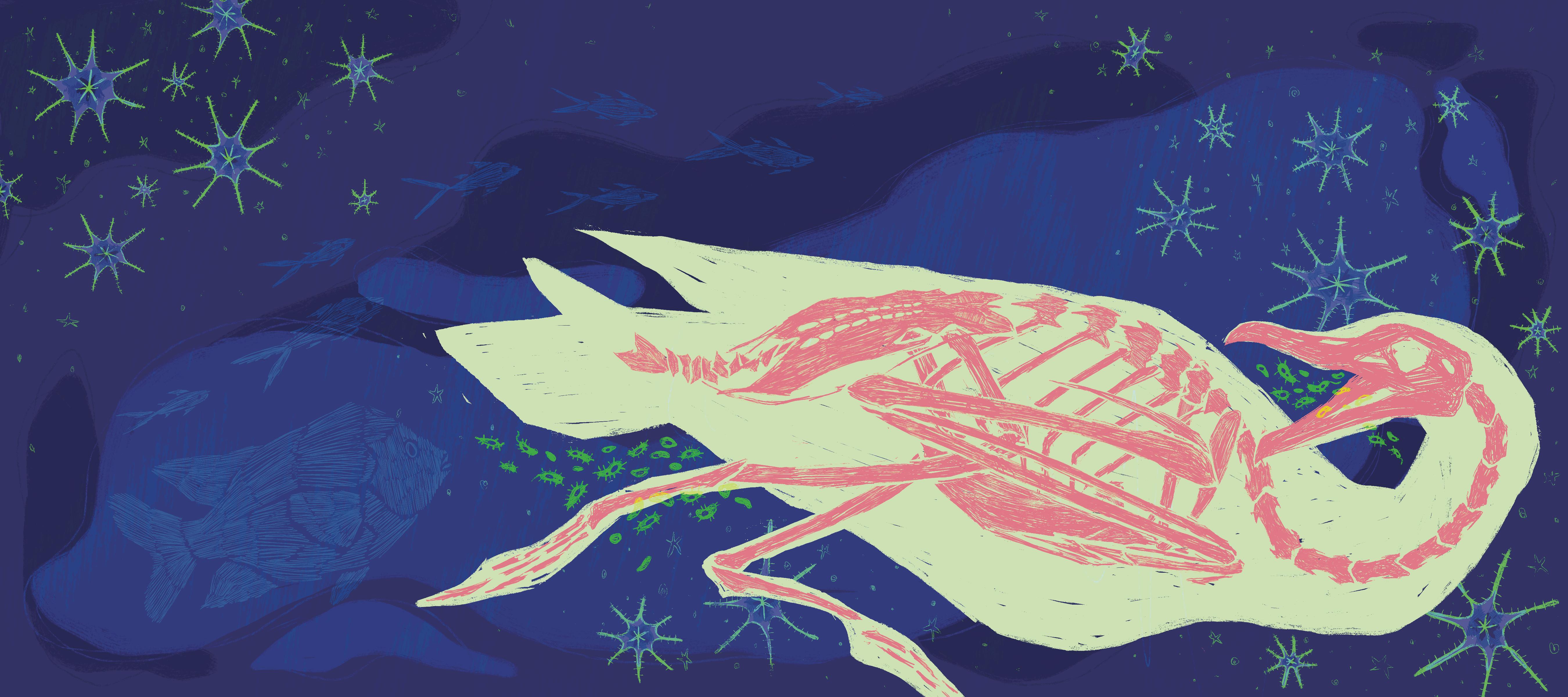

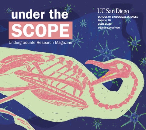
Under the Scope, like much of our work at Saltman Quarterly, is a product of passion. Each year, for the last 16 years, our student writers, editors, illustrators, photographers, and designers have been driven by this passion, coming together to form what is this publication. Writer pairs first discover pockets of exciting biological research at UC San Diego, which they then disseminate into digestible writing Then, the illustrators, photographers, and designers provide vibrant accompanying visuals for a tangible physicality to the evercomplex field of biology
In this year’s publication, through our features articles, we attempt to elucidate various ways in which our natural world, and ecosystem at large, intersects with our own. We begin at the junction of neuroscience and machine learning models, developing tools of computer vision to improve diagnostic accuracy, and continue through other unconventional sources of medical advancements, such as the untapped wealth of therapeutic compounds within our oceans From here, we venture into the delicate balance of our own biology, exploring potential reproductive dangers of dysregulated nervous and hormonal systems and the overlapping genetic and environmental factors that contribute to asthma And finally, we return to the intersection of computation and biology, investigating computation models used in accelerating tuberculosis drug development.

Beyond these articles, we end this publication with interviews of two faculty members and a pertinent discussion of the many downstream effects of recent, unprecedented cuts to federal research funding Their testimonies echo the same sentiment: not only does funding uncertainty hinder their own work, it prevents them from providing the necessary mentorship to bring students, just starting out in the field, into research.

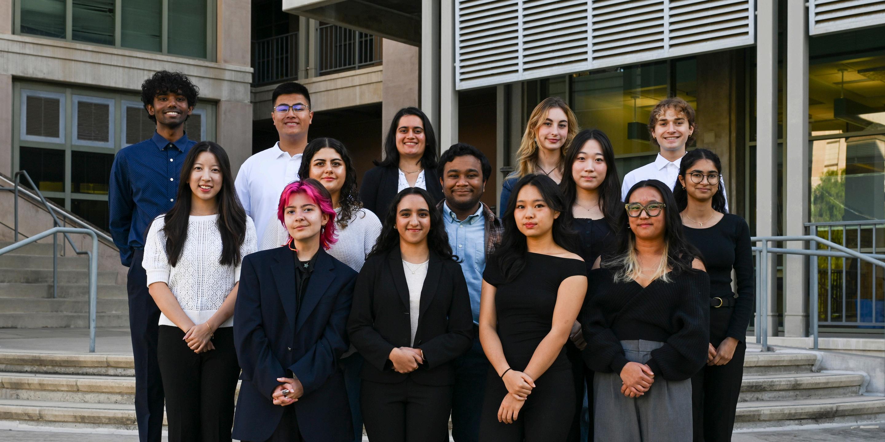
Editorial Board
Editors-in-Chief
GabbiBasa
EshaBhattacharya
ExecutiveEditor
MarkDavis
Editor-at-Large
ErinJang
FeaturesEditor
AdityaVerma
ProductionEditors
HimangiSharma
SanjiKumar
HeadTechnicalEditor
ShreyaAbhijit
TechnicalEditors
SiddharthBailkeri
MihikaDevireddy
LaurynGuan
NehaRoyKarintholil
EmilyLindemann
CarmenMolina
SunnySo
LaurenWu
FeaturesDesign
Editors
PatsyHsiao
TiffanyLiang
HeadIllustrator
JennaHughes
HeadPhotographer
InayaNicholls
CoreStaff
JiaBhavnani
BienAntonioDelaCruz
JinoEbrahimi
LanGao
RyanSinha
TavishaaVanjani
AaryaVishnu
EmmaWong
VaidehiZala
Head Advisors
Ashley Juavinett, Ph.D.
Associate Teaching
Professor of Neurobiology
LaTisha Hammond, Ph D
Milton Saier, Ph.D.
Julian Schroeder, Ph.D.
Elina Zuniga, Ph.D.
Writers
Nour Ali
Ratul Chakraborty
Terry Deng
Alexandra Gomez
Qinghan Jia
Shreya Joshi
Mridu Karanam
Miso Koo
Hannah Petyt
Sophia Perryman
Vivian Zhang
Photographers
Natalie Labatia
Dominic Tse
Illustrators
Erin Chickering
Julia Dingman
Shania Tayag
Amber Urena
Rebecca Zhang
Cover & Table of Contents
Illustrator
Leo Harris




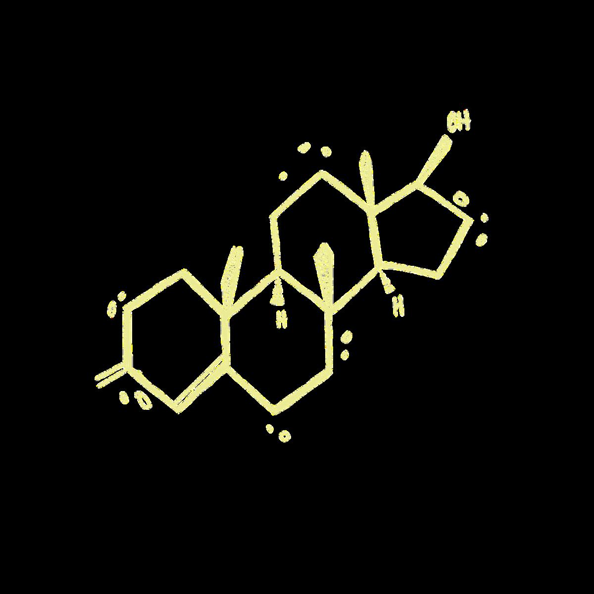



Illustrated by Shania Tayag

Written by Nour Ali and Alexandra Gomez
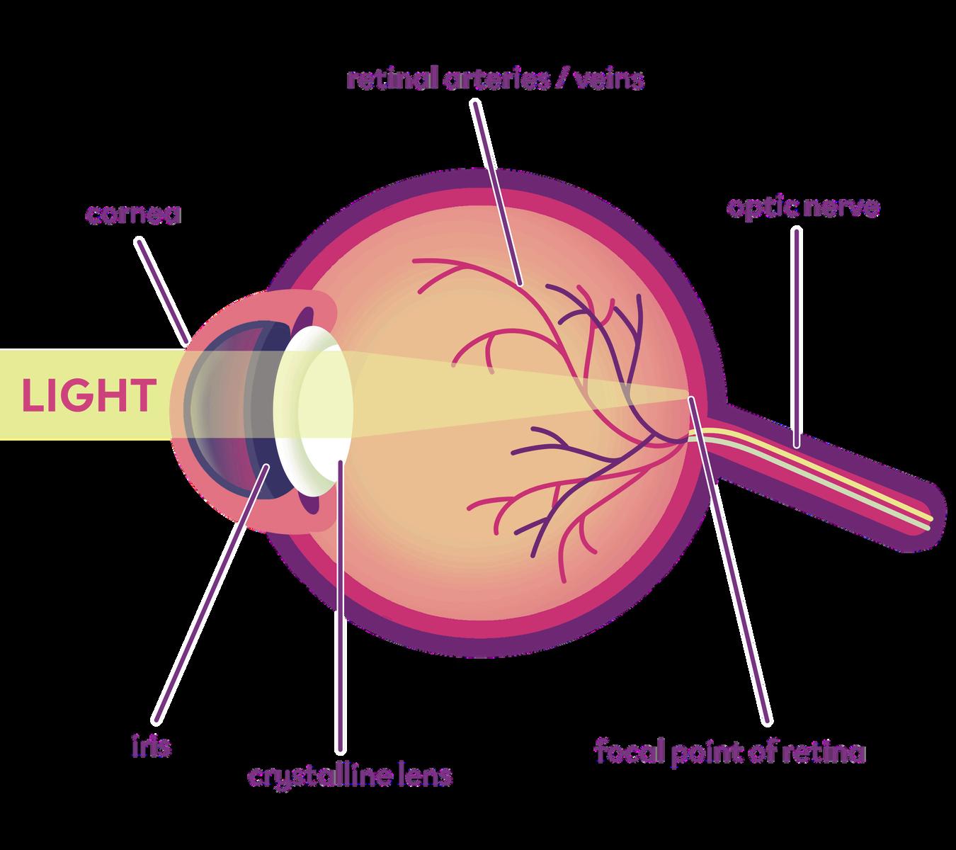
Have you ever found yourself entranced, wide-eyed, when considering the nature of sight? As your eyes dart around this page, light bounces off the paper and triggers a chain reaction inside your brain. Reflected light is composed of tiny packets of electromagnetic energy particles known as photons. These photons journey to the back of your eye, to an area known as the retina, which contains specialized lightdetecting cells known as photoreceptors. When photons interact with proteins in these cells, they initiate a chemical change that alters the cell’s electrical charge. As a result, electrical signals transmit through a bridge-like structure called the optic nerve, which then connects to your visual cortex, a region of the brain responsible for processing visual information. Different types of brain cells, or neurons, can then process this information in varying ways. This complex cascade rapidly unfolding within your visual system is what constructs your visual reality from mere particles of light.
When you read this page, you first recognize the individual letters, then words, and finally whole, meaningful sentences. This constructive process of building meaning from basic elements parallels an inherently biological system of hierarchical processing. In the visual cortex, hierarchical processing gradually builds our visual perception from basic features, such as lines and shadows, to recognizable shapes, and then finally to a coherent object.
If our brains require this complex network to transform light into images, how can a computer, which lacks eyes and neurons, “see” and identify objects in a scene? The answer lies in the parallels of biological and technological systems, both of which rely on certain computational mechanisms. Recent progress in visual neuroscience and artificial intelligence (AI) has enabled researchers to integrate biological principles into a previously uncharted territory: computer vision.

Computer vision is a field of AI that teaches computers to interpret and understand visual input. This work powers technology based on pattern recognition, such as self-driving cars, facial recognition, and medical imaging systems. Computer vision relies on artificial neural networks, which are algorithms modeled after the brain’s architecture of interconnected neurons. A convolutional neural network (CNN) is a computer program specifically designed to process visual information, acting as an image filter that detects simple features like edges, textures, and shapes. These features are incrementally overlaid and combined to form more complex images. This stepwise, hierarchical processing mirrors that of the human visual system, as neurons in the primary visual cortex essentially act in the same way as edge and orientation detectors.
Remarkably, computer systems have also been taught to understand the “semantic,” or meaningful, characteristics of an image that we assign to it. For example, an image of a red apple and a bonfire both roughly translate into a blob of red pixels, but computer vision systems will not treat the images as similar because pixel similarity does not capture meaning or structure. Instead of extracting only pixels, computer vision systems also extract features like the roundness of an apple or the sharp edges of bonfire flames. Thus, an image of a dog compared to one of a cat will be treated as similar by the computer system, as both images show four-legged mammals with similar body shapes and facial structures.
If computer systems and biological vision both rely on hierarchical processing to extract meaning from visual stimuli, can these shared principles be observed directly in the human brain? To address this, researchers often turn to electroencephalography (EEG), a non-invasive test that measures the brain’s ongoing electrical activity. Neurons in the brain communicate via electrical signals known as action potentials. Action potentials are allor-nothing spikes that travel along nerve cells to send messages to other neurons. When placed on the scalp, electrodes, which are electrical sensors, pick up the signals of thousands of neurons firing as an ensemble. The visual? A graphic representation known as a waveform, which depicts the brain’s average electrical activity across a period of time. When humans view semantically similar images, such as a dog and a cat, the resulting EEG waveforms are more similar than for meaningfully unrelated images like an apple and a bonfire. This mirrors how artificial computer vision systems group images by shared semantic features rather than pixel similarity.
The non-invasive nature of this technology comes with an important tradeoff: applying electrodes on the scalp results in noisy brainwave data. Utilizing EEG instead of a

local field potential implant — which records electrical activity from single neurons directly in the brain is comparable to listening to a concert from your apartment balcony: the sound is muffled and blended with background noise. However, the large-scale neural patterns that EEG picks up are more relevant in understanding how neurons coordinate with one another in a system compared to the isolated activity pattern of a single neuron.
In both biological and artificial systems, meaning emerges from the pattern of activity across vast and interconnected networks. Visual processing does not rely on individual neurons to encode basic features, patterns, and meaning, but rather, on the collaboration of neurons across different circuits. Understanding these distributed patterns is directly relevant to designing computer vision systems: just as the brain combines signals across many neurons, neural networks combine signals across many simple processing units, each analyzing a small part of a stimulus.
To observe the activity of neuronal ensembles in the visual cortex, researchers examine steady-state visually evoked potentials (SSVEPs), a type of brainwave signal detected by EEG when the retina is excited by a visual stimulus. SSVEP signals primarily come from the occipital lobe, which hosts the primary visual cortex responsible for constructing our visual world.

At the de Sa Lab at UC San Diego, the Brain Computer Interface (BCI) Division focuses on interpreting EEG signals to develop and improve BCI systems technology that provides a direct pathway between the brain’s electrical activity and an external device. BCIs have the potential to restore communication and mobility for individuals with paralysis or neurological conditions without the need for traditional motor input such as speaking, writing, or moving. Common BCIs include robotic limbs, which translate electric muscle signals into movements via sensors.
Within the de Sa Lab, graduate student Simon Fei works on linking SSVEP signals from the brain to computer commands. When a user focuses on a visual stimulus, such as a letter, that pulses at a specific frequency, the neurons in the visual cortex fire in sync with that frequency to produce an SSVEP. By detecting which frequency dominates the network of visual cortex neurons, the BCI can decode the user’s intent and generate the corresponding output on the screen. In other words, without any physical movement, a person can effectively “type” by simply looking at a series of letters flickering on a screen. Such technologies are especially useful for people with severe motor impairments, granting the ability to directly translate brain activity into communication.

In these early stages of widespread access to AI models featuring computer vision, the potential of the resource seems limitless. With each passing day, the extensive availability of the technology allows societies to upgrade current problemsolving methods into more efficient modalities. In the field of healthcare, the use of computer vision to conduct pattern recognition across different stages of medical diagnosis is becoming increasingly popular. Medical images like tissue slides, X-rays, and other scans can be analyzed using computer vision systems, assisting pathologists in identifying conditions faster and with greater accuracy. Reflecting upon the fascinating advancement of AI and its gradual integration into his work, Fei questions if and how there would be limits to such a powerful tool. If AI were to plateau, would it be due to the lack of technology able to measure existing boundaries? Or would it boil down to how humanity is continuing to grasp comprehension of AI?
Despite the extensive advancements, it’s still hard to say with current research just how much the combination of visual systems and AI models can launch humanity into new groundbreaking realms; hence, the beauty and fascination of it all. While there may not be answers to these questions as of today, the progress of artificial computer vision is one to keep an eye out for. As developing research elucidates the mysteries of the brain, the act of unveiling the mystery of the potential — or limits of AI will become clearer, like putting on a pair of glasses.
Nour Ali is a Neurobiology major graduating in 2028. Alexandra Gomez is a Human Biology major graduating in 2028.

Written
by
Miso Koo and Sophia Perryman
Illustrated by Julia Dingman
Photos by Inaya Nicholls

For much of human history, the search for natural remedies has been grounded, quite literally, on land. Foraging through terrestrial sources for medicine has long been the convention, as evidenced by modern drugs from the antibiotic penicillin originating from Penicillium mold to other plant-based cures, we constantly turn to the earth to source our medicine. By contrast, the ocean has remained a comparatively unexplored medicinal frontier, despite covering almost 70% of the Earth’s surface. So why is it that the abundance of chemical diversity produced by marine life has yet to be harnessed for medicine?
Many marine organisms synthesize chemical compounds for defense mechanisms. This helps them deter predators, prevent microbial colonization, and mediate ecological interactions with surrounding species. In recent years, researchers have recognized that such compounds contain anti-cancer and anti-inflammatory properties that can be utilized for therapeutic applications in humans. However, translational research and drug development has been slow due to challenges with harvesting marine organisms, issues with scalability, and difficulty in tracing these compounds back to the precise mechanisms responsible for their synthesis. Major ecological and sustainability hurdles have prevented further research on the marine organisms that produce these compounds. The Moore Lab at UC San Diego takes advantage of the oceanside campus to bridge this gap between marine biology research and medicine. They recently discovered a marine species known as octocorals, which are ancient multicellular organisms with soft bodies and eight tentacles. These creatures possess a unique chemical defense mechanism: biosynthetic gene clusters (BGCs). A BGC acts as a specific set of DNA instructions that provide octocorals a manual to build specialized secondary metabolites, which are bioactive compounds that serve as chemical weapons. BGCs are well documented in bacteria, fungi, and plants but were long thought to be rare or absent in animals, rendering this discovery a surprise that challenged preconceived notions about animal metabolism.
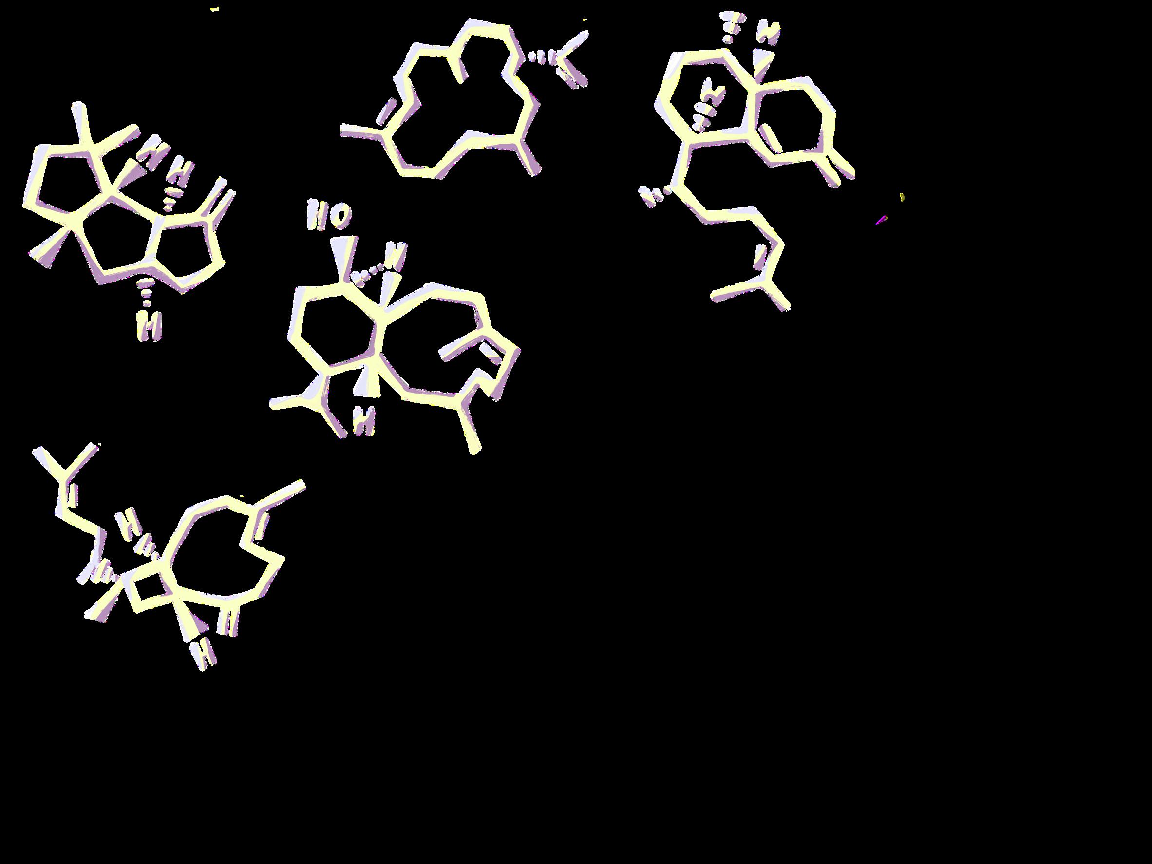
By sequencing the genomes of multiple distantly related octocoral species, the Moore Lab discovered that these clusters of five genes coded specifically for briarane diterpenoids. Briarane diterpenoids, previously only associated with coral reef chemistry, are a class of structurally intricate hydrocarbon compounds known for their diverse pharmaceutical roles and applications They possess anti-cancer and antiinflammatory properties through their effective inhibition of certain cytokines, which are cell-signaling proteins that initiate immune responses. Distinct from terrestrial diterpenoids, marine diterpenoids frequently exhibit unique chemical functions. One such function is halogenation, or the addition of halogen element atoms to a molecule, due to the ocean’s halogen-rich environment. Such modification confers different therapeutic capabilities to these molecules ranging from increased metabolic activity to binding affinity making marine-derived compounds potentially more potent and feasible for medicinal use than their terrestrial counterparts.The primary target of a briarane diterpenoid, however, is inflammation.

Inflammation is a complicated, double-edged sword of the immune system that scientists are constantly trying to balance. Inflammation can be thought of as a twoway train one route signals to immune cells to protect the body, resulting in swelling, and the other route inhibits those same immune cells to lessen the swelling effect, referred to as anti-inflammation. At UC San Diego, the Bertin Lab seeks to better understand inflammation’s dual nature and its underlying pathways, which may then reveal how these diterpenoids can positively modulate immune activity. Inflammasomes, which are immune receptors that sense harmful invaders in the body and trigger an inflammatory response, are known to work acutely, or short-term, to sense pathogens and prevent infection.
Dr. Bertin explains that inflammasomes play many important roles, yet when their effects become exaggerated or chronic, they may increase risks of cancer and even neurodegenerative diseases such as Alzheimer’s disease.
Inflammasomes also release cytokines to directly signal immune responses that facilitate inflammation. Understanding these signaling pathways is crucial for devising ways to prevent the negative effects of overactive inflammasomes. Interleukin-1, a cytokine also found in anti-inflammatory pathways in octocorals, is a target of study in the Bertin Lab for its chronic effects on lung cancer in response to common allergens. The lab is also investigating neutrophils, the immune cells that house inflammasomes, as another target for regulating cytokinemediated inflammatory responses. Inflammation pathways, whether they are provoked by allergens affecting the lungs studied at the Bertin Lab, or functioning as defense mechanisms in deep-sea soft corals, are currently being widely researched for their ability to work in equal and opposite ways.
In fact, it is the particular briarane diterpenoids biosynthesized by octocoral samples in the Moore Lab that are highly effective in inhibiting human neutrophils. The potential for these briarane diterpenoids to revolutionize antiinflammatory treatments in a medicinal pipeline is immense; however, translating marine chemistry into viable, accessible medicine creates a significant bottleneck. Historically, natural product research has been based on a process of tedious, labor-intensive collection of marine organisms and compound extraction practices neither practical nor environmentally sustainable. Beyond the environmental constraints, dealing with a vast mosaic of independent genes that code for the chemical entities utilized in therapeutics is much like “detective work,” as Dr. Bradley Moore emphasized. It is incredibly difficult to find the target collection of genes in a way that fully elucidates its therapeutic function, especially in an organism like octocorals that are not yet well understood

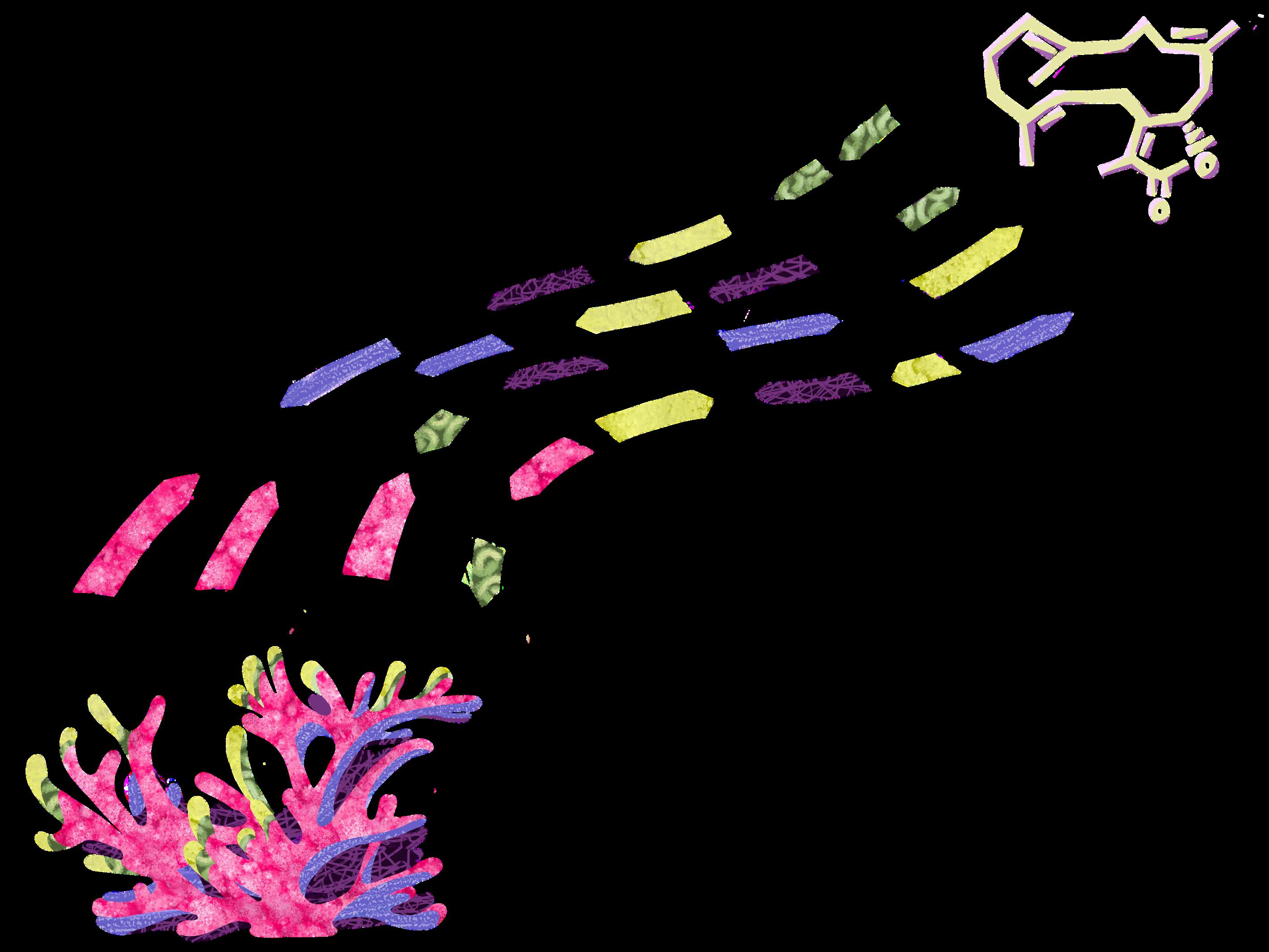
However, it’s essential to understand the original purpose of a compound in its host pathways to then appropriately evaluate their pharmaceutical applications. To then provide a more reasonable approach, sequencers, or automated machines with the ability to extract DNA and assemble genomes, have made this process much more efficient and achievable. With successful sequencing, elusive compounds such as diterpenoids can be traced back to their host organism’s biosynthetic genes. With a genomic blueprint of the octocorals to act as a “recipe,” scientists can produce exact copies of natural products at a larger and more sustainable scale. This is done through expression of the genomic blueprints in heterologous hosts such as E. coli and yeast, both of which are mediums that can be easily cultured and manipulated. This scaled production enabled the Moore lab and other research teams to further examine the compounds’ bioactivities with an ecological consideration.
As with all naturally-occurring forms of medicine, it is no surprise that these soft corals are highly sought after. However, as climate change has threatened our planet and oceans over the past few decades, a big question still remains what is the future of octocorals in medicine? In some regions where stony, reef-building corals have bleached and perished from rising temperatures and ocean acidification, octocorals have been found resisting or recovering from the same threats that destroyed others. By using DNA sequencing technology to isolate DNA fragments from soft coral samples, scientists have identified the presence of single-celled organisms called zooxanthellae that use sunlight to produce oxygen and nutrients. Zooxanthellae share a mutually beneficial relationship with corals. They use the coral’s materials to photosynthesize and, in return, provide the coral with sustenance. The absence of these single-celled organisms from hard corals, especially in poor environmental conditions, is what causes the bleaching process. This affliction strips them of nutrients and produces a white, “bleached” color that reveals their skeleton underneath. Octocorals host a diverse range of zooxanthellae cells different from those present in stony corals. As a result, this variety of mutualistic cells specific to soft corals may explain their ability to withstand and regenerate more in harsh environments compared to stony corals.
Octocorals are not completely resistant to human threat, however. Global warming damages soft and stony corals alike at unprecedented rates. Additionally, these scarce organisms have already been harvested for decades for both medicinal and commercial purposes, such as jewelry and the aquarium trade. Recognizing the endangered quantity of soft corals in nature, the Moore Lab has done extensive research in finding sustainable sampling methods for these organisms. Along with their discovery of diterpenoid production in octocorals, the Moore Lab has used tools like genome mining to search for and synthetically reproduce the genes that express valuable anti-inflammatory traits. Dr. Bradley Moore explained how just taking a “pinky-size” worth of soft coral samples from the ocean allows Scripps Oceanography and other research teams to conveniently access their DNA straight through their computers.

By treading more lightly on our planet, a future for octocorals in medicine appears not only more attainable to scientists, but also more optimistic for a sustainable and healthy environment. In the present day, we have advanced our technology, broadened our perspectives, and leaned into our curiosity in such a way that has allowed us to make groundbreaking medical discoveries within our fascinating, much uncharted oceans. With proper funding and devoted scientists, our university is on the forefront of solving problems and creating once unimaginable possibilities — right from our own watery backyard. Now, the next time you decide to catch a wave at Scripps Pier, consider the possibility that the future of medicine could be swaying right beneath your board.
Miso Koo is a General Biology major graduating in 2028. Sophia Perryman is a Human Biology major graduating in 2027.





Throughout popular culture, our brains are compared to the likes of computers, spider webs, cities, and even anthropomorphized emotions like Pixar’s film “Inside Out.” But these overused metaphors capture only a mere glimpse of how dynamic this organ really is, especially when it’s managing hormonal signals to regulate our menstrual cycles and fertility. At the heart of these coordinated processes is the HPG axis, which stands for our hypothalamus, pituitary glands, and gonads. Regulating the timing of the coordinated signal cascade connecting these organs is essential to how our body’s cells respond to their hormonal environment.
The onset of puberty, adulthood menstrual cycles, and ovulation is the culmination of several hormonal changes driven by the brain. The hypothalamus, an almondshaped gland at the base of the brain, acts as the primary control center overseeing hormonal signals. These hormones primarily serve as regulatory “messages,” traveling through the bloodstream to specific target organs and tissues, such as the ovaries. Once reaching their destination, they bind specific receptors to deliver “instructions” on how and when these organs and tissues need to function. However, hormones can fail to work, or even disrupt normal body functions, if they are released irregularly, at incorrect times, or in incorrect patterns. To stimulate their target tissues in a biologically timely manner, our glands release hormones in pulses rather than at a constant rate.
This messaging system allows the hypothalamus to work in conjunction with the pituitary gland, the secondary control center. The hypothalamus acts as the first responder to nerve signals, instructing the pituitary gland to trigger or inhibit the release of certain hormones. For example, the hypothalamus secretes gonadotropin-releasing hormones (GnRH) in response to low levels of sex steroids. When the pituitary gland receives the GnRH signal, it releases luteinizing hormones (LH) and follicle-stimulating hormones (FSH), which travel through the bloodstream and eventually bind to reproductive organs like ovaries and testicles. This binding triggers the further production of estradiol, a sex hormone that controls ovarian function and commences the menstrual cycle by stimulating the growth of eggs and the uterine lining. Through this continuous exchange of signals, the HPG axis functions as a self-regulating circuit that translates neural activity into balanced hormonal rhythms for reproduction.
However, this essential interaction between the hypothalamus and the pituitary gland would not be possible without the well-controlled regulation of the specific hypothalamic neurons that produce the GnRH hormone. Without proper neuron signaling, the HPG axis cannot be activated by the release of GnRH, preventing many key reproductive functions. However, hormones within the HPG axis do more than transmit stimulatory signals they can also inhibit reproductive function.

Interestingly, GnRH-releasing neurons do not expre androgen receptors and cannot be directly affect by testosterone. Without receptors, androgens a unable to modulate genes that regulate reproduct signaling In theory, androgens like testosterone c directly silence any neuron in the HPG axis with t help of androgen receptors. If there are no androg receptors on GnRH neurons for testosterone interact with, testosterone instead must act up earlier, “upstream” neurons in the GnRH signal pathway. Kisspeptin-releasing neurons, with t specific androgen receptors testosterone c interact with, are the specialized cells in t hypothalamus able to control “downstream” Gn neuron activity.
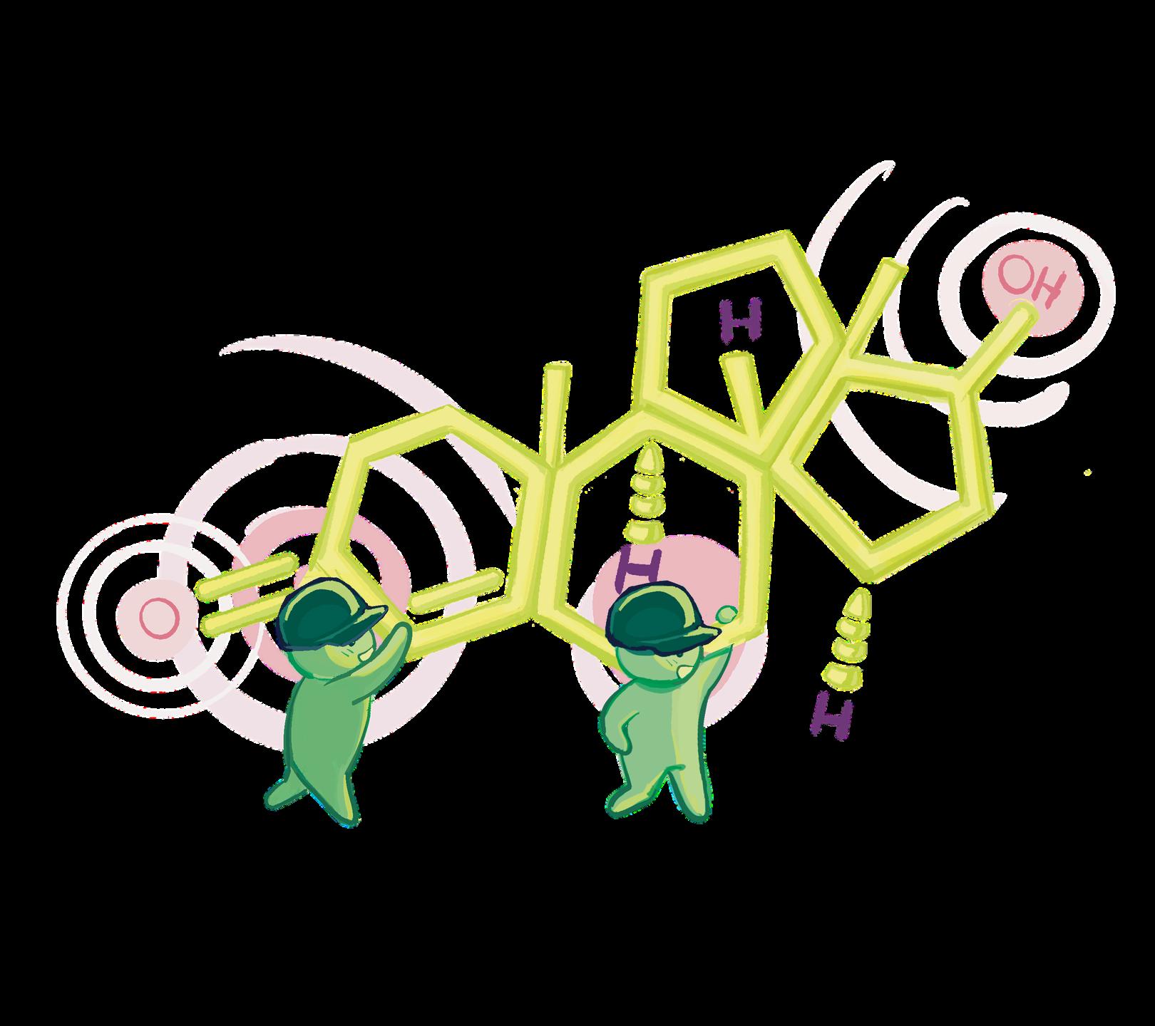
As members of the steroid hormone family, androge are fat-soluble, meaning they can diffuse direc
have the opposite effect: suppressing the reproductive system by inhibiting the testes in males and the release of GnRH, LH, and FSH in females. Due to this hormone depletion, reproductive events like sperm production in males and egg development and ovulation in females may not occur, leading to infertility and irregular menstrual cycles. Research at the Kauffman Lab at UC San Diego has helped uncover how this HPG axis suppression can occur. By manipulating hormone levels in mouse models and measuring downstream changes in hormone release, the Kauffman lab investigates how steroid hormones, like androgens, affect the HPG axis and reproductive function.
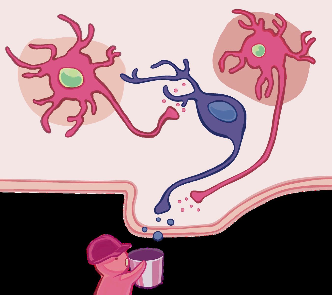
It’s these two proteins released by neurons, GnRH and kisspeptin, that the Kauffman Lab studies to understand how signals actually flow through the HPG axis. After fluorescently labeling GnRH neurons in female mouse models to track their identity, the researchers assessed changes in kisspeptin neurons by measuring gene expression using in situ hybridization. This technique uses labeled DNA probes to detect specific nucleic acid sequences. When they increased androgen levels by administering dihydrotestosterone (a potent androgen similar to testosterone) to the brain, they observed a drop in LH hormone release indicating suppression of the reproductive s GnRH neurons that directly regula gland and LH release showed n androgen exposure in the mice mo situ hybridization to measure Kiss1 the kisspeptin protein, within defin kisspeptin neuron populations, the marked reduction in kisspeptin synthesis. This suggests that an kisspeptin neurons to indirectly inhib FSH production.
This mechanism can be utilized for clinical applications, including testo and anabolic steroid use. Testos which supplies testosterone from o is used to improve energy, libido, m bone density for men with hypogonadism, a condition characterized by clinically low levels of testosterone. Similarly, anabolic steroids, synthetic derivatives of testosterone, can promote increased muscle growth and strength.
Investigations in the Kauffman Lab broadly demonstrate that excessive levels of androgen tell the brain to switch off the reproductive signals essential for ovulation and fertility. But what might happen when these levels are dysregulated entirely? The Thackray Lab at UC San Diego is currently investigating how the resulting hormonal imbalance contributes to a variety of symptoms, one of the most common in women being polyendocrine metabolic ovarian syndrom (PMOS).

ounts of androgen. blood into your cells, which your body either uses for energy or stores for later. Current treatments for PMOS include birth control, medications to block androgens, and drugs like metformin that help the body process excess insulin.


fman and Thackray labs, along with their tors, modeled PMOS in mice by giving letrozole, a drug that raises testosterone mimic the abnormally high testosterone in with PMOS. Through this technique, they the unusually fast and elevated LH pulses n diagnosed with PMOS in these PMOS-like understand why this was occurring, they d GnRH neurons in the hypothalamus that LH release from the pituitary gland. these neurons act like light switches, “on” or “off” by releasing stimulating or hormones depending on the current steroid feedback in the body. In PMOS-like mice, n neurons behaved as if the switch was “on,” constantly driving the reproductive and producing abnormally high levels of LH. urred despite the mice having high androgen hich, as noted above, can suppress the HPG refore, PCOS may involve a feedback loop in productive hormone release stays “on” and the condition.
ck-on” signaling helps explain why PMOS may ult to treat, but some therapies have still emerged to combat this feedback loop. Medications such as glucagon-like peptide 1 receptor agonists improve insulin resistance and cycle regulation, while others like artemisinin break down PMOS-stimulating enzymes. These treatments show promise in their selectivity and targeted repair. Our current understanding of treating conditions like PMOS requires further investigating the influence of stress and hormonal imbalances on our reproductive health.

Written
Breathing is incredibly simple: inhale and exhale, inhale and exhale, over and over again. It’s something that should be mundane, natural, an act you shouldn't have to think twice about doing. However, for asthma patients, this couldn’t be further from the truth: each breath has the potential to trigger chest tightness, coughing, and wheezing An asthma attack leaves its patient out of breath and scrambling for their inhaler. Only after the inhaler is the simple act of breathing restored.
Asthma affects over 300 million individuals worldwide and over 28 million individuals in the United States alone, cementing itself as one of the most prevalent airway conditions in the world. Despite its prominence, asthma remains a poorly understood condition with several unique causes, risk factors, and severity levels. Asthma attacks themselves have a plethora of triggers that elicit different immune responses, highlighting the need to categorize asthma on a spectrum rather than as a single, universal condition A closer look at risk factors underscores this idea: a dialogue between genetic and environmental factors shapes one’s individual predisposition.
Though asthma is categorized on a spectrum, we will focus on two broad classes that are the most persistent: type 2 and non-type 2. Type 2 asthma is identified by a specific immune response involving type 2 helper (Th2) cells. Helper cells are composed of critical white blood cells who detect issues and release signals (cytokines) in response to them. In this response, Th2 cells secrete cytokines, which are small proteins that immune cells use to communicate with each other. These cytokines and immune cells then induce airway inflammation, mucus hypersecretion (excessive mucus production in airways), and airway hyperresponsiveness (an exaggerated narrowing of your airway).
On the other hand, non-type 2 asthma is a catch-all term for forms of asthma that don’t traditionally fall into the type-2 category This includes asthma that isn’t caused by allergens (dust mites, pollen, etc.) and has fewer available treatments, making the development of effective non-type 2 treatments a primary focus for asthma researchers.
This brings us to a discussion on biologics, or therapies derived from living cells and proteins that reduce asthma inflammation by targeting specific stages in the inflammation process. Because the immune response consists of differing proportions of immune cell types between individuals, healthcare providers can tailor treatments to unique patients with a biologic that targets the most prevalent immune cell in each patient. The Doherty lab at UC San Diego focuses its research on developing these targeted therapies, which can only fully be understood after exploring asthma’s genetic and environmental basis.
Asthma has a strong genetic component, meaning that if someone’s parents have asthma, their children are more likely to also develop asthma. Four out of every five children diagnosed with asthma by the age of six are atopic, meaning they have a general genetic predisposition to asthma However, it is not simply determined by a single gene that encodes “asthma” — rather, multiple genes and their combined effects determine genetic risk. Rather than analyzing asthma in terms of discrete genes, it is more helpful to consider the sum of their effects.
There are a plethora of genes that each contribute to asthma risk in distinct ways. However, exploring these genes individually is deeply complex and nuanced; an easier approach is an exploration of the general mechanism that causes certain genes to increase asthmatic risk.
Rather than analyzing asthma in terms of discrete genes, it is more helpful to consider the sum of their effects.
“ “ “
We can summarize how a gene may influence asthmatic risk by looking at single nucleotide polymorphisms (SNPs), which are mutations of single nucleotide bases in an individual’s DNA. Put simply, one of the building blocks of DNA (an A, C, T, or G nucleotide) is “knocked out” and replaced with an erroneous nucleotide. Many SNPs are correlated with asthmatic traits, such as increased airway responsiveness and impaired lung function.
However, simply having asthmatic parents is not enough for an individual to develop asthma the other risk factor is more elusive and is closer than what it may seem to appear: a risk due to the environment. In other words, certain environmental cues can “pull the trigger” on an individual’s pre-existing genetic risks.
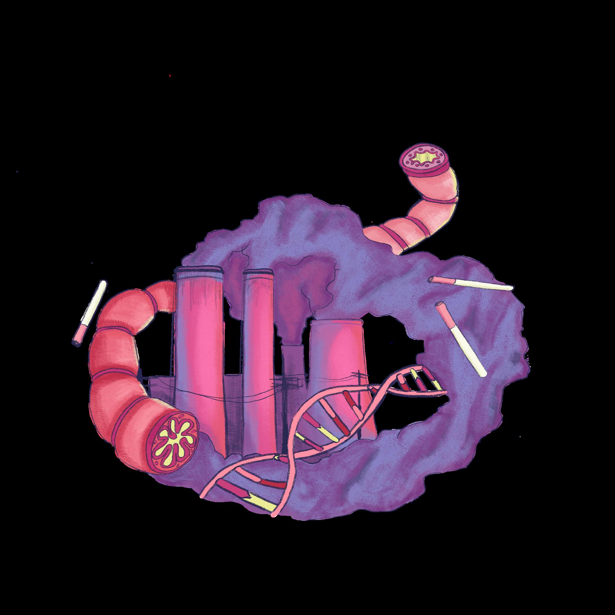
To see this clearly, consider the observation that industrially developed countries have asthma rates around 10%, while rates are under 1% in rural regions This is because large, urbanized areas have greater amounts of pollution compared to rural counterparts — due to industrial emissions and traffic exhaust which increase airway inflammation when inhaled. Tobacco smoke is another source of harmful particles that exacerbate inflammatory lung responses. Understanding the environmental factors is simple and can be explored firsthand by taking a walk around your community.
These factors may play a role in the disproportionately large number of cases in highly modernized countries (ex. United States) and other environmental influences such as access to nutritious food and availability of asthma treatments may also lead to varying rates within communities.
Circling back, the Doherty lab’s research on identifying therapeutic targets for asthma diseases does not focus solely on genetic or environmental factors contributing to asthma prevalence. The Doherty lab takes a multidisciplinary approach, developing both treatments that target specific genetic pathways and raising awareness about environmental risk factors and how to mitigate them on a broader scope.
The Doherty Lab specifically analyzed a therapeutic pathway involving eATP and the regulatory enzyme CD39. eATP is simply ATP (the energy currency that cells spend when performing cellular functions) that has been expelled out of the cell. eATP amplifies the immune response to asthma, and high levels of eATP outside of lung cells are a strong indicator of cell inflammation. Under normal conditions, CD39 modulates the levels of eATP in the lungs by converting eATP into other forms, but CD39 expression is reduced in the inflamed environment. Therefore, the Doherty Lab devised a treatment that raises CD39 expression levels during the immune response, which, in turn, downregulates eATP and reduces inflammation.
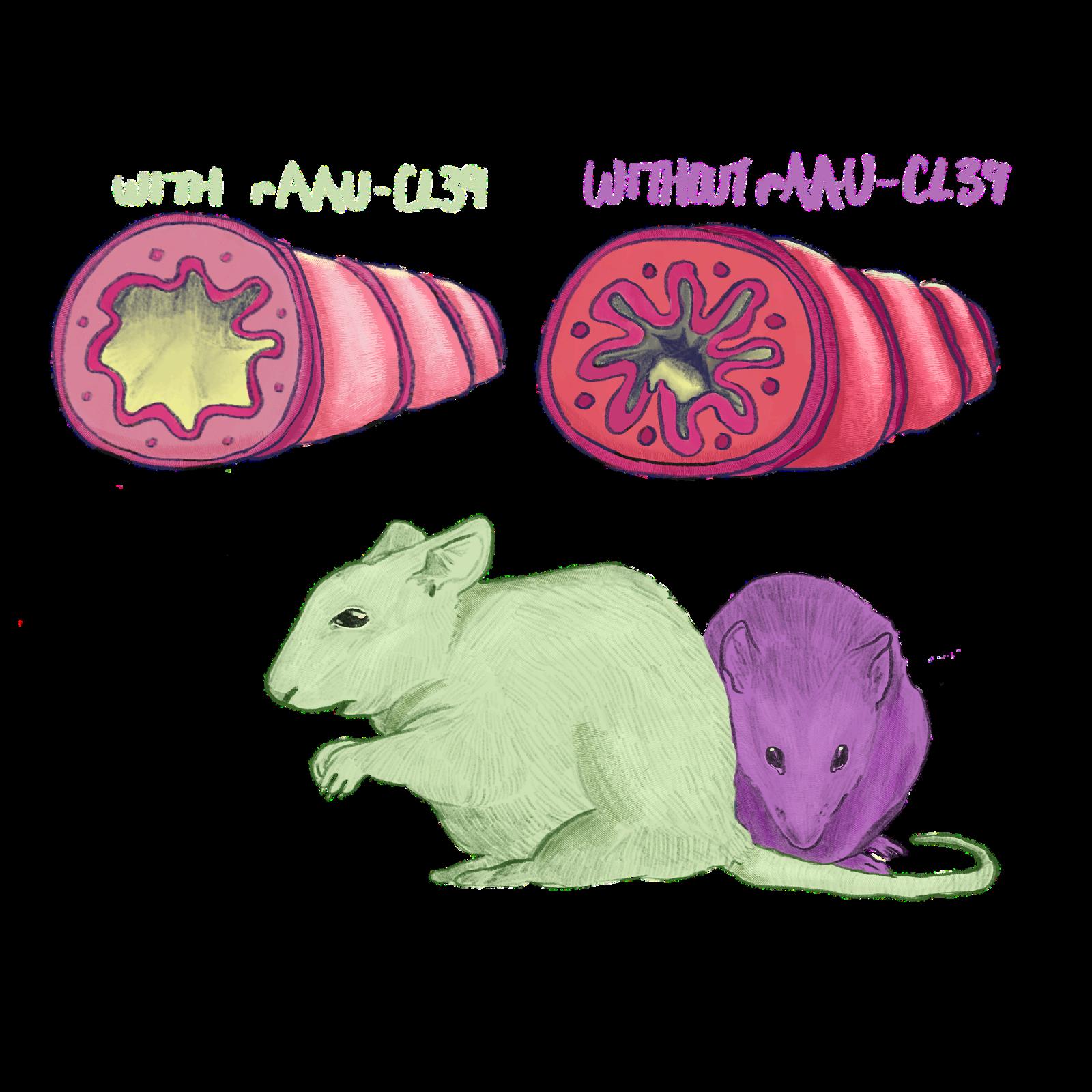
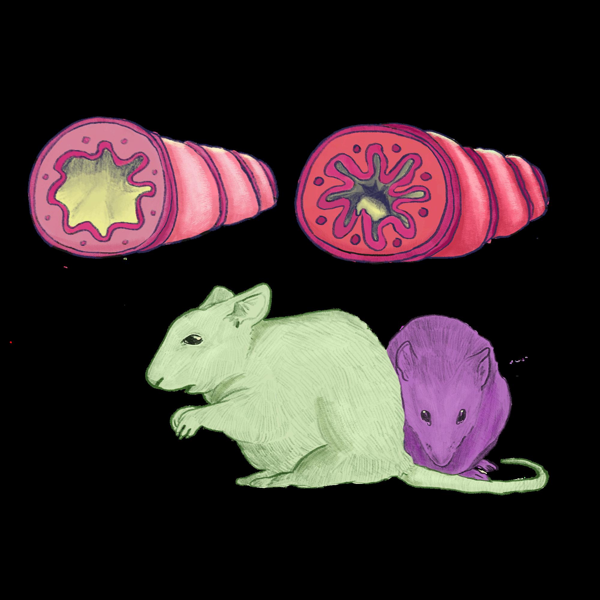
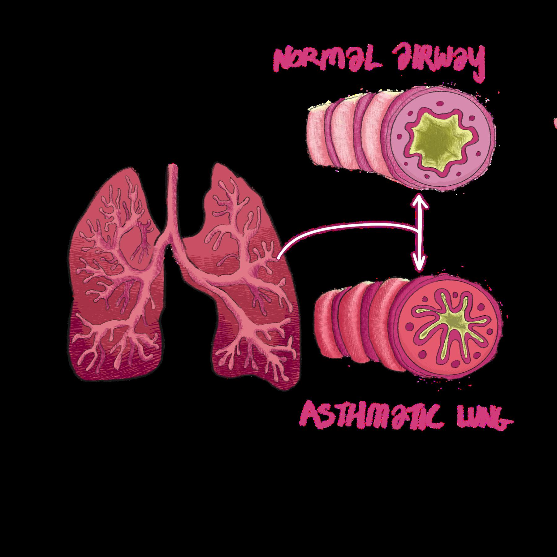
The treatment came in the form of rAAV-CD39: a viral vector that would carry the coding sequence of CD39 into the body and increase CD39 expression. Like a vaccine, a viral vector is a modified virus that carries therapeutic genetic material to its target cells. rAAV-CD39 binds to epithelial cells in the lung, restoring CD39 gene function and balanced eATP levels. Experimental testing found that the group of asthma-induced mice that were administered rAAV-CD39 showed increased expression of CD39, decreased levels of eATP, and fewer signs of airway inflammation. Upon examining the lung tissue of the treatment, there was less airway wall thickening present in the rAAVCD39-administered group, which stays consistent with the claim that the rAAV-CD39 treatment was successful in decreasing acute asthmatic symptoms. The Doherty lab’s research opens up the possibility of a new gene-based biologic for asthma: a vaccine-like viral vector that rescues CD39 expression in asthmatic patients and restores proper eATP regulation. Their findings align with the genetic basis of asthma, where the loss of function of certain genes predisposes individuals to asthma. Their research demonstrates that these losses of function can be readily restored with a detailed understanding of the biological mechanisms at play in asthmatic inflammation
Yet, these genetic pathways rely on an environmental contaminant to start the whole molecular cascade, leading to interesting links between environmental factors and asthma prevalence. For example, the Doherty lab cites wildfires as a key risk for individuals with a predisposition to asthma Smoke and ash particles in the air act as particles triggering an asthma attack. Risk is further exacerbated by climate change, which brings with it warmer temperatures and dryer conditions: perfect for wildfires. In a way, the very climate constantly modifies the risk levels for asthma.
Yet another case is the hygiene hypothesis, which argues that a lack of exposure to allergens at a young age predisposes the immune system to respond to these allergens later on in life, even if they are naturally harmless. While the hygiene hypothesis is neither proven nor disproven as of now, the Doherty
lab focuses their attention on the gut and lung microbiomes which consists of all the bacteria and other microorganisms that live in those organs. Microbiomes with an imbalance of bacteria types have been correlated to asthma, and certain treatments focus on restoring these imbalances through a combination of probiotic and dietary supplements.
Ultimately, what seems like a simple airway disease proves to be more complex and nuanced than meets the eye. A foundational understanding of asthma’s genetic and environmental risk factors is necessary for the development of quality treatments that seek to target specific pathways of asthma. Global trends such as climate change complicate matters: demonstrating asthma’s entanglement with our everyday lives. Although our investigation will not be over anytime soon, one thing is certain: we will continue piecing together the jigsaw that is asthma as we work towards a clearer understanding of one of the world’s most elusive diseases in order to impact and improve the quality of everyday life of many Allowing many to finally breathe without worry.
Shreya Joshi is a Neurobiology major graduating in 2028. Qinghan Jia is a Neurobiology major graduating in 2029.
Written
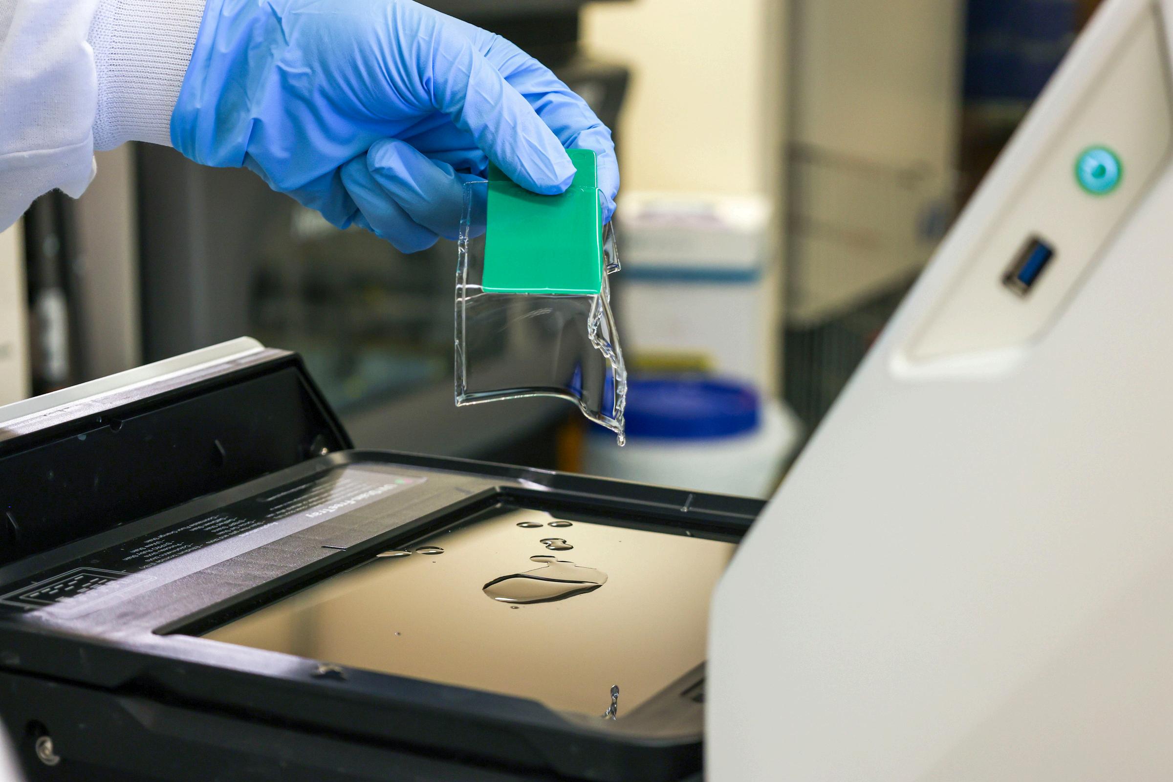
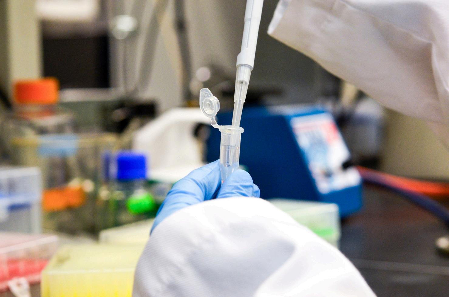
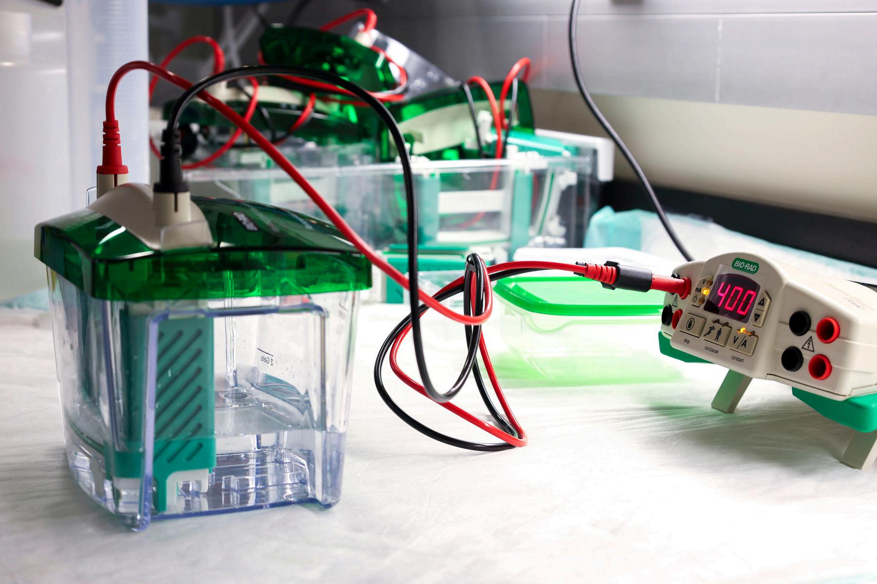
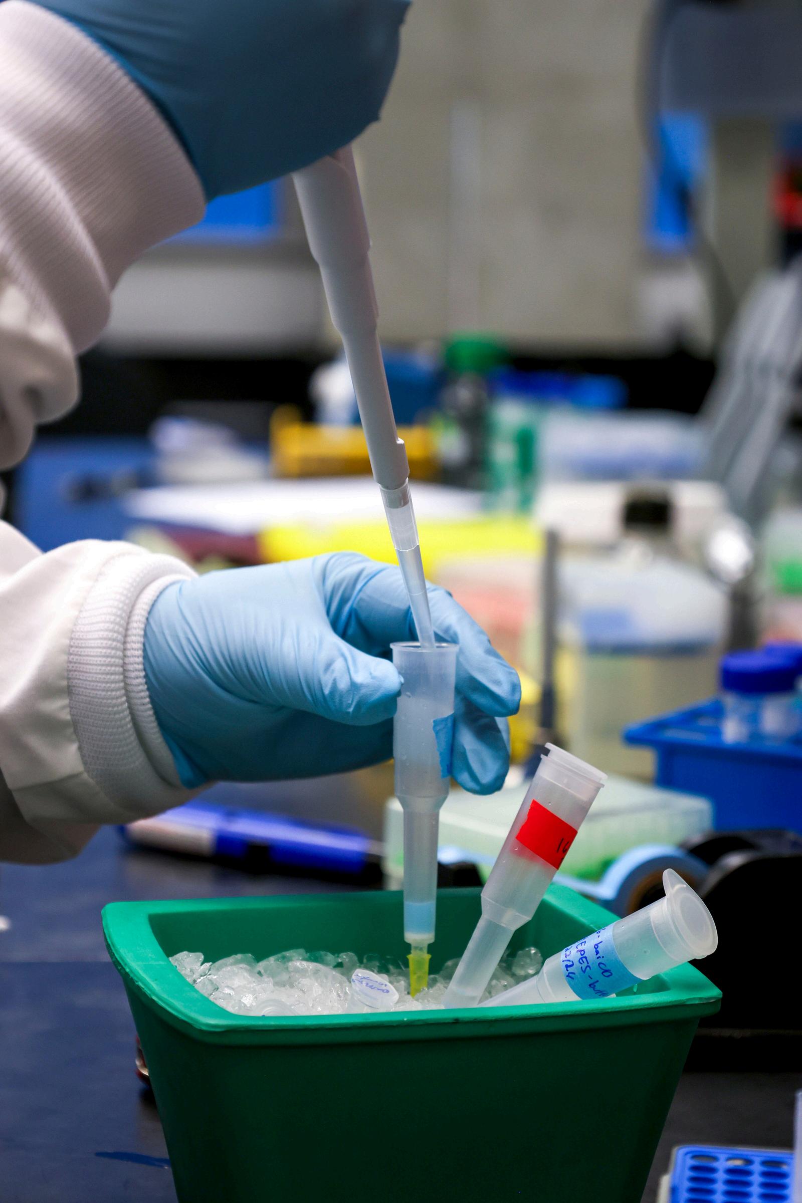
A disease as old as humankind itself, Mycobacterium tuberculosis (the bacteria that causes tuberculosis) has been the deadliest pathogen for the past 4000 years, claiming over a billion lives since its identification in 1882. Tuberculosis (TB) kills more people annually than malaria and HIV/AIDS combined, with nearly 1.23 million global deaths in 2025 and 10.7 million cases in 2024 according to the World Health Organization (WHO). When it infects the lungs, it can cause high fever, fatigue, loss of appetite, and severe respiratory symptoms like coughing up blood. Even after enduring the multi-month long recovery process, the bacteria can linger in patients’ lung tissues, laying dormant and posing the risk of reactivation later on.
Although diagnostics and treatments for tuberculosis have existed since the 1940s, the WHO reports a nearly 1.6 billion dollar funding gap across national tuberculosis programs worldwide. This deficiency in treatment resources leaves people in developing countries especially susceptible to the illness due to higher incidence rates. Even with regimens involving half-a-dozen antibiotics and lasting for months, scientists and doctors face new challenges in the form of drug-resistant strains. This presents an increasing necessity for discovering novel treatments. To meet this need, scientists can combine established laboratory procedures with novel computational techniques and accelerate the discovery of potentially cheaper drugs. The core of this search requires a deeper understanding of TB’s interactions with potential drugs.
Antibiotic resistance arises when changes in bacterial DNA allow the bacteria to produce proteins that break down, resist, or repair damage caused by antibiotics. This commonly occurs through natural selection. When bacteria are treated with an antibiotic, a subset of the population has the possibility of continuing to live thanks to random changes in their DNA that increase their resistance. If not treated, this can cause the resistant population to become dominant and render the initial drug ineffective. The typical prognosis for a tuberculosis patient lends itself to developing antibiotic resistance, with active symptoms lasting around four to six months with treatment. In addition to randomly developing resistance, bacteria can transfer mutated DNA between one another through a process known as horizontal gene transfer (HGT). HGT can occur via three mechanisms: conjugation, where two bacteria make direct contact and transfer genetic material; transduction, where a bacteriophage (a virus adapted to infect bacteria) injects and incorporates its DNA into a bacteria’s genome; and transformation, where a bacteria absorbs DNA from the environment and integrates it into its own genome.
While DNA mutations can produce a variety of defense mechanisms, one pronounced component is the development of proteins called enzymes. In your day to day life, the word “protein” may conjure up images of slabs of meat, a bowl of beans, or the selling point of a new line of “healthy” desserts. However, in biology, protein takes on a broader definition, serving as the fibers in your muscles, the carriers of the air you breathe, and accelerants for biochemical reactions. Proteins that fall into the final category are called enzymes. These proteins bind to molecules called substrates, which undergo relevant biochemical reactions. Like other bacteria, M. tuberculosis has evolved enzymes to break down molecules that pose a threat to its survival, such as antibiotics or compounds produced by the body’s immune system.
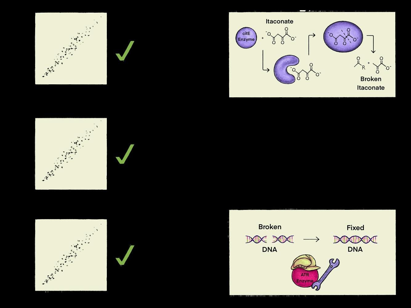
By combining traditional lab techniques with cutting-edge computing methods, the Jinich Lab at the Skaggs School of Pharmacy explores enzyme-substrate pair predictions in the defense mechanisms of tuberculosis against drugs and the immune system. In one experiment, they investigated the impact of itaconate, a chemical produced by the immune system that disrupts vital proteins involved in the removal of waste in tuberculosis. They did so by performing a co-essentiality test, which compares the impact of losing a gene on the survival or growth of a bacteria with the impact of losing a different gene. This was then applied to a section of the M. tuberculosis genome to identify the genes responsible for coding the proteins that tuberculosis uses to disarm itaconate. If removing two genes impacts the bacteria in a similar way, they are considered to be more interrelated. Among these results, they found clusters of genes that showed significant relatedness to one another. From the clusters, the lab verified that the genes identified were also established in previous research to be responsible for itaconate resistance in tuberculosis. The lab was then able to verify that the proteins encoded by these genes did occur with one another in mouse models. With protein modeling software, they also showed that genes with strong co-essentiality were more likely to produce proteins that interact with one another compared to randomly selected pairs of proteins.
This research ties into the broader problem of unannotated enzymes, which are enzymes that have been identified but not yet characterized by biochemical function. The most basic structure of any protein, also called the primary structure, is a long chain of amino acids. These amino acids act like links in a chain and will determine the chemical properties and the development pattern of the protein. Through DNA sequencing, we can determine the amino acid sequence of many proteins. Despite this technology, we don’t completely understand the function of many proteins, including over 80% of those involved in tuberculosis. By addressing this gap, we can better understand the fundamentals of how tuberculosis resists treatment, which can allow us to create drugs that target those processes.
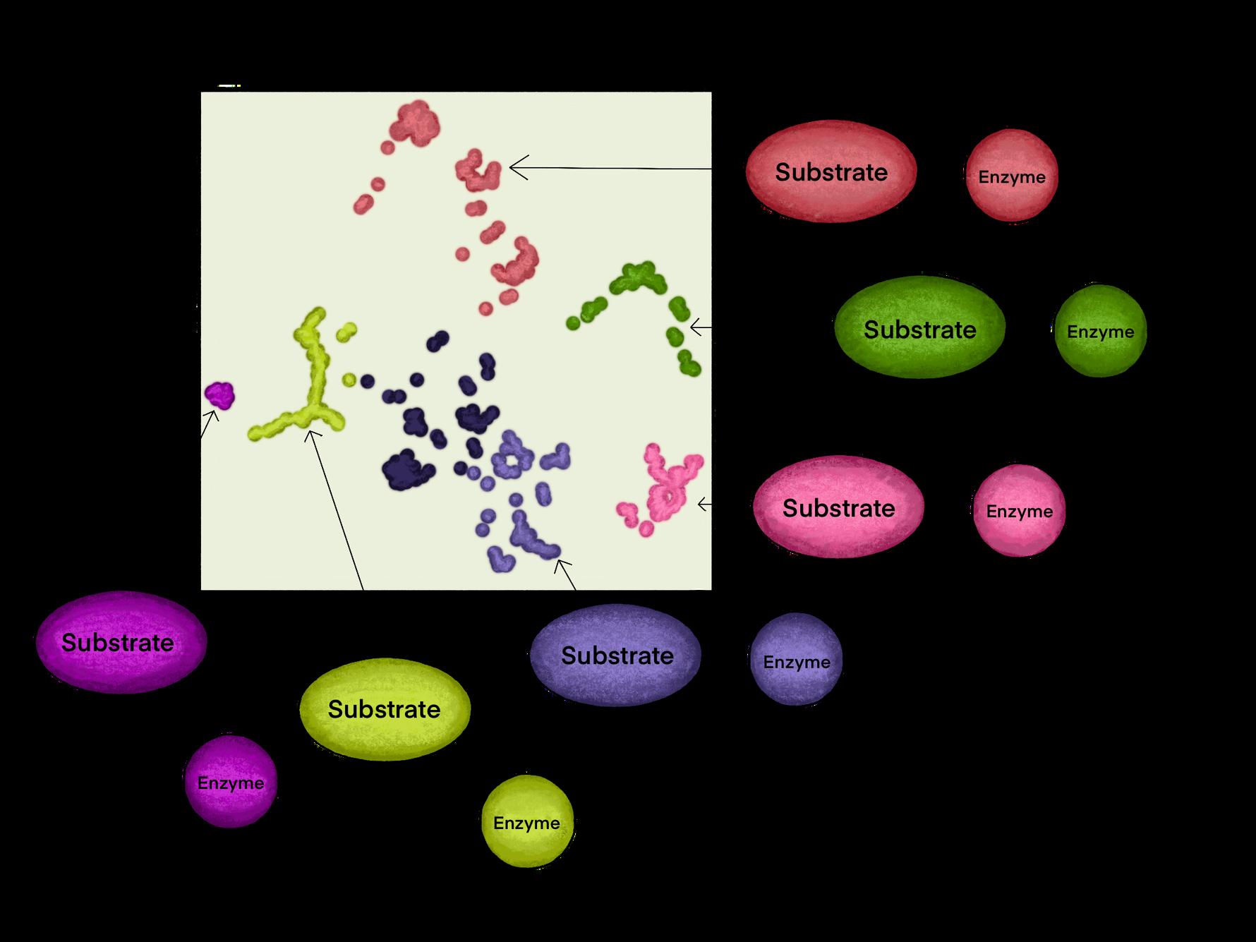
Protein language models are particularly well-suited to this type of problem. As their name suggests, they are a subclass of large language models (LLMs), which understand and generate human-like language. An LLM can be thought of as a machine taking sentences and breaking them into smaller chunks, or tokens. They then find patterns in these tokens and output the most fitting responses. By treating complex amino acid sequences as sentences, protein language models (PLMs) can predict protein structure and function by analyzing patterns present in the sequence and outputting predictions of a protein's function or interaction with substrates. The end result of this process is an embedding: a scatterplot of proteins where each point is a protein. If certain proteins function similarly, they will be closer to one another on the plot, which is then studied to find the relationship between various proteins.
This workflow combines the usage of computational methods, wet lab, and the newest generation of AI tools in biology to improve the efficiency and likelihood of success in experimental design. By using these techniques with known information about how these proteins interact with substrates, the Jinich lab was able to predict the substrate structure for two enzyme families key to TB survival: short-chain dehydrogenases and reductases (SDRs) and SAM-dependent methyltransferases (SAM-MTases).
Instead of stopping at just classification, this technique also provides information on possible substrate classes and their functions. Thus, these methods can narrow the experimental process down to testing just a small number of promising compounds in lieu of testing hundreds or even thousands of possible substrates.
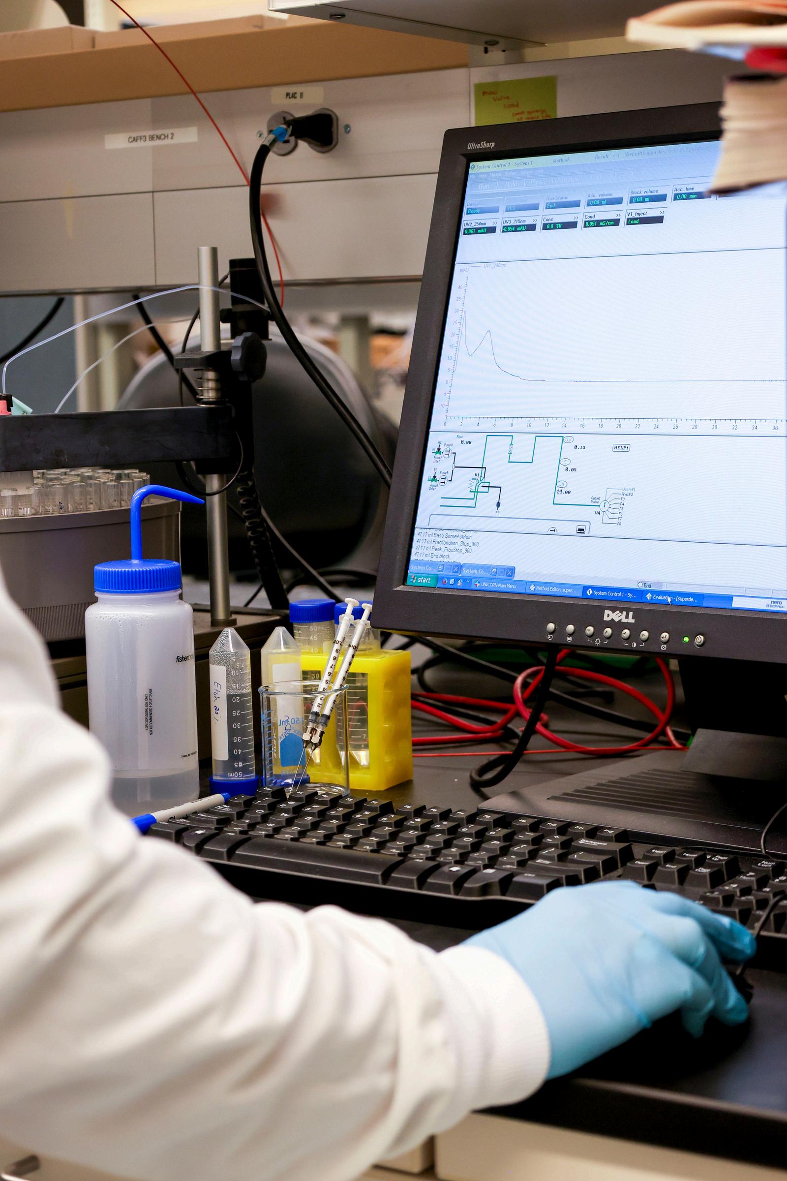
After the discovery of a new potential drug, it is essential to determine how it kills or inhibits tuberculosis. This is a drug’s mechanism of action, and it is critical information for predicting how toxic a drug may be to the body and whether it will interfere with other treatments. This is where the work of Joe Pogliano, a professor in the Department of Molecular Biology at UC San Diego and founder of the startup Linnaeus Biosciences, comes into play. Linnaeus specializes in biological assay research; one of their major projects, known as the Myco-BCP platform, advances the pre-existing bacterial cytological profiling (BCP) technology by supporting it with a convolutional neural network (CNN). A CNN is a computer vision model that takes the pixels in an image and tries to place identifiers on the different regions of the image. When using BCP, a researcher treats a bacterium with antibiotics, images it with fluorescent dyes that attach to different parts of the bacteria, and observes the changes in the cell that occur from the treatment. It then references the results with previous BCPs for specific mechanisms of action in the same bacterium. For a BCP test to yield accurate and interpretable results, individual cells need to be correctly identified and then referenced with a database. However, M. tuberculosis cells often vary in shape and tend to clump together, resulting in ineffective imaging and identification. The Myco-BCP platform addresses this fundamental issue through using a CNN. Instead of analysing individual cells, the CNN analyzes patterns within the entire image, which circumvents the necessity for cells to be imaged clearly and separately. As a result, when applied to BCP tests with anti-tuberculosis compounds, Myco-BCP was able to accurately identify their mechanisms of action and retroactively identified mechanisms of action that failed to be identified in previous studies.
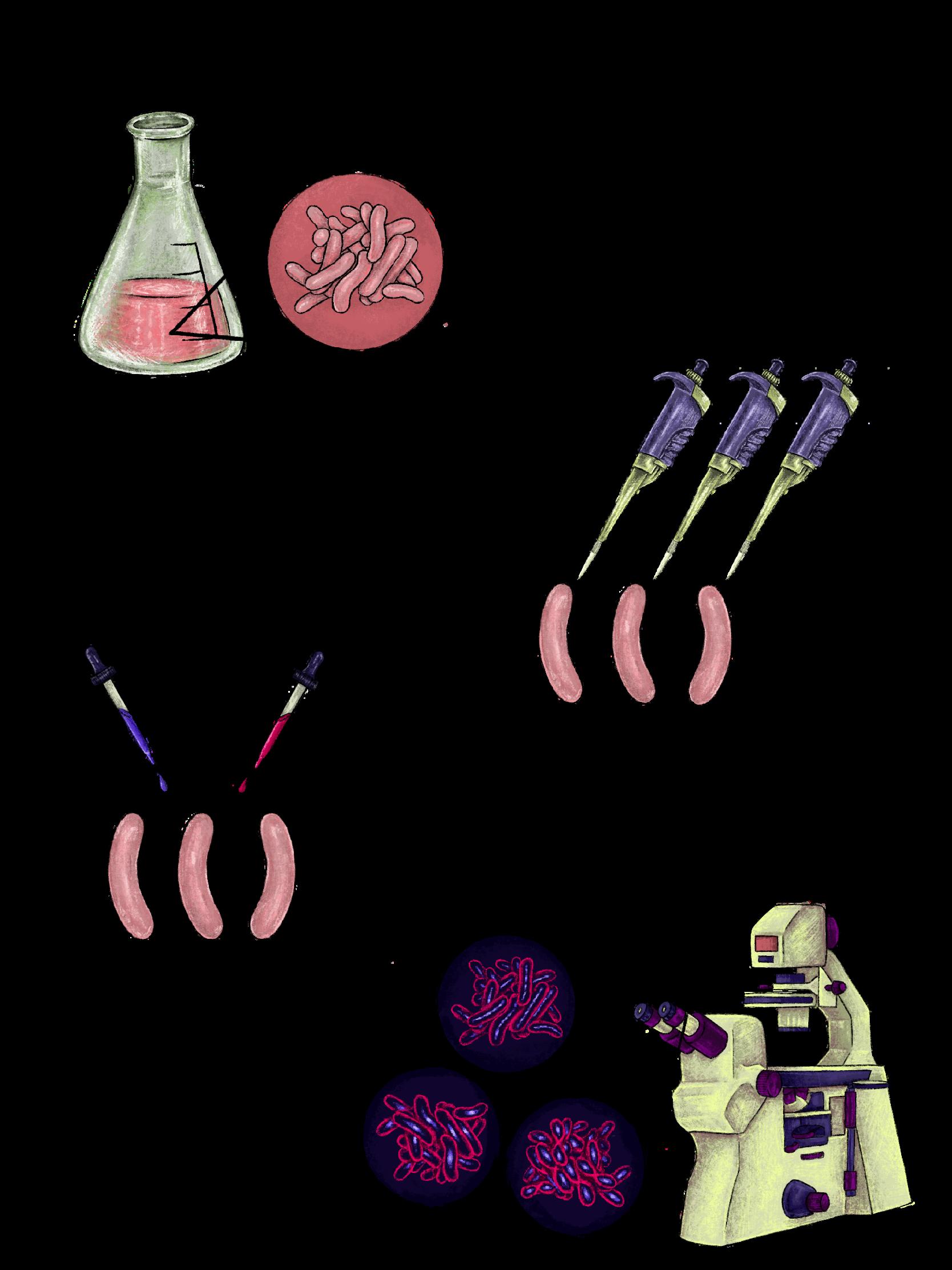

The work being done by the Jinich Lab and Linnaeus Biosciences are important frontiers in the continued fight against tuberculosis The necessity for novel treatments will be ever-present as drug resistance becomes more prominent. Hopefully, through these efforts, tuberculosis incidence and mortality will significantly reduce, and in the future, be eradicated. In this attainable future, these technologies will be ever-relevant as antibiotic resistance becomes more prominent with so-called “superbugs.” Furthermore, by refining these tools through experimenting on tuberculosis, later iterations could be applied to researching other diseases such as cancer, allowing for experiments to take on a new degree of precision.
Terry Deng is a Molecular and Cell Biology major graduating in 2029.
Ratul Chakraborty is a Mathematics major graduating in 2029.

In the face of federal research funding cuts under the Trump administration, universities and research institutions across the country have responded with growing concern and advocacy.
In conversation with SQ Core Staff members, UC San Diego biology faculty reflect on the impacts of an uncertain funding climate on their research, students, and scientific community. Through these interviews, professors share the actions they have taken to support academic research and their perspectives on the future beyond.
My group is really interdisciplinary. We are at the interface of biology, chemistry, pharmacology, computer science, and physics: use computational methods to understand how proteins work and try to develop new therapeutics against them or vaccine design. We've done a lot of work in computational virology, and we continue to work on a lot of viruses that have pandemic potential such as SARS-CoV-2, along with influenza and HIV.
There are a lot of different federal and philanthropic sources of grant funding, such as the National Institutes of Health (NIH) or National Science Foundation (NSF). Usually, they will announce: We're looking for people to work on X,” and then you submit a proposal to work on that particular topic.
As for the success rate, it varies case by case. The bottom line is that it's fairly competitive to get funding nowadays in the United States, and it's more common to be rejected than not. Although we could always argue that more funding would be better, we have to admit we have had a very good merit-based system for grant review. It's not perfect, but overall, it has served American science well.

Professor, UC San Diego Department of Molecular Biology
Interview by Lan Gao
It was pretty disruptive. I was part of a large effort in antiviral drug discovery centers called the AViDD program, which was formed in response to what happened with COVID. It was a coordinated effort to develop a shelf-ready set of molecules that could be deployed against a range of viruses such as Lassa, Ebola, and Nipah that could jump to humans, but for which we don’t have treatments. This program was supposed to be funded for five years, but ended up being funded for three. In March 2025, they abruptly terminated the program. I had just given a talk at a research conference in Italy when I got a stop-work order. All projects under this AViDD needed to stop working immediately. It was quite a disturbance.
One of the things that our group has noticed is that they are putting more funding into projects that are more applied, (with more specific deliverables) as opposed to basic discovery research, which is so key to making progress in transformative therapies. These grants are often shorter term, and more like a contract with a client, compared to an actual grant where you have some freedom in the research you do. It pays the bills, and people are really interested in that right now because you don't want to lose people but it creates a different kind of atmosphere.
When you have these unexpected budget shortfalls, there are real consequences people lose their jobs, services are shut down, or causes are made smaller. There have definitely been a lot of cuts, and one has to balance the books immediately. Universities are taking fewer graduate students, and I have had to let go of people. At the same time, we are always trying to diversify our funding sources. I think everyone is just trying their best to just ride this wave. Even if and when they do restore some of these things, a lot of people have already left and taken on other positions, or our equipment is no longer operational. Unfortunately, it's very easy to do damage to something, whereas it's very hard to build it back.
“Unfortunately, it’s very easy to do damage to something, whereas it’s very hard to build it back.”
When COVID hit, there was an infusion of funding, infrastructure, and appreciation for research in these areas. That was really good at first, but these current cuts will have a long-term impact on the field. We’re talking about sort of a generational injury to research in that less students will be trained what does this mean for the future of the scientific enterprise here?
We rely on bringing in the world's top talent to do science. America has been the top place to do research in the world, and that gives many good things, including a good standard of living and economics. But now, I don't know if we're number one anymore. We're basically giving our competitive advantage away [to other countries]. Hopefully, we can recover.
I think for students, it is important to keep abreast of what's happening and to advocate as much as possible for science within our communities. I think these are important things for students to do. As scientists, we need to commit to a life of advocacy for science and work harder to make our value known. The other thing that I would say is that there are a lot of people who are trying to get back to normalcy, and I am still optimistic that we're going to be able to recover from this.
Another thing is to be a little bit more brave in your [grad school] applications. You have to put yourself out there and take as many shots on goal as you can.
In general, I do think we will weather the storm and build back stronger with more protections and more knowledge of how to position science the way it ideally should be thought about.

Professor, UC San Diego and Director, Center for Empathy and Compassion
Training in Medical Education
Interview by Aditya Verma
The changes for NIH and NSF are completely nationwide, and there are a lot of universities that have been specifically targeted in California. There was a letter sent to UCLA threatening them with the loss of close to a billion dollars in government funding if they didn’t stop things like gender affirming care or allowing foreigners into the university, or uphold what were described as “Western values,” which were very difficult to define.
That challenge was met with a lawsuit. The Faculty Associations from the University of California, along with the American Association of University Professors, brought a lawsuit against the federal government to stop that letter from becoming real. They were able to get an injunction, and we learned recently that the government said they would not appeal it. So for now, we’re safe.
But this has been targeting higher education and research broadly throughout the country. It has very strong effects right here in California and at UCSD. Even when funding is not immediately cut, the instability is real.
“Research is very difficult to do when you don’t have a sense of where the next dollar is coming from.”
This is really being felt by students. I get at least two emails a week from undergraduates who want to work in my lab, but I can’t support that number of students. A lot of faculty are struggling in this way. They may have submitted grants but don’t know if they will get funded, so they can’t commit to supporting a student.
Undergraduate students then think, “Maybe research isn’t for me. Maybe I’ll go into business or something else where I don’t have to rely on a lab.” Pre-med students who feel research experience is important may decide they can’t pursue that path.
That is the trickle down effect of instability. One of the first things to go is the ability to take on additional trainees. Over time, we may see fewer people going into science, or people leaving California or even the United States for places where their work is more valued.
At that time, in March of last year, people felt really helpless. The university was communicating what was going on, but they weren’t giving much information about what the university community could do. Some folks were even being advised not to say too much, because it might bring more attention to the university.
Although I had experienced some impacts myself, I’ve seen a lot of impacts on my colleagues. Being someone who does have some funding that’s not federal, I felt that I sort of had the obligation, or the privilege, to do something.
I just thought it would be powerful to bring everybody together. We wanted it to be inclusive. We also had people there from SDSU and Mesa College. We wanted to talk about the impacts of the proposed cuts and delays on the economy, on the lives of scientists, and on students, workers, staff, all the people being affected.
Science funding has generally been a nonpartisan issue, and so we wanted to emphasize that this is an economic issue for San Diego. It is an issue of fairness. It is an issue of academic freedom. And we wanted the broader San Diego community to realize that this is not just a fight against “woke universities.” It’s something everybody has a stake in because of the medical advances and environmental protections that come out of research.
The Council of University of California Faculty Associations is an independent body made up of faculty from across the UC system. Because it is independent, it does not have the same restrictions as the Faculty Senate. The Faculty Senate has limitations on lobbying and on participating in lawsuits. The Faculty Associations do not.
That is why they were able to join lawsuits against the federal government. They can also organize grassroots support for legislation like SB 895.
I think it is important for legislators and the public to hear from individual scientists. It is easy to assume the university supports a bill because it benefits the institution. But when faculty step up and say, this is what I am seeing in my lab, this is the student I cannot train because I don’t know if funding will come through, that has power.
Students can sign petitions supporting SB 895. They can call their state representatives and ask whether they support the bill. They can attend rallies or press conferences.
Eventually this may come down to voter turnout, but students are the ones who can speak most directly to how difficult it is to find research positions right now, and how that affects whether they see science as a stable enough field to go into. Those voices are really important because students can speak to this experience the best.
“Stick with it. There are a lot of people fighting to make this a more viable field. Find something you are passionate about and be persistent with it.”
Stick with it. There are a lot of people fighting to make this a more viable field. Find something you are passionate about and be persistent. That is what we have to do as faculty with grant applications. You just keep putting them in.
Keep reaching out. Hopefully, you will find someone at the right time who can say yes. And speak out.