
Undergraduate Research Magazine Volume 15


Undergraduate Research Magazine Volume 15

Fifteen years of anything is no small feat, and this quindecennial edition of Under the Scope is no exception! Here, the exciting research brimming at UC San Diego is illuminated by student writers and editors, who have waded through technical jargon and endless acronyms to breach the intimidating world of research. Our photographers, designers, and illustrators transform these words into color and form, capturing the visual essence of each handcrafted story.
To pull the curtain back a little, vivid vignettes paint the year-long making of this volume: The to-and-fro of relocating our staff office from Revelle to Muir. Endless EtherNet cables and Adobe software
troubleshooting. Dark evenings in Geisel and Wong Avery, huddled around tables clearly not meant for ten. Their dedication culminates in a scientific journey—our first stop explores the neurological underpinnings of autism spectrum disorder. We then dive into aquatic ecologies, examining how traces of human activity are found even in the deepest depths of the ocean, before taking a look at how empathy should be intertwined in our approaches to medicine and scientific research. You’ll then read about the tumbling cascade of metabolic disorders, inflammation and impaired immunity, and end by unlocking the link between pancreatic cancer and diabetes.
We culminate this volume during a trying time for science. Impending financial slashes to research institutions on a federal level send shockwaves across the scientific community, with UC San Diego standing to lose millions of dollars in research funding and support. These actions affect not only the scientific community, but also the health and wellbeing of millions. It is now, more than ever, that we must stand with science, for Saltman Quarterly would be nothing without the brilliant minds who constantly redefine the future.
With these circumstances, our mission at Under the Scope remains ever more renewed: communicate science for all to understand. Saltman Quarterly buzzes with young and diverse voices, hoping to inspire future generations to explore and engage in science. We aim to continue celebrating innovation, foster a vibrant scientific community, and most importantly, keep science alive. In this spirit, it is our deepest pleasure to present you with Volume 15 of Under the Scope.
Best,

Chiraag Kambalimath and Malleeka Suy Editors-in-Chief of Saltman Quarterly 2024-2025
Editors in Chief
Chiraag Kambalimath
Malleeka Suy
Executive Editor
Amoolya Chandrabhatta
Editor at Large
Sammy Kawai
Features Editor
Meghana Hariprasad
Production Editors
Jacqueline Cheung
Erin Jang
UTS Design Editors
Tiffany Liang
Himangi Sharma
Head Technical Editor
Samia Rahman
Technical Editors
Shreya Abhijit
Deniz Erkok
Katherine Garcia
Lauryn Guan
Michelle Liang
Emily Lindemann
Selina Luo
Sarah Malko
Lauren Trinh
Samia Rahman
Head Illustrators
Leo Harris
Christina Zhao
Head Photographer
Inaya Nicholls
Ashley Juavinett, Ph.D.
Assistant Teaching Professor of Neurobiology
Adriana Elaine Scott, M.A.
Biology Student Engagement Coordinator
Andre Lin
Tommy Murphy
Claire Wolfenden
Illustrators
Sofie Blazejova
Mikkey Compuesto
Wirt Martin
Nethra Nair
Nour Ali
Mark Davis
Zaira Huq
Mridu Karanam
Joshua La
Ralph Greenspan, Ph.D.
Sara Jackrel, Ph.D.
Claire Meaders, Ph.D.
Michael David, Ph.D.
Kimberly Nguyen
Vickie Nguyen
Priscilla Schoenborn
Amber Ureña
Catherine Xu
Hannah Petyt
Samara Rathod
Keiki Sunderland
Aditya Verma
Abigail Yium

Just A Touch Away: Reaching for Social and Neural Connections in Autism Spectrum Disorder



Empathy as Scientific Method: The True Star Behind Scientific Research The Dual Frontsof Chronic Inflammation in Obesity


This article aims to explore the microscopic and macroscopic manifestations of Autism Spectrum Disorder (ASD), particularly the behavioral manifestations of ASD and the intricate neural foundations of this disorder. To further understand neurodevelopmental disorders, we must explore the crossroads of altered physiology and divergent social behavior.
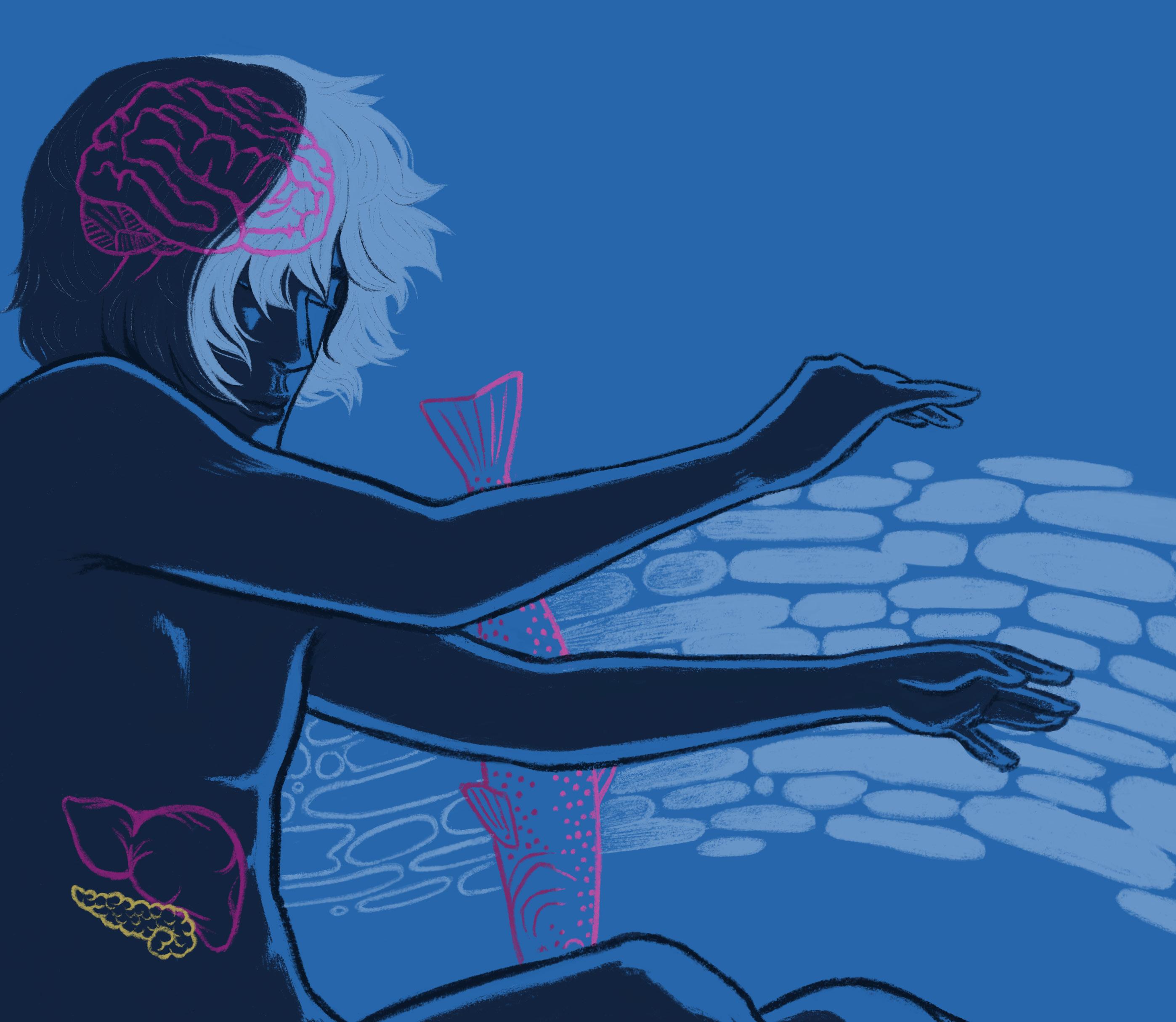
Between two outstretched hands lies a distance shaped by the forces of neural and social differences, pulling them apart even as they reach for connection. This dynamic mirrors the experiences of those with Autism Spectrum Disorder (ASD), a neurodevelopmental condition where neural differences underlie challenges in social communication, restricted interests, and repetitive behaviors. ASD symptoms are not fixed, but rather exist on a spectrum with affected individuals exhibiting a variety of behaviors that differ in type and intensity. Although common ASD behaviors are wellknown, the neural underpinnings of this disorder remain a puzzle, complicating efforts to develop effective treatments.
Nour Ali is a Neurobiology major graduating in 2028, and Abigail Yium is a General Biology major graduating in 2027.



According to Dr. Leslie Carver, the principal investigator of the Developmental Neuroscience Lab at UC San Diego, traditional perspectives on ASD often portray social difficulties as stemming from a “broken” social brain, characterized by difficulties in understanding or responding to social cues. Many modern researchers, like Dr. Carver, are not focused on “fixing” ASD with a cure or treatment, but are rather interested in better understanding the disorder itself. This perspective promotes the notion that the difficulties in social situations faced by autistic people arise from a mismatch in their neurological wiring and the demands of living in a largely neurotypical world where social cues and expectations are processed normatively. Specifically, individuals with autism require predictability in a socially unpredictable world. This makes social dynamics confusing and anxiety-inducing, leading to differences in behavior that are situational rather than intrinsic.
The key behavioral characteristics of ASD broadly fall into three categories: differences in language, social issues, and repetitive/restrictive behaviors. Regarding language, many of the differences pertain to awareness, or lack thereof, of contextual cues. For instance, children with ASD have difficulty understanding jokes or sarcasm and comprehending intentional differences in verbal tonality. Autistic children also typically present with flatter, or more monotone, language and often experience delays in language development. Individuals with ASD experience different social issues based on their age. Preadolescent children have
This attraction to familiarity clashes with the unpredictability of our social world, where interactions are fluid and often require adjustment.
difficulty sharing their attention with others, picking up on social cues, and typically do not respond to their own names. Older individuals with ASD have difficulty understanding and making predictions based on other people’s thoughts and intentions. Additionally, autistic individuals exhibit restrictive and repetitive behaviors as demonstrated by a narrow range of interests. For instance, they may develop an intense focus on specific objects, fascinated by their shape, texture, or motion. This attraction to familiarity clashes with the unpredictability of our social world, where interactions are fluid and often require adjustment.
Based on these patterns in social behavior, current research at the Carver Lab focuses on how six to nine-year old neurotypical and autistic children respond to social versus non-social stimuli through electroencephalogram recordings, or EEG. Social stimuli may include an image of a human face, while non-social stimuli may include an image of an inanimate object. Neurons, cells of the nervous system, orchestrate basic behaviors including our ability to move, talk, or process and react to information. When we think, groups of neurons fire in our brains to create waveforms. Measured by EEG, these waveforms are a representation of electrical activity in the brain and are characterized by their frequency, amplitude, shape, and location on the scalp. In neurotypical children, the waveform was more prominent when they predicted and thus shown the human face, as the observed activity was localized to reward centers in the brain associated with dopamine, the “feel-good” hormone. On the other hand, children with ASD exhibit reduced activation in the brain’s reward centers when anticipating a face in a sequence of images. The waveform measured in children with ASD is also nearly identical whether they view a face or an object. This difference between the children may be rooted in the anxiety-inducing unpredictability of social stimuli. This social disconnect, or inability to “fit in,” connects directly to the stigma around these neurodevelopmental disorders. These insights underscore the importance of rethinking how ASD is perceived— not as a condition that needs to be “fixed” but as a different way of experiencing and interacting with the world.

Figure 1: Neurotypical children exhibit increased activity in the reward systems of the brain when exposed to images of human faces, due to a sense of familiarity. On the other hand, children with ASD exhibit the same excitement when viewing inanimate, predictable objects, as it provides the same feeling.
Researchers at the Carver Lab are striving to paint a clearer picture of the social and behavioral aspects of ASD, but to fully explore its mysteries, we must zoom in on its neurological basis. Dr. Melissa Campbell, the principal investigator of the Campbell Lab in the Department of Neurosciences at UC San Diego is taking this microscopic approach by focusing on the neurons that shape how we think, feel, and behave.
Bridging sensory and motor neurons, interneurons receive signals from the nervous system and pass them along to the motor system, transferring messages from the brain to the rest of the body. Two primary types of interneurons are parvalbumin (PV) and somatostatin (SST). PV interneurons participate in synaptic depression, or the inhibition of neuronal activity, while SST neurons participate in synaptic facilitation, or the excitation of neuronal transmission. Neurons can also be broadly classified as either excitatory or inhibitory. Excitatory neurons initiate neural activity by releasing neurotransmitters that prompt neurons to fire, while inhibitory neurons act oppositely, blocking neuronal firing. Excitatory neurons are critical for the survival of interneurons during brain development. Deficits in PV interneurons, a feature commonly linked to ASD, are associated with abnormal excitatory neuronal connectivity in social and personality-based brain regions, specifically the prefrontal cortex.
The Campbell Lab investigates the neuronal framework of various neurodevelopmental disorders, with a particular focus on the mechanisms underlying ASD. Within the lab, post-doctoral scholar Dr. Yujie Cao focuses on receptor-like kinase (Ryk) proteins, which are crucial in determining PV interneuron cell fate. Ryk proteins facilitate the evolution of unspecified progenitor cells, which divide, mature, and specialize into specific interneurons. Fascinated by interneuron differentiation, Dr. Cao looks into cell transplantation, where


These insights underscore the importance of rethinking how ASD is perceived—not as a condition that needs to be “fixed” but as a different way of experiencing and interacting with the world.
embryonic stem (ES) cells that readily differentiate to any cell type are used to generate neural progenitor cells. These progenitor cells can then be transplanted into the brain, where they can migrate to appropriate regions and differentiate into interneuron subtypes like PV neurons. The ideal window of transplantation would likely be during the embryonic stage, which assists in rebuilding neuron plasticity, the ability of the nervous system to change its structures and functions. This process has shown some potential in mouse models, effectively altering the neural circuitry of the brain positively and allowing the possibility of adaptation. By manipulating Ryk proteins and promoting the development of PV interneurons, which are deficient in individuals with ASD, Dr. Cao’s work at the Campbell Lab helps uncover new strategies to address the neuronal imbalances seen in ASD.
The field of ASD research is vast but many questions remain unexplored. The Campbell Lab is working toward understanding the interactions that fail to give rise to sufficient PV interneurons in the brains of individuals with ASD. Alterations to the interneuron balance within these areas of the brain can result in behavioral shifts, as shown in individuals with ASD. However, there is no definite test to predict ASD during the embryonic stage, though family history and genetics can provide clues concerning the risk of later development. Those with ASD typically
perceive and interact with the world through a unique lens, resulting in an increased stigma and therefore a greater divide in social connection with others.
While innovations like cell transplantation may not “fix” ASD, they offer the potential for mitigating the neurological imbalances that are thought to result in the manifestation of the linguistic, social, and behavioral issues exhibited by individuals with ASD. Research at the Campbell Lab underscores the importance of investigating the neurological blueprint for ASD before neuronal imbalances become deeply established. Simultaneously, research at the Carver Lab emphasizes the importance of understanding the behavioral manifestations that shape this disorder. By approaching ASD from both neural and behavioral perspectives, the researchers at the Campbell Lab and the Carver Lab are moving towards a more comprehensive approach to understanding this disorder—one that goes beyond the idea of a cure and instead focuses on recognition, intervention, and a deeper understanding of ASD.
Figure 2: A deficiency in PV+ interneurons, represented by the tree’s one-sided leaf scarcity, is associated with abnormalities in the social and behavioral regions of the brain and is common in individuals presenting with ASD.

12 • Under the Scope • Vol 15
Keiki is a Ecology, Behavior, and Evolution and Literature/ Writing major graduating in 2025.
Samara is a General Biology major and Business Economics minor graduating in 2027.

From Sierra Nevada lakes beneath the hearths of mountains to sediment swirling along the seabed, aquatic ecosystems pool with life. Human interventions thrust these systems into peril. Deep sea environments marred by chemical pollution may take further damage from proposed deep sea mining and marine carbon dioxide removal initiatives. Underwater sounds from offshore wind farms, naval sonar, and seismic surveys block echolocation signals and deafen marine mammals. Meanwhile, in high-elevation lakes, newly introduced species and excess nutrients threaten lakebed communities.
Ill-advised human activities create barriers to aquatic ecosystem cohesion, presenting potentially catastrophic consequences for even the most remote bodies of water. At UC San Diego, researchers at the Levin, Shurin, and Scripps Acoustic Ecology labs examine these aquatic human-system conflicts and advise more mindful policies.
The deep sea is not a uniform void, but rather a mosaic of unique habitats, each finely tuned to its environment and highly vulnerable to disruptions.
“
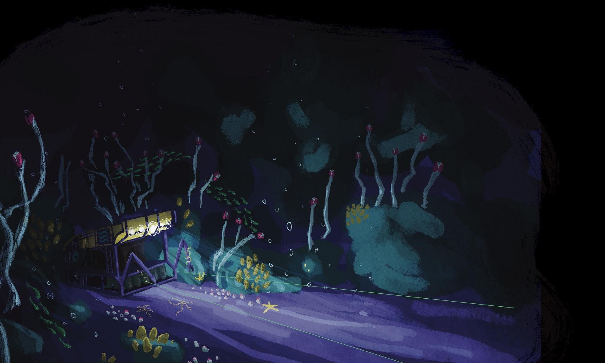
At the Levin Lab, part of the Integrative Oceanography Division at Scripps Institution of Oceanography, the mysteries of the deep sea come to light, one sample and one observation at a time. Led by Dr. Lisa Levin, the team explores the vast, largely uncharted ecosystems of the deep ocean. Among these are methane seeps, where escaping methane creates and sustains unique habitats that support deep-sea ecosystems. By studying these environments, researchers can gain insight into how human activities, such as deep-sea mining and climate change, impact fragile marine life.
Through meticulous sampling of sediment, rock, and marine life, the team investigates deep-sea ecosystems. By transplanting materials like bones and wood into methane seeps, they examine how specialized organisms colonize and break them down, revealing intricate symbiotic relationships and metabolic pathways that allow life to adapt and thrive in extreme environments.
Observations of these processes, alongside natural gradients in oxygen, depth, and temperature, offer valuable clues about the resilience and vulnerability of marine communities to future climate conditions. The team also investigates oxygendepleted ocean regions that endanger most marine organisms. Using cutting-edge tools like remotely operated vehicles and human-operated submersibles, the lab gathers data that not only illuminates the deep sea’s vibrant biodiversity, but also highlights its fragility in the face of human impact. The research holds a crucial
message: the deep sea is not a uniform void but a mosaic of unique habitats, each finely tuned to its environment and highly vulnerable to disruptions. A major focus of the lab’s research is the impact of ocean acidification and deoxygenation, two significant consequences of human activity that threaten deep-sea life. Ocean acidification, caused by the absorption of excess carbon dioxide, lowers seawater pH and disrupts the formation of calcium carbonate, a compound essential for species like mussels, crabs, and squid to build their shells and skeletons. Deoxygenation, driven by warming waters and pollution, further reduces oxygen availability, making survival in already low-oxygen environments even more challenging. To understand these effects, the team conducts experiments on larvae and embryos of affected species, exposing them to varying levels of salinity and temperature. These studies have revealed developmental abnormalities, such as impaired organ formation and reduced survival rates under stress conditions, offering crucial insight into how deep-sea organisms may struggle to adapt. The lab also identifies critical habitats known as Vulnerable Marine Ecosystems (VMEs), which are protected under international agreements. These habitats, sustained by corals, sponges, and other species indicative of ecosystem health, are vital for supporting marine biodiversity and fisheries. Dr. Levin emphasizes that conserving these ecosystems isn’t just about preservation–it’s about securing the future of the oceans and the countless species that depend on them, including us.
Another threat facing a different set of undersea organisms looms leagues above the deep sea. Sound travels faster and farther in water than in air, making it marine mammals’ favored communication strategy. Unfortunately, humans have unintentionally converted sound into a weapon. Short, intense noises, like those produced by naval sonar, elicit fearful behaviors in whales and porpoises. These man-made sounds can kill–medical examinations have reported brain bleeding in beached porpoises after nearby seismic surveys. Less severe instances of this phenomena can induce behavioral changes in marine mammals, including the elimination of hunting and calling. Understanding how marine fauna use sound in their environments helps researchers illustrate the disruptive impacts of anthropogenic noise on these precious spaces.
Figure 1: The Remotely Operated Vehicle SuBastian surveys a methane seep along the seafloor. While it collects samples of rocks and organisms, SuBastian records camera footage of the deep for Dr. Levin’s team to analyze. SuBastian is also equipped with temperature and oxygen sensors
At the Scripps Acoustic Ecology Lab (SAEL), Dr. Alba Solsona-Berga uses specialized microphones called hydrophones to listen to underwater soundscapes across the globe. Interested in studying marine mammal movement and behavior, Dr. Solsona-Berga triangulates sources for sounds by fastening four hydrophones together; this creates a 3D map of her subject’s activity. Machine learning engines model the hydrophone’s data by sorting its sound sources into species, depth, and temperature categories. With over 5 terabytes of data and recordings dating back to 2007, Dr. Solsona-Berga’s work pioneers bioacoustics research.
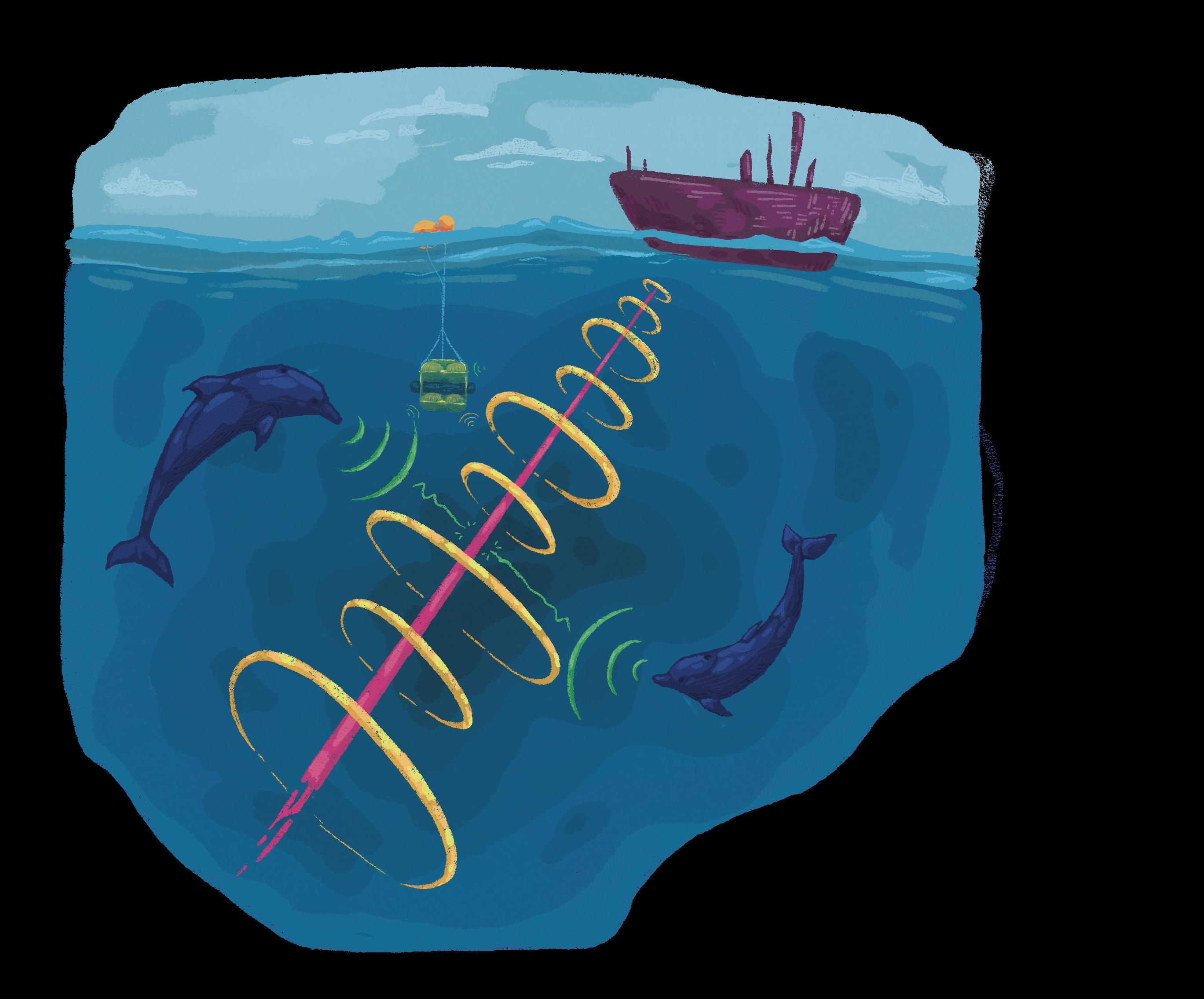
Figure 2: Sound from naval sonar, seismic surveys, underwater construction, and shipping traffic hinder marine mammals’ abilities to hunt and communicate. Hydrophones deployed in packs allow Dr. Solsona-Berga’s team to study anthropogenic noise’s impacts on the distribution and behaviors of marine mammals.
16 • Under the Scope • Vol 15
The SAEL collaborates with policymakers, the U.S. Navy, and energy companies to assess and infer the impacts of anthropogenic disturbances on marine life. Dr. Solsona-Berga’s hydrophones capture snapshots of life at proposed wind farm and seismic survey sites, modeling a baseline “normal” soundscape. Project managers assess these models to determine appropriate procedures to minimize their developments’ impacts on any at-risk species present. Dr. Solsona-Berga’s work allows companies and developers to anticipate and alleviate the noisy consequences of their plans for marine life.
Sometimes, Dr. Solsona-Berga’s work isn’t able to establish a baseline before tragedy strikes. Since just after the 2010 Deepwater Horizon oil spill, Dr. SolsonaBerga’s hydrophones have monitored animal behaviors in the Gulf of Mexico. Dr. Solsona-Berga’s data demonstrates a 14-year decline in species density in the region following the disaster. In order to advocate for legislation for the protection of this vulnerable area, Dr. Solsona-Berga anticipates using her library of behaviors to prove that the decline has been caused by an unnatural disturbance. Without baseline data from before the spill, Dr. Solsona-Berga continues to capture future trends that may help advise policymakers.
Sound from naval sonar, seismic surveys, underwater construction, and shipping traffic hinder marine mammals’ abilities to hunt and communicate.
“ “
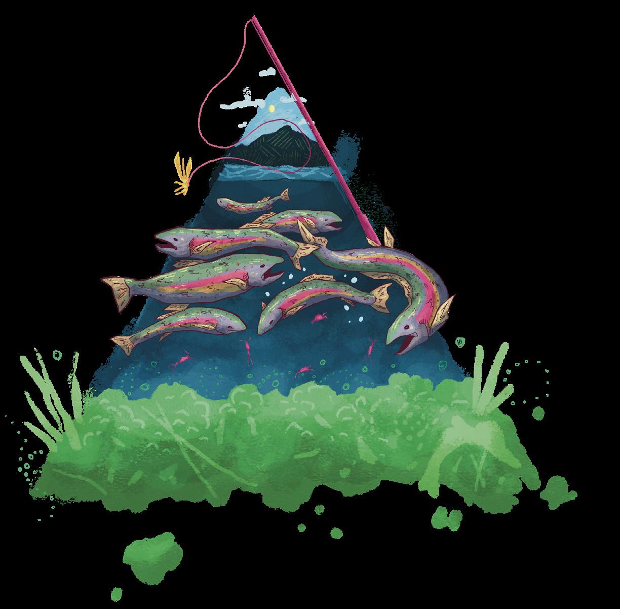
Inland from the Pacific, human activities pose different consequences for isolated Sierra Nevada lakes. Farm runoff and rainfall oversaturate lakes with nutrients, resulting in the unhealthy overpopulation of their algal communities. Humans also impact lakes by introducing non-native trout for fly-fishing. Trout consume the lakes’ larger zooplankton, disrupting the food chain and leaving opportunities for algae and phytoplankton producers to overpopulate. Trout also eat the eggs and tadpoles of the endangered Sierra Nevada yellow-legged frog, a species already endangered by an ongoing outbreak of the deadly chytrid fungus.
Within UC San Diego’s Ecology, Behavior, and Evolution department, Dr. Jonathan Shurin researches the changing population dynamics between 21 Yosemite lakes, sampling the elevation, temperature, and chemical properties of each lake yearly. The Shurin Lab observed that fish inhabiting warmer, lower elevation lakes tip the food chain in favor of producers like algae and away from prey like tadpoles, large zooplankton, and crustaceans. In colder, higher elevation lakes, these effects are less evident: while fish still prey on larger zooplankton, cooler aquatic temperatures inhibit the growth of phytoplankton and algae.
The Shurin Lab anticipates the potential of local climate warming trends to the warm-water producers. Warming temperatures across the Sierra Nevada range speed up the decomposition of plants and soil in lakes, resulting in increased concentrations of organic matter, or Dissolved Organic Carbon (DOC). Increasing DOC darkens lakes, reducing plankton’s ability to absorb sunlight. The Shurin Lab assesses plankton communities with nets and water samples, sorting out their species makeup. The lab then compares each species’ abundances with the temperatures, DOC contents, elevation, and nutrient compositions of each lake. These comparisons help Dr. Shurin assess whether the same species can adapt to changing local conditions between different interconnected lake populations. As temperatures warm and lake conditions fluctuate, this plasticity in temperature preference may keep lakes biodiverse.
Pollution, climate change, and invasive trout introductions make Yosemite and its slice of the Sierras a hotspot for research attention. Dr. Shurin and his team anticipate studying their lakes for another decade, uncovering ways that remote aquatic communities react to inadvertently induced habitat changes. Bridging the gap between human ambitions and ecological integrity, Dr. Shurin, Dr. Solsona-Berga, and Dr. Levin strive to ensure these vital ecosystems thrive for generations to come.
Figure 3: By introducing trout to highelevation lakes in Yosemite National Park, humans added another level to their isolated aquatic trophic pyramids. Trout prey on small organisms called zooplankton; without zooplankton, lakes can become overrun with photosynthesizing phytoplankton and algae.


In the performing arts world, a unique vernacular is used to acknowledge the role of the audience. To face “full front” is to directly address the audience, while “the house” refers to their seating. Between the stage and the seats lies the invisible zone commonly known as “the fourth wall.” Even when including the audience in conversations of stagecraft, there exists this invisible barrier between action and interpretation. This barrier, or fourth wall, is not unique to theater. The fields of both healthcare and research, despite their best efforts, often fail to breach the “fourth wall” that separates treatments from the communities they are designed to serve. The key to crossing this invisible barrier is simple: empathy.
the ability to understand another’s plays a major role in healthcare. Physicians spend years training to be able to care for people. In fact, the dynamic of empathy between care provider and patient defines the very structure of medicine. Patient-centered care emphasizes collaboration, stressing that the provider be an informative and empowering partner in the patient’s health, rather than
simply authoritative. This leads to better patient outcomes, greater satisfaction with the care they receive, and enhanced physician well-being. It even saves lives. According to Dr. Gerald Hickinson, a physician and expert on patient safety at Vanderbilt School of Medicine, patientcentered care prevents malpractice, or a patient’s death. Clearly, empathy must be a driving force in order for medicine to succeed in its aims.
Similarly, the inclusion of community perspectives in scientific research is vital to its impact. Participant-led research strives to center the needs of affected individuals. In science, the development of a treatment of any kind runs the risk of being formed obliquely by researchers, neglecting those it intends to serve. By centering these communities, inclusivity is improving in both science and healthcare. As a result, practitioners and researchers alike can become better guided by empathy and work to strengthen the integrity of public health as a whole. Two examples, one from education and one from research, demonstrate how empathy is innovatively integrated into these fields.
Mark Davis is a Microbiology major graduating in 2027.
Mridu Karanam is a Global Health major graduating in 2028.
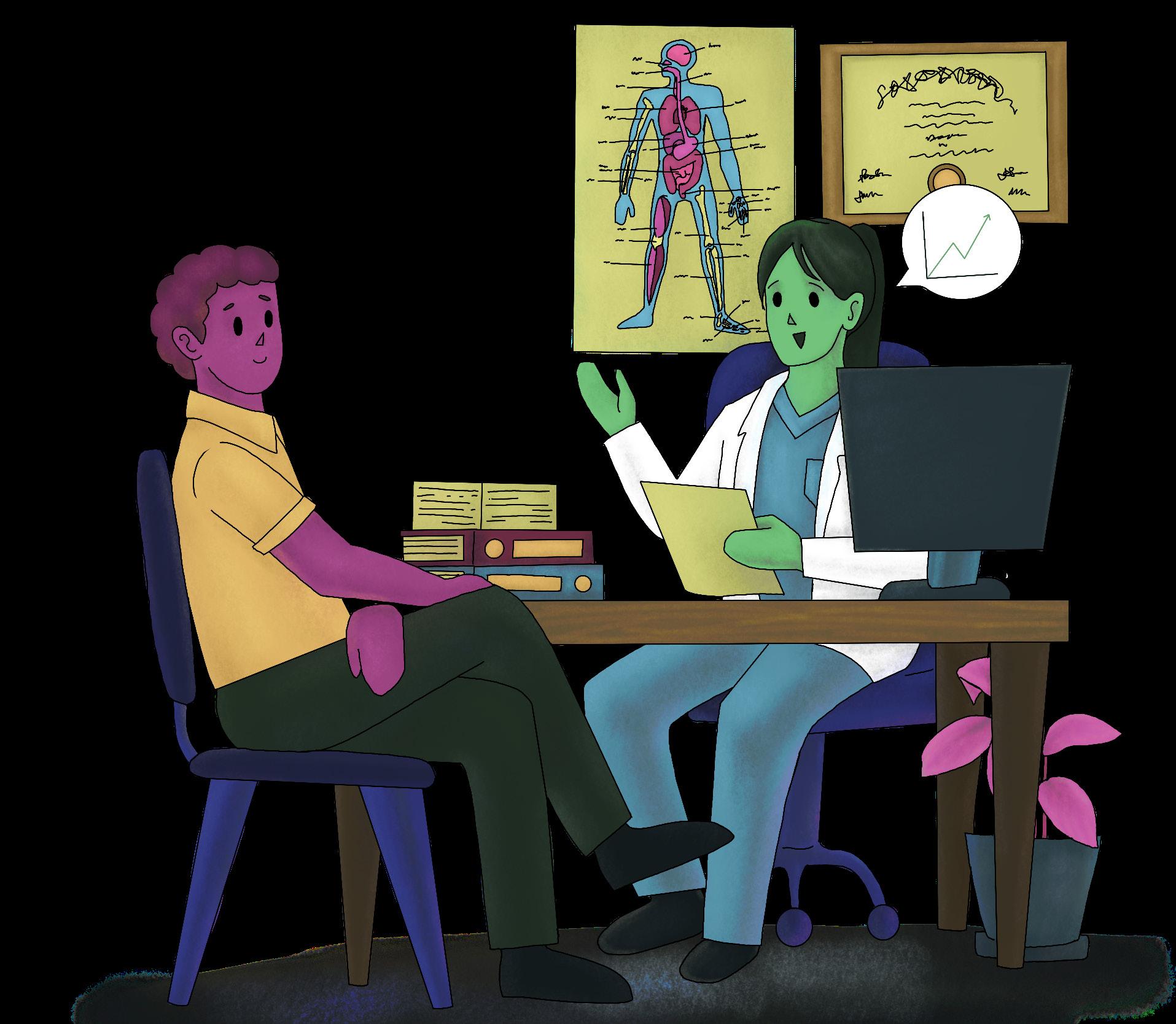
Figure 1: A physician engaging in an empathetic conversation with a patient by centering their needs can create a more inclusive and supportive healthcare experience for the patient.

The current medical paradigm falls short in its aims of empathy due to a lack of patient inclusivity. Patient inclusivity involves providing adequate care to those of all backgrounds, allowing providers to better meet patients’ needs. However, inadequate physician education has since prevented this its implementation. In the medical world, healthcare is changing to address this inequity. The work being done by Lauren Ibarra, a UC San Diego medical student, is one example of how education is intersecting with empathy. Ibarra is using co-design to develop a physician training curriculum that better accommodates Autism Spectrum Disorder (ASD) patients. This curriculum will be implemented as an intervention for physicians later in their careers.
Autism spectrum disorder (ASD) is a neurodevelopmental disorder that affects how people communicate, learn and behave. Formally classified as a disorder, ASD is perceived as a daunting diagnosis rather than a different way of thinking. Many providers assume that patients with ASD should adapt their sensory needs to the existing healthcare system, which can result in greater treatment complications. Sensory accommodations, such as dimming lights, are essential for individuals with ASD to feel comfortable during medical visits. Ibarra’s project addresses these specific needs through using co-design, which actively engages all users– healthcare providers, stakeholders, and ASD patients and their families– in the curriculum design process to ensure their perspectives shape the solution. Physicians provided input through volunteer-based interactions, such as focus groups, discussions, or survey feedback, that assessed understanding of ASD. These methods helped identify physician knowledge gaps or training needs that should be included in the
Ibarra is also currently working works to connect with advocacy organizations for adults with autism to advocacy organizations to include their voices directly in the process. The final curriculum will integrate feedback from healthcare providers and the ASD community, ensuring it will improved care for ASD patients. Ibarra’s research project has also helped identify gaps and stereotypes in medicine regarding ASD, including the preconception that ASD is a pediatric condition. Even if ASD is not perceived as an “adult condition,” it does not disappear in adults, and they deserve treatment that accommodates them. Parents of children with ASD have explained how providers might see “ASD” in their child’s developmental chart and choose not to treat them. The guides being developed by Ibarra aim to educate physicians about adapting care for ASD patients, the prevalence of ASD, and the necessary accommodations to improve patient experiences. By improving access to autism-related medical education, patients become centered in their healthcare, and their experiences improve.
In the future, Ibarra the project aims to connect with advocacy organizations for adults with ASD to further involve stakeholders in the research process. To implement the curriculum effectively, inperson training days will be created for physicians to understand the guides. , and the project Ibarra thus plans to will collaborate with Casa de Mosaic, a regional center involved with the autism community. By offering educational resources like adult visuals and addressing language and care needs, healthcare as a whole will be improved for individuals with ASD. Additionally, this practice of co-design in healthcare can be adapted beyond ASD patients to foster a more inclusive, comfort-focused approach for all patients, improving general healthcare practices.
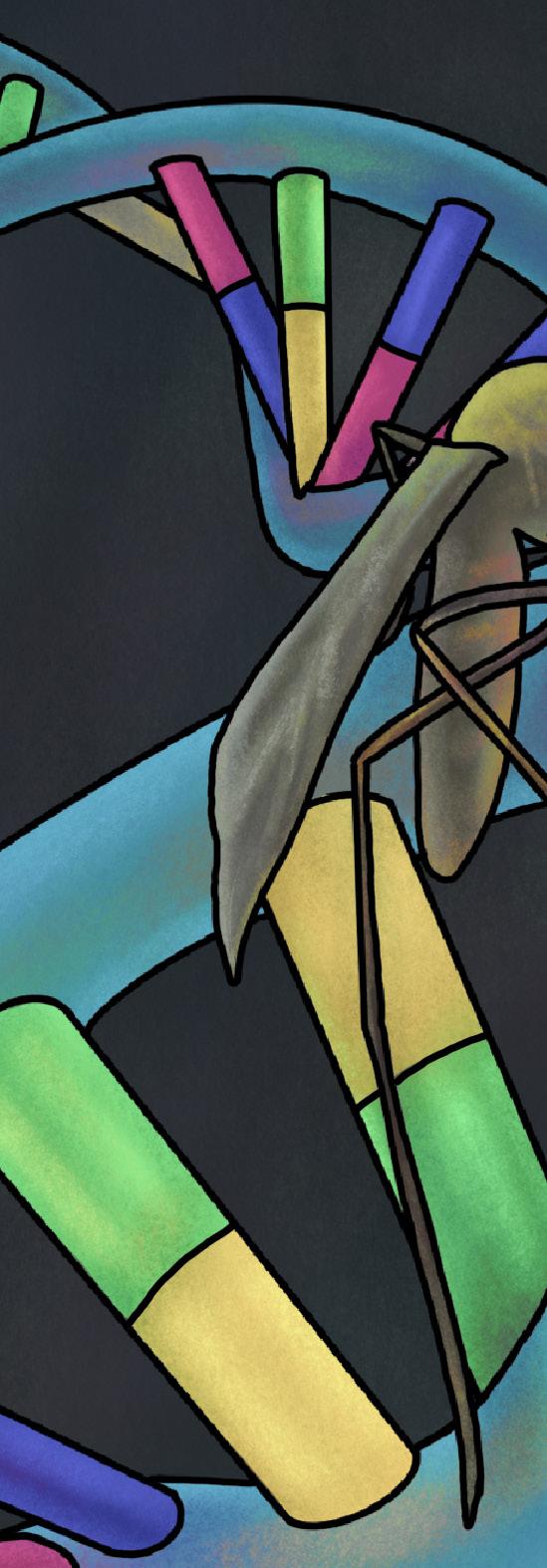
Empathy and inclusion hold a footing in scientific research as well. Gene editing technologies give scientists the ability to craft a world previously beyond the reach of human imagination. However, But wielding the power to irrevocably change an organism’s genetic code demands a significant degree of caution. For example, when it comes to malaria, a mosquito-borne disease that has plagued humanity for millennia, gene-editing technology affords us a pen to rewrite history. Geneticists can selectively delete malaria-related genes from mosquitoes and introduce these genetically altered mosquitoes into environments, a process known as a “gene drive.” However, driving a new, malaria-free genotype into the environment means that a community must now bear the risk of ecological changes since a new mosquito could alter delicate inter-species relationships.
Dr. Cinnamon Bloss, a researcher and professor at the Herbert Wertheim School of Public Health and Human Longevity Science at UC San Diego, has been working with scientists across the UC campuses (dubbed “Team California”) to determine how to implement these gene drives against mosquito-borne diseases like malaria. Dr. Bloss and her team conducted began to conduct focus groups with community members in order to seamlessly incorporate community ecological concerns. Consequently, the project became participant-led as community member input helped design the systems themselves. In the focus groups, the scientists explained the project to community members from counties known to have invasive mosquito species, and then asked what their concerns were. During the interview, questions were asked in both English and Spanish in order to include as many perspectives as possible. By understanding community concerns about gene editing, costs, and efficacy, Team California can structure a gene drive system rooted in empathy for impacted individuals.
Currently, Dr. Bloss aims to further elucidate the role of focus groups in genetic work. Under an NIH grant, she is expanding her investigation of public engagement to a nationwide study, where she is testing the assumption that people are willing to join focus groups for genetic work by sampling over one thousand participants across the U.S. By asking people what circumstances cause them to want to be engaged, she can discover the extent of inclusive science’s power in extending beyond the fourth wall, into an even brighter future.
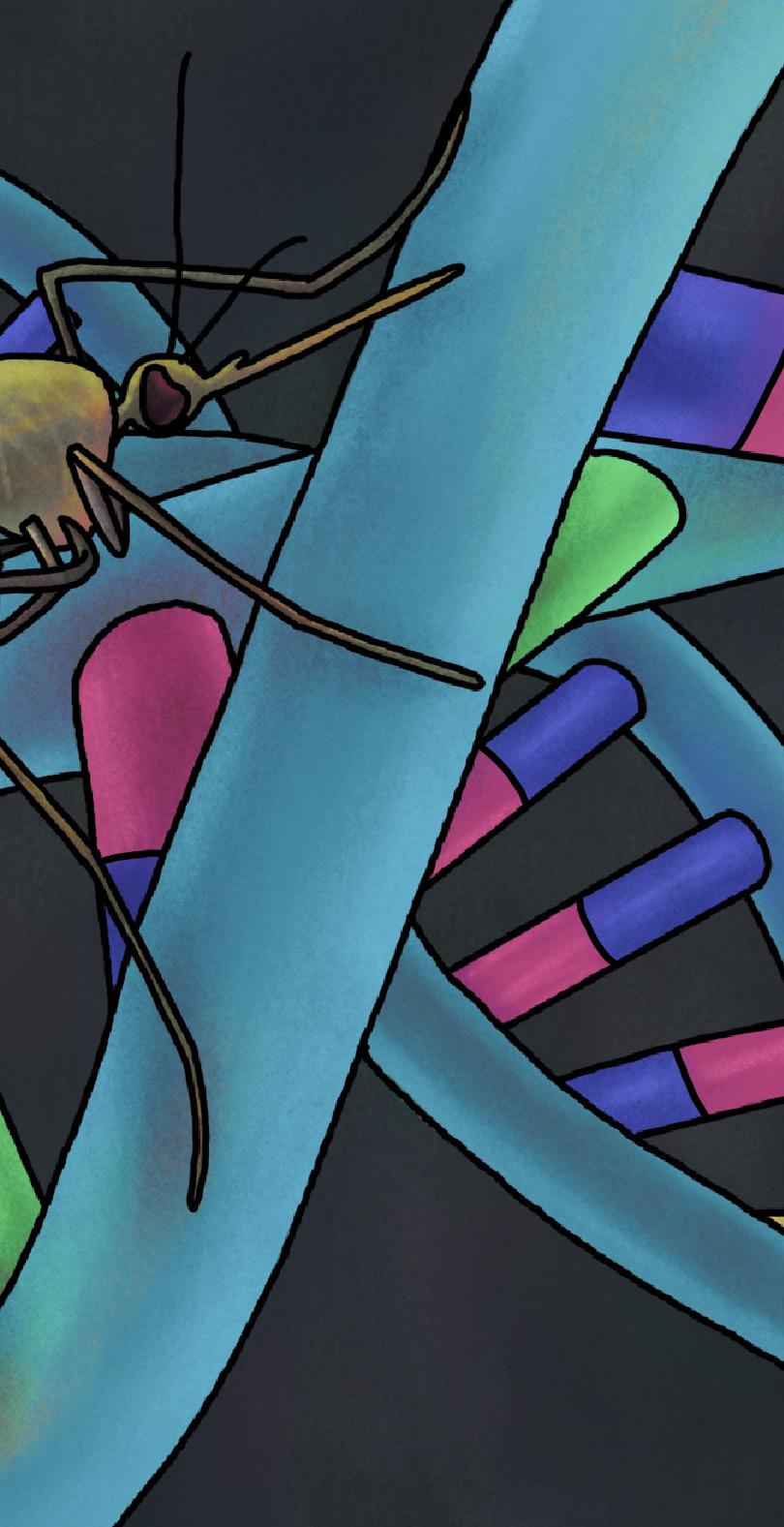
By centering empathy within healthcare communities, patient-provider relationships will improve as patients feel more valued and respected. With the proper approach, patients are more likely to engage with a care plan and follow medical recommendations catered to their needs. Medicine wields this transformative power in tandem with research. By leveraging public engagement to improve research outcomes, science can fine-tune itself to the needs of humanity. Between physician practices and public engagement in genetics, the modern scientific paradigm is flooded with spotlights, standing center stage. In order to breach the wall and reach the public audience, doctors and researchers alike must act with empathy.
incorporate community perspectives.
written by:
Aditya Verma
Zaira Huq
illustrated by:
Priscilla Schoenborn
photographed by:
Andre Lin
Julia Song
• Under the Scope • Vol 15
Aditya Verma is a Biology/Bioinformatics major graduating in 2027.
Zaira Huq is a Human Biology major graduating in 2027.
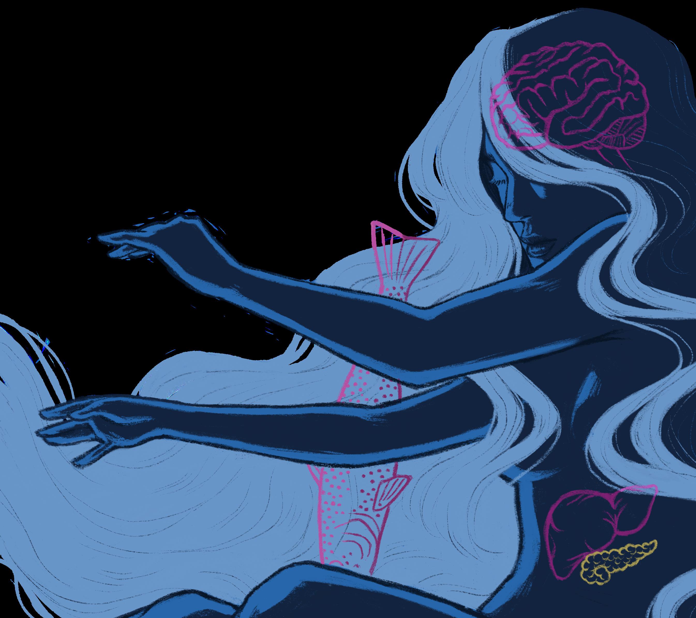
According to the National Heart, Lung, and Blood Institute, about one in every three American adults has a metabolic disorder, which were responsible for 33.1% of deaths in the United States in 2019. This silent yet profound epidemic that has swept the nation has been long in the making as metabolic risk factors, such as high blood pressure, high blood sugar, and excess fat, become increasingly prevalent in today’s society. These disorders are characterized by conditions such as obesity, diabetes, and lipid metabolism dysfunction. Not only do these disorders burden individuals with chronic health challenges, but they also harbor a more covert consequence: their impact on immune function. While often viewed as separate systems, the metabolic and immune systems share an intricate relationship, where disruption in one system can reverberate through the other. Understanding how metabolic disorders affect immune responses is critical to understanding their role in the rising incidence of infections and chronic inflammatory diseases.

Metabolism encompasses the chemical processes that sustain life, enabling the body to convert food into energy and support vital functions. Metabolic disorders arise when these processes are disrupted, leading to significant health complications. Obesity and Type 2 diabetes (T2D) are among the most prevalent metabolic disorders and share overlapping risk factors. For instance, chronic low-grade inflammation, a hallmark of obesity, makes individuals more susceptible to immune dysfunction. Similarly, high blood sugar levels associated with T2D can weaken an individual’s immune system by altering the body’s ability to fight infections and respond to injuries. These connections underpin the importance of understanding the relationship between metabolism and immunity, prompting the question: How do metabolic disorders affect the immune system?
The immune system, a complex network of cells and molecules, serves as the body’s primary defense mechanism against pathogens. Immune cells rely heavily on metabolic pathways for their activation, proliferation (rapid growth), and function, which are easily disrupted by metabolic disorders. Insulin resistance, which inhibits the uptake of sugar, also compromises immune cell signaling, and obesityinduced inflammation leads to the overactivation of immune pathways, contributing to tissue damage. Dysregulated metabolism further impairs the energy supply needed for proper immune responses, exacerbating inflammation and disease progression. Such findings highlight the dual role of metabolic disorders in both weakening immune defenses and perpetuating chronic inflammation.
Obesity triggers an immune response characterized by the activation of white blood cells called monocytes, increased circulating inflammatory markers, and the infiltration of macrophages into adipose tissue.

To uncover the link between metabolic disorders and the immune system, the Saltiel Lab at the University of California San Diego School of Medicine focuses on the intersection between obesity and inflammation, and their effects on adipose, or fat, and liver tissues. This includes studying their effects on Kupffer cells, the liver’s resident macrophages, which are immune cells that play a key role in maintaining liver health and regulating immune responses, allowing our bodies to maintain homeostasis and produce glucose for energy. The Saltiel Lab’s research redefines obesity as not merely excessive caloric intake, but as a systemic condition of persistent mild inflammation that significantly impacts immune and metabolic pathways.

Obesity triggers an immune response characterized by the activation white blood cells called monocytes, increased circulating inflammatory markers, and the infiltration of macrophages into adipose tissue. Using a high-fat diet model in mice, the Saltiel Lab has demonstrated how obesity alters the inflammatory properties of adipose tissue macrophages (ATMs) and impairs the ability of fat cells, or adipocytes, to store energy. Specifically, these proinflammatory ATMs release cytokines that reduce the insulin sensitivity of adipocytes, disrupting glucose and lipid metabolism, and contributing to the onset of diabetes. In lean mice, resident macrophages maintain tissue homeostasis while high-fat diets lead to the recruitment of pro-inflammatory M1 macrophages, which dominate the adipose tissue environment and perpetuate persistent mild inflammation. This sustained inflammatory response not only disrupts immune equilibrium, but also has systemic consequences. Similarly, Kupffer cells undergo a shift toward a pro-inflammatory state in obesity, amplifying immune dysfunction. Their hyperactivation fuels liver
inflammation and disrupts insulin signaling in the liver, increasing metabolic dysregulation. Pro-inflammatory cytokines released by M1 macrophages trigger immune dysregulation, including the activation of T-cells, further amplifying inflammation. Chronic inflammation also diminishes the ability of regulatory, antiinflammatory immune cells like M2 macrophages to resolve inflammation, creating a feedback loop of immune overactivation. This dysregulated immune state exacerbates insulin resistance, suppresses energy expenditure, and weakens immune defense against pathogens, making individuals more vulnerable to infections and delayed wound healing. In the liver, this imbalance is amplified as Kupffer cells lose their homeostatic function and contribute to the infiltration of additional liver macrophages, perpetuating the inflammatory environment that drives insulin resistance and metabolic dysfunction.
The Saltiel Lab seeks to pinpoint the early molecular events that initiate inflammation in obesity. By identifying triggers and signal cascades, the lab aims to uncover new strategies for preventing the progression of metabolic disorders, focusing on the cross-talk between immune cells and adipocytes to understand how chronic inflammation in obesity drives systemic metabolic dysfunction. This integrative research provides a foundation for innovative therapeutic strategies aimed at breaking the cycle of metabolic and immune dysfunction.
Building on the exploration of inflammation’s role in metabolic disorders, Dr. Ishtiaq Jeelani’s work in the Olefsky Lab at the UC San Diego School of Medicine investigates the immune mechanisms underlying metabolic inflammation in conditions like nonalcoholic steatohepatitis (NASH), a cluster of metabolic disorders characterized by the accumulation of excess fat deposits in the liver. More specifically, Dr. Jeelani observes the activity of Kupffer cells during proinflammatory responses of
Kupffer cells are master strategists, constantly interpreting metabolic and immune cues to decide whether to fight or remain at peace. But in chronic disease, their precise judgment falters, leading to unintended destruction.
recruited macrophages activated in NASH. Kupffer cells are master strategists, constantly interpreting metabolic and immune cues to decide whether to fight or remain at peace. But in chronic disease, their precise judgment falters, leading to unintended destruction.
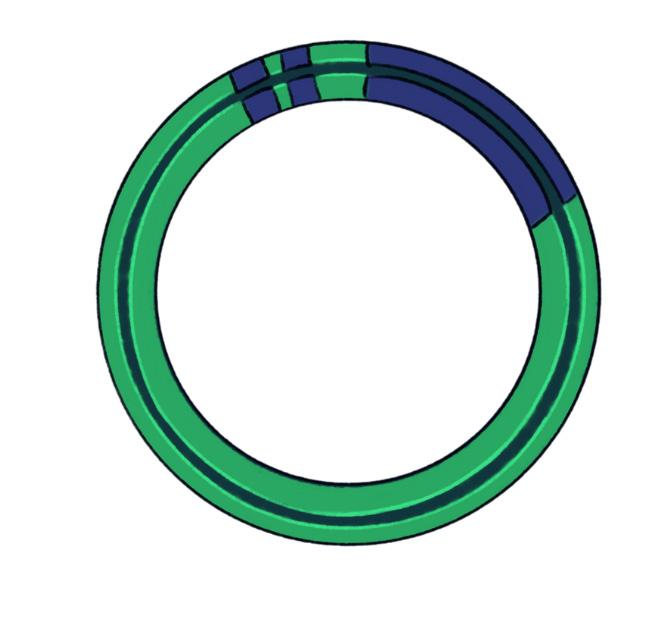
In NASH, Dr. Jeelani observed that Kupffer cells and M2 macrophages are killed due to hypoxia, or lack of oxygen, and increased metabolic demands, contributing to severe health problems such as fibrosis. While investigating the culprit behind the Kupffer cell killers, researchers at the Olefsky Lab uncovered HIF-2α, a protein responsible for adaptation to low oxygen levels. Using CRISPR, a genetic editing tool that enables targeted gene disruption, Dr. Jeelani specifically knocked out HIF-2α from a cohort of mice without impacting any other genes. Two groups of mice, one with HIF-2α and one without HIF-2α, were then fed a diet consisting of fatty, unhealthy foods dubbed the “Western Diet.” As expected, the livers of mice lacking HIF-2αwere abnormally enlarged and characterized by tumors, while mice with the HIF-2α protein had healthy livers. However, mice with the HIF-2α experienced significant Kupffer cell death, indicating that HIF2α directly caused Kupffer cell death, which accelerated liver disease in NASH-afflicted mice. Thus, knocking out HIF-2α using CRISPR could potentially enhance the metabolic health of the liver and mitigate liver damage in mice exhibiting NASH in future experiments.
By leveraging cutting-edge tools like CRISPR, Dr. Jeelani’s work in the Olefsky Lab has provided critical insights into the intersection of immune and metabolic systems, particularly in the context of liver diseases like NASH. Their research demonstrates how immune cell dysfunction contributes to metabolic imbalances, paving the way for novel therapeutic strategies and for the exploration of how immune cells, such as macrophages, regulate metabolic processes by influencing inflammation.
Metabolic disorders like obesity, diabetes, and NASH are not just isolated health issues; they are part of a larger systemic imbalance that intertwines with immune dysfunction, driving chronic inflammation and disease progression. Research at UC San Diego is illuminating this critical relationship, offering hope for therapies that target the root causes of these interconnected epidemics and can reshape the future of metabolic and immune health.
Cancer
written
by:
Hannah Petyt
Joshua La illustrated by: Christina Zhao
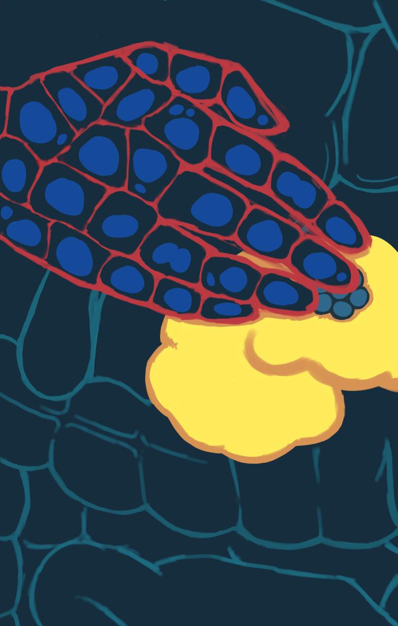
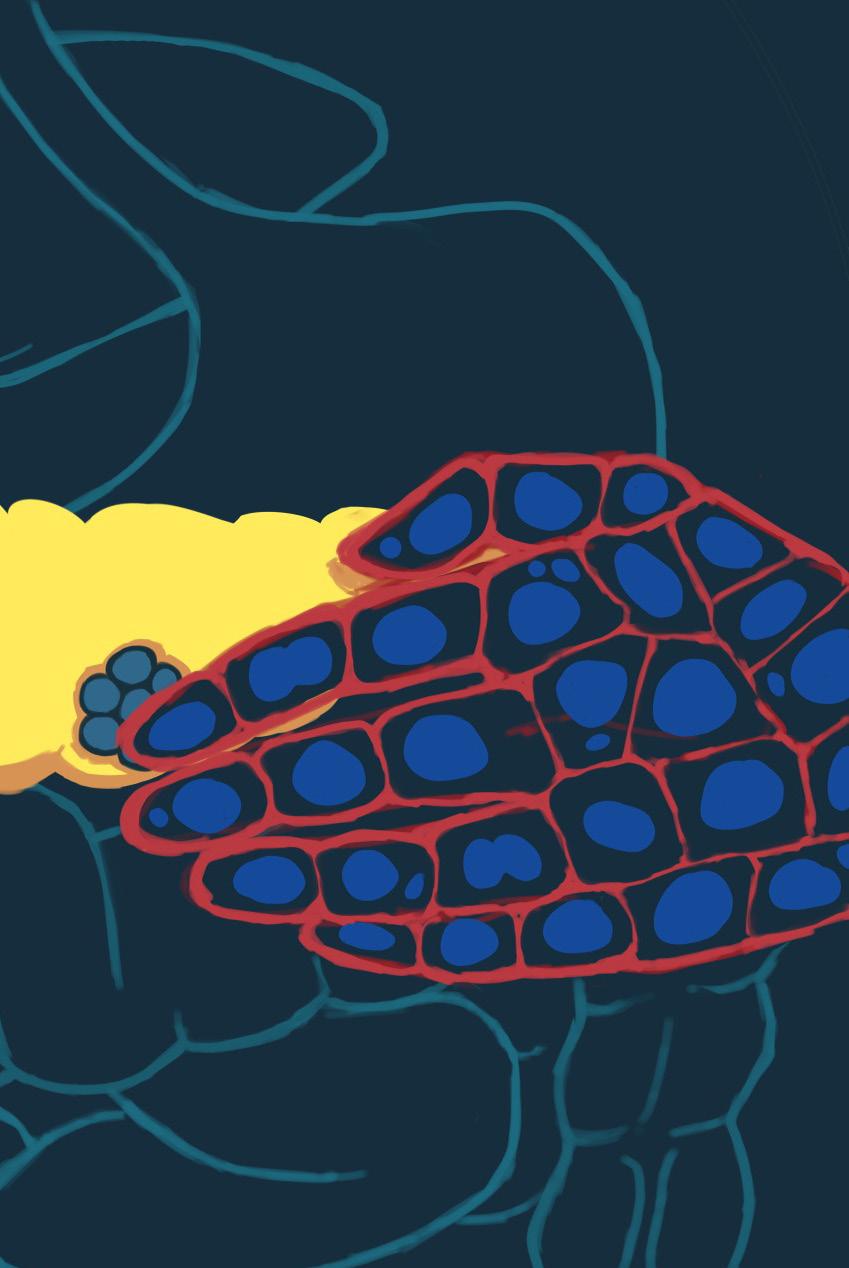
Pancreatic ductal adenocarcinoma (PDAC) is an aggressive cancer that has a five-year survival rate of only 13% due to late diagnosis and treatment resistance. This article investigates PDAC’s relationship with inflammation and diabetes and advances in novel research, such as organoid models and targeted therapeutics, to highlight attempts at developing new effective treatments and improving patient outcomes.
Hannah is a Molecular and Cell Biology major graduating in Spring 2026. Joshua is a Neurobiology major graduating in Spring 2025.
When asked to name common organs, people often think of the skin, brain, and liver. While these are all vital organs, they tend to overshadow another extremely important organ that is responsible for enabling digestion and regulating blood sugar: the pancreas. While the pancreas works normally most of the time, sometimes pancreatic cells cease function. Some pancreatic cells undergo tumorigenesis, the process during which normal, healthy cells turn into cancerous ones. What triggers this change? The answer lies in the dysfunction of specialized pancreatic cells, leading to the development of pancreatic ductal
Pancreatic ductal adenocarcinoma (PDAC), the third leading cause of cancerrelated deaths in the United States, originates from digestive enzyme cells that line the pancreatic duct. These specialized cells within the pancreas are responsible for breaking down carbohydrates, fats, and proteins, while also facilitating nutrient absorption. When these cells develop into PDAC, they become resistant to enzyme action, thereby creating greater regenerative properties for the cancer cells. With this enhanced regeneration, these cancerous digestive enzyme cells are able to withstand damage and continue to grow rapidly. The disease goes undetected because patients usually suffer from generic symptoms that are common in other conditions, leading to 80% of diagnoses being made when the disease is already advanced. As a result, the five-year survival rate is one of the lowest of all cancers,
Dr. Michael Karin researches pancreatic cancer at UC San Diego. After unfortunately losing a friend to the disease, he believes greater education and training is required for physicians to improve patient care and survival outcomes. He describes how a multitude of deaths could be prevented if healthcare providers promoted more frequent familial cancer screenings and were hypersensitive to the generic symptoms indicative of PDAC, such as weight loss, abdominal or back pain, and jaundice. These issues
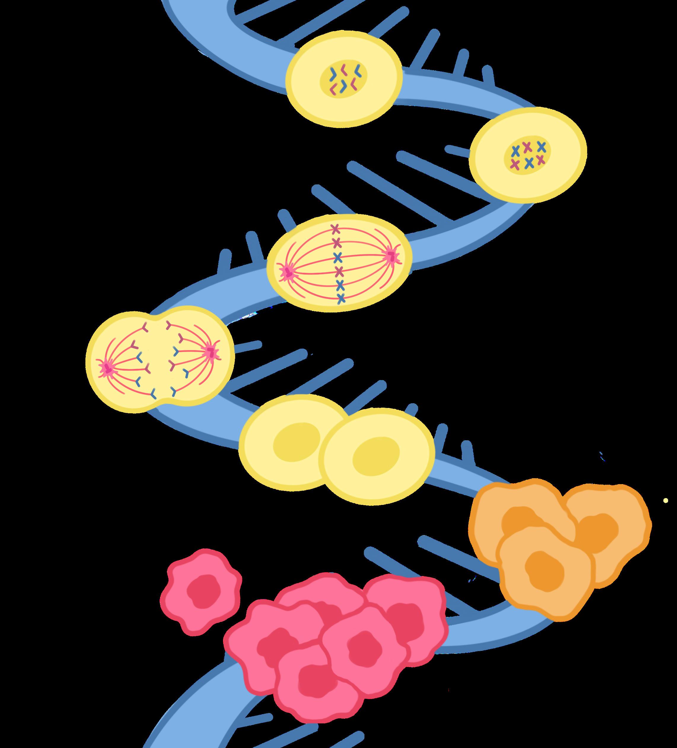
Figure 1: What goes wrong? Cancer cells undergo mitosis, just like healthy cells. However, cancer cells divide too quickly, leading to tumorigenesis, represented by the red cells.
motivated the need for further research and treatment development, thereby inspiring Dr. Karin’s work to fully understand pancreatic cancer, from its causes and risks to its molecular mechanisms and pathways.
The Karin lab uses in vitro pancreatic cancer organoids to gain a greater understanding of the disease and to research the efficacy of current treatments in order to develop novel ones. In-vitro organoids are 3D organ models that mimic an organ’s composition and functionality and are utilized outside the body. Organoids derived from pancreatic tumors harness the stem potential of these tumors, which is the ability to develop into a diverse range of cell types. This remarkable potential enables differentiation, where cells specialize into distinct types, allowing researchers to replicate tumors on a smaller scale and experiment with them ex-vivo, outside the organism. This specialized 3D culture technique creates more accurate and personalized research, allowing us to better understand the disease and how to treat it.
Dr. Karin started by focusing on the relationship between inflammation and cancer, noticing that high levels of inflammation lead to increased mitotic cell division rate, or the speed at which cells divide, and subsequently tumorigenesis. This initial finding was met with skepticism, as most experts believed mutations, or changes in DNA, and oncogenes (cancer-causing genes) were the driving forces behind cancer. However, once experimental controls were established, the researchers were able to produce more reliable data than previously obtained, leading to the conclusion that inflammation was also a risk factor for tumor development.
As previously mentioned, when pancreatic cells have an increased mitotic rate, they undergo tumorigenesis. These cells differentiate into pancreatic cancer stem cells (PCSCs), which further aid tumorigenesis and the cancer’s resistance to chemotherapy, rendering most therapies against PDAC ineffective. This has led to the development and usage of novel therapies that directly target and inhibit the genes associated with pancreatic cancer and PCSCs’ mechanism of action. In recent studies regarding the genetic activity of pancreatic cancer, antibiotics have proved promising in stopping tumorigenesis and metastasis, due to gene inhibition (switching off genes that promote cell growth). Specifically, the antibiotic salinomycin has been shown to inhibit PDAC tumor growth in mouse models. Another antibiotic, azithromycin, inhibits tumor formation by binding to a subunit of the bacterial ribosome.
Current treatments include surgery resection, which removes diseased areas of the pancreas, radiation, a cocktail of chemotherapy drugs, immunotherapy, and ablation. According to Dr. Karin, the preferred line of treatment is pancreatic resection, currently the least invasive treatment available. Using this treatment, the patient can live without a completely intact pancreas by taking enzyme pills and insulin. However, in most cases, the disease is so aggressive and has progressed to a point where a resection is not completely effective and full eradication of pancreatic tumors is not possible.
A variety of drugs that specifically target PCSCs and decrease their viability have also been effective in treating pancreatic cancer. For example, aspirin has been found to lower the activity of ALDH1, an enzyme responsible for fundamental cellular activities within PCSCs. Aspirin inhibits ALDH1’s involvement with NF-κB signaling, a pathway responsible for expressing pro-inflammatory genes that contribute to cancer development. Additionally, disulfiram, the drug used to treat alcoholism, minimizes the expression of cellular signalling pathways, like the NF-κB pathway, which are crucial in the development of cancer. The initial results are promising since a variety of drugs are effective in stopping tumor growth by targeting specific molecular components of the tumor. If effective, these therapies could sustainably cure PDAC and make it one of the most curable cancers.
Along with inflammation, ongoing research has established a bidirectional relationship between pancreatic cancer and the development of diabetes.
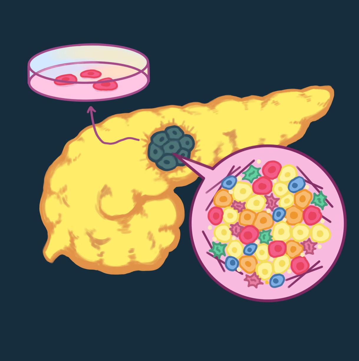
2: From the body to a petri dish; Pancreatic Cancer Stem Cells (PCSCs) are taken from an affected pancreas to be used by labs like the Karin and Sander labs at UC San Diego to conduct life saving research. 34 • Under the Scope • Vol 15

Along with inflammation, ongoing research has established a bidirectional relationship between pancreatic cancer and the development of diabetes. Across different studies, researchers have shown that some patients who develop pancreatic cancer have already had diabetes or were considered prediabetic, which raises the possibility of a potentially strong link between diabetes and cancer. The Maike Sander Lab studies diabetes and the molecular mechanisms that affect insulin-producing beta-cells (β-cells) in the pancreas. These insulin-producing β-cells play a key role in secreting insulin, a hormone responsible for lowering blood glucose levels. People with diabetes either fail to produce enough insulin or develop a resistance to insulin, disrupting the regulation of blood glucose levels. This interrupts a healthy homeostasis, an equilibrium required in all bodily systems for proper functioning.
The onset of diabetes has been linked to pancreatic cancer growth, as resistance to insulin drives the pancreas to produce more insulin in an attempt to help regulate blood sugar levels. However, insulin also promotes cell growth, which creates a counterproductive “one step forward, two steps back” relationship, where the excess insulin produced to compensate for the deficit fuels the growth of cancer. Winnie Gong and Erika Agena, a master’s researcher and undergraduate student at the Sander lab, study the underlying molecular mechanisms of pancreatic β-cells. They use a variety of stem-cell islets, or disease-modeling cells for research, within stem cell models to represent what exactly occurs in diabetic patients. They look for metabolic pathways which produce ATP, the primary energy source for living things. The lab uses a sevenstage process to help differentiate the rate at which ATP is being produced in β-cell islet models. By testing specific pathways’ ATP output, they are able to identify and select which pathways to stop and manage insulin production.
The Sander Lab at UC San Diego has identified the folate pathway as a pathway of interest. While it is a metabolic pathway that is not a significant producer of ATP, it is responsible for regulating essential cell functions and metabolism. If this folate pathway is unchecked, it can lead to cellular mayhem, specifically affecting islet β-cells in the pancreas. The Sander Lab found that inhibiting the folate pathway improved insulin production levels. Their research’s findings have been replicated in a clinical trial where a patient with Type 1 diabetes regained insulin secretion ability in their cells, allowing the regulation of blood glucose levels. This research could potentially be used in developing a treatment for Type 2 diabetes and contribute to the ongoing research about the relationship between diabetes and pancreatic cancer.
The research conducted at the Karin and Sander labs at UC San Diego draws an interesting connection between pancreatic cancer and diabetes—two vastly different yet seemingly interconnected diseases. The relationship between diabetes and pancreatic cancer is complex; while we have no definitive answers just yet, this critical journey into understanding the complexities of these diseases allows us to develop treatments and cures. The groundbreaking research being conducted today, exemplified by the extraordinary efforts at the Karin and Sander labs, will carve a healthier and more informed tomorrow. Thanks to this remarkable research, we move closer to a future where the issue of pancreatic cell dysfunction is no longer a threat—a testament to the invaluable work being done at the Karin and Sander labs.


ERIKA K AGENA
& DR. MAIKE SANDER
Folate Cycle Inhibition Improves In Vitro Differentiation of Pancreatic Beta-Cells from Human Stem Cells
CLAIRE ELAYNE ANDOLINA & MELISSA HOON
Job Hazard Analysis Management Project
VARSHA BELDONA & DR. WEI YING
Crosstalk Between Macrophages and Adipocyte Precursors That Contributes to a Healthy Adipose Tissue Expansion
JACQUELINE CHEUNG
& DR. SONYA NEAL
Investigating the Role of RHBDL4 on Bone Development in Zebrafish
LINDSEY CHINA
& DR. OLIVIER GEORGE
Validation of a Mouse Model of Nicotine Dependence Induced By Electronic Cigarette Inhalation
JACKLYN CHIU
& DR. DUC DONG
Impact on Liver Regeneration in Biliary Atresia Zebrafish Model
REBECCA ANNE CARDOSO CLAGUE & DR. DAVID CHERESH
KRAS driven tumors hijack injury recovery pathway to promote an aggressive behavior
KATHERINE DEIKO & DR. STANLEY LO
Examining First-Generation College Student Extracurricular Experiences and Science Trajectory in STEM Using Science Identity
ASHLEY NICOLE EINHORN & MELISSA HOON
Oral Drug Formulation and Development
NATHAN JOSEPH FANG & MELISSA HOON
Ensuring Product Excellence: Insights from Thermo Fisher Scientific
SALLY GUAN
& DR. TARIK BENMARHNIA
Transportation use and extreme heat: identifying the most susceptible populations in San Diego, California
MEGHANA HARIPRASAD
& DR. JESSE DIXON
Exploring the Role of CTCF in the Formation of Topologically Associated Domains in Metazoans
ALINA HO & MELISSA HOON
The Learning Curve of Tablet Development
NATALIE NORA KAPLANYAN & DR. LOUISE C LAURENT
Characterization of Extracellular Vesicles in BeWo: Identifying Biomarkers in an Epithelial Placental Cancer Cell Line
KATHERINE KAZULINA & DR. SHIRI GUR-COHEN
Sexually Dimorphic Niche Organization Dictates Mammary Gland Development.
KATHRYN KERTIS & MELISSA HOON
Research and Development in Pharmaceutical Manufacturing at Catalent
TU GIA LA
& DR. OLIVIER GEORGE
Validation of a Mouse Model of Nicotine Dependence Induced By Electronic Cigarette Inhalation
HAN GIA MAI
& DR. ANUJA JINDAL
Using Co-Design To Develop an Education Guide on Inclusive Care for Patients with Autism Spectrum Disorder (ASD)
VIVI NGUYEN & MELISSA HOON
Driving Innovation, Efficiency, and Growth in Biotech Manufacturing
LAWRENCE HERMOGINO ONG & DR. KATHLEEN FISCH
Applying Spatial Deconvolution to Determine Cell Type Compositional Differences Across Sampling Tissues, Sampling Regions, Fetal Sex, and Mode of Delivery
KELLY XIN PENG & DR. JOE POGLIANO
A Journey Through My Co-Op Internship Experience: Thermo Fisher Scientific
BEAT PEREIRA DOS GUIMARAES PEIXOTO & DR. DAVID CHERESH
Glutamate transporter, EAAT1: a novel therapeutic target to overcome Osimertinib resistance
CHEN QIAN
& DR. DAVID CHERESH
Targeting Fibronectin-Binding Integrins α5β1 and αvβ3 on Cancer-Associated Fibroblasts Blocks Fibronectin Collagen Matrix Assembly and Tumor Initiation
JULIE QIAN
& DR. WEI YING
A crosstalk between ATM and APC promotes metabolically healthy adipose tissue expansion
KARTHYAYANI SAJEEV
& DR. GRAHAM MCVICKER
Single-Cell Edit Capture: Direct Sequencing of Genome Perturbations and Transcription
RYAN JOEL SASSOWER
& DR. JOE POGLIANO
Liquid Chromatography is Essential for Quality Control Testing of Drug Products
ASHLEY BROOKE SEDGWICK & MELISSA HOON
Dissolution and Chromatography of Tablets to Determine the Quality of Pharmaceuticals
SEPENTA JAFARZADEH SHIRVAN
& DR. ARDEM PATAPOUTIAN
Quantifying sensory innervation of the mouse kidney cortex
ANDREI SHEYDAYI
& DR. JOE POGLIANO
Spray Drying at Catalent
LAWRENCE CHAN
& DR. AARON CARLIN
Role of SREBPs in Regulating Human Inflammatory Signaling
ELSA CHEN
& DR. JOSE PRUNEDA-PAZ
Investigating Systemic Transcriptional Changes Induced by a Localized Bacterial Infection in Plants
SUHANA SINGH
& DR. NEWEN WANG
Pediatric Leading Causes of Death
Among Asian American and Native Hawaiian Pacific Islanders (Aggregated and Disaggregated Subgroups) Compared to Non-Hispanic Whites (2005-2019)
RUBY TSENG
& DR. KAY TYE
Neuromodulated mixture of experts: A prefrontal cortex inspired architecture for lifelong learning
VARSHA VINOD
& DR. MICHAEL CASTLE
This way, Axon: Guided Axonal Regeneration for Spinal Cord Injury
EMILY CATHERINE WHITE & DR. AMY KIGER
Investigation of PI3KC2 function in autophagy flux during muscle cell remodeling
CATALINA ZARAGOZA FOGG
& DR. TREY IDEKER
Using Prime Editing Sensor Screens to Target Oncogenic Mutations of the Nuclear Pore Complex
SAMVEL SARKIS GABOYAN
& DR. LAURA CROTTY ALEXANDER
Inflammatory And Anti-inflammatory Effects Of E-cigarette Aerosol Inhalation On Allergic Inflammatory Airways Disease In Mice
JULIA KEUM
& DR. ANDREW MUROYAMA
Measuring the impact of stomatal alignment on pore opening in Arabidopsis thaliana
RYAM LAM
& DR. PENGZHE LU
Co-deletion of PTEN/SOCS3 induced Robust Graft-derived Axonal Extension in a Severe Spinal Cord Injury Mouse Model
SOLOMON JINRI KIM
& DR. KAY CHUNG
Elucidating the Role of CXCL16 Isoforms in T-Cell Differentiation
YANG LIU
& DR. JOSEPH GLEESON
Deciphering the Role of Somatic Mosaicism in Malformations of Cortical Development: A Genetic Exploration of Focal Cortical Dysplasia
YUFEI LIU
& DR. ENFU HUI
Differential effects of ligand binding on LAG3 clustering in CD4+ and CD8+ T-cells
BEIYING LU
DR. MICHAEL CASTLE
Enhanced Cortex-Specific Promoters for Brain-Derived Neurotrophic Factor Gene Therapy in Alzheimer’s Disease
HANH YEN NGUYEN
& DR. DONG-ER ZHANG
Characterizing the Functional Significance of Peptidylarginine Deiminases in t(8;21) Acute Myeloid Leukemia
LAURA MARIE NORONHA
& DR. NICOLA J. ALLEN
Dysregulated Cholesterol Metabolism in Alzheimer’s Disease
YUHAN CHEN
& DR. JULIE LAW
A Family of DNA Binding Proteins
Control CLSY3-Dependent Sexual-Lineage-Specific DNA Methylation in Plants
TYLER DIEP
& DR. BINHAI ZHENG
Assessing the impact of DLK/ LZK deletion on injured corticospinal tract neurons
VERINA HYMN-MONG LEUNG
& DR. LI YE
Volumetric CATCH: In Vivo 3D
Drug-Target Profiling Across MultiOrgan Systems
ERIN LI
& DR. STEFAN LEUTGEB
Neuronal firing patterns during working memory retention across hippocampus dorsal-ventral axis
VEDA PALAPARTY
& DR. KARL J. WAHLIN
Developing an Approach for Adeno-Associated Viral Vector Mediated Cellular Reprogramming
TEJASVI PATIL
& DR. JOSE PRUNEDA-PAZ
Characterizing the role of TCP1 in Arabidopsis Clock-Controlled Growth and Development
FARID REZAYAT
& DR. YULIYA SKOROBOGATKO
The role of Brown Fat Glucose Uptake in Weight Loss induced by β3 Adrenergic Agonist
SHARANYA SRIRAM
& DR. MATTHEW WORTHAM
Characterizing expression of post-transcriptionally regulated ribosomal genes in pancreatic alpha and beta cells
SONIA VICTORIA STAN
& DR. MICHAEL KARIN
Identification of Combination Therapies Targeting Macropinocytosis in Pancreatic Ductal Adenocarcinoma (PDAC) Stem Cells
TIMOTHY ANDERS SWAIN
& DR. DAVID HOLWAY
Insectivorous bird use of ornamental vegetation: tipu trees, tipu psyllids, and warblers in suburban San Diego
NAYANA TELLAKULA
& DR. WILLIAM B. KRISTAN, JR.
Planarian Transection and Regeneration Modify Behavioral Responses
LUCY WANG
& DR. ALAN R. SALTIEL
Investigating the role of MAVS in the adipose tissue
JINGYAO WANG & DR. ALON GOREN
Prod-seq, a Multiplex Measurement of Protein Abundance and Interactions
ELLEN WRIGHTSMAN
& DR. GENE YEO
RNA Binding Proteins Caprin bind Synaptic RNA in Human iPSC-derived Neurons
QINGCHEN ZHOU
& DR. QINGFEI JIANG
Exploring the role of Matrin-3 in double-stranded RNA sensing in T-cell Acute Lymphoblastic Leukemia
WINNIE ZHOU
& DR. ANDREAS ERNST
Characterizing the mechanisms of TFG oncogenic fusion proteins
SUNNY ZHU
& DR. NICHOLAS WEBSTER
Exploring the role of alternative splicing of Insulin Receptor to IR-B in IGF-2 driven liver fibrosis and inflammation
MICHAEL PAUL CHOI & DR. HEIDI COOK-ANDERSEN
ZFP36L2 is a novel regulator of the mouse pluripotent state and germ layer specification
CRISTINA KEIKO DIAZ
& DR. DONG-ER ZHANG
Analysis of pathogenic RUNX1 C-terminus truncation in HL-60 promyelocytic leukemia cell line models
LEANNE ELIZABETH DUGAN
& DR. LAURA CROTTY ALEXANDER
HIF Expression in Pulmonary Endothelial Cells Modulates Immune Cell Recruitment in a Murine Asthma Model
LILI NICOLE FOLLETT
& DR. ANDREW MUROYAMA
Dissecting the mechanistic basis of nuclear migration during stomatal formation in Arabidopsis thaliana
MAX ALBERT GRUBER
& DR. CHRISTINA SIGURDSON
BERNICE HWANG
& DR. JEFFREY D. ESKO
Developing a Reliable Assay for Heparanase
HANNAH KAM & DR. STANLEY LO
The Role of Job Advertisements in Building Diverse UC Faculties: An Automated Study with ChatGPT-3.5
ALISA PANICHKINA
& DR. TONY L. YAKSH
Social Defeat Stress induces chronic pain in juvenile mice involving TLR4 activation in dorsal root ganglia
ALEXANDRIA XENA SINGAPAN
& DR. LISA MCDONNELL
Aligning beliefs and actions: Do students’ perceptions versus actual utilization of study strategies align?
Prion Protein-induced Neurodegeneration in the Central Nervous System of Drosophila Melanogaster: Building a Model ANDY TRINH
& DR. STANLEY LO
Asian American Undergraduate Student Experiences with Racism in STEM: Resistant capital through an AsianCrit Lens
ANN TRUONG
& DR. JING YANG
Unraveling the Role of ITCH: Investigating Invadopodia and Metastasis in Cancer Cells