Geoffroy's spider monkey (Ateles geoffroyi) Santa Rosa National Park, Costa Rica, 2025
Inside Cover
Photo by Inaya Nicholls
Olive Ridley sea turtle (Lepidochelys olivacea) mass nesting Ostional, Costa Rica, 2025
Back Cover
Photo by Leo Harris
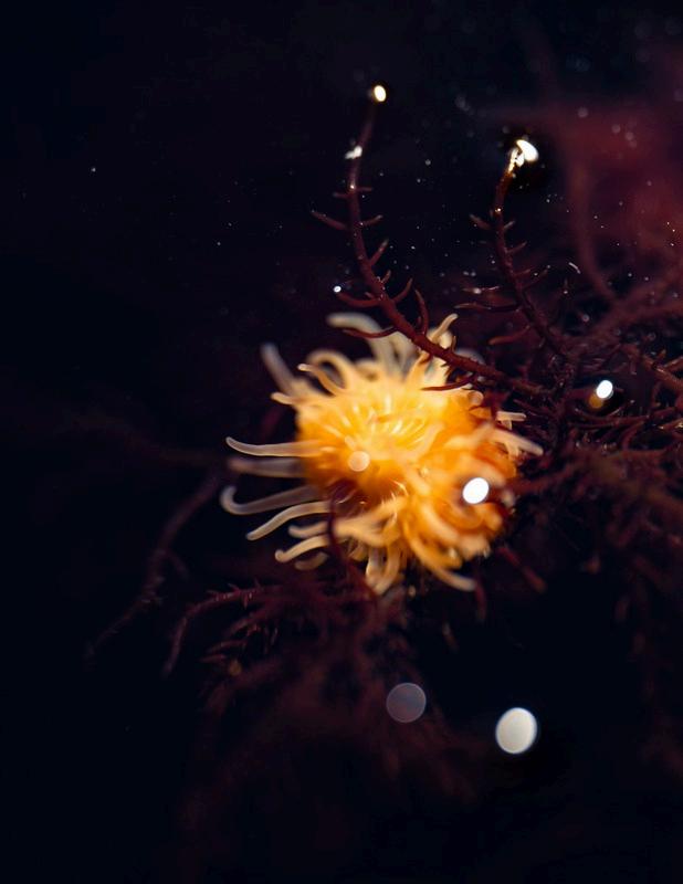
Orange anemone (Diadumene cincta) in tidepool Sunset Cliffs, 2024
The views expressed in this publication are solely those of Saltman Quarterly, its principal members, and the authors of the content of this publication. While the publisher of this publication is a registered student organization at UC San Diego, the content, opinions, statements, and views expressed in this or any other publication published and/or distributed by Saltman Quarterly are not endorsed by and do not represent the views, opinions, policies, or positions of the ASUCSD, GSAUCSD, UC San Diego, the University of California and the Regents or their offices, employees, or agents. The publisher of this publication bears and assumes the full responsibility and liability for the content of this publication.
In an effort to involve the UC San Diego community, Saltman Quarterly holds an annual photo contest. The winners of this contest have their images featured on the cover and interior pages of the journal.
AND
UC SAN DIEGO SCHOOL OF BIOLOGICAL SCIENCES
Letter from the Editors Letter from the Editors
Dear Reader,
It is our utmost pleasure to introduce this year’s edition of Saltman Quarterly, our annual flagship publication dedicated to spotlighting novel biological research at UC San Diego You hold in your hands the culmination of a year’s work of undergraduate scientific communication, collaboration, and creativity As you flip through this issue, we invite you, for a moment, to become a student of biology and to immerse yourself in fascinating stories unfolding within the field
Paired with synergistic visuals and figures, our journalistic features highlight how biological principles scale and echo across systems from glycans and evolutionary biology, which illustrate ho species, to p biology shap research sec which span ecological cr honored to Honors Thes the highest c
In a time o institutions, at SQ and stu of higher edu much inform cycle a doze grow into the
surely, we begin to take on the responsibilities that come with being scientists: not just to pursue knowledge but to share it meaningfully with the world and advocate for its continued support. This is why we do what we do at SQ, channeling our passions, frustrations, and hopes for the future into impactful and rigorous science journalism
This year, our mission extended beyond creative publication alone From expanding our STEM education outreach to support elementary, middle, and high schools in San Diego to hosting a biology student research symposium, we aimed to encourage students to see themselves reflected in science and research In many ways, SQ is our way of giving back, of building the scientific and intellectual community that we want to see for years to
Photo
TABLE OF CONTENTS
Saltman Dedication
Saltman Quarterly thanks the Saltman Family for their generosity and support Their contributions have allowed SQ to continue to spread Dr. Paul Saltman’s ideals of science communication and education
Glycans by Smriti Narayanan
The Biological Inevitabiliity of Antibiotic Resistance by Monica Wu
Brain Invader by Ioanna Andritsogianni
Astrocyte Plasticity Across the Lifespan by Keya Patel
How Expectation Shapes Pain by Aneesha Asthana Features
Research
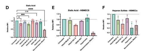
Characterizing the Impact of Classical and Novel Human Astrovirus Spike Proteins on the Endothelial Glycocalyx of Tissue-Specific Barrier Cells by Finn Coughlin
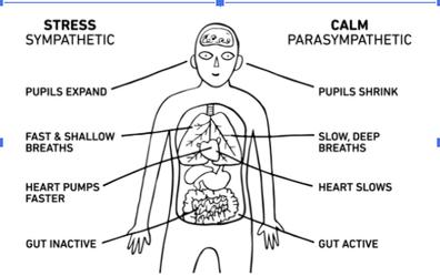
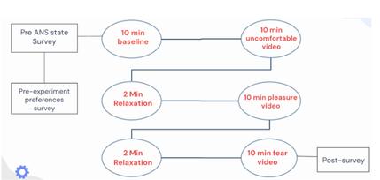
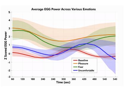
How We Involuntarily Convey Our Emotions through the Autonomic Nervous System by Suchita Rao
The Aral Sea Crisis: Environmental Consequences and Mitigation Policies by Jieyin Jiang Advancing Oncology Treatments through Genetic Engineering by Sanchali Tiwari
Undergraduates in the Biology Senior Honors Thesisprogram are required to complete a written thesis detailing their scientific research The Senior Honors Theses section, which presents the topics of their individual theses, highlights achievements of accomplished undergraduate researchers.
Meet the members of the 2025-2026 Saltman Quarterly staff who worked throughout the year to bring you this issue, as well as our online content, quarterly Insider magazines, and community outreach initiatives Staff
Dr. Paul Saltman
Professors often shape the course of our lives. Whether through lectures or time spent in the lab, they leave lasting impressions that guide our educational journeys. Chances are, you’ve had a professor who inspired you. At UC San Diego, alumni who have studied here from the late ’60s through the ’90s will likely share the same answer: Dr. Paul Saltman.
Dr. Saltman’s life was marked by resilience and impact. From an outside perspective, his early circumstances did not suggest a future in academia He was born on April 11, 1928 to an immigrant family and faced profound loss at a young age his mother passed away when he was three, and his father when he was twelve Despite these challenges, he was inspired by a high school chemistry teacher, setting him on a path that would ultimately define hiscareer.
He went on to earn his PhD in Biochemistry from the California Institute of Technology in 1953 From there, he would later become a faculty member at UC San Diego during its infancy as the provost of Revelle College, with the expressed goal of improving the standards of undergraduate education to match that of the graduate programs of the university. In 1972, he assumed the role of ViceChancellorforAcademicAffairs.
In 1980, Dr. Saltman returned to research with a notably different approach than many of his academic peers. Rather than embracing an elitist mindset and perpetuating the ideathatacademiaisasecluded‘ivorytower,’hepositioned himself rather as a researcher ‘for the public.’ He committed himself to accessibility and education for all. This was reflected in his appearance on “Patterns of Life,” which was a program produced for National Education Television, where he helped translate complex scientific concepts into easily digestible information that could benefit everyday people
Saltman’s research focused heavily on nutritional sciences, particularly his discovery of the nutritional role of trace metals like zinc and copper His work contributed to improvements in dietary strategies to reduce risk of heart disease and anemia. At the same time, he was deeply critical of the prevailing dietary advice of his era, which he often saw as misguided or insufficiently grounded in evidence. With this work in mind, he published The University of California San Diego Nutrition Book, aimed at the general public in informing proper dieting and fighting off “faddish beliefs.” He would go as far as to say, “There is no such thing as junk food; only good or bad choices,” to reiteratetheimportanceofeatingwell.
This combination of communication and pursuit for scientific knowledge made Dr. Paul Saltman a beloved professor among students So beloved in fact, that one student wrote "Paul Saltman is the most charismatic, altruistic,caringandnoblepersonIhaveeverknown Ihave neverrespectedanyonemorethanIrespecthim Heisloved by thousands of students” He won many teaching awards at his time at the University of Southern California, as well as from Revelle, Muir, Warren, and Marshall Colleges at UC SanDiego.
Outside of the academic world, Dr. Saltman was an avid lover of sports. Starting with basketball and boxing in high school, he later took up skiing, surfing, and tennis. He was an original member of the San Onofre Surfing Association, living up to his roots as a Southern California native
“
“ Every piece of scientific writing published... is a tribute to his pursuit for public understanding of biology and health.
In 1999, Paul Saltman sadly passed away. He left behind a profound impact on his students and biology as a whole. To honorthislegacy,5yearsafterhispassingin2004,Saltman Quarterly was founded by a group of undergraduate students.
More than two decades after his passing, Dr. Saltman’s legacy continues to shape UC San Diego. Every piece of scientific writing published in Saltman Quarterly is a tribute to his pursuit for public understanding of biology and health His memory, beyond his role as a researcher, vice chancellor, or provost and as a husband, father, and friend, is lived on by Saltman Quarterly Not by just honoring his unwavering pursuit of science, but by also adopting the mission of making research accessible to a broad audience, as Dr. Paul Saltman worked towards throughhisdedicationtodemocratizingscience.
Written by Aarya Vishnu
U R E S
F E A T U R E S
Stauffer's Tree Frog (Scinax staufferi) Guanacaste, Costa Rica, 2025
Photo by Inaya Nicholls
GLYCANS GLYCANS
Written by Smriti Narayanan
Illustrated by Elena Yu
THE KEY TO CELL AND ORGANISMIC EVOLUTION THE KEY TO CELL AND ORGANISMIC EVOLUTION
In the same way that humans use different languages, cells display different surface sugars and proteins based on their function, shape, and host organism. Sugars make up the linguistic and logistic basis of all food webs: they store energy and serve as a tool for cells to communicate and interact with the environment. Unique to other macromolecules, sugars possess immense structural complexity and can incorporate proteins and lipids into their signaling. All living cells are coated with a layer of sugars, called a glycocalyx, which allows a cell to identify itself and interact with its environment, much like a language. Across all domains of life, organisms use and produce thousands of unique monosaccharides, or the simplest form of a sugar These basic sugar units are linked together to form sugar chains, oligosaccharides, or polysaccharides, essentially creating a code composed of “glycoletters ” Within the same organism, cell surface sugars signal the organism and its immune system receptors that each cell is “self” and not damaged (as in the case of infected or cancerous cells), much like how members of the same language group communicate efficiently Though between hosts and pathogens, glycan patterns used by the host can detect “non-self” cells, like the recognition of different languages
What is especially remarkable about these complex sugar chains, commonly referred to as “glycans,” is their ability to serve as a multipurpose macromolecule. The range of glycan function spans from structural roles to complex interactions crucial for organism development, growth, and maintenance Cellulose and chitin are examples of structural glycans in plants, while sialic acids are cell surface signaling glycans found on vertebrate cells Cell-to-cell recognition, pathogen evasion, and immune system regulation are all mediated by the intricacies of glycan function Without any modulation
mechanism in place, cells would succumb to loss of protection, decreased structural integrity, and the eventual takeover of control-seeking viruses.
Though an essential biological component, sugars do not act in isolation They are often linked to either lipids or proteins, which act as cellular chaperones to protect sugars from degradation. While all living organisms have cells lined by glycans, glycan structures are not directly coded for inside the genome Rather, environmental factors such as diet, aging, and evolution directly influence the development and makeup of each organism’s glycocalyx, leading to immense diversity across all glycan forms
Situated at the surface of cells, glycans are the first point of contact between different cell types They are involved in the cellular recognition of numerous infectious and genetic diseases, as well as inflammation and infection. Many cells also secrete glycans into the extracellular matrix, a network of proteins and sugars surrounding cells in tissues and organs Beyond immunology, studying the path of glycan evolution has allowed researchers to inform different fronts of research, establishing an immunological and dietary link from modern humans to other primates Aside from sharing large brains, complex social behaviors, and a majority of our protein-coding genetic information with other primates, humans also share a number of glycans and glycoconjugates (proteins or lipids bonded to glycans) responsible for immune regulation and general extracellular communication. Glycobiology, the study of the biosynthesis, structure, and function of glycans, is the lens through which Dr Pascal Gagneux investigates immunological interactions The name of the game, according to Dr Gagneux, is understanding that without glycobiology, there would be a gap in the current understanding of cellular identity and communication
His lab in the Department of Pathology at UC San Diego studies the evolution of cell surface glycans and glycoproteins with respect to the evolutionary influence on hostpathogen interactions
IMMUNOLOGICAL APPLICATIONS OF GLYCOBIOLOGY
By studying molecular mechanistic changes to sialic acid structures on the surface of vertebrate cells, Dr Gagneux’s lab gained an acute understanding of the consequences of sialic acid variation in conditions such as metastatic cancers, age-related macular degeneration, and sperm-egg interactions, modeling a “survival of the luckiest approach ”
Any two organisms living within the same environment compete for resources, space, and overall population growth Just as a stealthy cat preys on a mouse, host-pathogen interactions follow an exceedingly competitive dynamic. But there are many cases, such as in gut microbiome host interactions, where host-secreted mucins both feed and exclude based on perception of threat. The cat and mouse analogy is best used to describe the evolutionary competition within the immune system The immune system is the cat, on the hunt yet always catching up to the mouse, or the pathogen In the case of metastatic cancers, the cat and mouse dynamic exists between the metastatic cancer cells and natural defense mechanisms in the tissue and immune system
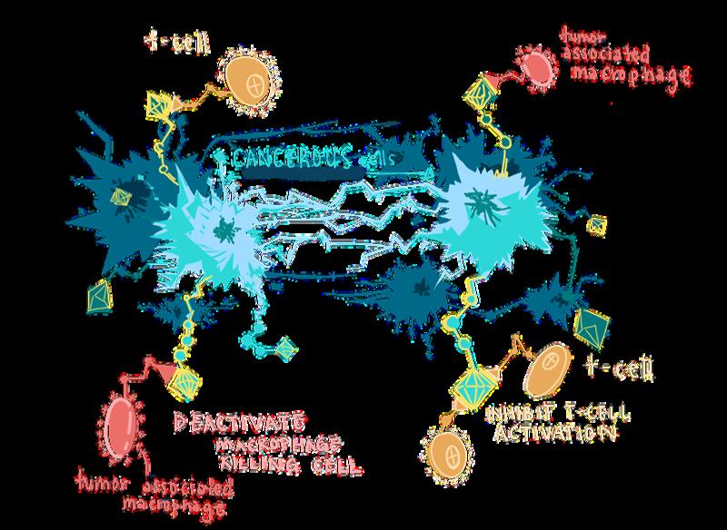
Unfortunately, in many cases the metastatic cancer may be more fit than the host immune system and can eventually give rise to a hostile tumor environment that suppresses and hijacks immune system machinery Thus, researchers are beginning to treat cancer as an immunological phenomenon, capable of evolving and dominating a given system analogous to an infectious disease
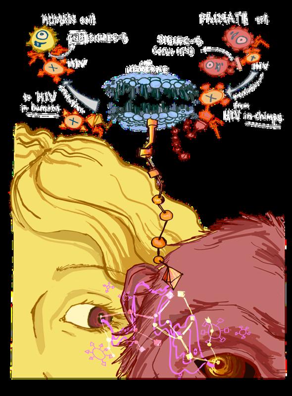
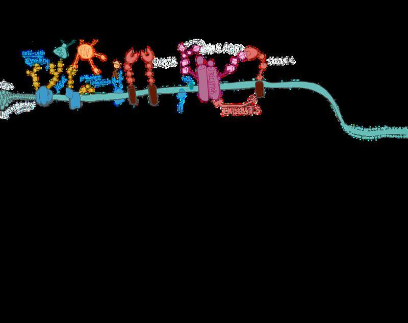
Figure 1 The long sugar sialic acid caps the end of a glycoprotein on the outer membrane of cells The SIGLEC cell surface receptor binds to sialic acids on glycoproteins to determine ‘self’ and ‘non-self’ molecules. .
When introduced to an infectious disease, the body releases multiple lines of defense The innate immune system acts as a general front line to initiate contact with a pathogen Then, the adaptive immune system stores this information in memory B cells and follows up with antibodies, offensive molecules specific to the particular pathogen In cases where this immune system defense fails, the security of an organism’s cells is compromised. This allows pathogens to manipulate cell surface glycans and glycoconjugates to gain entry into a host cell Glycans, which initially served as a receiving and signaling molecule to warn the cell of a potential threat, can now be exploited by a particularly evasive pathogen, with the pathogen becoming the fittest survivor in this case. Pathogens that have evolved the capacity to mimic host glycans are analogous to replicating the accent of another language group. Though not a product of knowing the language, an accent can still be very convincing to a listener 1
An area of focus from the immunological perspective is how sialic acids shape both cancer progression and virus evolution Sialic acids act as both critical building blocks and terminal caps for longer, more complex glycans In cancer, affected cells have been shown to produce abnormally high amounts of glycans with sialic acid caps, a phenomenon known as hypersialylation Cancer-associated antigens on the surface of cancer cells are recognized as an anomaly of sialic acids Hypersialylation, the extreme sialic acid presence, is caused by a dysregulation in the expression of enzymes In tumor cells, enzyme and protein expression may change as a result of cell differentiation or rapid cell growth In comparison to normal cells, which also have sialic acids on their surface, the concentration of sialic acid molecules in cancer cells is far greater, frequently decreasing cell-to-cell adhesion and interfering with cell signaling. While hypersialylation can sometimes reduce intercellular adhesion, it can also promote cell-to-extracellular-matrix adhesion, increasing binding to blood vessels in the surrounding tissue In the case of fastgrowing cancers such as breast cancer, polysialic acid (PSA), a long chain form of sialic acid which helps cells to move, allows cancer cells to become metastatic, or mobile beyond their initial site of growth The movement of cancer cells is what allows for the formation of new tumor sites across bodily systems, eventually leading to the complete organismal takeover of the disease
Fascinatingly, many infectious dise as influenza A and B and some co implement the same hypersialylatio
Sialic acids play a major role in i signaling, namely through specific sialic acid binding immunoglobulin (SIGLECs) and selectins SIGLE family of cell surface proteins th sialic acids on the surface of othe viral cell presents itself with hype it could trick SIGLEC on a norm recognizing a “self” molecule, e potential immune response T example of an “immune cloaking” by which a virus hides under the d native cell to evade a rapid an immune response Researchers ha SIGLECs as “double-edged sword of preventing autoimmunity again cells while easily being exploited cells and pathogens. As it turns complex immune battles take pla organisms, and recent research evolution reveals a likeness betwe diseases
THE CROSSROADS BETWEEN PRIMATOLO AND IMMUNOLOGY
Comparative biology serves as a central lens through which the Gagneux Lab approaches glycobiology, allowing researchers to identify evolutionary changes on a molecular level
The function, molecular structure, and interactions of cells with external stimuli are all determined by the host organism’s unique biological niche The types of glycans and glycoconjugates are uniquely tailored to the various foreign molecules and cells inside or outside an organism So in actuality, cellular identity exists on both a macro and micro scale, forcing researchers to consider a cell’s environment has a remarkable impact on its role
Dr Gagneux and Dr Ajit Varki of UC San Diego’s School of Medicine were among the first researchers to dive headfirst into the glycobiology of infectious disease evolution, particularly in research on the progression of the human immunodeficiency virus (HIV) to acquired immunodeficiency virus (AIDS) They studied how the first strains of HIV were able to shift their primary host from chimpanzees to humans, despite a clear difference in virus specificity and reactivity among the two species
independently evolved multiple times within chimpanzees, evading the adaptive and innate immune systems in the process Only after four distinct strains evolved did one lineage successfully cross over species into humans The HIV-1 strain translated into humans as a direct result of differential expression of the evolutionarily conserved inhibitory receptor SIGLEC-5 found on CD4-T cells CD4-T cells are “helper” white blood cells that stimulate other immune cells to create antibodies and record memory of infection. 8
The SIGLEC-5 receptor pathways differ between the two species in the level of immune system inhibition This inhibition level changes how viruses must adapt before crossing over to another species. The SIGLEC-5 receptor was hypothesized by researchers, namely Dr Varki, to protect T cells from HIV-induced cell death In this same study, Dr. Varki observed that 99% of chimpanzee CD4-T cells expressed SIGLEC5 Quite starkly, 99% of human CD4-T cells did not express SIGLEC-5 did While chimpanzees and humans can both contract 9
p infection by this virulent pathogen This indicates a key species difference, marking a divergence at some point in primate immune system evolution
This cross-species comparison with our notso-distant relatives lends insight as to why certain pathogens are detrimental, causing infection at the level of epidemics in our world This style of evolutionary and immunological interdisciplinary research is quickly becoming a method through which many biologists might find the answers to their questions about human identity
There are almost no limits to studying the role that evolution plays in anthropology and glycobiology. Thus it is important to uplift interdisciplinary collaboration, especially at research-centered institutions like UC San Diego Many publications produced by the Gagenux lab result from international collaborations with researchers from the Max Planck Institute and other labs at UC San Diego. A common sentiment glycobiologists share is the need for a better understanding of our sialome and the role evolution plays, both on a molecular level and an organismic level.
REFERENCES he name of the game, ding to Dr. Gagneux, s understanding that without glycobiology, would be a gap in the ent understanding of cellular identity and communication.””
USION
arded from gels in sequencing biologists are taught to wash s due to the messiness they might dataset. Dr. Gagneux feels that gists today still don’t grasp that might be producing a clean data e throwing away a monumental molecular information that reveals details about organismal
glycans is a deeply understudied molecular biology that has the answer longstanding questions cancer cell irregularities and vasion from the immune system, all through studying what occurs at the cell surface With genomics at the forefront of the biotech industry and 21st-century organismal discoveries, many scientists tend to focus on what is going on inside of a cell, forgetting about the crucial interactions that drive the cascade of foreign object interactions
What begins as a cell-to-cell contact culminates in a series of chain reactions that impacts an organism’s immune response, regulatory processes, and overall balance with its environment The manner in which we communicate with one another using language parallels how cells utilize their language of sugars to express chemical interactions to the outside world While we develop much of our conception of human identity externally through how we interact with the surrounding environment, glycobiology emphasizes to biologists, students, and the general public that the key to understanding human identity begins on a molecular level.
[1] Gagneux, P , Hennet, T , & Varki, A (2022)
Biological functions of glycans - Essentials of Glycobiology - NCBI bookshelf National Center for Biotechnology Information. https://www ncbi nlm nih gov/books/NBK57998
4/
[2] Cao, H , & Chen, X (2024, July 30) General consideration on sialic acid chemistry PMC Home https://pmc ncbi nlm nih gov/articles/PMC1128
8308/
[3] Vliet, S J , & Kooyk, Y (n d ) Sialic acids in cancer biology and immunity recent advancements PMC Home https://pmc ncbi nlm nih gov/articles/PMC1249 4545/[4] Drummond-Guy, O , Daly, J , Wu, A , Stewart, N., Milne, K., Duff, C., Nelson, B., Williams, K , & Wisnovsky, S (2025, March 20) Polysialic acid is upregulated on activated immune cells and negatively regulates anticancer immune activity PMC Home https://pmc ncbi nlm nih gov/articles/PMC11965 634/
[5] Habeeb, I , Alao, T , Delgado, D , & Buffone, A (2024, November 19) When a negative (charge) is not a positive: Sialylation and its role in cancer mechanics and progression PMC Home https://pmc ncbi nlm nih gov/articles/PMC116118 68/
[6] Zhu, W , Zhou, Y , Guo, L , & Feng, S (2024, September 30). Biological function of sialic acid and sialylation in human health and disease Nature.
https://www nature com/articles/s41420-02402180-3
[7] Raïch-Regué D, Resa-Infante P, Gallemí M, Laguia F, Muñiz-Trabudua X, Muñoz-Basagoiti J, Perez-Zsolt D, Chojnacki J, Benet S, Clotet B, Martinez-Picado J, Izquierdo-Useros N Role of Siglecs in viral infections: A double-edged sword interaction Mol Aspects Med 2023 Apr;90:101113 doi: 10 1016/j mam 2022 101113 Epub 2022 Aug 16 PMID: 35981912; PMCID: PMC9923124
[8] Binning, J , Chesarino, N , Emerman, M , & Gross, J. (2019, December 11). Structural Basis for a Species-Specific Determinant of an SIV Vif Protein toward Hominid APOBEC3G Antagonism. Cell Host and Microbiome: A Cell Press Journal https://www.cell.com/cell-hostmicrobe/fulltext/S1931-3128(19)30538-4? returnURL=https%3A%2F%2Flinkinghub elsevier com%2Fretrieve%2Fpii%2FS1931312819305384% 3Fshowall%3Dtrue
[9] Soto, P , Karris, M , Spina, A , Richman, D , & Varki, A (2021, September 4) Cell-intrinsic mechanism involving siglec-5 associated with divergent outcomes of HIV-1 infection in human and chimpanzee CD4 T cells PubMed
Smriti Narayanan is a Molecular and Cell Biology major graduating in 2027.
THE BIOLOGICAL THE BIOLOGICAL INEVITABILITY OF INEVITABILITY OF ANTIBIOTIC ANTIBIOTIC RESISTANCE RESISTANCE
Written by Monica Wu
Illustrated by Alisa Liao
A Antibiotic resistance is well known as one of the most pressing modern crises According to the World Health Organization, antimicrobial resistance contributed to approximately 1 14 million deaths globally in 2021 alone,
making it one of the leading issues in global public health
1
However, resistance is often misunderstood as a modern development, with the assumption that bacteria only evolved resistance due to overprescription and misuse of antibiotics While these are major contributing factors, the mechanisms behind bacterial resistance are ancient strategies that have been refined over billions of years of microbial warfare.
Due to the nature of bacterial reproduction, genetic mutations arise almost constantly in bacterial populations. Through the genetic insertions, deletions, and substitutions that occur during the process of reproduction, a continuous supply of potentially beneficial mutations allow bacteria to resist changes in their environment that would otherwise lead to their extinction. In theory, resistance should be manageable through the simple evolutionary principle that selection pressure drives trait selection If clinicians use antibiotics sparingly while developing new antibiotics faster than resistance can spread through a bacterial population, bacterial survival should remain at a minimum. In reality, however, pervasive infections, which harbor bacteria long enough to develop resistance to consistent antibiotic use, and unsupervised antibiotic use, which kills susceptible bacteria to die and allows resistant strains to proliferate, have accelerated bacterial evolution beyond the rate at which new drugs can be developed
In 2016, an experiment at Kishony Lab at Harvard Medical School made the inevitability of bacterial resistance impossible to ignore To visualize bacterial evolution in real time, the researchers constructed a massive petri dish with nine zones of escalating antibiotic concentrations, from those that were barely lethal to E coli at the edges to concentrations 1,000 times the deadly dose at the center Researchers placed E coli at the antibiotic-free outer edge and observed what happened Within 11 days, the bacteria had spread across the entire Petri dish, surviving even the deadliest band. Since only resistant bacteria could have survived the antibiotic zones, the spread revealed that resistant mutants had emerged and were advancing The Kishony Lab made clear the harrowing truth that the evolution of antibiotic resistance in bacteria is biologically inevitable But the experiment left a critical question unanswered: what mechanisms allow bacteria to adapt so rapidly? While the Harvard study focused on antibiotic stress, understanding bacterial adaptation to any environmental pressure could reveal fundamental principles of rapid evolution 2
BACTERIA’S MULTIPLE ROUTES TO RESISTANCE
The key principle underlying resistance is that bacteria do not rely on a single “lucky” resistance gene; rather, even bacteria of the same strain will independently evolve different resistance strategies, whether by degrading the antibiotic, expelling it from the cell, or modifying the drug’s target protein Additionally, bacterial evolution is not constrained to evolution within a single bacterium; survival genes can be traded
horizontally, or between different
organisms via small rings of DNA called plasmids Instead of waiting for resistant offspring to multiply, bacteria can share resistance genes directly with their neighbors and spread survival strategies across lineages within hours 4
Bacteria also possess the ability to overwhelm antibiotic drugs through gene amplification, a brute-force mechanism of overproducing enzymes evolved to neutralize compounds toxic to bacteria, including antibiotics Under intense antibiotic pressure, the stress disrupts normal DNA replication and causes the replication machinery to stall. When bacteria attempt to repair their stalled replication machinery, errors in the repair process can accidentally duplicate entire sections of DNA If this contains resistance genes, the bacteria have now created multiple copies of this gene. Each gene copy can be transcribed into messenger RNA and translated into a protein, so multiple copies produce proportionally more enzymes For example, a bacterium with a beta-lactamase gene, which codes for an enzyme that breaks down penicillin, can quadruple beta-lactamase production under stress to effectively neutralize four times the amount of antibiotic. Yet maintaining so many gene copies comes at a cost The extra energy required for increased replication and protein production comes at the expense of growth When antibiotic pressure is removed, bacteria often shed the extra copies of resistance genes to conserve energy, making gene amplification a temporary but effective survival strategy
5, 6
Beyond gene multiplication, bacteria can remove drugs from the cell entirely Antibiotics can be removed from bacterial cells through efflux pumps, or molecular pumps that normally expel other toxic substances from bacterial cells In E coli, one of the most well-studied efflux systems is the AcrAB-TolC pump, a protein complex that spans both the inner and outer bacterial membranes When antibiotics enter the cell, regulatory proteins such as MarA, SoxS, and Rob detect the threat and activate transcription of efflux pump genes. Bacteria can upregulate AcrAB-TolC pump production through mutations in regulatory genes, further increasing the number of pumps and boosting antibiotic export By expelling antibiotics faster than the drugs can accumulate, efflux pumps keep intracellular concentrations below lethal levels and allow the bacterium to survive. If bacteria had only one path to resistance, its evolution may have occurred more slowly and the problem more manageable via antibiotics 7 However, resistance evolves rapidly through simultaneous employment of multiple mechanisms, along with the sharing of resistance genes across populations Selection explains how beneficial mutations spread through bacterial
populations once mutations arise, but not why adaptive mutations appear so readily when bacteria face stress A deeper question emerges: are these mutations truly random in the first place, or do bacteria possess systems that make adaptive changes more likely under stress?
“A deeper question emerges: are these mutations truly random in the first place, or do bacteria possess systems that make adaptive changes more likely under stress?”’
TRANSPOSONS AS EVOLUTIONARY ACCELERATORS
Transposons are mobile DNA sequences that can relocate within a genome Standard gene regulation works without changing the genetic sequence, where transcription factors reversibly bind to the DNA strand to turn genes on or off by physically controlling the access of transcription machinery to the gene Transposons work differently by physically relocating DNA segments to new genomic locations, where the movement and structuring of the genome can potentially activate nearby genes. What remains unclear is whether transposon insertions occur randomly or as a response to environmental conditions
At UC San Diego, the Saier Lab uses E coli as a model organism to investigate the importance of these mobile genetic elements, their discoveries challenging the conventional evolutionary theory of random mutations. Research led by Jonathan Onstead from the Saier Lab reveals that stress can influence where these genetic elements insert, suggesting bacteria may have more control over genetic changes than previously thought Rather than landing randomly across the genome, the Saier Lab discovered that environmental stress destabilizes and unwinds certain DNA doublehelix structures, a process called stress-induced duplex destabilization (SIDD) Transposons preferentially insert into these SIDD regions, creating hotspots for genetic changes This stressguided insertion mechanism is known as transposon-mediated directed mutation
The Saier Lab focuses on Insertion Sequence 5 (IS5), one of the smallest transposons 8 IS5’s simplicity and presence in E coli, a well-studied model organism, make IS5 an ideal system for studying transposon responses to environmental stress In a 2017 study on IS5-mediated gene activation under starvation, the Saier lab found that starved E. coli in the presence of glycerol, an unconventional E coli metabolite, dramatically increased IS insertion rates Specifically, the stress of starvation destabilized the DNA near the glpFK operon, which codes for the metabolism of glycerol The destabilized DNA then allowed IS5 insertions, which in turn activated the exact genes needed to
metabolize glycerol and survive While the transposon 9 does not seek out beneficial genes, its preference for stress-destabilized DNA regions increases the likelihood that mutations occur when the bacterium is under pressure, whether from starvation (as in the glycerol study) or from antibiotic exposure Although this mechanism has been observed across multiple bacterial phyla, transposon insertion remains slower, riskier, and less efficient than routine transcriptional regulation, making transposons more of an “ emergency response ” than a preferred adaptation dtrategy The Saier Lab’s research on transposons complicates the traditional evolutionary picture: bacteria don’t directly control when mutations occur, but stressresponsive systems make beneficial genetic changes more likely when bacteria need them most. 3
METABOLIC FLEXIBILITY: THE FINAL BARRIER
Most antibiotics target a single bacterial process. Penicillin blocks transpeptidases, preventing cell wall synthesis Fluoroquinolones inhibit DNA gyrase and topoisomerase IV, preventing DNA replication during cell division 10, 11
Moving beyond individual genetic and regulatory mechanisms of bacteria, their ultimate defense lies in metabolic flexibility and interconnected backup pathways What makes bacterial resistance difficult to overcome is how these defenses work together as a coordinated system When one pathway is blocked, bacteria reroute critical functions through backup routes To address the network resilience issue, the logical approach would be designing drugs that attack multiple pathways at once
The network-based approach to attacking bacteria’s antibiotic resistance makes sense in theory If a drug disrupts multiple essential pathways concurrently, each individual bacterium would need to have multiple genetic adaptations to survive one drug However, creating antibiotics with multiple targets is much harder to practically achieve Antibiotic resistance is not a simple, single-gene change, but a complex, system-coordinated physiological defense that protects the bacteria’s essential metabolic network The systemic level of coordination has presented a challenge for drug design to target antibiotic resistance Unfortunately, even if scientists deploy network-based approaches to design drugs that target multiple bacterial pathways, bacteria may still reroute their metabolism or switch to alternative pathways to keep essential functions running Bacterial adaptability continues to outpace our understanding 3 3
Beyond the challenge of bacterial flexibility, multi-target drugs face another fundamental barrier in human biology When discussing multi-faceted antibiotics, practicality and human safety also come into question Dr Medrano-Soto mentions that while attacking bacteria on multiple fronts sounds promising, such strategies could have devastating effects on human cells.3
and human cells share fundamental metabolic processes, including protein synthesis, DNA replication, and ATP production. Human mitochondria, which 12 evolved from ancient bacteria, retain bacterial-like ribosomes that are particularly vulnerable to drugs targeting protein synthesis A drug aggressive enough to disrupt multiple bacterial networks may interfere with mitochondrial function, damaging organs like the heart and liver. The systematic design of bacterial resistance may make multi-target antibiotics a double-edged sword
The Saier Lab’s research and the Harvard MegaPlate experiment demonstrate that no matter how hostile the environment, bacteria will always employ multiple complementary strategies This includes horizontal gene transfer, stress-induced mutation mechanisms, and flexible metabolic networks, together rerouting essential functions and ensuring survival. Bacterial multilayered defenses make antibiotic resistance a formidable challenge that demands sustained, long-term research efforts
Yet, the gap between what we know and what bacteria can do remains vast Dr Medrano-Soto notes that despite E coli being the most extensively studied bacterium, decades of research have revealed only about 15 percent of its biology. By studying how bacteria regulate genes, transport molecules, and adapt their 3 metabolism at a systems level, this basic research is building the foundation for future breakthroughs Ongoing studies continue to reveal how bacteria adapt so rapidly, creating opportunities for smarter interventions. For now, the question shifts from “Can we prevent resistance?” to “How do we stay ahead of it?” And, the answer lies in combining scientific humility with determined innovation, acknowledging how much we still do not understand. Understanding the inevitability of resistance is the first step toward learning how to live with it, manage it, and eventually, outsmart it
Monica Wu is a Molecular and Cell Biology major graduating in 2027.
“For now, the question shifts from ‘Can we prevent resistance?” to ’How do we stay ahead of it?” And, the answer lies in combining scientific humility with determined innovation, acknowledging how much we still do not understand.”’
REFERENCES
[1] WHO 2025 Global antibiotic resistance surveillance report 2025 World Health Organization [accessed 2026 Jan 16]; https://www who int/publications/i/item/9789240116337
[2] Harvard Medical School 2016 The evolution of bacteria on a "MegaPlate" Petri dish YouTube [accessed 2026 Jan 17]; https://www.youtube.com/watch?v=plVk4NVIUh8.
[3] Wu M, Saier MH Jr, Medrano-Soto A 2025 Saltman Quarterly Journal Feature article interview on antibiotic resistance.
[4] Scuttik S 2016 See for yourself: A giant petri dish models antibiotic resistance CNN Health [accessed 2026 Jan 16]; https://www cnn com/2016/09/08/health/giant-petri-dish-antibioticresistance
[5] Silva, KPT, Khare, A 2024 Antibiotic resistance mediated by gene amplifications npj Antimicrob Resist [accessed 2026 Jan 16]; https://doi org/10 1038/s44259-024-00052-5
[6] Slack A, Thornton PC, Magner DB, Rosenberg SM, Hastings PJ 2006 On the Mechanism of Gene Amplification Induced under Stress in Escherichia coli PLOS Genetics [accessed 2026 Jan 16]; https://doi org/10 1371/journal pgen 0020048
[7] Bahaj SS, Al-Dhubaibi MS, Noman A et al 2025 Expression of multidrug efflux pump gene acrAB in Escherichia coli: a systematic review and meta analysis BMC Infectious Diseases [accessed 2026 Jan 22]; https://doi.org/10.1186/s12879-025-11778-6.
[8] Onstead J, Zhang Z, Huo J, Ord JW, Smith S, and Saier MH Jr 2024
Investigating How Genomic Contexts Impact IS5 Transposition Within the Escherichia coli Genome Microorganisms [accessed 2026 Jan 16];
https://doi org/10 3390/microorganisms12122600
[9] Humayun, MZ, Zhang Z, Butcher AM, Moshayedi A, Saier MH Jr 2017
Hopping into a hot seat: Role of DNA structural features on IS5-mediated gene activation and inactivation under stress PloS one [accessed 2026 Jan 16]; https://pmc ncbi nlm nih gov/articles/PMC5493358/
[10] Bertonha AF, Silva CCL, Shirakawa KT, Trindade DM, Dessen A 2023
Penicillin-binding protein (PBP) inhibitor development: A 10-year chemical perspective Experimental biology and medicine [accessed 2026 Jan 22]; https://doi org/10 1177/15353702231208407
[11] Hooper DC, Jacoby GA 2016 Topoisomerase Inhibitors: Fluoroquinolone Mechanisms of Action and Resistance Cold Spring Harbor Perspectives in Medicine. [accessed 2026 Jan 22];
https://doi org/10 1101/cshperspect a025320
[12] Cooper GM. 2000. The Cell: A Molecular Approach. 2nd edition. National Library of Medicine [accessed 2026 Jan 22];
https://www ncbi nlm nih gov/books/NBK9841/
BRAIN INVADER BRAIN INVADER
Written by Ioanna Andritsogianni
Illustrated by Yoyo Lu
HOW BLOOD LEAKAGE DRIVES HOW BLOOD LEAKAGE DRIVES
NEUROINFLAMMATION IN NEUROLOGICAL DISEASE NEUROINFLAMMATION IN NEUROLOGICAL DISEASE
RIVERS IN THE HUMAN BODY LANDSCAPE
The harmonious fluidity of nature often conceals its reliance on structure Behind the dancing movements of animals, plants, cells, and molecules alike lies a carefully evolved organization of biological systems. When this organization falters, even briefly, systems begin to break down, and the fluid movements are no more
In ancient Greece circa 300 B C , Aristotle laid the foundation for viewing and studying the body as a collection of distinct organs through his investigations of anatomy; yet, since antiquity, scientific advancement has revealed that biological systems are not so clear-cut The interconnected nature of such systems is particularly embodied by our vascular system Like a river network that transports water from mountaintops to villages, the vascular network connects far places in an organism’s landscape via a constant flow of blood
In the same way that a river is contained by its banks, blood circulation in the human body is restricted to certain regions Although the brain and spinal cord are densely supplied by blood vessels, circulating blood is normally contained within those vessels and does not spill into the surrounding neural tissue The exclusion of toxins and unwanted substances in blood from neuronal tissues is enforced by the blood-brain barrier (BBB), a selective interface composed of tightly connected endothelial cells, which are thin, flat cells that make up the inner lining of blood vessels A physical and chemical seal, the BBB restricts which molecules and cells can enter the central nervous system (CNS)
In its healthy state, the BBB acts as a selectivity filter for the brain Small molecules such as oxygen and glucose can pass through via diffusion or specialized transporters, while larger proteins, immune cells, and any potentially toxic blood-borne molecules are actively barred from entry
BBB regulation of CNS entry is crucial for neural signaling, appropriate immune activation, and protection from inflammatory or metabolic stress By controlling the brain’s chemical environment, the BBB preserves the conditions required for proper synaptic transmission and neural circuit stability
1
2
Despite the protection afforded by the BBB, physicians have still observed blood leaks in the brain and spinal cord in association with severe and chronic neurological diseases and traumatic brain injury Observations suggest that when neurological disease manifests, the integrity of the BBB is compromised and blood components are allowed to enter regions of the brain where they are normally excluded. For decades, this was the prevailing hypothesis: neurological damage caused an uncontrolled opening of the BBB, and blood proteins entered the brain only as a consequence of disease. Under this view, research framed blood leakage as a symptom of ongoing brain pathology rather than an active contributor to it This interpretation persisted largely unchallenged until one simple but remarkable experiment began to unravel it
While doing his postdoctoral studies in Dr Katerina Akassoglou’s lab at UCSF, Dr Andrew Mendiola, now a UC San Diego professor, injected one microliter of blood from a healthy mouse into the brain of another healthy mouse The next day, the lab analyzed cellular changes in gene expression at the injection site They observed the activation of immune-related transcriptional pathways in microglia, the resident immune cells of the CNS More specifically, the injected blood had reprogrammed microglia toward gene expression profiles like those seen in neurological disease.
Microglia are largely responsible for immune surveillance, debris clearance, and the maintenance of tissue homeostasis 3
In their pathologically active state, rather than performing routine maintenance and repair, microglia are tuned to oxidative stress production and inflammatory immune responses. 2
In other words, the presence of blood alone was sufficient to push healthy microglia into a disease-associated state, altering how they sensed and responded to their environment
The lab repeated this experiment with one alteration: they removed fibrinogen, a bloodclotting factor, prior to injection. Fibrinogen, abundant in circulating blood, assists in blood coagulation and wound healing The lab chose to remove fibrinogen to test whether a 4 specific blood protein, rather than blood as a whole, was driving the previously observed microglial changes When fibrinogen was absent, the blood-induced gene expression changes in microglia failed to occur This result revealed that microglial activation was not driven by blood exposure in general but by a specific molecular signal. By identifying fibrinogen as sufficient in inducing 2 disease-associated immune states once it enters the brain, this experiment reframed blood leakage as an active biological process linking the nervous, immune, and circulatory systems through one misplaced interaction.
BLOOD-MICROGLIA INTERACTIONS
Microglia constantly interpret molecular cues in their environment, and those cues shape whether they remain in a stable “homeostatic” state or shift toward damaging immune activation Microglial activation can support repair and cleanup after injury, and that response is often beneficial early on However, problems tend to arise when activation becomes prolonged or redirected toward pathways that injure neurons and myelin. What surprised researchers was not simply that blood could activate microglia, but that individual blood proteins could drive fundamentally different immune behaviors through the same receptor
When fibrinogen enters the brain, it binds to the microglial receptor CD11b, also known as integrin αMβ2, a surface receptor involved in immune signaling and cell adhesion In this context, fibrinogen-CD11b interactions activate 5 gene programs associated with oxidative stress, including pathways that increase reactiveoxygen species (ROS) along with interferon signaling and inflammatory activation Oxidative stress refers 6 to the production of reactive oxygen species, which can be protective at controlled levels but damaging when excessive or prolonged What made 7 this finding especially striking was that CD11b was already well-known to immunologists The receptor had long been studied for its role in immune functions, including clearance of debris and regulation of inflammation Yet in the brain, engagement of CD11b by fibrinogen 5 produced a response that was pathological instead of protective
Herein lies a deeper question: was the CD11b receptor on the microglial surface itself the problem, or did the outcome depend on which molecular signal activated it? The answer emerged when researchers examined another CD11b ligand known as iC3b, a complement protein part of the innate immune system that tags pathogens and damaged cells for removal Like fibrinogen, iC3b binds 8 the CD11b receptor; however, the iC3b complement protein binding activates pathways linked to phagocytosis, meaning debris clearance Rather than act 6 the m
an interpretive checkpoint that translates different molecular inputs into distinct microglial behaviors
Researchers did not expect that the same receptor on microglia could mediate such opposing outcomes In the brain, the difference between fibrinogen and the iC3b complement proteins is not subtle: one accelerates immune-driven damage, while the other supports maintenance This contrast revealed that blood-induced pathology is not caused by immune receptor activation alone but by which blood-derived signals gain access to the brain and how microglia respond to them To understand whether fibrinogen was unique in driving these responses, researchers began testing what happened when other major components of blood were removed Removing albumin, the most abundant protein in circulation, had no effect on microglial activation so long as fibrinogen remained present Thus, simply allowing blood proteins into the brain was not enough to trigger immune dysfunction. The same logic applied to complement proteins
Even when immune-related proteins were removed, microglial inflammation persisted if fibrinogen was still in circulation. Only when fibrinogen itself was absent did bloodinduced transcriptional rewiring in microglia diminish.6
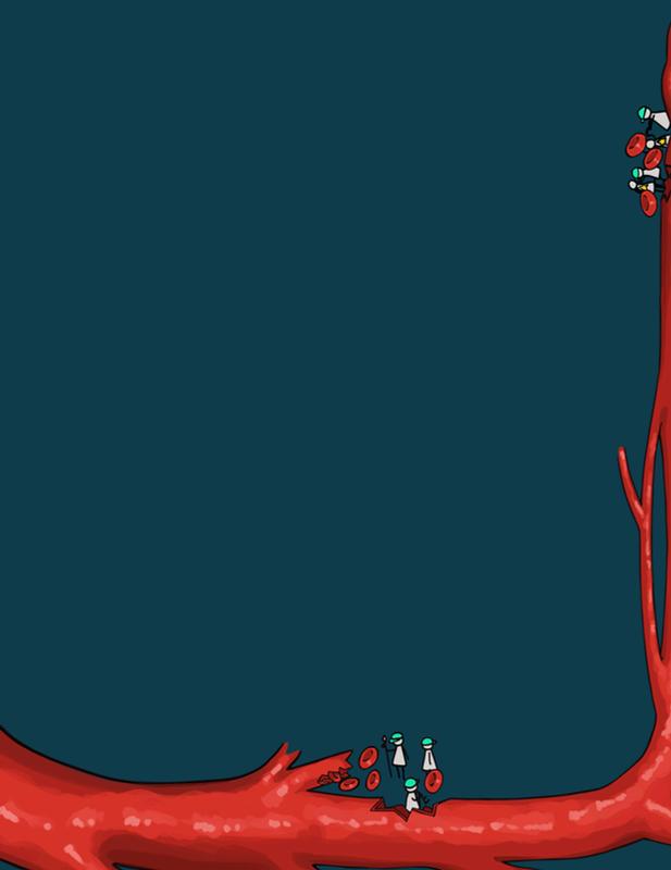
In a healthy brain, the BBB functions like a riverbank, keeping powerful biological signals confined to their proper channels When that boundary breaks, the current carries signals which reshape the landscape surrounding it
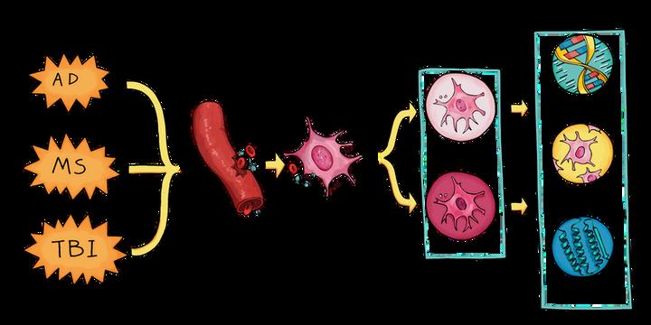
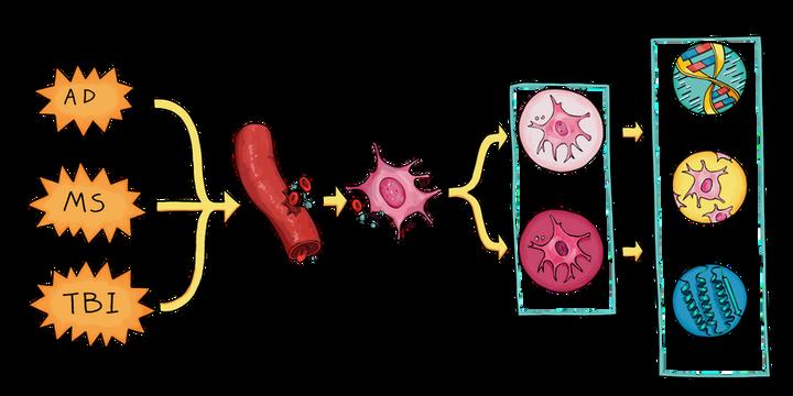
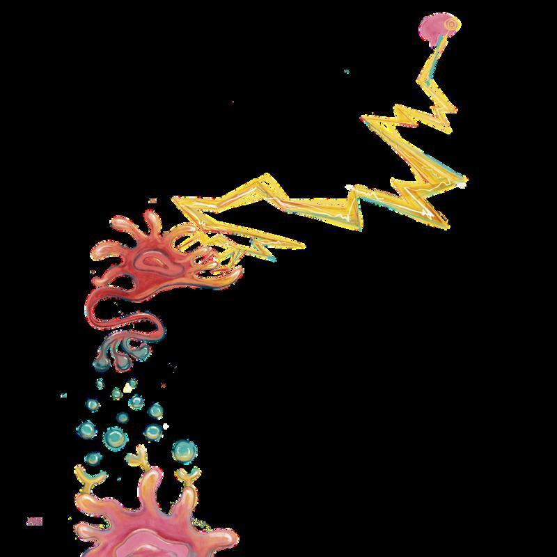
Figure 2 Across Alzheimer’s disease, multiple sclerosis, and traumatic brain injury, blood proteins enter brain tissue and engage microglia. Tox-seq sorts microglia by reactive oxygen species output, and multi-omics links oxidative microglial states to shared diseaseassociated signatures
As a brain invader, fibrinogen behaves like a foreign current, redirecting immune pathways that are helpful in peripheral tissues, where they support clotting and repair, into drivers of inflammation and oxidative damage inside vulnerable neural tissue
SHARED MICROGLIAL FINGERPRINTS ACROSS
NEUROLOGICAL DISEASE
Across different neurological diseases, blood exposure leaves behind a molecular fingerprint on microglia A “fingerprint” is a recurring pattern of gene expression and immune behavior that appears when microglia encounter blood-derived proteins What makes this pattern striking is its consistency across otherwise distinct diseases In models of Alzheimer’s disease, multiple sclerosis, and traumatic brain injury, microglia exposed to blood converge on nearly the same transcriptional programs, which exhibit heightened oxidative stress activity and inflammatory signaling Despite differences in disease onset, affected brain regions, and clinical symptoms in disease onset, the immune response at the level of microglial gene expression looks remarkably similar. Rather than acting as an original cause of disease, blood exposure appears to accelerate processes already in motion In this framework, fibrinogen functions less like a trigger and more like a gas pedal, amplifying
2 immune activation after other pathological processes are underway The molecular fingerprint left by blood exposure helps explain why diverse n
shared inflammatory outcomes Thus, by targeting blood-derived signals that drive a shared immune response, it may be possible to intervene in multiple neurological conditions at once, rather than addressing each disease individually
TOOLS TO STUDY THE NEUROVASCULAR
INTERFACE
Identifying shared microglial fingerprints across neurological disease raises a practical question: how can researchers reliably detect and compare immune states across conditions? To approach this question of practicality, researchers use multi-omic approaches. Multi-omics combines large-scale biological measurements, such as transcriptomics (all expressed RNA molecules) and proteomics (all translated proteins), to capture systemwide cellular states rather than focusing on single pathways 10
OPEN QUESTIONS AND EMERGING DIRECTIONS
Moving forward, one open question that remains is how blood-induced microglial changes evolve over time Current experimental models mostly focus on acute exposure or early responses, but in human disease, blood-brain barrier disruption can be intermittent, chronic, or cumulative For example, traumatic brain injury involves acute vascular damage that can lead to long-term neuroinflammation and increased risk of neurodegeneration It remains unclear whether repeated blood leaks compound microglial dysfunction, whether microglia retain a form of “ memory ” of past exposure, or whether later perturbations further destabilize existing immune states
Another emerging inquiry concerns how blood toxicity integrates with other biological axes, including metabolism and systemic physiology Microglia are long-lived cells with high energetic demands, and metabolic state influences whether immune activation supports repair or drives damage Dysregulated metabolism may therefore exacerbate blood-driven immune responses by sustaining oxidative stress and inflammatory signaling 2
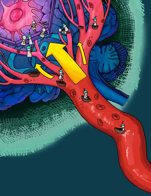
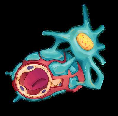
Figure 1 A microglial cell patrols alongside a brain blood vessel When the BBB weakens, blood proteins can escape the vessel and enter neural tissue, exposing microglia to signals they rarely encounter.
Humans continue to build beside water because resource outweighs certainty Biology, time and time again, makes that same choice
Ioanna Andritsogianni is a Molecular and Cell Biology major graduating in 2026.
REFERENCES
[1] Daneman R, Alexandre P 2004 The bloodbrain barrier Cold Spring Harb Perspectiv Biol 7(1)
approach allows researchers to connect molecular fingerprints directly to known pathogenic immune states, rather than
In practice, multi-omic analyses compare defined biological conditions, such as microglial exposure to blood proteins or disease model immune cells to healthy controls Multi-omic comparisons make it possible to identify groups of genes and proteins that change together in response to specific biological perturbations A major challenge with system-wide datasets is deciding which changes matter most. Researchers use Tox-seq to address this problem Tox-seq is an experimental and computational after which multi-omic profiling is applied to each population. This 2 inferring disease relevance from gene expression changes alone
Although originally developed for 6 neuro-immunology research, Tox-seq can be utilized beyond the brain
Similar strategies can be applied to tissue-resident immune cells in other organs, where oxidative stress also plays a central role in diseaseprogression
Tools like Tox-seq translate shared immunefingerprints into measurable, comparablestates, allowing attention to shift from simply detecting blood-driven immune responses to understanding their timing, progression, and potential points 2 of intervention
More broadly, work on neurovascular immunology contributes to a shift in how the brain is conceptualized. Rather than a protected organ occasionally breached in disease, the brain emerges as a system in constant negotiation with the vascular and immune systems Disruptions in sleep, infection, stress, or aging may subtly alter the system’s balance long before overt disease develops. This perspective shifts attention from single points of breakdown to the larger question of how biological systems manage flow, protection, and risk at the same time Nature holds itself together through balance, not rigidity Rivers bring life by moving freely, even as their banks quietly bear the risk of overflow The human body makes the same tradeoff Blood circulates to sustain life, while the brain depends on boundaries that carefully shape that current
When those boundaries weaken, signals meant for one landscape spill into another, and familiar systems begin to behave in unfamiliar ways For biologists, meaning lies in these tensions.
[2] Andritsogianni I, Mendiola AS 2025
Saltman Quarterly Journal features article interview on blood-induced microglial changes in neuropathology.
[3] Colonna M, Butovsky O 2017 Microglia Function in the Central Nervous System During Health and Neurodegeneration Annu Rev Immunol 35:441-468
[4] Petersen M, Ryu J, Akassoglou K 2018 Fibrinogen in neurological diseases: mechanisms, imaging and therapeutics Nat Rev Neurosci 19, 283-301
[5] Schmid MC, Khan SQ, Kaneda MM et al 2018 Integrin CD11b activation drives anti-tumor innate immunity Nat Commun 9(5379)
[6] Mendiola AS, Yan Z, Dixit K et al 2023
Defining blood-induced microglia functions in neurodegeneration through multiomic profiling Nat Immunol. 24, 1173-1187.
[7] Pizzino G et al 2017 Oxidative Stress: Harms and Benefits for Human Health Oxidative medicine and cellular longevity Oxid Med Cell Longev 8416763
[8] Ueda T, Rieu P, Brayer J, Arnaout MA 1994 Identification of the complement iC3b binding site in the beta 2 integrin CR3 (CD11b/CD18)
Proc Natl Acad Sci USA 91(22):10680-10684
[9] Uribe-Querol E, Rosales C 2020
Phagocytosis: Our current understanding of a universal biological process Front Immunol 11.
[10] Hasin Y, Seldin M Lusis A 2017 Multi-omics approaches to disease. Genome Biol 18, 83
ASTROCYTE ASTROCYTE PLASTICITY PLASTICITY
ACROSS ACROSS
THE THE LIFESPAN LIFESPAN
Written by Keya Patel
Illustrated by Hailey Chee
Since the emergence of modern neuroscience in the 1960s, neurons alone were thought to hold the brain’s secrets Learning, memory, and age-related decline were all pinned on these electrically excitable cells But what if the brain’s true power lies not just in neurons, but in the cells that surround them?
In the late twentieth century, through the work of neuroscientist Ben Barres and others, a growing field of research revealed that neurons do not act alone in shaping brain circuits Instead, focus shifted towards glia Glia, meaning “glue” in Greek, perform crucial functions such as helping form the blood–brain barrier, regulating neurotransmitters and neural communication, and supporting neurons throughout the brain More specifically, researchers began examining astrocytes, special star-shaped glial cells that were initially thought of as only support units of the brain. They have been discovered to orchestrate the brain’s environment, influencing everything from memory to attention 1
The role of astrocytes in the brain has become a topic of immense research, and astrocytes are now known to play a part in memory, attention, and various brain disorders by controlling the maturation of synapses, a junction where two neurons communicate These insights challenge the neuron-centric model, showing that understanding cognition requires looking beyond the nerve cell 2 Brain flexibility, specifically functional and neural connectivity, generally peaks in young adulthood before declining with age Astrocytes are increasingly recognized as key regulators of this broader neural coordination, helping to maintain the delicate equilibrium that allows brain circuits to function effectively Highly flexible circuits and interconnected networks of neurons allow the brain to learn from experience, refine sensory processing, and recover from injury. On the other hand, increased stability helps preserve learned information and facilitate reliable neural communication When the balance is disrupted, circuits either become far too rigid to adapt to changes or too unstable to function Understanding how the brain regulates this transition is crucial for explaining learning, aging, and vulnerability to neurological disease
ASTROCYTES AS THE BRAIN’S HIDDEN CONDUCTORS
At the Salk Institute for Biological Studies, Dr Nicola Allen and her lab are studying how astrocytes release certain molecular signals that help determine when neural connections remain flexible and stable Dr Laura Sancho Fernandez, a senior member of the Allen Lab, studies astrocytic gene expression and its relationship with regulating critical period plasticity. Astrocytes assume an active role in maintaining specific, time-limited developmental windows in which neural circuits are malleable to environmental experience Her research focuses on how the functional, genetic, and molecular activities within astrocytes control the visual critical period, a specific developmental window when the brain's visual cortex is highly flexible and rewires itself based on sensory input During critical developmental periods, neurons show remarkable flexibility by rapidly forming and pruning vast networks of synaptic connections in response to experiences, processes known as
synaptogenesis and synaptic pruning, respectively, which allow for efficient learning Astrocytes actively regulate the timing of circuit development by controlling the molecular environment where synapses form and stabilize Instead of allowing neurons alone to determine when plasticity windows are “ open ” or “closed,” astrocytes act as molecular guards that regulate when growth should slow and when stability should be prioritized 7 8
Because astrocytes and neurons constantly exchange molecular cues, shifts in plasticity are often difficult to trace back to a single cell type. To determine whether astrocytes actively drive changes in plasticity rather than merely responding to neurons, the Allen Lab selectively manipulates astrocytes by increasing or suppressing specific astrocytic molecular cues The Allen Lab asks causal questions: which molecules promote synapse formation, which restrict further remodeling, and how do signals change across development and aging? Viral tools with astrocyte-specific promoters allow researchers to modify astrocytes’ molecular output specifically, isolating astrocytes’ independent contributions to circuit timing. If plasticity changes under these conditions, the effect must arise from astrocytic activity, allowing the lab to directly test astrocytes’ role in regulating circuit timing Experimental advancements have allowed the lab to identify astrocyte-derived proteins that both encourage growth during development and restrict plasticity in adulthood.
A MOLECULAR BRAKE ON PLASTICITY
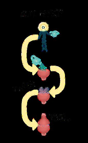
A key protagonist in the Allen Lab’s selective manipulation of astrocytes is the cellular communication network factor 1, better known as CCN1, an astrocyte-secreted protein that regulates critical cellular processes More specifically, it regulates AMPA receptor trafficking, a process that determines how strongly neurons communicate at synapses and whether connections are maintained over time By controlling when and where AMPA receptors are located at the synapse, astrocytes influence whether a synapse becomes stronger or short-lived When astrocytes release CCN1, this induces synapses to become settled and longlasting instead of remaining flexible and easily altered. Astrocytic control of the receptor showcases a key mechanism where neural circuits are able to transition from flexibility to long-term stability
10
The Allen Lab investigated how the astrocyte-secreted protein CCN1 influences neural circuit plasticity in mice In juvenile mice, researchers overexpressed CCN1 by utilizing a virus engineered to carry the CCN1 gene into astrocytes so the cells produce more of the protein They found that higher levels of CCN1 cause connections to stabilize earlier and reduce the amount of change the circuit can undergo In contrast, when researchers lowered CCN1 expression in adult mice using a different genetic approach, previously rigid circuits regained flexibility typically seen during early development In other words, high levels of CCN1 signal for circuits to stabilize, while low levels allow synapses to remodel and form new connections These findings demonstrate that CCN1 acts as a molecular “brake” on plasticity. By showing that adult neural circuits are not permanently fixed, this work positions CCN1 as a key factor through which astrocytes actively regulate plasticity and suggests potential therapeutic strategies for enhancing circuit flexibility in adulthood 9
CCN1 secretion has on regulating flexibility and stability By carefully controlling CCN1 protein levels in lab mice, scientists Khaspekov and Frumkina discovered that windows of plasticity can be reopened well into adulthood and successfully (yet temporarily) restored the brain’s ability to adapt and rewire itself For many neurological conditions, such as stroke, traumatic brain injury (TBI), or neurodegenerative diseases like Alzheimer’s, this finding is pivotal When neural circuits become too rigid, slowly reintroducing neural flexibility in a controlled manner could help them regain previous functionality without destabilizing well-established connections Studying the CCN1 protein provides insight into how the brain remains adaptable through injury and disease without compromising circuits maintained throughout life, helping us understand cognitive function at the molecular level If similar mechanisms exist in humans, targeting CCN1 or related pathways in mice could one day allow scientists and therapies alike to refine plasticity, enhance recovery, slow cognitive decline, and improve the brain’s ability to adapt to new experiences If astrocytes regulate plasticity throughout development, an important question follows: how does this regulatory system change as the brain ages?
ASTROCYTE AGING AS MORE THAN PASSIVE DECLINE
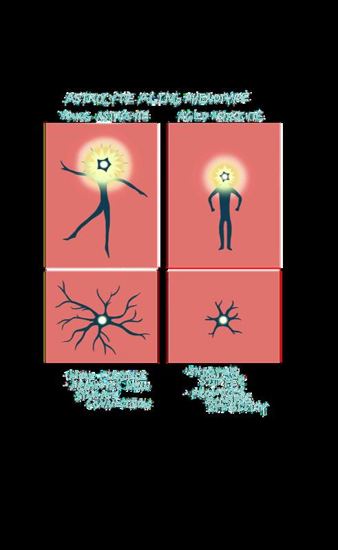
Figure 1. Astrocyte aging reprograms neural environments. As astrocytes age, changes in gene expression shift them from promoting flexible, growing synapses to reinforcing stable, less adaptable connections, emphasizing how aging actively reshapes how these cells support neural circuits rather than simply reflecting gradual decline.
“Astrocytes are not simple bystanders in agerelated cognitive decline, but instead actively remodel the synaptic environment within the brain and impact the degree of functional loss.””
Building on the idea that astrocytes actively regulate neural plasticity, the Allen Lab has uncovered that astrocytes go through set, systematic, and biologically programmed changes themselves, a phenomenon known as the “astrocyte aging phenotype ” Rather than a steady, passive decline, astrocytes experience coordinated shifts in gene expression and secretion patterns that fundamentally change how astrocytes support synapses For example, aging astrocytes increase the production of molecules associated with inflammation and synapse pruning, ultimately influencing the stability and function of neural circuits Understanding molecular changes is crucial because it tells us that astrocytes are not simple bystanders in age-related cognitive decline, but instead actively remodel the synaptic environment within the brain and impact the degree of functional loss 12 where ce from eith can take higher le cytokines and wea the entir prone t astrocyte to diseas the progr
Transcriptomic studies, which examine patterns of gene expression by measuring RNA levels, show that astrocytes undergo systematic molecular changes with age, with aging astrocytes exhibiting altered expression in pathways related to inflammation and synapse elimination, which may potentially contribute to ageassociated cognitive decline. The idea that older astrocytes exhibit different expression behaviors than their younger counterparts challenges the assumption that neuronal aging on its own is responsible for cognitive decline In this way, astrocyte aging reframes brain aging itself as a shift in circuit equilibrium, not merely neuronal wear and tear 11
A NEW VISION OF THE BRAIN
Research on astrocytes and proteins like CCN1 is reshaping how we understand brain plasticity Once thought to be passive support cells, astrocytes are now recognized as active regulators that communicate with neurons and other glial cells to refine connections throughout life These discoveries suggest that targeting astrocytic signals could one day enhance recovery from stroke, TBI, or other neurodegenerative diseases, helping us better understand the molecular basis of learning and cognition
As glial biology intertwines with mainstream neuroscience, researchers will approach plasticity with new eyes to reimagine disease and recovery By linking fast synaptic activity to slower, network-wide modulation, astrocytes challenge the traditional neuron-centric model of neural computation and suggest that cognition emerges from both electrical signaling and glial coordination. Overall, the field is moving more towards a nuanced view of the brain, where plasticity is not fixed but can be continuously modulated, which offers exciting possibilities for both basic neuroscience and future therapies
REFERENCES
[1] Goldman, B (2017, December 27) Neuroscientist Ben Barres, who identified crucial role of glial cells, dies at 63 News Center https://med stanford edu/ news/all-news/2017/12/neuroscientist-ben-barresdies-at-63 html
Keya Patel is a Human Biology and Cognitive Psychology double major graduating in 2028.
Figure 2. CCN1 drives synaptic maturation and stability. Astrocyte-secreted CCN1 acts as a synapse-strengthening growth signal by promoting AMPA receptor insertion, helping synapses become strong and longer-lasting. The protein’s secretion illustrates how astrocytes actively nurture the balance between flexibility and circuit stability in response to developmental and environmental cues.
[2] Clarke, L. E., & Barres, B. A. (2013, May) Emerging roles of astrocytes in neural circuit development Nature reviews Neuroscience https://pmc ncbi nlm nih gov/articles/PMC4431630/
[3] Miller, E K , & Tye, K (2014, May 13) How the brain is flexible enough for a complex world (without being thrown into chaos) Picower Institute https://picower mit edu/news/how-brain-flexibleenough-complex-world-without-being-thrown-chaos
[4] Yamamoto, M (2025) Astrocyte-mediated plasticity: Multi-scale mechanisms linking synaptic dynamics to learning and memory Cells | Free Full-Text https://www mdpi com/2073-4409/14/24/1936/review report
[5] Patel K, Fernandez L 2025 Saltman Quarterly Research Features article interview on Astrocytic Regulation of Neuronal Synapse Formation.
[6] Jerome, R (2025, October) Astroglial regulation of critical period plasticity in the developing brain. ScienceDirect. https://www sciencedirect com/science/article/pii/S0959438825001230
[7] Kolb, B (2025, July 16) Critical and sensitive periods in brain development Oxford Research Encyclopedia https://oxfordre com/psychology/display/10 1093/acrefore/ 9780190236557 001 0001/acrefore-9780190236557-e-700
[8] Ribot, J , & Breton, R (2021, July 2) Astrocytes close the mouse critical period for visual plasticity Science (New York, N Y ) https://pubmed ncbi nlm nih gov/34210880/
[9] Verkhratsky, A , Parpura, V , Li, B , & Scuderi, C (2021) Astrocytes: The housekeepers and guardians of the CNS Advances in neurobiology https://pmc ncbi nlm nih gov/articles/PMC9004589/
[10] Khaspekov, L G , & Frumkina, L E (2023, April) Molecular mechanisms of astrocyte involvement in synaptogenesis and brain synaptic plasticity. Biochemistry Biokhimiia https://pubmed ncbi nlm nih gov/37080936/
[11] Sancho, L., Boisvert, M. M., Eddy, T., Burgado, J., Contreras, M., Labarta-Bajo, L , Wang, E , Tatsumi, L , & Allen, N J (2025, December 17) Astrocyte CCN1 stabilizes neural circuits in the adult brain Nature News https://www nature com/articles/s41586-025-09770-w
[12] Sancho, L , Boisvert, M , & Allen, N (2025, December 19) How do brains stay stable, and when might a dose of flexibility be helpful? Salk Institute for Biological Studies https://www salk edu/news-release/howdo-brains-stay-stable-and-when-might-a-dose-of-flexibility-be-helpful/
[13] Palmer, A L , & Ousman, S S (2018, October 25) Astrocytes and aging Frontiers https://www frontiersin org/journals/agingneuroscience/articles/10 3389/fnagi 2018 00337/full
[14] NJ;, B M G M (2018, January) The aging astrocyte transcriptome from multiple regions of the Mouse Brain Cell reports https://pubmed.ncbi.nlm.nih.gov/29298427/
[15] Lee, G A , Chang, Y -W , Lai, J -H , Chang, T -H , Huang, S -W , Shen, TA., Lin, W.-L., Wu, Y.-C., Tseng, L.-W., Tseng, S.-H., Chen, Y.-C., Chiang, Y.H , & Chen, C -Y (2024, July 19) CCN1 is a therapeutic target for repressed ischemic brain injury - translational stroke research. SpringerLink. https://link springer com/article/10 1007/s12975-02401279-0
HOW EXPECTATION HOW EXPECTATION SHAPES PAIN SHAPES PAIN
Written by Aneesha Asthana
Illustrated by Alivia Gao
n clinical and research settings, physicians and scientists often conceptualize pain as a passive consequence of a harmful event: a
signal traveling from injured tissue to the brain, demanding attention. This framework has shaped how pain is diagnosed and treated, offering a seemingly straightforward explanation of how bodily injury becomes conscious experience. However, clinical and experimental evidence have long suggested that describing pain as a signal alone paints an incomplete picture
Patients receiving placebo treatments, or treatments with no chemically active effect, can report substantial analgesia, or pain relief, and these responses can be blocked by opioid antagonists If patients can experience relief after receiving medications with no pharmacological action, then pain perception cannot be understood solely as a bottom-up sensory process Instead, part of the experience of pain must rely on internally derived neurochemical mechanisms that regulate how negative input is processed 1
behavioral responses to noxious stimuli when those cues were presented alone This approach uniquely allows a complex human phenomenon to be recreated in animals, where the underlying neural circuits can be directly manipulated and measured Using this paradigm, the lab identified the descending pain modulatory system (DPMS) as a key pathway linking expectation to analgesia Connecting the cerebral cortex with the midbrain, the DPMS is a well-characterized network that engages endogenous opioid signaling to regulate how noxious sensory input is perceived and how organisms respond behaviorally (Figure 1)
3
and emotional responses that accompany pain
2 1,2
Central to this regulation are expectations, which arise from prior experiences, associative learning, contextual signals, and the social communication that surrounds care. Expectations function as learned predictions about pain and relief, influencing how incoming sensory information is interpreted and whether signals are amplified or suppressed Placebo analgesia is therefore not a cognitive illusion or myth but a conserved and useful neurobiological mechanism, one that relies on specific neural circuits to shape the experience of pain through learned predictions.
TREATING THE PLACEBO EFFECT AS A NEUROBIOLOGICAL PROCESS
Rather than treating placebo analgesia as a cognitive overlay on sensation, the Banghart Lab at UC San Diego investigates the placebo effect as a neurobiological process Using a reverse-translated placebo paradigm in mice, the researchers modeled the expectation-driven pain relief observed in humans by conditioning animals to associate specific contextual cues with analgesia and then measuring
Central to this system is the periaqueductal gray (PAG), a midbrain structure that integrates sensory and contextual information and coordinates analgesic output through downstream brainstem nuclei These nuclei are clusters of neurons that relay signals to the spinal cord to suppress ascending nociceptive input Anticipation of pain activates neurons in frontal cortical regions involved in cognition and emotion, including the medial prefrontal cortex and anterior cingulate cortex, which project directly to the PAG While these pathways have long been known to contribute to pain modulation, the Banghart Lab demonstrated that they are specifically required for placebo analgesia This then links expectation to the release of endogenous opioids, the brain’s naturally produced pain-relieving
2 3 molecules, within the PAG. Once released, these opioids suppress ascending nociceptive signals and alter the behavioral
In the Banghart Lab’s study, mice were conditioned to associate morphineinduced analgesia with a specific context, such as visual cues (diagonal stripes or dots) paired with olfactory cues (citrus or banana scents) When subsequently exposed to that same context without receiving morphine, the mice displayed reduced pain behaviors Most importantly, the placebo-like analgesia depended on endogenous opioid signaling within the PAG; a pharmacological blockade of opioid receptors eliminated the effect To identify the “upstream” drivers of this effect, the Banghart lab combined in vivo calcium imaging, optogenetic manipulation, and circuit tracing. In doing so, they discovered that neuronal projections from the medial prefrontal cortex (mPFC) and anterior cingulate cortex (ACC) to the PAG were necessary for placebo analgesia. The medial prefrontal cortex (mPFC), located in the frontal lobe, can suppress the perception of painful stimuli through its extensive connections with other cortical regions and its role as a major cortical input to the periaqueductal gray (PAG), which is critical for pain modulation The anterior cingulate cortex (ACC) contributes to the emotional and affective aspects of pain, and altering ACC activity changes how pain is experienced rather than its sensory detection When signaling from both the 3 mPFC and ACC to the PAG is inhibited, the analgesic effect is lost, indicating that both pathways are necessary for pain relief
The frontal lobe isn’t the only region involved in signaling pain modulation and responses The insula, a region situated deep in the cerebral cortex, is
consistently activated during pain perception and evaluation; however, disrupting insular cortex inputs did not affect nociceptive suppression The results from the Banghart Lab’s mouse experiments suggest that expectation-driven analgesia is initiated by cognitive-emotional cortical regions rather than primary sensory areas. Interoceptive regions, which monitor the body’s internal physiological state such as visceral sensation and autonomic activity, do not appear to be the primary drivers of this effect In this framework, placebo analgesia does not emerge from altered sensory encoding per se but from top-down modulation that reshapes how nociceptive signals are gated.3
One of the Banghart Lab study’s central insights concerns how expectation interacts with sensory input Cortical activity alone was insufficient to activate the DPMS Instead, endogenous opioid release in the PAG depended on the arrival of harmful sensory information, suggesting a feedback-based mechanism When presented with a noxious stimulus, the PAG integrates this sensory input with top-down signals from the mPFC and the ACC. If expectation predicts relief, endogenous opioids are released, and nociceptive signals are inhibited from ascending through the DPMS for higher processing and perception in the cortex.3
Our brains modulate pain dynamically in response to sensory input rather than suppressing it preemptively, allowing the system to remain sensitive to injury while still incorporating learned predictions This model aligns with broader theories of predictive coding, in which sensory perception reflects an interaction between prior experiences and incoming data Predictive coding proposes that the brain continuously anticipates incoming sensory information, evaluating sensory signals against these predictions to construct perception based on the degree of alignment between expectation and sensory input. By identifying the specific circuitry through which expectation shapes nociceptive processing, the Banghart Lab provides a mechanistic bridge between predictive theory and the neural architecture observed in humans 3
PAIN AS A REGULATED PROCESS: EXPECTATION, CIRCUITS, AND CONTEXT BROADER
Although the Banghart lab conducted their experiments in mice, human imaging studies implicate parallel circuit architecture involving the prefrontal cortex, ACC, and PAG in placebo analgesia Importantly, the lab’s work provides causal evidence that learned predictions can directly drive endogenous opioid release through defined neural pathways, which has broader implications for how placebo responses are understood in clinical settings The Banghart lab's findings challenge the view that placebo analgesia is a psychological artifact to be treated as 2 3 a confound in drug trials. Rather than merely altering subjective reporting, a placebo engages conserved neuromodulatory systems that regulate 1 pain at the circuit and molecular level. From this 1,2 perspective, placebo responses reveal something fundamental about pain itself: it is not a passive readout of tissue damage, but a regulated process shaped by cognition and context
Anticipation surrounding pain and relief develops over time through experience rather than arising as fixed attributes of individuals, particularly in medical settings Therefore, communication, trust, and prior experience all influence how patients anticipate pain and relief. If expectation 4,5 can recruit endogenous opioid release via prefrontal PAG pathways,3 then physician-patient interactions play a deeper role in the efficacy of treatments Language, reassurance, and context may influence pain, not only through psychological comfort, but by modulating neural
circuits that control nociceptive processing, highlighting how clinical communication is part of the therapeutic context in which drugs act. A patient’s preconceptions may amplify or constrain treatment efficacy by engaging or failing to engage certain endogenous modulatory systems 5 4
CONNECTIONS
Building on the neurobiology research described here, it is clear that compassionate clinical communication is an active component of care to effectively treat pain long-term Therapeutic interventions operate within a neural landscape shaped by prediction and learning, and the clinical 6 encounter contributes to that landscape
Medications, procedures, and conversations all interact with the same regulatory systems that shape pain perception. Communication, perceived competence, and reassurance influence how patients anticipate pain and relief, shaping the predictions the brain generates in response to noxious stimuli
Understanding these brain mechanisms highlights why trust is ethically important
to cultivate in medical care. Dr. Banghart notes that placebo effects may be weaker in individuals who do not trust medical institutions or clinicians In such cases, 7 expectations of relief are harder to form, altering how pain is anticipated before it is even experienced
“Pain care disparities, then, cannot be understood solely as differences in diagnosis, access, or treatment choice, but must also be situated within the broader social and institutional contexts that influence how pain is anticipated and regulated.’’
alth
Photo by Leo Harris
CHARACTERIZING THE IMPACT OF CLASSICAL AND NOVEL
HUMAN ASTROVIRUS SPIKE PROTEINS ON THE ENDOTHELIAL GLYCOCALYX OF TISSUE-SPECIFIC BARRIER CELLS
FINN COUGHLIN
SCHOOL OF BIOLOGICAL SCIENCES, UNIVERSITY OF CALIFORNIA, SAN DIEGO
ABSTRACT 1
Human astrovirus (HAstV) is an under-researched RNA virus known to cause gastroenteritis in infants, the immunocompromised, and the elderly Up until the last few years, there have been 8 distinct subtypes of the “classical” clade Recently, the novel clades, MLB (Melbourne-based) and VA (Virginia-based) have been discovered. These novel clades seem to have an expanded reach, infecting distal organs such as the brain and lungs and potentially causing encephalitis and meningitis. It is unknown why these novel clades display this expanded tropism Previous research has proposed that viral spike proteins degrade the endothelial cell barriers that line the blood vessels of the body. To test this, I added viral spike proteins from the novel clades to cell monolayers from endothelial cell types consistent with the expanded tropism Via an endothelial glycocalyx layer (EGL) immunofluorescence assay, I determined that the novel clade MLB showcased degradation of key glycoproteins and proteoglycans in predominantly lung endothelial cells and the novel clade VA showcased degradation of the same glycoproteins and proteoglycans in predominantly brain endothelial cells. These findings offer insight into how these novel clades have a greater reach than the classical strains of human astrovirus seen typically in the gut. 1
INTRODUCTION 2
Human astrovirus (HAstV) is a common yet understudied cause of pediatric gastroenteritis. First described in 1975, it is most prevalent in the elderly, the immunocompromised, and young children With a seropositivity rate of ~90% by the age of five [8], HAstV is widespread. As of now, there are 8 classical serotypes of HAstV. Astrovirus is transmitted fecal-orally The stability of the HAstV allows it to survive through water treatment , leading to the infection of both mammalian and avian species. Recently, novel clades of HAstV have been discovered in Melbourne (MLB) and Virginia (VA), exhibiting dissimilarities of sequence and structure between the classical serotypes and the novel clade1. These novel clades have an expanded tropism of the central nervous system (CNS), possibly contributing to encephalitis and meningitis This differs from the classical clades which showcase the typical gut tropism, with epithelial permeability causing an influx of fluid entering the gut rather than cellular inflammation leading to gastroenteritis6 More specifically, some tight and adherens junction proteins (TJ/AJ) between epithelial cells such as occludin are degraded by the virus, increasing the permeability between epithelial cells Novel clades of HAstV have been identified in the CNS, indicating that HAstV travels from its site of origin in the gut to distal organs. It is unknown how they are able to do this Viruses must pass endothelial barriers, lining the interior of the blood vessels found in humans, to infect new organs. This can potentially be done by (1) infecting the cells or (2) breaking down the TJ/AJ proteins between cells, causing endothelial dysfunction and barrier hyperpermeability Alongside the junctions
4 5 7 9
2,3
holding together cells, an endothelial glycocalyx layer (EGL) also plays a critical role in barrier permeability Glycoproteins and proteoglycans such as sialic acid and heparan sulfate maintain the structural integrity of the endothelial cell barrier and, when degraded, leave the cell vulnerable to infection
Previous work has found that certain viral proteins can degrade the TJ/AJ proteins and the EGL , which are both very important in barrier strength I hypothesize that HAstV capsid spike protein may also have the capacity to disrupt endothelial barriers potentially due to the novel clades and classical clades having very different spike proteins Each clade encodes a spike protein of varying size, folding topology, and overall shape The sequence identities have proven to be dissimilar1, implying different tropisms for each unique clade. I have already observed the novel clades demonstrating degradation of the TJ/AJ proteins in both lung and brain endothelial cells I hypothesize that novel HAstV spike may also cause degradation of the EGL To determine this, I employed an EGL immunofluorescence assay to visualize and calculate cell barrier integrity, utilizing viral proteins from classical strains, novel strains, and both positive and negative controls.
MATERIALS AND METHODS 3
3
Spike Proteins Utilized: Classical HAstV types 1 and 8 alongside novel types of MLB 1, 2, and VA 1 The spike proteins of interest were added at a concentration of 10 µg/mL in 500 µL The enzymatic controls heparinase, used for heparan sulfate, and neuraminidase, for sialic acid, were also included with 1 µL of heparinase added and 5 µL of neuraminidase added The positive control for Dengue Virus’ NS1 protein was also included in some trials at the same concentration as the spike proteins of interest but was omitted in later trials
Cell Culture and Passaging: I used human pulmonary endothelial cells (HPMECs) and human brain endothelial cells (HBMECs) Cells were thawed and placed in 5 ml of endothelial growth media-2 (EGM2). These passages were kept until confluency, or when 100% of the flask area was covered with cells, with media changes after day 2 Upon confluency, which occurred commonly on day 3, the cells were passaged with 2 mL of protease enzyme TrypLE then centrifuged at 1500 rpm for 5 minutes The cells were then seeded at a 1 to 5 ratio of the previous confluent media
Preparation of Coverslips and Treatment: In a 24 well plate, glass coverslips were added and treated with a 0 2% gelatin solution to better emulate the extracellular matrix I seeded 1e5 cells in 500uL of media and allowed them to grow 3-4 days or until confluent. The media was changed every day except the day of treatment On the day of treatment, 10ug/mL of HAstV spike protein or DENV NS1 was added to each well. I fixed cells at the optimized time point of 6 hours post treatment with 4% formaldehyde and stored them in phosphatebuffered saline (PBS) until staining
Immunofluorescence, Visualization, and Quantification: A monoclonal mouse-anti heparan sulfate IgM antibody was applied to bind to the remaining heparan sulfate at a 1 to 100 dilution in blocking buffer A secondary monoclonal antibody, a polyclonal goat-anti mouse antibody conjugated to a 488 fluorophore, was attached 24 hours later at a 1:1000 dilution in blocking buffer The lectin wheatgerm-agglutinin conjugated to a 647 fluorophore was utilized to bind to sialic acid. To visualize cells, Hoechst nucleic acid stains were added at 1:1000 dilution in the blocking buffer, allowing for easy visualization of the cells seen on the coverslip After treatment of the coverslips, ProLong Gold Antifade Reagent was used to mount coverslips on glass slides and to prevent signal loss over time To visualize and take representative photos of the coverslips, an Invitrogen EVOS M5000 Imaging System with the ability to capture many different channels individually and as a whole was utilized A total of 5 images were taken of each coverslip The mean fluorescence intensity (MFI) was calculated with the program ImageJ. A decrease in MFI is indicative of decreased signal from the applied antibodies and degradation of a particular protein found on the EGL
PREVIOUS WORK 4
Previous work on endothelial barrier disruption was conducted in the Biering Lab via a Trans Endothelial Electrical Resistance (TEER) assay For this assay, endothelial cells are cultured for around 4 days before seeding into a transwell within a 24 well plate. After these wells reach confluency, the electrical resistance measured in Ohms (Ω) is calculated via two ends of an electrode with one in the cell confluent transwell and the other in the 24-plate well. An electrical current is applied with a healthy endothelial cell barrier being reflected with a larger value A decrease from timepoint 0 indicates barrier dysfunction. After collecting baseline data, viral proteins from the viruses of interest are applied to the transwell media, along with a Dengue Virus (DENV) NS1 protein From these methods, the Biering lab found that, compared to untreated values, the electrical resistance found in both HPMECs and HBMECs was discernably lower after treatment with both novel clades’ MLB1 and VA1 spike proteins (Fig 1) This indicates that the viral spike proteins found from the novel HAstV clades play a role in both the degradation of tight and adherens junctions and in the barrier dysfunction of endothelial cells Overall, this work elucidates the importance of spike proteins in the process of viral dissemination. This is especially important as a way to further understand these newer forms of human astrovirus
Figure 1. TEER assay 6 hours post spike protein treatment indicated barrier dysfunction : (A) A representative image of the TEER assay (B) The data 6 hours post treatment indicating novel HAstV spike proteins degrade the TJ/AJ proteins seen on endothelial cells
Figure 2 Novel HAstV Spike Proteins induce degradation of sialic acid in a tissue specific manner. (A) Nucleic acid stain (DAPI) and sialic acid fluorescence only in HPMECs (B) DAPI and sialic acid fluorescence only in HBMECs (C) DAPI and heparan sulfate fluorescence only in HBMECs. (D) MFI of sialic acid in HPMECs for each added spike protein and enzyme relative to an untreated control The data is from n=3 replicates with (F) MFI of heparan sulfate in HBMECs for each added spike protein and enzyme relative to an untreated control The mean fluorescence intensity (MFI) was calculated with the program ImageJ and was analyzed using GraphPad Prism.
Sialic Acid For pulmonary endothelial cells, I found that the MLB1 spike protein from the novel clade MLB displayed a decrease in fluorescence of the amount of Sialic Acid as compared to an untreated control (Fig 2A, D) Interestingly, I noticed that the classical HAstV strain 1 also showcased a decrease in MFI in HPMECs (Fig 2D), indicating that its effectiveness at barrier degradation might not be restricted to the typical gut tropism seen with the classical clade Classical HAstV strain 8 returned to what was expected, with an MFI consistent with the untreated control condition. The novel clade VA, with its VA1 spike protein, was also consistent with the untreated control values My control values, NS1, heparinase, and neuraminidase all showcased expected values. NS1 protein has been observed to cause EGL degradation and dysfunction, and an evident MFI lower than the untreated MFI is consistent with this expectation Heparinase is a protease, specifically towards Heparan Sulfate, and should not degrade glycoproteins such as sialic acid As expected, the
MFI of Heparinase was consistent with the untreated cells and our expectations Neuraminidase on the other hand, degrades sialic acid, consistent with a decrease in MFI. For brain endothelial cells, I found that the VA1 spike protein, from the novel clade VA, displayed a decrease in sialic acid MFI, differing from what was observed in HPMECs and the MLB clade (Fig. 2D, E). The MLB1 spike protein was found to be consistent with the untreated MFI in this circumstance NS1 protein again displayed a lower MFI than untreated, and the enzymatic controls Heparinase and Neuraminidase were consistent with what was seen in HPMECs (Fig 2B, E)
Heparan Sulfate. For the readings on the MFI of heparan sulfate, preliminary data suggests that both VA1 and MLB1 spike proteins saw a decrease in the MFI compared to untreated values in HBMECs NS1 protein saw a consistent MFI decrease as expected. The enzymatic controls, however, brought up some concern The Heparinase led to a decrease in MFI as expected, but the addition of Neuraminidase had the same effect despite being able to degrade sialic acid Even though there is not enough data to come to a consistent conclusion, each data point follows a similar pattern with the difference for each data point being equal for each condition (Fig 2C, F) For HPMECs, inconclusive data was collected, necessitating more trials in order to come to any conclusion For this trial, all classical clades, VA, and NS1 proteins showcased an uncharacteristic increase in MFI, indicative of experimental control. The MLB1 spike protein showcased an MFI that was similar to the untreated control group, which is notable as the novel clade MLB was seen affecting barrier permeability in HPMECs for sialic acid but not for heparan sulfate. I believe that an uneven layer of heparan sulfate caused this effect, yet further inquiry is needed Another point of concern was that there were only 2 data points, with MFI values varying for each point
DISCUSSION
figure2 5
My research project sheds light onto the ability for the spike proteins of both classical and novel clades to degrade critical components of the EGL, potentially facilitating barrier dysfunction and viral dissemination. My findings correlate with previous studies that found that viral spike proteins can cause degradation of the EGL and TJ/AJ proteins2,3, both important components of the endothelial cells that line blood vessels Specifically, I found that the novel clade MLB was able to cause sialic acid degradation in HPMECs and heparan sulfate degradation in HBMECs In agreement with my hypothesis, I also found that novel clade VA was able to cause the degradation of sialic acid of HBMECs and heparan sulfate of HBMECs. It appears each clade is more effective at causing barrier dysfunction and viral dissemination in different cell types, consistent with my hypothesis, but it requires further research to determine if the MLB clade is more adept at viral dissemination into the lungs and if the VA clade is better equipped to travel to the brain Unfortunately, uneven barrier cell growth caused the heparan sulfate on HPMECs to be unrepresentative of cellular conditions and showcased an unrealistic strengthening of cell barriers in the lung endothelial cells With some more trials, I should be more equipped to deal with the recently discovered strains of human astrovirus. It is especially important to understand them now since the ability of Astroviridae to infect such a large quantity of mammals and birds could potentially lead to zoonotic spillovers and the introduction of a more dangerous strain . 7
In conclusion, I found that HAstV utilizes spike proteins to degrade the barriers found within blood vessels to travel from their gut origin to distal organs such as the lung and the brain The recently discovered novel clades of HAstV, MLB and VA are seen to have some
specificity of cell type The MLB clade was seen to be more effectivein lung endothelial cells, and the VA clade was seen to be more effective in brain endothelial cells. This is consistent with where each novel HAstV clade was discovered5 The VA clade has been seen in the brain tissue of patients, while the MLB clade has been more evident in pulmonary cells. For future research I will collect more data on heparan sulfate and HPMECs and utilize gut epithelial cells (CaCo2s) to determine if these novel clades also display the classical gut tropism.
ACKNOWLEDGEMENTS 6
Thank you to Rebecca Kirby and Dr Scott Biering for their mentorship and openness to working in the Biering Lab Thank you to Ms. Wendy Kwok for sponsoring this research throughout the summer of 2025
REFERENCES 7
[1.] Ghosh A, Delgado-Cunningham K, López T, Green K, Arias CF, DuBois RM Structure and antigenicity of the divergent human astrovirus VA1 capsid spike. Rey FA, editor. PLoS Pathog. 2024; 20(2):e1012028
[2 ] Biering SB, Akey DL, Wong MP, et al Structural basis for antibody inhibition of flavivirus NS1– triggered endothelial dysfunction. Science 2021; 371(6525):194–200
[3 ] Biering SB, Gomes De Sousa FT, Tjang LV, et al SARS-CoV-2 Spike triggers barrier dysfunction and vascular leak via integrins and TGF-β signaling. Nat Commun. 2022; 13(1):7630.
[4 ] Méndez E, Salas-Ocampo E, Arias CF Caspases Mediate Processing of the Capsid Precursor and Cell Release of Human Astroviruses. J Virol. 2004; 78(16):8601–8608.
[5.] Janowski AB Beyond the Gastrointestinal Tract: The Emerging and Diverse Tissue Tropisms of Astroviruses Viruses 2021; 13(5):732
[6.] Moser LA, Carter M, Schultz-Cherry S. Astrovirus Increases Epithelial Barrier Permeability Independently of Viral Replication J Virol 2007; 81(21):11937–11945
[7.] Wohlgemuth N, Honce R, Schultz-Cherry S. Astrovirus evolution and emergence Infection, Genetics and Evolution 2019; 69:30–37
[8 ] Koopmans MPG, Bijen MHL, Monroe SS, Vinjé J Age-Stratified Seroprevalence of Neutralizing Antibodies to Astrovirus Types 1 to 7 in Humans in The Netherlands Clin Diagn Lab Immunol 1998; 5(1):33–37
[9 ] Vu D-L, Bosch A, Pintó R, Guix S Epidemiology of Classic and Novel Human Astrovirus: Gastroenteritis and Beyond. Viruses. 2017; 9(2):33
“It [his research] definitely made me more of a germaphobe,” Finn admitted. “But it also made me appreciate how much these small discoveries contribute to bigger goals like vaccines and disease prevention. These small steps add up.”
Want more? Read the research feature article “Breaking Barriers: How New Human Astrovirus Strains Invade the Brain and Lungs” by Natalie Botello at sqonline ucsd.edu.
HOW WE INVOLUNTARILY CONVEY OUR EMOTIONS THROUGH THE AUTONOMIC NERVOUS SYSTEM
¹UNIVERSITY OF CALIFORNIA, SAN DIEGO, ²DEPARTMENT OF BIOGINEERING, STANFORD UNIVERSITY
ABSTRACT 1
The human gut contains over a 100 million neurons that are in constant communication with the brain through the gut’s enteric nervous system (ENS), commonly known as the “second brain ” The ENS not only communicates to the brain digestive function and gut activity, but even emotional states such as stress, fear, and anxiety For those with gut disorders such as Irritable Bowel Syndrome (IBS), the communication between the brain and gut is disrupted, turning everyday stress and anxiety into chronic physical pain While previous studies have researched the relationship between gut-microbiota and the gut-brain axis, few have dived into the emotional connection between the ENS and the autonomic nervous system (ANS), which regulates involuntary physiological processes such as respiration, digestion, and arousal. Therefore, this study aims to investigate how emotions influence gastrointestinal and cardiac activity, and how the autonomic nervous system translates emotional states into physiological responses
It was hypothesized that feelings of pleasure would activate the participants’ parasympathetic nervous system, leading to an increase in gut activity, whereas discomfort and fear would stress the body and activate the sympathetic nervous system, decreasing gut activity To test this, participants were each shown three videos designed to evoke emotional responses for fear, discomfort, and pleasure. Their cardiac and gut activity were simultaneously measured using an electrograstrography and electrocardiography setup The results showed that the hypothesis was mainly accurate in accordance with gut activity levels, but there were surprising deviations in heart rate activity The gut-brain axis remains a gray area where further data is needed to fully comprehend the autonomic nervous system’s integral role in the physiological effects of gastrointestinal and cardiac activity Finding the true root of such connections can significantly advance treatments for gut-related disorders and lead to a deeper understanding of the connection between mental and physical health.
INTRODUCTION 2
The autonomic nervous system is a crucial part of the nervous system that controls involuntary bodily functions such as breathing, heart rate, and digestion The ANS plays a central role in maintaining homeostasis by adjusting bodily functions in response to internal or external stimuli These processes are regulated through neural pathways connecting the central nervous system with organs, allowing for a rapid connection of cardiovascular, respiratory, and gastrointestinal functions 2
Unconsciously, emotions modulate autonomic activity through neural signaling between brain regions involved in emotional regulation, such as the hypothalamus and amygdala. As a result, external stimuli can physiologically influence the body For example, stress and fear increase heart and respiration rate, whereas calmness lowers both As for the gut, stress and anxiety can suppress digestion, while relaxation promotes motility and secretion which allows digestion to
become more active and coordinated. Functionally, the autonomicnervous system is divided into two complementary subdivisions that dictate bodily responses - the sympathetic and parasympathetic nervous systemThe sympathetic nervous system activates the body’s “fight or flight” response, increasing heart rate and breathing while slowing digestion On the other hand, the parasympathetic nervous system triggers the “rest and digest” state, lowering heart rate and respiration, while increasing digestive and gut activity Together, these two systems work to regulate physiological a responses and maintain internal balance in response to fluctuating emotional and environmental factors. 2
Figure 1 The physical & internal states that result from activation of the parasympathetic and sympathetic nervous system (Source) On the left side of the figure, the activation of the sympathetic nervous system is displayed with pupil expansion, increased heart and breath rate, and an inactive gut. The right side distinguishes the activation of the parasympathetic nervous system through pupil shrinkage, decreased heart and breath rate, and an active gut 3
PREVIOUS WORK
Emotions can have a powerful impact on digestive function For example, individuals often experience gastrointestinal urgency before stressful events such as an interview or exam When anxiety or stress takes over, the physiological responses in the body can affect the GI system, one of the many causal factors of stress-related disorders such as IBS This relationship is mediated through the gut-brain axis, involving the enteric nervous system, endocrine signaling, and autonomic regulations. Because the brain and gut are closely connected through neural and hormonal signaling pathways, changes in emotional state can prove to have a strong influence on digestive processes. Studying and understanding these connections may provide valuable insight into new treatments that focus on targeting both the brain and gut, instead of one or the other A review of existing literature online indicated that this specific topic was largely unexplored and not extensively analyzed, further supporting the need to investigate it
It was hypothesized that an emotion such as pleasure would activate the parasympathetic nervous system because it would calm the body down, which would lead to a slower heart rate and increased gut
activity. Fear and discomfort, on the other hand, would stress the body and activate the sympathetic nervous system, therefore increasing the heart rate and decreasing gut activity
PROJECT METHODOLOGY 3
This study focuses on how the autonomic nervous system and the gut are interconnected using devices such as electrogastrograms (EGG) and electrocardiograms (ECG). Electrogastrography measures the electrical activity of the stomach to assess gastric and digestive activity Electrocardiography records the heart’s electrical signals, allowing for analysis of heart rate and autonomic regulation. The setup of these devices involved the placement of 8 electrodes on the participants’ abdomens, where the electrodes measuring the highest gut and heart activity were utilized in analyzing the EGG results ECGs and EGGs were integral to the research study and shaped how findings were interpreted
The ultimate goal of the experiment was to stimulate particular emotional responses in participants, then measure the subsequent heart and gut activity to ascertain any existing correlation Participants were first asked a series of questions to determine what scenarios brought them calm, discomfort, and fear These three emotions were selected as they are identifiable through using video, unlike other emotions such as anger and love. Using participants’ answers, 10 minute videos were made to stimulate emotional activity, and therefore, the predicted gut activity Three 10 minute videos were created for each participant: one each for pleasure,fear, and discomfort.
The night before the experiment, participants were told to fast for at least 12–13 hours and not drink water in the morning so that their baseline gut activity would be low enough to measure accurately and to ensure that any changes during the study would be attributed to the experiment rather than prior digestion. Right before the experiment, participants were surveyed to measure their initial anxiety, hunger, stress, and awareness levels This survey, referred to as the pre-ANS state survey, was given to see how individual-specific factors such as the amount of sleep the participant received and morning hunger levels could influence their gut activity, since some people are more accustomed to fasting or sleep deprivation than others.
Figure 2. Experimental workflow of how the experiment was run from the top down The participants were given a pre-experiment preferences survey a day or two before the experiment so that videos could be formatted to the participants’ preferences Right before the experiment, the Pre ANS state survey was given to the participants to measure their energy, arousal, stress, and hunger levels. The experiment then began with a 10 minute baseline of white noise, directly following into the 3 videos with 2 minute breaks in between each After the final video, a post-experiment survey was given to measure the participants’ final energy, arousal, stress, and hunger levels
SALTMAN QUARTERLY
Following the survey, subjects were given noise-canceling headphones to wear, and lights were turned off in order to create an immersive environment The trial started with 10 minutes of white noise to calm the participant and lower their heart rates and anticipation levels. Then, one of the three videos played, followed by a 2-minute relaxation period to reset the participants’ gut and heart activity This process was repeated twice with each of the three videos. After the experiment, participants were surveyed to rate their emotion levels after watching the videos
The experimental workflow for each participant was the same except for the order of the videos and the type of videos shown Participants were also not aware of the type or content of videos before their experiment to ensure that they would be alert and react genuinely to the videos
RESULTS 4
Figure 3. EGG power results These results represent the overall magnitude of gastric activity across different emotional states The x-axis measures time (in seconds) and the y axis is z-scored EGG power (in decibels) which represent how far given power values deviate from the baseline mean gut activity The average EGG power spanned all participants for each emotion over the 10-minute period.
The first variable analyzed was EGG power (Figure 3), which refers to the signal output Z-scored values were utilized for the y-axis of each figure in order to standardize measurements across subjects and compare how emotions deviated from baseline activity. Z-scored EGG power represents how far a given EGG power value deviates from the baseline mean, otherwise calculated as (valuemean)/standard deviation This particular statistical analysis allows comparison of gut activity and heart rate across all participants, which it was utilized for analyzing results The mean for heart rate and gut activity was taken from the participants’ baseline measurements in the first 10 minute segment of the experiment. The shaded region around each trend line represents the standard error of the baseline activity 5
Looking at Figure 3, the EGG Power for pleasure levels was consistently higher than all other emotions and the baseline Fear and discomfort levels were lower, aligning with less GI activity and indicating activation of the sympathetic nervous system
The second variable analyzed was the average heart rate, which was recorded with a respiration belt (Figure 4) Utilizing z-score heart rate, the average heart rate was compared to the mean baseline activity for all three emotions across every participant
Figure 4. Average heart rate across each emotion at a given point of time recorded with a respiration belt The x-axis is time in seconds, and the y axis is the z-scored heart rate in beats per minute The average heart rate spanned all participants for each emotion over the 10-minute period. Clear spikes can be observed in particular for emotions such as fear and pleasure
Looking at Figure 4, fear and discomfort levels were lower than pleasure and baseline levels for the majority of the 10 minute period These results contradict the initial hypothesis, which predicted that the heart rate for these two emotions would be higher Spikes in heart rate for fear and discomfort are likely due to jumpscares or specific scenes in the videos that elicited fear or disgust in the participants, which was expected Heart rate may also have been influenced by the participants’ awareness of artificial stimuli or individual tolerance to uncomfortable situations, which could explain why heart rates for these emotions were lower than expected
Figure 5. Average standard deviation of normal to normal intervals (SDNN) across all emotions tested in order to measure heart rate variability The heart rate variability was averaged across 10 minutes across all participants. The x-axis represents the 4 emotions tested and the y-axis measures the SDNN in seconds The SDNN for pleasure is observed to be the highest in comparison to the other emotions, whereas the lowest SDNN belongs to discomfort.
Furthermore, heart rate variability was analyzed in order to see how activation of the parasympathetic and sympathetic nervous how
systems impacted short-term fluctuations of heart rate frequency. Standard deviation of normal-to-normal intervals (SDNN) is a measure of heart rate variability (HRV), or variations in the time intervals between consecutive heartbeats. SDNN entails the combined influence of both the sympathetic and parasympathetic branches of the autonomic nervous system, making it a useful indicator of autonomic balance. In this context, SDNN is expected to be lower during fear and discomfort, when sympathetic activation increases, and higher during pleasure, when parasympathetic activity is increased This aligns with the results, as shown in Figure 5 6
Higher SDNN values indicate that the heart is adapting flexibly to the body’s changing physiological demands, reflecting healthy heart rate variability and effective autonomic regulation. In contrast, lower SDNN suggests a more rigid heart rhythm, which is often associated with physiological stress and fatigue SDNN values during baseline are typically moderate and depend on the participant’s resting state. If the baseline condition is truly relaxed and unstimulated, SDNN is expected to remain relatively stable 6
In the pleasure condition, positive emotional states associated with calmness, safety, or social connection are linked to increased parasympathetic activity, which generally elevates HRV As a result, SDNN would be expected to be higher than baseline when the pleasurable stimulus is relaxing, such as soothing music or pleasant imagery However, if the pleasurable stimulus is more exciting or arousing, SDNN may instead decrease.
The fear condition activates the sympathetic nervous system and the body’s fight-or-flight response. This suppresses heart rate variability, leading to a decrease in SDNN relative to baseline Similarly, the uncomfortable condition often reflects heightened stress or tension, which reduces parasympathetic influence and increases sympathetic drive SDNN is therefore expected to be lower than baseline and may be comparable to, or lower than, levels observed during fear
Overall, these results mostly aligned with the initial predictions based on autonomic nervous system activity and parasympathetic and sympathetic function
For EGG power, pleasure-related emotions showed the highest GI activity, consistent with parasympathetic activation In contrast, emotions such as fear and discomfort lowered GI activity, reflecting that the sympathetic nervous system was activated For heart rate, pleasure was associated with lower levels as expected However, fear and discomfort also had relatively low heart rates. In terms of heart rate variability measured by SDNN, pleasure had a positive effect, leading to a higher heart rate variability, while discomfort and stress reduced it. Fear triggered sympathetic activity which caused a greater drop in Heart Rate Variability (HRV) than seen with the pleasure levels Increased arousal may have driven the fear HRV even further below the baseline values
FUTURE DIRECTIONS 5
In the future, this project could explore how other emotions create significant bodily responses. Because this study was conducted with a limited sample size over a short time period, future research would benefit from adding a more diverse group of participants and investigating a larger range of emotions. A greater sample size would improve the reliability of the results, allowing determination of consistent patterns across a larger population A larger sample size would also reduce the influence of individual variability and help
distinguish outliers. By encompassing a broader range of physiological responses across participants, future studies could be more accurate in determining if the relationship between emotional states and the gut responses are generalizable, rather than specific to a small data set. Furthermore, since the respiration data wasn’t analyzed, it would be useful to compare specific breathing patterns to the corresponding video scenes in order to see how emotions are linked to specific spikes or surges. Virtual reality headsets could also be implemented to elicit stronger emotions in participants and see how a fully immersive atmosphere affects emotional response and gut activity.
CONCLUSION 5
Emotions are more than just fleeting feelings They have a significant effect on the autonomic nervous system and therefore all bodily functions. Specifically, they influence the gut and heart activity, revealing how closely inner feelings are linked to physical health Higher GI activity may mean stronger contractions or movement in the stomach, but may also be a result of pleasure, happiness, or calmness This study shows that simply watching videos can leave a lasting subconscious physiological imprint It paves the way for future studies to explore how emotional states influence physical health in order to treat gut conditions such as IBS and IBD Effective treatments that address both the physiological and emotional aspects of these disorders can not be developed without fully investigating the cause and ultimate mechanisms between the gut-brain axis. This connection reinforces the fact that emotions are key factors in how people function, feel, and stay healthy By becoming more aware of the different emotional states, we can shape physical health and cultivate a more balanced, resilient well-being 7
REFERENCES 7
[1] The Gut-Brain Connection Harvard Health (2023, July 18) https://www health harvard edu/diseases-and-conditions/the-gutbrain-connection
[2] Patel, Y A , & Pasricha, P J (2020) Enteric Neuromodulation for the Gut and Beyond Cold Spring Harbor perspectives in medicine, 10(1), a034355. https://doi.org/10.1101/cshperspect.a034355
[3] Tpoh (2021, April 15) Polyvagal theory – the real root cause of addiction The Pursuit of Health https://thepursuitofhealth.co.uk/2021/04/13/polyvagal-theorythe-real-root-cause-of-addiction/
[4] Nadar, Y (2024, January 22) Decoding the Differences between ECG, EKG, and EEG Tests TenTabs Medical Devices https://tentabs.in/blogs/news/decoding -the-differencesbetween-ecg-ekg-and-eeg-tests?
[6] Shaffer, F , & Ginsberg, J P (2017) An Overview of Heart Rate Variability Metrics and Norms Frontiers in public health, 5, 258 https://doi.org/10.3389/fpubh. 2017.00258
[7] Staudacher, H M , Black, C J , Teasdale, S B , Mikocka-Walus, A , & Keefer, L (2023) Irritable bowel syndrome and mental health comorbidity - approach to multidisciplinary management. Nature reviews Gastroenterology & hepatology, 20(9), 582–596
https://doi org/10 1038/s41575-023-00794-z
“The results of Rao’s research demonstrate the influence that our emotions can have on our GI activity and heart rate. The feelings we experience are not simply fleeting, but can leave footprints that result in physiological changes in our bodies.”
Want more? Read the research feature article “A Gut Feeling“ by Sophia Chau at sqonline.ucsd.edu.
THE ARAL SEA CRISIS: ENVIRONMENTAL
CONSEQUENCES AND MITIGATION POLICIES
JIEYIN JIANG
Located on the border between southern Kazakhstan and western Uzbekistan, the Aral Sea was once the world's fourth largest inland lake ¹ However, beginning in the 1960s, it rapidly shrank as Soviet-era irrigation policies diverted water from its two main inflow rivers¹ This paper examines how policies from the Soviet through the post-Soviet era shaped the fate of the Aral Sea. Through three representative case studies, this paper examines the environmental impacts brought about by these policies, as well as the extent to which the subsequent restoration and adaptation measures have addressed these issues. It argues that the outbreak of the Aral Sea crisis was not only due to unsustainable water resource allocation, but also resulted from the policy orientation of prioritizing short-term economic production over long-term ecological stability By reviewing these cases, this paper emphasizes the importance of sustainable water resource governance and regional cooperation in addressing large-scale environmental degradation
2
As a closed-basin lake that lies on the border of Kazakhstan and Uzbekistan, the Aral Sea was once one of the largest inland bodies of water in central Asia, spanning nearly 68,000 kilometers squared before the 1960s.¹² The lake’s ample water made it an important fishery for Central Asia, with annual catches once reaching 30,000–35,000 tonnes ³
Because the lake depended almost entirely on inflow from the Amu Darya and Syr Darya, it was highly vulnerable to reductions in river input Beginning in the 1960s, extensive water diversions for agricultural irrigation caused the Aral Sea to further shrink.⁴ Consequently, by the late 1980s, the lake had split into the North Aral Sea and the South Aral Sea ⁴ In the early 21st century the South Aral Sea was further diminished, and by 2001 the eastern and western divisions had become completely disconnected ⁵ The eastern South Aral had completely dried up by 2014, now known as the Aralkum Desert.⁵ The now exposed lakebed contributes to salinization, dust storms, and broader ecological decline ⁶⁷ ,
3
SOVIET-ERA WATER POLICIES
Large-scale Soviet irrigation projects are a root cause of the Aral Sea Crisis In the late 1920s the USSR launched massive collectivization and irrigation campaigns in Central Asia, centralizing agricultural production and linking land and water use to national production goals ⁸ In the early 1930s, the Soviets began constructing preliminary irrigation infrastructure in the Aral basin to boost cotton production ⁸ This developed into the 1939 construction of the Main Fergana Canal to irrigate cotton fields in the highly agricultural region of Fergana Valley in the Syr River Basin.¹⁰ Remarkably, this canal was built in just 45 days, dramatically reducing the water flow of the Syr Darya it drew from.¹¹
However, the turning point came in the 1960s, when continued irrigation expansion began to divert the Amu Darya and Syr Darya away from the Aral Sea ⁴ In 1954, the Soviet Union began constructing the 1,400 kilometer Karakum Canal that diverted the Amu Darya to irrigate desert cotton fields.¹² In Uzbekistan, a growing network of diversion channels similarly tapped the Syr Darya to support intensive agriculture in the Fergana Valley ¹³ Although beneficial economically, they had profound environmental impacts. Soviet planners consistently prioritized meeting cotton production targets over conserving water or protecting downstream ecosystems Many irrigation canals were poorly constructed, resulting in huge water loss. Almost 50 percent of the water carried through the Karakum Canal was lost each year to seepage and evaporation, while surplus water was often drained into the surrounding desert ¹⁴
As a result, freshwater inflow to the Aral Sea plummeted and caused a dramatic decline in the lake’s surface area and volume With evaporation far exceeding inflow, the remaining water vastly increased in salinity. The total inflow into the lake decreased from 235 kilometers cubed in 1966-1970 to 10 kilometers cubed in 1981-1985 ¹⁵ Between 1960 and 1990 the Aral Sea’s surface area shrank from about 67,500 kilometers squared to 39,734 kilometers squared, and by 1995 the sea had lost nearly 75 percent of its water.¹⁶
The increase in salinity became the primary diver behind the Aral Sea ecosystem’s destruction Of the more than twenty original fish species, including species such as trout and sturgeon, almost all were lost. Rising salinity placed severe physiological stress on these freshwater fish, disrupting osmoregulation, reducing reproductive success, and making survival increasingly difficult ³¹ Fisheries collapsed and commercial catches were driven nearly to zero ¹⁶ The vast belts of reeds and freshwater algae that once covered the lake also vanished; by the late 1980s only a few salt-tolerant algae remained ¹⁶ The Aral Sea region had also historically served as a major stopover and breeding ground for migratory Central Asian birds, such as pelicans, flamingos, and various waders The retreat of the lake and wetlands, along with the feeding grounds this provided, thus
Figure 1. Shrinkage of the Aral Sea, 1960–2014 ⁴
thus eliminated a critical habitat and altered migration routes for these birds.¹⁶
In addition to the increase in salinity, this ecological collapse was also deeply affected by land degradation, namely desertification and the following salt storms, due to the shrinking sea The newly formed Aralkum Desert in the eastern South Aral has severe topsoil erosion, with a yearly 1 5 to 2 centimeters of topsoil erosion by wind ¹⁷
However, political instability and economic stress meant that water remained closely tied to only energy, agriculture, and security concerns While countries like Kazakhstan tried to revitalize parts of the Aral Sea, other countries like Uzbekistan continued to operate large-scale cotton irrigation systems ²⁴ , ²⁷ Overall, post-independence policy priorities remained focused on agricultural expansion and energy supply, causing regional water allocation to fundamentally not improve
Figure 2. Before the regression crisis, the Aral Sea supported substantial biodiversity, including 20 fish species, 195 free-living invertebrates, 71 parasite species, more than 640 microalgae taxa, 24 angiosperms, 6 charophytes, and about 40 macroalgae The data in this chart is summarized from ¹⁶
This topsoil erosion causes the salt and residual agricultural chemicals in the dried lakebed to be picked up by strong winds, creating frequent and intense salt-dust storms These storms, which disperse these salt-laden particles hundreds of kilometers, have increased rates of chronic respiratory illnesses among local populations ¹⁸ The particles settle on farmland and natural vegetation, causing a buildup of salt and pollutants in the soil which further inhibit both agricultural and grassland growth in the region ¹⁹
The disappearance of the Aral Sea has even altered the local climate, amplifying seasonal extremes Summers have become hotter and drier, while winters are colder and with stronger winds Large lakes normally buffer nearby climate, both by cooling surrounding areas in summer and releasing stored heat in winter. With the lake’s area drastically reduced, its moderating effect has weakened In addition, a smaller surface area can also reduce evaporation and atmospheric humidity, thus ultimately decreasing regional precipitation.²¹
Decades of water-diversion policies have caused massive damage to the Aral Sea ecosystem on multiple fronts. In the late 1980s the Soviet government did attempt to address the issue by signing a watersharing agreement among the five Central Asian republics, but this agreement was not effectively supervised or enforced, preventing any substantial improvement ²²
KAZAKHSTAN’S LOCALIZED WATER MANAGEMENT SUCCESS
005, with World Bank support, Kazakhstan attempted to protect North Aral Sea with the construction of the 12 kilometer long -Aral Dam.²⁴ It physically separated the North Aral Sea from the th Aral Sea, capturing much of the flow from the Syr Darya to ease the water retention of the North Aral Sea ⁵ The goal of policy to balance downstream irrigation needs and ecological oration by retaining more water inflow in the North Aral Sea, ng water levels and gradually revitalizing the recovering system ²⁴ These efforts, causing decreasing salinity and increasing er levels, helped restore the delta wetlands and the associated rient cycles, reconstructing the habitats and the aquatic food web that supports fish populations ²⁸
The ecological and climatic benefits are the most evident The expansion of the water surface and the partial restoration of nearby wetlands primarily helped mitigate local desertification. Increased evaporation from the restored North Aral Sea could potentially lead to more rain-bearing clouds and improve irrigation conditions for local agriculture.³³ Vegetation on the formerly arid lands has also begun to recover slowly, secondarily reducing the stress on the soil and the ecosystem ³⁴ In addition to the general ecological benefits, the total volume of water in the Aral system has increased by 42 percent, reaching about 27 billion cubic meters ²⁹ As a result, the lake’s salinity declined from 30 grams per liter to 7 grams per liter ³⁰ This has caused annual fish catches in the North Aral Sea to increase more than fivefold in the last decade: from about 1,360 tonnes in 2006 to 7,106 tonnes in 2016 ³² This remarkable increase not only revived the local fishing economy, but also brought direct benefits to related industries and fishermen in southern Kazakhstan. In short, the dam project has brought multiple ecological benefits: improved water quality and biodiversity, a partial recovery of fisheries that supported the local economy, and some relief from drought-related effects.
However, while the Kok-Aral Dam had a positive impact on the Kazakh-controlled North Aral Sea, some analyses suggest that it has effectively “shut off” the flow from the Syr Darya into Uzbekistan’s portion of the basin in the South Aral Sea ⁵ As one report noted: “Without a dam, the ecological situation here is more dire than up north. All that [Uzbek region] has to rely on for water is the Amu Darya, which has been dwindling for decades”³⁵ The South Aral Sea continues to shrink, with the eastern basin entirely dry from 2014 Unlike the localized recovery in the North Aral, the people of Uzbekistan have not enjoyed the same ecological respite; their environmental and social problems remain very serious ²⁶
After the Soviet Union’s collapse in 1991, regional water management entered a new phase In 1992, the five Central Asian states established the Interstate Coordination Water Commission (ICWC) with inherited Soviet-era water-allocation quotas ²³
Kazakhstan’s experience shows that strong national policy combined with international support can achieve partial environmental recovery, even in what was once considered an “irreversible” disaster. t the same time, this case also serves as a reminder of the limits of
unilateral action – long-term resolution of a transboundary environmental problem like the Aral Sea requires coordinated cooperation among all related countries
Unlike Kazakhstan, Uzbekistan is a downstream country on the Amu Darya and has historically depended heavily on cotton production Although the post-independence government has called for diversification of their agricultural industry, much of their prominent crop-production has remained irrigation-intensive Inevitably facing water shortages, Uzbekistan began promoting some water-saving practices in the 2010s, including more efficient cotton farming and drip irrigation ³⁷ However, adoption remained limited, and most cotton fields still relied on traditional flood irrigation, with only 12% of farm irrigation channels being waterproofed in 2012.³⁸ This inefficient irrigation system worsened the problems of secondary salinization and declining soil fertility, continuing the over-extraction of the Amu Darya.³⁹ Fertilizer runoff also further contributed to downstream eutrophication and declining water quality ⁴⁰
This reduction in freshwater inflow, combined with nutrient pollution from agriculture, accelerated the ecological deterioration of the South Aral Sea The lake surface has kept shrinking, with only scattered remnants remaining By 2007, its water area had fallen to about 10% of its original size, while salinity had risen from roughly 14 grams per liter in the 1960s to over 100 grams per liter now ⁴¹ Under these conditions, the South Aral Sea has virtually lost its freshwater fish population. As a result, only a few salt-tolerant organisms, including euryhaline species, halophiles, and brine shrimp, remain ¹⁶
The degradation of the Aral Sea ecosystem has had profound effects on local people’s health and livelihoods The exposed lakebed increases the rates of salt and pesticide dust, air quality has lowered and increased respiratory stress on local populations ⁴² Respiratory disease rates in the Karakalpakstan region of Uzbekistan are far above the national average Chronic illnesses such as bronchitis and asthma are particularly common, and even incidence of tuberculosis has increased.⁴³ In conjunction with impacted health outcomes, water shortages and soil salinization threaten local livelihoods by negatively impacting the agriculture industry and general food security ⁴⁴
Due to these various stressors, the Uzbek government has taken measures in recent years to mitigate the crisis The 2015 Comprehensive Plan focused on improving water management, protecting public health, and restoring ecosystems ⁴⁵ In biological terms, this included increased conservation efforts for salt- and drought-tolerant species and re-establishment of vegetation and wetland habitats to reduce salt-dust emissions and support local biodiversity In 2017, the government also launched a State Programme aimed at improving living conditions through investments in infrastructure, healthcare, and environmental management.⁴⁶ , ⁴⁷
While not instantaneous, recent developments in Uzbek policy concerning the Aral Sea demonstrate that after Soviet collapse Uzbekistan’s development model became increasingly incompatible with environmental sustainability These policies, even if small, did help alleviate some environmental and societal stressors in the region. For many years, the country’s entire economy heavily relied on waterintensive cotton cultivation, placing enormous pressure on the Amu Darya’s resources. Despite these challenges, Uzbekistan has taken proactive ecological restoration measures and engaged in
international cooperation to mitigate the crisis’s effects. In recent years, Uzbekistan has made progress in stabilizing sands, promoting water-saving irrigation technologies, and improving social welfare for local residents – steps that have helped create conditions that reduce the impact of this environmental disaster.
The cases above demonstrate the powerful impact of policy on the Aral Sea. During the Soviet era, irrigation policies aimed at economic output led to the Aral Sea’s shrinkage; after the Soviet collapse, each country followed different paths based on its own interests The Aral Sea clearly demonstrates that once the salinity exceeds the critical tolerance limit, the aquatic ecosystem will undergo rapid changes: the increase in salinity will impose severe osmotic regulation stress on freshwater species and disrupt the food chain. The Kazakh example shows that targeted dam construction and management measures can bring rapid local hydrological and ecological improvement , including lower salinity and partial fishery recovery. Indeed, the water policies of Tajikistan, Turkmenistan, and Kyrgyzstan have also had significant effects, making the international situation quite complex Overall, whether considering the catastrophic historical lessons or recent water-management successes, it is clear that national policies play a crucial role in cross-border environmental issues
Governance must be guided by a scientific, open-minded approach and incorporate all countries’ policy directions into a shared framework of sustainable development Under global climate change and population pressure, challenges of water scarcity will persist, and countries need to learn from the Aral experience and strengthen international cooperation In line with the 2021 UN General Assembly’s initiative to declare the Aral Sea region a zone of ecological innovations and technologies, all countries should invest more funds and innovation, using technology to seek solutions Future governance should emphasize basin-wide equity, ecological priority, and sustained international support. Only through coordinated and long-term water management can similar crises be prevented elsewhere
Ultimately, the history of the Aral Sea reminds us that policy choices can push the ecosystem beyond its biological tipping point, but targeted and implementable water resource management can also create conditions for the local restoration of the ecosystem
BACKGROUND
REFERENCES 8
[1] Che, X , Feng, M , Sun, Q , et al (2021) The Decrease in Lake Numbers and Areas in Central Asia Investigated Using a LandsatDerived Water Dataset (2000–2015) Remote Sensing, 13(5), 1032
[2] Narbayep, Marat and Pavlova, Vera The Aral Sea, Central Asian Countries and Climate Change in the 21st Century. United Nations ESCAP, IDD, April 2022 Bangkok
[3] United Nations General Assembly (2013, September 16) Letter dated 12 September 2013 from the Permanent Representative of Uzbekistan to the United Nations addressed to the SecretaryGeneral (A/68/383)
[4] Encyclopaedia Britannica (n d ) Aral Sea Retrieved January 11, 2026, from https://www britannica com/place/Aral-Sea
[5] NASA Earth Observatory. (n.d.). World of Change: Shrinking Aral Sea Retrieved January 11, 2026, from https://science nasa gov/earth/earth- observatory/world-ofchange/aral-sea/
[6] United Nations Development Programme (UNDP) Multi-Partner Trust Fund Office (2018, December 3) Terms of reference and annexes – Aral Sea (PDF) https://mptf undp org/sites/default/files /documents/30000/20181203 tors and annexes-aral sea pdf
[7] Waehler, R., & Pohl, E. (2017). Respiratory health and quality of life in adults living near the Aral Sea in Kazakhstan International Journal of Environmental Research and Public Health, 14(2), 226 https://www.mdpi. com/1660-4601/14/2/226
[8] Tarr, D., & Trushin, E. (2004). Did the desire for cotton selfsufficiency lead to the Aral Sea environmental disaster? A case study on trade and the environment (World Bank working paper) World Bank
[9] Ashirbekov, U. A., & Zonn, I. S. (2003). Aral: The history of dying sea Executive Committee of the International Fund for Saving the Aral Sea (IFAS).
[10] Stronski, P (2010) Tashkent: Forging a Soviet city, 1930–1966 University of Pittsburgh Press
[11] Davis Center for Russian and Eurasian Studies, Harvard University (n d ) Shock work: Building the Great Fergana Canal
[12] Encyclopaedia Britannica. (n.d.). Karakum Canal. Retrieved January 11, 2026, from Britannica website
[13] Akramov, K T (1998) Cotton Industry in Uzbekistan: Structure and Current (Beltwide Cotton Conferences proceedings paper) National Cotton Council of America
[14] Ovezberdyyeva, A. (2009). Sustainable water management in Turkmenistan: Challenges and solutions (Master’s thesis, ErnstMoritz-Arndt University of Greifswald) CAWater-Info https://www.cawater-info.net/bk/iwrm/pdf/ovezberdyyeva e pdf
[15] Vinogradov, S , & Langford, V P E (2001) Managing transboundary water resources in the Aral Sea Basin: In search of a solution *International Journal of Global Environmental Issues, 1* (3/4), 345–362 https://www cawater info.net/bk/water law/pdf/vinogradov langford.pdf
[16] Plotnikov, I S , Aladin, N V , Zhakova, L V , Mossin, J , & Høeg, J T (2023) Past, present and future of the Aral Sea: A review of its fauna and flora before and during the regression crisis Zoological Studies, 62, e19. https://doi.org/10.6620/ZS.2023.62-19
[17] Agency of the International Fund for Saving the Aral Sea (IFAS) (2019) 25 years of activities International Fund for Saving the Aral Sea … (report).
[18] Whish-Wilson, P (2002) The Aral Sea environmental health crisis Journal of Rural and Tropical Public Health, 1, 29–34
[19] NASA Earth Observatory. (2020, March 25). A Dusty Day Over the Aral Sea
[20] Singer, A , et al (2003) The PM10 and PM2 5 dust generation potential of soils/sediments in the Southern Aral Sea Basin, Uzbekistan Atmospheric Environment
[21] Khan, V M , Vilfand, R M , & Zavialov, P O (2004) Long-term variability of air temperature in the Aral Sea region Journal of Marine Systems, 47(1–4), 25–33. https://doi org/10 1016/j jmarsys 2003 12 006
[22] Horsman, S (2005) Afghanistan and transboundary water management in Central Asia: The Amu Darya Basin (PDF). CAWaterInfo
[23] Interstate Commission for Water Coordination (ICWC) (n d ) Interstate Commission for Water Coordination of Central Asia. Retrieved January 11, 2026, from http://www icwc-aral uz/
[24] World Bank. (2005, September 1). Saving a corner of the Aral Sea. World Bank https://www worldbank org/en/results/2005/09/01/saving-acorner-of-the-aral-sea
[25] World Bank. (2017, August 8). World Bank and Kazakhstan: 25 years of partnership World Bank https://www worldbank org/en/news/feature/2017/08/08/world -bank-kazakhstan-25-years-of-partnership
[26] NASA Earth Observatory. (2014, September 25). The Aral Sea loses its eastern lobe NASA Science https://science nasa gov/earth/ earth-observatory/the-aral-sea-loses-its-eastern-lobe-84437/
[27] Conrad, C , Schönbrodt-Stitt, S , Löw, F , & Dech, S (2016) Analysing
irrigated crop rotation patterns in arid Uzbekistan by the combined use of MODIS time series and Landsat imagery Journal of Arid Environments.
[28] Kirillin, G B , Shatwell, T , & Izhitskiy, A S (2025) Consequences of
the Aral Sea restoration for its present physical and chemical structure Hydrology and Earth System Sciences, 29, 3569–3588 https://doi org/10 5194/hess-29-3569-2025
[29] Kazinform. (2025, June 3). The volume of water in the Small Aral Sea
increased by 42% (news report)
[30] Micklin, P (2018) The Aral Sea, Uzbekistan and Kazakhstan: Present, problems and prospects (presentation).
[31] Uddin, M H , et al (2023) Effects of salinity on fish growth (review/technical document)
[32] White, K. D. (2016). Kazakhstan’s Northern Aral Sea today: Partial ecosystem restoration and economic recovery In E Freedman & M Neuzil (Eds ), Environmental Crises in Central Asia: From steppes to seas, from deserts to glaciers (pp. 129–140). Routledge.
[33] He, H , Hamdi, R , Luo, G , Cai, P , Zhang, M , Shi, H , Li, C , Termonia, P , De Maeyer, P , & Kurban, A (2022) Numerical study on the climatic effect of the Aral Sea. Atmospheric Research, 268, 105977 https://doi org/10 1016/j atmosres 2021 105977
[34] Cui, M , Zheng, X , Li, Y , & Wang, Y (2023) Analysis of NDVI Trends and Driving Factors in the Buffer Zone of the Aral Sea. Water, 15(13),
2473 https://doi org/10 3390/w15132473
PREVIOUS WORK
[35] Kipnis, V (2024, October 30) Central Asia’s Ticking Time Bomb: Water. NPR. https://apps.npr.org/aral-sea-shrinking-map/ [36] World Bank (2024) Uzbekistan: Policy perspectives for irrigation and drainage sector reform (PDF) World Bank https://documents1.worldbank.org/curated/en/099022824155511 451/pdf/P1732501ad71e00301b2fa1dc0dd0e51758 pdf
[37] Chinese Academy of Sciences (2023, December 4) China contributes water-saving technology to protecting Aral Sea (News release) CAS
[39] World Bank (2024) Water security in Uzbekistan: General country assessment (Water Security Diagnostic Initiative) World Bank https://documents1.worldbank.org/curated/en/0990624241210 35288/pdf/P170030-356bad97-a463-43e5-9ce8-fc808f542aeb pdf
[40] U S Geological Survey (n d ) Nutrients and eutrophication Retrieved January 11, 2026, from https://www.usgs.gov/missionareas/water-resources/science/nutrients-and-eutrophication
[41] Aladin, N. V., Plotnikov, I. S., Micklin, P., & Ballatore, T. (2009). The Aral Sea: Water level, salinity and long-term changes in biological communities of an endangered ecosystem Past, present and future Natural Resources and Environmental Issues, 15 https://www.cawater info.net/bk/water land resources use/ english/english ver/pdf/aladin-et-al-2008 pdf
[42] O’Hara, S L , et al (2000) Exposure to airborne dust contaminated with pesticide in the Aral Sea Region. The Lancet (PubMed record)
[43] Anchita, et al (2021) Health Impact of Drying Aral Sea: One Health and Socio-Economic Perspectives. Water, 13(22), 3196.
[44] Wæhler, T A , & colleagues (2017) The vanishing Aral Sea: health consequences of an environmental disaster. Tidsskrift for Den norske legeforening
[45] Cabinet of Ministers of the Republic of Uzbekistan (2015, August 29). Comprehensive program of measures for 2015–2018 to mitigate the consequences of the Aral Sea disaster, and to restore and promote socio-economic development of the Aral Sea region (Resolution No. 255). CIS Legislation.
[46] President of the Republic of Uzbekistan (2017, January 18) On the State Program for the Development of the Aral Sea Region for 2017–2021 (No RP-2731) LexUZ
[47] Ministry of Ecology, Environmental Protection and Climate Change of the Republic of Uzbekistan (2024) National State of the Environment Report: Uzbekistan (PDF) International Institute for Sustainable Development (IISD)
BACKGROUND
“We like to think to ourselves, ‘What would changing my habits do for the environment? End climate change?’ Your individual effort may seem insufficient to improve the environment we live in. However, if everyone in the world adopts this mindset, it could have a negative collective environmental impact due to individual choices.”
Want more? Read the research feature article “The Aral Sea: History and Environmental Consequences” by Anusha Prasad at sqonline.ucsd.edu.
ADVANCING ONCOLOGY TREATMENTS THROUGH GENETIC ENGINEERING
SANCHALI TIWARI
Cancer recurrence remains a critical limitation of conventional cancer treatments such as surgery, chemotherapy, and radiation, which often fail to eliminate the genetic mutations driving malignancy Advances in genetic engineering, particularly gene editing and immune cell reprogramming, are transforming oncology by enabling therapies that target the molecular origin of cancer This paper explores how these innovations can redefine cancer care while emphasizing the ethical and regulatory responsibilities that accompany them
The case of Jesse Gelsinger, who died in an early gene‐therapy trial for ornithine transcarbamylase deficiency (OTCD), underscores the dangers of inadequate oversight and conflicts of interest His death prompted sweeping reforms by the U.S. Food and Drug Administration (FDA) and Institutional Review Boards (IRBs), including stricter safety monitoring, conflict‐of‐interest disclosures, and long‐term follow‐up requirements In contrast, the case of Layla Richards, the first patient successfully treated with universal chimeric antigen receptor (CAR) T‐cell therapy, demonstrates the ability of gene editing to overcome immune rejection and achieve complete remission in otherwise incurable leukemia.
These developments illustrate how rigorous regulation and sustained funding ensure that innovation progresses ethically and safely. Federal investment not only accelerates scientific discovery it maintains the infrastructure that protects patients, data-safety monitoring boards, ethical review processes, and ongoing surveillance of gene-editing trials Ultimately, the future of oncology depends on balancing innovation with responsibility With sustained investment, cancer treatment can shift toward early genetic intervention, immune system reprogramming, and personalized prevention, which would completely change what it means to survive cancer
INTRODUCTION 2
Despite advances in oncology, recurrence limits the efficacy of conventional therapies in many malignancies Acute myeloid leukemia (AML) recurs in 9-29% of patients, pancreatic cancer in 46%, bladder cancer in 50%, breast cancer in 30%, and glioblastoma and soft tissue sarcoma at almost 100% 1 These figures reveal a critical limitation of conventional treatments such as surgery, chemotherapy, and radiation therapy: They can temporarily suppress malignancy, but they frequently fail to eliminate its underlying genetic drivers
Advances in genomic sequencing have transformed cancer care by enabling mutation-specific treatment strategies Building on this foundation, genetic engineering technologies such as gene editing and immune cell reprogramming offer the possibility of targeting cancer at its molecular origin. Since cancer is fundamentally driven by genetic alterations, the ability to directly rewrite or reprogram cellular 2
DNA represents a shift from broadly attacking rapidly dividing cells to intervening at the level of the mutation itself Gene editing refers to techniques that allow scientists to precisely modify DNA sequences within living cells, either by disabling harmful genes, correcting mutations, or inserting therapeutic genetic material Therapies such as engineered CAR T-cells demonstrate how modified immune cells can be directed to recognize and destroy malignant cells with unprecedented precision ³ Unlike traditional treatments, these approaches do not aim to merely reduce tumor burden, but to reconfigure the biological systems that allow cancer to persist and return
However, the ethical deployment and clinical success of these technologies depend on more than scientific innovation alone Gene editing requires sustained federal investment to fund rigorous trials, long-term safety monitoring, and the regulatory infrastructure necessary to protect patients Inconsistent support leads to progress and patient safety being compromised Therefore, strengthening federal investment in genetic engineering research is essential not only to accelerate breakthroughs in oncology but ensuring that emerging therapies develop under robust ethical and regulatory oversight.
This review examines the role of genetic engineering in addressing these limitations, drawing on historical and contemporary cases to highlight the importance of rigorous oversight and sustained research support SALTMAN QUARTERLY
Figure 1 Diagram of the urea cycle highlighting ornithine transcarbamylase deficiency (OTCD), the genetic condition that affected Jesse Gelsinger. The figure illustrates the biochemical pathway that converts ammonia, a toxic byproduct of protein metabolism, into urea for safe excretion The defective OTC enzyme, located in the mitochondria of liver cells, disrupts this cycle and causes a dangerous buildup of ammonia in the bloodstream This metabolic failure explains the target of the gene therapy used in Gelsinger’s clinical trial, in which researchers attempted to deliver a functional OTC gene to restore normal urea metabolism 5
PREVIOUS WORK
While gene editing shows transformative promise in oncology, its risks extend beyond cancer applications. Early gene therapy trials in non-oncologic contexts revealed serious safety challenges, demonstrating that genetic intervention requires rigorous oversight to prevent catastrophic outcomes.
One of the most widely cited cases is of Jesse Gelsinger, who had ornithine transcarbamylase deficiency (OTCD), a rare genetic disorder caused by mutations in the OTC gene, which encodes the enzyme ornithine transcarbamylase This enzyme plays a critical role in the urea cycle, a metabolic pathway that converts ammonia a toxic byproduct of protein metabolism into urea for safe excretion When the OTC’s function is impaired, ammonia accumulates in the bloodstream to neurotoxic levels 4
immune complications. Recognizing both the efficacy and limitations of genetic intervention supports the argument that increased investment is essential to not only expand its potential in cancer care, but to ensure its safe, ethical, and responsible development
BACKGROUND
In 1999, Gelsinger participated in a gene therapy trial at the University of Pennsylvania that sought to deliver a corrected version of the OTC gene to liver cells using a modified adenovirus vector.6 Adenoviruses are efficient gene delivery vehicles that are also capable of provoking strong immune responses Shortly after infusion, Gelsinger experienced a massive systemic inflammatory reaction known as a cytokine storm, in which the immune system releases excessive inflammatory signaling molecules This response led to widespread tissue damage, liver dysfunction, and jaundice, or a yellowing of the skin and eyes caused by an accumulation of bilirubin a red blood cell breakdown product in the bloodstream As his liver function deteriorated, multiple organ systems failed, and Gelsinger died four days later.6
Gelsinger’s death was not trivial, nor was it an isolated complication it exposed significant deficiencies in trial oversight, informed consent procedures, adverse event reporting, and the management and disclosure of conflicts of interest Rather than undermining the legitimacy of gene editing entirely, Gelsinger’s case underscored the necessity of stronger regulatory infrastructure Gene editing technologies today operate within a far more structured ethical and regulatory framework than in 1999. In the years that followed, federal oversight of gene therapy expanded substantially The FDA implemented stricter adverse-event reporting requirements, and IRBs adopted tighter conflict-of-interest policies In addition, high-risk gene-editing trials undergo multi-layered review, including independent Data and Safety Monitoring Boards (DSMBs) that continuously evaluate patient outcomes and halt studies if safety thresholds are exceeded. Long-term follow-up requirements, extending 15 years or more, are now standard for gene therapy trials to monitor delayed adverse effects These safeguards directly address the mechanisms that contributed to Gelsinger’s immune reaction by requiring dose-escalation controls, immune-response monitoring, and transparent risk disclosure before clinical approval 7,8 7, 8
The lesson of the Gelsinger case is not that genetic intervention is inherently untenable, but that powerful biomedical technologies demand proportional investment in safety systems. The evolution from early viral vector trials to modern precision gene-editing platforms demonstrates how risks can be mitigated through systematic oversight and sustained institutional support Continued federal funding supports not only scientific innovation but also the monitoring boards, long-term follow-up studies, and regulatory enforcement mechanisms that reduce the likelihood of severe
The advancement of gene editing from high-risk experimentation to targeted clinical success is exemplified by the case of Layla Richards, who was diagnosed at three months old with acute lymphoblastic leukemia (ALL), an aggressive bone marrow cancer Despite undergoing multiple rounds of intensive chemotherapy, her disease relapsed soon after treatment, and a subsequent bone marrow transplant provided only a brief remission before the leukemia returned again 9 Her condition was particularly complex because her marrow was producing large numbers of immature B cells. B cells normally mature in the bone marrow before entering the bloodstream to produce antibodies and support immune defense When leukemic B cells fail to mature, they proliferate uncontrollably and disrupt immune regulation This abnormal immune environment increased the risk that traditional donor T-cell therapies would trigger graftversus-host disease (GVHD), a potentially fatal complication in which donor immune cells attack the patient’s healthy tissues
3 10 3
To address this challenge, physicians at Great Ormond Street Hospital administered an experimental form of “universal” CAR T-cell therapy 3 Unlike conventional CAR-T therapies, which are created by modifying a patient’s own T cells, universal CAR-T cells are derived from healthy donors and genetically engineered to remove receptors that would normally cause immune rejection or GvHD In Richards’ case, gene editing was used to eliminate specific immune recognition markers in donor T cells, allowing them to be safely administered without attacking her tissues These engineered cells were further modified to target CD19, a protein expressed on leukemic B cells 3 Following treatment, physicians detected no remaining leukemia.9 Her case illustrates how gene editing can both enhance therapeutic precision and reduce immune complications, demonstrating the value of continued investment in developing safer, more adaptable cellular therapies 3
Figure 2. Comparison of allogeneic and autologous CAR-T approaches The diagram illustrates two major CAR-T cell manufacturing strategies. Autologous CAR-T therapy (right) involves collecting a patient’s own T cells through leukapheresis, genetically modifying them using viral or gene-editing vectors, expanding them in culture, and reinfusing them after conditioning therapy Allogeneic CAR-T therapy (left), by contrast, utilizes healthy donor-derived or stem-cell-derived T cells that are genetically engineered to prevent graft-versus-host disease (GvHD) and immune rejection before being infused into the patient 11
Lillian L. Siu, MD, the president of the American Association for Cancer Research, reinforces this sentiment, noting that technological innovations are fueling “rapid advances” in early cancer detection and the development of precise treatments that have the potential to both “extend life and preserve its quality” Sustaining this momentum requires policymakers to provide strong annual funding for the lifesaving research supported by the National Institutes of Health (NIH) Crucially, this investment is also what maintains the regulatory infrastructure that keeps research ethical NIH funding supports data‐safety monitoring boards, long‐term follow‐up systems, and the regulatory policy needed to identify risks before they reach patients. While risks remain, continued research and investment are essential to reducing complications and developing safer, more effective geneediting treatments.
[7.] Shanks P. 2019 Sep 17. Twenty years later, what have we learned from Jesse Gelsinger’s death? Center for Genetics and Society [Internet] Available from: https://www geneticsandsociety org /biopolitical-times/twenty-years-later-what-have-we-learnedjesse-gelsingers-death
[8 ] MedBound Times n d Jesse Gelsinger: Gene therapy, death, and ethics [Internet]. Available from: https://www medboundtimes com/biotechnology/jesse-gelsingergene-therapy-death-ethics
[9 ] Layla Richards: One-year-old girl becomes world’s first person to receive therapy to cure ‘incurable’ cancer. 2015 Nov 5. The Independent [Internet] Available from: https://www independent co uk/news/people/layla-richards-oneyearold-girl-becomes-worlds-first-person-to-receive-therapy-to-cure-incurable-cancera6723226 html
The cases of Gelsinger and Richards illustrate both the therapeutic potential of gene editing and the critical need for well-established ethical and regulatory standards Richards’ recovery illustrates how gene editing can redefine survival itself, transforming once-terminal diagnoses into curable conditions through precise molecular intervention Conversely, Gelsinger’s case reminds us that innovation without oversight can lead to severe adverse outcomes, and that ethical progress must evolve alongside scientific discovery. These narratives converge on a key insight: progress in oncology relies on integrity, infrastructure, and ingenuity
BACKGROUND
Sustained federal investment ensures that the breakthroughs born in research laboratories are tested, regulated, and delivered safely to patients It funds the institutions that uphold ethical standards, monitor long-term outcomes, and prevent the repetition of past failures By reinforcing both innovation and accountability, continued support transforms gene editing from a promising technology into a reliable cornerstone of modern oncology. With sustained investment, the future of oncology could focus on early genetic intervention, immune system reprogramming, and personalized prevention, redefining the possibilities for patients once considered beyond the reach of effective therapy
[1 ] Blevins AS 2018 Nov 30 Cancer recurrence statistics Cancer Therapy Advisor [Internet]. Available from: https://www cancertherapyadvisor com/factsheets/cancerrecurrence-statistics/
[2.] Kulkarni A, Carley H. 2016 Dec. Advances in the recognition and management of hereditary cancer Ther Adv Med Oncol [Internet] Available from: https://pubmed ncbi nlm nih gov/27941041/
[3 ] Gene editing helps a baby battle cancer n d Science News [Internet]. Available from: https://www.sciencenews.org/article /gene-editing-helps-baby-battle-cancer
[4 ] ProteinJug n d Ammonia: Byproduct of protein metabolism [Internet]. Available from: https://proteinjug.com/ammoniabyproduct-of-protein-metabolism/
[5 ] Unknown author n d OTC deficiency image [Internet] Available from: https://pdf4pro.com/view/otc-deficiency-255997094.jpg
[6.] Steinbrook R The Gelsinger case In: Emanuel EJ, editor The Oxford Textbook of Clinical Research Ethics [Internet] Oxford University Press; 2008. Available from: https://pdf4pro.com/view /the-gelsinger-case-uab-674a41 html
[10 ] National Cancer Institute n d B cell In: NCI Dictionary of Cancer Terms [Internet]. Available from: https://www.cancer.gov/ publications/dictionaries/cancer-terms/def/b-cell
[11 ] Zong J, Li Y-R iPSC technology revolutionizes CAR-T cell therapy for cancer treatment. Bioengineering (Basel). Available from: https://doi org/10 3390/bioengineering12010060
[12 ] Rally for Medical Research Sponsor Quotes n d Rally for Medical Research [Internet] Available from: https://rallyformedical research.org/sponsors/sponsor-quotes/
[13 ] Best of MD Anderson 2019: Words of wisdom from our cancer survivors 2019 Dec 20 MD Anderson Cancer Center [Internet] Available from: https://www.mdanderson.org/cancerwise/ mantras-and-words-of-wisdom-from-cancer-patients-andsurvivors h00-159308568 html
[14.] Hughes L, Taylor R, Beckett A, Lindner O, Martin A, McCulloch J, Morgan S, Soanes L, Uddin R, Stark D 2024 Mar 29 The emotional impact of a cancer diagnosis: A qualitative study of adolescent and young adult experience. J Adolesc Young Adult Oncol [Internet]. Available from: https://pmc ncbi nlm nih gov/articles/PMC11010824/
“Cancer patients already shoulder immense uncertainty, but continued investment in genetic engineering offers a path toward treatments designed not only to extend life, but to protect it.”
Want more? Read the research feature article “Why Cancer Keeps Coming Back — and How Genetic Engineering Could Change That” by Omar Mokhashi at sqonline.ucsd.edu.
PI: Lars Eckmann, M.D., UCSD School of Medicine, Department of Medicine
Screening Macrocyclic Proteasome Inhibitors for Activity Against Trichomonas vaginalis Trichomonas vaginalis (Tv), the protozoan cause of trichomoniasis, is a common non-viral sexually transmitted disease. Current treatment relies only on nitroheterocyclic drugs such as metronidazole, but treatment failure and resistance highlight the need for new therapeutics Recent work identified the β5 catalytic subunit of the 20S proteasome as a promising drug target and discovered several inhibitors with submicromolar activity and excellent selectivity over human cells but are predicted to be unstable This project will evaluate stable macrocyclic proteasome inhibitors for activity against Tv. Parasites will be exposed to serial dilutions of each compound After 24 h incubation, parasite viability will be measured using a luminescence-based ATP assay to generate dose-response curves and determine the 50% growth inhibitory concentration (GI50) This screening will identify the most potent compounds for further optimization as potential treatments for Tv infection.
Richard Nguyen
UCSD School
of Biological Sciences, Department of Cell and Developmental Biology
Investigation of Apocarotenoid Regulators in Physcomitrium Patens Development
Understanding the metabolic pathways that regulate stem cell differentiation and development remains a central challenge in developmental biology. To investigate these processes, we used Physcomitrium patens as a model, which allows for clear visualization of developmental patterns under metabolic perturbation Apocarotenoids, signaling molecules that are crucial to several developmental processes in plants, were examined for their potential role in regulating stem cell activity To test this, we applied D15, a synthetic chemical inhibitor of apocarotenoid biosynthesis, in varying concentrations Through a combination of short-term and long-term D15 growth assays, we found a significant reduction in moss perimeter and area, leading us to investigate phenotypic deviations in sexual organ maturation These findings underscore the importance of an unidentified apocarotenoid(s) in P patens development and define the temporal window during which this apocarotenoid influences growth and reproductive capacity, ultimately highlighting its critical role in the survival and propagation of the species.
Daniella Bandari
John Muir College Bioengineering: Biotechnology and Molecular and Cell biology majors
PIs: Tony Yaksh, Ph.D. and Sara Dochnal, Ph.D., UCSD School of Medicine, Department of Anesthesiology
Calcium Imaging-Based Functional Assessment of a Human Macrophage-Nociceptor CoCulture System
Macrophages are recognized as modulators of neuropathic pain through interactions with nociceptive neurons Animal studies suggest macrophages enhance nociceptor excitability, but this has not been recapitulated in human models. This project establishes a human co-culture model using THP-1–derived macrophages and HD10 6 nociceptive neurons by optimizing THP-1 differentiation and assessing the impact of macrophage co-culture on neuronal activity. THP-1 cells differentiated under 50 nM PMA for 48 hours demonstrated optimal adherence as well as positive staining for macrophage marker CD14 In the presence and absence of co-culture with differentiated THP-1 cells, the excitability of HD10 6 neurons was measured using calcium imaging following stimulation with 10 mM KCl Calcium imaging confirmed depolarization-induced increases in maximal ΔF/F₀ in HD10 6 neurons following 10 mM KCl stimulation compared to buffer control (p = 0 0046) However, maximal ΔF/F₀ during 10 mM KCl stimulation was not significantly changed upon co-culture with macrophages
Roger Revelle College Human Biology major
Seventh College Microbiology major
PI: Veronica Shubayev, M D
Stephanie Luisa Encarnacion
PI: Alexandra Dickinson, Ph D , UCSD School of Biological Sciences, Department of Cell and Developmental Biology
Differential genotype x environment interactions govern the Arabidopsis response to the metabolite itaconate
Plants are known to rely extensively on small molecule metabolites for regulating growth and environmental responses. Itaconate is a TCA cycle metabolite localized to the differentiation zone. Itaconate has been well characterized in metazoans but still remains largely uncharacterized in plants To address this gap, I am helping to characterize the impacts of itaconate on Arabidopsis growth by investigating its effects on various ecotypes of Arabidopsis Each ecotype has adapted to a particular environment, meaning each ecotype may have a unique itaconate response. Comparing ecotypes to Col0 as a standard, four ecotypes have been identified as itaconate-sensitive (Sha, Tsu-0, Sav-0, and Kin-0), and four ecotypes as itaconate-tolerant (Ler, Bur-0, C24, and Tsu-0). From these characterizations, RNAseq of Col-0, itaconate-sensitive ecotypes, and itaconate-tolerant ecotypes will give further insight into the differential genomic interactions with itaconate This knowledge will help evaluate the use of itaconate’s potential use in both agricultural and environmental preservation settings
Lena Gyulbekyan
, UCSD School of Medicine, Department of Anesthesiology
MBP(84-104)’s Role in PIP2 Metabolism
Neuropathic pain following peripheral nerve injury is associated with the degradation of myelin basic protein (MBP), a key protein of myelin sheath produced by Schwann cells Our lab has previously shown that the MBP84-104 fragment (MBPd) drives nociceptive sensitization through induction of IP3 receptor (IP3R)-Ca²⁺ signaling; pharmacological IP3R blockade reverses MBPd-induced pain Because myelin stability depends on interaction between cationic MBP and anionic phosphatidylinositol 4,5bisphosphate (PIP₂), we hypothesized that MBPd induces IP3R activity through PLC-mediated PIP₂ hydrolysis (PIP₂→IP₃), and that driving PIP₂ metabolism towards PI3K/Akt-mediated phosphorylation (PIP₂ →PIP₃) activates adaptive repair without development of pain In cultured Schwann cells, PI3K/Akt signaling has pro-survival function. Preliminary evidence shows an MBPd-mediated increase in total Akt protein after 24 hours of treatment in Schwann cells, consistent with its transcriptional regulation of Akt in the nerve. Ongoing work examines experimental conditions and whether MBPd induces Akt phosphorylation at 15-60 minutes
John Muir College
Bioinformatics major, Computer Science minor
Anshul Reddy Govindu
PI: Tal Einav, Ph.D., La Jolla Institute for Immunology, UCSD School of Medicine
Leveraging 10 Years of Electronic Health Records to Quantify the Determinants of Influenza Immunity
A central goal of influenza research is to determine why some individuals obtain better vaccine-elicited protection than others Despite intensive study of the many complex biological pathways triggered by vaccination, we still lack frameworks that quantify for each person what actions or which vaccine formulation would best augment their immunity This study uses influenza vaccination and infection outcomes from patients in the UC Health medical system to estimate vaccine effectiveness while adjusting for confounders like age, geographic region, and underlying conditions Applying this framework reveals significantly higher protective effects among key subgroups: vaccine-elicited protection increased by 10% for individuals younger than 18 years old, with no prior flu vaccines, or current smokers The impact of sex, vaccine formulation, and comorbidities were smaller Such results underscore which groups would especially benefit from targeted intervention, transitioning from universal recommendations toward data-driven, personalized vaccination strategies
Yuntian Zhu
PIs: Anjana Rao, Ph D , UCSD School of Medicine, Department of Pharmacology and Patrick Hogan, Ph D , La Jolla Institute for Immunology
Mapping NFAT’s Transcriptional Interactions via Proximity Labeling
The recent development of Chromatin Immunoprecipitation Sequencing (ChIP-seq) allows the investigation of interactions between transcription factors and DNA in a high-throughput way However, a major limitation of ChIP-seq is that it can only examine the interaction between a single protein and DNA, rather than capturing protein–protein interactions that occur in the context of DNA binding Proximity labeling is an emerging technique that is traditionally used in identifying the interactions between substrate proteins and enzymes In this project, we seek to combine the advantages of ChIP-seq and proximity labeling for characterizing protein-protein interactions in the context of DNA binding in a high-throughput way As a biologically relevant test case for this integrated approach, we will focus on NFAT, a crucial transcription factor for T cell-mediated immune responses, and its transcriptional partners, including NR4A, c-Jun, JunD, c-Fos, and FosL2
Roger Revelle College Neurobiology major
Thurgood Marshall College General Biology major
Seventh College Molecular and Cell Biology major
Bamdad Zareh
PI: Sunil J. Advani, M.D., UCSD School of Medicine, Department of Radiation Medicine and Applied Sciences
Assessing the Potential of Tisotumab Vedotin as a Targeted Radiosensitizing Agent for Cervical and Head and Neck Cancers
Conventional chemotherapies lack tumor specificity, resulting in off-target toxicity. Antibody drug conjugates (ADCs) address this limitation by delivering cytotoxic payloads directly to cancer cells via antibody-mediated targeting of cell surface receptors. Cervical and head and neck cancers, both human papillomavirus (HPV) positive and negative, overexpress tissue factor (TF), making them candidates for TF-targeting ADCs like tisotumab vedotin (TVC). Here, we show by immunoblotting that TF expression is maintained following radiation in both HPV-positive and -negative cells, supporting TVC’s use with radiotherapy. We demonstrate that combined treatment with TVC’s cytotoxic payload, monomethyl auristatin E (MMAE), and radiotherapy resulted in significantly increased γH2AX foci, a marker of DNA double-strand breaks, compared to either treatment alone (p<0 05) Notably, radiosensitization was broader across tested dose ranges in HPV-positive cell lines These findings support the rationale for TFtargeted ADCs as tumor-selective radiosensitizers, motivating further in vivo investigation of TVCradiation combination therapy
Jerica Jiaqi Ju
PI: Shanshan Wang, M.D., Ph.D., UCSD School of Medicine, Department of Anesthesiology
Systemic Neuronal Caveloin-1 Overexpression Mitigates Motor Neuron and Neuromuscular Degeneration in a TDP-43 Mouse Model of ALS
Amyotrophic Lateral Sclerosis (ALS) is a fatal neurodegenerative disease characterized by progressive motor neuron (MN) loss with no current effective treatments This study examined whether systemic neuronal overexpression of Caveolin-1 (Syn Cav1), a membrane/lipid raft scaffolding protein that regulates pro-survival signaling, provides neuroprotection in the TDP-43A315T classical mouse model of ALS. Our results showed that TDP43-SynNull mice exhibit reduced neuromuscular junction (NMJ) occupancy and decreased MN soma size compared to transgenic negative mice (TGN), whereas TDP43 SynCav1 mice exhibited preserved NMJ structure and MN soma size Furthermore, ultrastructural analysis of ventral horn MNs showed that TDP43-SynNull mice exhibited a high percent of damaged mitochondria compared to TGN and SynCav1 mice Together, these findings suggest that SynCav1 affords neuroprotective effects against neurodegenerative changes observed in a TDP-43 ALS mouse model; future study is needed to evaluate whether this histological preservation results in functional benefits
Thurgood
Marshall College
Molecular and Cell
Biology
major, Human Developmental Studies minor
Naomi Peisajovich
Seventh College Neurobiology major
PI: Alessandro Sette, Ph.D., La Jolla Institute for Immunology
Decoding immunogenic regions recognized by CD8 and CD4 T cells following poliovirus vaccination
Millions of people in the United States are affected by Enteroviruses yearly, yet poliovirus, which causes poliomyelitis, is the only one with FDA-approved vaccines available in the US: Inactivated Poliovirus Vaccine (IPV) and Oral Poliovirus Vaccine (OPV). Little is known about T cell responses in the context of poliovirus vaccination Our study aims to pinpoint the immunogenic, highly conserved poliovirus epitopes recognized by both CD4+ and CD8+ T cells. Beginning with CD4+ T cells, we explored the advantage of increasing our sample size in quantifying the T cell response following in vitro stimulation with peptides covering the entire poliovirus proteome. This data will also allow us to identify possible differences between the mentioned vaccines In parallel, to enhance the characterization of CD8+ T cell responses, we develop a novel protocol that includes a more complex dual-fluorospot assay to improve the detection of their reactivity and define their polyfunctionality
Molecular and Cell Biology major, Chemistry and Cognitive Science minors
Yonhee (Irene) Eu
PI: Alejandro Chavez M D , Ph D , UCSD School of Medicine, Department of Pediatrics
Optimized methods for the endogenous tagging of gene pairs
Protein tags enable tracking of protein localization, movement, and interactions within the cells
However, endogenously tagging genes across the genome has been slow and technically difficult when studying large sets of proteins Recently our lab created a method called HITAG, High-throughput
Insertion of Tags Across the Genome HITAG uses CRISPR-Cas9 and non-homologous end joining to insert protein tags at the C-terminus of target genes While the current system enables efficient tagging of a single gene per cell, tagging two different genes within the same cell to study protein–protein interactions remains elusive. To facilitate tagging two genes in a single cell, an optimized vector that enables dual gRNA expression (one for each gene) was adopted Ongoing efforts are focused on identifying the optimal donors and experimental conditions for dual gene tagging.
Abhyudaya Srivastava
PI: Bingren Hu, M.D., Ph.D., UCSD School of Medicine, Department of Emergency Medicine
Development of a Neuron-Specific Tricolor Reporter Mouse to Visualize Organelle Dynamics
The endolysosomal system (ES) is responsible for removal of toxic organelles such as damaged mitochondria The neuronal ES in the soma degrades all axonal and dendritic toxic organelles and thus is extraordinarily active and highly susceptible to pathological events Neuronal mitochondrion requires constant turnover via the ES Dysfunctional ES leads to buildup of toxic mitochondria, resulting in cell death In this study, we developed a neuron-specific transgenic tricolor reporter mouse dubbed “Kaleidoscope” to simultaneously visualize size, distribution, and cellular dynamics of the ES, mitochondria, and microtubules. While generating this mouse, we observed neuronal overexpression of lysosomal membrane protein, Lamp1, causing ES defects, resulting in buildup of damaged mitochondria
This fluorescent model is a powerful tool to study mechanisms of neuronal injury, and ongoing work ve ES leads to buildup damaged mitochondria and to simulate ith ES defects such as stroke and lysosomal storage disorders.
Kaia Robinson
PI: Chris Benedict, Ph.D., La Jolla Institute for Immunology
Human Biology major, Spanish Language Studies minor
Cross-Species Characterization of the HCMV Immunoevasin UL141 in Murine Cytomegalovirus
Human cytomegalovirus (HCMV) infects most of the global population and can cause severe disease in newborns and immunocompromised individuals HCMV possesses one of the largest genomes among human viruses, encoding numerous proteins that facilitate viral entry and suppress host immune defenses One such glycoprotein, UL141, suppresses antiviral immunity by retaining host receptors, including TRAIL death receptors and CD155, within infected cells, blocking immune-mediated apoptosis and natural killer cell recognition Recent studies show that UL141 participates in a viral entry complex termed the GATE complex Here, we examine UL141 function in a heterologous viral system using recombinant murine cytomegalovirus expressing UL141 (MCMV-UL141). Preliminary results confirm UL141 incorporation into MCMV virions To determine how UL141 behaves in this context, we perform co-immunoprecipitation with anti-UL141 monoclonal antibodies followed by mass spectrometry of purified virions to identify viral or host proteins that associate with UL141 in MCMV Understanding how UL141 functions in a cross-species viral system will help evaluate its functional conservation and inform vaccine strategies targeting cytomegalovirus entry and immune evasion
Saranya Vohra
PI: Subhojit Roy, M D , Ph D , UCSD School of Medicine, Department of Pathology
Global spread of Parkinson’s pathology after single nasal injury
Early olfactory pathology and loss of smell is an established feature of Parkinson’s disease, but it is unclear if this is a cause or consequence of the disease. Surprisingly, we found that ZnSO4 irrigation of the mouse nasal cavity – a single, relatively modest and reversible nasal injury – triggered a sustained increase in pathologic markers of Parkinson’s disease throughout the mouse brain, that persisted even after expected resolution of the local nasal insult Moreover, our pilot experiments suggest a timedependent spread of inflammation from the olfactory bulb to other brain regions such as the striatum, a key area where neurons degenerate in Parkinson’s disease Accordingly, we hypothesize that relatively mild nasal injuries can induce Parkinson-like changes in the mammalian brain, a concept that has broad implications for therapeutic development and public health My ongoing experiments are exploring mechanistic links between nasal injury, spread of pathology, and functional outcomes in this rodent injury model
Eleanor Roosevelt College
Thurgood Marshall College
Eleanor Roosevelt College Molecular and Cell Biology major
Eleanor Roosevelt College Neurobiology major, Theatre minor
Phoo Nay Chi
PI: Mariko Horii, M D , UCSD School of Medicine, Department of Pathology
Characterization of induced trophoblast stem cells to model preeclampsia
Preeclampsia (PE) is a hypertensive pregnancy disorder associated with impaired extravillous trophoblast (EVT) differentiation and function Existing trophoblast models derived from induced pluripotent stem cells recapitulate EVT abnormalities; however, these cells lack chromosome 19 microRNA cluster (C19MC) expressed by trophoblasts To overcome this limitation, we characterized induced trophoblast stem cells (iTSCs) derived from three PE and three healthy control umbilical cord mesenchymal stem cells, which were previously reported to express C19MC To date, four iTSC lines (two PE, two control) were analyzed at TSC and EVT using flow cytometry and Matrigel invasion assay. Upon differentiation, PE EVT exhibited reduced EVT marker expression and decreased invasive capacity compared to controls. Future work includes qPCR, cell proliferation assay, MMP2 ELISA, and RNA sequencing analysis to complete the phenotypical assessment on all six iTSC lines These findings will identify the best in vitro model for investigating mechanisms underlying impaired trophoblast differentiation in PE
Sara Bernstein
PI: Hiromi Wettersten, DVM, Ph.D., MPVM, UCSD School of Medicine, Department of Pathology
GLAST (SLC1A3), A Novel Therapeutic Target for Overcoming Osimertinib Resistance in Lung Cancer
Lung cancer is the leading cause of cancer-related death in the United States In EGFR-mutant non–small cell lung cancer (NSCLC), osimertinib is the preferred first-line therapy, but resistance inevitably develops and is often associated with more aggressive disease Current strategies mainly target genetic alterations but provide limited clinical benefit, highlighting the need to investigate non-genetic resistance mechanisms Our data show that the glutamate transporter GLAST (SLC1A3), a critical protein regulating glutamine metabolism, is upregulated in osimertinib-resistant NSCLC patients and that high GLAST expression correlates with poor survival. We tested the hypothesis that GLAST inhibition is a novel therapeutic approach for osimertinib-resistant NSCLC Using paired osimertinib-naive andresistant cells derived from PC9 NSCLC xenografts, in which osimertinib-resistant tissues have increased GLAST, we found that osimertinib-resistant cells are sensitive to GLAST inhibition Together, these findings identify GLAST as a therapeutically targetable, non-genetic mechanism of osimertinib resistance in EGFR-mutant NSCLC
Roger Revelle College
Neurobiology major, Literature minor
Paige Sumowski
Molecular and Cell Biology major, Business and Global Health minors
PI: Mark Tuszynski, M.D., PhD., UCSD School of Medicine, Department of Neurosciences
Characterizing a Novel hTau-APPKI Alzheimer’s Mouse Model
To better simulate the localization and timing of Tau neuropathology observed in human Alzheimer’s Disease (AD), our study characterizes a novel mouse model that employs Adeno-Associated Virus (AAV)induced expression of mutant human Tau (hTau) in the existing APPKI amyloid mouse model A cortical neuron-specific promoter restricts hTau expression and pathology to the cortex of aged mouse brains
To determine optimal AAV dosage and timing of delivery, we retroorbitally injected wild-type mice with escalating doses of AAVPhP.eb-hTau and observed hTau expression at the lowest dose 6 weeks postinjection We then injected APPKI mice with this dose and analyzed brain tissue 3 and 6 months postinjection using immunohistochemistry and Western blot. We observed hyper-phosphorylated and soluble Tau oligomers throughout the cortex by 3 months post-injection and synapse loss in the entorhinal cortex by 6 months post-injection. Future research will determine the trajectory of cognitive impairment in this AD model
Roger Revelle College Molecular and Cell Biology major, Global Health minor
John Muir College
Eleanor Roosevelt College Neurobiology major
Laura Liang
PI: Karl J Wahlin, Ph D , UCSD School of Medicine, Department of Ophthalmology
Construction of a Temporal microRNA Atlas of Human Retinal Development Using Transcriptional Reporter-Guided 3D Organoid Models
MicroRNAs (miRNAs) are increasingly recognized as critical regulators of gene expression during neural and retinal development, yet their temporal dynamics and interactions with key transcription factors remain incompletely understood Using hPSC-derived retinal organoids, my thesis investigates how miRNA expression changes across stages of retinal differentiation and how these patterns correlate with transcription factors that drive cell fate specification During organoid development from day 90 to day 200, we expect miRNA expression changes to correspond with the transcription factors Cone-Rod Homeobox (CRX) and Arrestin-3 (ARR3), which regulate photoreceptors and cone phototransduction Because miRNAs negatively regulate mRNA through transcript degradation or translational repression, integrating small and large RNA sequencing should reveal inverse relationships between miRNA abundance and cone-associated gene expression. Together, identifying differentially expressed miRNAs will provide insight into post-transcriptional regulatory mechanisms that shape retinal cell lineage specification and contribute to a developing genetic atlas of the eye, informing future strategies for studying and treating retinal degenerative diseases
Rebecca Tseng
PI: Kyle Gaulton, Ph.D., UCSD School of Medicine, Department of Pediatrics
Machine Learning of Single Cell Genomics to Predict HIV-infected Microglia
Human Immunodeficiency Virus (HIV) infects immune cells throughout the body, although relatively little is known about how infection affects specific immune populations in the brain. In this project, we combined single cell genomics profiling with machine learning to distinguish HIV-exposed and unexposed microglia. We used single-nucleus RNA-sequencing data generated across different brain regions from the Single Cell Opioid Responses in the Context of HIV (SCORCH) consortium Through this, we hope to classify learn molecular features associated with HIV exposure. We applied least absolute shrinkage and selection operator (LASSO) regression to remove trivial features and then machine learning methods such as gradient boosting to train models to classify HIV infected and uninfected cells across individuals To evaluate key genes driving the model, we then use SHapley Additive exPlanations (SHAP) analysis to identify genes with high feature importance Finally, we will use these models to predict HIV infected cells in large, independent datasets and characterize their properties
Eleanor Roosevelt College Neurobiology major
Joon Lee
Seventh College Bioinformatics major, Computer Science minor
PI: Eiman Azim, Ph.D., Salk Institute for Biological Studies, Molecular Neurobiology Laboratory
Determining corrective strategies for forelimb reaching in mice
Complex movement coordination is vital for performing diverse actions. Forelimb movements, such as reaching and grasping, are particularly important and often modified on the fly Corrective adjustments throughout a forelimb reach are vital in making precise and accurate movements, and experimental and clinical data have shown the importance of the cerebellum in the precision and smoothness of reaching movements, with damage to the cerebellum leading to significant deficits. To understand these corrections, we examined the possible corrective strategies mice utilize in response to a moved target We trained head-fixed mice to reach to multiple targets and performed detailed kinematic analysis from high-speed multi-camera video data to characterize the types of exhibited motor corrections and understand the corrective strategies used to reach to shifting target positions These findings will serve as a foundation for assessing the neurophysiological underpinnings of movement corrections in the cerebellum, hopefully informing ways to treat movement disorders
Rita Davda
PI: Amir Zarrinpar, M.D., Ph.D., UCSD School of Medicine, Division of Gastroenterology
Circadian Regulation of Pro-Angiogenic Gene Expression Influences Timed Anti-VEGFR Therapy in a MC38 Colorectal Cancer Model
Circadian rhythms, the internal biological clock controlled by light and core clock genes, regulate rhythmic transcription of cancer-driving genes Pathogenic angiogenesis is a therapeutically targeted hallmark of cancer, though the relationship between circadian timing and angiogenesis remains unexplored Here, we investigate circadian expression of pro-angiogenic genes in colorectal tumors and whether aligning anti-angiogenic therapy with these rhythms improves treatment efficacy
Building on prior findings that transgenic colorectal tumors exhibit rhythmic pro angiogenic gene expression, we aim to test timed anti-angiogenic therapy in the MC38 colorectal cancer (CRC) mouse model. To verify circadian gene regulation in this model, we implanted MC38 tumors into mice before tumor collection at six timepoints across a 24-hour period RT-qPCR of tumor tissue assessed rhythmicity of pro-angiogenic genes Vegfr1 and Vegfr2 and established target timepoints for chronotherapy We will perform timed-administration therapy with a VEGF receptor family inhibitor to optimize chronotherapy strategies to improve CRC treatment outcomes.
Thurgood Marshall College
Molecular and Cell Biology major, Chemistry and Global Health minors
Yongjia Wang
PI: Dannielle Engle, Ph D , Salk Institute for Biological Studies
CA19-9 Promotes Liver Metastasis of Pancreatic Cancer through E-selectin-mediated Seeding
Pancreatic ductal adenocarcinoma (PDAC) is characterized by early liver metastasis, which remains the primary driver of patient mortality. As the most widely used serum biomarker for PDAC, CA19-9 is elevated in the majority of patients, yet its functional role in metastatic progression has been unclear Here, we investigated whether CA19-9 promotes liver metastasis through interaction with endothelial Eselectin Using murine and human CA19-9-positive PDAC cell lines, we demonstrate that CA19-9 expression significantly enhances tumor cell adhesion to human umbilical vein endothelial cells in vitro
In vivo splenic injection models confirmed that CA19-9-positive cells exhibited increased hepatic seeding compared to CA19-9-negative cells E-selectin knockout mice showed markedly reduced early tumor infiltration and long-term metastatic burden Antibody neutralization of either target similarly impaired metastatic seeding These findings establish CA19-9 as a promoting factor of PDAC liver metastasis through E-selectin-mediated endothelial adhesion, reframing CA19-9 as a potential therapeutic target beyond its diagnostic role
Elysia Kuo
PI: Dr. Dannielle
Engle, Ph.D.,
Salk Institute for Biological Studies, Department of Molecular Biology
Examining Mechanism of Resistance to RMC-6236 through the S1P Receptor Pathway in Pancreatic Ductal Adenocarcinoma
Pancreatic ductal adenocarcinoma (PDAC) is projected to be the second leading cause of cancerrelated deaths in the U S by 2040, with a 5-year survival rate of 13% Treatment options remain limited with surgery and chemotherapy being standard of care. Recently, the development of RAS inhibitors poses a promising direction for treatment RMC-6236 is a pan-RAS inhibitor that targets the commonly mutated RAS oncogene. However, resistance mechanisms to RMC-6236 remain to be elucidated To examine this, we have generated two RMC-6236 resistant mouse PDAC 2D cell lines Through qPCR analysis, we observed an increase in gene expression of the sphingosine-1-phosphate receptor (S1PR) family, specifically S1PR-5, in RMC-6236 resistant cells We hypothesize that signaling through S1PR-5 could be a resistant mechanism, as S1PRs are also capable of activating proliferative pathways common in cancer Understanding S1PR-5 can potentially help in identifying a target to combat the development of RMC-6236 resistance.
Jenna Michelle DeWit
Earl Warren College Molecular and Cell Biology major, Psychology minor
PI: Xianjin Zhou, Ph.D., UCSD School of Medicine, Department of Psychiatry
Quantification of Isotype-Specific Anti-NMDAR1 Autoantibodies to Examine Pro-Cognitive Effects in Mice
Anti-NMDAR1 encephalitis is a rare disease that occurs when synaptic NMDA receptors are inhibited by anti-NMDAR1 IgG autoantibodies IgM anti-NMDAR1 autoantibodies are too large to enter the synaptic cleft and thereby selectively bind extrasynaptic NMDA receptors to be potentially neuroprotective Natural anti-NMDAR1 autoantibodies, mostly IgM isotype, have been suggested to be neuroprotective in both Alzheimer's patients and subjects with TBI-associated depression and PTSD. In order to analyze the effects of the IgM anti-NMDAR1 autoantibodies on cognition, we need to develop a protocol to quantify levels of the IgM and IgG isotypes separately My project’s main research question is: How can we quantify IgM and IgG anti-NMDAR1 autoantibody isotypes separately in mice? A novel quantification assay using Gaussia luciferase has been tested with blood plasma samples from immunized wildtype C57 mice The assay was also validated in immunized AICDA mutant mice that only produce the IgM isotype anti-NMDAR1 autoantibodies
Earl Warren College Human Biology major
Seventh College Neurobiology major, Cognitive
minor
Earl Warren College
Neurobiology and Cognitive Science: spec Machine Learning and Neurocomputation majors, Japanese Studies minor
Jiangnan Shou
PI: Nicola Allen, Ph D , Salk Institute for Biological Studies
Generation and characterization of Protein-A-deficient human iPSC-derived astrocytes Alzheimer’s disease (AD) is a neurodegenerative disease characterized by progressive cognitive decline in aging population, and investigation on its mechanism in human cell-based models is important for translational relevance Clinical studies suggested brain Protein-A level is correlated with AD progression and Allen Lab’s previous studies found that Protein-A decreases synapse number in human iPSC-neurons. To determine the role of astrocytes-secreted Protein-A in synapse function, Protein-A-deficient human iPSC line (KOLF2.1J) were first generated by CRISPR-Cas9 and then differentiate into astrocytes by transcription factor-based method Successful knockout of Protein-A in iPSC was confirmed by DNA sequencing and Western blotting, and maintenance of pluripotency was verified by RT-qPCR and immunohistochemistry (IHC) analysis Doxycycline-inducible NFIA-NFIB-SOX9 cassette was then introduced into the genome through the piggyBac system to differentiate the cell into astrocytes In future work, astrocyte conditioned media will be collected from Protein-A-deficient astrocyte cultures to assess their effects on human iPSC-neuron’s synapse function
Yik Hong Ma
PI: Deborah Yelon, Ph.D., UCSD School of Biological Sciences, Department of Cell and Developmental Biology
Role of smarcc1b in AVC Formation and Heart Development
The atrioventricular canal (AVC) is essential for chamber separation and valve formation, and its disruption causes cardiac defects In zebrafish, mutation of smarcc1a results in a heart tube that remains linear and fails to exhibit refinement of gene expression patterns specific to the AVC Smarcc1a is a conserved core component of the BAF chromatin-remodeling complex, but its function in this complex can also be performed by its paralogue Smarcc1b However, the role of the smarcc1b in heart development is unknown To evaluate this, we are creating smarcc1b mutant alleles that lack the promoter region and the first two exons, which contribute to its Chromo domain The phenotypes of smarcc1b mutants will be assessed by analyzing AVC morphology and AVC gene expression Future work will examine smarcc1a;smarcc1b double mutants to determine whether smarcc1b compensates for loss of smarcc1a. These studies may provide insight into how chromatin regulation influences heart formation
Seventh College Molecular and Cell Biology major
Yuxuan Jiang
Sixth College Molecular and Cell Biology major
PI: Cristina Llorente, Ph D , UCSD School of Medicine, Department of Medicine
Compensatory Preservation of Intestinal Homeostasis Prevents Fucosyltransferase 2 Deficiency from Aggravating Ethanol-Induced Liver Injury in Mice Expressing Constitutively Active gp130 in Intestinal Epithelial Cells
Alcohol-associated liver disease (ALD) is one of the most prevalent and devastating liver diseases worldwide Our recent studies show that constitutive activation of IL-6 signal transducer (IL6ST, also known as glycoprotein 130; gp130) in intestinal epithelial cells (IECs) in mice (gp130Act/IEC) attenuates ALD by limiting bacterial translocation through the IL-22 induction and antimicrobial REG3 lectin production Fucosyltransferase 2 (Fut2), which mediates intestinal α1,2-fucosylation and supports commensal bacterial colonization, contributes to gut homeostasis Since IL-22 upregulates Fut2 gene expression, we hypothesized that Fut2 plays a role in gp130-mediated intestinal homeostasis We demonstrate that constitutively active gp130 in IEC upregulates intestinal Fut2 expression. Intriguingly, Fut2 knockout does not aggravate liver injury in gp130Act/IEC mice This is explained by gp130 compensating for Fut2 deficiency by sustaining Il-22 and Reg3 expression, thereby preserving intestinal barrier function These findings indicate that gp130 signaling maintains intestinal homeostasis independently of Fut2, highlighting its therapeutic potential in ALD.
Abril A. Chimy Limon
PI: Richard Daneman, Ph.D., UCSD School of Medicine, Department of Pharmacology
Understanding the Differences and Similarities Between the Blood-Brain Barrier and the Blood-Nerve Barrier to Improve Drug Delivery
The Blood-Brain Barrier (BBB) is a series of properties found in endothelial cells (ECs) of the central nervous system (CNS), designed to limit the entry of blood-resident molecules into CNS However, this protective function prevents many medications for neurological disorders from reaching the CNS Similarly, ECs in the peripheral nervous system (PNS) form the Blood-Nerve Barrier (BNB) Although both barriers share several properties, it remains unclear how they differ and whether strategies used to bypass the BBB can be applied to the BNB Using scRNA sequencing from publicly available datasets, we compared cortex and sciatic nerve EC transcriptomes Immunohistochemistry was performed on differentially expressed genes that may aid therapeutic strategies to bypass these barriers. Slco1c1, an organic anion transporter, was the most differentially expressed gene, and immunohistochemical analysis confirmed significantly lower expression in the PNS compared to the CNS. These findings highlight molecular differences aimed at optimizing therapeutic delivery
Roger Revelle College Neurobiology major
Thurgood Marshall College Neurobiology and Psychology: Specialization in Human Health majors
PI: Lilia M
Iakoucheva,
Giulia Tranquilini Borges dos Santos
PI: Alysson R Muotri, Ph D , UCSD School of Medicine, Department of Pediatrics
Cellular and Structural Consequences of FOXG1 Deficiency in Patient-Derived Organoids and Mouse Models
The transcription factor FOXG1 is an essential regulator of early forebrain development, orchestrating the critical balance between neural progenitor proliferation and neuronal differentiation. Pathogenic mutations in FOXG1 precipitate a severe neurodevelopmental disorder marked by microcephaly, depleted neuronal output, and profound cortical abnormalities. This study utilizes a dual-model approach, integrating patient-derived induced pluripotent stem cell (iPSC) cerebral organoids and FOXG1 mouse models, to elucidate the cellular and molecular mechanisms driving this pathology. Through highresolution confocal imaging and quantitative immunofluorescence, we characterize the disruption of cortical-like organization and shifts in cell density across both in vitro and in vivo systems Furthermore, this research evaluates the potential for phenotypic rescue using antisense oligonucleotide (ASO) therapeutic strategies By correlating molecular expression profiles with structural outcomes, this work identifies key drivers of neurodevelopmental failure and highlights novel therapeutic targets for clinical intervention in FOXG1 syndrome
Ashley Nicole Einhorn
Ph D , UCSD School of Medicine, Department of Psychiatry
Assessing the Impact of Polyethylene Nanoplastics on Early Neurodevelopment Using Human Cortical Organoids
Nanoplastics (NPs) are ubiquitous environmental contaminants that accumulate in human tissues, yet their effects on human neurodevelopment remain unknown Polyethylene (PE), the most abundant environmental plastic, has been detected at high concentrations in blood serum, placenta, and brain tissue To investigate the neurodevelopmental impact of PE, we exposed human induced pluripotent stem cell-derived cortical organoids to fluorescently labeled PE NPs (50-65nm diameter) at environmentally relevant concentrations Organoid growth was monitored longitudinally, and image analysis revealed a dose-dependent reduction in organoid size relative to vehicle. Fluorescence imaging demonstrated progressive accumulation and sustained retention of PE within organoids, even after exposure termination. Notably, apoptosis assays at day 30 showed no increase in cell death, indicating that PE exposure may disrupt early cortical development through non-cytotoxic mechanisms Together, these findings establish a human-relevant organoid model for investigating nanoplastic neurodevelopmental toxicity and indicate that environmentally relevant polyethylene exposure can perturb early brain development.
Human
Jenna Louise Hitchcock
PI: Pradipta Ghosh, M D , Ph D , UCSD School of Medicine, Department of Cellular and Molecular Medicine
Earl Warren College
General Biology major, Law and Society minor
GIV-Dependent Integration of RTK and Gαi Signaling Drives Tumor Growth in Breast Cancer Cells
Receptor tyrosine kinases (RTKs) and G protein–coupled receptors (GPCRs) are classically viewed as parallel signaling systems: GPCRs activate heterotrimeric Gα-proteins, whereas RTKs signal through Tyrbased signal cascades Yet this separation is incomplete because multiple growth factors can noncanonically activate Gαi-protein downstream of RTKs, a pathway enabled by the multimodular protein, GIV, and linked to tumor progression Hormone-positive breast cancer cells, including MCF7, express little endogenous GIV and remain therapy-responsive until they acquire GIV from stromal sources, a shift associated with relapse We hypothesize that stromal GIV creates a dominant RTK→Gαi signaling interface via a “signal steal” mechanism in which phosphorylation of Gαi (Y320) privileges growth factor inputs over GPCR signals Using GIV-null and GIV-reconstituted MCF7 cells, we will quantify signaling and tumor phenotypes in 2D and 3D models. These studies test whether the tumor microenvironment installs a molecular “short circuit” that rewires signaling logic and therapeutic vulnerability
Thurgood Marshall College
Biology major, Chemistry minor
Seventh College
Molecular and Cell Biology and Global Health majors
Grace Lu
PI: Shaochen Chen, Ph.D., Jacobs School of Engineering, Department of Chemical and Nano Engineering
3D Bioprinting
Glioblastoma for Disease Modeling
Glioblastoma (GBM) is the most aggressive primary malignant central nervous system tumor, with a fiveyear relative survival rate of 6 8% Despite extensive research, treatment advances remain limited, and prognosis is poor. Traditional GBM models rely on methods with limited reproducibility or 2D methods, which fail to capture complex interactions within the brain microenvironment that influence tumor behavior and therapeutic response. This project uses digital light processing (DLP)-based 3D bioprinting to create mechanically and structurally biomimetic GBM models that better replicate native tumorsupport cell interactions. We utilize patient-derived TS576 GBM cells to assess the consistency of our model across neural cell types Quantitative PCR (qPCR) is used to evaluate gene expression, focusing on resistance-associated markers such as CXCL12, a chemokine linked to tumor migration, immune evasion, and drug resistance These biomimetic models offer a promising platform for future therapeutic testing and the development of more effective GBM treatments
Ashlynn
Chloe-Casimira Canfield
PI: Binhai Zheng, Ph.D., UCSD School of Medicine, Department of Neurosciences
Uncovering the Role of LZK-Induced Astrogliosis in Appetite and Metabolism
Appetite is regulated by complex neuronal circuits distributed across multiple brain regions. Although these circuits are well characterized, how pathological changes in glial cells such as astrogliosis modulate neuronal activity remains largely unknown Previous studies in our lab observed that induced astrogliosis in astrocytes is associated with reduced body weight in mice To quantify this effect, I systematically measured body weight and food and water intake I found that astrocyte activation reduced food intake during the first week and produced sustained weight loss These mice also exhibited reduced adipose tissue, suggesting broader metabolic effects Dietary manipulation revealed that high-calorie diets delayed weight loss, whereas more palatable diets exacerbated it. Because pronounced astrogliosis was observed in the arcuate nucleus (ARC), ongoing immunostaining across the ARC, lateral hypothalamus (LHA), and parabrachial nucleus (PBN) will determine how astrocyte activation alters neural circuits controlling body weight and metabolism
Leo Harris
PI: Maria Vernet, Ph.D., Scripps Institution of Oceanography
Explorations of Plastics and Prey in Gut Contents of Northern Fulmars (Fulmarus glacialis)
Plastic movement in marine ecosystems is a critical anthropogenic threat to monitor. Northern Fulmars (Fulmarus glacialis) provide a unique look into pollution and ecosystem health due to their feeding biology. This project expands preexisting fulmar gut content data by analyzing stomachs of fifteen beached birds found in San Diego from 2020-2025 Contents were sorted by material to quantify the number and volume of plastics and prey items in each sample via touch, shape, and UV-fluorescence In addition, a literature review of plastics and prey items in Northern Fulmars was conducted to further investigate these relationships and compare our results to published data These two facets of the project were combined to create a comprehensive analysis of plastic load, plastic type, and prey in Northern Fulmar stomachs across various geospatial scales Gut plastics were found in 100% of the examined birds, supporting that fulmars are heavily impacted by environmental plastic pollution.
Ella Angelina Rust
PI: Lindsey Burnett, Ph D , M D , UCSD School of Medicine, Department of Obstetrics, Gynecology and Reproductive Sciences
Differential Effects of Pregnancy and Obesity on Skeletal Muscle
Insulin Signaling and Glucose Uptake
Maternal glycemic regulation is essential for healthy pregnancy outcomes Although pregnancy is often characterized as insulin resistant, obesity is highly prevalent among reproductive-aged women and independently disrupts glucose homeostasis The independent and combined effects of pregnancy and obesity on skeletal muscle insulin signaling under physiologic conditions remain unclear. Female C57BL/6 mice were fed a low-fat diet (LFD) or 60% high-fat diet (HFD) prior to mating to generate lean and obese, non-pregnant and late-pregnant groups. Skeletal muscle glucose uptake was measured using radiolabeled 2 deoxyglucose, and insulin signaling proteins were quantified by automated western blotting after physiologic insulin stimulation. Insulin increased pAKTSer473 in LFD mice regardless of pregnancy, but this response was blunted in HFD mice IR abundance was unchanged across groups, while IRS-1 increased with insulin only in late pregnancy. Together, these findings demonstrate distinct effects of pregnancy and obesity on skeletal muscle insulin signaling and metabolic adaptation j y
Thurgood Marshall College Ecology, Behavior and Evolution major, Marine Sciences minor
Roger Revelle College Human Biology major
Sixth College
Molecular and Cell Biology major, Mathematics minor
Qinlin Jiang
PI: David A. Cheresh, Ph.D., UCSD School of Medicine, Department of Pathology
Immune Mimicry via IL-7Rα Promotes Chemoresistance in Pancreatic Ductal Adenocarcinoma
Pancreatic ductal adenocarcinoma (PDAC) is a lethal pancreatic cancer with poor survival in part due to chemotherapy resistance Understanding mechanisms driving chemoresistance is critical to improving patient outcomes Our preliminary data show that chemotherapy induces interleukin-7 receptor alpha (IL-7Rα) expression in primary tumor cells from PDAC patients and established PDAC cell lines Although IL-7Rα is essential for immune cell survival, it is not normally expressed in pancreatic epithelial cells To investigate its function, we induced endogenous IL-7Rα expression in PDAC cell lines with tumor-associated stresses, such as hypoxia and inflammatory cytokines, or ectopically expressed IL-7Rα and treated with cognate ligands IL-7 and TSLP. These ligands hyperactivate tumorigenic transcription factors STAT1, STAT3, and STAT5 and upregulate multiple anti-apoptotic BCL-2 family members. We hypothesize that PDAC cells express IL-7Rα as a stressresponsive effector representing a form of “immune mimicry,” allowing the activation of pro-survival pathways to enhance tumor fitness and drive chemoresistance.
Harshitha Palacharla
PI: Christopher Glass, M D , Ph D , UCSD School of Medicine, Department of Cellular & Molecular Medicine
Investigating the Ligand-Dependency of the LXR Proteomic Complex in Transcriptional Regulation of Macrophagic Lipid Metabolism
The liver X receptor (LXR)–a member of the nuclear receptor transcription factor family–is a promising therapeutic target for chronic conditions associated with lipid metabolism and inflammation LXR activation by its endogenous ligand, desmosterol, mitigates atherosclerosis by inducing cholesterol eAlux and concurrently inhibiting SREBP fatty acid synthesis pathways in the lipid-laden macrophage cells of arterial plaques GW3965–a synthetic LXR agonist–also upregulates cholesterol eAlux, but unlike desmosterol, unfavorably triggers SREBP1c-driven triglyceride production in hepatocytes. GW3965’s therapeutic limitations underscore the promise of desmosterol mimetic DMHCA, which promotes cholesterol homeostasis without increased risk for hypertriglyceridemia-associated fatty liver. We hypothesize that diAerential lipid regulation under GW3965 and DMHCA is due to agonist-dependence of the LXR complex’s proteomic makeup. Using co-immunoprecipitation mass spectrometry (co-IP-MS), this project aims to identify LXR protein-protein interactions specific to GW3965 and/or DMHCA treatment, and consequently, elucidate signaling mechanisms required for eAective LXR-mediated therapy of lipid metabolic diseases
John Muir College
Molecular and Cell Biology major
Grisha Tamazyan
PI: Andrew McCulloch, Ph D , Distinguished Professor of Bioengineering and Medicine, UC San Diego Jacobs School of Engineering
Atrial Cardiomyocyte Differentiation from Second Heart Field Human Induced Pluripotent Stem Cells (hiPSCs) Progenitors
Atrial cardiomyocytes (aCMs) possess distinct electrophysiological and transcriptional properties that play key roles in atrial fibrillation and other supraventricular arrhythmias. During human cardiac development, the second heart field (SHF), particularly posterior SHF progenitors, gives rise to most atrial myocardium, yet current cardiomyocyte differentiation protocols primarily generate first heart field–like cells This limits the study of lineage-specific cardiac development and mechanisms of atrial disease. To address this, we developed a directed differentiation strategy that generates cardiomyocytes through defined SHF progenitor pools by temporally modulating Wnt and bFGF signaling FACS analysis indicates that these progenitors include both anterior and posterior SHF populations. Building on this, Wnt and retinoic acid signaling are further modulated to bias progenitors toward an atrial fate Resulting cells are validated using marker analysis and patch-clamp electrophysiology This platform enables the generation of human atrial cardiomyocyte models for studying atrial development, arrhythmias, and therapeutic responses
Meera Sharma
PI: Gene Yeo, Ph.D., MBA, UCSD School of Medicine, Department of Cellular and Molecular Medicine
Screening novel anti-sense oligonucleotides for Hutchinson-Gilford Progeria Syndrome
Hutchinson-Gilford Progeria Syndrome is a fatal childhood disease characterized by rapid, premature aging, leading to eventual death from cardiovascular complications Progerin, the toxic protein isoform of lamin A responsible for progeria, is generated by aberrant splicing of exon 11 of the LMNA gene Antisense oligonucleotides (ASOs) can be used to regulate splicing by sterically blocking splicing regulatory elements on a given transcript Previous studies have screened ASOs targeting the Exon 11/12 junction
This project seeks to screen novel ASOs that also target upstream regulatory elements by quantifying isoform expression at the transcript level We anticipate that our study will identify and validate previously unexplored ASOs and splicing regulatory sites for the rescue of the progeria phenotype Eleanor Roosevelt College Molecular and Cell Biology major, Data Science Minor
Sophia Trujillo
PI: Jill Wildonger, Ph.D., UC San Diego, Department of Pediatrics and School of Biological Sciences
Disrupting Acetylation of alpha-Tubulin K394 Affects the Microtubule-Binding Protein Tau in Neurons
Microtubule behavior directly impacts the structure and function of developing neurons. Previous work from our lab has shown that mutating an ⍺-tubulin lysine residue to block acetylation (K394R) disrupts axon outgrowth in the Drosophila melanogaster mushroom body (MB), a brain region critical for learning and memory Defects in axon extension can be assessed by analyzing levels of midline crossing between the MB beta lobes. Data from our lab demonstrates that elevating levels of the microtubulebinding protein Tau in K394R mutants severely disrupts MB morphology, while eliminating Tau rescues the MB phenotype. This work suggests that an interaction between Tau and ⍺-tubulin K394 acetylation regulates proper axon outgrowth To test the model that ⍺-tubulin K394 acetylation affects Tau's binding to microtubules, I’ll manipulate Tau’s affinity to microtubules by examining a mutation (P251L) that reduces its affinity for microtubules and altering Tau phosphorylation levels via the kinase GSK3β in K394R mutants
Seventh College Molecular and Cell Biology major
Eleanor Roosevelt College Molecular and Cell Biology major