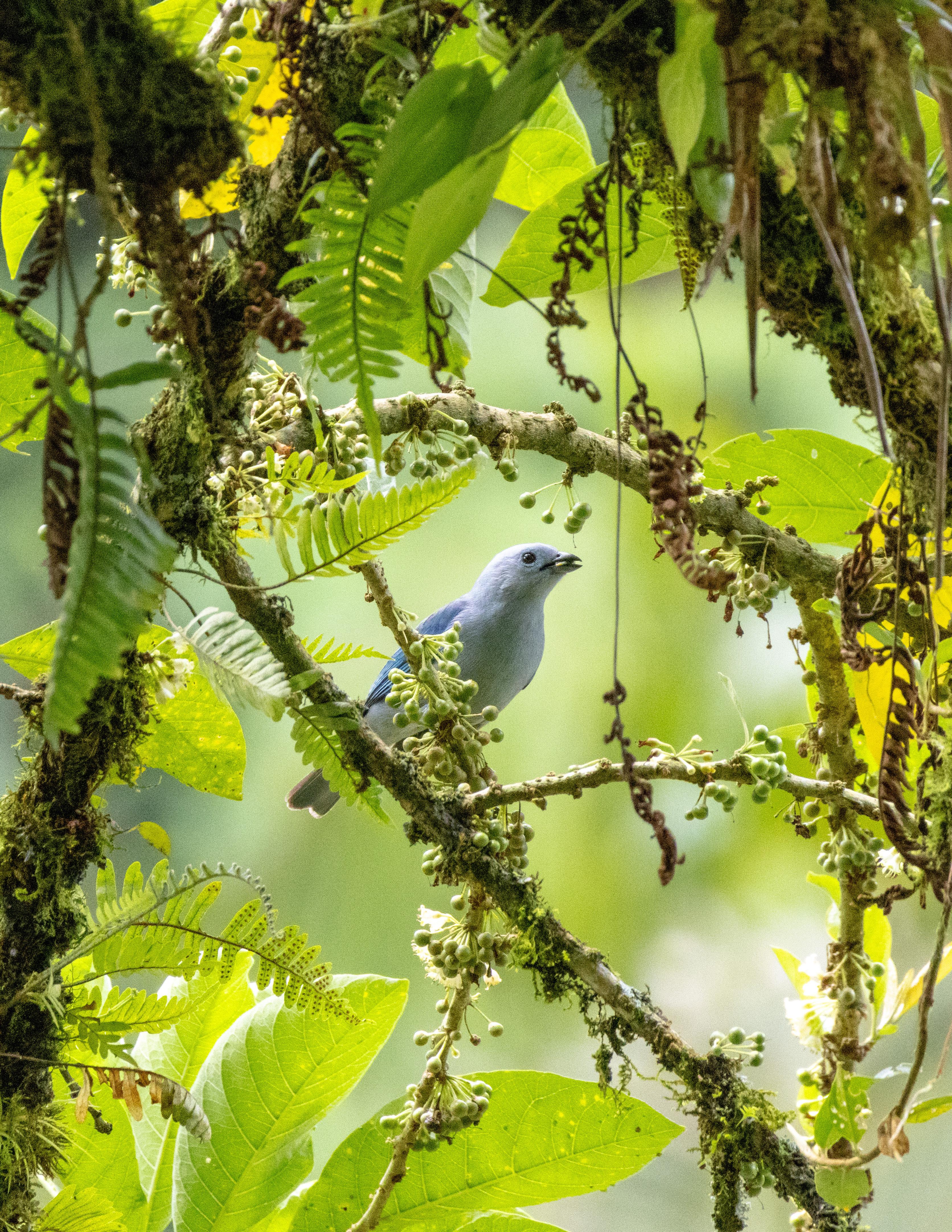
Blue-grey Tanager (Thraupi episcopus), eating thrush fruit (Iochroma arborescens)
Photo by: Spencer Andersen
Inside Cover
Brandt’s Cormorant (Phalacrocorax penicillatus) bathing in golden sunlight at La Jolla, CA
Photo by: Inaya Nicholls
Back Cover
Prickly pear cactus (Opuntia ficus-indica) on Scripps Institution of Oceanography campus, overlooking Scripps Pier, 2024
Photo by: Inaya Nicholls
SALTMAN FAMILY AND SUPPORTED
The views expressed in this publication are solely those of Saltman Quarterly, its principal members, and the authors of the content of this publication. While the publisher of this publication is a registered student organization at UC San Diego, the content, opinions, statements, and views expressed in this or any other publication published and/or distributed by Saltman Quarterly are not endorsed by and do not represent the views, opinions, policies, or positions of the ASUCSD, GSAUCSD, UC San Diego, the University of California and the Regents or their offices, employees, or agents. The publisher of this publication bears and assumes the full responsibility and liability for the content of this publication.
In an effort to engage the UC San Diego Community, Saltman Quarterly holds an annual photo contest. The winners of this contest have their images featured on the cover and interior pages of the journal.
DEAR READER,
As one might imagine, the appearance of Saltman Quarterly has evolved over the years. The first volume, published in 2005, was printed almost entirely in black and white and featured a charming immunology crossword puzzle on its back cover. With each subsequent volume came a little more color, a few more illustrative pieces, and a gradual, though unmistakable, gravitation to an ever-sharpening embrace of artistry and flair. Before long, the journal displayed a thoroughly rich character of visual art and creativity, showcasing vibrant tapestries of illustrations and photographs. Needless to say, if you were to hold this volume up against Vol. 1, you would almost certainly spot more differences than you would similarities.
Still, a feature of Saltman Quarterly that’s remained—and will for yet countless years—is its connection to a broader moment. In 2020, for example, that moment was COVID. And in the years that followed, the awkward emergence of a new normal—and its myriad implications—characterized the time. Today, our moment is one of deepening disarray—in which the bonds between truth and objectivity fray ever more and the tenor of collective discourse coarsens still further. In this atmosphere, science has suf-
fered under growing pessimism and distrust. Celebrating science’s practitioners and institutions has never been more important—and this volume, the combined expression of aspiring scientists, is a striving testament to that aim.
In these pages, you will find a diverse ensemble of scientific journalism and literature. You’ll encounter a charming and exquisitely-illustrated spread of feature articles, which illuminate groundbreaking and innovative research across several biological laboratories right here on campus. You’ll journey into the incredible original research of four UC San Diego undergraduates, whose manuscripts span a wide spectrum of biological research—and uncover new and exciting investigative frontiers. And you’ll meet over forty senior undergraduate researchers and receive a glimpse of their findings and discoveries over the last year.
Saltman Quarterly is a vast collaborative effort by a large and diverse team of students. Over the last year, these students—writers,
editors, illustrators, photographers, and production designers—volunteered their time and energies, amid the demands of countless other obligations, to this truly intensive and demanding undertaking. This incredible devotion to crafting and publishing insightful and accessible scientific journalism is a striking tribute to the essential enterprise and cause of science. And though the journey was not without setbacks and hiccups, challenges and difficulties, we persisted because our collective voice matters—because Saltman Quarterly matters. And so, amid this moment, when the future is all but certain, we are proud to claim, without equivocation, that our work will never cease.
We hope you enjoy the 22nd volume of Saltman Quarterly
Saltman Quarterly thanks the Saltman Family for their generosity and support. Their contributions have allowed SQ to continue to spread Dr. Paul Saltman’s ideals of science, communication, and education.
FEATURES
Wiring Our Brains and Biofilms: Untangling Bioelectrical Signaling by Gabbi Basa
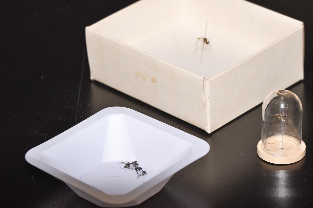
Mosquitos: an Unrequited Love by Rohan Purohit
The Root of the Matter by Sannidhi Krovvidi
CATCHing Cancer With Bacterial Biosensors by Kalisa Kang
Knowing Your Heart: Deep Tissue Monitering by Siya Jatia
RESEARCH, BREVIAS, & REVIEWS
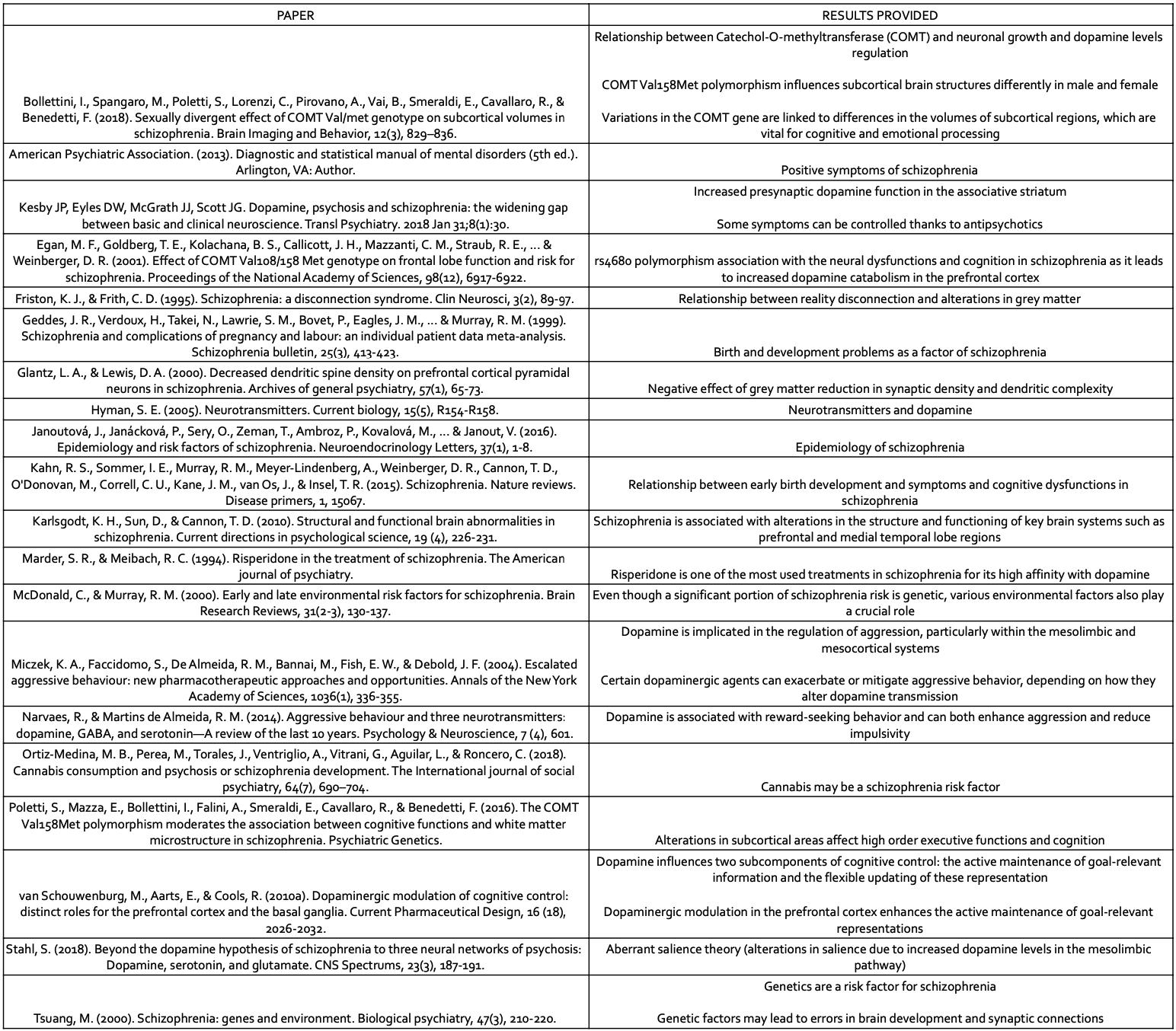
The Role of Dopamine in Schizophrenia: Systematic Review by Paula Alconchel Albelda
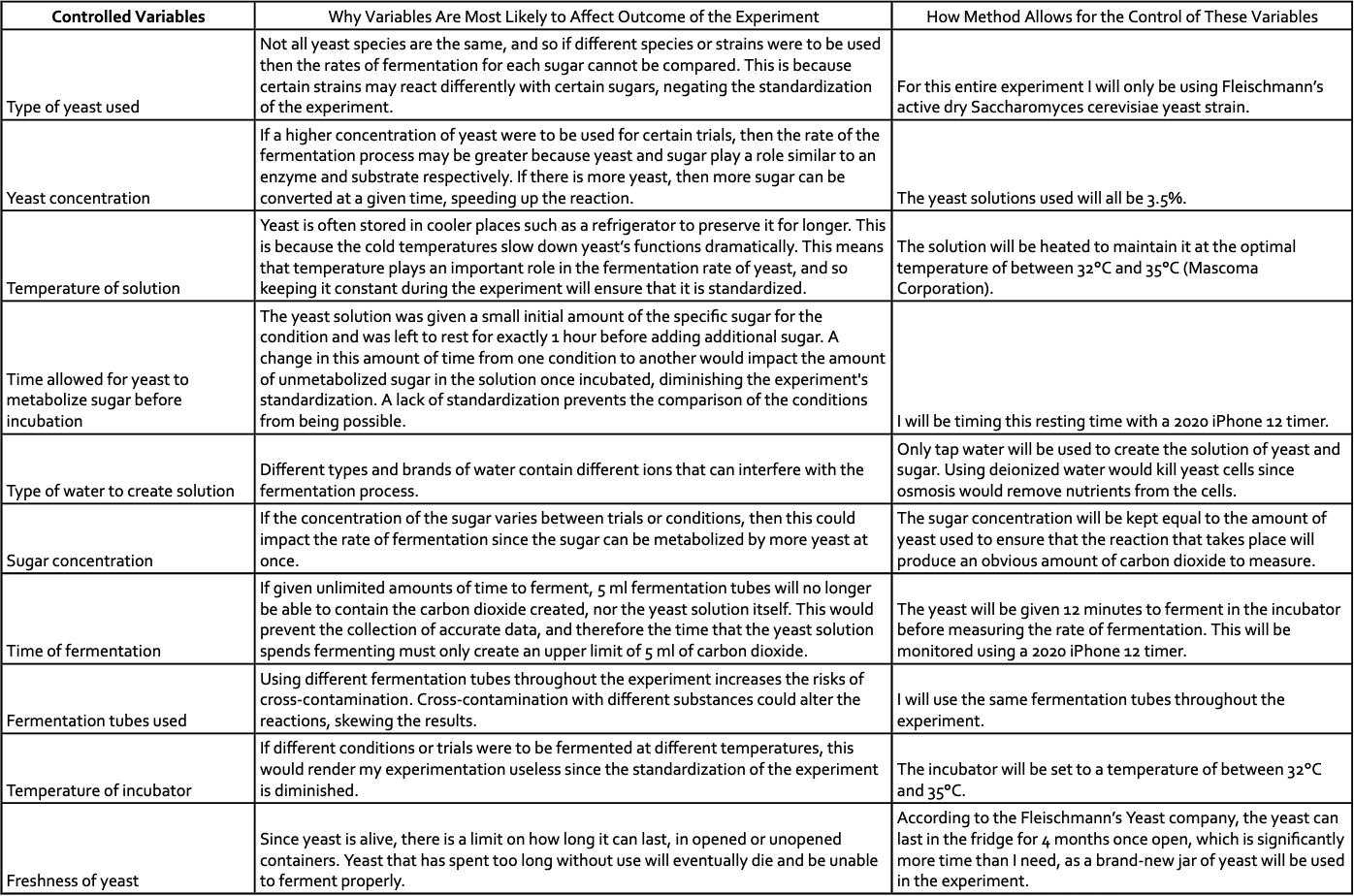
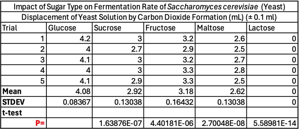
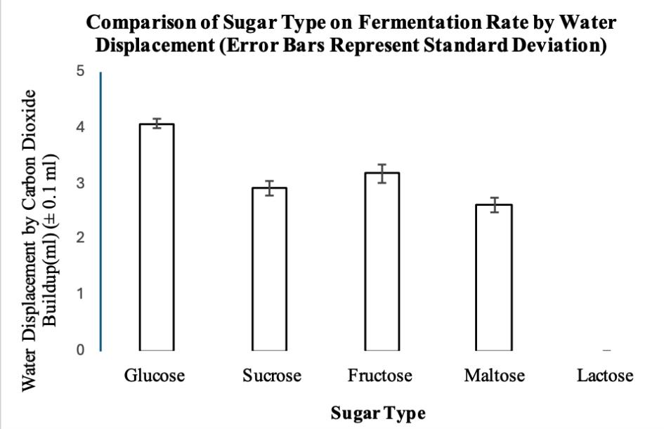
Investigating Different Types of Sugar on the Fermentation Rate of Saccharomyces cerevisiae (Yeast) by Carbon Dioxide Production by David Georges
Linking Carbon Storage to Climate Action: The Case for Kendall-Frost Marsh and Estuarine Restoration by Jeff Finn Schwartz
Potential of clinal variation in seed viability and dormancy in Eschscholzia californica as indicator of plant species’ adaptability to climate change by Rachel K. Brown
SENIOR HONORS THESIS
Undergraduates in the Biology Honors program are required to complete a written thesis detailing their scientific research. The Senior Honors Theses section, which presents the abstracts of their individual theses, highlights the achievements of accomplished student researchers.
STAFF
Meet the members of the 2023-2024 Saltman Quarterly staff who worked throughout the year to bring you this issue, as well as our online content, quarterly insiders, and community outreach initiatives.
DR. PAUL SALTMAN
Learning,
teaching, and service—the tenets of Dr. Paul Saltman that live on posthumously through Saltman Quarterly. His enthusiasm for scientific communication forever motivates our organization to promote the discussion of scientific knowledge. Even 26 years after his passing, Dr. Saltman is still remembered as an innovator in nutritional biochemistry, a beloved professor, and a dedicated mentor who pushed his students to tackle and solve biological enigmas. Dr. Saltman’s legacy, the lasting impact of a career devoted to expanding biological mysteries, remains omnipresent, reminding us that science is better when it is more accessible and helps everyone learn new information.
" science is better when it is more accessible and helps everyone learn new information "
Dr. Saltman's education began at the California Institute of Technology, where he earned his B.S. in Chemistry in 1949 and his Ph.D. in Biochemistry in 1952. After receiving his doctorate, he moved abroad, teaching at the University of Copenhagen and Murdoch University in Australia. Dr. Saltman then returned to California and served as a faculty member in the Keck School of Medicine at the University of Southern California. In 1967, he became Provost of Revelle College at UC San Diego, where he would later serve as Chancellor for Academic Affairs.
Dr. Saltman forever advanced the field of nutritional biochemistry, uncovering how trace metals like zinc, iron, copper, and manganese support immune function
and bone health. His work directly improved national recommended dietary plans and better revealed how nutritional supplements affect human health, touching and improving the lives of many. He guided patients to display clinical improvements in their skeletal metabolism, reductions in heart damage, and preventions in anemia. He also took his expertise to literature, authoring two books, The University of California Nutrition Book and The New Nutrition, both of which sought to extend scientific information by teaching the general public about nutritional health. His clinical achievements not only motivates current scientists and researchers to uncover new biological breakthroughs, but also inspires students looking to build a healthier society.
While furthering public understanding of nutrition and human physiology, Dr. Saltman grew concerned with how to effectively educate elementary and secondary school students in various scientific fields. After learning that a significant portion of U.S. elementary school teachers had not taken a year of college-level science, he used his position on the NSF Committee on Science Education to create the Program for Teacher Enhancement in Science and Technology. Soon, K-12 teachers spent their summers on UC San Diego’s campus learning about the fundamentals and frontiers of the basic sciences. Dr. Saltman continued to pro-
mote student accessibility to learning as Provost of UC San Diego’s Revelle College, where he developed “The Frontiers of Sciences”, a lecture series intended to foster science literacy among UC San Diego’s students and faculty. Dr. Saltman’s then-unconventional emphasis on bettering student experience of science education initially elicited strong backlash from some faculty members and San Diego residents. However, his persistent efforts in maintaining his science education programs eventually earned him high praise among faculty, with UC San Diego Chancellor Robert C. Dynes referring to Saltman as “a role model for students and faculty alike”.
With his scientific education programs expanding not only at UC San Diego but also throughout the state, Dr. Saltman turned his attention to enhancing scientific literacy among individuals of all academic backgrounds. Throughout the early 1960s he collaborated with the National Educational Television to create “Patterns of Life”. This half-hour video series, which offered approachable discussions of various molecular and biochemical pathways in organisms, still exists in the media collections of many universities and was archived by the Library of Congress. Aside from television projects, Dr. Saltman frequented radio networks and newspapers to explain various scientific concepts
to the general public. In fact, he was one of the first to publish an educational “course by newspaper.” Titled “America and the Future of Man”, this 322-page booklet consisted of 20 lectures which covered topics ranging from history to biophysics. Whether through television or local newspapers, Dr. Saltman’s passion for bridging the gap between scientific research and public awareness remained ever present through his practical explanations.
Just as Dr. Saltman sought to broaden the frontiers of science by increasing its educational accessibility, Saltman Quarterly hopes to do the same for its readers. Since 2004, SQ has taken its own multifaceted approach to uplift a diverse range of original manuscripts from undergraduate biology scholars at UC San Diego. As of 2025, SQ has connected with thousands of science and nonscience majors to cultivate an inviting space to explore the biological sciences. Whether it is providing mentorship through our educational events with local high schools or broadening the accessibility of undergraduate biological research findings to all interested students, Saltman Quarterly looks to Dr. Saltman’s legacy as a scholar and educator to push the boundaries of science literacy within our communities while also reaching outward to all who are eager to learn.
written by Leanne Liaw and Nicolas Bello
FEATURES
UC San Diego is at the forefront of scientific discovery and exploration as a hub of biological research. The Features section highlights some of the groundbreaking work accomplished by researchers affiliated with the UC San Diego campus.
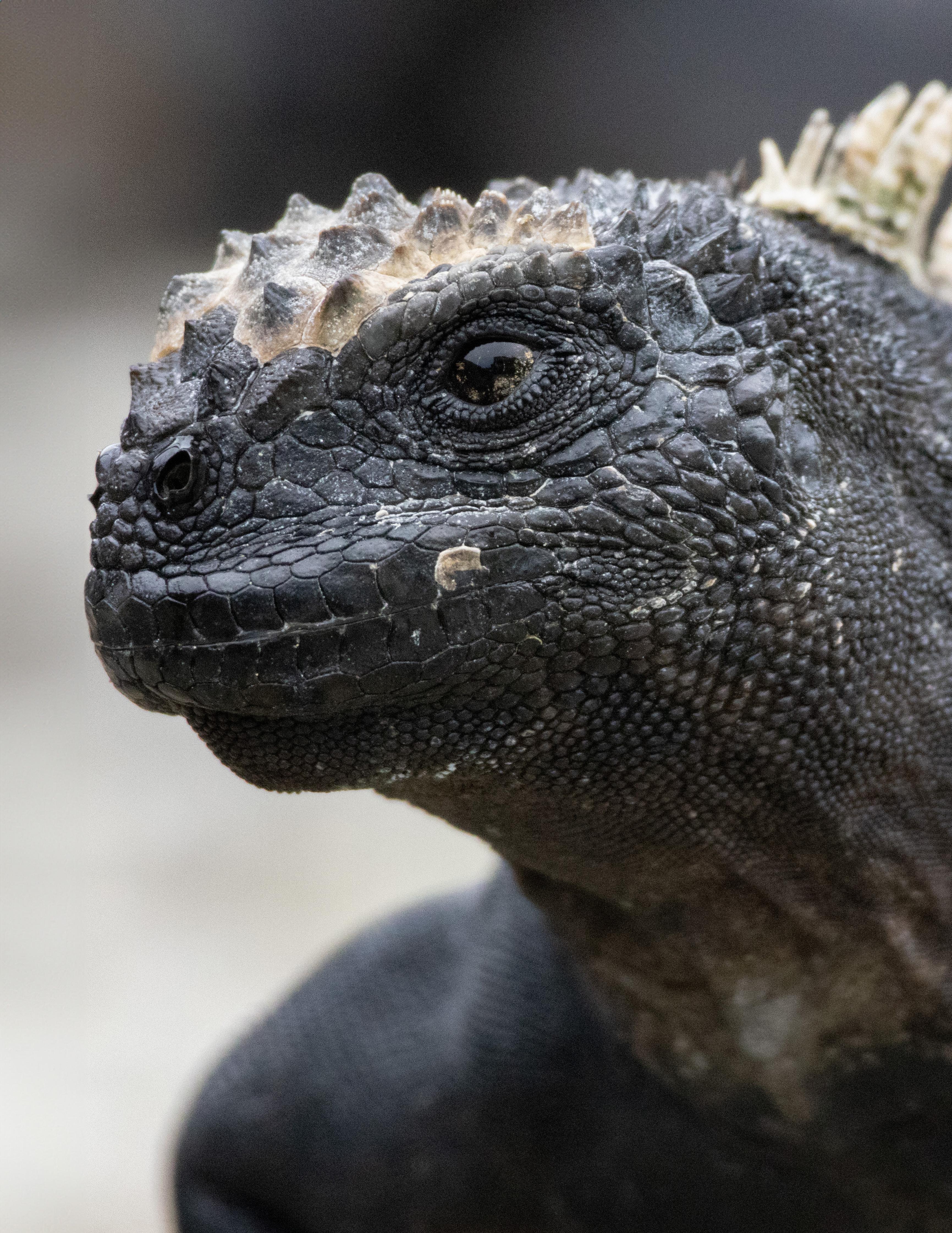
A Marine Iguana(Amblyrhynchus cristatus) at the Charles Darwin Research Station
photo by: SPENCER ANDERSON
Wiring Our Brains and Biofilms
Written by Gabbi Basa
Illustrated by Maia Lazor
Untangling Bioelectrical Signaling
Imagine a bustling city with sprawling infrastructure and busy constituents. In any high-functioning environment, communication is key to running things smoothly. Across human cities, or on a macroscopic scale, electricity facilitates the mass transfer of information, powers communication, and maintains order. Just as electricity runs our world, it is also critical for life on a microscopic scale, permeating biological systems in both familiar and unexpected ways. Electrical signaling between neurons may be the most intuitive example of electricity shaping life, but it is far from the only one; mycobacterium tuberculosis attacking a lung, a predatory venus flytrap closing its trap, a heart pumping blood through a body—these distinct biological events all rely on electrical signaling to different degrees. If one looks closer and a little further back in evolutionary history, it becomes apparent that bioelectrical signals permeate all forms of life in patterns that mirror over time.
Fig. 1
Bacterial Biofilms: Ancient or Advanced?
Despite their relatively simplistic biology, basic bacterial systems have been found to share functional links with the most advanced biological systems yet to have evolved. For example, bacterial cells contain genes that are homologous, or functionally similar, to eukaryotic genes that code for neuron-specific features, such as voltage-gated ion channels and synaptic proteins. In fact, one hypothesis suggests that voltage-gated channels first evolved from ancient prokaryotes’ need for water and ion regulation.1 In our bodies, voltage-gated sodium and potassium channels rapidly open and close in response to fluctuations in a neuron’s membrane potential, or electric charge difference, generating an electric signal known as the action potential—one of the most complex forms of biological communication. This organization is incredibly useful for transmitting signals across long distances, whether in multicellular organisms or in communities of smaller organisms like bacteria. While bacteria are often thought of as solitary, free-floating organisms, they lead social lives out in nature, often forming into biofilms, or “cities” of their own. A biofilm is a densely packed community of bacteria with enhanced functions and protections for individual cells and growing colonies.2 Much like an urban landscape, a biofilm is a diverse population of cells, all with different roles and metabolic states that work together to support its growth and adaptability. Biofilms form in nutrient-limited environments when a foundational group of bacterial cells attaches itself to a moist surface using built-in hair-like appendages known as pili and fimbriae.
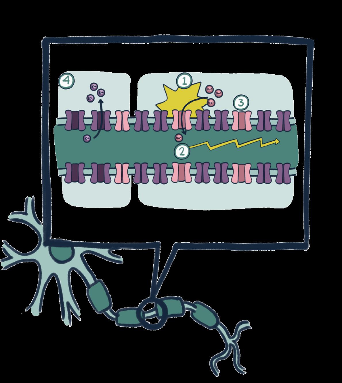
Membrane depolarization creates a signal in neurons and bacterial biofilms. To generate an action potential in a neuron, positively charged sodium ions rush into the cell and create an imbalance in charge or the propagating electrical signal. In bacterial cells, the efflux of potassium similarly depolarizes the cell and the cells around it, as sodium does not play a role in membrane potential oscillations.
"Recognizing bacteria as communal organisms rather than isolated cells opens the door to new ways of researching their behavior, communication, and pathogenicity."
These foundational cells then multiply to form microcolonies, which are similar to what you might observe on a petri dish. Simultaneously, the growing biofilm secretes an extracellular matrix, like a bacterial glue containing a wide variety of proteins and other biomolecules. This slimy substance shields the internal microcolonies from the environment and forms a three-dimensional system that maintains the flow of nutrients and waste. The extracellular matrix is also believed to support the heightened antibiotic resistance of biofilms by providing a physical barrier that blocks and neutralizes antibiotics. Once the numerous microcolonies mature, they detach from the biofilm as motile, or independent, cells to initiate biofilm formation on new surfaces.3
Various estimates show that anywhere from 40-80% of bacterial species naturally aggregate into biofilms because of the significant survival advantages afforded by the extracellular matrix.4 You may be more familiar with bacterial colonies grown on petri dishes, but in fact, only around 1% of all bacterial species are culturable, meaning that they aggregate into observable colonies under laboratory settings. A biofilm exhibits many collective mechanisms, such as quorum sensing for coordinated gene expression and cell differentiation; this complexity likens it to a multicellular organism. Yet, biological education
and research practice traditionally view the functional unit of bacteria as the cell, not the biofilm. Recognizing bacteria as communal organisms, rather than isolated cells, opens the door to new ways of researching their behavior, communication, and pathogenicity. One lab pioneering this focal shift is the Gurol Suel Lab at UC San Diego, which has spent the last decade investigating electrical signaling and emergent behaviors in biofilms. In their groundbreaking 2015 publication, “Ion channels enable electrical communication in bacterial communities,” the Suel Lab provided the first evidence that bioelectrical signaling is not exclusive to excitable cells like neurons or muscles. Rather, bacterial biofilms undergo a phenomenon of synchronized oscillations in their metabolic rates to proliferate and defend themselves. This is accomplished through a mechanism of ion-channel-mediated-signaling that bears striking similarities to neuronal communication in animals.
Our Shared Sparks of Life
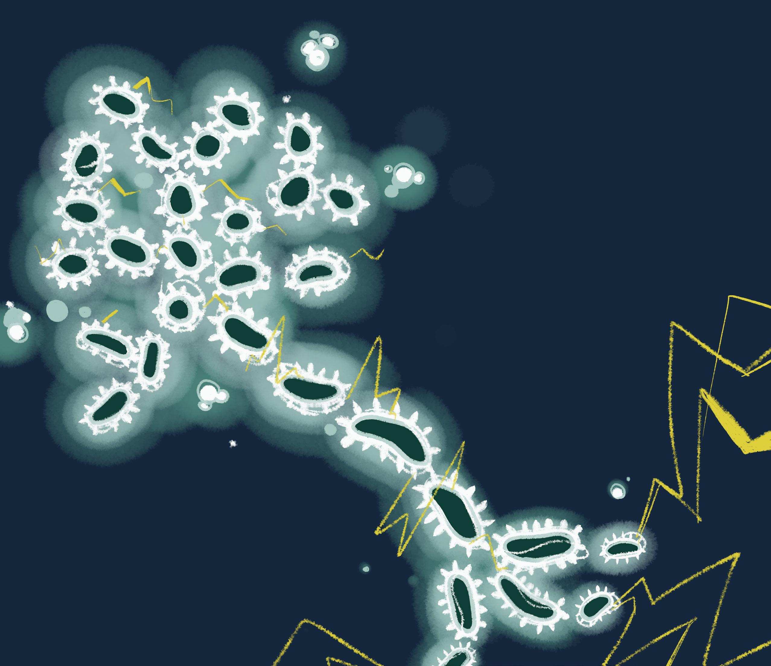
In our brains, neurons are the electric highways of life, rapidly firing signals that coordinate our thoughts, words, and actions. When traffic is light, the inside of a neuron is negatively charged compared to the outside. But when an electric signal is received, voltage-gated channels open, allowing sodium ions to rush in and make the inside of the neuron more positive. This sudden depolarization, or positive shift in voltage, travels down the neuron like a wave, passing the signal to the next neuron or a target cell. In the wake of the action potential, potassium channels open to restore the negative charge inside the cell and prepare for the next stimulus.5 Similarly, biofilm cells utilize ion channels to propagate their own signals for survival. However, unlike neuronal signals, which are highly precise and carry specific information from one neuron to the next, biofilm electrical signaling is a broader, population-wide phenomenon. Under metabolic stress, specifically a lack of glutamate, YugO channels on the membranes of interior bacterial cells will open to release potassium ions. These channels function similarly to neuronal potassium channels and cause the cell to depolarize. This produces a positive change in the local extracellular current, triggering nearby cells to also depolarize until the signal reaches the biofilm’s periphery (Figure 1).6 Both systems are elegant examples of electricity on a biological microscale, but how does bioelectrical signaling benefit biofilms?
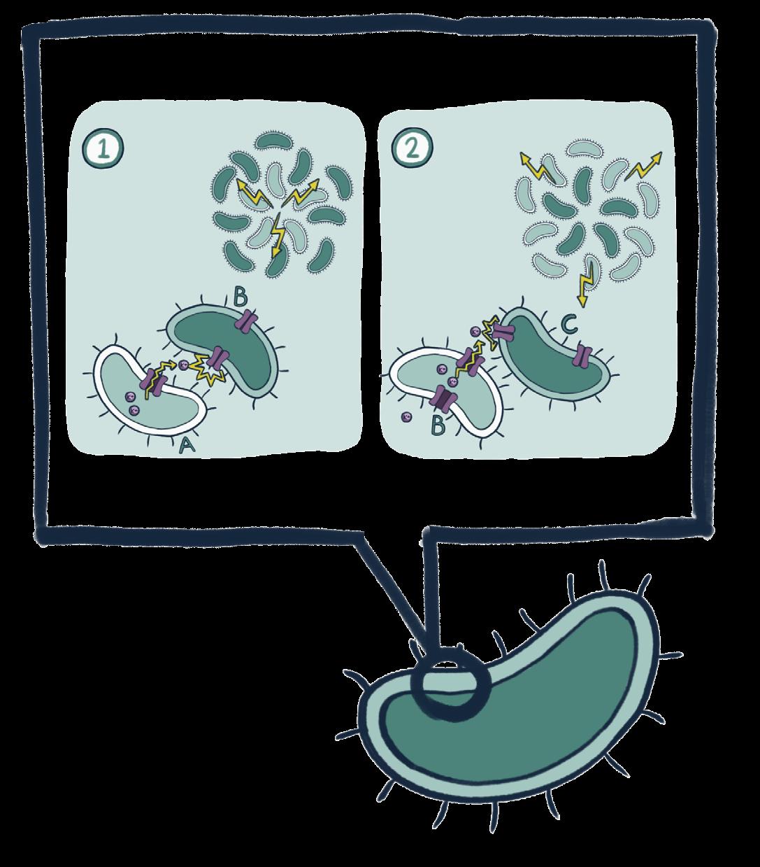
Interior and periphery biofilm regions fluctuate their metabolic rates. The periodic oscillations in a biofilm's metabolic rates and membrane potential occur in tandem. Propagating outwards from the interior, a modulatory depolarization signal enables a biofilm’s interior and periphery cells to exchange ammonium and glutamate, respectively.
Think of a biofilm as a bacterial “city” in which an electric grid dictates everything from resource allocation (who gets nutrients) to construction plans (which areas should grow or shrink).
Just as a city’s grid has the ability to reroute electricity to maintain overall function, a biofilm utilizes electrical signaling to modulate the growth rates of different regions in response to metabolic stress. In particular, cells on the periphery of Bacillus subtilis biofilms will periodically stop growing to prevent the starvation of interior cells. The fluctuation in metabolic rates is the result of a limited supply of glutamine, an amino acid and essential bacterial nutrient that is manufactured from the charged metabolites glutamate and ammonium. Interior cells contain enzymes that regularly produce ammonium, which eventually diffuses to cells on
Fig. 2
"The Suel Lab realized that control over the electrical dynamics of bacterial cells would allow for the manipulation of bacterial biofilm morphology."
the biofilm’s periphery. However, glutamate can only be sourced from the environment, giving peripheral cells easier access to glutamate and a metabolic advantage. To redistribute the available nutrients, peripheral cells halt their growth to allow glutamate to diffuse to the interior cells (Figure 2). Interior cells can then produce ammonium from a new supply of glutamate and begin the cycle anew.7 In contrast to a neuron encoding distinct messages using ion flow, a biofilm adjusts its “power grid” to balance energy demands across regions and fortify its structure.
In a proof-of-concept study to support this decade of findings, the Suel Lab combined forces with the Rolandi Research Group from UC Santa Cruz who specialize in bioelectronic systems and devices. Together, the researchers successfully integrated Bacillus subtilis biofilms with bioelectronic ion pumps that deliver potassium ions regionally within the biofilm. In their previous studies of biofilm oscillations, the Suel Lab used a microfluidic device, often described as a “lab on a chip,” for precise fluid measurements and ion delivery. A biofilm sat inside a chamber with two openings where an ionic solution flowed in then out, eliciting whole-biofilm growth and oscillations. This device, however, did not allow for spatiotemporal control over biofilm growth. Since the chamber delivered ions to the entire biofilm, there was no way to tell how separate biofilm regions would respond to ion delivery. In contrast to the microfluidic chamber, the bioelectronic ion pump, consisting of four glass capillary tubes lined with a hydrogel, facilitated ion delivery to specific spots on the biofilm. Once potassium ions were delivered into the extracellular fluid, YugO channels would open to depolarize the cells in that region. This promoted biofilm growth in an anisotropic, or non-uniform, manner, while the “control” biofilm, without the added device, experienced isotropic, or uniform, growth. Through this research, the Suel Lab realized that control over the electrical dynamics of bacterial cells would allow for the manipulation of bacterial biofilm morphology.8
Frying the Lines
Maintaining a balance of metabolic rates is shown to maximize biofilm robustness against external attacks. Because of their low metabolic rates and nested position within the biofilm, interior cells are well-equipped to resist antibiotics and guarantee the biofilm’s long-term survival. Severing the communication lines between the interior and periphery would therefore prevent the biofilm’s meticulous metabolic cycle, sabotaging its normal growth and function. Bacterial infections involving biofilms, such as cystic fibrosis and chronic wound infections, are notoriously difficult to treat due to a biofilm's ability to shield bacteria from both the immune system and antibiotics. Based on the Suel Lab’s past research, a new, drug-free method of treatment that exploits bacterial excitability could be effective in treating harmful biofilms. Let’s return to the neuron’s action potential: to transmit one signal after the other, a neuron must rapidly depolarize and hyperpolarize, or shift to a negative voltage. In between transmissions, there is a mandatory refractory, or recovery, period before the neuron can re-enter an excitable state. Bioelectronic Localized Antimicrobial Stimulation Therapy (BLAST), a device created by the Suel Lab,
leverages this concept in Staphylococcus epidermidis, a common skin bacteria that forms into a biofilm when it infects skin or contaminates medical devices. In its virulent state, S. epidermidis—a leading cause of hospital infections— forms antibiotic-resistant biofilms on implants, making it a critical target for treatment. First, exposure to an acidic pH of 5, typical of healthy skin, excites the biofilm and makes it responsive to external electric stimuli. Then, BLAST taps into S. epidermidis’ “electric grid” and delivers weak 1.5 volt signals to the bacteria, inducing a kind of refractory period. During this period of hyperpolarization, signals cannot be transmitted, preventing the coordination of metabolic rates between the interior and periphery. When tested on pig skin inoculated with S. epidermidis, BLAST decreased biofilm size by tenfold after an 18-hour treatment cycle and decreased the expression of antibiotic resistance genes. As antibiotic resistance continues to threaten public health, innovations like BLAST offer a promising, drug-free strategy to outsmart biofilms and protect against bacterial infections.9
The Concluding Transmission
From neurons firing in our brains to bacterial biofilms coordinating survival, bioelectrical signaling serves as the power grid that fuels life across biological domains. What was once thought to be exclusive to excitable cells has now been uncovered as a key mechanism driving the endurance of bacterial communities, further revealing strong functional links between microbes and complex life. The Suel Lab’s discoveries of metabolic coordination and ion-channel-mediated signaling have not only expanded our understanding of biofilm communication but have also paved the way for innovative bioelectronic therapies like BLAST. By recognizing the similarities between the communication systems of vastly different domains of life, we can gain new biological perspectives and apply this knowledge to solving problems in medicine, biotechnology, and beyond. Biofilms will continue to evolve, but so will our innovations to disrupt or tap into their power.
Sources
[1] Kristan W. 2016. Early evolution of neurons. Current Biology. [accessed 2024 Dec 12]; https://www.cell.com/current-biology/fulltext/S0960-9822(16)30489-4
[2] Muhammad MH. 2020. Beyond risk: Bacterial biofilms and their regulating approaches. Frontiers. [accessed 2024 Dec 12]; https://www.frontiersin.org/journals/microbiology/articles/10.3389/fmicb.2020.00928/full
[3] Penesyan A. 2021.Three faces of biofilms: A microbial lifestyle, a nascent multicellular organism, and an incubator for Diversity. Nature News. [accessed 2024 Dec 12]; https://www.nature.com/articles/s41522-021-00251-2
[4] Flemming H-C, Wuertz S. 2019. Bacteria and archaea on Earth and their abundance in biofilms. Nature Reviews Microbiology. [accessed 2024 Dec 12]; https://www.nature.com/articles/s41579-019-0158-9.
[5] Grider M. 2023. Physiology, Action Potential. National Institutes of Health. [accessed 2025 February 22]; https://www.ncbi.nlm.nih.gov/books/NBK538143/
[6] Prindle A. 2015. Ion channels enable electrical communication in bacterial communities. Nature. [accessed 2024 Dec 12]; https://www.nature.com/articles/ nature1570
[7] Liu J. 2015. Metabolic co-dependence gives rise to collective oscillations within biofilms. Nature. [accessed 2024 Dec 12]; https://www.nature.com/articles/ nature14660
[8] Prinzi A, Rohde R. 2023. The role of bacterial biofilms in antimicrobial resistance. ASM.org. [accessed 2024 Dec 12]; https://asm.org/articles/2023/march/ the-role-of-bacterial-biofilms-in-antimicrobial-re
[9] Kim S. 2024. Bioelectronic drug-free control of opportunistic pathogens through selective excitability. Current Biology. [accessed 2024 Dec 12]; https:// www.cell.com/device/fulltext/S2666-9986(24)00542-8
Written by Rohan Purohit
Mosquitos
Illustrated by Sofie Blazejova
Photographed by Andre Lin
an Unrequ ited Love
Mosquitoes, often underestimated, are one of the deadliest animals on the planet. Their impact on human society is staggering—shaping public health, economies, and even the course of history. Recently, San Diego has seen a concerning spike in mosquito populations as the Aedes Aegypti (Ae. Aegypti) mosquito, an invasive species, has entered the area, raising alarms for potential increases in mosquito-borne diseases.1 However, not all mosquitoes have a taste for human blood. Of the 3,500 mosquito species that exist today, only a few have evoWlved to specialize in biting humans. Understanding how these species diverged from others to become human-specialists—and whether their numbers could continue to rise—is crucial. Considering the rise of human-specialist mosquitoes, and its potential for future disease outbreaks, there is a need for researchers to better understand these species and take the necessary steps to prepare for their further spread. As humans transform the world through expanding cities and inducing climate change, the evolving resilience of mosquitoes reveals nature's persistent ability to adapt and push back.
Development of an Invasive Species
Firstly, there are many reasons why mosquitoes are such an effective disease vector in comparison to other animals. Female mosquitoes require blood feeding to nourish their eggs, making these host interactions highly frequent. Some mosquito species also have a large flight range of up to three miles, allowing them to expand their range for finding and passing on diseases.2,
3 Biologically, what allows a mosquito to function as a disease vector is their ability to successfully hold an arbovirus before transmitting it to another human in their future blood meals. Arboviruses, like the Zika virus, are viruses that are passed to a vertebrate from the bite of an arthropod, such as a mosquito.
After an arbovirus infects the mosquito, it develops and infects its salivary glands. Saliva is then injected into a human when the mosquito feeds, passing on the virus . However, many arboviruses, such as West Nile Virus, have co-evolved with a specific mosquito species (Culex in this case) to optimize its life cycle around that particular mosquito. This co-evolution makes certain mosquitoes more effective at transmitting certain arboviruses than others.4
The Ae. Aegypti mosquito is one of the most studied human-specialist mosquitoes acting as arbovirus vectors, affecting around 700 million people annually due to diseases like dengue, Zika, chikungunya, and yellow fever.5 The invasive form of this species originates from the Sahel region directly south of the Sahara desert, a semi-arid region that acts as a transition between the desert and savanna. Over 5,000 years ago, the ancestors of Ae. Aegypti thrived in pools of water within the then wetter and more lush landscape of the Sahel region. These stagnant pools of water are critical to the mosquito life cycle, serving as the primary
breeding sites where females lay their eggs. The eggs hatch into larvae, which remain aquatic and feed on organic matter in the water until they mature into adults.. The abundance of natural water pools in the ancient Sahel likely provided ample breeding grounds for Ae. Aegypti ancestors. These mosquitoes thus acted as generalists, living in tree holes and rock pools and feeding on a broad range of species, both human and non-human.
However, a few thousand years ago, the Sahel underwent a dramatic drying event and transformed into the arid desert it is today, receiving only three months of annual rainfall. This ecological shift rendered the region inhospitable for the ancestors of Ae. Aegypti, due to their life cycle revolving around stagnant water pools. Water stored by human societies for agricultural practices was now the only yearround reliable water source. Due to this acute shift in its habitat, Ae.Aegypti had to evolve to favor different kinds of hosts. “Hosts,” in this case humans, are a group of organisms that mosquitoes rely on for blood and, therefore, reproduction. Due to their newly increased proximity, humans became the most available host and shifted Ae. Aegypti into a new ecological niche. Over time, this adaptation led to the distinct human-specialist population of Ae. Aegypti, now genetically suited to thrive in human-dominated environments.
Global trade and urbanization further led to the spread of this human-specialized species outside of just the Sahel. For example, during the Atlantic Slave Trade, many water kegs were stored upon ships. Ae. Aegypti mosquitoes naturally gravitated to still water in these kegs, allowing the slave trade to facilitate distribution of these mosquitoes out of Africa. The mosquitoes’ close relationship with human populations enabled it to thrive in urban environments, further establishing its role as a vector for disease.6
Ecological Niche of Invasive Ae. Aegypti
Researchers are now intrigued by how the human-specialist Ae. Aegypti differ from generalist mosquitoes in their risk of disease transmission. Dr. Noah Rose, a researcher in the Department of Ecology, Behavior, and Evolution at UC San Diego, was working on a research project in the Cook Islands. However, progress was continuously interrupted by outbreaks of mosquito-borne infections in the region that posed a threat to the team. Dr. Rose grew curious of how such a small insect could pose such an immense obstacle to humans and refocused the scope of his research towards understanding the evolution of human-specialist mosquitoes.10
This research from the Rose Lab has shed light on the specific reasons for why certain mosquito populations prefer feeding on human populations over others. Major factors driving this preference are urbanization and climate
change, which extend dry seasons and allows human-specialist mosquitoes to outcompete generalist species due to their usage of man-made water sources. As more regions urbanize, particularly in Africa, mosquitoes that once only thrived in natural environments are increasingly adapting to human-dominated landscapes. Urbanization fragments original habitats and reduces natural breeding grounds, forcing Ae. Aegypti to exploit human-made water sources just as Ae. Aegypti responded to the drying of the Sahel.
"With climate change altering habitats and extending dry seasons, regions that previously did not see human-specialist mosquitoes may begin to experience an influx of Ae. Aegypti."
In addition to ecological factors, one significant behavioral trait that enhances Ae. Aegypti's human preference is its heightened attraction to human pheromones. The primary method for detecting humans are specialized nerve cells, called cpA neurons, with receptors designed to detect carbon dioxide, allowing them to sense the air humans exhale. However, mosquitoes are also attracted to humans, and specifically their skin, even in the absence of carbon dioxide. To determine how mosquitoes are able to detect human skin, researchers at UC Riverside constructed an experiment where they briefly exposed mosquitoes to a chemical that blocked their carbon dioxide receptors, rendering them unable to respond to exhaled carbon dioxide.7 Then, to test their attraction to skin pheromones, the team placed the mosquitoes in a wind tunnel with glass beads infused with the scent of human foot odor, obtained by wearing socks over the beads for several hours. After exposure to the reAe. Aegypti mosquitoes showed significantly reduced attraction to the scented beads. These results demonstrated that the cpA receptors are not only able to detect carbon dioxide but also play a key role in sensing human skin pheromones.
In a related study by Princeton researcher
quitoes were given the choice between entering a compartment with a hu man or guinea pig scent. McBride found that inva sive Ae. Aegypti mosquitoes would fly towards the human scent 90% of the time. as to the factors that determine whether a mosquitoes would prefer a human scent over an animal scent, Dr. Rose conducted his own study. He and his team sought to collect mosquitoes from twenty-seven different locations in Sub-Saharan Africa with varying human population densities and climates, including highly seasonal and semi-arid. Dr. Rose hoped that this wide sampling would give clues as to the types of environments human-specialist mosquitoes tend to thrive in, hypothesizing that distance from large human populations and weather being good potential indicators. Egg traps were placed in heaps of plastic and concrete in some locations and within shrubbery in others, attempting to capture a wide range of genetic and behavioral diversity within Ae. Aegypti populations for this experiment.
The captured mosquitoes were then tested for odor preference on an olfactometer, which observed a mosquito’s preference for scents between a single human and two guinea pigs.8 Preference for a certain odor was then determined by the amount of time a mosquito spent in a particular arm. Results showed that human-specialist species spent much more time in the arm holding the human odor rather than that of the guinea pigs. These findings were then further supported when the researched used a different human subject and quails to ensure accurate results. A linear model developed from these results showed that the number of humans living within a twenty to fifty kilometer radius from the mosquito collection site was a strong predictor of preference. Specifically, the model identified that as population density increased, so did preference for human scent.9
Climate variables were also determined to explain 65% of mosquito preference, with a longer dry season showing strong correlation with preference for human scent.9 Among these climate variables, low precipitation levels during the warmest season were the strongest predicting factors for human-specialist mosquitoes. Dr. Rose and his team believe that these two factors together highlight the struggle mosquitoes face during longer dry seasons and why some have evolved to be human-specialists. While eggs in continuous wet conditions can hatch immediately, those laid at the end of the rainy season and in regions with extensive dry periods must pause their development to survive these extended dry periods. However, human-stored water provides a wet habitat that lasts year round even in harsh climates, allowing for the development of larvae regardless of environmental constraints.
Dr. Rose's study highlights a significant distinction between human-specialist mosquitoes and generalist species, particularly in their scent preferences and egg-laying behaviors. Human-specialist groups sacrifice access to more organic material to consume, in order to survive a longer dry season. This underscores a clear behavioral divergence be-
Dr. Lindy McBride, mos-
tween the two groups, as generalist species favor natural, bacteria-rich pools that signal abundant food for their larvae. Generalist mosquitoes also develop slightly faster as larvae, possibly due to the greater predation risks in their more dangerous natural habitats. This speed gives them a survival advantage in environments with higher predator pressure, allowing them to mature before they are consumed. However, this increased speed would not impact human-specialist species, who tend to live in different areas than generalist species.10 This contrast in habitat preference, development speed, and survival strategies highlights the evolutionary trade-offs that shape mosquito behavior, reinforcing the distinct ecological roles of human-specialist and generalist species.
Response to Risks
These insights into mosquito behavior and adaptation are not only critical for understanding their evolutionary path but also have significant public health implications. With climate change altering habitats and extending dry seasons, regions that previously did not see human-specialist mosquitoes may begin to experience an influx of Ae. Aegypti and other populations, such as Culex. This shift increases the risk of disease spread in urbanizing regions, most notably Africa. Diseases that have historically had less of an impact in Africa, such as yellow fever, could now become more widespread as Ae. Aegypti enter cities. Understanding the genetic, be havioral, and environmental factors that influence mosquito preference for human hosts helps researchers predict which species may evolve similar behaviors in the future, potential ly allowing for proactive control measures
Currently, the Rose Lab uses technological advances, such as gene-editing techniques like CRISPR, to investigate methods of mitigating the threat posed by human-specialist mosqui toes. Broadly, CRISPR uses a guide RNA to find a particular sequence of DNA and then uses the Cas9 enzyme to cut the DNA at that location.11 The Rose Lab utilizes this tech nology to selectively knock out, or inactivate, regions in the genomes of human-specialist mosquitoes and observe its effects on preference of host. The ultimate goal of this re search focus is to gain an understanding of the genomic re gions of human-specialist mosquitoes that contribute to their unique nature.
It has become clear that the territories of human-specialist mosquitoes will continue to expand in the coming years. In fact, the problem is closer than many think, as the invasive
Ae. Aegypti has now reached San Diego. These mosqui toes introduce various new threats to the environment and highlight the need for quick action. Ae. Aegyti threatens the health of millions. Without an effective method of interven tion, it’s clear that the invasive human-specialized Ae. Aegypti can profoundly impact the health and livelihoods of millions worldwide. Without an effective plan of intervention, it will continue to rise, with the cost of inaction already being made so clear. As individuals, and as a society, it is critical to take action to curb their spread—whether through innova tive technologies, public health initiatives, or personal efforts to reduce mosquito breeding grounds.
References
[1] Invasive Aedes mosquitoes. County of San Diego. [accessed 2024 Dec 5]. https://www. sandiegocounty.gov/content/sdc/deh/pests/ aedes.html
[2] Diseases > mosquitoes. Link to the Entomol ogy Extension’s website. [accessed 2024 Dec 5]. https://extension.entm.purdue.edu/publi chealth/diseases/mosquito.html#:~:text=Numer ous%20factors%20determine%20the%20abili ty,%20vector%20capacity.
[3] County C. Mosquito control. Chatham County Mosquito Control Department. [accessed 2024 Dec 5]. https://mosquitocontrol.cha thamcountyga.gov/Faq/MosquitoFAQs#:~:text=Mosquito%20spe cies%20preferring%20to%20breed,in%20search%20of%20a%20 bloodmeal.
[4] Lewis J, Gallichotte EN, Randall J, Glass A, Foy BD, Ebel GD, Kading RC. Intrinsic factors driving mosquito vector competence and viral evolution: A Review. Frontiers. 2023 Dec 8 [accessed 2025 Jan 5]. https://www.frontiersin.org/journals/cellular-and-infec tion-microbiology/articles/10.3389/fcimb.2023.1330600/full?utm_ source=chatgpt.com
[5] Piovezan-Borges AC, Valente-Neto F, Urbieta GL, Laurence SGW, Roque F de O. Global trends in research on the effects of climate change on Aedes Aegypti: International Collaboration has increased, but some critical countries lag behind - parasites & vec tors. BioMed Central. 2022 Sep 29 [accessed 2025 Feb 24]. https:// parasitesandvectors.biomedcentral.com/articles/10.1186/s13071022-05473-7#:~:text=8%20Altmetric,Results
[6] Sokol J. Slave trade records help reveal when first yellow fe ver mosquitoes bit humans. Science. 2023 Mar 27 [accessed 2024]. https://www.science.org/content/article/slave-trade-records-helpreveal-when-first-yellow-fever-mosquitoes-bit-humans#:~:tex t=Some%20500%20years%20ago%2C%20a,ripple%20through%20 the%20 colonial%20world.
[7] How mosquitoes detect people. National Institutes of Health. 2022 Oct 26 [accessed 2025 Jan 5]. https://www.nih.gov/newsevents/nih-research-matters/how-mosquitoes-detect-people
[8] Fuller Wright L. Why do mosquitoes choose us? Lindy McBride is on the case. Princeton University. 2018 [accessed 2025 Jan 5]. https://www.princeton.edu/news/2018/10/23/why-do-mosquitoeschoose-us-lindy-mcbride-case
[9] Rose NH, Sylla M, Badolo A, Lutomiah J, Ayala D, Aribodor OB, Ibe N, Akorli J, Otoo S, Mutebi J-P, et al. Climate and urbanization drive Mosquito preference for humans. Current biology : CB. 2020 Sep 21 [accessed 2024 Dec 5]. https://pmc.ncbi.nlm.nih.gov/arti cles/PMC7511451/
[10] Purohit R, Rose N.. 2024. Saltman Quarterly Journal features article interview on the evolution of human-specialist mosquitoes. [11] November 21 2024, November 6 2024, October 17 2024. HOW CRISPR is Changing Cancer Research and treatment. NCI. [ac cessed 2025 Feb 24]. https://www.cancer.gov/news-events/can cer-currents-blog/2020/crispr-cancer-research-treatment#:~:tex t=With%20other%20versions%20of%20CRISPR,and%20RNA%20 from%20cancer%20cells.
The Root of the Matter
Written by Sannidhi Krovvidi
Illustrated by Julia Song
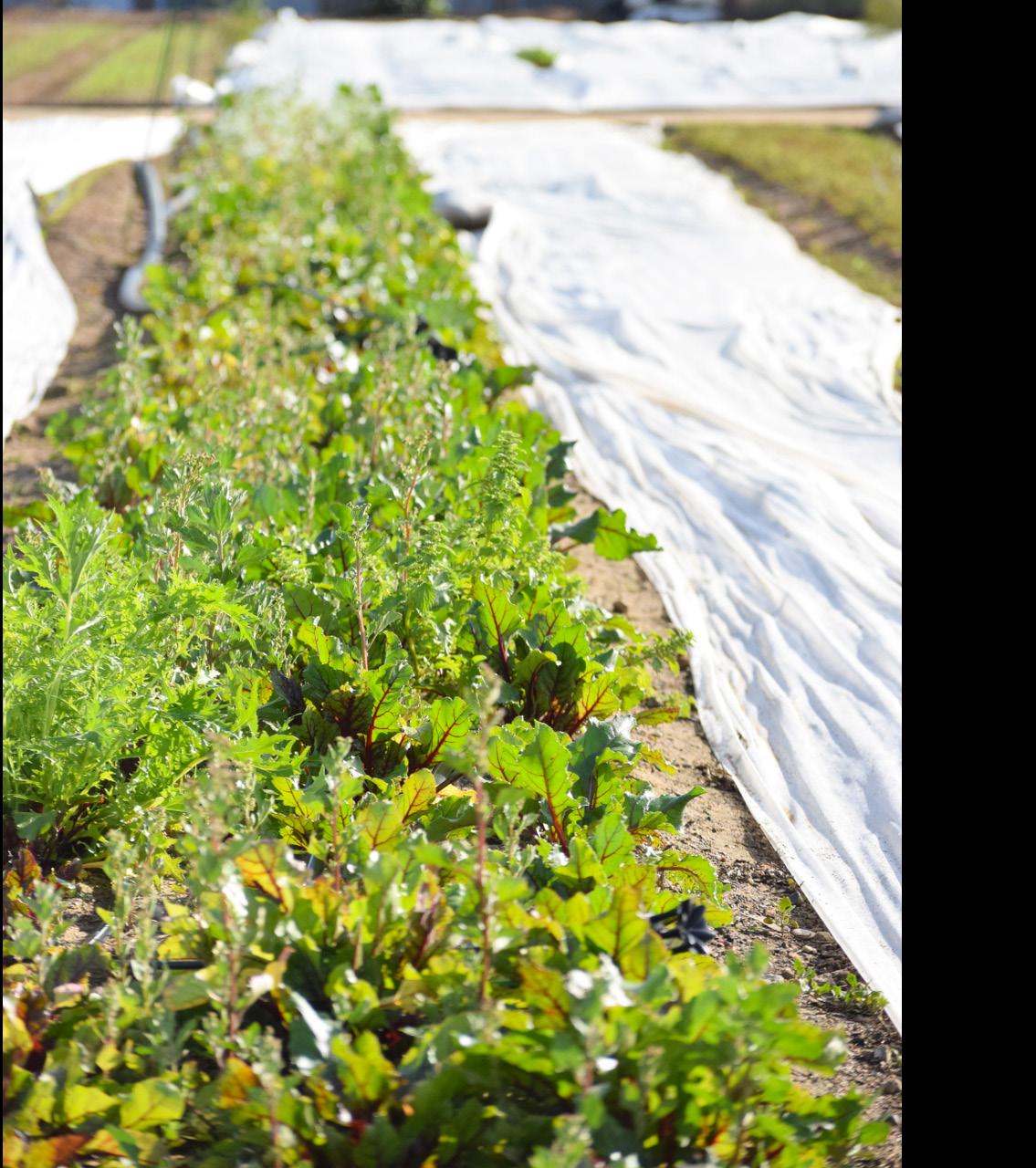
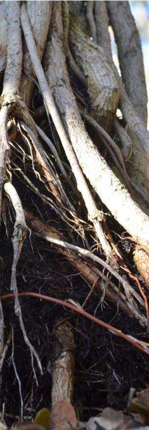
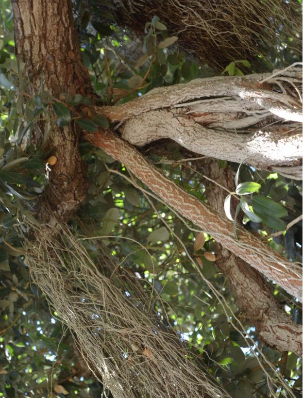
Photographed by Inaya Nicholls and Dominic Tse
Imagine your typical trip to the grocery store, walking through aisles filled with your usual shopping list—produce, dairy, snacks, and other items—comfortably picking up what you need. You can rely on the availability of produce and grains as staples in many households. However, this convenience is
The Fragility of Food Security
a privilege of our time. There may come a day when we can no longer take such abundance for granted as a result of the effects of the current climate change trajectory on the environment. Especially staple grains like corn, rice, and wheat could become seasonal or scarce, forcing consumers to carefully plan every meal around what’s available. The potential for empty grocery aisles and the absence of essential foods stand to serve as a stark reminder of how climate change has the potential to fully reshape our world. This shift would not only disrupt local food systems but also signal a global loss of stability and security, upending the very foundation of how we feed communities around the world.
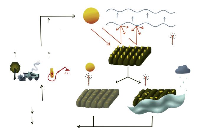
Agriculture is an important pillar of society, continually used as bargaining chips in international relations and international wars. Beyond its relevance at the global stage, 75% of the world’s population earn most of their living through farming.1 Increasing and promoting agricultural productivity is widely viewed as a way to improve economic development and reduce poverty. Climate change, however, has enormously, and multidimensionally, impacted agricultural harvest. The immediate effects of the environmental stressors of climate change, such as droughts and floods, are reduced crop yields and soil fertility. These environmental stressors can also lead to a cascade of problems affecting the overall health of an ecosystem. In a comprehensive study published in Science by Yang et al from China’s Chongqing University alongside numerous international institutions, researchers found that this decline in crop yields and soil fertility could force farmers into clearing more land for agriculture to still meet their yearly harvest requirements. However, this would further result in the loss of wildlife habitats and biodiversity from the now cleared land.2, 3 Simultaneously, climate change has worsened environmental and soil conditions, increasing our reliance on fertilizers and pesticides to sustain crop production (Figure 1). This overuse can similarly harm surrounding ecosystems, affecting birds, fish, beneficial insects, non-target plants, water quality, and even human health.2, 4
It is well known that the negative effects of climate change on our planet are extensive and damaging. Climate and temperature changes due to rising carbon emissions have had a wide-ranging effect on the global agriculture sphere. As per the June 2024 climate report from the National Centers for Environmental Information, the average temperature in the United States alone between January and June 2024 hit its second highest record yet.5 Additionally, the U.S. experienced a much drier season, resulting in low-moisture soil. Other regions, including South America, Europe, and Africa, also recorded their warmest temperatures yet, leading to similar effects.5 Cumulatively, these factors lead to drought stress, creating extensive detrimental implications for agricultural crops, predominantly maize, which loses around 15% of its global yield due to drought conditions.6 Given that the United States is the world’s largest producer, consumer, and exporter of maize— planting around ninety million acres annually—this issue is of immense national and international concern.7 Thus, acknowledging its importance and impact, Dr. Alexandra Jazz Dickinson’s Lab, in the School of Biological Sciences at UC SanDiego, chose to focus their study on maize.8
Scientists, like those in the Dickinson Lab, have been studying the complex interplay between climate and agriculture to better prepare crops to withstand future changes in weather and climate. Researchers are currently studying ways to develop deeper, broader, and stronger plant root networks to specifically address low-moisture soil. This process allows plants to forage for nutrients and water deep in the soil, po-
tentially increasing their resistance to drought and other climate-related stresses. They are tackling this study at the molecular level, focusing on the metabolites that influence root growth. By combining small molecule interactions and plant biochemistry, the Dickinson Lab investigates plant root metabolites to track corresponding stem cell activity and behavior, and thus control over root growth. A metabolite is a substance made or used when an organism breaks down food, drugs, and chemicals during metabolism. Notably they are instrumental in cell growth as different metabolites are increased during cell growth and division versus dormancy or death. Thus, by studying the relative concentrations of different metabolites, we can better understand the metabolic patterns of growth and stress resilience to engineer crops with stronger root systems. This knowledge can then be applied to maximize harvests, increase the sustainability of agriculture, and modify the plants themselves to combat the effects of climate change.
Strengthening Roots to Fight Climate Impact
Roots are the “heart” of the plant. Plant root systems that can dig deeper to acquire nutrients, fertilizer, and retain water live longer. Crops cultivated for human agriculture originate from wild species
"Roots are the 'heart' of the plant. Plant root systems that can dig deeper to acquire nutrients, fertilizer, and retain water live longer."
that were capable of “digging deeper”. These species were tough and able to survive without assistance or supplementation. Unfortunately, due to human intervention during the process of domestication for agriculture, these traits have been unintentionally selected against since these plants no longer actively compete for their own nutrients and water. Thus, the Dickinson Lab seeks to understand the mechanisms behind the strengthening of plant root growth, due to the potential of rediscovering these traits in crops that have weakened roots due to domestication. With this research they aim to understand how to not only to mitigate the current effects of climate change, but also how to engineer plants to continue withstanding future harsh climate conditions.
The Dickinson lab has specifically studied the plant root system in regards to the particular metabolites of the tricarboxylic acid (TCA) cycle. The TCA cycle, commonly known as the Krebs cycle, provides the energy necessary to sustain life across all plants and animals. In humans, TCA cycle intermediates play a significant role beyond just their traditional metabolic functions, influencing various signaling pathways impacting physiology and modulating innate and adaptive immune systems.9 In the plant root system, the TCA metabolites play an essential role in regulating root growth, making it possible to visualize several cell growth patterns through multiple stages of development by tracking TCA cycle metabolites. While this is an excellent focus for study, the TCA cycle is particularly tricky to study. For the study of most biological mechanisms, the preferred methodology is knocking out, or disabling, genes responsible for the function of interest and examining the physiological response. However, in the TCA cycle every metabolite is required to be present in order for the cycle to cascade and proceed forward. Each step is needed for the next, meaning the isolation and study of individual steps is difficult to pursue. As a result, diverse methods such as mass spectrometry imaging, chemical treatments, and tissue-specific genetic engineering are used.
At the Dickinson Lab, researchers specifically used mass spectrometry imaging to spatially visualize and map the metabolites present. Mass spectrometry imaging is an analytical tool used to measure spatial distribution of molecules.10 DESI-Mass Spectrometry Imaging, the type used by these researchers, involves spraying a cryosection, or a frozen thin slice, of plant tissue with a stream of charged droplets of a specified solvent. The solvent interacts with the molecules of interest, in this case metabolites, manipulating them to maximize their presentation in the imaging data collected by the mass spectrometer. This imaging data then determines the relative distribution of different me-
Low moisture soil and drought
Crop Yeilds
Soil Fertility [Wilting]
Increased evaporation, rainfall, and flooding [Greenhouse gases raise global tempatures]
[Plants uprooted]
tabolites regionally.11 The varied distribution of different metabolites in each area of the root can help guide the understanding of what chemical composition is necessary to stimulate and hinder root growth and differentiation – those present at higher concentrations towards the tip and branching points likely push root stem cells to divide and grow and those in dormant areas likely do the opposite. A combined understanding of this entire metabolite-map of the root system of a plant, including the tips, branching points, and dormant areas, can act as a first step in figuring out how to engineer root systems of important crops.
Understanding Root Growth Through Metabolites
For both maize and Arabidopsis (a common plant model), different TCA metabolites were found in opposing developmental areas (i.e., in areas of growth and division as well as dormancy). Two primary metabolites, succinate and aconitate, were found to accumulate in distinct regions of the root. Succinate was found to be concentrated in the meristem, at the tip of the root which holds the undifferentiated stem cells that divide and grow into different cell types.11 In contrast, aconitate, and other related TCA metabolites like malate and fumarate, were generally concentrated in the root differentiation zone, where cells begin to mature and specialize.11 Therefore, it was concluded that while succinate encourages rapid expansion of the root through stimulating proliferation of stem cells in the tip, aconitate guides the transition of these cells into their final, specialized forms as they move toward the differentiation zone. Interestingly, it was found that the distribution and development of TCA metabolites in relation to root stem cell growth were largely uncorrelated with ATP levels.11 This suggests that, unlike many metabolic processes typically linked to ATP production, TCA metabolites may have roles that extend beyond basic energy maintenance. Specifically, these metabolites appear to play additional, localized, non-canonical roles in regulating different stages of root development outside of those tied to energy production, further supporting the hypothesis that TCA metabolites help the plant modulate growth in specific developmental contexts outside of only metabolism. Understanding these metabolite concentrations can help us identify the key factors needed to grow deeper roots, essential for improving plant resilience, and provide a pathway into engineering plant roots able to survive in the face of climate change. Ultimately, the hope is to maintain food security despite a rapidly changing climate, especially for staple grains like
Figure 1: Climate Change and Agriculture. Rising global temperatures have resulted in diminishing soil fertility and integrity, as well as reduced crop yields, due to insufficient water and nutrients, and extreme climate events. The resulting increase in fertilizer and pesticide use, to address these challenges, has further exacerbated climate change.
maize, which play a major role in global food systems. Food security could be severely exacerbated unless research is accelerated to address the negative impacts of environmental change on agriculture. The research conducted by Dr. Alexandra Jazz Dickinson’s Lab offers promising pathways for a sustainable agricultural future. By studying the TCA cycle, which is conserved across a wide variety of plant types, the lab’s work aims to enhance crop resilience better withstand drought stress and other climate-related challenges.11 Human activities have caused the climate warming which has inflicted stress upon agricultural crops. However, with research geared towards exploring plant growth and development, it may also be human involvement through science that helps us enrich sustainable crop production, ensuring that our grocery shopping runs remain as accessible and affordable as possible.
References
[1] Agriculture and rural development. World Bank. (n.d.). https://www. worldbank.org/en/programs/knowledge-for-change/brief/agriculture-and-rural-development
[2]Growing threats: How climate change will exacerbate agriculture’s impacts. The Nature Conservancy. (n.d.). https://www.nature.org/en-us/ newsroom/growing-threats-how-climate-change-will-exacerbate-environmental-impacts-agriculture/
[3]Yang, Y., Tilman, D., Jin, Z., Smith, P., Barrett, C. B., Zhu, Y.-G., Burney, J., D’Odorico, P., Fantke, P., Fargione, J., Finlay, J. C., Rulli, M. C., Sloat, L., Jan van Groenigen, K., West, P. C., Ziska, L., Michalak, A. M., Lobell, D. B., Clark, M., … Zhuang, M. (2024). Climate change exacerbates the environmental impacts of Agriculture. Science, 385(6713). https://doi.org/10.1126/ science.adn3747
[4]Aktar, W., Sengupta, D., & Chowdhury, A. (2009). Impact of pesticides use in agriculture: Their benefits and Hazards. Interdisciplinary Toxicology, 2(1), 1–12. https://doi.org/10.2478/v10102-009-0001-7
[5]NCEI.Monitoring.Info@noaa.gov. (n.d.). June 2024 global climate report June 2024 Global Climate Report | National Centers for Environmental Information (NCEI). https://www.ncei.noaa.gov/access/monitoring/monthly-report/global/202406#:~:text=The%20average%20temperature%20of%20 the%20contiguous%20U.S.%20for%20the%20January,on%20record%20 for%20this%20period
[6]Kim, K.-H., & Lee, B.-M. (2023). Effects of climate change and drought tolerance on maize growth. Plants, 12(20), 3548. https://doi.org/10.3390/ plants12203548 Environmental Protection Agency. (n.d.). EPA.
[7]Feed grains sector at a glance. USDA ERS - Feed Grains Sector at a Glance. (n.d.). https://www.ers.usda.gov/topics/crops/corn-and-other-feedgrains/feed-grains-sector-at-a-glance/
[8]Krovvidi S, Dickinson A. 2024. Saltman Quarterly Journal Features article interview on plant root stem cell activity for crop engineering.
[9]Martínez-Reyes, I., & Chandel, N. S. (2020). Mitochondrial TCA cycle metabolites control physiology and disease. Nature Communications, 11(1). https://doi.org/10.1038/s41467-019-13668-3
[10]Murayama, C., Kimura, Y., & Setou, M. (2009). Imaging mass spectrometry: Principle and application. Biophysical Reviews, 1(3), 131–139. https:// doi.org/10.1007/s12551-009-0015-6
[11]Zhang, T., Noll, S. E., Peng, J. T., Klair, A., Tripka, A., Stutzman, N., Cheng, C., Zare, R. N., & Dickinson, A. J. (2023). Chemical Imaging reveals diverse functions of tricarboxylic acid metabolites in root growth and development. Nature Communications, 14(1). https://doi.org/10.1038/s41467-02338150-z
[12]Environmental Protection Agency. (n.d.). EPA. https://www.epa.gov/ climateimpacts/climate-change-impacts-agriculture-and-food-supply [13]NCI Dictionary of Cancer terms. Comprehensive Cancer InformationNCI. (n.d.). https://www.cancer.gov/publications/dictionaries/cancer-terms/ def/metabolite
Written by Kalisa Kang Illustrated by Nethra Nair
The Evolutionary Arms Race
Bacteria are fierce competitors, constantly evolving and adapting to survive. Many bacteria are “naturally competent,” meaning that they can actively transport environmental DNA fragments across their cell membrane(s) and into their cytoplasm. Once in the cytoplasm, the DNA integrates into the bacterial genome in a process called recombination. Bacterial recombination is facilitated by horizontal gene transfer (HGT), the non-sexual movement of genetic information between organisms.1 For billions of years, bacteria have engaged in this evolutionary arms race, exchanging genetic material to evade predators, phages, and host immune systems. As in Lewis Carroll’s Through the Looking-Glass, when the Red Queen informs Alice that “here, you see, it takes all the running you can do to keep in the same place,” so too have bacteria constantly adapted through recombination to keep them one step ahead of their adversaries. This endless race has equipped bacteria with remarkable tools. But what if this evolutionary advantage, honed over eons, could be redirected to help humans in a different kind of battle: the fight against cancer?
cerous DNA. Together, these components enable CATCH to identify oncogenes.
The CATCH system targets the kras gene. This gene encodes for the K-Ras protein, which regulates cell growth, differentiation, and gene expression. Unfortunately, kras can be mutated to become an oncogene, driving the progression of colorectal adenomas (benign tumors) into advanced carcinomas (malignant tumors).7 The most frequently mutated codon in the K-Ras protein of carcinomas is the twelfth codon, where glycine is replaced by aspartic KRASG12D).8
"But what if this evolutionary advantage, honed over eons, could be redirected to help humans in a different kind of battle: the fight against cancer?
Dr. Cooper uses A. baylyi biosensors to detect this single-codon mutation, creating a powerful tool for diagnosing colorectal cancer.
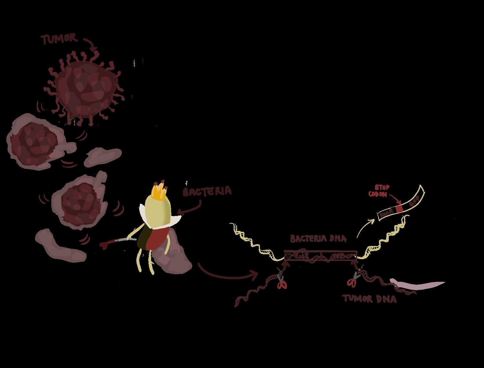
The first component of CATCH is to ensure that A. baylyi can intercept DNA from human cells. To do this, Dr. Cooper engineered the kras gene into A. baylyi and colorectal cancer cells. He designed a gene cassette, or small segment of DNA, containing a kanamycin resistance gene (kanR), a stop codon for kanR, and a green fluorescent protein (GFP). Then, he inserted this gene cassette in the middle of the kras gene. The segmented gene fragments serve as ‘homology arms,’ a pair of DNA sequences that are identical to corresponding regions in another gene cassette. Alignment of these pairs of homology arms allows for homologous recombination to occur. During homologous recombination, the sequences in between the pairs of homology arms are exchanged. The recipient genome incorporates the donor sequence, while the donor genome receives the recipient’s sequence.
To visualize the CATCH system in action, the cassette was first introduced into the A. baylyi genome. Then, the same cassette, this time without the kanR stop codon, was transduced into colorectal cancer cells. As DNA is constantly shed from cells due to the natural process of cell turnover, these DNA fragments act as genetic debris scattered across the battlefield. When the A. baylyi biosensors come into close proximity with DNA shed from engineered human colorectal cancer cells, the kras homology arms from both species line up, and homologous recombination occurs. The donor A. baylyi genome loses the kanR stop codon and can now express resistance to kanamycin, providing a clear readout for cancer detection (Figure 1).
The second component of CATCH wields A. baylyi’s native CRISPR-Cas system to detect the KRASG12D mutation. In bacteria, CRISPR functions as an adaptive immune system.9 It
stores small DNA segments from viruses (spacers) into an array, enabling the bacteria to recognize and defend against future attacks. The array is transcribed into guide RNAs, which pair with Cas proteins to form a Cascade complex. This complex scans DNA for a matching se quence next to a short conserved sequence called PAM. If the PAM site is present and the sequence matches the spacer, the Cascade complex binds and degrades the viral DNA, effectively killing the virus.
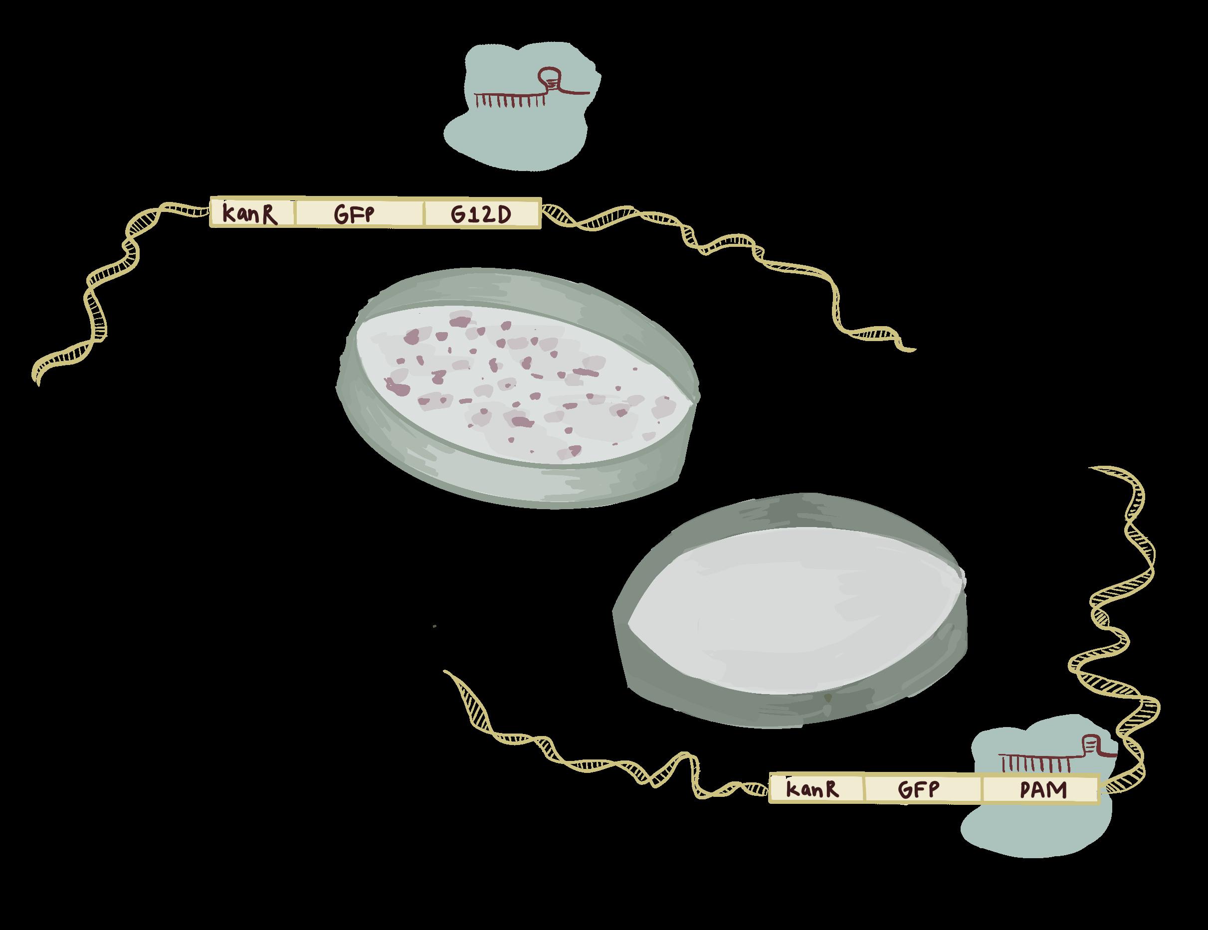
Following homologous recombination between A. baylyi and colorectal cancer cells, the CRISPR-Cas system is deployed. If the incorporated kras sequence is wild-type, the PAM site remains intact. The Cascade complex then recognizes and cleaves the wild-type DNA, killing the A. baylyi cell. Conversely, if the incorporated sequence contains the KRASG12D mutation, then the PAM site is disrupted. The Cascade complex cannot cleave the mutant DNA, allowing these A. baylyi expressing kanR to survive and grow. Their survival serves as a battle flag raised in victory, signaling the presence of the KRASG12D mutation and providing a clear readout for cancer detection (Figure 2).
CATCHing Cancer In Vitro and In Vivo
The A. baylyi biosensors successfully detected KRASG12D mutations in both engineered cancer cell lines on an agar plate and engineered tumorigenic organoid lines. Notably, the biosensors detected not only purified DNA, but also raw, unpurified DNA.7 In the chaos of a frenzied battleground, the A. baylyi biosensors wield a sword of precision. By recognizing a very specific mutation in an environment filled with contaminants, DNases, and other debris, the A. baylyi biosensors open the door to in vivo applications, where environments are similarly highly contaminated. To assess the A. baylyi biosensor in vivo, the engineered bacteria were rectally delivered to mice with and without colorectal tumors. The contents inside the gastrointestinal tract of the mice were analyzed for kanamycin-resistant A. baylyi by quantifying the number of colonies that grew on kanamycin selection agar plates. These results demonstrated that CATCH successfully distinguished between mice with or without colorectal cancer.
CATCHing Non-Engineered Cancer DNA
The CATCH system represents a breakthrough in live bacterial therapeutics, coupling evolutionary ingenuity with modern
biotechnology. Detecting engineered cancer DNA in vitro and in vivo is one achievement, but identifying non-engineered cancer DNA in patients presents an even greater challenge. In a new A. baylyi biosensor to detect non-engineered cancer DNA, a gene that encodes a tetracycline repressor protein (tetR) was inserted in the middle of the kras gene. Initially, the tetR gene is constitutively transcribed, continuously producing the repressor protein. Therefore, an output gene, in this case kanamycin resistance, is repressed.
Once again, when the biosensor encounters target DNA, homologous recombination initiates a tactical transfer. The tumor DNA delivers its kras gene to A. baylyi. If the kras sequence is wild-type with an intact PAM site, the Cascade complex recognizes and binds the sequence, degrading the kras DNA and output gene. This leaves the battlefield quiet, with no signal. However, if the kras sequence carries the KRASG12D mutation, the PAM site is altered and “camouflaged,” eluding detection. The Cascade complex is no longer able to degrade the DNA, allowing the output gene to produce a clear signal, a triumphant flare, marking the successful identification of colorectal cancer.7
Important Considerations with the Current Biosensors
"By recognizing a very specific mutation in an environment filled with contaminants, DNases, and other debris, the A. baylyi biosensors open the door to in vivo applications, where environments are similarly highly contaminated."
Bacterial biosensors represent a promising new weapon in the fight against cancer, but their development is still in the early stages. They are not yet poised to replace colonoscopies, which remain the gold standard for detecting polyps and early-stage colorectal cancers. Instead, these biosensors offer a less invasive, supplementary method for detection. However, this is not
"Just as bacteria have engaged in the Red Queen’s relentless race for survival, constantly evolving to outpace their adversaries, we can now harness this evolutionary momentum for human health."
yet a battle-ready solution; current studies remain proof-ofconcept and require further validation to ensure their effectiveness across diverse patient populations. To sharpen their battlefield precision, biosensors capable of detecting multiple genetic mutations—not just those in kras—would allow for a more comprehensive analysis of tumor DNA to identify multiple oncogenes or mutations within the same sample. Rigorous biocontainment measures, such as genetic "kill switches," could prevent unintended consequences, ensuring biosensors retreat once their mission is complete.10 Optimizing delivery strategies is another key objective. While rectal delivery has been effective in animal studies, oral delivery would make these tools more practical for patients.
Positive Outlooks for the Biosensors
The vision for bacterial biosensors extends far beyond colorectal cancer detection. With further development, these systems could be adapted to detect a variety of cancers including breast, liver, and blood, or even other diseases char acterized by specific genetic markers. Their adapt ability and precision could open doors to appli cations in personalized medicine. Just as bacteria have engaged in the Red Queen’s relentless race for survival, constantly evolving to outpace their adversaries, we can now harness this evolutionary momentum for human health. By redirecting the tools bacteria have used over billions of years, we may turn their survival strategies into powerful allies in the fight against cancer. Though still in their infancy, bacterial biosensors exemplify the potential of evolution’s arsenal not to keep humanity running in place, but to bring us clos er to victory in the ongoing battle against cancer.
References
[1] Vos M. Why do bacteria engage in homologous re combination? Trends in Microbiology. 2009;17(6):226–232. doi:10.1016/j.tim.2009.03.001
[2] Zhang J, Hasty J, Zarrinpar A. Live bacterial ther apeutics for detection and treatment of colorectal can cer. Nature Reviews Gastroenterology & Hepatology. 2024;21(5):295–296. doi:10.1038/s41575-024-00901-8
[3] Din MO, Danino T, Prindle A, Skalak M, Selimkhanov J, Allen K, Julio E, Atolia E, Tsimring LS, Bhatia SN, et al. Syn chronized cycles of bacterial lysis for in vivo delivery. Nature. 2016;536(7614):81–85. doi:10.1038/nature18930
[4] Fu T, Coulter S, Yoshihara E, Oh TG, Fang S, Cayabyab F, Zhu Q, Zhang T, Leblanc M, Liu S, et al. FXR regulates intestinal cancer stem cell proliferation. Cell. 2019;176(5). doi:10.1016/j.cell.2019.01.036
[5] Nguyen TT, Ung TT, Kim NH, Jung YD. Role of bile acids in colon carcinogenesis. World Journal of Clinical Cases. 2018;6(13):577–588. doi:10.12998/wjcc.v6.i13.577
[6] Cheng Y-Y, Chen Z, Cao X, Ross TD, Falbel TG, Burton BM, Ven turelli OS. Programming bacteria for multiplexed DNA detection. Nature Communications. 2023;14(1). doi:10.1038/ s41467-023-37582-x
[7] Cooper RM, Wright JA, Ng JQ, Goyne JM, Suzuki N, Lee YK, Ichinose M, Radford G, Ryan FJ, Kumar S, et al. Engineered bacteria detect tumor DNA. Science. 2023;381(6658):682–686. doi:10.1126/science.adf3974
[8] Vogelstein B, Fearon ER, Hamilton SR, Kern SE, Preisinger AC, Leppert M, Smits AMM, Bos JL. Genetic alterations during colorectal-tumor development. New England Journal of Medicine. 1988;319(9):525–532. doi:10.1056/nejm198809013190901
[9] Doudna J. CRISPR in Nature. Innovative Genomics Institute. 2024 Nov 23 [accessed 2024 Nov 26]. https://innovativegenomics.org/crisprpedia/crispr-in-nature/
[10] Kang K, Cooper R. 2024. Saltman Quarterly Journal features article interview on live bacterial therapeutics for colorectal cancer diagnosis.
Fig. 2: CRISPR Selection for KRASG12D Mutation
If wild-type kras is incorporated, the PAM site stays intact and the Cascade complex binds and cleaves the DNA, thereby killing the A. baylyi. However, if the KRASG12D mutation is present, the PAM site remains and the Cascade complex cannot cleave, allowing kanR-expressing A. baylyi to survive on kanamycin agar plates.
Knowing Your Heart
Written by Siya Jatia
Wong
Illustrated by Kristiana
Deep Tissue Monitoring
For more than a hundred years now, cardiovascular diseases (CVDs) have been the leading cause of death in the US.1 CVDs include, but are not limited to, coronary heart disease (CHD), stroke, heart failure, arrhythmias (abnormal heart rhythm), and high blood pressure. All CVDs originate from subtle vascular changes that often serve as harbingers of disease long before any prominent physiological symptoms emerge. The ability to monitor these changes continuously and in real-time could dramatically shift the paradigm for early detection, treatment, and management of CVDs. However, traditional diagnostic tools such as cardiac catheterization or blood pressure monitors and cuffs are either too invasive or inadequate for capturing the nuances of cardiovascular health. Enter wearable health technologies—innovations designed to integrate seamlessly into daily life while providing critical health data. Research in wearable health technology is a global endeavor, developing rapidly in the last few years with significant contributions coming from both academic and industry labs. These innovations are poised to redefine how we approach cardiovascular health monitoring and management, offering non-invasive, real-time insights into vital hemodynamic parameters.
and pulse pressure, accelerating vascular damage and the progression of diseases like hypertension and atherosclerosis. Monitoring CBP offers valuable insights into these factors, enabling better diagnosis and tracking of cardiovascular disease progression.3
However, conventional tools for CBP monitoring face limitations, making them less suitable for routine clinical use. Cardiac catheterization, an invasive procedure where a catheter is
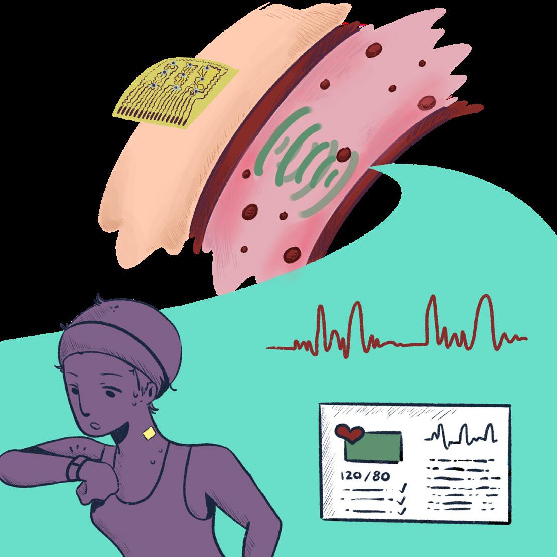
Fig. 1: A wearable ultrasound patch for continuous cardiovascular monitoring. The illustration depicts how the device adheres to the skin, transmitting ultrasound waves into deep tissues to detect arterial movements and blood flow dynamics. Additionally, it highlights real-time physiological data collection, including central blood pressure waveform analysis, enabling the tracking of cardiovascular health during physical activity.
Cardiovascular Diseases and Role of Wearable Tech
For diagnosis and prognosis of cardiovascular diseases, healthcare professionals currently use the central blood pressure (CBP) waveform (representing the pressure changes in the central arteries, such as the aorta, over a cardiac cycle), which is an indication of blood pressure in primary arteries near the heart and brain. On the other hand, peripheral blood pressure (PBP) measures blood pressure in peripheral arteries.2 Unlike PBP, CBP reflects the true load or stress on vital organs and the elasticity of central arteries, which play key roles in diagnosing and tracking disease progression.3 This distinction is crucial because CBP provides a more accurate representation of cardiovascular stress and arterial health. As a result, the heart is forced to work harder because of increased load, such as heightened afterload from arterial stiffness. This contributes to conditions like left ventricular hypertrophy and heart failure. Similarly, reduced arterial elasticity amplifies central blood pressure
Research in wearable health technology is a global endeavor, developing rapidly in the last few years with significant contributions coming from both academic and industry labs.
inserted into blood vessels to measure CBP directly, provides highly accurate readings but is impractical for regular monitoring due to patient discomfort and risks such as infection.4 Photoplethysmography (PPG) is a non-invasive method that uses light absorption to detect blood volume changes in tissues. It is commonly employed for peripheral vascular assessments, but lacks the depth penetration required for central arterie s.5 Despite its ability to monitor arrhythmias and peripheral hemodynamics, PPG is also highly susceptible to motion artifacts (distortions or inaccuracies in the data caused by movement during the measurement process), providing inconsistent results in dynamic environments 3 Similarly, tonometry, which estimates arterial pressure by compressing the skin over a superficial artery, is influenced by tissue stiffness and movement, as well as dependent heavily on operator skill.6 These limitations highlight the need for advanced CBP monitoring technologies. Wearable ultrasound devices, in particular, show promise by addressing these gaps through continuous, non-invasive, and accurate monitoring of central blood pressure.
One such advancement is a wearable ultrasound device developed by Dr. Sheng Xu’s lab at UC San Diego. This device holds transformative potential for cardiovascular health management by enabling continuous, non-invasive monitoring of CBP and other vital hemodynamic parameters. By overcoming the limitations of conventional tools, the Xu Lab’s wearable ultrasound device offers a novel approach to understanding and managing cardiovascular diseases, paving the way for more precise and proactive care.
Highlighting Research at the Xu Lab
To understand the applicability of the wearable ultrasound device, it is first and foremost necessary to learn about its structure. At its core, the device relies on an array of piezoelectric transducers that generate and receive ultrasonic waves. “Piezoelectronic” refers to electricity caused by pressure, and piezoelectric transducers convert the electrical charges produced by certain solid materials into tangible energy. A piezoelectric transducer works similarly to the body of an acoustic guitar. When you pluck a guitar string, the vibrations travel through the guitar’s body, amplifying the sound. Similarly, when pressure is applied to a piezoelectric material, it transforms that mechanical energy into electrical energy.7 These transducers are constructed using multiple piezoelectric microrods embedded within an epoxy resin matrix (providing structural support, adhesion, and durability). This design enhances acoustic coupling, enabling the efficient transfer of sound energy between the ultrasound device and biological tissues. The anisotropic configuration of the microrods—arranged in different directions—further improves the device’s ability to couple with tissues and ensures effective wave transmission for more precise measurements. The device is remarkably thin, measuring only 240 μm (just about the height of 3 sheets of printer paper), and highly stretchable, with mechanical properties that closely mimic human skin. This skin-like flexibility ensures intimate and stable contact with the body, even during intense physical activity. To further enhance usability, the device is encapsulated in elastic silicone elastomers, which not only provide a soft and comfortable interface but also protect the device from moisture and sweat. This encapsulation is equivalent to (and eliminates the need for) a traditional ultrasound gel, making the device more practical for everyday use.
Fig. 2: Applications of wearable ultrasound technology in cardiovascular health. At the very top, an animated physical architecture of the flexible ultrasound patch is depicted alongside its many applications in diverse user groups—including pregnant individuals, athletes, and patients with cardiovascular conditions.
Functionally, the researchers found an optimum balance between resolution and tissue penetration by operating the device at a high frequency of 7.5 MHz. Higher frequencies provide better resolution, allowing for more precise measurements of vessel dynamics, but they are less effective at penetrating deeper tissues. A frequency of 7.5 MHz is optimal for capturing detailed vascular information while still being able to penetrate the skin and underlying tissues effectively. The device utilizes ultrasonic waves to measure changes in blood vessel diameter and calculate blood pressure waveforms with exceptional precision. When placed on the skin, the waves penetrate biological tissues and reflect off interfaces, or the surface where two different materials or structures meet, like vessel walls. The reflected waves are captured by the transducers on the device itself.
Next, the time-offlight (TOF) data of the reflected waves is then analyzed to determine vessel health with sub-millisecond temporal resolution and micron-scale spatial accuracy.
The device is remarkably thin, measuring only 240 μm (just about the height of 3 sheets of printer paper), and highly stretchable, with mechanical properties that closely mimic human skin.
TOF refers to the time taken for an ultrasonic wave to travel from the transducer to a target (such as a vessel wall) and back to the transducer. This time is used to calculate the distance to the target, which is essential for determining vessel dynamics with high precision. Health parameters are processed from the raw data through advanced algorithms. For instance, systolic and diastolic blood pressure values are derived from vessel diameter measurements, while pulse wave velocity (PWV)—a key indicator of arterial stiffness—is calculated by correlating ultrasound data with electrocardiographic (ECG) signals. Additionally, researchers analyze the shape of the waveforms to provide more insights into vascular health. For example, the systolic peak represents when the heart's left ventricle contracts and pumps blood into the arteries, and other waveform characteristics reflect vascular resistance, elasticity, and cardiac output.
Despite its groundbreaking capabilities, developing this wearable ultrasound device required overcoming several challenges. Motion artifacts posed a significant hurdle since maintaining stable data collection during movement is essential for accurate measurements during regular daily use. The researchers addressed this through the device’s conformal design, which adheres closely to the skin. Biocompatibility was another critical consideration; extensive testing ensured that the materials used in the device were safe for prolonged use without causing irritation or other adverse effects on the skin and underlying tissues.3 Furthermore, energy efficiency was optimized to allow extended use without frequent recharging or excessive power consumption, making it suitable for long-term monitoring applications. By addressing these challenges with innovative solutions, this wearable ultrasound system sets a new standard for continuous health monitoring, offering unprecedented precision and convenience while paving the way for future advancements in personalized healthcare technologies.
Current and Future Applications
In its present form, the wearable ultrasound device revolutionizes hypertension management by enabling CBP monitoring. This real-time data allows clinicians to fine-tune antihypertensive therapies (focusing on lowering high blood pressure), tracking physiological parameters with unprecedented precision and improving patient outcomes with more personalized treatment plans. In post-surgical care, the device's ability to track vascular dynamics in real-time has proven crucial for early detection of potential complications such as thrombosis or vascular occlusion, significantly improving patient safety during recovery. Its utility extends beyond clinical settings, such as by finding valuable applications in sports medicine by providing detailed insights into cardiovascular responses during exercise to help athletes and trainers make informed decisions about training intensity and recovery.
Looking towards the future, the potential applications of this wearable ultrasound technology are vast and exciting. Researchers are exploring multimodal integration, combining ultrasound with other sensing technologies like PPG and ECG to create comprehensive cardiovascular profiling systems. This integration could provide a more holistic view of cardiovascular health, enabling more accurate diagnoses and treatment strategies. The incorporation of artificial intelligence (AI) and machine learning (ML) algorithms into health data analysis holds promise for bettering its interpretation. While the device collects real-time data, the integration with other sensing technologies, as well as the incorporation of AI and ML algorithms, typically requires external computational power to identify patterns, predict potential health issues, and provide more accurate and actionable insights. These advanced analytics could potentially predict the onset of cardiovascular diseases based on subtle changes in waveform patterns, allowing for earlier interventions and improved patient outcomes.
By bridging biology and engineering, wearable technologies like this are paving the way for a healthier, more proactive future–where healthcare is accessible at the individual level.
Furthermore, the continuous data collection enabled by this wearable technology opens up possibilities for extensive longitudinal studies (conducted over an extended period over a fixed group of subjects). These studies could offer unprecedented insights into how vascular parameters evolve over time in response to aging, lifestyle changes, and various medical interventions, potentially reshaping the current understanding of cardiovascular health trajectories.
Beyond cardiovascular applications, researchers in the Xu lab are also exploring the device's potential in other areas of medicine. There is growing interest in adapting the technology for monitoring liver and kidney function, which could revolutionize the management of chronic diseases affecting these organs. Additionally, the device's ability to assess blood flow could have significant implications in oncology. Monitoring tumor vasculature for cancer management could provide valuable information about tumor growth and response to treatments. As the technology continues to evolve and miniaturize, it may
also find applications in neurological monitoring, fetal health assessment during pregnancy, or even in vascular health monitoring of astronauts during long-term space missions. The non-invasive nature of the device, along with its ability to provide continuous, high-resolution data, positions it as a transformative tool for personalized and preventive medicine.3 By enabling early detection of disease markers and facilitating timely interventions, wearable ultrasound devices have the potential to significantly improve health outcomes and promote more proactive, patient-centered healthcare practices.
Conclusion
The wearable ultrasound device developed by Dr. Xu’s lab exemplifies the power of interdisciplinary innovation within the field of wearable health technology. By addressing limitations in traditional cardiovascular monitoring, this device provides a non-invasive, scalable solution for continuous deep tissue monitoring. As this technology matures, its potential to revolutionize personalized medicine becomes clear: from allowing for early detection to guiding therapy and empowering patients to take control of their health, the impact on cardiovascular care could be groundbreaking. By bridging biology and engineering, wearable technologies like this are paving the way for a healthier, more proactive future–where healthcare is accessible at the individual level.
References
[1] All Leading Causes of Death. 2022. Injury Facts. https:// injuryfacts.nsc.org/all-injuries/deaths-by-demographics/allleading-causes-of-death/.
[2] Ranjan AK, Gulati A. 2023. Controls of Central and Peripheral Blood Pressure and Hemorrhagic/Hypovolemic Shock. Journal of Clinical Medicine. 12(3):1108. doi:https:// doi.org/10.3390/jcm12031108. https://www.mdpi.com/20770383/12/3/1108.
[3] Wang C, Li X, Hu H, Zhang L, Huang Z, Lin M, Zhang Z, Yin Z, Huang B, Gong H, et al. 2018. Monitoring of the central blood pressure waveform via a conformal ultrasonic device. Nature Biomedical Engineering. 2(9):687–695. doi:https://doi. org/10.1038/s41551-018-0287-x.
[4] Cardiac Catheterization Handbook. 2015. Google Books. [accessed 2025 Jan 8]. https://books.google.co.in/ books?hl=en&lr=&id=7GByCgAAQBAJ&oi=fnd&pg=PP1&dq=cardiac+catheterization&ots=pq5xpIR7Nx&sig=KHXl3w13xA68oD89HkwY6_j45LI&redir_esc=y#v=onepage&q=cardiac%20catheterization&f=false.
[5] Alian AA, Shelley KH. 2014. Photoplethysmography. Best Practice & Research Clinical Anaesthesiology. 28(4):395–406. doi:https://doi.org/10.1016/j.bpa.2014.08.006. [accessed 2020 Nov 15]. https://www.sciencedirect.com/science/article/pii/ S1521689614000755?via%3Dihub.
[6] Sato T, Nishinaga M, Kawamoto A, Ozawa T, Takatsuji H. 1993. Accuracy of a continuous blood pressure monitor based on arterial tonometry. Hypertension. 21(6_pt_1):866–874. doi:https://doi.org/10.1161/01.hyp.21.6.866.
[7] What Is A Piezo Transducer? americanpiezo. https:// www.americanpiezo.com/knowledge-center/piezo-theory/ whats-a-transducer/.
RESEARCH
Biology students at UC San Diego often choose to enrich their educational experience by joining labs and conducting their own research. This section showcases original research manuscripts and review papers produced and written entirely by undergraduate students.
photo by: LEO HARRIS
Spider of the Orbweaver species at the Fauna Forever Field Station, Tambopata, Madre De Dios, Peru 2023
The Role of Dopamine in Schizophrenia: Systematic Review
Paula Alconchel Albelda | UC San Diego, Department of Psychology
abstract
The term schizophrenia can refer to one or more chronic psychotic disorders. Although it has been extensively studied by researchers, neuroimaging techniques and observations have not been enough to discover the exact cause of this condition. MRI studies have shown that patients with schizophrenia have decreases in gray matter volume in prefrontal, medial temporal, and superior temporal regions of the cerebral cortex. Gray matter is involved in functions such as short-term and episodic memory, or decision-making. The neurotransmitter dopamine has been the subject of increasing schizophrenia research in recent years due to its influence on certain behaviors. Specifically, the rs4680 (Val108/158Met) polymorphism has been associated with schizophrenia, neurological dysfunctions, and cognitive impairments. For this review, PubMed, the widely known database, was used to select 20 related papers using a chronological criteria. Although the neuroanatomical approach is valuable to research the relationship between schizophrenia and dopamine, neurophysiological approaches and the development of neuroimaging technology can help discover the exact matter between dopamine and schizophrenia.
introduction
Over the years, various researchers have focused their efforts in the study of schizophrenia, a complex mental health disorder that develops through various factors, relying on a combination of genetics, environmental situations, and developmental abnormalities, among others, that influence individuals.1 It is characterized by symptoms like delusions, disorganised speech or behaviour, hallucinations and cognitive impairments. Terms such as ‘madness’ or ‘spiritual affliction’ were used in the past to refer to this disorder until Kraepelin, a psychiatrist who identified schizophrenia as a distinct disease in 1896, classified it as dementia. Although the exact cause of schizophrenia is yet to be discovered, researchers have described an apparent relationship between this condition and the levels of one of the most potent and important neurotransmitters: dopamine.
materials and methods
For this systematic review, the widely known database PubMed was used. The exact search words used included “schizophrenia dopamine.” A chronological filter was then applied, leading to a selection of relevant literature from the past ten years (2013-2023). This filter was performed to ensure the information taken into account was as up-to-date as possible. The next step consisted of selecting a total of 47 relevant papers. Subsequently, a further selection took into account the content of the papers that specifically addressed the role of dopamine (DA) in schizophrenia. Eventually, out of those 47 papers, just 20 were included in this review.
results
Most research in schizophrenia analysis has been done using neuroimaging techniques, which have helped identify abnormalities in the neuroanatomy and neurophysiology of affected patients. Brain imaging, both functional and structural, have highlighted reductions in grey matter in areas such as the prefrontal cortex, accompanied by decreased synaptic density and dendritic complexity.2 For instance,
decreased dendritic spine density on prefrontal cortical pyramidal neurons has been consistently observed.3 Observational studies have also documented positive symptoms, such as hallucinations (vivid sensory experiences, like hearing, seeing, or smelling things that aren’t there but often feel like actual perceptions) and delusions (strong belief that someone holds onto even when there’s clear evidence it’s not true and it often seems strange to others). Imaging has shown that these positive symptoms, as well as negative symptoms and cognitive dysfunctions related to grey matter reduction and altered subcortical volume, respectively, are linked to hyperactive dopamine signaling in the mesolimbic pathway.4 Furthermore, most imaging was performed through in vivo neuroimaging, though some researchers have opted for a post-mortem approach to confirm these abnormalities at a histological level.
Genetic analyses have further refined the understanding of schizophrenia. The Catechol-O-methyltransferase (COMT) Val158Met polymorphism has been extensively studied for its role in dopamine metabolism and its sexually divergent effects on subcortical brain volumes. This genetic variation has been associated with frontal lobe dysfunction and an increased risk for schizophrenia. It also moderates the relationship between cognitive functions and white matter microstructure, linking genetic factors to structural neural alterations.4
Several studies have expanded beyond the traditional dpamine hypothesis, proposing a more integrative approach involving serotonin and glutamate to explain the complexity of schizophrenia’s neural and cognitive dysfunctions. Despite these advancements, antipsychotics like risperidone, known for their high affinity for dopamine receptors, remain a cornerstone of symptom management, especially for controlling positive symptoms such as hallucinations and delusions (Figure 1) .
discussion
SCHIZOPHRENIA
Schizophrenia is commonly diagnosed at the time of the first psychotic episode, which happens between ages 15 to 25 for men and 25 to 35 for women. In children, an official diagnosis is more rare, as the cases are often very severe and difficult to treat. It is not a deadly condition; however, schizophrenia symptoms can lead to harmful behaviors. It is estimated that around one third of patients´ symptoms worsen due to lack of treatment response. Overall, patients can live a mostly normal life. Individuals are said to be “in remission” when symptoms do not return, but they can return unpredictably and only have had a few episodes (around 10% of patients). About another 20% have an overall positive outcome: they respond to treatment, have manageable symptoms, and will live their life in a standard way.
Although schizophrenia is characterized by several symptoms, positive symptoms are believed to be more linked to dopamine levels (Figure 1) and they represent unusual changes of thought and feeling during a psychotic episode (eg. hallucinations). These positive symptoms can be controlled by the administration of antipsychotics in most patients. However, antipsychotics are not as effective in repairing social cognition, neurocognitive dysfunction, and negative symptoms5 which are a loss of functions or behaviors. One of the most common symptoms among patients with schizophrenia is the experience of disconnecting from reality. This is thought to be linked with the effects of the alterations in gray matter, such as reduction of dendritic complexity and synaptic density, which can affect interneuronal communication.6 Other examples are development and birth cir-
Table 1: A table of each of the papers selected following the mentioned criteria with their reference number and what information they provided to the review.
cumstances, such as mothers with gestational diabetes, preeclampsia or malnutrition7 or environmental factors such as extreme stress.8,9
Researchers have linked several factors to schizophrenia such as recreational drug use, specifically in large amounts and in early life.10 Some drugs, like cannabis, have been closely studied but haven’t been determined to represent a direct cause or a contributing factor.8
Numerous studies described morphological and functional changes of the brain cortex in individuals with schizophrenia. Neuroimaging allowed for this observation, as it kept track of some of the modifications occurring in this multifactorial disorder. For example, during the early phases of the disorder, there is an association between a reduction in the volume of gray matter and the disruption of white matter integrity.9 The results of MRI studies revealed that gray matter volume decreases in prefrontal, medial temporal,
and superior temporal regions of the brain, influencing brain functions like short-term and episodic memory, and decision-making.10
Furthermore, post-mortem studies demonstrated that gray matter reduction negatively affected the synaptic density and dendritic complexity but not loss of cells.11 These morphological and functional alterations affect the subcortical areas. Due to their connectivity with the prefrontal regions, subcortical areas play an essential role in high-order executive functions and cognitive functions, such as attention and learning.12,15
DOPAMINE
Neurotransmitters are signaling chemical messengers in the nervous system that are essential for transporting information between neurons13, allowing certain behaviors to exist. These neurotransmitters play an essential role in regulating mood, cognition, and perception, which are often disrupted in psychiatric conditions like schizophrenia. One of the most common antipsychotics used in schizophrenia treatment is risperidone (RPD)14, a benzisoxazole derivative that presents a very high binding affinity to dopamine receptors.15 These receptors are specific to individual neurotransmitters in the synap-
tic cleft. Recently, dopamine has been the subject of increasing research lines over its influence on certain behaviors. In fact, many studies have associated dopamine increase with aggressive behavior and stress coping.16 However, it also plays an important role in many cognitive functions such as memory, learning and motivation.7
RELATIONSHIP BETWEEN DOPAMINE AND SCHIZOPHRENIA
Recent studies have associated COMT, an inactivating catecholamines enzyme, with the regulations of the DA levels and neuronal growth.17 Specifically the rs4680 (Val108/158Met) polymorphism in COMT has been associated with schizophrenia, neurodysfunctions, and cognition issues.18 The COMT Val108/158Met substitutes a methionine (Met) for valine (Val) at codon 158.10 This amino acid substitution plays a role in lowering DA levels. However, the role dopamine plays in schizophrenia is more complex. One theory known as Aberrant salience consists of alterations in salience due to increased dopamine levels in the mesolimbic pathway.19 This could explain some of the symptomatic manifestations of schizophrenia, as increased dopamine activity in the mesolimbic pathway is thought to amplify neural signaling. This leads to the overstimulation of certain brain regions and results in positive symptoms such as hallucinations and delusions. The Aberrant salience theory is just one of the many theories proposed by researchers over the past years.
conclusion
Genetic and neurobiological factors play a crucial role in early brain development. Abnormalities lead patients to experience a range of symptoms, ranging from delusions to hallucinations and dysfunctions in cognition and behavior. Numerous studies have described several changes in the morphology and functions of the cortex and the connections between its regions. Furthermore, it has been demonstrated in post-mortem studies that this gray matter reduction negatively affected the synaptic density and the dendritic complexity, but not cell abundance. These morphological and functional alterations affect the subcortical areas, which due to their connectivity with the prefrontal regions, play an essential role in high-order executive functions or cognitive ones such as attention or learning. The exact cause of this lifelong condition is yet to be discovered. However, we already know that there are some factors that researchers in recent years have said to somehow be associated.
Recent studies have associated the Catechol-O-methyltransferase (COMT), an inactivating catecholamine enzyme, with the regulations of the DA levels and neuronal growth. They can be controlled by the administration of antipsychotics in most patients, but they are not as effective in treating deficits in social cognition, neurocognitive dysfunction, and NS. One of the most popular phenomena among patients with schizophrenia is the experience of some sort of disconnection from reality. This is thought to be linked with the effects of the alterations in gray matter, such as reduction of dendritic complexity and synaptic density, which can affect interneuronal communication. In children, schizophrenia is more rare, but the cases are often very severe and harder to treat. However, its effects on the patient can lead to harmful behaviors.
acknowledgements
I would like to express my sincere gratitude to Dr. Carrasco, whose invaluable guidance and expertise have been instrumental in shaping this literature review. Their feedback and support throughout this process have been greatly appreciated. Finally, I would like to acknowledge my family and friends for their unwavering support and encouragement throughout this endeavor.
references
[1] Patel KR, Cherian J, Gohil K, Atkinson D. Schizophrenia: overview and treatment options. P T. 2014 Sep;39(9):638-45. PMID: 25210417; PMCID: PMC4159061.
[2] Coppack, N. (2024b, August 15). The history of schizophrenia. MentalHealth.
[3] Jablensky A. The diagnostic concept of schizophrenia: its history, evolution, and future prospects. Dialogues Clin Neurosci. 2010;12(3):271-87. doi: 10.31887/ DCNS.2010.12.3/ajablensky. PMID: 20954425; PMCID: PMC3181977.
[4] American Psychiatric Association. (2013). Diagnostic and statistical manual of mental disorders (5th ed.). Arlington, VA: Author.
[5] Kahn, R. S., Sommer, I. E., Murray, R. M., Meyer-Lindenberg, A., Weinberger, D. R., Cannon, T. D., O'Donovan, M., Correll, C. U., Kane, J. M., van Os, J., & Insel, T. R. (2015). Schizophrenia. Nature reviews. Disease primers, 1, 15067. https://doi. org/10.1038/nrdp.2015.67IF: 65.038 Q1
[6] Karlsgodt, K. H., Sun, D., & Cannon, T. D. (2010). Structural and functional brain abnormalities in schizophrenia. Current directions in psychological science, 19 (4), 226-231.
[7] Glantz, L. A., & Lewis, D. A. (2000). Decreased dendritic spine density on prefrontal cortical pyramidal neurons in schizophrenia. Archives of general psychiatry, 57(1), 65-73.
[8] Poletti, S., Mazza, E., Bollettini, I., Falini, A., Smeraldi, E., Cavallaro, R., & Benedetti, F. (2016). The COMT Val158Met polymorphism moderates the association between cognitive functions and white matter microstructure in schizophrenia. Psychiatric Genetics. doi:10.1097/YPG.0000000000000130
[9] van Schouwenburg, M., Aarts, E., & Cools, R. (2010a). Dopaminergic modulation of cognitive control: distinct roles for the prefrontal cortex and the basal ganglia. Current Pharmaceutical Design, 16 (18), 2026-2032.
[10] Hyman, S. E. (2005). Neurotransmitters. Current biology, 15(5), R154-R158.
[11] Narvaes, R., & Martins de Almeida, R. M. (2014). Aggressive behaviour and three neurotransmitters: dopamine, GABA, and serotonin—A review of the last 10 years. Psychology & Neuroscience, 7 (4), 601.
[12] Marder, S. R., & Meibach, R. C. (1994). Risperidone in the treatment of schizophrenia. The American journal of psychiatry
[13] Miczek, K. A., Faccidomo, S., De Almeida, R. M., Bannai, M., Fish, E. W., & Debold, J. F. (2004). Escalated aggressive behaviour: new pharmacotherapeutic approaches and opportunities. Annals of the New York Academy of Sciences, 1036(1), 336-355.
[14] Bollettini, I., Spangaro, M., Poletti, S., Lorenzi, C., Pirovano, A., Vai, B., Smeraldi, E., Cavallaro, R., & Benedetti, F. (2018). Sexually divergent effect of COMT Val/met genotype on subcortical volumes in schizophrenia. Brain Imaging and Behavior, 12(3), 829–836. https://doi.org/10.1007/s11682-017-9748-1
[15] Egan, M. F., Goldberg, T. E., Kolachana, B. S., Callicott, J. H., Mazzanti, C. M., Straub, R. E., ... & Weinberger, D. R. (2001). Effect of COMT Val108/158 Met genotype on frontal lobe function and risk for schizophrenia. Proceedings of the National Academy of Sciences, 98(12), 6917-6922.
[16] Stahl, S. (2018). Beyond the dopamine hypothesis of schizophrenia to three neural networks of psychosis: Dopamine, serotonin, and glutamate. CNS Spectrums, 23(3), 187-191. doi:10.1017/S1092852918001013
[17] Kesby JP, Eyles DW, McGrath JJ, Scott JG. Dopamine, psychosis and schizophrenia: the widening gap between basic and clinical neuroscience. Transl Psychiatry. 2018 Jan 31;8(1):30. doi: 10.1038/s41398-017-0071-9. PMID: 29382821; PMCID: PMC5802623.
[18] Friston, K. J., & Frith, C. D. (1995). Schizophrenia: a disconnection syndrome. Clin Neurosci, 3(2), 89-97.
[19] Janoutová, J., Janácková, P., Sery, O., Zeman, T., Ambroz, P., Kovalová, M., ... & Janout, V. (2016). Epidemiology and risk factors of schizophrenia. Neuroendocrinology Letters, 37(1), 1-8.
[20] Tsuang, M. (2000). Schizophrenia: genes and environment. Biological psychiatry, 47(3), 210-220
[21] Ortiz-Medina, M. B., Perea, M., Torales, J., Ventriglio, A., Vitrani, G., Aguilar, L., & Roncero, C. (2018). Cannabis consumption and psychosis or schizophrenia development. The International journal of social psychiatry, 64(7), 690–704. https:// doi.org/10.1177/0020764018801690
[22] Geddes, J. R., Verdoux, H., Takei, N., Lawrie, S. M., Bovet, P., Eagles, J. M., ... & Murray, R. M. (1999). Schizophrenia and complications of pregnancy and labour: an individual patient data meta-analysis. Schizophrenia bulletin, 25(3), 413-423.
[23] McDonald, C., & Murray, R. M. (2000). Early and late environmental risk factors for schizophrenia. Brain Research Reviews, 31(2-3), 130-137.
[24] Page MJ, McKenzie JE, Bossuyt PM, Boutron I, Hoffmann TC, Mulrow CD, et al. The PRISMA 2020 statement: an updated guideline for reporting systematic reviews. BMJ 2021;372:n71. doi: 10.1136/bmj.n71
Want more? Read the new research feature "Putting the Pieces Together: Unraveling the Complex Puzzle of Schizophrenia" by Jimena Davalos on sqonline.ucsd.edu.
Investigating Different Types of Sugar on the Fermentation Rate of Saccharomyces cerevisiae (Yeast) by Carbon Dioxide Production
David Georges1, Stephen Smith2 | [1] UCSD, Seventh College, Human Biology, 2028; [2] Department of Biology, John F. Kennedy High School, La Palma, California, United States
molds are used to create different foods.7 For example, yeast is widely used to make bread while molds are used to make cheeses.
Beer, wine, and bread are all products that depend on the yeast fermentation process to produce their key features, that being alcohol or the texture of leavened bread. The aim of this study was to uncover the sugar (either glucose, fructose, sucrose, maltose, or lactose) that allows Saccharomyces cerevisiae to best ferment. Glucose’s simple structure was hypothesized to metabolize relatively efficiently and therefore be optimal in food production. In order to determine how efficient the yeast was at metabolizing each sugar, standardized procedures were implemented to ensure consistent sugar concentrations, incubation times, and temperature conditions across all trials, allowing for accurate comparison of fermentation rates based on carbon dioxide production. Fermentation tubes were used to measure the relative production of CO2 as a waste product by displacement of the yeast solution. The results supported the hypothesis, with glucose leading carbon dioxide production with an average of 4.08 mL of water displacement, 28% greater than the second leading sugar, fructose, and around 40% greater than the third, sucrose. Sucrose is the most common sugar used in baking, but the results of the study demonstrate that, in regards solely to leavening potential, sucrose is far from the best choice. By using glucose, less sugar would be needed to get the same effect. A possible benefit of sucrose over glucose could be that using more sugar to leaven bread may improve overall taste, but more research is necessary to understand the full impact. The displacement of the yeast solution with lactose, a meager zero milliliters, has important implications. The inability of Saccharomyces cerevisiae to metabolize lactose demonstrates the absence of the lactase enzyme, making the yeast lactose intolerant. Although lactose is a disaccharide made of glucose and galactose, the inability to break this down explains why the addition of milk to dough is not enough to allow bread to rise.
abstract introduction
There are many kinds of yeasts that can be found just about anywhere. Yeast is most abundant in natural sugary mediums like flower nectar and fruit. Yeasts found in nature carry the role of decomposers in the ecosystem.3 They are extremely important in the decomposition of organic matter, which generates biomass that other organisms can use as a nutritional source. Although yeasts such as Candida albicans, Rhodotorula rubra, Torulopsis, and Trichosporon cutaneum are found on human skin, they are generally not used in the production of food and alcoholic beverages.4 Saccharomyces cerevisiae is a single-celled yeast organism that is often used in food production.5 Both yeasts and molds are classified as fungi but differ in characteristics. According to the National Library of Medicine, characteristics of yeast include the ability to cause superficial, cutaneous, subcutaneous, systemic, or allergic diseases.6 In addition to this, yeasts also reproduce by budding, a type of asexual reproduction. The distinction between yeast and mold is important in the context of food production. Even though molds can be used for the fermentation of food products, yeasts and
The process of fermentation is commonly used in the production of certain consumables, such as baked goods (i.e. bread) and alcoholic beverages. Yeast’s special metabolism is what makes this possible. Yeast consumes sugars including glucose, fructose, sucrose, and maltose. According to Koschwanez et al., yeasts secrete many enzymes that metabolize these monosaccharides and disaccharides.8 For example, the secretion of invertase breaks down sucrose into glucose and fructose. However, not all sugars are equally viable for the fermentation of yeast. For example, glucose is best for yeast fermentation because it is easiest to break down, as there are less bonds to break compared to other sugars (Homrok et al., 2019).9,10 Research from the Mascoma Corporation revealed that yeast converts the sugar into ethanol alcohol and carbon dioxide at the optimal temperature between 32°C and 35°C.11 Fermentation has an optimal temperature because there are active enzymes that best metabolize sugars at certain temperatures. The chemical reaction that takes place in this process is:12 C6H12O6 --> 2C2H5 + CO2
In this reaction, one molecule of glucose acts as a reactant to produce two molecules of ethanol and one molecule of carbon dioxide.
The entire process of fermentation takes place in two parts: Primary Fermentation and Secondary Fermentation.13 Primary Fermentation is a very short time frame, where the yeast microbes act upon the raw ingredients it is presented with to convert the available sugar into the products of alcohol and carbon dioxide. This stage is where the impact of different kinds of sugars can be identified.
The following stage, Secondary Fermentation, is more relevant in the contexts of brewing alcohols. Secondary Fermentation lasts from days to several weeks, but is also self-destructive. Over the long time period, the alcohol level gradually rises; this alcoholic environment is not suitable for yeast growth. Research from Yoshida et al. demonstrated that under laboratory conditions, a 10% concentration of ethanol denatures proteins in the yeast, resulting in the collection of insoluble proteins.14 The denaturation of proteins is an extremely significant detriment to the function of yeast because they are responsible for the structure and almost every process that occurs within a cell. The specific impacts this has on the yeast is investigated in Stanley et al., which describes that yeast exposure to excess alcohol has several significant impacts on yeast physiology that ultimately destroys the cells.15 This includes the inhibition of endocytosis, preventing the absorbance of external material, a decrease in cell volume, and the inhibition of cell growth and division among others. A high alcohol concentration also signifies that the sugar has declined, and the yeast is running out of available energy.
research question
What impact do different types of sugar have on the fermentation rate of Saccharomyces cerevisiae (yeast)?
Although I am not personally involved in baking or brewing alcoholic beverages, the process of baking is still extremely important
in my life. I often watch as my mother bakes bread, and each time I am fascinated by how the raw dough bakes and begins to inflate, almost becoming spherical, before my mother removes it from the stove. I know that this is caused, at least in part, by the process of yeast fermentation, and so this investigation piqued my interest. At the same time, I am interested in yeast as an organism, in addition to its metabolic processes. Despite being microscopic, the yeast sold to the public looks almost like large grains of sand because it clumps together. I don't often find the opportunity to interact with microorganisms (that are harmless in the case of baking) like I do with yeast. Because I have become more familiar over the years with yeast as a microorganism, my goal is to determine whether a certain kind of sugar would be desirable for yeast in the context of baking.
hypothesis
If different types of sugars are used to ferment Saccharomyces cerevisiae (yeast), such as glucose, fructose, sucrose, maltose, and lactose, then the fermentation rate produced by glucose will be greater because its simpler chemical structure due to relatively minimal bonds may be easiest for yeast to convert through cell respiration.
materials and methods
The materials needed in order to create the yeast solution include the Saccharomyces cerevisiae yeast strain, glucose, fructose, sucrose, maltose, and lactose sugars, tap water, a 500 mL beaker, Flinn Scientific weighing
scale, weighing boat, Corning hot plate, and magnetic stir bar. An incubator and fermentation tubes are needed to test the effect of each sugar.
In preparation of the yeast solution, place 200 mL of deionized water into the 500 mL beaker, and place the magnetic stir bar in the beaker to stir at around 500 rpm. Meanwhile, begin heating the beaker on a hot plate to maintain a temperature between 32°C and 35°C. Weigh 7.0 grams of yeast and 2.0 grams of the specified sugar and add them to the water. Once the solution is properly mixed, lower the stir rate to 130 rpm to keep the solution aerated. After 1 hour passes, measure another 2.0 grams of the same sugar and add it to the solution. Waiting an hour is necessary for the yeast to activate. Yeast is dormant until it meets warm water. Then, fill up the fermentation tubes with 20 mL of the yeast solution and quickly place it into the incubator. The solution will incubate for 12 minutes at around 35°C. Once the time in the incubator is up, remove the fermentation tube and measure how much CO2 was produced based on the displacement of the solution in the tube. The fermentation tube allows for the collection of gases at the top of an inverted tube. The addition of gases creates an air pocket, which also describes the displacement of the yeast solution. Repeat all steps for each condition and trial.
results
The raw data in Table 2 suggests that glucose causes the greatest production of carbon dioxide in Saccharomyces cerevisiae, with an average displacement of 4.08 mL, compared to the other sugars. The statistical significance of this is demonstrated through the use of the t-test done
Table 1: Importance of certain variables that were controlled during the experiment, in order to maximize the validity and reliability of the experiment. The method of controlling each variable promotes the validity of the experiment.
between the two leading sugars. A p-value of 4.4 x 10⁻⁶ was obtained, which is much less than the critical value of .05, suggesting statistical significance. Ultimately, this suggests that the results did not simply occur by chance in these specific trials, but rather because glucose is a superior energy source for yeast fermentation compared to the other sugars.
were created, 20.0 mL samples were poured into fermentation tubes and allowed to incubate for 12 minutes. Once the time passed, the resulting displacement of the solution caused by the buildup of carbon dioxide was measured. Each condition was fermented at once. As predicted, glucose had the highest displacement of an average of 4.08
Table 2: Observed differences in the displacement of the yeast solution by the fermentation with different sugars. This demonstrates that glucose is indeed the sugar best metabolized by the Saccharomyces cerevisiae yeast strain and that there are statistically significant differences between the other sugars.
Graph 1: Represents the displacement of the yeast solution resulting from the cell respiration of yeast. The use of different sugars impacted the formation of carbon dioxide resulting in variations in displacement.
conclusion
The research question investigated was “What impact do different types of sugar have on the fermentation rate of yeast?” It was hypothesized that if different types of sugars were used to ferment S. cerevisiae (yeast), then the fermentation rate produced by glucose would be the greatest because its simple structure is easiest for yeast to break down and make use of in respiration. 5 yeast solutions were created using 5 sugars (glucose, sucrose, fructose, maltose, and lactose) while keeping all other factors constant and controlled. Once the solutions
discussion
mL, and therefore the hypothesis was correct. This was followed by fructose, sucrose, maltose, and lactose, whose average displacements were 3.18 ml, 2.92 ml, 2.62 ml, and 0.0 mL respectively.
It can be noted that there was no displacement of the yeast solution when lactose was used. This means that the yeast lacks the tool necessary to digest it, that being the enzyme lactase. Without lactase, the yeast cannot use the lactose sugar in fermentation to create any displacement. In the context of baking, these results also demonstrate that milk is ineffective when making dough because sugars in the milk are non-metabolizable by yeast, which prevents bread from fully rising. A research study conducted by Angustia et al investigated the effect glucose and fructose monosaccharides and sucrose and maltose disaccharides had on the rate of carbon dioxide production by S. cerevisiae (yeast). 18The researchers found that both fructose and glucose produced the highest amounts of carbon dioxide, but there was no significant difference between the two conditions. Between the results from this study and those from the aforementioned researchers, there are some important similarities and differences. Both experiments concluded that glucose and fructose resulted in the most carbon dioxide production compared to the other sugars. However, the experiment in this study did not show that carbon dioxide production rivaled that of glucose. In fact, between glucose and fructose, there was a 0.9 mL difference between the average displacement, with glucose being greater. By comparing the error bars on the processed data of Graph 1, it becomes clear that there is no overlap between glucose, which was predicted to result in the greatest displacement, and any of the other conditions. This, in addition to the p-value of 4.4 x 10⁻⁶ < 0.05 between glucose and the next most efficient sugar of fructose (in terms of carbon dioxide production) demonstrates that glucose had a much more significant effect on carbon dioxide production, and in turn, water displacement, than any other sugar.
LIMITATIONS
There were a few key limitations that could have impacted the data. For example, placing the fermentation tubes in the incubator meant that the door to the incubator remained open for a few seconds. This allowed air to enter the incubator and drop the temperature. This could have impacted the data because a portion of the incubation time included bringing the temperature back up. This may be significant because fluctuations in temperature could impact the rate of fermentation within those intervals, threatening the precision of the measurements. Another limitation presented was that the fermentation process began immediately upon yeast introduction to the fermentation tubes before the tubes were placed in the incubator. To clarify, it took time to transfer the yeast solution to each fermentation tube, and while
this was ongoing, the yeast solution was fermenting and producing carbon dioxide. By the time I was able to pour the yeast solution into each fermentation tube and place them in the incubator individually, they had begun to collect carbon dioxide at different times Some modifications that could have made the data collection and procedure more precise would have been testing one trial at a time. This would have remedied both limitations of the study. By testing only one trial at a time, the incubator door would be open for less time, preventing the drop in the internal incubator temperature. Additionally, testing individual trials one trial at a time would decrease the time it takes to place the tubes in the incubator, preventing additional carbon dioxide formation before incubator introduction.
acknowledgements
The author expresses thanks to Stephen Smith for his excellent mentorship and for providing the necessary supplies for the experiment.
references
[1] Angustia J, Chan M, Dinneen D, Hortamani S, Mutabaruka D. 2013. The effect of different sugars in the medium on carbon dioxide production in Saccharomyces cerevisiae. https://ojs.library.ubc.ca/index.php/expedition/article/ view/184804/184481.
[2] Baseadmin. 2022 Mar 14. Where Is Yeast Found in Nature? Explore Yeast. https://www.exploreyeast.com/what-is-yeast/where-is-yeast-found-in-nature/.
[3] Georges, D., UCSD, Seventh College, Human Biology, 2028
[4] H. Koschwanez J, R. Foster K, W. Murray A. 2011. Sucrose Utilization in Budding Yeast as a Model for the Origin of Undifferentiated Multicellularity. PLoS Biology. 9(8). doi:https://doi.org/10.1371/journal.pbio.1001122. https://www. ncbi.nlm.nih.gov/pmc/articles/PMC3153487/#:~:text=In%20nature%2C%20 the%20budding%20yeast.
[5] Hill M. 2024 Feb 2. How to Activate Yeast. Culinary Hill. https://www.culinaryhill.com/how-to-activate.
[6] Homrok C, Woltjen J, Weaver H, Bhupathiraju V, Felder M. 2019. Monosaccharides Yield Higher Fermentation Rates than Disaccharides in Saccharomyces cerevisiae. Journal of Undergraduate Biology Laboratory Investigations. 2. https://undergradsciencejournals.okstate.edu/index.php/JUBLI/article/ download/9667/1997.
[7] Kovač B, Raspor P. 1996 Dec 18. The Use of the Mould Rhizopus oligosporus in Food Production. Ftbcomhr. https://www.ftb.com.hr/archives/919-the-useof-the-mould-rhizopus-oligosporus-in-food-production.
[8] Ledesma-Amaro R. 2022. Academic Editors: Farshad Darvishi. Yeasts Inhabiting Extreme Environments and Their Biotechnological Applications. 10(4). doi:https://doi.org/10.3390/microorganisms10040794. https://www.mdpi. com/2076-2607/10/4/794/pdf.
[9] Maicas S. 2020. The Role of Yeasts in Fermentation Processes. Microorganisms. 8(8):1142. doi:https://doi.org/10.3390/microorganisms8081142. https:// www.ncbi.nlm.nih.gov/pmc/articles/PMC7466055/.
[10] Mascoma Corporation. 2013. Temperature Is Key to Fermentation Success. https://www.lbds.com/wp-content/uploads/2013/06/LBDSMascoma_ThermostabilityDocument.pdf.
[11] McGinnis MR, Tyring SK. 1996. Introduction to Mycology. National Library of Medicine. https://www.ncbi.nlm.nih.gov/books/NBK8125/.
[12] Pellegrinelli C. 2019 Nov 16. What Is Yeast and How Is It Used in Baking? The Spruce Eats. https://www.thespruceeats.com/basic-yeast-information-304312.
[13]Smith, S., Biology department, John F. Kennedy High School, La Palma, California, United States
[14] Stanley D, Bandara A, Fraser S, Chambers PJ, Stanley GA. 2010. The ethanol stress response and ethanol tolerance of Saccharomyces cerevisiae. Journal of Applied Microbiology. 109(1). doi:https://doi.org/10.1111/j.13652672.2009.04657.x. https://onlinelibrary.wiley.com/doi/pdf/10.1111/j.13652672.2009.04657.x.
[15] Tarziu C. 2023 Oct 5. Why Is Glucose the Best for Yeast Fermentation? Oculyze. https://www.oculyze.net/why-is-glucose-the-best-for-yeast-fermentation/.
[16] The Ultimate Guide To The Fermentation Process. 2023 Dec 6. Atlas Scientific. https://atlas-scientific.com/blog/fermentation-process/.
[18] Yoshida M, Furutani N, Imai F, Miki T, Izawa S. 2022. Wine Yeast Cells Acquire Resistance to Severe Ethanol Stress and Suppress Insoluble Protein Accumulation during Alcoholic Fermentation. Howell KS, editor. Microbiology Spectrum. 10(5). doi:https://doi.org/10.1128/spectrum.00901-22.
Want more? Read the new research feature "Fermentation Grand Prix: How Glucose Takes the Cake" by Aarya Vishnuon on sqonline.ucsd.edu.
Linking Carbon Storage to Climate Action: The Case for Kendall-Frost Marsh and Estuarine Restoration
Jeff Finn Schwartz1, Matthew Costa2, Andrew Meyer3 | [1] UCSD, Sixth, Environmental Systems,
2023; [2] Scripps Institute of Oceanography, UCSD, USA; [3] San Diego Bird Alliance, San Diego, California, USA
abstract
In California, estuarine environments have declined approximately 90% since 1850. Despite the loss, salt marshes are estimated to bury approximately 87.2 million tonnes of carbon at just 0.1-2% of Earth’s land area.1 This study seeks to determine the carbon stock currently found within Kendall Frost Marsh Reserve at Mission Bay, San Diego, which has experienced a sharp decline in wetland environments over the last century. In an effort to better approximate carbon stock, this study looked into how carbon stock differs depending on habitat and elevation. Thirteen sediment cores were collected from both salt marsh and mudflat environments. Samples of these cores were then analyzed for carbon content; their resulting carbon stocks were compared across habitats and elevations. This study’s results found that neither habitat nor elevation predict variation in carbon stock. Since habitat and elevation do not appear to control variation in carbon stock, an approximate carbon stock can be inferred for the entire wetland using acreage. These calculations can be applied to the proposed restorations plans for northwestern Mission Bay to estimate how many additional metric tons of long-term carbon storage would be achieved by each plan.
introduction
What determines carbon stored within Kendall-Frost Marsh? Protecting wetlands provides tremendous socio-economic value that includes benefits such as water filtration, nurseries for commercial fish, sea-level rise protection, carbon sequestration, and research opportunities for academics such as the Kendall-Frost Marsh managed by the University of California Natural Reserve System.2 As rivers drain through the sea, they create what are known as estuarine environments, or the regions in which freshwater and saltwater mix to create unique ecosystems. These environments provide a natural water filtration system potentially cleaning the notoriously dirty waters of Mission Bay, San Diego and benefiting the city between $79,750 - $264,000 in increased tourism.3 Due to warm waters and a lack of large predators, estuarine environments provide favorable conditions for the rearing of juvenile commercial fish. These aquatic safe havens allow fisheries to thrive, with the potential to increase potential yields by millions in USD to local fisheries.3 Restoring estuarine environments provides cities an opportunity to allow natural spaces to thrive within city boundaries and create recreational opportunities for local communities.3 With sea levels projected to rise 4 to 8 inches4 and 1 to 2 meters specifically in San Diego5, estuarine environments provide natural protection to developed properties at risk. Most pertinent to this study, wetlands sequester significant amounts of carbon from the atmosphere and are rich carbon sinks.6 In estuarine environments, the accumulation of sediment buries organic matter over long periods of time, creates anoxic conditions, and prevents decay.6 When organic matter decays, which is composed from carbon dioxide acquired from the air and incorporated into plant tissue via photosynthesis, it releases carbon back into their surroundings.7
Estuarine environments prevent this decomposition restricting the release of carbon and creating conditions perfect for rich carbon sinks.7 This natural process, particularly regarding coastal wetlands, allows substantial amounts of carbon to be removed from the atmosphere, which on average significantly exceed the amount of carbon per unit area sequestered within forests.8 At about 5.5% of total land coverage in the contiguous US, wetlands are estimated to store approximately 2.9 billion metric tons of carbon and sequester 4.8 million metric tons of carbon from the atmosphere per year.9 The protection of long-term carbon stocks is one important way in which wetlands provide immense socio-economic value, and further research can help us better estimate the magnitude of these values, in mass of carbon stored per unit area. Ultimately, these studies will allow us to suggest restoration efforts to help create resilient ecosystems that also serve as rich carbon sinks.
Currently, it is understood that carbon stock and sequestration is largely determined by accretion rates of incoming sediment and soil depth.6 Since 1850, roughly 90% of historical wetland extent has been lost in California.10 Specifically, the Southern California Bight –which extends from Point Conception to the Mexican border experienced a 50% decline of wetlands while San Diego county itself has experienced a decline of 31%.10 With the implementation of San Diego’s Climate Action Plan (CAP), San Diego intends to restore and create approximately 700 acres of wetlands by 2035.11 This renewed push to restore estuarine environments is due to a better understanding of the socio-economic value that these wetlands provide.
San Diego County has 11 unique watersheds with corresponding coastal wetlands. Throughout the Southern California bight, 70% of all wetlands are found within San Diego County. Although 70% of historical wetlands persist within San Diego County, this decline has not been equitable, with the most significant declines occurring within Mission Bay.10 Starting in the 1950s, massive construction projects were undertaken to deepen the channels present at Mission Bay and allow for water recreation within bay waters.2 Massive development adjacent to the bay coincided with the newly deepened channel, ultimately leading to the heavily developed marine environment seen today. Kendall-Frost Marsh Reserve (KFMR) is the last remaining natural wetland found in Mission Bay, standing at just 40 acres. Historically, whereas Mission Bay contained 4,000 acres of wetland.2 The diversion of Rose Creek has potentially lowered the accretion rates of sediment in KFMR, imperiling the ability of the marsh to continue growing, although further study must be conducted to confirm this.
The relationship between carbon stock and elevation was investigated to better understand the distribution of carbon storage throughout KFMR. It was hypothesized that within KFMR, carbon stock increases with increasing elevation, based on the relative increase in vegetation and biomass per unit area found within KFMR, with mudflats at lower elevations and increasing vegetation at higher elevations. Additionally, the difference in carbon stock between mudflat and southern coastal salt marsh habitat within the reserve was analyzed. It was hypothesized that the southern coastal salt marsh would have a larger carbon stock than the mudflat due to the increased vegetation when compared to mudflats.
materials and methods
OVERVIEW OF KENDALL-FROST MARSH
Owned and operated by the UC San Diego division of the UC Natural Reserve System, the Kendall-Frost Marsh Reserve (32.79 °N, 117.23 °W) is found within Mission Bay in the city of San Diego. KFMR is characterized as a predominantly tidal system with little freshwater flow.2 Arid conditions are found in KFMR, with approximately 9 inches of rain per year.2 The dominant vegetation found within KFMR is Salicornia bigelovii, commonly known as pickleweed, along with Spartina foliosa, or California cordgrass, and several other species.12 Within KFMR, 16 soil cores from unique locations within the KFMR were sampled and analyzed. To ensure representation of different habitat types, 10 sites represent a southern coastal salt marsh and three represent a mudflat. The remaining samples represent open water, a tidal channel, and disturbed areas of salt marsh habitat, respectively. Elevation for each site was established using a data source provided by NOAA13, with elevations at the sampling sites ranging between 0.41-1.70 meters above sea level per the North American Vertical Datum of 1988 (NAVD88), a standardized vertical datum for sea-level throughout North America and utilized by NOAA.
FIELD METHODS
Within KFMR, coring sites were spread across all 40 acres. Coring sites were established in a grid with each point 0.001 degree north or west from the last (Figure 1). Many sites were accessible by foot, while access to some required kayaks. All sites were located with a GPS device. While 38 coring sites have been established, this study only includes 16 cores, which are those with complete data available for analysis in spring of 2023.
Following the methods of Costa et. al (2022)14, a Russian Peat Corer was used to collect soil cores (Aquatic Research Instruments). The peat corer collected 50cm semi-cylindrical core sections, with an approximate diameter of 5cm. This process was repeated to depths greater than 50cm using extension rods, allowing successively deeper sampling at 50cm intervals. This process was continued until the subsurface was reached, preventing further extraction. Samples were then transferred to a halved 50cm PVC pipe. To prevent spillage, all samples were wrapped in plastic to transport to the lab for further preparation and analysis.
LABORATORY METHODS
To prevent decay between collecting samples and preparing them for analysis, any samples that could not be processed immediately were stored in a 35 °F walk-in refrigerator. After removal from the refrigerator and allowing the partially frozen cores to soften, 5cm high samples were cut and placed in a labeled glass jar. Each visibly distinct soil horizon was identified and its depth range recorded. Samples were taken from every 20cm with depth in the cores, and additional samples were taken to capture any soil horizons too narrow to fall into those predetermined sample depth ranges. Each sample jar was weighed and placed open in a drying oven at 60 °C. Each sample was weighed daily and remained in the oven until the weight change was less than 0.1g from the previous day. The mass of the fully dried samples was divided by the known sample volume (i.e. the cross-sectional area of the core times the vertical extent of the sample) to determine bulk density. To prepare the samples for elemental analysis, they were homogenized
Figure 1. All 16 cores are represented in the map of KFMR provided below. KFMR 16, 23, and 28 are found within mudflat habitat and colored orange. KFMR 3 is found within a disturbed salt marsh habitat and colored red. KFMR 6 is found within a tidal channel and colored green. KFMR 37 is found within open water and colored yellow. KFMR 4, 5, 5.5, 7, 8, 14, 20, 26, 30, and 34 are found within a southern coastal salt marsh and colored brown. (Google Earth 2022)
using a mortar/pestle and a mixer mill (Spex). This process was repeated until the soil would pass through a 500µm sieve. To remove inorganic carbon (CaCO3) from the soil, samples were HCl-fumigated following procedures established by Ramnarine et al. (2011).15 Each sample had 6 – 9mg weighed precisely into small tin capsules and ran in an ECS 4010 CHNSO Elemental Analyzer at the University of California San Diego Scripps Institute of Oceanography Aluwihare lab to measure mass percentage of carbon. As no inorganic carbon was present, these results represent the mass percentage of organic carbon in the samples.
DATA ANALYSIS
Analysis was conducted using Minitab® Statistical Software, and figures were synthesized with the same program combined with Microsoft Excel® (2023). Multiplying the bulk density by the percent of organic carbon gives us the mass of carbon per unit volume, or carbon density. To calculate the carbon stock within each soil core, the carbon densities of the samples from within each sediment horizon were averaged. This average was then multiplied by the measured depth interval of that horizon. The carbon stocks of each horizon were then summed to arrive at the carbon stock of the entire core. Variation in carbon stock in the 16 locations cored were also analyzed to gauge predictors of habitat type and elevation. To test for an effect of elevation on carbon stock, we conducted an analysis using Pearson’s Correlation with a P value of less than 0.05 suggesting a significant relationship between the two variables. To test for the effect of habitat type, we used ANOVA, with α = 0.05.
results
Utilizing 16 cores collected from 6/29/2019 to 10/27/2022, as seen in Table 1, the carbon stock at each site was estimated in metric tons per hectare. These cores represent five different habitats found within Kendall-Frost Marsh Reserve (KFMR) as represented spatially in Figure 1; these habitats include Disturbed Salt Marsh, Southern Coastal Salt Marsh, Tidal Channel, Mudflat, and Open Water. Averaging all 16 cores’ carbon stock values and multiplying that average by the total acreage of KFMR, an estimate of the total belowground carbon stock found within KFMR of approximately 2440 metric tons was determined.
These 16 cores were then used to create a scatter plot (Figure 2), plotting the carbon stock for each individual core against the elevation
where those cores had been obtained13 and running a Pearson correlation test. With R = 0.27, there is a slight positive relationship between the two variables. The resultant R2 of 0.073, however, indicates that very little of the variance in carbon stock can be attributed to the elevation per NAVD88, and this correlation is not significant (P > 0.05).
As only one core represented each of the disturbed salt marsh, open water, and tidal channel habitats, these habitats were omitted when comparing the carbon stock among habitat types. Comparing cores found in the mudflat to those found in the southern coastal salt marsh yielded very little difference between the two with the following statistical results, as seen in Figure 3 (ANOVA, NMudflat = 3, NSouthern Coastal Salt Marsh = 10, F = 1.22, P > 0.05). On average, mudflats have a carbon stock of approximately 100Mg/ha as compared to the 180Mg/ha found in the southern coastal salt marsh. Although this is a difference of 80Mg/ha, the cores found in the southern coastal salt marsh have a large amount of variability encompassing the values found within the mudflat.
discussion
Overall, the data suggest that habitat and elevation do not help in predicting carbon stock variation of Kendall-Frost Marsh Reserve (KFMR). Considering elevation, the data does not allow rejection of the null hypothesis, suggesting that determining the carbon stock as a function of elevation is not fruitful. Attempting to predict variation in carbon stock by habitat led to a similar result, as neither the mudflat or the southern coastal salt marsh differed significantly in their carbon content. This finding is likely due to the large inter-core variance inherent in the carbon stock data. Considering that carbon stock does not appear directly to be predicted by elevation or habitat type, the best method to approximate the total carbon stock of the site is simply to multiply the average carbon stock per unit area by the total acreage of the site. Within KFMR, we may estimate that 151 metric tons of carbon are stored belowground per hectare. At 40 acres, KFMR stores approximately 2440 metric tons of carbon.
To improve the accuracy of carbon stock estimates for KFMR, more data must be collected for analysis. Out of the 38 cores sites established in KFMR, only 16 have been collected and analyzed as of spring 2023. It is important to note that, as more data become available, the findings of this research will be refined and patterns of carbon stock variation at KFMR may come into focus. An interesting question that this study could not answer with confidence is: how long does it take for carbon stock to build to the levels we currently observe? Future studies could potentially look into this question by dating cores via radiocarbon and comparing their age to carbon stock. As accretion rates, linked to changes in relative sea-level rise (SLR), may contribute to variation in carbon stock14,16, dating cores allows us to pinpoint an accurate accretion rate to each core, leading to a better understanding of how sediment accumulation has varied across KFMR. Additionally, it would be interesting to understand the residence time of carbon within this estuarine environment. A potential study looking to answer this question must delve into the soil chemistry in cores such as the ones in this study and investigate processes of both carbon preservation and release into the atmosphere.17
With only 40 acres and approximately 2440 metric tons of carbon currently stored in KFMR, restoring and protecting wetland environments found throughout the world should be a top priority. Ensuring that these environments are climate-resilient, including via promoting their ability to sequester carbon through healthy accretion rates, is vital for the benefits that wetlands provide to extend to future generations. Thankfully, the city of San Diego already plans to restore upwards to 700 acres of wetlands by 2035.11 As seen in Figure 4, the city of San Diego’s preferred restoration proposal plans to restore approximately 138 acres of wetlands at a site just east of KFMR, currently known as Campland and De Anza Cove.18 With 138 acres, San Diego has the potential to store upwards of 8,420 metric tons of carbon over the lifetime of the restored environment, while also recon-
Figure 2. Using a scatter plot, this graph summarizes the relationship between elevation above sea level as per NAVD88, in feet, and carbon stock in metric tons per hectare. The relationship between carbon stock and elevation is not statistically significant (R = 0.27, R2 = 0.073, P > 0. 05).
Figure 3. This box and whisker plot shows variation in carbon stock in metric tons per hectare in mudflat vs. southern coastal salt marsh habitats. The boxes represent the interquartile range of the range with the median corresponding with the line in the middle of the boxes. The bottom and top line express the 1st and 3rd quartiles respectively, and the whiskers show the 95% confidence intervals. There is no significant difference in carbon stock by habitat type (ANOVA, NMudflat = 3, NSouthern Coastal Salt Marsh = 10, F = 1.22, P > 0.05).
necting Rose Creek to the rest of restored wetland. Although this proposal is promising, it fails to consider the implications of SLR, with a projected increase of 1–2 meters by 21005. With the city’s preferred plan, a 1 – 2 meter SLR would drown approximately 69 acres of the restored wetlands.19 Based on the results of this study, the city of San Diego should consider what’s known as the “wildest” proposal to restore Mission Bay, which focuses on the restoration of wetlands.19 As seen in Figure 5, this plan would restore 277 acres and thus has the potential to store upwards of 16,900 metric tons of carbon over the lifetime of the restored environment. Considering sea level projections, the “wildest” option would have approximately 117 acres of restored
wetlands by 2100. This option provides the resilience that these ecosystems need in the face of climate change and would allow future generations to enjoy the plethora of benefits that wetlands provide.
conclusion
Given the stark decline in the extent of wetland from 1900 to present day, the restoration and protection of wetlands are of utmost importance. With their ability to sequester and store large amounts of carbon, coastal wetlands provide a unique opportunity to provide large-scale climate mitigation while protecting natural spaces within urban proximity. KFMR currently stores approximately 2440 metric tons of carbon at only 40 acres. Further studies should be conducted to better map accretion rates and carbon turnover at KFMR. Carbon stock at KFMR is best estimated simply by averaging carbon stock estimates across core sites, as elevation and habitat did not significantly predict the variation in carbon stock observed at KFMR. Therefore, this dataset can project that KFMR stores approximately 151 metric tons of carbon per hectare. These results allow prediction of potential additional long-term carbon burial under the proposed restoration plans for Mission Bay.
acknowledgements
A special thanks to the following: Kellie Uyeda, Heather Henter, Andrew Meyer, Rebekah Loveless, Elijah Kahn, Isabelle Kay, Elizabeth Hetherington and Carlos Callado. Thanks for your invaluable contributions to the work conducted for this study. Funding for this project was provided, in part, by the San Diego Bird Alliance. The sediment core raw data were generated by and used with permission of Matthew T. Costa, Ph.D.
references
[1] McLeod, E., Chmura, G. L., Baker, M. E., & Hinkel, J. 2011. A blueprint for blue carbon: toward an improved understanding of the role of vegetated coastal habitats in sequestering CO2. Frontiers in Ecology and the Environment. 9(10): 552-560. https://doi.org/10.1890/110004
[3] Reese, Sean. Economic Feasibility of the Wildest Option of the ReWild Mission Bay Plan MS diss., University of California, San Diego, 2021.
[4] NOAA. Global and Regional Sea Level Rise Scenarios for the United States. NOAA Technical Report NOS CO-OPS 083. Silver Spring, MD: National Ocean
Service, Center for Operational Oceanographic Products and Services, 2017. https://tidesandcurrents.noaa.gov/publications/techrpt83_Global_and_Regional_SLR_Scenarios_for_the_US_final.pdf.
[5] Hurley, Brad. “City of San Diego State Lands Sea Level Rise Vulnerability Assessment,” 2017
[6] Lovelock, Catherine E., and Carlos M. Duarte. “Dimensions of Blue Carbon and Emerging Perspectives.” Biology Letters 15, no. 3 (March 2019): 20180781. https://doi.org/10.1098/rsbl.2018.0781.
[7] Gorham, Eville. "Northern Peatlands: Role in the Carbon Cycle and Probable Responses to Climatic Warming." Ecological Applications 1, no. 2 (1991): 182195. https://hdl.handle.net/11299/125845.
[8] Duarte, Carlos M., Isabel J. Losada, Inmaculada E. Hendriks, Ignacio Mazarrasa, and Núria Marba. "The Role of Coastal Plant Communities for Climate Change Mitigation and Adaptation." Nature Climate Change 3, no. 10 (2013): 961–968. https://doi.org/10.1038/nclimate1965.
[9] Pew Trusts. "Coastal 'Blue Carbon': An Important Tool for Combating Climate Change." 2021. https://www.pewtrusts.org/en/research-and-analysis/ reports/2021/03/coastal-blue-carbon-an-important-tool-for-combating-climate-change.
[10] Stein, Eric, Cayce, Kristen, Salomon, Micha, Bram, Danielle, Mello, Danielle. “Wetlands of the Southern California Coast - Historical Extent and Change Over Time.” Southern California Coastal Water Research Project 826, no. 1 (2014): 1-58
[11] Muto, Alyssa. Buso, Shelby. Werner, Heather. Reeser, Kristy. Ayala, Krystal. et. al. 2023. “Climate Action Implementation Plan.” City of San Diego, March 8, 2023.
[13] NOAA. 2015. National Oceanic and Atmospheric Organization. https:// noaa-nos-coastal-lidar-pds.s3.amazonaws.com/laz/geoid18/8611/supplemental/San_Diego_LiDAR_Delivery_Lot9_Summary_Report.pdf
[14] Costa, Matthew T., Exequiel Ezcurra, Paula Ezcurra, Pelayo Salinas-deLeón, Benjamin Turner, Joy Kumagai, James Leichter, and Octavio Aburto-Oropeza. “Sediment Depth and Accretion Shape Belowground Mangrove Carbon Stocks across a Range of Climatic and Geologic Settings.” Limnology and Oceanography 67, no. S2 (November 2022). https://doi.org/10.1002/lno.12241.
[15] Ramnarine, R., R. P. Voroney, C. Wagner-Riddle, and K. E. Dunfield. “Carbonate Removal by Acid Fumigation for Measuring the δ 13 C of Soil Organic Carbon.” Canadian Journal of Soil Science 91, no. 2 (May 2011): 247–50. https:// doi.org/10.4141/cjss10066.
[16] Weston, Nathaniel B., Elise Rodriguez, Brian Donnelly, Elena Solohin, Kristen Jezycki, Sandra Demberger, Lori A. Sutter, James T. Morris. "Recent Acceleration of Wetland Accretion and Carbon Accumulation Along the U.S. East Coast." Earth's Future 11, no. 3 (2023): e2022EF003037. https://doi. org/10.1029/2022EF003037.
[17] Moseman-Valtierra, Serena, Omar I. Abdul-Aziz, Jianwu Tang, Khandker S. Ishtiaq, Kate Morkeski, Jordan Mora, Ryan K. Quinn, Rose M. Martin, Katharine Egan, Elizabeth Q. Brannon, Joanna Carey, and Kevin D. Kroeger. "Carbon Dioxide Fluxes Reflect Plant Zonation and Belowground Biomass in a Coastal Marsh." Ecosphere 7, no. 11 (2016): e01560. https://doi.org/10.1002/ ecs2.1560.
[18] Roberts, Annie, Schalo, Lizzie, Sheehan, Lindsey. “Technical Review Memorandum for the De Anza Natural Amendment to the Mission Bay Park Master Plan Draft Program Environmental Impact Report” ESA (2023): 1-17
[19] SDAS. 2023. “Restoring Mission Bay with ReWild.” ReWild Mission Bay. May 4, 2023. https://rewildmissionbay. org/2023/05/04/restoring-mission-bay-how-rewilds-wildest-plan-would-shape-san-diegos-environmental-efforts/.
Want more? Read the new research feature "From Mud to Metrics: Carbon Stock Insights at Kendall-Frost Marsh" by Natalie Botello and Pallavi Singamsetty on sqonline.ucsd.edu.
Figure 4. In the proposal currently laid out by the city of San Diego, 138 acres of coastal wetlands will be restored in the area now known as Campland and De Anza Cove. Recreation, parklands, beaches (in yellow), and visitor accommodation will fill out the remaining acres11 (Muto 2023).
Potential of clinal variation in seed viability and dormancy in Eschscholzia californica
as indicator of plant species’ adaptability to climate change
Rachel K. Brown | UCSD, Scripps Institution of Oceanography
abstract
Future projections of climate have suggested that temperatures in California will continue to increase, leading Northern California to become more like Southern California in its characteristics of temperature and aridity. The adaptability of a species in a changing climate and local conditions is key to its survival. As environmental conditions change, the functional trade-offs of life strategies must benefit the species’ fitness the most. Seed dormancy is a common strategy for plants to escape the threats of harsh environmental conditions, such as drought and periods of unsurvivable temperature. Seeds can remain viable throughout dormancy, then germinate when conditions are ideal and spread their emergence through many years. To investigate the possible clinal variation of dormancy in herbaceous species, 200 maternal lines of Eschscholzia californica (California poppy) sourced from 20 populations from a gradient of latitude and aridity in California were analyzed. In a controlled lab environment, a non-linear relationship was determined between latitude and time to germination, with the fastest germination at mid-latitudes. More arid sites displayed the fastest germination timing, aligning with the belief that more arid populations must germinate quickly to avoid the hotter months of the growing season. Assays were conducted to determine seed viability in the remaining ungerminated seeds and to calculate rates of dormancy. Results showed that southern populations exhibit the highest rates of seed dormancy and northern populations show the lowest. The study’s results provide evidence that the California poppy exhibits clinal variation in seed dormancy and germination timing, and suggest that other herbaceous species may have the capability to adapt to changes in local conditions as well.
introduction
Average global temperatures and drought intensity are predicted to increase as a result of greenhouse gas emissions.13 As ecosystems continue to be affected by the changing climate, natural organisms will need to adapt to their new environmental conditions to survive. Although restoration and conservation measures are being taken to avoid drastic environmental changes, some change is inevitable.25 One of the many concerns about the effects of climate change regards the ability of plants and animals to react to the changes in their habitat. Environmental conditions control the ability of plants to reproduce and survive. Being able to estimate how organisms are going to react and adapt to these changes is crucial in creating more efficient and effective restoration methods. Because plants are sessile organisms and cannot move to different geographical locations to avoid harmful climate changes, research must be done to understand how plants are adapting to a changing environment.
As an effect of adaptation to local environmental conditions, plant species often display variations in genetic and phenotypic diversity. The level of intra-species variation, referred to as clinal variation, is amplified by species that range over broad demographics with varying
climate gradients.4 As these gradients continue to change, immobile plant species must change their spatial distribution, exhibit phenotypic flexibility within a single generation, or undergo genetic adaptation over multiple generations. Populations that are unable to respond to climatic changes will have a lower fitness than populations that can respond to the changing environmental pressures.29 Evidence from previous mass extinctions resulting from global climatic alterations indicate that such disturbances result in low-diversity ecosystems. These ecosystems are typically dominated by a few species that are capable of adapting across broad geographical and demographic regions.6
Specialized species, such as native angiosperms, are responsible for much of an ecosystem’s health and biodiversity.24 Environmentally and economically critical pollinators rely on the abundance of wildflower resources. This resource availability is dependent on floral health and survival.22 Native wildflowers are also known to benefit their ecosystems’ soil health and prevent harmful erosion.2 High relative ecosystem productivity—the rate at which a system generates biomass—is reliant on its native plant composition, therefore making it necessary to protect and promote the survival of native flower populations throughout a changing climate.24,28 Previous research has found that plants have three strategies that help them survive through periods of drought.15 First, drought avoidant plants often display high stomatal conductance and can remain productive during periods of water stress.12 Second, drought tolerant plants may have evolved physiological traits, such as tap roots in shrubs, that help them stay productive during periods of stress.16 Third, plants that escape drought can use seed dormancy as a way to completely avoid periods of high temperature14,15 and low water availability.18,30
In this paper, the strategy of dormancy for seeds to escape drought was examined. Temperature patterns and water availability in a seed’s environment are largely responsible for controlling whether a seed will germinate.26 However, recorded traits of seed dormancy and germination timing differ among sites with different species and ecosystems.18 Evidence of this could suggest that native flower species may display clinal variation in their fraction of seeds that remain dormant through a growing season9.14,31 Previous studies differ, however, in their direction of these effects depending on their study species and their placement in their species range. Some studies have found that increasing temperatures can break seed dormancy in genera such as Arabidopsis that exhibit physiological dormancy.10,14 Research on other species, like Artemiesis tridentata and Thymelaea hirsuta, has found that increased temperatures may increase seed dormancy.5,17 Earlier germination with increasing temperatures could be used as a strategy for seeds to germinate, grow, and reproduce before intense summer and drought seasons begin. The environmental cues that a seed needs to break its dormancy are dependent on its maternal environment. This development is how seed populations can be locally adapted to germinate at the best time7 depending on their direct environment.1,8 This ability for plant populations to respond to conditions in their local environment has caused clinal variation across demographic variable populations of the same species.
California’s state flower, Eschscholzia californica (California poppy), is a well-known native wildflower with a widely distributed range across
the state, displaying phenotypic variation across populations. California has a broad range of climatic regions, with the northern regions of the state often receiving more rainfall and lower annual temperatures than the southern regions of the state. Clinal variation of seed dormancy in the California poppy was studied because of its ability to survive across the state’s climate gradient, thriving in regions of both low and high drought stress.3 Given that the poppy can survive through periods of intense drought, the species could be using seed dormancy as a strategy to escape the pressures of intense drought in southern and more arid regions. The flower also exhibits traits of both annuality and perenniality and has many of the fundamental behaviors that other native wildflowers portray.3 Because of this, the California poppy was chosen as a proxy to examine how other native wildflowers could also be responding to the same changing climatic conditions. This study worked to understand how the California poppy has adapted to its variable conditions on a population level. Traits of seedling germination timing and seed dormancy were tested to identify differences across regions of differing latitudes and aridity. It was hypothesized that (1) seeds from southern and more arid populations will have a lower germination rate and a higher fraction of dormant seeds than northern and less arid populations to avoid seasons of intense drought, and (2) seeds from southern and more arid regions will be quicker to germinate than seeds from northern and less arid regions, as southern and more arid populations experience increasing heat that may break their dormancy.
methods
SEED COLLECTION
Seeds were collected and studied from 20 different populations of varying latitudes and aridities across California. This was done to analyze clinal variation in response to latitude and aridity through phenotypic traits of germination timing and rates of dormancy (Figure 1). Seed samples were collected from a gradient of latitudes and aridity indexes to determine the effect of these variables on the traits of the flower (Table 1). At each population, seeds from 10 maternal lines (flowering plants) were collected in the spring and summer of 2023. Seeds were collected by bagging seed heads during the flowering stage in the spring. Seed heads are formed after floral pollination and after floral petals are dropped, with the flowers’ ovules forming a seed “pod” that contain all offspring of the plant. Bags were left on the seed pods until the summer when the pods will naturally fall off of the plant to disperse the seeds. Seeds that were taken from each maternal line were kept in individual envelopes at room temperature until testing.
VIABILITY TEST
10 seeds were tested from each maternal line, providing 100 seeds to test from each population. Maternal lines were kept separate in petri dishes with filter paper under consistent darkness to simulate their natural environment. Lab temperatures were kept consistently at a neutral 20°C. Deionized (DI) water was added to each petri dish as needed to not let seeds dry out. Seeds were observed every day to track the day of germination for each seed. Germinated seeds were counted and recorded every day and were then removed from the petri dish to prevent mold growth.11 Observations were concluded by 25 days as seeds had appeared to have finished germinating. Cut tests to assess the viability of the ungerminated seeds were conducted to determine dormancy.19 Cut tests were performed by cutting seeds in half with a razor blade under a dissecting microscope. Seeds that were firm with a white interior were determined to be alive but dormant. Seeds that were not firm with a red or moldy interior were determined to be dead seeds.
analyses
Latitudes and longitudes of each site were found using coordinates that were recorded during each seed collection. Monthly precipitation
Figure 1. Map of California representing the California poppy populations that were collected as yellow stars.
Table 1. Populations and their corresponding abbreviation (SITE), latitude, longitude, and aridity index.
and mean temperature data from each site were collected from the PRISM software using a 4 kilometer resolution for each of the sites.21 Water years (an annual calendar utilizing data from October 1st to September 30th each year) were used to calculate annual precipitation for each site instead of calendar years, due to the assumption that precipitation from the later months of the calendar year may not drain out of the watershed until the following spring.32 Each population’s aridity index was calculated with the Thornthwaite equation by using annual precipitation and mean temperature data from the previous 30 water years before seed collection in the summer of 2023. The Thornthwaite equation was used to estimate the potential evapotranspiration from each site by using the monthly mean temperature and latitude.27 The aridity index of each site was then calculated by dividing the annual water year precipitation by the potential evapotranspiration.
The average day of germination for each population was calculated by averaging the germination day of seeds from each maternal line from each population (Table 2). The fraction of dormant seeds from each population was calculated by dividing the number of dormant seeds by the total number of seeds from each population (100). Linear regression models were run to determine the effects of latitude on the average number of days to seed germination and the fraction of dormant seeds from each population. Regression tests and ANOVAs were used to analyze the effects of one continuous variable on another: (1) the populations’ aridity index on (2) their fraction of dormant seeds and their timing of germination. A Cook’s Distance test was run to determine if there were any outliers in the dataset. Angelo Coast Reserve (ANGE) was determined to be an outlier in its aridity index value (3.1637) and was excluded from both germination timing and dormancy analyses. All calculations and analyses of data were done using R programming version 4.3.2.
Table 2. Sites abbreviated with corresponding Aridity Index and their mean days to germination and mean proportion of seeds dormant. Standard deviation (SD) for both days to germination and proportion of dormancy are also included in the table.
results
Sites ranged greatly in latitudes, from the most northern site of Angelo Coast Reserve (ANGE) with a latitude of 39.73 to Elliott Reserve (ELLI) with a latitude of 32.89. The aridity of sites also had a wide variation, with
the driest site being Motte Rimrock Reserve (MOTT) with an aridity index of 0.3059 and the wettest site being Angelo Coast Reserve (ANGE) with an aridity index of 3.1637. Because Angelo Coast Reserve was determined to be an outlier through the Cook’s Distance test, it was excluded from the analysis of aridity’s effects on seed dormancy and germination timing. The wettest site analyzed was then considered to be Hopland Research and Extension Center (HOPL) with an aridity index of 1.5457.
Statistical analyses exhibited a negative relationship between latitude and the fraction of seeds that remained dormant based on their latitudes (F₁,₇₉₁ = 70.42, p < 0.001, R² = 0.26). Populations from the lowest latitudes had the highest rates of dormancy, while those from highest latitudes had the lowest rates of dormancy (Figure 2). The aridity of sites also had a significant effect on the fraction of seeds that remained dormant, with a negative correlation between aridity index and fraction of dormancy (F₁,₁₉₈ = 50.1, p < 0.001, R² = 0.20). Seeds from more arid environments show a higher dormancy rate, while those from wetter environments had lower fractions of dormant seeds (Figure 3).
Population sites differed significantly in their mean days to germination based on their latitudes (F₁,₇₉₁=6.635, p=0.0102, R² = 0.007). Although a positive relationship between latitude and germination timing was initially predicted, analyses showed that there was not a linear relationship between the two variables. Populations from mid-latitude sites had the fastest germination time, while populations from higher latitudes had a wide range of timing by either germinating quickly or by taking the longest to germinate. Populations from the lowest latitudes had germination timings in between those from mid-latitudes and high latitudes (Figure 4). Seeds’ germination timings were linearly and positively correlated with their sites’ aridity indices. Sites from more arid environments had the fastest germination timing, with populations from wetter sites having a longer time to germination (Figure 5, F₁,₇₉₁ = 43.67, p < 0.001, R² = 0.05).
Seeds from Fort Ord (FORT) were the quickest to germinate with an average of 3 days to germinate with a mid-latitude of 36.69 and a lower aridity index of 0.0618. Seeds from the Bodega Bay (BODE) population had the highest days to germination with an average of 12.71 days to germinate, a higher latitude of 38.32, and a higher aridity index of 1.3198. Seeds from Elliott Reserve (ELLI) seeds displayed the highest fraction of dormancy at 1.00. Elliott seeds were sourced from the site at a low 32.89 latitude and a low 0.4177 aridity index. Seeds from Canyon Valley Preserve Placer Land Trust (CAVP) displayed the lowest fraction of dormancy at 0.11. CAVP seeds were sourced from a higher latitude at 38.93 and less arid sites with an aridity index of 1.2342.
discussion
The study’s hypothesis proposed that California poppy seeds from more southern and arid environments would use dormancy as a method of escaping seasons of warmer temperatures and extreme drought conditions. As expected, the fraction of seed dormancy decreased with increasing latitude, meaning that populations from more southern sites exhibited dormancy at a higher rate than northern populations (Figure 2). Populations from arid environments prone to drought were also observed to exhibit more seed dormancy than less arid environments. These results support the hypothesis that seeds from southern and more arid populations can use dormancy as a method of survival. When growing conditions are less ideal, the study’s results concur with previous studies suggesting that seeds can remain dormant through a less-ideal growing season.9,14,31 As these conditions of drought and higher temperatures are more prevalent in southern and more arid environments, California poppy seeds have adapted to survive in their local conditions by altering their state of dormancy. These results are consistent with previous research that suggests that species will exhibit clinal variation to survive in their direct environments.18
Figure 2. Population level traits of the fraction of dormant seeds is affected by latitude of the population site. Fraction of seed dormancy decreases with increasing latitude. Points on the graph are marked with the site abbreviation that the seeds were collected from Table 1. The blue line represents the line of best fit, with the gray shading as the standard error. Figure 3. Population level variance of the fraction of dormant seeds is dependent on the population site’s aridity index. Populations’ fraction of dormant seeds is affected by their aridity index, with drier sites having a higher fraction of dormant seeds than wetter sites. Lower values on the aridity index represent drier environments, with higher values representing wetter environments. Points on the graph are marked with the site abbreviation that the seeds were collected from Table 1. The blue line represents the line of best fit, with the gray shading as the standard error
Figure 4. Trait variance at the population level of the mean days for seeds to germinate is affected by population site latitude. Populations of the lowest latitude showed mid-range days to germinate. Populations of mid-latitudes were the quickest to germinate. Populations in the highest latitudes displayed a wide range of germination rates, with some populations germinating quickly and others germinating at the slowest rates. Points on the graph are marked with the site abbreviation that the seeds were collected from Table 1. The blue line represents the line of best fit, with the gray shading as the standard error. Figure 5. Sites’ aridity index is shown to have an effect on populations’ mean days to germination. More arid sites have a lower aridity index, with more wet sites having a higher aridity index. Populations from more arid sites germinate faster than more wet populations. Points on the graph are marked with the site abbreviation that the seeds were collected from Table 1. The blue line represents the line of best fit, with the gray shading as the standard error.
This study also hypothesized populations from more southern and arid environments would be quicker to germinate when compared to populations from more northern and wetter regions, as high temperatures are known to break dormancy in seeds. A positive correlation between time to germination and latitude, as well as between time to germination and aridity index of the population’s site was thus anticipated. Although results show that germination time varied across latitude, it was not a directly positive correlation. The most northern sites had a wide range of germination timings, with some sites taking the longest to germinate while others germinated quickly. Populations from mid-latitude sites took the shortest amount of time to germinate. Germination timing from seeds sourced from southern sites fell around the midpoint of all germination timings. While there was not a linear relationship between latitude and germination timing of seeds, there was a positive correlation between the aridity index of
sites and the speed of seed germination. Seeds from more arid sites were quicker to germinate than populations from less arid environments. This finding complies with the study’s hypothesis that more arid populations would germinate faster than less arid populations. The range in results from the effects of latitude on germination timing data could be due to other environmental factors that the seeds face in their habitat, such as soil pH or availability of nutrients23, as explained in other previous research.20 The availability of nutrients in an ecosystem fluctuates throughout the year. Nutrients20 and soil acidification23 affect the release of seed hormones that cause the germination or dormancy of the seed. It is possible that the timing of seed germination in the California poppy could be more dependent on these factors than purely on the latitude of their environment. The scope of this study did not include the effects of soil nutrients and
acidity on seed dormancy; however, the relevance of certain soil nutrients and acidity could be responsible for breaking dormancy cues and should hence be considered in future studies of clinal variation.
Since this study only focused on the effects of latitude and aridity on native wildflower dormancy rates and germination timing, there were numerous confounding factors that seeds react to in their natural environment that could not be considered. Our study was conducted in a laboratory setting which remained at a neutral temperature of 20 and offered the same amount of light and water availability to all seeds. This uniformity in temperature, sunlight, and nutrient availability may differ from the conditions that they would experience in their natural environments. This study thus only focused on the genetic component of seed dormancy and germination timing traits. These limitations may have caused the seeds to display different behaviors than they would have in their actual environments. Plastic abilities that seeds may contain to respond to their immediate conditions were not able to be studied in this experiment.
Future studies could consider these additional variables either in a laboratory setting or by placing and studying the seeds in their natural environment. Studies could be continued by placing seeds in a mesh-like bag in their natural ecosystem and observing their rates of dormancy and germination timing. With this experimental method, researchers could determine if traits of seed dormancy and germination are influenced only by genetics, or if immediate environmental factors also have an effect. Future studies could also be done in a laboratory setting by adding more variables of nutrients, light availability, and temperature to the seeds. The addition of these variables could give insight into what seeds from different latitudes and aridities need to break dormancy, therefore helping us understand what factors limit germination and could put native wildflowers at risk in a changing climate.
Native species must be able to respond to environmental changes as Northern California’s climate begins to shift to conditions similar to Southern California’s higher temperature and aridity.13 Populations that are unable to adapt as quickly as our climate is changing will not be able to survive. Continuing the study of native flora and traits controlling seed dormancy and germination is vital in predicting how species will be affected by future changes in climate. These predictions will help researchers and land managers understand how species will react to climatic alterations in their specific environments, which will prove to be crucial in the conservation and restoration efforts of native species.
acknowledgements
Thank you to Elsa Cleland, Stuart Schwab, Karagan Smith for their help and mentorship during this study. Thank you to Jay Sexton, Peter Nguyen and Joseph Kesler for their contribution to seed collection. Populations were sourced from reserves in the UC Natural Reserve System, Antelope Valley State Park, and Santa Margarita Ecological Reserve.
references
[1] Biere, A. 1991. “Parental Effects in Lychnis Flos‐cuculi . I: Seed Size, Germination and Seedling Performance in a Controlled Environment.” Journal of Evolutionary Biology 4(3):447–65. doi: 10.1046/j.1420-9101.1991.4030447.x.
[2] Burel, Françoise. 1996. “Hedgerows and Their Role in Agricultural Landscapes.” Critical Reviews in Plant Sciences 15(2):169–90. doi: 10.1080/07352689.1996.10393185.
[3] Cook, S. A. (1962). Genetic System, Variation, and Adaptation in Eschscholzia californica. Evolution, 16(3), 278. https://doi.org/10.2307/2406277
[4] DeMarche, Megan L., Kathleen M. Kay, and Amy L. Angert. 2016. “The Scale of Local Adaptation in Mimulus Guttatus : Comparing Life History Races, Ecotypes, and Populations.” New Phytologist 211(1):345–56. doi: 10.1111/nph.13971.
[5] El‐Keblawy, A. A., Shaltout, K. H., Doust, J. L., & Doust, L. L. (1996). Maternal effects on progeny in Thymelaea hirsuta. New Phytologist, 132(1), 77–85. https://doi.org/10.1111/j.1469-8137.1996. tb04511.x
[6] Erwin, Douglas H. 1998. “The End and the Beginning: Recoveries from Mass Extinctions.” Trends in Ecology & Evolution 13(9):344–49. doi: 10.1016/S0169-5347(98)01436-0.
[7] Fernández Farnocchia, Rocío B., Roberto L. Benech-Arnold, and Diego Batlla. 2019. “Regulation of Seed Dormancy by the Maternal Environment Is Instrumental for Maximizing Plant Fitness in
Polygonum Aviculare.” Journal of Experimental Botany 70(18):4793–4806. doi: 10.1093/jxb/erz269.
[8] Fernández-Pascual, Eduardo, Borja Jiménez-Alfaro, Juli Caujapé-Castells, Ruth Jaén-Molina, and Tomás Emilio Díaz. 2013. “A Local Dormancy Cline Is Related to the Seed Maturation Environment, Population Genetic Composition and Climate.” Annals of Botany 112(5):937–45. doi: 10.1093/ aob/mct154.
[9] Finch‐Savage, William E., and Gerhard Leubner‐Metzger. 2006. “Seed Dormancy and the Control of Germination.” New Phytologist 171(3):501–23. doi: 10.1111/j.1469-8137.2006.01787.x.
[10] Footitt, S., Douterelo-Soler, I., Clay, H., & Finch-Savage, W. E. (2011). Dormancy cycling in Arabidopsis seeds is controlled by seasonally distinct hormone-signaling pathways. Proceedings of the National Academy of Sciences, 108(50), 20236–20241. https://doi.org/10.1073/pnas.1116325108
[11] Godoy, O., & Levine, J. M. (2014). Phenology effects on invasion success: Insights from coupling field experiments to coexistence theory. Ecology, 95(3), 726–736. https://doi.org/10.1890/131157.1
[12] Ilyas, M., Nisar, M., Khan, N., Hazrat, A., Khan, A. H., Hayat, K., Fahad, S., Khan, A., & Ullah, A. (2021). Drought Tolerance Strategies in Plants: A Mechanistic Approach. Journal of Plant Growth Regulation, 40(3), 926–944. https://doi.org/10.1007/s00344-020-10174-5
[13] IPCC, 2023: Climate Change 2023: Synthesis Report. Contribution of Working Groups I, II and III to the Sixth Assessment Report of the Intergovernmental Panel on Climate Change [Core Writing Team, H. Lee and J. Romero (eds.)]. IPCC, Geneva, Switzerland, pp. 35-115, doi: 10.59327/IPCC/ AR6-9789291691647
[14] Klupczyńska, Ewelina A., and Tomasz A. Pawłowski. 2021. “Regulation of Seed Dormancy and Germination Mechanisms in a Changing Environment.” International Journal of Molecular Sciences 22(3):1357. doi: 10.3390/ijms22031357.
[15] Kooyers, N. J. (2015). The evolution of drought escape and avoidance in natural herbaceous populations. Plant Science, 234, 155–162. https://doi.org/10.1016/j.plantsci.2015.02.012
[16] Lindh, M., Zhang, L., Falster, D., Franklin, O., & Brännström, Å. (2014). Plant diversity and drought: The role of deep roots. Ecological Modelling, 290, 85–93. https://doi.org/10.1016/j. ecolmodel.2014.05.008
[17] Meyer, S. E., & Monsen, S. B. (1991). Habitat‐Correlated Variation in Mountain Big Sagebrush Seed Germination Patterns. Ecology, 72(2), 739–742. https://doi.org/10.2307/2937214
[18] Montague, J. L., S. C. H. Barrett, and C. G. Eckert. 2008. “Re‐establishment of Clinal Variation in Flowering Time among Introduced Populations of Purple Loosestrife ( Lythrum Salicaria , Lythraceae).” Journal of Evolutionary Biology 21(1):234–45. doi: 10.1111/j.1420-9101.2007.01456.x.
[19] Ooi, Mark, Tony Auld, and Rob Whelan. 2004. “Comparison of the Cut and Tetrazolium Tests for Assessing Seed Viability: A Study Using Australian Native Leucopogon Species.” Ecological Management & Restoration 5(2):141–43. doi: 10.1111/j.1442-8903.2004.201-6.x.
[20] Osuna, D., Prieto, P., & Aguilar, M. (2015). Control of Seed Germination and Plant Development by Carbon and Nitrogen Availability. Frontiers in Plant Science, 6. https://doi.org/10.3389/ fpls.2015.01023
[21] PRISM Climate Group [Computer software]. (2024). Retrieved from https://prism.oregonstate.edu/explorer/bulk.php
[22] Riedinger, Verena, Oliver Mitesser, Thomas Hovestadt, Ingolf Steffan-Dewenter, and Andrea Holzschuh. 2015. “Annual Dynamics of Wild Bee Densities: Attractiveness and Productivity Effects of Oilseed Rape.” Ecology 96(5):1351–60. doi: 10.1890/14-1124.1.
[23] Roem, W. J., Klees, H., & Berendse, F. (2002). Effects of nutrient addition and acidification on plant species diversity and seed germination in heathland. Journal of Applied Ecology, 39(6), 937–948. https://doi.org/10.1046/j.1365-2664.2002.00768.x
[24] Sgrò, Carla M., Andrew J. Lowe, and Ary A. Hoffmann. 2011. “Building Evolutionary Resilience for Conserving Biodiversity under Climate Change.” Evolutionary Applications 4(2):326–37. doi: 10.1111/j.1752-4571.2010.00157.x.
[25] Swart, N. C., Cole, J. N. S., Kharin, V. V., Lazare, M., Scinocca, J. F., Gillett, N. P., Anstey, J., Arora, V., Christian, J. R., Jiao, Y., Lee, W. G., Majaess, F., Saenko, O. A., Seiler, C., Seinen, C., Shao, A., Solheim, L., von Salzen, K., Yang, D., … Sigmond, M. (2019). CCCma CanESM5 model output prepared for CMIP6 ScenarioMIP (Version 20230220) [Application/x-netcdf]. [object Object]. https://doi. org/10.22033/ESGF/CMIP6.1317
[26] Ten Brink, H., Gremer, J. R., & Kokko, H. (2020). Optimal germination timing in unpredictable environments Ecology Letters, 23(4), 620–630. https://doi.org/10.1111/ele.13461
[27] Thornthwaite, C. W. (1948). An Approach toward a Rational Classification of Climate. Geographical Review, 38(1), 55. https://doi.org/10.2307/210739
[28] Tilman, D., Reich, P. B., & Isbell, F. (2012). Biodiversity impacts ecosystem productivity as much as resources, disturbance, or herbivory. Proceedings of the National Academy of Sciences, 109(26), 10394–10397. https://doi.org/10.1073/pnas.1208240109
[29] Trisos, Christopher H., Cory Merow, and Alex L. Pigot. 2020. “The Projected Timing of Abrupt Ecological Disruption from Climate Change.” Nature 580(7804):496–501. doi: 10.1038/s41586-0202189-9.
[30] Vidigal, Deborah S., Alexandre C. S. S. Marques, Leo A. J. Willems, Gonda Buijs, Belén Méndez‐Vigo. 2016. “Altitudinal and Climatic Associations of Seed Dormancy and Flowering Traits Evidence Adaptation of Annual Life Cycle Timing in Arabidopsis Thaliana.” Plant, Cell & Environment 39(8):1737–48. doi: 10.1111/pce.12734.
[31] Vleeshouwers, Leo M., and Harro J. Bouwmeester. 2001. “A Simulation Model for Seasonal Changes in Dormancy and Germination of Weed Seeds.” Seed Science Research 11(01):77–92. doi: 10.1079/SSR200062.
[32] Wasko, C., Nathan, R., & Peel, M. C. (2020). Trends in Global Flood and Streamflow Timing Based on Local Water Year. Water Resources Research, 56(8), e2020WR027233. https://doi. org/10.1029/2020WR027233
Want more? Read the new research feature "Seeds of Resilience: How the California Poppy Adapts to a Changing Climate" by Ellen Wu on sqonline.ucsd.edu.
Antelope Canyon, Arizona, 2022, geologic processes carve the canyon into a unique ecosystem.
UC San Diego's Senior Honors Thesis Program allows undergraduate biology majors to work one-on-one with faculty mentors to pursue independent lab research. These are the abstracts of all the exceptional research projects conducted by honors students this past year.
SENIOR HONORS THESIS
photo by: ALIVIA GAO
Seventh College Molecular and Cell Biology Major
WILLIAM CHAN
PI: Diane Simeone, M.D., UCSD Moores Cancer Center
Investigating POLQ as a Therapeutic Target in BRCA2-Deficient Pancreatic Cancer
Pancreatic ductal adenocarcinoma (PDA) is an aggressive malignancy often associated with BRCA2 mutations that impair homologous recombination (HR) repair and worsen survival. These HR defects induce dependence on polymerase theta (POLQ)-mediated alternative end-joining, making POLQ a promising therapeutic target. While BRCA2-deficient PDA is susceptible to PARP inhibitors, resistance remains a major challenge. Using KPC-BRCA2-/- (KPCB), KPC-POLQ-/- (KPCQ), and KPC-BRCA2-/-; POLQ-/- (KPCBQ) mice, we found that BRCA2 or POLQ knockout alone reduced survival (p<0.001, p<0.05). However, BRCA2/POLQ double knockout markedly prolonged survival compared to KPCB mice (T50: 15.5 vs. 9.5 weeks, p<0.001). Immunohistochemical analysis revealed increased CD8+ T-cell and reduced macrophage infiltration in KPCBQ tumors. Building on these findings, the efficacy of POLQ inhibitors in combination with PARP inhibitors was tested in BRCA2-mutant and wild-type PDA cell lines. Overall, these results establish POLQ as a critical therapeutic target and provide insight into the mechanisms driving synthetic lethality in BRCA2-deficient PDA.
SHREYA CHANDRASEKHAR
PI: Sonya E Neal, Ph.D.,
UCSD School of Biological Sciences, Department of Cell and Developmental Biology
Investigating the role of mammalian Derlins in regulating Golgi protein dynamics
One-third of all proteins are translated in the Endoplasmic Reticulum (ER), where they are folded and shuttled to appropriate cellular compartments. However, protein misfolding is a frequent occurrence, and cells rely on ER protein quality control (ERpQC) pathways to eliminate these misfolded proteins. Derlins are a subset of proteins that play a key role in removing misfolded proteins in the ER for degradation. A proteomic study in The Neal Lab found that a select population of Golgi proteins were upregulated in Derlin knockout cells, suggesting a link between Derlins and the Golgi. This observation opens avenues to explore how Derlins affect protein dynamics in the Golgi. To investigate this, I used a two-part approach where I studied the dynamics of Golgi proteins and explored Derlin localization in the Golgi using confocal microscopy. The results from these experiments will provide valuable insights into the interplay between Derlins and other cellular compartments."
Revelle College Molecular and Cell Biology Major
HSUAN-FAN CHEN
PI: Corina E. Antal, Ph.D., UCSD School of Medicine, Department of Pharmacology Investigating POLQ as a Therapeutic Target in BRCA2-Deficient Pancreatic Cancer
Pancreatic cancer remains one of the deadliest malignancies, with a five-year survival rate of only 13%. Identifying novel therapeutic targets is crucial for improving patient outcomes. Our lab has identified a metabolic enzyme that also functions as an RNA-binding protein (RBP), as a potential regulator of pancreatic cancer progression. To elucidate its function, we examined the effects of RBP loss in mouse and human pancreatic cancer cell lines, revealing a decrease in cellular proliferation and protein synthesis. Additionally, in a pancreatic cancer transplant model, loss of the RBP markedly reduced tumor growth, highlighting its critical role in tumor progression. To further dissect its function, ongoing studies include mutagenesis of the active site and RNA-binding domain, and TurboID proximity-labeling to map its protein interactome. Our findings suggest that this dual-function RBP might coordinate cellular metabolism with protein synthesis, unveiling a novel regulatory mechanism with potential therapeutic implications for pancreatic cancer.
SINCLAIR CHIENG
PI: Joseph Pogliano, Ph.D., School of Biological Sciences, Department of Molecular Biology
Investigating the Role of Conserved Proteins in the Nucleus-forming E.coli Phage Goslarics
Bacteriophages have long been thought to infect and randomly assemble inside their host cells during infection. Bacteriophages in the family Chimalliviridae are capable of assembling a proteinaceous, nucleus-like structure inside the host cell during infection. This structure is analogous to the eukaryotic nucleus where DNA is separated from the cytoplasm. The phage nucleus protects viral DNA frombacterial defense systems. A group of conserved proteins of unknown function are thought to be important for the life cycle of these phages but their specific functions here remain poorly understood. To investigate the function of these proteins, we utilized CRISPR-Cas technology to study phage replication when these conservedgenes are not expressed. Through fluorescence microscopy, we observed that loss of these proteins significantly inhibited progeny formation at the late stage of infection. In the future, we will utilize this method to characterize other genes of unknown function in the nucleus-forming phage Goslar.
Seventh College Molecular and Cell Biology Major
John Muir College Molecular and Cell Biology Major
Seventh College
Molecular and Cell Biology Major Data Science Minor
SALONI DANGRE
PI: Douglas Bartlett, Ph.D., Scripps Institute of Oceanography, Department of Marine Biology Investigating the Genetic Mechanisms Behind Pressure Tolerance in a High PressureEvolved E. coli strain
The majority of the Earth’s microbial biomass persists at high pressure. These abundant microbes are better known as piezophiles or are considered piezotolerant. However, genetic adaptations that allow microbes to grow under high-pressure conditions are virtually unknown. In order to better understand the molecular mechanisms operating in high pressure adaptation in bacteria, we investigated the genes responsible for the pressure-tolerant phenotype of the previously evolved Escherichia coli strain AN62– capable of growing at pressures up to 62 MPa. Using genetics techniques, we have found that mutations involved in nitrogen assimilation and transcription regulation are critical for AN62’s ability to grow at elevated pressure. Additionally, using label-free proteomics we observed increased concentrations of proteins associated with the nitrogen starvation response and anaerobic respiration pathways in AN62 compared to its parental strain. Currently, we are continuing our investigation of other mutations in AN62 that may be critical for its piezo-tolerant phenotype."
ELIEE FARKASH
PI: Amy Kiger, Ph.D., Department of Cell and Developmental Biology, University of California, San Diego
Roles for Rab21 GTPase in Muscle Membrane Remodeling
The plasma membrane of muscle cells contains highly organized domains, including Transverse (T)-tubule networks and integrin adhesion complexes (IACs), essential for muscle contraction. In Drosophila, these structures undergo regulated disassembly upon muscle remodeling during development, a process initiated by membrane-associated PI3-kinase (PI3KC2) activity and dynamin GTPase vesiculation. However, the molecular mechanisms that coordinate T-tubule and IAC disassembly remains unclear. In non-muscle cells, we showed that Rab21 GTPase interacts with PI3KC2 and dynamin, and plays conserved membrane trafficking roles from flies to humans. Our preliminary data suggests that Rab21 is required in muscle for regulated disassembly of T-tubules and IACs. To investigate this, I generated new genetic conditions to determine the role of Rab21 in regulated muscle remodeling, including potential function(s) in ashared pathway with PI3KC2 and dynamin. By leveraging these tools, this study will clarifymechanisms that coordinate T-tubule and IAC disassembly important for muscle membrane remodeling.
Revelle College Neurobiology Major
Revelle College Microbiology Major
DYLAN FERNANDEZ
PI: Eric Zorrilla, Ph.D., Scripps Research Institute, Department of Molecular Medicine
The Effect of KOR Antagonist CYM-53052 on Stress-induced Relapse of Postdependent CIE Rats and Identifying Proteins Associated with Protracted Abstinence
Chronic alcohol use affects the neuropharmacological plasticity in stress-related circuits, including kappa opioid receptors (KOR), which manifests as negative emotional symptoms and sleep disturbances during abstinence and motivates relapse through negative reinforcement. Utilizing a Wistar rat self-administration model, this study tests the hypothesis that a KOR antagonist will reduce stress-induced reinstatement of ethanol-seeking or post-dependent drinking and that these effects will be greater in subjects showing greater sleep disturbance or irritability during acute withdrawal. The proteins connected with protracted abstinence will also be investigated using Bioorthogonal Noncanonical Amino Acid Tagging (BONCAT) in post-CIE mice. So far, results have shown that high dosage CYM-53052 (30 mg/kg) significantly reduces voluntary ethanol intake and preference in male, but not female, rats and has greater effects in subjects with fewer sleep bouts. These findings support the hypothesis that blocking KOR receptors can reduce post-dependent drinking, especially in individuals with sleep disturbances."
KAIA FOSTER
PI: Maria Carolina Marchetto, Ph.D., UCSD Department of Anthropology and Salk Institute for Biological Studies
The Role of Astrocytes in Synaptic and Electrophysiological Neoteny
Human brains develop more slowly than those of closely related non-human primates (a phenomenon called neoteny), and thus human neurons are slower to reach functional maturity. In a previous project, the earlier production of certain maturation-associated metabolites was observed in non-human primate astrocytes compared to human astrocytes. However the extent of astrocytic influence on the developmental rate of neurons is unknown. In this project, human neurons are treated with the conditioned media of several primate species' astrocytes and their synaptic maturation is compared. Synapse formation is assayed using immunostaining for synaptic markers and branching morphology, and electrical maturity is measured using multi-electrode array recordings and patch clamp electrophysiology. These experiments address the extent to which the slower development rate of human neurons is cell-autonomous, or driven by astrocytic signaling.
Eleanor Roosevelt College Neurobiology Major Chemistry and Biological Anthropology Minors
TANIA FRANK
PI: Ulrich F. Müller, Ph.D., UCSD Department of Chemistry and Biochemistry
How can RNAs form catalytic complexes with non-specific DNA cofactors?
The RNA world hypothesis proposes that RNAs once functioned as both genetic carriers and the first genomically encoded catalysts. To mimic prebiotic evolution, researchers use in vitro selection to identify catalytically active RNA sequences. Six RNA sequences identified from a recent selection experiment were biochemically tested, and four were found to form catalytic complexes with a randomized pool of DNA 20mers. This project focuses on the biochemical characterization of one such RNA, R285. By analyzing its secondary structure with a known DNA cofactor and systematically modifying its sequence, this study aims to identify the sequence space of DNA cofactors that can form a catalytic complex with R285 and determine what information is required for catalysis. These insights will contribute to our understanding of fundamental catalytic processes relevant to the origin of life, because early catalytic complexes could have been adaptable to the sequence variation present in a random, prebiotic environment. Seventh College General Biology Major Computer Science Minor
DARAUDOM NHEM
PI: Michael Lam, M.D., Ph.D., UCSD School of Medicine, Division of Pulmonary, Critical Care, and Sleep Medicine. Department of Medicine
Advancing Endotyping for Patients with Acute Respiratory Failure: Profiling Immune Transcription Factor Activity for Precision Medicine
Acute Respiratory Distress Syndrome (ARDS) remains a leading cause of ICU mortality. Despite medical advancements, targeted therapeutics have failed due to the disease’s complexity and the lack of precision in identifying patient-specific mechanisms. To address this, we profiled cis-regulatory element (cis-RE) activity, a proxy for transcription factor (TF) function, in immune cells of ARDS patients. TF activity patterns stratified patients’ disease severity. To extend these findings, we profiled transcriptomic and cis-RE activity from the same patient samples, applying machine learning to identify genes whose expression predicts TF activity. This enables the reinterpretation of public ARDS transcriptomic datasets through TF functional levels. We then refined this predictive gene set using Recursive Feature Elimination with Cross- Validation (RFECV) and Logistic Regression, laying the foundation for bedside diagnostic given the fast profiling time for gene expression. Moreover, because TF are therapeutic targets, this framework may advance the precision of ARDS treatment and personalized medicine.
Warren
College Molecular and Cell Biology Major
RONGJUN HAN
Revelle College Bioinformatics Major Computer Science Minor
PI: Julie A. Law, Ph.D, Salk Institute for Biological Studies
Investigation of DNA Methylation Inheritance Patterns in clsy3 and clsy4 mutants in Arabidopsis thaliana
DNA methylation is an essential mechanism for repressing transposons and genes in cells. In plants, RNA-directed DNA methylation (RdDM) has been identified as the de novo DNA methylation pathway. Previous studies have found that a family of proteins called the CLASSYs (CLSY1-4) are putative chromatin remodelers involved in the production of 24-nt small interference RNAs (24-nt siRNAs) in Arabidopsis thaliana. Loss of either CLSY3 or CLSY4 causes global decreases in 24-nt siRNAs and DNA methylation. However, it also results in increases in CHH methylation at particular genomic regions, termed hyperCHH loci. It remains unclear whether these hyperCHH loci are inherited across generations. By analyzing the hyperCHH sites after methylation-sensitive enzyme digestion in both leaves and flower buds from first-generation clsy3 and clsy4 homozygous plants and the progenies of homozygous plants crossing back to wild-type plants, we can determine the hyperCHH inheritance patterns in clys3 and clsy4 mutants in Arabidopsis thaliana.
MEGHANA HARIPRASAD
PI: Jesse Dixon M.D., Ph.D., Salk Institute for Biological Studies, School of Biological Sciences
Exploring the Role of CTCF in the Formation of Topologically Associating Domains in Metazoans
Genes are regulated across species by the interplay of transcription factors, enhancers, andpromoters that enable gene expression. In vertebrates, genomes are organized in 3D space into self-interacting regions called Topologically Associating Domains (TADs), which facilitate interactions between distal enhancers and their target genes. TAD formation in certain species is unexplored despite these species harboring the CTCF protein. My research aims to address two key questions: Can CTCF proteins from all species contribute to TAD formation? and Is the introduction of CTCF sufficient for TAD formation in species where it has been lost? To address this, we express CTCF from different species in human cell lines otherwise lacking endogenous CTCF and perform Hi-C experiments to test for the ability to form TADs. We expect that if CTCF is capable of driving TAD formation in a given species, CTCF derived from that species would form TADs in our assay.
Revelle College
Molecular and Cell Biology Major Chemistry and Psychology Minors
SABA HEYDARI SERADJ
PI: Li Ye, Ph.D., Scripps Research, Department of Neuroscience Investigating the terminal structure of fat-innervating sensory neurons
Adipose tissues play a key role in maintaining whole-body homeostasis through bidirectional communication with the central nervous system (CNS). Conventionally, the brain-fat crosstalk is thought to involve hormone release from fat and noradrenergic sympathetic output from the CNS. We recently demonstrated the anatomical presence and physiological importance of somatosensory innervation of adipose tissue in mice. Yet, the terminal structure of these sensory neurons, the cell types they are interacting with, and whether this interaction is synaptic remains entirely unknown. To tackle these questions, we are testing and modifying various molecular tools, such as split fluorescent proteins and split enzymes which have been used to visualize synapses in the brain. Trafficking and concentrating the exogenous proteins to sensory terminals in the fat has proven challenging but adding synaptic domains to the cargo seems to improve this. Once established, our framework could be adapted to studying neuronal interaction with non-neuronal cells in other systems. Seventh College Neurobiology Major
SEAN K. HSU
PI:
Nicole F. Steinmetz, Ph.D., UCSD Aiiso Yufeng Li Family Department of Chemical and Nano Engineering
Melt-extruded PLGA Implants Prepared with Internally Crosslinked Virus-Like Particles
"Plant virus-like nanoparticles (VLPs) are a versatile system for vaccine development and cancer therapy. Combined with melt-extruded PLGA-based implants, VLPs can be administered with sustained release, eliminating need for multiple doses. One of the challenges in melt-extrusion is the harsh manufacturing conditions, which include high melting temperatures and shear stress during extrusion. To overcome these issues, an internal polymer crosslinking strategy using maleimide-PEG-maleimide is applied to Physalis Mottle Virus (PhMV), a model VLP, to protect them from stressors. The effects of the melt-extrusion process on particle integrity are compared between native VLPs and internally crosslinked VLPs with dynamic light scattering and size-exclusion chromatography to assess stability provided by the crosslinking strategy. Functional integrity of the VLP is tested with dot blot. The dispersion of VLPs in the PLGA/PEG matrix, as well as the effect of different base polymers on the release rate are studied.
Seventh College Molecular and Cell Biology Major
seniors honors thesis
Eleanor Roosevelt College Microbiology Major
DRAKE JIMENEZ
PI: Galia Debelouchina, Ph.D., Department of Chemistry and Biochemistry, University of California San Diego
Studying the Interactions of the HSPB1 Chaperone with a Client Protein using Fluorescence Microscopy
Protein aggregation is involved in neurodegenerative conditions such as Alzheimer’s disease. HSPB1 is a chaperone that cells use to prevent aberrant protein aggregation. This protein chaperones intrinsically disordered clients like FUS, which can phase separate into highly condensed protein droplets. In these droplets, FUS can slowly transition from a disordered state into amyloids. HSPB1 maintains the liquid-like state of FUS and prevents its transition. This project explores HSPB1’s interactions with FUS through fluorescence microscopy. While cysteines are often used to fluorescently label proteins, HSPB1’s cysteine residue is structurally important. We will use genetic code expansion to insert unnatural amino acids with reactive chemical handles into HSPB1. We will then use bioorthogonal chemistry to attach fluorophores to the handles. Finally, we will perform fluorescence recovery after photobleaching (FRAP) experiments to measure HSPB1’s mobility within FUS droplets. These experiments will reveal information regarding how HSPB1 works to prevent undesired FUS aggregation.
SANNIDHI KROVVIDI
PI: Dr. Pradipta Ghosh, M.D., UCSD, Department of Cellular and Molecular Medicine
Understanding the role of Guanine Exchange Factor Ric8A in Binding, Activating or Stabilizing Gαi that is Phosphorylated Upon Growth Factor Stimulation. Receptor Tyrosine Kinase (RTK) and G-Protein Coupled Receptor (GPCR) signaling are two major pathways in mammalian systems. While G proteins are traditionally regulated by GPCRs, recent studies have shown that growth factor stimulation can phosphorylate specific residues of the Gαi protein (Roy et al., 2024). Ric8A, a Guanine Exchange Factor (GEF) and chaperone, is also known to regulate G protein signaling (Srivastava et al., 2019). Our research focuses on understanding how phosphorylation of Gαi might impact its regulation by Ric8A. Using structural data from protein databases, we identified key residues at the interface of Ric8A and one critical phosphorylated residue on Gαi and are currently validating these interactions through pull-down assays with recombinant proteins to elucidate their functional significance.
Thurgood Marshall College
Neurobiology and Biochemistry
Majors
Linguistics Minor
AMY LI
Thurgood Marshall College
Molecular and Cell Biology Major Computer Science Minor
PI: Matthew Banghart, PhD, UCSD School of Biological Sciences, Department of Neurobiology
Characterizing photo-activatable ketamine blockade of N-methyl-D-aspartate receptors (NMDARs)
In photopharmacology, light-activated drugs are used to study biological processes with greatly improved spatiotemporal precision. These tools are particularly applicable to neuroscience, where photo-activatable drugs can provide insight into receptor signaling kinetics, functions of specific brain regions, and more. One drug of interest is ketamine, a dissociative anesthetic that has promise as a fast-acting antidepressant for treatment-resistant depression. Ketamine is known to act on the N-methyl-D-aspartate receptor (NMDAR) as an open-channel blocker. Here, we describe the blockade of NMDARs by a novel photo-activatable ketamine in ex vivo brain slice electrophysiology. This photo-activatable ketamine could help advance understanding of the synaptic- and circuit-level mechanisms of ketamine's antidepressant effects.
EMILY LIPPMAN
PI: James C. Nieh, PhD., UC San Diego School of Biological Sciences, Department of Ecology, Behavior and Evolution
Heatballing and Thermal Responses to Aggression in Honey Bees
The evolution of heat balling, a defensive behavior employed by honey bees to kill predators, is not well understood. In this behavior, guard bees, older bees that act as defenders of the hive, form a ball-shaped cluster around the predator, releasing alarm pheromones that encourage other bees to join the cluster, and attempt to sting the attacker. Inside this ball, bees generate heat, carbon dioxide, and block the spiracles of the predator, disabling or killing it. Apis mellifera, unlike Apis cerana, does not form large heat balls that are capable of disabling or killing hornets and wasps. However, other studies have shown that they can form small heat balls. We hypothesize that colonies exhibit different levels of heat balling tied to their aggressiveness. Our results showed that there were significant differences in the threatened Apis mellifera’s thoracic temperatures: attackers were hotter. Seventh College Microbiology Major Global Health Minor
Warren
College Molecular and
Cell Biology Major
Chemistry and Psychology Minors
BIANCA LOPEZ
PI: Andrew Muroyama, Ph.D., UCSD School of Biological Sciences, Department of Cell and Developmental Biology
Designing Genetically Encoded Tools for Local Cytoskeletal Disruption
Filamentous actin (F-actin) is a key component of the cytoskeleton in plant cells, governing cell polarity, shape, and organelle dynamics. However, dissecting its roles during specific phases of plant development remains challenging due to its critical involvement in vital pathways. To overcome this, we designed genetically encoded constructs targeting actin-modifying polypeptides (DeActs) to organelles to locally disrupt F-actin. Using confocal microscopy and transient expression in Nicotiana benthamiana, we evaluated the effects of targeting DeActs to the outer nuclear envelope (NE) and mitochondria. DeActs expression led to G-actin sequestration at the NE, while mitochondria exhibited filamentous elongation upon F-actin disruption. In ongoing work, we are testing construct efficiency with cell type-specificity in Arabidopsis thaliana. Ultimately, we plan to leverage these new tools to investigate how the cytoskeleton regulates stomatal formation in A. thaliana, which has important implications for plant growth and stress response.
MAGGIE MA
PI: Emma Farley, Ph.D., UCSD School of Biological Sciences, Department of Medicine and Molecular Biology
Investigating the Impact of Non-Coding Mutations in Putative Enhancers on gene regulation and Melanoma Progression
Receptor Tyrosine Kinase (RTK) and G-Protein Coupled Receptor (GPCR) signaling are two major pathways in mammalian systems. While G proteins are traditionally regulated by GPCRs, recent studies have shown that growth factor stimulation can phosphorylate specific residues of the Gαi protein (Roy et al., 2024). Ric8A, a Guanine Exchange Factor (GEF) and chaperone, is also known to regulate G protein signaling (Srivastava et al., 2019). Our research focuses on understanding how phosphorylation of Gαi might impact its regulation by Ric8A. Using structural data from protein databases, we identified key residues at the interface of Ric8A and one critical phosphorylated residue on Gαi and are currently validating these interactions through pull-down assays with recombinant proteins to elucidate their functional significance.
Revelle College
Molecular and Cell Biology Major
Revelle College Biology with Specialization in Bioinformatics Major Cognitive Science Minor
ISABELLA MARANAN
PI: Nicholas Webster, PhD., M.A., UCSD School of Medicine, Department of Endocrinology & Metabolism
The Role of Insulin-Receptor Signaling on the Entrainment of Tumor Circadian Rhythms to Inhibit Tumor Growth
Epidemiological studies have identified obesity as a key risk factor for breast cancer in pre- and postmenopausal women. Preclinical studies by our lab show that time-restricted eating (TRF) corrects obesity-driven hyperinsulinemia, normalizes aberrant circadian rhythms in tumors, and reduces tumor growth in postmenopausal obese mouse models. However, the mechanism by which TRF inhibits tumor growth remains unclear. We aim to investigate whether insulin acts directly on tumors through its own proliferative pathway or indirectly by serving as the entrainment signal on the tumor circadian clock. To this end, we have generated two insulin receptor knockout (IRKO) breast cancer cell lines, and through time course analyses, we will measure the cyclic expression of circadian genes and activation of insulin signaling pathways in vitro and in mouse tumors. Our findings will provide further insight as to how simple dietary interventions can mitigate breast cancer incidence and severity, an approach that has not been rigorously studied in human trials.
PARSA FARNAD
PIs: Randy Hampton and Matthew P. Flagg, UCSD School of Biological Sciences, Department of Cell and Developmental Biology
Determining the Effect of Mutation Location on Pharmacological Chaperoning
The ubiquitin-proteasome system degrades misfolded proteins regardless of their ability to retain function. This can cause genetic disorders: while the ΔF508-CFTR allele exhibits partial function, lower steady-state levels due to the ubiquitin-proteasome system results in cystic fibrosis. However, small molecule allosteric effectors can restabilize these mutated proteins in a process called “pharmacological chaperoning”. Using chorismate mutase as a model protein, we explored if a mutation’s distance from the allosteric binding site (ABS) affects the efficacy of a pharmacological chaperone. By randomly generating mutations and screening for proteasome-induced degradation, we found the mutation L250G. This mutation is highly destabilizing, distant from the ABS, and retains partial function. When treating L250G-ARO7 with allosteric regulator tryptophan, a known pharmacological chaperone for chorismate mutase mutations near the ABS, steadystate levels did not increase, suggesting that this distant mutation was not stabilized. This approach will be used to test the generality of the near-ABS hypothesis for pharmacological chaperone function.
Revelle College General Biology and Theatre Majors
seniors honors thesis
Thurgood Marshall College Neurobiology Major
NASEEM MOUSSA
PI: Dr. Kay M. Tye, PhD, Salk Institute for Biological Studies, Howard Hughes Medical Institute Investigator
Analyzing the Effects of Social Relocation on Rodent Behavior and Medial Prefrontal Cortex (mPFC) Dynamics
Social instability has been shown to induce behavioral alterations that may contribute to psychiatric disorders. Recent studies have indicated a higher rate of depression in those who moved once or more as a child. In mice, the medial prefrontal cortex (mPFC) has been implicated in processing social information, such as rank and isolation. I utilized a novel relocation paradigm in which group-housed mice were relocated into cages with established social hierarchies, and used in-vivo epifluorescent calcium imaging to assess the effects of relocation on the mPFC. In relocated mice, I found an anxiogenic effect with no changes in social rank compared to control. Additionally, future cohorts will elucidate the effects of relocation on mPFC responsiveness during hierarchy-based social interaction. These results demonstrate a novel method of social instability that induced anxiogenic effects in mice, as well as opening the door for further research into the mPFC’s response to social instability.
MING TAK NGAN
PI: Matthew Lovett-Barron, Ph.D., UCSD School of Biological Sciences, Department of Neurobiology
The Timescales and Neural Basis of An Odor-Evoked Persistent Internal State in Larval Zebrafish
Internal states elicited by transient sensory stimuli, including fear and arousal, can persistently influence animal behavior and physiology, allowing adaptations to changing circumstances. However, persistence generation mechanisms remain elusive. In larval zebrafish engaging in a visuomotor behavior, 1-minute exposure to cadaverine (an innately aversive odor) induces a persistent internal state, characterized by 15-20 minutes of elevated heart rate (after 1-mM but not 0.1-mM cadaverine) and movement suppression. Functional imaging data revealed neuronal activities across short, intermediate, and long timescales in olfactory bulb, monoaminergic neuron enriched diencephalic areas, and hindbrain, respectively (after 1-mM but not 0.1-mM cadaverine), suggesting that long-timescale signal propagation produces persistent behavior. Our preliminary data suggests that movement suppression is diminished in fish with ablation of dopaminergic and noradrenergic neurons. Together, we show state-induced visuomotor, physiological, and neuronal effects across minutes-long timescales, giving insights into neuronal mechanisms for persistent physiology and behavior.
Sixth College
Molecular and Cell Biology Major
Spanish Language Studies Minor
Seventh College Neurobiology Major Cognitive Science Minor
MATTHEW NUNES
PI: Deborah Yelon, Ph.D., UCSD School of Biological Sciences, Department of Cell and Developmental Biology
osr1 acts synergistically with hand2 to promote cardiomyocyte production in zebrafish Heart formation relies on precise coordination of myocardial differentiation, since ineffective cardiomyocyte production can lead to organ dysfunction. The basic helix-loop-helix transcription factor hand2 is crucial for cardiomyocyte production, and its loss causes significant cardiomyocyte deficits. We demonstrate that osr1, a zinc finger transcription factor, acts synergistically with hand2 to regulate cardiomyocyte development in zebrafish. While hand2 mutants and hand2;osr1 double mutants show comparable cardiomyocyte numbers at 18 hours post-fertilization (hpf), double mutants display a striking reduction by 26 hpf. Live imaging is underway to investigate cellular mechanisms underlying this cardiomyocyte loss within the critical 18-26 hpf developmental window. Concurrently, we are analyzing the dynamic expression of osr1 in the anterior lateral plate mesoderm, where osr1 and hand2 appear to have initially overlapping expression patterns that become complementary by mid-somitogenesis. These expression patterns will inform our developing model of how osr1 interacts with hand2 to regulate cardiomyocyte production.
THRISHA PRAVEEN
PI: Prashant Mali, Ph.D., UCSD Shu Chien - Gene Lay Department of Bioengineering Lineage Biasing in hESC derived Teratomas via TF overexpression
One obstacle in building transplantation-scale organ tissue has been a lack of strategies to introduce blood vessels into these tissues. One potential approach leverages teratomas grown from iPSCs in immunodeficient mice, which develop into vascularized, differentiated human tissue. Targeted gene overexpression can enrich specific cell or tissue types within these teratomas. Notably, GATA6, a transcription factor involved in organogenesis, has been shown to promote endoderm differentiation, a key precursor to pancreatic and liver cells (Guye et al., 2015). My research investigates whether GATA6 overexpression in iPSCs injected into mice enhances the definitive endoderm population in teratomas. To test this, iPSCs are transfected with a PiggyBac transposon vector carrying GATA6 and analyzed via qPCR and immunofluorescent staining. Future work will characterize GATA6 expression and tissue composition. If supported by sufficient evidence, the hypothesis could enable efficient sculpting of endoderm-derived tissues from teratomas, paving the way for vascularized, transplantable organs.
Eleanor Roosevelt College Molecular and Cell Biology Major Computer Science Minor
Thurgood Marshall College Biology with Specialization in Bioinformatics Major Computer Science and Environmental Systems Minors
EVELYN QUAN
PI: Gen-Sheng Feng, Ph.D., Department of Pathology, School of Medicine, and Department of Molecular Biology, School of Biological Sciences, UCSD
Characterization of CD133+ Vesicle Function in Liver Regeneration and Cancer Recurrence Through Transcriptomic Profiling
CD133 (prominin 1) is a transmembrane glycoprotein and known cancer stem cell marker whose expression is closely associated with tumor recurrence, metastasis, and drug resistance. Previous experiments in the Feng Lab have identified a type of intracellular vesicle enriched with CD133 in hepatocytes during liver regeneration. This vesicle has been shown to recuperate the intracellular diversity of mitogenic mRNAs through cell-to-cell exchange of its cargo contents in Shp2-deficient cells. However, the precise mechanisms by which this cargo subsequently promotes the restoration of proliferative signaling in cells remain unclear. Here, through RNA-seq analyses of human hepatoma and glioma cell lines, we aim to uncover common gene expression patterns and functionally enriched pathways associated with the RNA components of CD133+ vesicles. Through the characterization of vesicle functionality and upon further experimental validation of results, these findings may provide insights into the development of future cancer therapeutic strategies, especially for the recurrent tumors in patients.
KATELYN RODE
PI: Miranda Koloski, Ph.D., UCSD School of Medicine, Department of Psychiatry
Neurofeedback, a type of biofeedback in which participants can learn to volitionally modulate their own neural activity, is a potential alternative treatment for many neuropsychiatric disorders including depression. Compared to current forms of brain stimulation treatments, neurofeedback would optimally engage functional circuits to prevent unwanted side effects in a cost/ time efficient manner. Toward that goal, we trained 7 female rats on a neurofeedback paradigm. After rats demonstrated successful learning defined by 3 consecutive training sessions with over 30% increase of brain modulation during real (paired with tone) trials compared to fake (no tone) trials, they were perfused and immunohistochemistry was performed on brain slices targeting c- fos protein expression. There appears to be a difference in c-fos expression from sample images of learners and non-learners. Future studies will evaluate other markers of cell activation and examine the spread of neurofeedback activation and long changes in brain states last.
Sixth College Neurobiology Major
Revelle College Microbiology Major Japanese Studies Minor
IAN ROUSSEAU
PI: Ethan Bier, Ph.D., UCSD School of Biological Sciences, Department of Cell and Developmental Biology
A Genetic Screen Identifies DNA Repair Genes Regulating Cas9 and Nickase-induced HTR in D. Melanogaster
CRISPR-based gene editing is initiated by a DNA break or nick generated by the Cas9 nuclease or its D10A Nickase variant at a specific site defined by the sequence of a guide RNA. In some contexts, the cellular machinery uses genetic information from the homologous chromosome to repair the DNA lesion, a process termed HTR, for Homologous chromosome-Templated Repair. We designed a phenotype-based system in D. melanogaster in which HTR at the white locus restores gene activity, producing red clones in otherwise white eyes. RNAi knock-down of several identified DNA repair genes either suppressed or enhanced HTR, shedding light on mechanistic aspects of the DNA repair process. Using this system, we have identified genes within the Fanconi Anemia pathway that specifically affect Nickase-induced but not Cas9-dependent repair. These results could guide the design of future safe gene editing strategies to correct disease-causing alleles in human patients.
JULIAN SCHALK
PI: James W. Golden, Ph.D., UC San Diego School of Biological Sciences, Department of Molecular Biology
Heterologous Expression of Horseradish Peroxidase in the Cyanobacterium Synechococcus elongatus
Horseradish peroxidase (HRP) has been shown to catalyze polymer formation in phenol-rich compounds and is active against a variety of environmental contaminants, such as textile dyes and phenol. Heterologous expression of HRP is desirable due to the resources and time required to extract it from horseradish, but often results in a misfolded enzyme with poor activity, necessitating co-expression of helper genes. This study investigates the heterologous expression of HRP in the cyanobacterium Synechococcus elongatus PCC 7942, leveraging its photosynthetic metabolism for cost-effective biomanufacturing. To facilitate proper folding, HRP was fused to periplasmic targeting signal peptides that utilize the general secretory (Sec) pathway. Enzymatic activity was observed for all tested forms of HRP via colorimetric oxidation of 2,2'-azino-bis(3-ethylbenzothiazoline-6-sulfonic acid) (ABTS). These results demonstrate that HRP has been successfully expressed and retains enzymatic activity in S. elongatus, enabling future applications in environmental remediation and biomaterial production.
Revelle College Neurobiology Major
Revelle College Molecular and Cell Biology and Marine Biology Majors
GUHA SUNDARAM
PI: Hiruy Meharena, PhD., UCSD School of Biological Sciences, Department of Neurobiology
Modulating Cellular Senescence in Aging and Neurological Disorders
Cellular senescence is a hallmark of aging, and identifying mechanisms to reduce or reverse this state is crucial for developing anti-aging therapies. Additionally, elevated senescence has been observed in Down syndrome and Alzheimer’s disease, underscoring the need for identifying therapeutic interventions to alleviate the impact of senescent cells. My research focuses on identifying novel senomorphic drugs that can restore senescent cells to a healthy state. Here, I propose to identify and investigate small molecules that modulate senescence in patient-derived neural models of neurological diseases. I have identified small molecules that either reverse senescence or induce a transient senescent-like state, offering tools to study the underlying mechanisms as well as serve as potential senomorphic therapies. These findings open new avenues for decoding the molecular basis of senescence entry and exit and provide potential strategies for targeting senescence to improve the quality of life for individuals living with senescence associated disorders.
MALLEEKA SUY
PI: Julie A. Law, Ph.D., Salk Institute, Department of Plant Molecular and Cellular Biology
Investigating cofactors targeting CLSY3 and CLSY4 in plant reproductive tissue
Gene regulation has popularly been discussed in human contexts, but it is just as important in plants. DNA methylation is an epigenetic regulation mechanism that silences genes and transposons. In plants, DNA methylation is established by the RNA-directed DNA methylation (RdDM) pathway, in which small interfering RNAs (24nt-siRNAs) are produced via the RNA POLYMERASE IV (Pol-IV) complex to target DNA methylation. Pol-IV is recruited by the CLSY family (CLSY1-4) of putative chromatin remodeling factors. At loci controlled by CLSY1 and CLSY2, Pol-IV targeting relies on SAWADEE homeodomain homolog 1 (SHH1). However, the cofactor(s) targeting Pol-IV to loci regulated by CLSY3 and CLSY4 remains largely unknown. Here, we conducted a forward genetic screen, revealing several putative genes acting with CLSY3 to target DNA methylation. This work illuminates potential novel targeting mechanisms for DNA methylation and reveals the complexities of DNA methylation in plants.
Seventh College Human Biology Major History and Philosophy Minors
John Muir College
Microbiology Major
Bioethics Minor
JASMINE A. TAYLOR
PI: Omar S. Akbari, M.S., PhD, University of Reno, UCSD, School of Biological Sciences
Aedes Eqyptii Or15 knockouts
Mosquitos are an ever-present foe that kills millions per year through disease. Unfortunately, with rising temperatures, their range will widen, impacting and burdening more across the globe. As mosquitos navigate the world, they use their olfactory receptors for host attraction and nectar acquisition. Understanding the mechanisms at which they are attracted to hosts or nectar can be targeted by researchers to decrease their disease spread, feeding behaviors, or attraction. Thus, we created a knockout for an olfactory receptor, Or15, and found through sequencing to have two differing populations, one of a ~5 base insertion and ~40 base deletion. This then prompted us to create two differing lines; insertion and deletion by way of DNA extraction, PCR, gel electrophoresis and gel extraction allowing for sequencing. By creating two homozygous knockout lines we can further research if there are behavioral differences in host or nectar seeking behavior compared to wildtype mosquitos.
ARUKSHITA TIWARI
PI: Robert A.J. Signer, Ph.D., Division of Regenerative Medicine, Department of Medicine, Sanford Stem Cell Institute Discovery Center, Moores Cancer Center, UC San Diego
Determining the Timing of Heat Shock Factor 1 Activation in Aging Hematopoietic Stem Cells
Maintaining protein homeostasis (proteostasis) has emerged as critically important for hematopoietic stem cell (HSC) fitness and function. Aged HSCs exhibit reduced regenerative activity, which contributes to development of blood disorders in older people. Loss of proteostasis is a hallmark of aging, and aged HSCs activate Heat shock factor 1 (Hsf1), the master regulator of the heat shock response – a key proteostasis stress pathway. Hsf1 promotes expression of chaperones that help maintain proteostasis and its activation in aging HSCs promotes self-renewal activity. In contrast to old HSCs, Hsf1 is largely inactive and dispensable for young HSCs. This raises the question of when Hsf1 gets activated in aging HSCs. In this work, I am systematically assessing Hsf1 activation in HSCs throughout life by assessing its nuclear localization. These findings set the stage for uncovering how proteostasis is challenged in aging HSCs and to determine proteostasis pathways that promote HSC function throughout life. Eleanor Roosevelt College Molecular and Cell Biology Major
ROHAN VANHEUSDEN
PI: Pavel Pevzner, Ph.D., UC San Diego, Department of Computer Science and Engineering
Detection of microinversions and their role in human genetic disease
Inversions are a type of genetic mutation in which a genomic segment is replaced with its reverse complement. Inversions can occur at various scales, including “microinversions” shorter than 100 nucleotides in length. Previous studies have shown that short inversions can be used to perform reconstruction of mammalian phylogeny, and even evolutionary history in humans. However, detection of short inversions is challenging because the accumulation of other mutations can quickly mask the presence of a small inversion. In order to address this problem, we propose a novel method based on condensed de Bruijn graphs for identifying inversions, with sufficient sensitivity to detect microinversions as small as 20 nucleotides in length. Using this method, we intend to investigate the distribution of microinversions within the human genome to assess whether or not microinversions are significantly implicated in genetic disease. Seventh College Biology with a Specialization in Bioinformatics Major Computer Science Minor
ANNIE X. WANG
PI: Jill Wildonger, Ph.D., UCSD School of Medicine, Department of Pediatrics, and UCSD School of Biological Sciences, Department of Cell & Developmental Biology
Investigating cofactors targeting CLSY3 and CLSY4 in plant reproductive tissue
The distribution of dendritic ion channels allows sensory neurons to relay information about external stimuli to the central nervous system. Our lab is investigating the functional significance of variations in ion channel density in neuronal dendrites, using peripheral sensory neurons in the developing fly as a model. These neurons, and our ion channel of interest (Pickpocket), have been implicated in mediating responses to harsh mechanical stimuli. Here, we describe a protocol to analyze the functional implications of variations in Pickpocket distribution in response to mechanical stimuli in intact larvae. Our protocol uses GCaMP as a fluorescent reporter of neuronal activity. We monitor changes in GCaMP fluorescence in dendrites before and after stimulation with a Von Frey fiber that exerts different calibrated mechanical pressures. This assay enables researchers to monitor neuronal responses to mechanical stimuli in intact animals without causing overt damage, providing insight into how neurons normally perceive external stimuli.
Seventh College Neurobiology Major Business Minor
Warren College
Neurobiology Major Chemistry and Health-Care: Social Issues Minors
DAIWEI WANG
PI: Tenio Popmintchev, Professor, UC San Diego, Department of Physics, Center for Advanced Nanoscience, and Vienna University of Technology, Photonics Institute, Austria
Visible laser beams with orbital angular momentum and waveguiding characterization for efficient coherent X-ray generation
"Guiding laser beams in photonic crystal fibers is critical for bioimaging and biomedical applications where direct free-space illumination is not feasible. Our experiments investigate transmission of visible laser light with purely quantum properties carrying orbital angular momentum through hollow core and novel antiresonant waveguides. We use a perfect Gaussian beam in the visible spectral range as input. The waveguiding is intended to produce coherent X-rays via the high harmonic generation process, improving the upconversion efficiency by extending the focal region of high intensity. Results show that using laser beams in hollow core fibers, with and without encoded orbital angular momentum, yield characteristic donut spatial profiles, sensitive to the coupling parameters of the light. Meanwhile, coupling into antiresonant fibers is more invariant to the classical and quantum parameters of the input. Future phase-profile characterization via interferometry will be essential to test if the orbital angular momentum is perfectly preserved during propagation.
ELLICE WANG
PI: Alon Goren, Ph.D., UCSD Department of Medicine, Division of Genomics & Precision Medicine
Investigating the impact of short tandem repeats on gene expression
Short tandem repeats (STRs) are genomic sequences consisting of 1-6 nucleotides repeated adjacent to one another, and are prone to polymorphisms. Our lab has identified associations between polymorphic STRs and gene expression, blood and serum biomarkers, and other complex traits. To systematically study the effects of STRs on gene regulation, I optimized a massively parallel reporter assay (MPRA) to characterize a series of potential causal human-STRs that were identified by analysis of expression quantitative trait loci (eQTL) and genome-wide association studies (GWAS). Unlike episomal plasmids, lentivirus-based MPRA enables the integration of STRs to the genome, thus providing in-genome readouts and the flexibility to infect a diverse range of cell types. Episomal and lentiviral methods showed that highly expressed variants are typically highly-enriched for GC-rich repeat units. Overall, we studied STRs using a lentivirus-based MPRA, allowing us to investigate the impact of differing repeat numbers on transcriptional regulation and genome function.
Eleanor Roosevelt College
Human Biology Major
Chinese Studies Minor
Seventh College Molecular and Cell Biology Major Chemistry Minor
KELLY WANG
PI: Cole Ferguson, M.D., Ph.D., UCSD School of Medicine, Department of Pathology
BAP1 regulates enhancer-associated histone ubiquitination in neuronal euchromatin Histone post-translational modifications within nucleosomes represent a major mechanism by which cells enact specific programs of chromatin and gene regulation required for development. Deposition of ubiquitin onto histone 2A by the Polycomb Repressor Complex 1 generates the histone modification H2AK119ub which is poorly understood in the brain. Preliminary data suggests that PRC1-dependent H2AK119ub undergoes dramatic remodeling during neurodevelopment, becoming progressively enriched within active enhancers. To further characterize the function of H2AK119ub in the brain, we perturbed the Bap1 (BRCA1- associated protein 1) deubiquitinase enzyme in the brain to constrain H2AK119ub abundance and distribution. Encouragingly, we found that BAP1 loss in the mouse forebrain resulted in developmental delay and severe behavioral abnormalities, paralleling deficits recently identified in a BAP1-associated neurodevelopmental disorder. Application of NGS chromatin mapping methods also indicated dysregulation of H2AK119ub within active enhancers (marked by H3K27ac), highlighting the essential role of Bap1 in neurodevelopmental chromatin regulation.
JOANNE WU
PI: Binhai Zheng Ph.D, UCSD School of Medicine, Department of Neuroscience Understanding LZK-Mediated Reactive Astrogliosis in Neuronal Repair Using Mouse Molecular Genetics and Transcriptiomic Profiling
After spinal cord injury (SCI), astrocytes undergo reactive changes in a process called astrogliosis. These reactive astrocytes surround the lesion, forming an astrocyte scar border. Astrogliosis is regulated by numerous genes, including leucine zipper kinase (LZK), which was recently identified by the Zheng lab. However, the functional role of LZK-mediated astrogliosis remains unexplored. We are investigating how the overexpression and knockout of astrocytic LZK influence locomotor recovery in a mouse model of SCI. Current results in wild-type mice indicate that a force of 60-kilodyne produces a contusion injury that allows for moderate improvement in functional recovery. This force will be used in future experiments with astrocytic LZK knockout and overexpression mice to assess the impact of LZK manipulation on functional recovery. This study will provide key insights into the cellular and molecular mechanisms of astrogliosis and its functional consequences in SCI.
ZIYANG XIAO
Revelle College Human Biology Major Psychology Minor
PI: Stefan Leutgeb, Ph.D., UCSD School of Biological Science, Department of Neurobiology
Control of Circuit Computations for Cue-Guided Movement Initiation by Dopamine
My project challenges the established view of basal ganglia function by investigating the dorsal striatum’s role in movement control and determining whether direct and indirect pathways are co-activated during movement. Using toxin- and genetic-based Parkinson’s disease (PD) models—the 6-hydroxydopamine (6-OHDA) and MitoPark mice—we examine basal ganglia dysfunction across different stages of dopamine depletion. Our experiments involve head-fixed Neuropixels recordings in awake mice navigating a virtual linear track for sugar rewards. We aim to reveal how dopamine regulates striatal neuron firing patterns during movement initiation and cessation, hypothesizing that adaptive mechanisms stabilize population firing properties more effectively in MitoPark mice, where dopamine depletion is gradual, compared to the abrupt depletion in 6-OHDA mice. Our study will provide insights into how large-scale neural activity and oscillation patterns in the dorsal striatum contribute to motor and cognitive function, advancing our understanding of basal ganglia dysfunction in PD.
HANNA ZIA
PI: Michael Castle, Ph.D., UCSD School of Medicine, Department of Neurosciences
Long-Term Response to AAV2-NGF Gene Therapy in Alzheimer’s Disease
Growth factor gene therapy is a promising approach for treatment of human Alzheimer’s disease that could potentially slow neurodegeneration and rebuild synapses. The first phase 1 clinical trial and the first randomized phase 2 clinical trial of gene therapy for Alzheimer’s disease were performed at UC San Diego and involved direct brain injections of an adeno-associated virus (AAV) vector carrying the nerve growth factor (NGF) gene into the basal forebrain. Postmortem analysis can determine if NGF expression remains detectable after several years and if there are long-term immune responses to AAV-NGF. I performed histological analyses on treated and sham patients to stain for NGF gene expression and endogenous response to NGF (p75-labeled fibers attracted to sites of gene delivery), as well as potential gliosis (microglia and astrocytes), neuron loss, and immune response (CD45-positive immune cells). The results support the long-term safety of AAV-based gene delivery in human Alzheimer’s disease.
Sixth College Neurobiology Major Clinical Psychology Minor
Seventh College Neurobiology Major Public Service and Music Minors
EDITORS-IN-CHIEF
Chiraag Kambalimath
Malleeka Suy
EDITOR AT LARGE
Sammy Kawai
EXECUTIVE EDITOR
Amoolya Chandrabhatta
HEAD PRODUCTION
EDITORS
Jacqueline Cheung
Erin Jang
FEATURES DESIGN EDITOR
Flannery Houck
SPECIAL SECTIONS DESIGN
EDITOR
Tamanna Gandhi
RESEARCH DESIGN
EDITOR
Sanji Kumar
RESEARCH EDITOR
Leanne Liaw
RESEARCH FEATURES
EDITOR
Sarah Mirsaidi Madjdabadi
STAFF
RESEARCH FEATURES
WRITERS
Ellen Wu
Jimena Davalos
Kaia Foster
Pallavi Singamsetty
Natalie Botello
Aarya Vishnu
FEATURES EDITOR
Esha Bhattacharya
FEATURES WRITERS
Gabrielle Basa
Siya Jatia
Kalisa Kang
Sannidhi Krovvidi
Rohan Purohit
HEAD TECHNICAL EDITOR
Samia Rahman
TECHNICAL EDITORS
Lauren Trinh
Sarah Malko
Michelle Liang
Selina Luo
Deniz Erkok
Katherine Garcia
Emily Lindemann
Lauryn Guan
Shreya Abhijit
HEAD ILLUSTRATOR
Christina Zhao
Leo Harris
ILLUSTRATORS
Wirt Martin
Nethra Nair
Mikkey Compuesto
Amber Ureña
Catherine Xu
Kimberly Nguyen
Sofie Blazejova
Rebecca Zhang
Priscilla Schoenborn
Vickie Nguyen
HEAD
PHOTOGRAPHER
Inaya Nicholls
PHOTOGRAPHERS
Tommy Murphy
Claire Wolfenden
Andre Lin
PODCAST MANAGER
Thrisha Praveen
PODCAST TEAM
Tavishaa Vanjani
Lan Gao
Sriya Bairy
Mia Otoski
Alekhya Pushadapu
WEB DIRECTOR
Anasofia Vildosola
WEB ASSISTANTS
Tasha Nguyen
Andre Lin
Cathy Tan
ONLINE EDITOR
Vaidehi Zala
ONLINE REPORTERS
Smriti Narayanan
Manas Mantri
April Lin
Jadyn Tunnell
Aeva Dey
Zainab Fatima
BLOG EDITOR
Jihyun In
BLOGGERS
Sarah Huynh
Kaya Moss
Irene Eu
Aeint Shinn Thant
Sanat Mahajan
COMMUNITY
OUTREACH CHAIR
Meghan Nguyen
Anisah Moutra
COMMUNITY OUTREACH COMMITTEE
Samantha Nasser
Syni Solanki
Shriya Krishna
Kamala Paspuleti
Sierra Vasquez
Bien Dela Cruz
Cathy Tan
Michelle Tang
Maryam Qadir
MEDIA DIRECTOR
Meghna Pulacode
MEDIA TEAM
Jino Ebrahimi
Janhavi Joshi
Silvana Santamaria
Tiffany Nguyen
REVIEW BOARD MANAGER
Kelly Wang
REVIEW BOARD MEMBERS
Amy Peng
Heidi Wong
Saee Jadhav
Alishba Qadri
Paula Alconchel
Chloe Kim
Kate Ruiz
Bryan Liao
Julie Barbosa
Lindsay Le
Ryan Sinha
Carolyn Foo
Emma Wong
Francesca Felip
SPECIAL THANKS TO PATERA DESIGN
HEAD ADVISORS
Ashley Juavinett,Ph.D. Assistant Teaching Professor of Neurobiology
Adriana Elaine Scott, M.A. Biology Student Engagement Coordinator