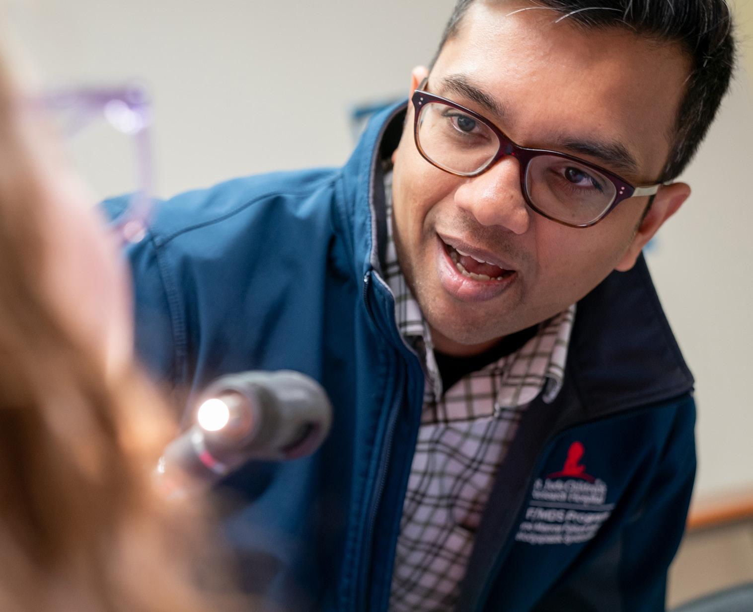
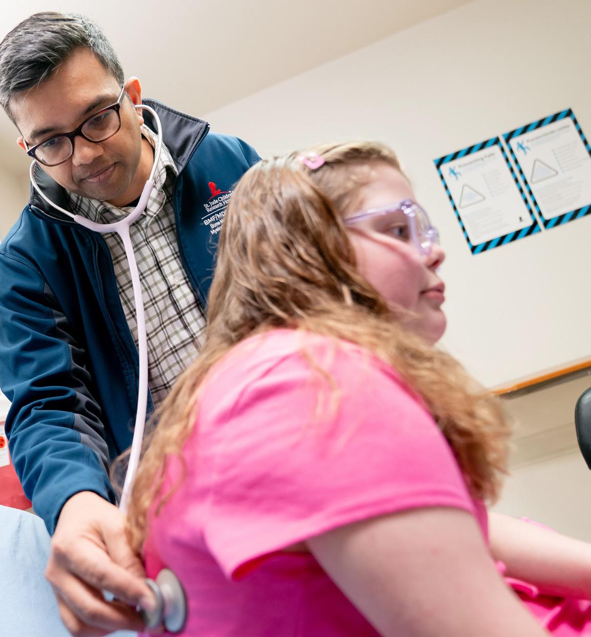
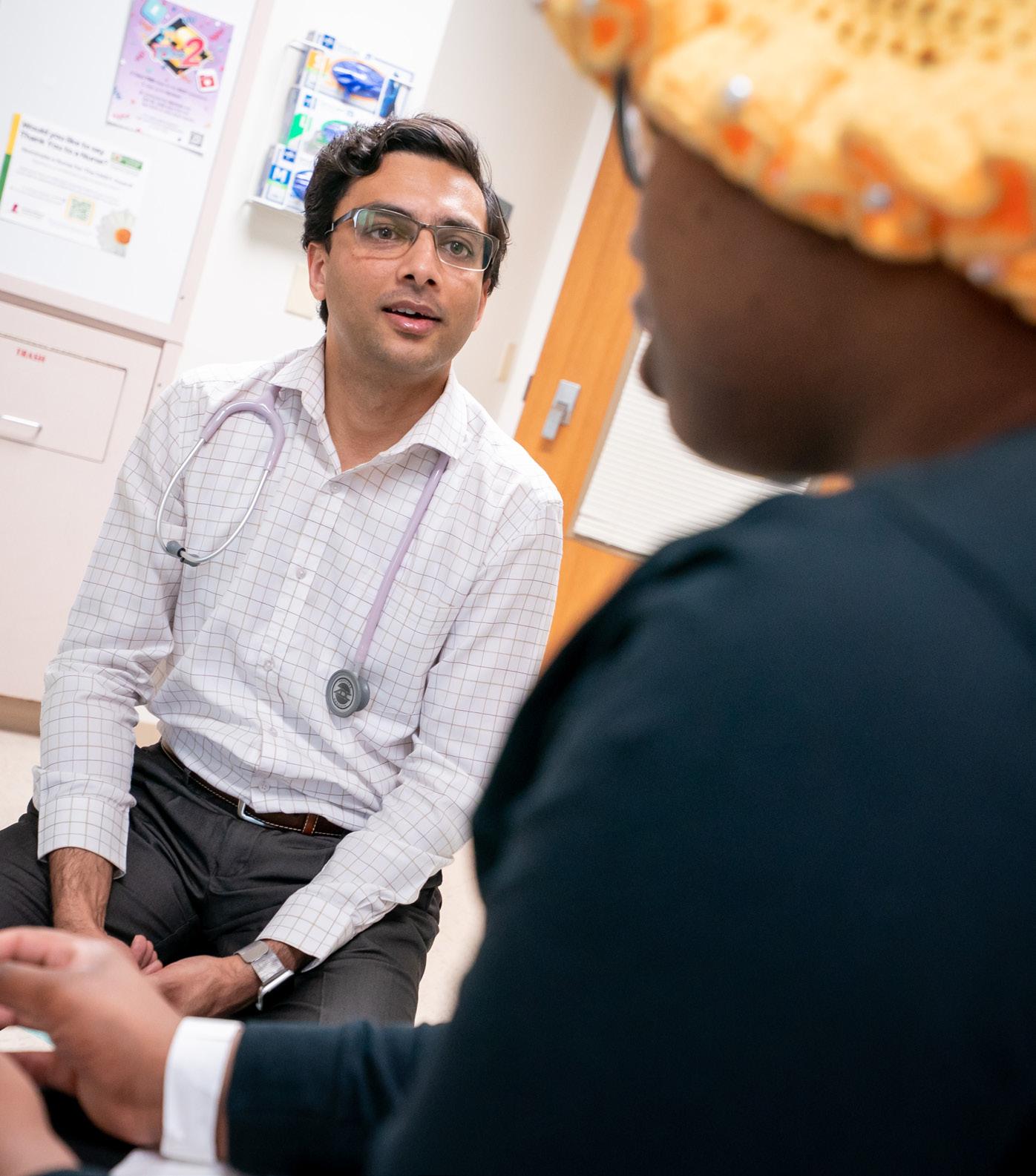
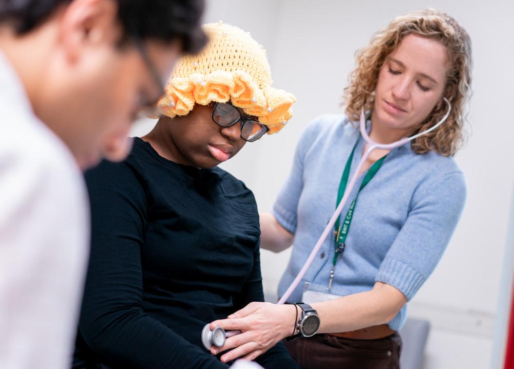
St. Jude researchers, backed by extraordinary resources and support teams, are focused on making big discoveries.
INITIALL clinical trial creates new paradigms for acute lymphoblastic leukemia treatment..........................................13 Reaching new milestones in SMA treatment................................
23 Shining a light into the shadows of protein structure and function.......
Technologies level up to tap into biology’s hidden information.............................................................................33
Advancing the story of discovery in blood disorders................................
Repurposed ALS drug presents a noninvasive diagnostic approach for neurodegeneration............................................................75
Studies identify SMARCAL1 as a novel osteosarcoma predisposition gene...........................................................................................76
A dynamic data visualization tool provides nuanced clinical trial insights............................................................................................................77
Hacking resistance mechanisms to combat antibioticresistant bacteria................................................................................................79 Cancer treatments and genetic predisposition contribute to second cancer risk.............................................................................................80
Reducing the risk of relapse after immunotherapy in pediatric leukemia..............................................................................................81 Comprehensive kinase atlas uncovers new layers of gene regulation....................................................................................................82
Targeting tumor mitochondria to enhance immunotherapy effectiveness.........................................................................................................83
Hearing ability linked to cognitive outcomes in young children with brain tumors......................................................84
SAGES1 employs the powerful potential of gene editing for sickle cell disease........................................................45 47 Bird flu in the barnyard: Tracking the spread of avian influenza and its risk to humans.................................
Revolutionary approach of Global Platform for Access to Childhood Cancer Medicines transforms care worldwide......................................................................................55 35
Mapping the microworld to reveal biology’s hidden networks
Identifying cellular drivers of DICER1 predisposition in pediatric rhabdomyosarcomas.................................................................85
aging and neurocognitive outcomes in survivors of childhood cancer................................................................................................86
57
The condensate landscape of health and disease..........67
A legacy grounded in stewarding the St. Jude mission: James R. Downing, MD.....................................................................69
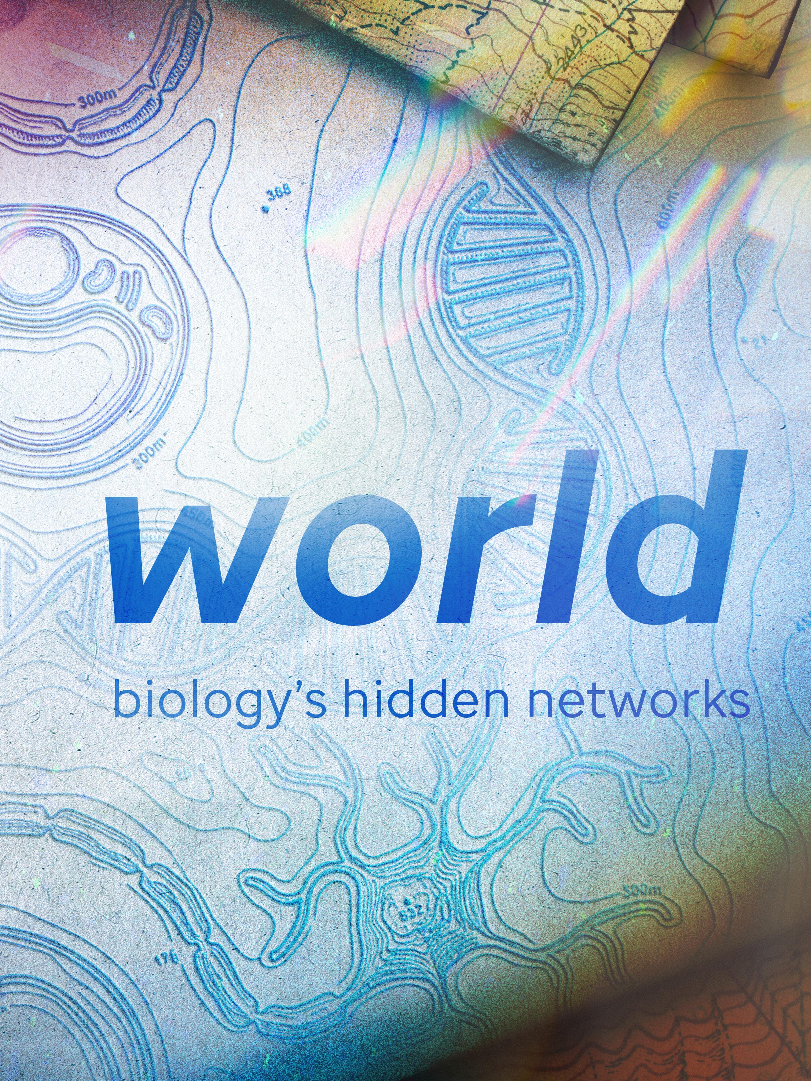
Behind the Cover
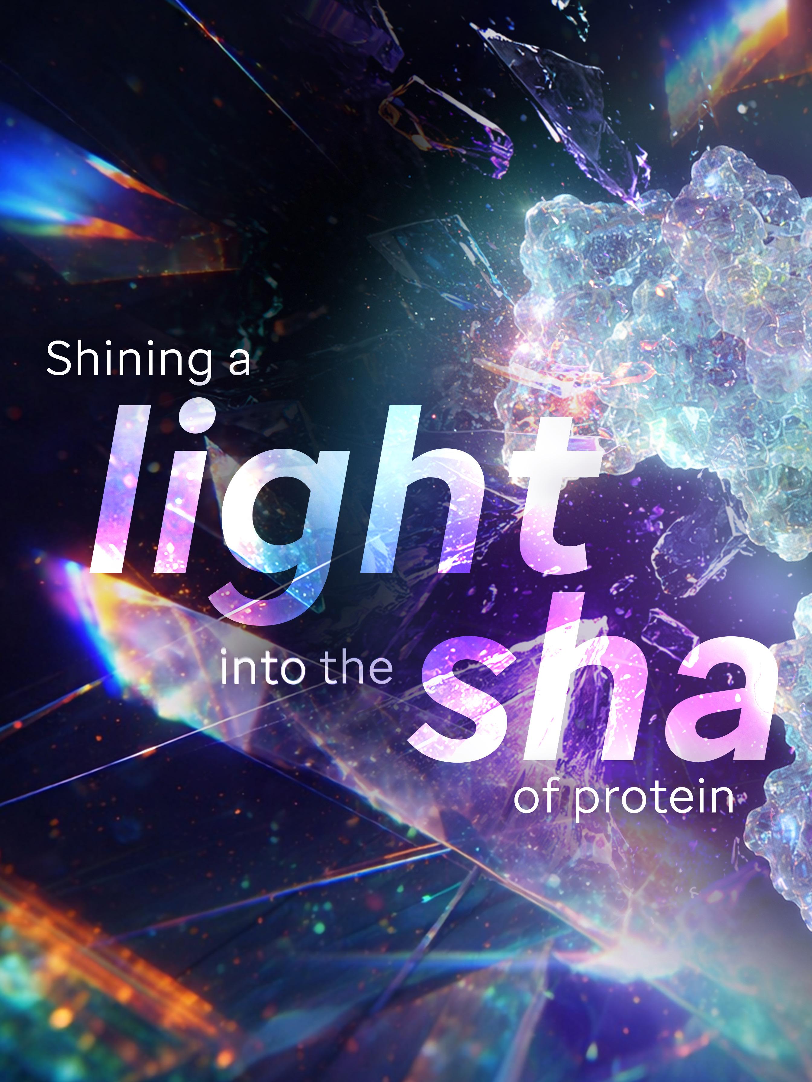
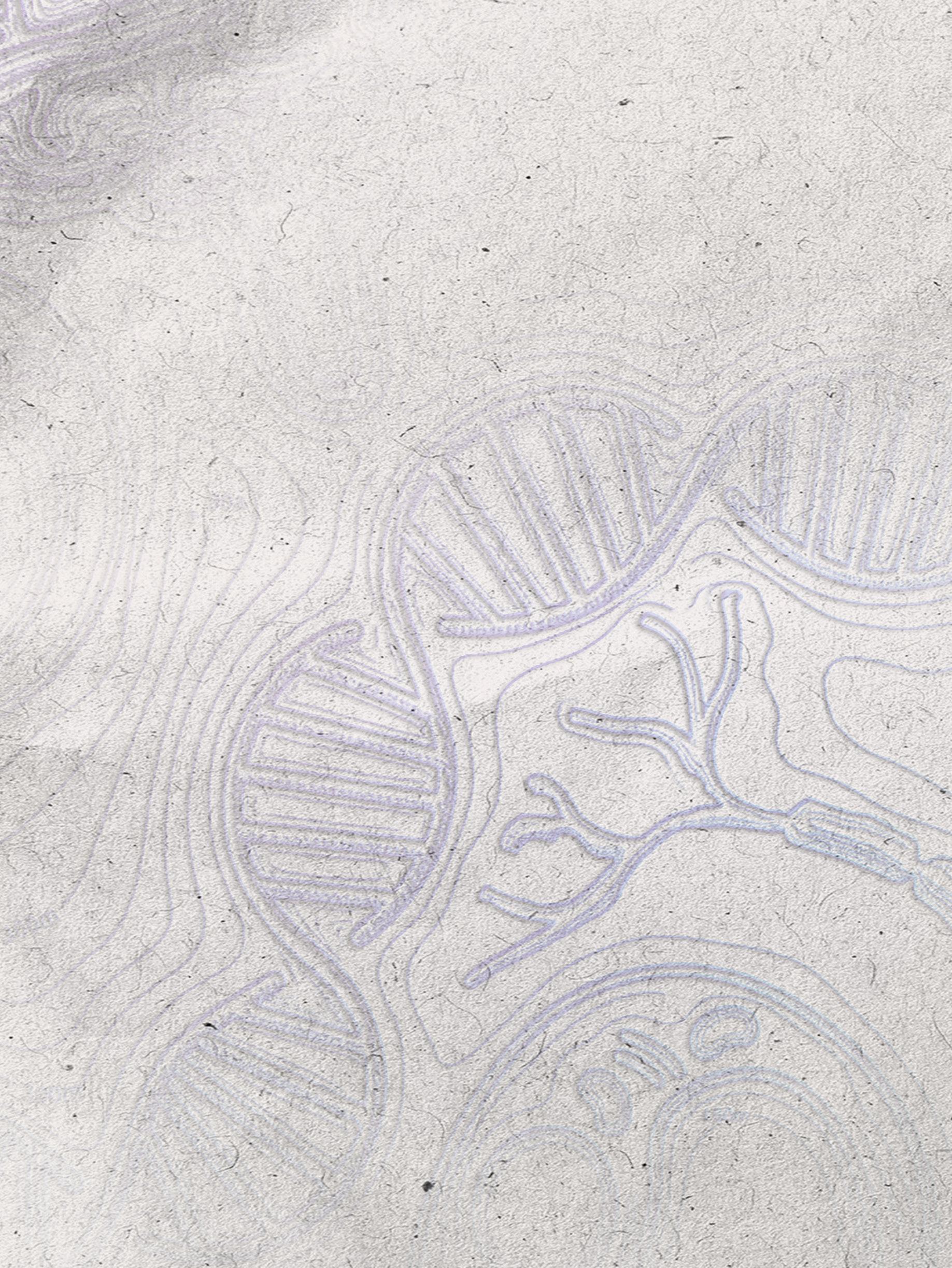
With the rapid advancement of technological capabilities, biomedical science is entering a new era. Researchers are stepping through the looking glass of discovery – revealing the hidden states that define biological function. This illustration pays homage to the evolving power of imaging sciences, depicting past insights into protein biology, the technologies accelerating innovation, and the discoveries yet to come.
SHAPING THE FUTURE OF RESEARCH AND CARE
When I first joined St. Jude Children’s Research Hospital 40 years ago, I knew this was a special place. At St. Jude, everyone understood the mission. We knew that the discoveries we made would advance cures and means of prevention for catastrophic childhood diseases.
That same spirit lives on at St. Jude today. Our scientific legacy is not defined by a single breakthrough, but by a culture of rigorous science committed to asking bold questions and sharing knowledge freely.
Over the past six decades, this culture has allowed us to make tremendous progress. Yet significant challenges remain today. To treat the untreatable and cure the uncurable, we must understand normal biology and how it goes wrong in pediatric catastrophic diseases, and from that, how we can develop new therapeutic approaches. This requires not just creativity but commitment and collaboration, often on a global scale.
The 2026 Scientific Report showcases some of those discoveries and how they’re transforming pediatric medicine. Across disciplines, our teams are pushing the boundaries of what is possible — from fundamental discoveries that deepen our understanding of biology to translational advances that are improving outcomes, reducing suffering, and accelerating cures. We celebrate their commitment to advancing the mission and honoring Danny’s vision.
In this report, you’ll read about how we’re mapping decision-making in cells and molecular systems, improving prediction and probing of protein structures, and redefining what’s possible for children and families affected by spinal muscular atrophy by advancing treatment before and beyond symptoms. We’ll share studies that have shown that malignancy emerges within prewired cellular systems that shape tumor initiation, evolution, and
therapeutic response. And we’ll look at advances in computational and experimental methods that are improving how researchers predict and probe protein structures.
Reviewing my decade as president and CEO of St. Jude, I am incredibly grateful to have been a part of this team and proud of what we have achieved together over the past four decades. I have had the privilege of watching this unique place of discovery grow in scientific ambition, global impact, and clinical collaboration.
As St. Jude looks ahead to its next strategic plan and how it continues to drive its mission forward, I am filled with excitement at our opportunities to generate and share knowledge that will save children everywhere.
James R. Downing, MD President and Chief Executive Officer St. Jude Children’s Research Hospital
Cells contain intricate signaling circuits that tightly control all biological processes, including metabolism, growth, and survival. Cancers alter that wiring to emerge and grow, and in some cases, to resist treatment.
St. Jude scientists have been studying cellular signaling in depth to understand how malignant cells develop and evade death, revealing vulnerabilities in that compromised circuitry that can be targeted with novel therapeutic approaches.
Timing of cancer emergence in developing B cells impacts leukemia treatment
Even in cancers where most patients respond well to treatment, such as the most common childhood cancer, B-cell acute lymphoblastic leukemia (B-ALL), understanding how a patient’s specific form of the disease rewires cells has value. By uncovering these alterations, physicians can predict who will and will not respond to treatment and alter their approach.
Published in Nature Cancer, scientists at St. Jude and University Health Network’s Princess Margaret Cancer Centre in Toronto found that when a B cell becomes cancerous during development affects treatment outcomes, enriching researchers’ understanding of the cancer’s origin. They made this discovery by developing
a robust single-cell reference atlas of normal human B-cell development and cross-referencing single-cell B-ALL data and outcomes data. They used that comparison to align the different B-ALL subtypes — and subgroups within subtypes — to different developmental stages, advancing fundamental understanding and association with outcomes. This enabled the researchers to find potentially actionable differences between types of B-ALL.
“At St. Jude, we have large acute lymphoblastic leukemia cohorts that have been genomically profiled with very rich underlying metadata, including outcomes,” said co-corresponding author Charles Mullighan, MBBS (Hons), MSc, MD, Comprehensive Cancer Center senior deputy director, Department of Pathology member.
Using that data, the researchers pinpointed the cell states in blood development from which B cells are diverted to become leukemia cells in patient samples. Many cancer samples arose from cells arrested in the pre-B and pro-B cell stages, which was anticipated, but there was much more diversity than expected. The data showed that less mature arrested cells retain features allowing lineage changes to occur, which can enable the cancer to evade therapy. They
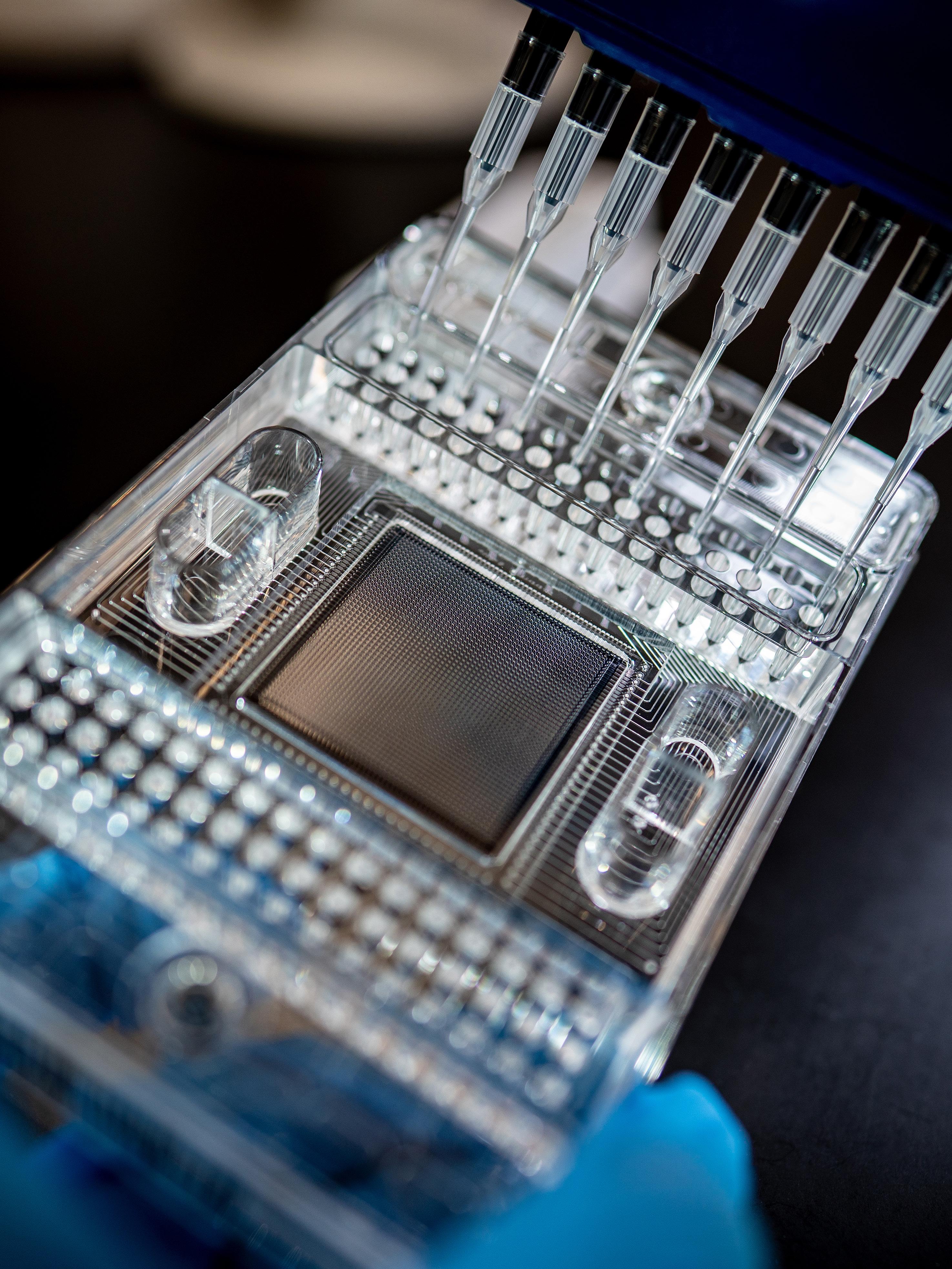
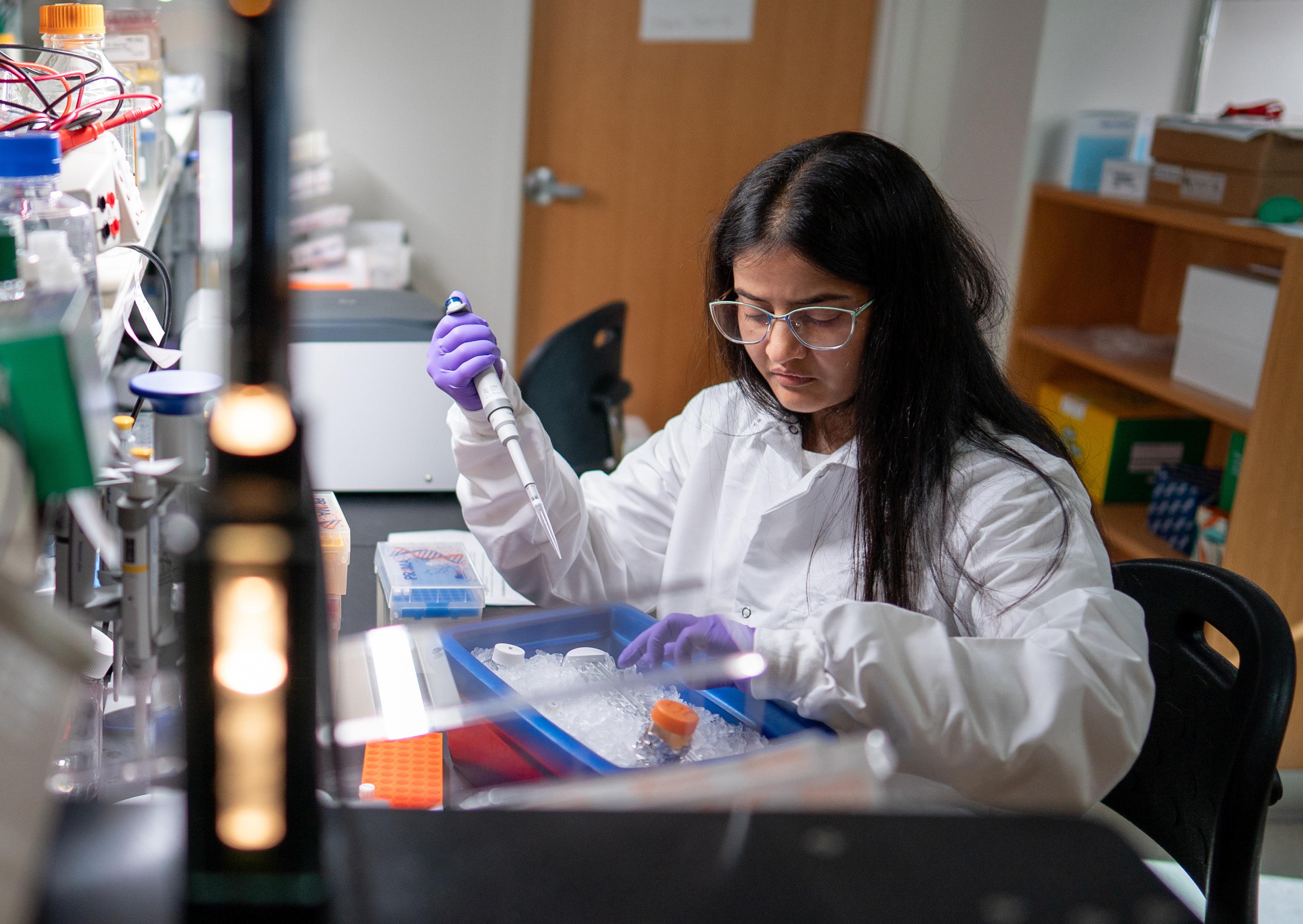
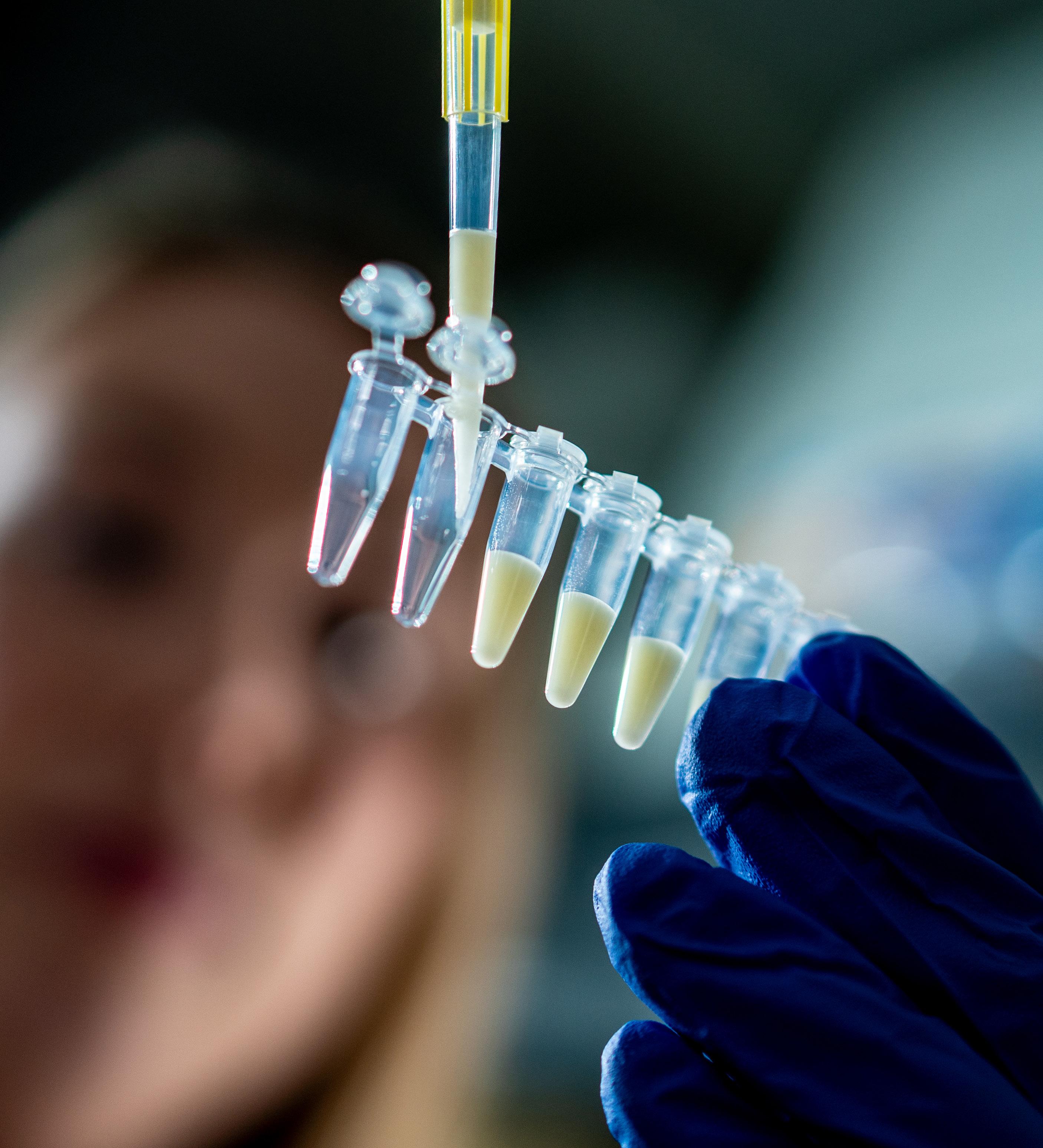
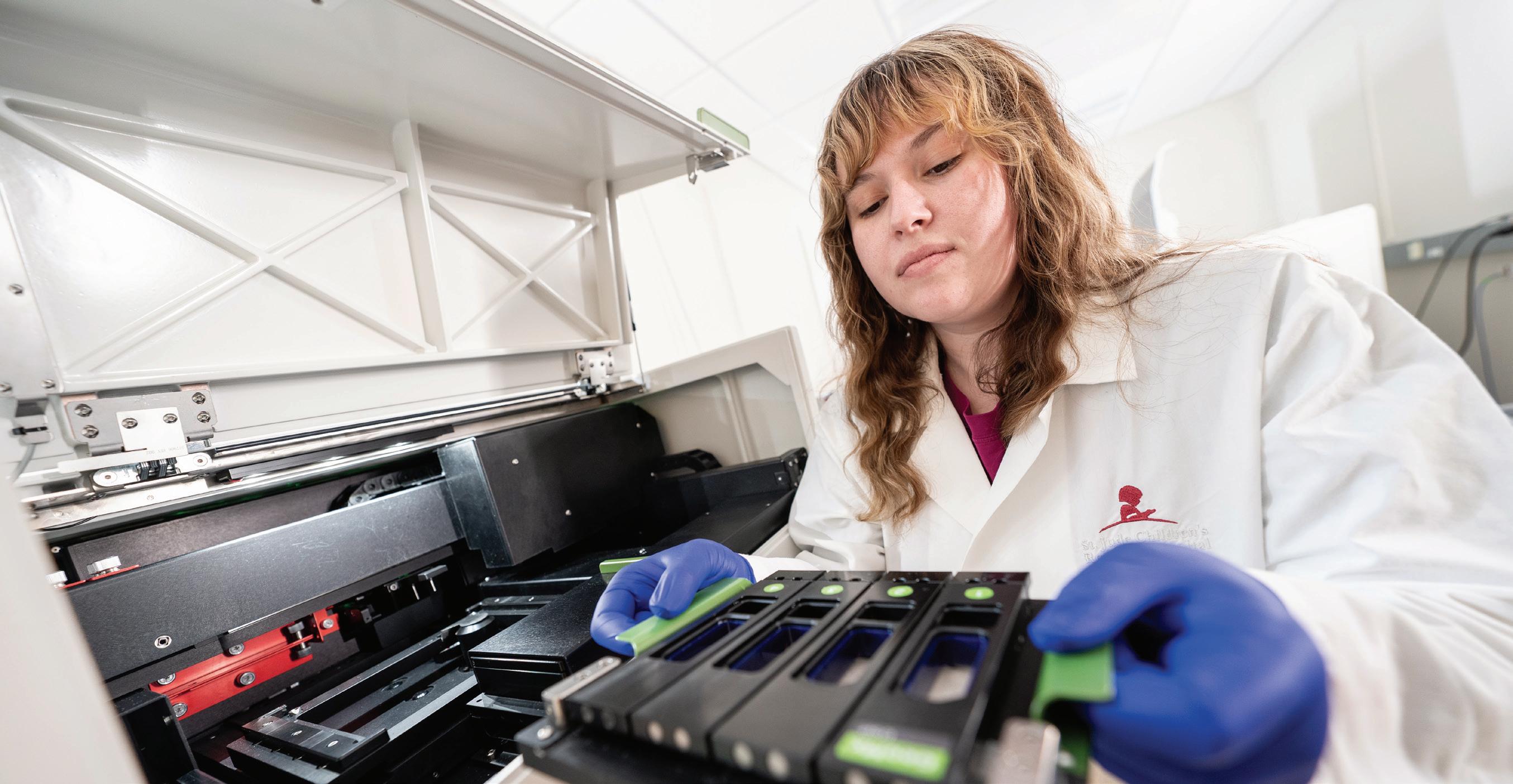
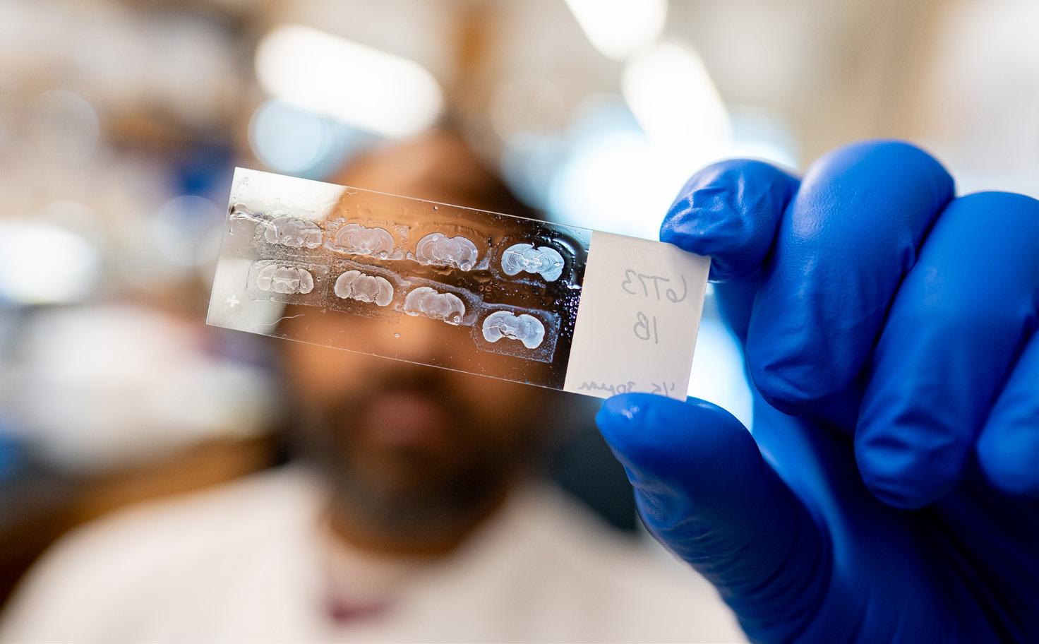
A scientist prepares a chip for single-cell DNA sequencing of cancer cells to explore their circuitry.
then developed those findings into a “multipotency score” that accurately predicted outcomes on a tested cohort of independent samples.
A vulnerability in an aggressive myeloproliferative neoplasm
While usually scientists look at how cancers emerge from normal development gone awry, they also study how existing cancer cells can be rewired to fuel a different and more aggressive form of the disease. This occurs in myeloproliferative neoplasms (MPNs), which develop when a person’s bone marrow creates too many of a specific blood cell type. These cancers have two phases, starting with a slow-moving chronic phase, then progressing to the acute blast phase (BP-MPN), with rapid growth of the neoplasm and a dismal prognosis, often just several months. The mechanisms of progression have been unclear, but massive genomic changes in chromosomes appear frequently in the blast phase.
St. Jude and University of Oxford scientists successfully untangled those massive genomic changes, finding that an amplification of a region of chromosome 21 was present in a subset of 64 patients with BP-MPN. Published in Nature Genetics, the researchers used CRISPR to individually study the genes within that region in BP-MPN cells, revealing that they depended on the gene DYRK1A
“We uncovered that DYRK1A is driving chronic-MPNs into leukemia,” said corresponding author John Crispino, PhD, MBA, Division of Experimental Hematology director and Department of Hematology member. “By mapping how it rewired these cells, we also revealed a potential therapeutic vulnerability in BP-MPNs.”
We uncovered that DYRK1A is driving chronic-MPNs into leukemia. By mapping how it rewired these cells, we also revealed a potential therapeutic vulnerability in BP-MPNs.
John Crispino, PhD, MBA Department of Hematology
The researchers found that DYRK1A indirectly upregulates BCL2, a gene that prevents cell death. When the researchers tested a BCL2 inhibitor, navitoclax, in combination with a DYRK1A inhibitor in cells, they saw substantial synergy, providing a new potential therapeutic approach.
A targetable mechanism behind a prevalent medulloblastoma
predisposition gene
In contrast, researchers often already know that a mutant gene rewires cells to become malignant, but do not know how. This was the case for children who inherit a deficient ELP1 gene, which increases their risk of developing SHH-medulloblastoma, a subtype of malignant pediatric brain tumor. In Cancer Cell, St. Jude researchers uncovered how ELP1 deficiency leads cells to turn off the major cellular “circuit breaker” for cancer development, the tumor suppressor protein p53.
“We made the initial discovery of ELP1 deficiency predisposing children to SHH-medulloblastoma
seven years ago, but now we’ve worked through its mechanism to an actual pharmacological intervention,” said co-corresponding author Paul Northcott, PhD, Center of Excellence in Neuro-Oncology Sciences director and Department of Developmental Neurobiology member. “We started with a gene in a spreadsheet and translated that into a potential targeted treatment.”
Protein p53 loss can fuel SHHmedulloblastoma. The researchers showed that ELP1 deficiency reduces p53 activity, putting neuronal progenitor cells at risk of developing into SHH-medulloblastoma. The protein MDM2 also reduces p53 activity by sparking the destruction of the p53 tumor suppressor. When the researchers prevented p53’s destruction by blocking MDM2, the increased p53 reestablished its normal circuit breaker function, killing cancerous cells. MDM2 inhibition significantly increased the survival of mice implanted with ELP1-mutant SHH-medulloblastoma patient-derived xenografts, counteracting the effects of ELP1 loss.
“We showed pharmacologically restoring p53 activity using MDM2 inhibition is an exciting avenue toward a targeted therapy for ELP1associated SHH-medulloblastoma,” Northcott said. The strategy is being evaluated for inclusion in clinical trials.
Mitochondria can drive cancer emergence and treatment resistance
Studying how cancers emerge can provide novel therapeutic opportunities. However, learning how cancers rewire themselves to resist treatments offers another compelling area of investigation to complement existing therapies and improve outcomes.
One unexpected source of treatment resistance comes from mitochondria, which power cells and have their own DNA (mtDNA) separate from the chromosomes and DNA in the nucleus. Until now, technical challenges have limited scientists’ understanding of how mtDNA mutations affect cancer growth.
“Each cell contains hundreds of copies of mitochondrial DNA. So, a mutation might be present at low levels in many cells, or at high levels in just a subset of cells,” explained corresponding author Mondira Kundu, MD, PhD, Department of Cell & Molecular Biology.
To overcome this challenge, Kundu’s team combined powerful computational tools, statistical analyses, bulk whole-genome sequencing, and single-cell studies. Published in Science Advances, the researchers first found that some mtDNA mutations occur before a B cell turns cancerous — and that these mutations are not always random. It
appears that in some cases, cancer cells actively “select” for a mix of normal and mutated mitochondrial DNA.
Each cell contains hundreds of copies of mitochondrial DNA. So, a mutation might be present at low levels in many cells, or at high levels in just a subset of cells.
Mondira Kundu, MD, PhD Department of Cell & Molecular Biology
Kundu’s team then deployed a computational tool, called NetBID2, created by Jiyang Yu, PhD, Department
of Computational Biology interim chair. With this tool, the researchers discovered that certain mtDNA mutations are not only associated with activation of pathways linked to cell growth, which helps explain their selection, but also with pathways linked to resistance to glucocorticoids, a common therapy for ALL. Further analysis suggested that this type of mtDNA mutation may make leukemia cells more likely to resist treatment, opening a new area of investigation to leverage that information to help patients.
Overcoming menin treatment resistance in NUP98 leukemia subtypes
Instead of searching for cancer-related mutations, scientists sometimes already know the exact mutation that a malignancy uses to rewire cellular signaling; they just cannot target it
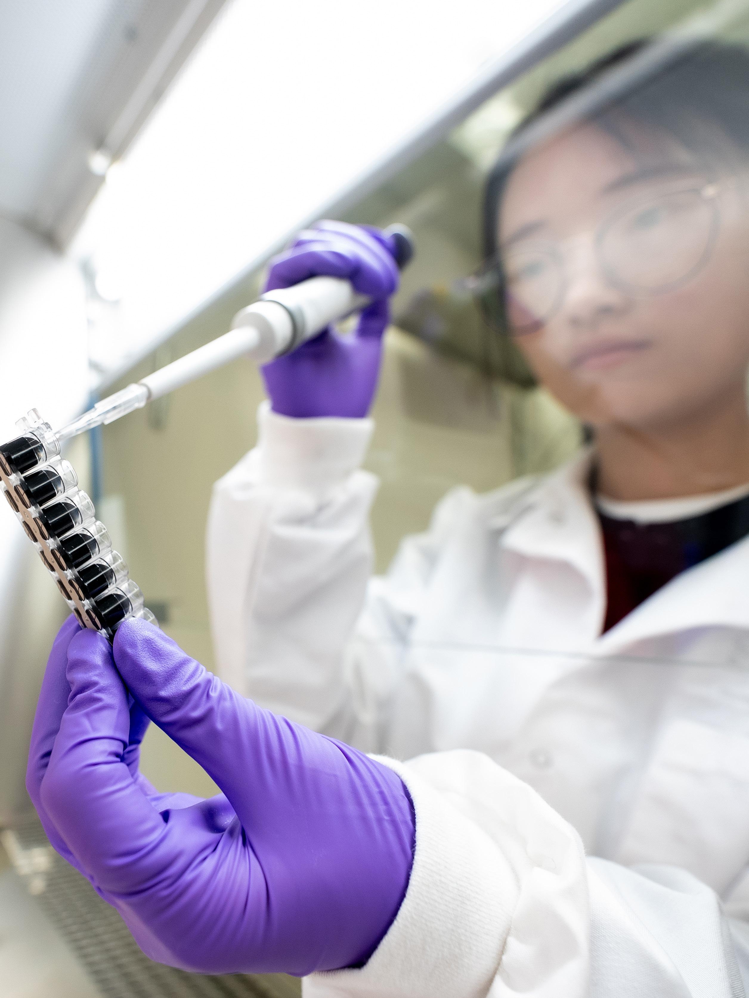
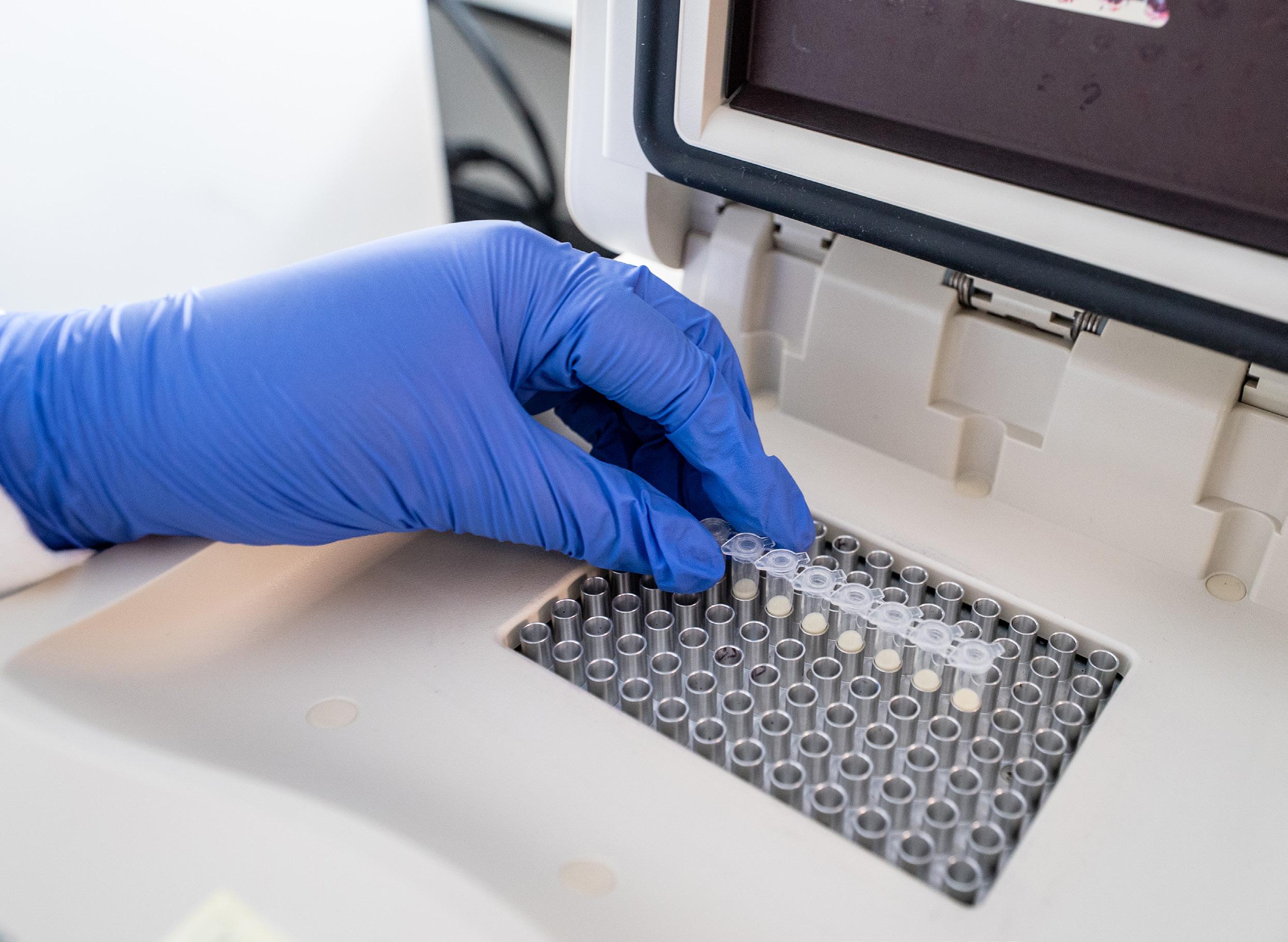
A researcher prepares single-cell RNA sequencing samples for analysis.
due to the potential collateral damage to normal cells. This is true for many fusion oncoproteins, which comprise two abnormally joined proteins, thus giving the hybrid new functions that can promote cancer-driving genes. Because the two original proteins still exist in the body, targeting them directly can cause adverse effects. Scientists have looked for a workaround, targeting the proteins that fusions interact with instead.
Genomic rearrangements involving NUP98 result in a fusion protein often associated with poor prognosis and relapse in acute myeloid leukemia (AML). St. Jude and Dana-Farber Cancer Institute researchers systematically leveraged proteomic and CRISPR gene editing technologies to identify and study the proteins interacting with NUP98 fusions and DNA, which cannot be directly targeted, in AML models to find another vulnerability.
Published in Cancer Discovery, they identified the acetyltransferases MOZ/ KAT6A and HBO1/KAT7, which help form a complex that activates pro-cancer gene expression. The scientists then combined MOZ/KAT6A and HBO1/ KAT7 inhibitors with a menin inhibitor (menin works with NUP98 fusion proteins to foster leukemogenesis), greatly increasing survival in patient-derived mouse models.
“We found NUP98 fusions drive leukemia by assembling these proteins into a complex to switch on the expression of genes that turn normal cells into leukemia cells,” said corresponding author Mullighan.
“We showed these inhibitors can stop the assembly of the switch, preventing activation of these cancer-driving genes, which is a novel therapeutic vulnerability in AML.”
Although most common in AML, NUP98 rearrangements have been linked to a wide spectrum of hematologic malignancies, including T-cell ALL and myelodysplastic neoplasms. How NUP98 fusion oncoproteins lead
We showed these inhibitors can stop the assembly of the switch, preventing activation of these cancerdriving genes, which is a novel therapeutic vulnerability in AML.
Charles Mullighan, MBBS (Hons), MSc, MD Department of Pathology
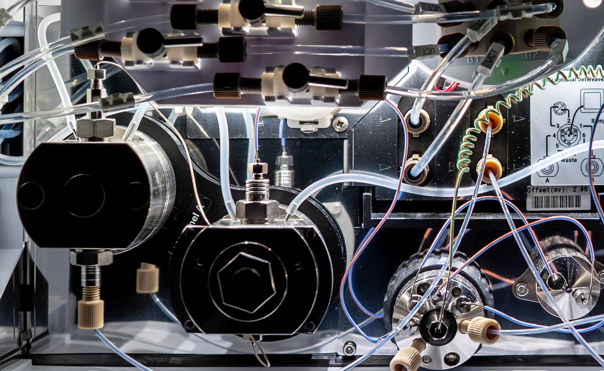
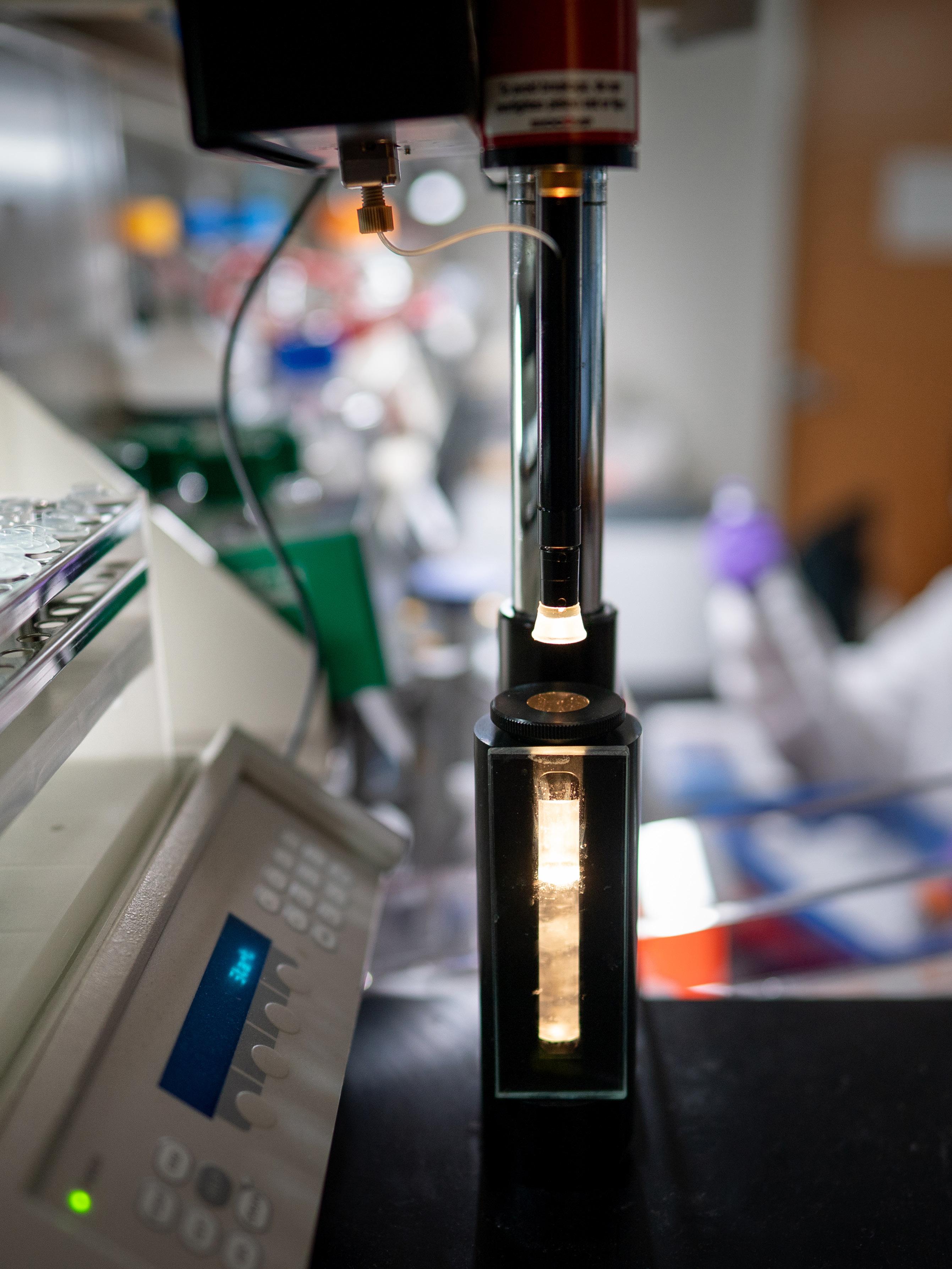
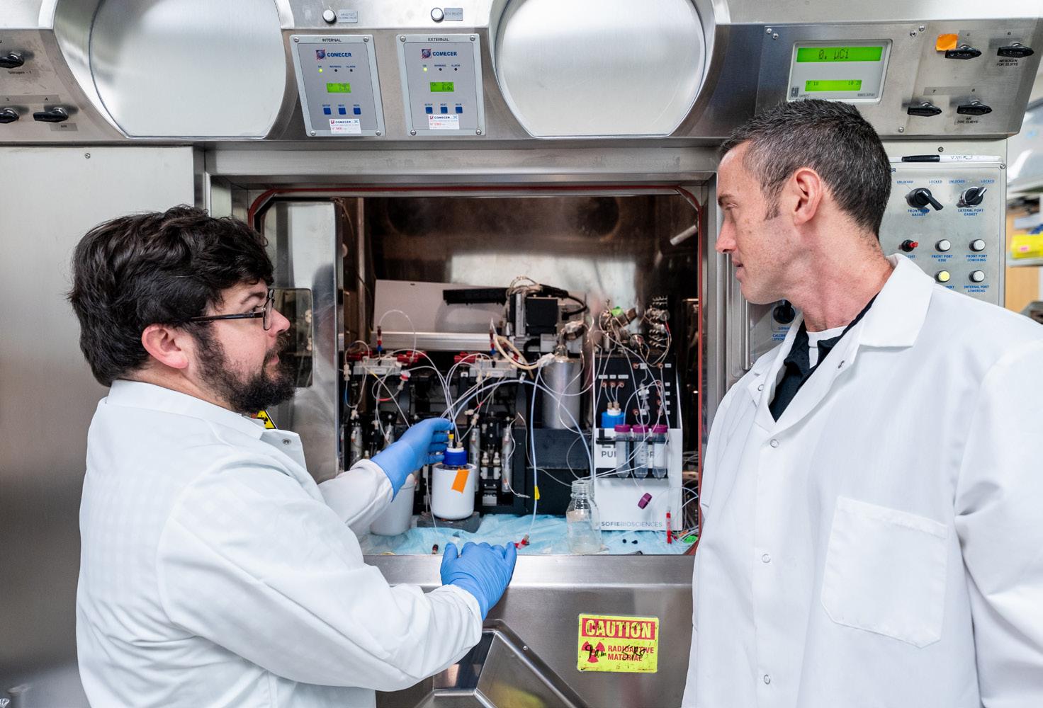
The internal structure of a machine that analyzes cancer cells’ metabolic circuitry.
to such a broad range of diseases remains poorly understood, though it could have lessons for treatment.
Published in Blood, St. Jude researchers studied the genomic background of NUP98-rearranged leukemias.
The team found that cooperating mutations, those which co-occur with specific NUP98 rearranged fusion oncoproteins, are linked to specific disease types. They then established models of NUP98-rearranged leukemias and showed that each NUP98 fusion oncoprotein induces blood cell lineages differentially, while cooperating alterations promote the survival of cells or block differentiation at distinct blood cell development stages.
“Our results show that we need to consider the impact of mutations beyond the driver fusion when studying these cancers,” said corresponding author Jeffery Klco,
MD, PhD, Department of Pathology. “We need to think about potential resistance arising from sources outside of NUP98 rearrangements.”
Their models showed that cells with cooperating mutations may become less sensitive to menin inhibition. These findings indicate a need to view menin inhibitor effects through the lens of differentiation or cooperating mutations, to identify patients who are less likely to respond and guide alternative therapeutic strategies.
Finding the vulnerability in an AML treatment resistance pathway
Another set of difficult-to-treat AML cancers contains mutations in the genes IDH1 and IDH2. These mutations metabolically rewire cancer cells, resulting in a massive accumulation of a
molecule known as an “oncometabolite” that helps fuel cancer growth, (R)2-hydroxyglutarate (R-2HG).
AML often rapidly develops resistance to existing IDH inhibitors, resulting in a re-accumulation of R-2HG and further malignant growth. In Blood, St. Jude scientists reported how metabolic rewiring induced by mutant IDH was used to find a better target. They created cell lines representing the breadth of potential IDH mutations with CRISPR-gene editing, then profiled their gene expression to find a potential vulnerability.
“We consistently saw the cell surface protein CD44 upregulated in cells with IDH mutations,” said corresponding author Jian Xu, PhD, Department of Pathology. “That was exciting, because we could block CD44 and see its effect on this type of leukemia.”
When Xu’s group inhibited CD44 in leukemia cells with IDH mutations, they saw a significant decrease in cell growth, and a greater decrease when mixed with IDH inhibitors. They observed the same effect on xenografts of patient cancer samples, including those from patients who had developed resistance to IDH therapy in the clinic.
“Inhibiting CD44 and IDH at the same time can significantly enhance antileukemia activity, reducing leukemia burden and prolonging survival in these model systems,” Xu said. “This gives us confidence that the combination therapy could have the potential to outperform the single agent IDH inhibitor therapy currently used in the clinic, which suggests that it should be further evaluated.”
The right tools to study neuroblastoma’s wiring
Before testing new potential treatments, researchers need a model system that faithfully represents how the disease appears in people. For rare cancers, scientists create bespoke models to understand how they are driven by altered signaling in hopes of finding vulnerabilities. In the solid tumor neuroblastoma, approximately 50% of high-risk cases amplify thedriver oncogene MYCN. However, “a subset of high-risk neuroblastoma patients do not have MYCN amplification in their cancer, but instead highly express the oncogene c-MYC,” said correspondingauthor Jun Yang, MD, PhD, Department of Surgery.
Yang found there was no research model available to study c-MYC–overexpressing neuroblastoma, so he created one, which he published in Cancer Research. To trigger the expression of c-MYC in neuroendocrine cells, the researchers used an enzyme called Cre recombinase, which is used to manipulate gene expression in specific
cell types in model organisms. However, the model did not behave as expected.
“We observed tumor growth but found that it developed unexpectedly in the pancreas, which turned out to be a very rare neuroendocrine tumor called somatostatinoma,” Yang said. “For this model, we used tyrosine hydroxylase Cre, which is classically used to generate MYCN tumors. So, we tested other types of Cre lineages and, eventually, landed on an improved dopamine β-hydroxylase Cre. Finally, we got neuroblastoma tumor growth.”
The model successfully reflected key features of neuroblastoma, including sensitivity to drugs used to treat the disease. This gives researchers a better way to study this cancer and develop novel therapies.
Hijacking of embryonic development circuitry leads to retinoic acid sensitivity
Once a model is developed, most cancer research focuses on finding new vulnerabilities to target, but St. Jude scientists recently addressed the question in reverse for neuroblastoma. Decades ago, physicians found that adding retinoic acid to high-risk neuroblastoma treatment increased survival by 10%-15%, but how it was altering cancer cells’ circuitry to trigger their demise was unclear.
Intriguingly, the effect was only evident when retinoic acid was added to the later parts of therapy, after the main tumor had largely been eliminated. In a study published in Nature Communications, St. Jude scientists revealed why.
“We’ve come up with an explanation for a decades-long contradiction about why retinoic acid works in late, but not early,
therapy,” said senior co-corresponding author Paul Geeleher, PhD, Department of Computational Biology. “Retinoic acid’s activity heavily depends on the cellular microenvironment.”
The researchers first found a subset of neuroblastoma cell lines that were extremely sensitive to retinoic acid. When they deleted candidate genes, the researchers found that bone morphogenetic proteins (BMPs), genes normally highly upregulated in the bone marrow that help drive embryonic bone and related tissue development, were responsible for that sensitivity. The scientists further uncovered how retinoic acid killed these cancer cells: If there are a lot of BMPsignaling pathway–related proteins already on the DNA, retinoic acid signaling combines with those proteins to “hijack” a normal development pathway and promote downstream cell death–related gene expression.
As the bone marrow microenvironment causes neuroblastoma cells to have higher BMP pathway activity, it neatly explains why retinoic acid is effective at treating metastasized cells in the bone marrow but not the primary tumors during up-front treatment.
Turning vulnerabilities in cancer’s wiring into tomorrow’s opportunities
These examples demonstrate that cancers have a variety of mechanisms to rewire cellular circuitry to promote their own growth and survival. However, St. Jude researchers are finding how those pathways are altered across pediatric malignancies, uncovering and testing novel therapeutic vulnerabilities introduced by that rewiring, moving one step closer to improving outcomes for children with these cancers.
A scientist prepares a set of cancer samples for metabolic analysis.
St. Jude innovated the “total therapy” combination of chemotherapy, which forms the backbone of modern treatment for pediatric acute lymphoblastic leukemia (ALL). While many children have benefited from the approach, researchers continue to investigate how to de-intensify treatment for those who respond well to prevent adverse late effects, and to improve treatments for those who do not respond well.
“We’re approaching an upper limit to what giving a single therapy regimen to all patients can do to improve overall survival,” said Seth Karol, MD, MSCI, Department of Oncology. Karol is the principal investigator of a new clinical trial for ALL called INITIALL.
“St. Jude created Total Therapy, and now, with INITIALL, we are building on what we learned about improving the outcome for children with ALL and moving to a new approach to treatment and a new trial design.”
Stratifying by susceptibility
St. Jude scientists have collected a wealth of genetic and epigenetic information, clinical features, and outcomes on pediatric cancers. That information enabled researchers to classify ALL into distinct subtypes
CLINICAL TRIAL INITIALL
CREATES NEW PARADIGMS FOR ACUTE LYMPHOBLASTIC LEUKEMIA TREATMENT
With INITIALL, we are building on what we learned about improving the outcome for children with ALL and moving to a new approach to treatment and a new trial design.
Seth
Karol, MD
Department of Oncology
and identify features that predict susceptibility or resistance to current therapies, including targetable vulnerabilities. INITIALL aims to leverage these insights.
“What makes INITIALL’s design special is combining the knowledge of specific genomic alterations and tumor characteristics, then separating patients into a standard of care or an experimental treatment group based on their specific cancer’s risk profile and likelihood of response to therapy,” Karol explained. “The use of cancer genomics to dramatically change induction therapy is unique and allows us to offer new treatments only to those patients most likely to benefit from it, while offering standard treatment to those least likely to need newer therapies to be cured.”
Instead of testing a single treatment for all participants, INITIALL stratifies patients based on the features of their cancer to different treatment arms that are likely to provide the most benefit. Most patients will have lowor standard-risk B-cell ALL (B-ALL) or B-cell lymphoblastic lymphoma and will receive therapy intended to minimize adverse late effects based on Total Therapy’s most recent iteration, Total 17. The almost 40% of children with either T-cell ALL (T-ALL) or highrisk B-ALL will receive a new therapy combination changing the current standard mix of chemotherapy drugs used in the first month of treatment. Most notably, patients with B-ALL will receive inotuzumab and blinatumomab during induction, moving these forward into the earliest parts of therapy. If any patients in the standard risk group have a slow response, they can be transferred to the experimental combination and receive these agents on the trial after induction.
Experimental combinations
The drugs inotuzumab and blinatumomab in the experimental combination are antibody-based treatments that have been approved by the Food and Drug Administration (FDA) for relapsed disease, but this is their first use in the early induction phase of treatment for pediatric cancer. Inotuzumab is an antibodychemotherapy drug conjugate, with the antibody binding to a cancer-specific protein to bring the drug close enough
to kill the cancer cells, enabling a lower dose of the drug to reduce unwanted side effects. Blinatumomab also binds to a cancer-specific protein, but it acts as a bridge for anticancer T cells to find and destroy malignant cells.
In contrast to B-ALL, most patients with T-ALL will receive an experimental combination, venetoclax or dasatinib, added to standard therapy. Both have already been FDA–approved for use in adults; this will be the first time venetoclax and dasatinib will be tested in pediatric T-ALL.
Based on previous work from St. Jude, patients will receive therapy targeting nearly mutually exclusive sensitivity to venetoclax, which counters antiapoptotic BCL-2 signaling, or dasatinib, which counters prosurvival T-cell receptor signaling, in combination with other chemotherapies.
“INITIALL builds on the strength of fundamental and clinical research at St. Jude to test whether a treatment works or not and understand why it performed the way it did, from characterizing the genomics of unexpected toxicities to navigating logistical challenges, while being nimble enough to come up with solutions,” said Karol. “That expansive view is what will enable us to continue improving therapy for every child with ALL by providing a platform for comprehensive patient-centered assessments regardless of the therapy selected.”
Collaborators essential to the success of INITIALL include: Tomi Mori, PhD, Department of Biostatistics chair; Cheng Cheng, PhD, Department of Biostatistics and lead study statistician; Elizabeth Fox, MD, Clinical Trials Research senior vice president; Julie Park, MD, Department of Oncology chair and Comprehensive Cancer Center, Translational Research associate director; Sarah Elitzur, MD, Leukemia/Lymphoma Division director and Hematological Malignancies Program co-leader; Ching-Hon Pui, MD, Department of Oncology; Hiroto Inaba, MD, PhD, Pediatric Hematology Oncology Fellowship Program director and Department of Oncology member; Jeffery Klco, MD, PhD, Division of Hematopathology & Molecular Diagnostic director and Department of Pathology member; Charles Mullighan, MBBS (Hons), MSc, MD, Comprehensive Cancer Center senior deputy director and Department of Pathology member; Jun Yang, PhD, Pharmacy Sciences director and Department of Pharmacy & Pharmaceutical Sciences vice-chair; and Lisa Jacola, PhD, ABPP-CN, Department of Psychology & Biobehavioral Sciences.
Spinal muscular atrophy (SMA), a progressive neuromuscular disease affecting 1 in 15,000 babies in the United States, was known for being devastating and uniformly fatal. In 2016, the first effective treatments rewrote the narrative.
SMA is caused by loss or mutation of both copies of the gene SMN1. Untreated, this loss leads to reduced levels and activity of the survival motor neuron 1 (SMN1) protein, subsequent irreversible motor neuron loss, progressive motor function loss, and, in more severe Type 1 cases, death by 2 years of age. Although it manifests across a range of characteristics, when SMA occurs in infants, it is especially debilitating, requiring treatment to prevent further deterioration.
At St. Jude, the Pediatric Translational Neuroscience Initiative is tackling the question of how to optimize treatment for diseases such as SMA by leveraging expertise in experimental therapeutics, decades of knowledge about clinical trial design and implementation, and a collaborative spirit to work with a variety of stakeholders. These three arms of the program are led by the Center for Pediatric Neurological Disease Research, the Center for Experimental Neurotherapeutics, and the Office of Strategy and Alliance, respectively.
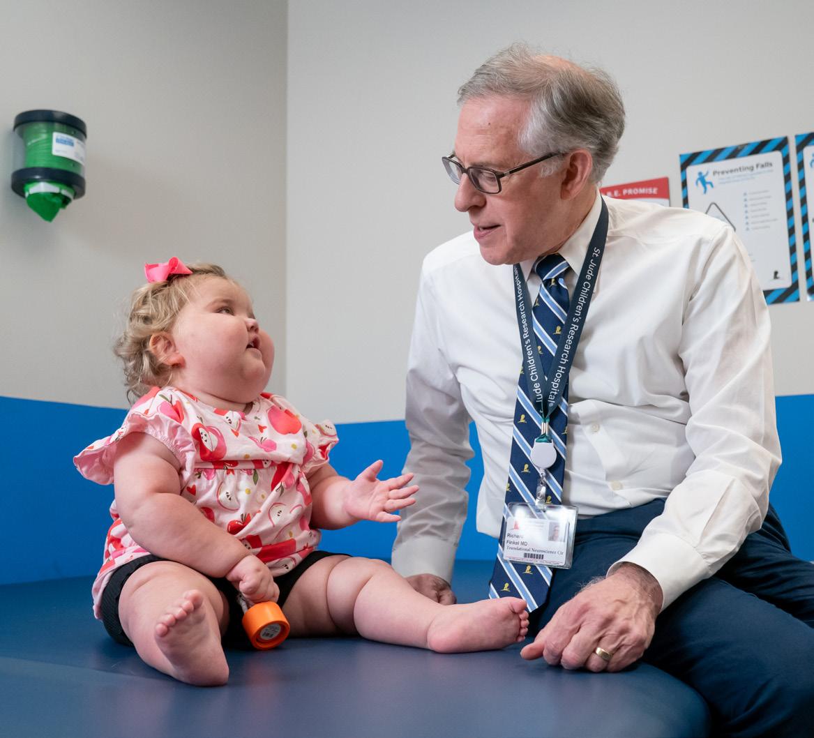
Richard Finkel, MD, Center for Experimental Neurotherapeutics director and Department of Genomic & Translational Neuroscience member, is a world-renowned expert in the treatment of SMA. Finkel joined St. Jude in 2020, expanding the institution’s clinical care for pediatric neurologic diseases. The recently published results of clinical trials led by Finkel demonstrate a commitment to continuous innovation — not simply accepting the progress made in 2016 with the first SMA therapies but pushing to make those therapies better and improve outcomes for children and families.
Providing SMA treatment before symptoms begin
SMA causes the loss of motor neurons responsible for activating muscles. Without those neurons, muscle tissue atrophies. The oral drug risdiplam improves motor function and increases survival by preventing atrophy from accumulating over time. However, risdiplam had only been Food and Drug Administration (FDA)–approved for patients aged 2 months and older.
An international consortium, co-led by Finkel, tested whether treating younger children with SMA with
risdiplam, before symptoms appeared, could delay or prevent disease progression. The results of the phase 2 clinical trial were published in the New England Journal of Medicine
Babies with the genetic mutations that cause SMA were all started on daily risdiplam in the first six weeks of life, before definitive features of SMA appeared; the researchers then followed their progress for two years. Of the eight children genetically predisposed to SMA Type 1, seven were able to sit up at 12 months of age, and five could walk by the end of the study’s reporting period, with no fatalities reported. Of the 18 children who had a mutation predicting less
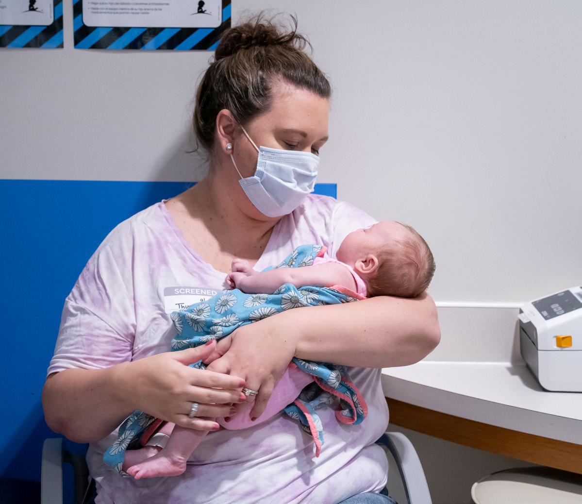
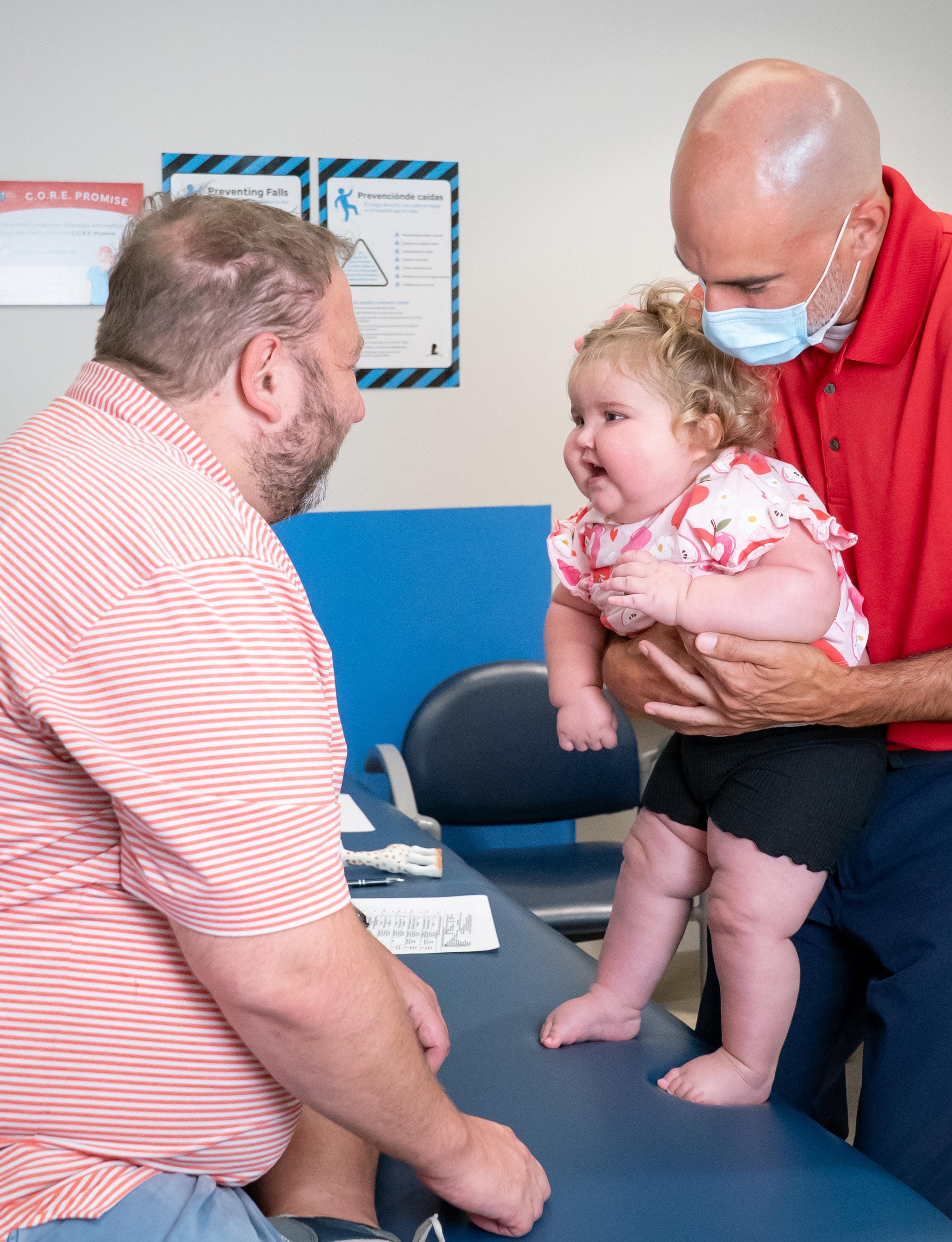
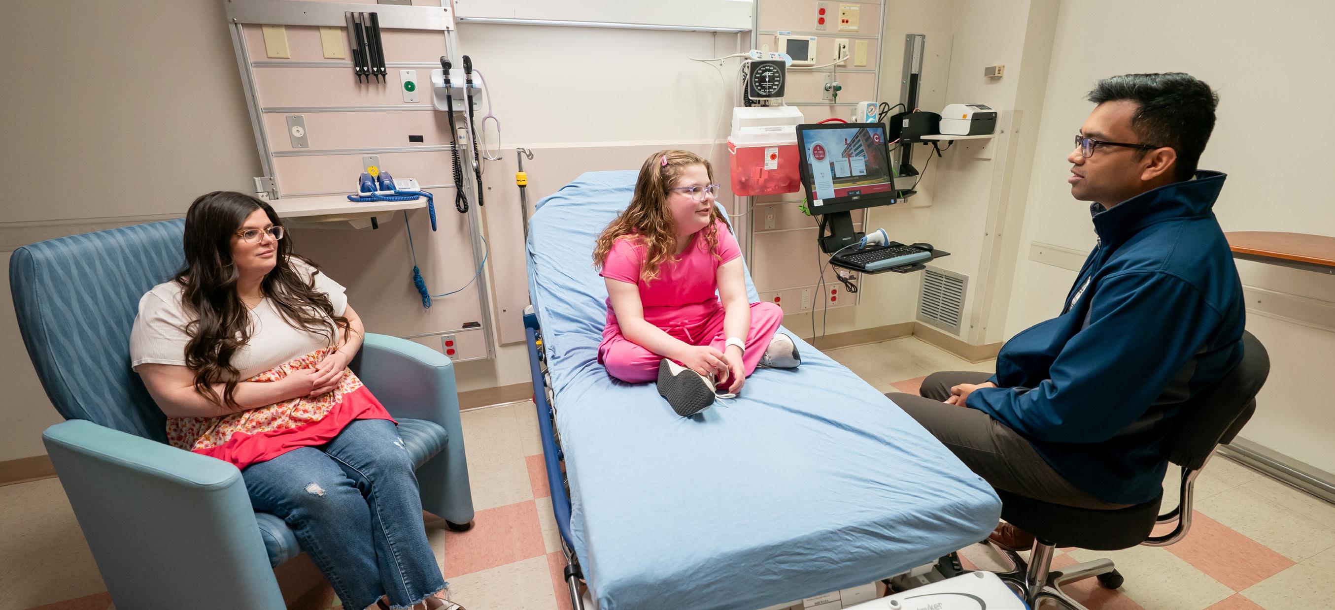
A St. Jude patient cuddles her mother during an appointment.
severe disease, all achieved sitting by 12 months and walking by 24 months, with most reaching these milestones in timeframes comparable to those of typically developing children. None of the children experienced any major treatment-related adverse events.
“We demonstrated in this study that with treatment shortly after birth, risdiplam maintained a good safety profile and generated a favorable clinical response,” said Finkel, who was co-first and corresponding author on the study. “Importantly, data from this study supported the request for the FDA to change the label for risdiplam’s use, extending it to younger children.”
While risdiplam slows and may even halt disease progression, there is still no cure for SMA. However, the success of this approach in newborns suggests that early intervention provides substantial benefits.
“The treatment of babies right after birth is an important milestone,” said Finkel, “but we will continue to investigate potentially better ways to give these kids a chance at a normal life.” To that end, Finkel has already begun testing risdiplam in the prenatal environment, which showed promising results in a single patient case report, also published in the New England Journal of Medicine
The treatment of babies right after birth is an important milestone, but we will continue to investigate potentially better ways to give these kids a chance at a normal life.
Richard Finkel, MD Department of Genomic & Translational Neuroscience
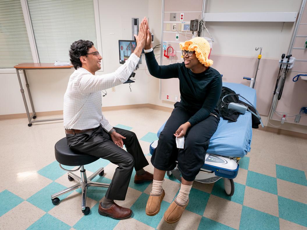
A clinician checks a St. Jude patient’s progress.
A St. Jude physical therapist works with a patient, while her father cheers her on.
N-of-1: A case study of prenatal risdiplam
The SMN1 protein is most needed in the third trimester of fetal development and the first three months of life after birth. Because SMN1 deficiency affects motor neuron survival, SMA symptom severity is closely linked with early implementation of effective interventions. Finkel launched a unique clinical study to assess the impact of prenatal treatment of risdiplam in a single patient to determine the feasibility of treating a fetus in utero for SMA Type 1 — a first-of-its-kind protocol.
In this case, both parents carried a pathogenic SMA genetic variant and previously had an infant born with SMA Type 1 who died at 16 months of age. At that time, treatment did not exist. While expecting their next child, genetic testing conducted through amniocentesis confirmed the fetus had no copies of SMN1, which, in combination with the family history and other genetic information, was highly predictive of this infant being born with SMA Type 1.
Risdiplam was administered to the expectant mother during the final six weeks of pregnancy. Since birth, the child has been periodically observed at St. Jude, and, thus far, has no observable signs of SMA. While some developmental delays and abnormalities were observed, these were believed to have resulted from deficits in early development, before risdiplam was given.
As this child continues to grow, she has made steady, albeit slower than normal, progress in her motor development. She has met major developmental milestones, including eating and sitting up independently, breathing without additional support, using words and gestures to convey her wants, and reaching the age of 3 years — a significant feat for children with SMA Type 1.
“Our primary objectives were feasibility, safety, and tolerability, so we’re very pleased to see that the parent and child are doing well. The results suggest it would be worthwhile to continue investigating the use of prenatal interventions for SMA,” said corresponding author Finkel.
The study’s promising results, including the child’s development and milestone achievements, support running a more comprehensive prenatal study. To date, 11 other patients have been treated prenatally using the same model established by Finkel and his colleagues at St. Jude.
Expanding therapeutic options to older patients
While risdiplam is effective at slowing disease progression, chronic administration of the drug presents
Richard Finkel, MD, connects with his patient during a checkup appointment.
challenges, including potential noncompliance. Onasemnogene abeparvovec (OAV101) offers a potential solution to this problem as a one-time gene replacement therapy that provides sustained survival motor neuron protein expression. However, OAV101, as a single intravenous administration, was limited to children under the age of 2 years old in the United States or those weighing less than 21 kilograms in European countries.
Through a pharmaceutical company–sponsored clinical trial in which Finkel had a leading role, researchers aimed to expand access to the drug to older children with milder, but still lifethreatening, forms of SMA. The study examined the safety and efficacy of intrathecal administration (injection into spinal fluid) to children and teens between the ages of 2 and 18 years old with SMA Type 2, who could sit, but not walk, independently. While SMA Type 2 is less severe than Type 1, with life expectancy stretching into adulthood, longevity is still reduced if untreated, emphasizing the need for a one-time safe and effective gene therapy option within this group.
Finkel co-designed the branch of the OAV101 study focused on children who had never received treatment before and published the findings in Nature Medicine. Patients treated with OAV101 demonstrated a statistically significant improvement in motor function, as measured by the Hammersmith Functional Motor Scale-Expanded score, a tool used to evaluate the physical abilities of people with SMA, compared to the control group.
“The results of this study were persuasive to the FDA to approve this intrathecal administration of the gene therapy for all patients over 2 years of age,” said Finkel, who was the corresponding author. “This approval provides potential access to this gene therapy throughout a person’s lifespan.”
A continuum of innovation to advance SMA therapeutics
The development of the first viable SMA treatments offered a glimmer of hope for patients and their families. A decade later, innovation and expansion of therapeutic options are giving researchers and clinicians the chance to halt disease progression and are opening a world of possibilities for children of varying ages and disease severity.
Ongoing improvements to therapy reflect the efforts of researchers, such as Finkel, to continue moving the needle in treatment efficacy and expand the patient population who can be treated with available therapies. Even after a decade of consistent improvement, the work will not be done until SMA no longer poses a barrier to health, mobility, or quality of life.
A St. Jude patient plays with Samuel Hughes, Center of Experimental Neurotherapeutics operations manager, during her checkup. Feature Photos by: Ann-Margaret Hedges and JP Parobek
Advancements in biophysical techniques such as nuclear magnetic resonance spectroscopy have enabled researchers to observe fleeting transition states central to protein function.
A revolution in protein imaging capabilities is allowing biomedical researchers to probe what was previously invisible — hidden-in-plainsight features, blink-and-you’ll-missit moments, and dynamics unfolding on time scales once beyond detection.
By capturing fleeting time frames and forms, St. Jude scientists are gaining new insights into protein function and revealing previously unidentified avenues for therapeutic design. With advances in biomedical engineering and computational analyses to match, St. Jude is well positioned to push the boundaries of discovery in protein sciences.
GPCR intermediate structures reveal the fine tuning of signaling responses
Propelled by a period of microscopy and spectroscopy innovation referred to as the “resolution revolution,” researchers have teased out a richer understanding of some of the most important proteins in the body. This is exemplified by the mechanistic insight gained into G protein-coupled receptors (GPCRs), membrane proteins that are vital to numerous cellular processes, ranging from metabolism to neurotransmitter signaling. Extracellular molecules bind to GPCRs, which activate signaling pathways inside the cell to generate an appropriate response to the bound molecule.
GPCRs are not just on/off switches, however. Modulation of the amplitude of the signal allows for more variable responses. Limited insight into how different molecules produce varying levels of response has hindered rational drug development for GPCRs, even though one-third of Food and Drug Administration (FDA)-approved drugs target these receptors. To find answers, St. Jude researchers examined the protein’s fleeting intermediate steps in detail.
In a study published in Nature, the scientists used time-resolved cryoelectron microscopy, molecular dynamics simulations, and singlemolecule fluorescence approaches to catch a glimpse of the statesbetween-states that GPCRs move through to elicit a response, to observe the intermediate steps of GPCR dynamics after a molecule binds, but before the signal relay concludes.
They successfully detailed how three very different drugs for the μ-opioid GPCR fine-tune receptor signaling, demonstrating that each molecule moves the GPCR through its intermediate steps in different ways, with strongly binding molecules promoting faster and more efficient transitions than weaker ones.
“We found that for weaker GPCR stimulants, the system slows down as it gets stuck in specific steps while changing shape during activation,” said corresponding author Georgios Skiniotis, PhD, Center of Excellence for Structural Cell Biology director and Department of Structural Biology member. “Regardless of how strongly a binding molecule activates the system, the steps remain the same, but the weaker drugs take longer to move through those steps, correlating with their efficacy.”
The intermediate states described in the study reveal the inner mechanics of GPCR signaling at the atomic level. By knowing these details, scientists can design next-generation drugs that are more refined in modulating signaling and maximize safety while maintaining efficacy.
Hidden kinase mechanism revealed
The ability to capture intermediate protein states in such fine detail allows researchers to observe all the shapes a protein can take, called its “conformational landscape.”
Charalampos Babis Kalodimos, PhD, Department of Structural Biology chair, is exposing previously invisible states within the conformational landscape of the second largest group of therapeutic targets: protein kinases.
Kinases are critical regulators of protein function, removing a phosphate group from adenosine triphosphate (ATP) and attaching it to a target protein in a process called phosphorylation. Many biological functions, such as cell migration, rely on processive phosphorylation, in which multiple phosphates are adhered to different sites in a single protein.
Processive phosphorylation requires the kinase to transition between different conformations rapidly. While some states of this conformational landscape are well established, a study from corresponding author Kalodimos, published in Science, used nuclear magnetic resonance
spectroscopy to investigate the shortlived intermediate states adopted by members of the Src kinase family.
The researchers found that Src has a hidden state that acts like a quick-release mechanism, ensuring the by-product of ATP, adenosine diphosphate (ADP), is rapidly discarded from the kinase. This allows the kinase to successively phosphorylate without needing to disengage and reengage the protein each time.
There
are likely other hidden or invisible states that we have not yet detected.
We’ve
only scratched the surface. Many more invisible states remain to be revealed.
Charalampos Babis Kalodimos, PhD Department of Structural Biology
While the fleeting appearance of this hidden state explains its elusiveness, its conservation across other Srcfamily kinases, such as Lck and Hck, underscores its importance for cell function. When the researchers abolished the state using targeted mutations, they found that cell migration (controlled by Src and Hck) and T-cell regulation (controlled by Lck) were significantly impaired.
The researchers are now exploring hidden states across other kinases to understand this essential protein family better. “There are likely other hidden or invisible states that we have not yet detected,” Kalodimos said. “We’ve only scratched the surface. Many more invisible states remain to be revealed.”
Jude researchers leverage advanced assay development technologies, highly sophisticated integrated robotics, and large-scale data processing and visualization platforms to unlock a protein’s hidden potential in drug discovery.
St.
St. Jude researchers use cryo-electron microscopy to reveal protein conformational landscapes in unprecedented detail.
Integrated approach reveals heat-shock protein assembly
Pursuing the elusive within protein conformational landscapes is not restricted to kinases. In a study published in Molecular Cell, corresponding author Kalodimos and his team presented the first fulllength structures of two molecular chaperone proteins assembled together, revealing key structural features regulating their function.
Heat-shock proteins are molecular chaperones essential in organisms ranging from bacteria to humans. Two of these proteins, Hsp40 and Hsp70, help incorrectly folded proteins attain their correct shape, thereby promoting cellular homeostasis.
Exactly how Hsp40 and Hsp70 work as an assembly to fix misfolded proteins was unknown; no one had been able to overcome the technical hurdles involved in capturing the massive and highly dynamic complex.
Kalodimos and his team combined cryo-electron microscopy, nuclear magnetic resonance spectroscopy, and X-ray crystallography to finally reveal the structure of the assembled bacterial chaperone complex. They captured multiple forms of the assembly, consisting of dimers (pairs) of Hsp70 interacting with dimers of Hsp40, in several different states of association with misfolded proteins.
“We obtained key structures of the active and inhibited states bound to an incorrectly folded protein, but we also solved multiple smaller pieces,” Kalodimos said. “By patching them together, they’ve given us a new model of how the Hsp40 and Hsp70 assembly really functions.”
The researchers paid particular attention to a section of Hsp40 called the G/F region. They found that this region binds the incorrectly folded protein first, then latches onto the Hsp70 active site. Hsp40 then pulls the incorrectly folded protein into
the active site, displacing itself, but remaining bound to Hsp70 at a different location. After the handoff, Hsp70 then refolds the incorrectly folded protein.
“We now understand mechanistically how the chaperones and protein clients form this very big assembly, how they are released, and how they cycle through this conformational landscape,” Kalodimos said. “Using that knowledge, we have a starting place to look for therapeutic interventions to compensate for disease-causing mutations, exploit as drug targets in cancers, or inspire next-generation antibiotics.”
Hidden nuclear receptor pocket gives new life to PXR drug discovery
Structural studies have unlocked new therapeutic potential in key targets such as GPCRs, kinases, and heat shock proteins. However, drug design is the first step on a long road to clinical benefit. Once exposed to the internal miasma of a cell, optimized therapeutics may be degraded or removed before they can have the desired effect. This is the role of pregnane X receptor (PXR), a nuclear receptor that binds to potential toxins and drugs, including chemotherapeutics, triggering the production of enzymes responsible for their removal.
Blocking PXR is beneficial for the efficacy of many cancer treatments, but different types of nuclear receptors are structurally similar. This makes it difficult to design a drug to target a particular receptor — precisely the problem Taosheng Chen, PhD, PMP, Department of Chemical Biology & Therapeutics, set out to solve.
“My lab has had great success in developing PXR inhibitors, but a subtle chemical change or a point mutation in PXR can convert an inhibitor to an activator,” explained Chen. “This is dangerous, because the inhibitor itself, if converted to an activator, could not only lose efficacy but have the opposite effect. A different approach,
such as a PROTAC that destroys the protein, wouldn’t have this issue.”
PROTACs are bifunctional drugs that bind protein targets with one end and recruit protein-degrading machinery with the other, presenting a promising alternative to inhibitors. However, the deep PXR binding pocket traditionally targeted for small molecule drug discovery has hindered the rational design of effective PROTACs. Chen explored already available PROTACs to determine if any could be repurposed to bind and degrade PXR.
In a study published in Nature Communications, corresponding author Chen and his team identified a potential candidate, MD-224, a PROTAC designed for anticancer therapy. MD224 effectively degraded PXR but had an unexpected twist: It bound to a previously unnoticed binding pocket, which allowed it to recruit proteindegrading machinery more effectively.
While MD-224 also bound other nuclear receptors, only those that shared close similarities with PXR were affected. This could also be tuned, suggesting it is possible to achieve selectivity. Ultimately, a newly discovered targetable binding pocket offers new avenues for drug discovery.
“Our angle is PXR, but the study is very relevant for those who think about other receptors too,” Chen said. “There are many exciting opportunities for other researchers stemming from what we’ve uncovered for this protein family.”
Structural insight of Fanzor2 nuclease demonstrates new bioengineering potential
Understanding the structural variation among closely related proteins, such as nuclear receptors, allows researchers to understand their unique features better. While Chen explores this protein family for exploitable features in drug discovery, Elizabeth Kellogg, PhD, Department
of Structural Biology, has uncovered the potential for a bioengineering breakthrough in an ancient family of DNA-editing proteins called Fanzors.
CRISPR-Cas9 technology is derived from a naturally occurring genomeediting system called an RNA-guided nuclease that bacteria use as a defense mechanism. Its discovery ushered in a new paradigm in genomic engineering capabilities. Recently, another large protein family found in bacteria, called TnpB, was discovered to be a functional, but distinct, predecessor to the Cas12 family of RNA-guided nucleases. The Fanzor1 and Fanzor2 proteins are the eukaryotic counterparts to TnpB and are encoded by transposable elements embedded in the genome.
It’s important, not only to know the functional constraints that make something an RNA-guided nuclease, but also how you apply those principles and harness them in engineering.
Elizabeth Kellogg, PhD Department of Structural Biology
In a study published in Nature Structural & Molecular Biology, corresponding author Kellogg obtained the structure of Fanzor2 to chart how these systems have evolved, offering key insights into the relationship between structure and function for these RNA-guided nucleases. The work revealed that RNA’s role in
structuring the active site of Fanzor2 differs from other similar proteins, suggesting that the RNA and protein co-evolved on a separate evolutionary branch from the Cas12 family.
Kellogg hopes this structure will be a launchpad for engineering the next generation of RNA-guided nucleases. “The structural diversity of these assemblies is something that we have no understanding of at all,” she emphasized. “That’s where I think it’s important, not only to know the functional constraints that make something an RNA-guided nuclease, but also how you apply those principles and harness them in engineering.”
One key factor that makes TnpB and Fanzor proteins so exciting is their size: They are significantly smaller than Cas9 and Cas12. In terms of genome engineering, minimizing the size of the protein offers more functionality. “Fanzor2 is already pretty minimal, but its structure suggests there’s malleability in terms of how it can function with the RNA,” Kellogg said. “It hints that we could reduce its size further, but there’s a lot more to do to understand that.”
AI unlocks potential in tandem CAR T–cell therapy
Capturing the Fanzor2 structure is vital not only for understanding the functional constraints of RNA-guided nucleases but also for revealing how the design principles that regulate their activity can be applied in bioengineering. While this journey is just beginning, computational advances and artificial intelligence (AI) are taking such bioengineering pursuits to previously unattainable heights.
St. Jude scientists have already shown how AI can revolutionize the design of another bioengineering innovation: chimeric antigen receptor (CAR) T cells. These engineered T cells are a type
of immunotherapy that reprograms a patient’s existing immune cells to target a tumor-specific protein marker. While CAR T cells have successfully treated some blood cancers, they have not been as effective in targeting solid and brain tumors. One reason is that cancer cells do not uniformly express the same surface proteins. This means CAR T cells targeting a single antigen can miss malignant cells that do not express that protein.
Scientists have tried to create CARs that target two proteins at once, called tandem CARs. However, they have encountered problems, including poor CAR expression on the surface of T cells and suboptimal cancer-killing ability.
A study published in Molecular Therapy by co-corresponding authors Giedre Krenciute, PhD, Department of Bone Marrow Transplantation & Cellular Therapy, and M. Madan Babu, PhD, FRS, senior vice president of data science, Center of Excellence for Data-Driven Discovery director, and Department of Structural Biology member, set out to address these challenges.
For CAR T cells, surface expression of the CAR is vital to ensure that the cell contacts its target protein on tumor cells. In their pursuit of tandem CARs, the team generated 24 variations, but none of them reached the cell surface.
“We repeated everything multiple times to ensure we weren’t missing anything and eventually acknowledged that our approach needed to be adjusted,” Krenciute said. “We needed AI input.”
The team developed and validated an AI-informed computational pipeline that significantly accelerated tandem CAR design with improved surface expression and anti-tumor function. Specifically, the pipeline predicted a better tandem CAR design if targeting two proteins expressed in pediatric brain tumors: B7-H3 and IL-13Rα2. The researchers tested their optimized CAR T cells and compared them to several single-target CARs in mouse-
tumor models that reflected the mixture of expression seen in patients: cells with both targets, one target or the other, or neither target.
The AI-informed tandem CAR completely cleared tumors in four out of five mice. In contrast, all tumors treated with CARs targeting a single antigen grew back. The results provide evidence that leveraging AI can help the design of other tandem CARs and be used to develop more efficacious designs more quickly than by manual testing.
“Researchers can use our approach to help screen and create better tandem CARs, bringing us closer to
the day we can successfully treat challenging tumors, such as pediatric brain cancers,” Krenciute said.
Continuing to explore biology’s hidden states
As researchers continue to probe the unknown spaces of biological function, novel approaches to treating catastrophic diseases are revealed. The technological leaps taken to uncover unmet potential within proteins, combined with the dawn of AI-informed analysis, have brought once elusive conformational capabilities into full light.
As discoveries change what is known about protein motion and activity, and as novel technologies and innovations in approach make it possible to do this work faster and more efficiently, St. Jude is well poised to continue this important pursuit. Investigators are already working to capture new protein states, pulling back the curtain even more and revealing never-before-seen views into the hidden dynamics of life.
An AI-informed computational pipeline co-developed by Giedre Krenciute, PhD, and M. Madan Babu, PhD, FRS, accelerates tandem CAR design and enhances anti-tumor function.
Technologies level up to tap into biology’s hidden information
Advanced imaging and computational capabilities are rewriting the boundaries of what can be observed in biology. But as increasing amounts of raw data are generated, care must also be taken to ensure this data is used to its maximum potential. St. Jude researchers are ensuring no stone is left unturned by enhancing the capabilities of existing key technologies to extract a deeper understanding of biology’s fundamental processes. This steadfast commitment ensures scientific breakthroughs are not outpaced by their pursuit and that no valuable information is neglected along the way.
AlphaSync keeps protein structure prediction up to date
In 2022, the AlphaFold Protein Structure Database launched, providing predictions for nearly all known and cataloged protein sequences. However, AlphaFold does not automatically update when new protein sequences are discovered, nor when an existing sequence is corrected based on new data. This means the quality of the predicted models can decrease over time, leading to out-of-date predicted structures and potentially cascading errors.
Published in Nature Structural & Molecular Biology, co-corresponding author M. Madan Babu, PhD, FRS, senior vice president of data science, chief data scientist, Center of Excellence for Data-Driven Discovery director and Department of Structural Biology member, and first and co-corresponding author Benjamin Lang, PhD, formerly Department of Structural Biology, created AlphaSync, a free database that improves upon existing protein structure prediction resources through continuous revision.
“In a rapidly evolving scientific landscape, having access to the most current and accurate information on protein structural models is essential for breakthroughs in medicine and biology,” said Babu. “With AlphaSync, we ensure predicted protein structures stay continuously updated, and we also enrich the models with key information such as amino acid interaction networks, surface accessibility, and disorder status, so that researchers can move from sequence to insight faster than ever before.”
In addition to updating structures, AlphaSync provides pre-computed data and other ease-of-use features, including a modernized data format, to keep research free of bottlenecks and ensure discoveries are built upon the latest available information.
In a rapidly evolving scientific landscape, having access to the most current and accurate information on protein structural models is essential for breakthroughs in medicine and biology.
M. Madan Babu, PhD Department of Structural Biology
ColdBrew taps into protein water networks to inform drug discovery
As research endeavors become more data-intensive, putting as much information as possible to work is at the core of many innovations. This approach motivated Marcus Fischer, PhD, Department of Chemical Biology & Therapeutics, to bring a frequently overlooked feature of experimentally obtained protein structures into focus: their water network.
Proteins have evolved to fold precisely according to the repulsion and attraction of their amino acid building blocks to each other and to water. Water is also key to protein activity since it helps guide other molecules, including drug molecules, to bind effectively. Drug discovery efforts often use techniques such as X-ray crystallography and cryo-
electron microscopy for structure determination. However, these techniques involve freezing, or “cryogenic” temperatures, which can distort how water molecules appear in structural models. The severity of this practice is currently unappreciated, as water molecules are often seen as an inconvenience and routinely thrown out of analyses.
The importance of water in drug efficacy motivated tapping into this network. Published in Nature Methods, a computational tool called ColdBrew puts water to work by accurately predicting the likelihood that cryogenic water molecules would also appear at physiological temperatures within experimental protein structures. Importantly, the ColdBrew metric captures the ability of drugs to displace these water molecules.
“To enable the wide use of ColdBrew, we pre-calculated probabilities for over 46 million water molecules across 100,000 proteins in the Protein Data Bank,” Fischer, the study’s corresponding author, said. “Remarkably, our results show that drug designers unknowingly avoid tightly bound waters, so actually knowing which ones to avoid could guide the process.”
Parallel Rapid Exchange takes sample prep noise out of singlemolecule data
As Fischer recaptures neglected information from X-ray crystallography and cryo-electron microscopy data, the complementary technique of single-molecule fluorescent imaging has matured into a powerful way to
observe the finer details of protein dynamics. However, the technical issues that arise from working at such a small scale have become major barriers to observing fleeting states in single-molecule motion.
To address this, a technique called Parallel Rapid Exchange (PRE), published in Nature Methods, is bringing transient protein dynamics into clear view. Single-molecule imaging techniques usually require independent measurements to be taken. PRE introduces parallel data collection, which means multiple measurements can be set up at once under identical conditions, allowing subtle functional dynamics to be measured.
PRE eliminates the minute differences in experimental setup and execution, such as sample concentrations and noise, which traditionally mask equally minute molecular motions, giving greater sensitivity to the single-molecule data and higher experimental throughput.
“We found ways to improve the efficiency, reproducibility, sensitivity, and reliability of single-molecule microscopes,” explained corresponding author Scott Blanchard, PhD, Department of Structural Biology.
“This method forms the basis of a generalizable and scalable approach for parallelized singlemolecule imaging that we hope will reveal subtle, yet fundamentally important, functional distinctions in the molecules that support life at unprecedented resolution.”
With this approach, the researchers could uncover previously unseen
protein dynamics behind fundamental processes. In one example, they studied how a major class of cell-surface proteins, called G protein-coupled receptors (GPCRs), convert signals from outside the cell into internal responses. GPCRs are managed by regulatory proteins, including β-arrestin1. By observing four β-arrestin1 sensors simultaneously, the researchers obtained the full picture of how the regulatory protein moves when it is activated.
“We knew there were three big structural changes that occur within β-arrestin1, but they have largely been reported as concerted events,” Blanchard said. “With parallelization, we could show that they don’t happen simultaneously; instead, two of them occur at about the same time, and only then, the slower, rate-limiting rotation, is allowed to happen. This was not possible to decisively determine before.”
Maximizing the value of data
These studies show how new technologies can deepen our understanding of biology’s most fundamental processes. As data becomes easier to generate, scientists must be mindful that what they collect is not only meaningful but also used to its fullest potential.
Technology improvements, such as AlphaSync, ColdBrew, and Parallel Rapid Exchange, ensure that any valuable information that might be overlooked along the path to discovery does not remain overlooked forever.
ADVANCING THE STORY OF
IN BLOOD DISORDERS
For St. Jude, the story of discovery in blood disorders — conditions such as leukemia, sickle cell disease, DiamondBlackfan anemia, β-thalassemia, and GATA2 deficiency — is both the foundation of the institution and an area of continued innovation.
To advance this narrative of exploration, St. Jude researchers have focused their investigations on the underlying mechanisms of disease and the science of gene therapy to advance the story of discovery and treat blood disorders at their source.
Rewriting treatment possibilities for Diamond-Blackfan anemia
Diamond-Blackfan anemia (DBA) is a bone-marrow failure syndrome caused by mutations in genes that encode the cell’s protein factory, the ribosome. These mutations prevent the body from producing enough red blood cells, leading to potentially life-threatening complications. Senthil Bhoopalan, MBBS, PhD, Department of Bone Marrow Transplantation & Cellular Therapy, and Michell J. Weiss, MD, PhD, Department of Hematology chair, have been leading efforts to find a curative treatment for DBA: one that directly alters the ribosome genes that house these mutations.
“Fifty years ago, the standard of care [for DBA] was corticosteroids and blood transfusions across a person’s lifetime, both with significant
long-term side effects. The only curative therapy was bone marrow transplantation from a healthy donor, which remains true today, but we are now one step closer to another curative option,” said Bhoopalan.
In results published in Molecular Therapy, Bhoopalan and Weiss focused on the most common dysfunctional gene in DBA, RPS19, which encodes a key component of the ribosome. The researchers developed a gene therapy approach by engineering a lentiviral vector, a genetically modified form of a lentivirus, to integrate functional RPS19 into blood-forming stem cells. The team demonstrated that adding a functional copy of RPS19 to RPS19mutated human blood–making stem cells rescued ribosome function, increased red blood cell production, and allowed corrected stem cells to survive over noncorrected DBA stem cells.
“The rescued cells had a competitive survival advantage over the uncorrected stem cells,” said first and co-corresponding author Bhoopalan.
“In some patients, blood-forming cells that have spontaneously lost the mutated RPS19 gene and duplicated the healthy copy of RPS19 can expand over time. Thus, our data suggest that lentiviral vector–corrected stem cells will expand over time and take over
the bone marrow. That means we may be able to use low-intensity conditioning for future gene therapy studies, which will reduce treatmentrelated toxicity for these patients.”
To test this hypothesis, Bhoopalan is planning a St. Jude–led clinical trial to assess this gene therapy approach in patients with DBA.
Weiss credits the groundbreaking work of the late Brian Sorrentino, PhD, who developed a curative gene therapeutic approach for treating X-linked severe combined immunodeficiency that laid the foundation for this potential DBA therapy.
“As an institution, we have built the expertise and infrastructure to cure devastating blood disorders with
As an institution, we have built the expertise and infrastructure to cure devastating blood disorders with lentiviral vector–based gene therapy. We are now positioned to cure DBA using this approach.
Mitchell Weiss, MD, PhD Department of Hematology
lentiviral vector–based gene therapy. We are now positioned to cure DBA using this approach,” said senior and co-corresponding author Weiss.
Revising gene editing approaches for blood disorders
As technologies have advanced, new treatment solutions for blood disorders have shifted to precision approaches, including the use of CRISPR-Cas9–based gene editing. Sickle cell disease and β-thalassemia, blood disorders caused by mutations in the HBB gene that encodes the β-globin subunit in adult hemoglobin, have a commercially available, Food and Drug Administration (FDA)–approved gene editing approach. However, investigators are working to
Research efforts conducted in Senthil Bhoopalan’s, MBBS, PhD, laboratory are being used to develop a St. Jude clinical trial to assess a gene therapy approach in patients with Diamond-Blackfan anemia.
better understand the mechanisms by which the gene therapy works.
By silencing BCL11A, a gene responsible for switching hemoglobin production from fetal to adult forms, the commercially available CRISPR-Cas9 gene therapy approach allows a patient to maintain a high fetal hemoglobin level. This lessens the severity of both disorders by inhibiting the production of mutant sickle hemoglobin in sickle cell anemia or compensating for deficient β-globin subunit gene production in β-thalassemia.
Leading the way in advancing the understanding of how this CRISPRCas9 gene therapy approach works, investigations led by principal investigator Jian Xu, PhD, Department of Pathology, and co-investigator Weiss set out to determine how CRISPR-Cas9 reactivates fetal hemoglobin.
“Our motivation for this study was twofold,” said corresponding author Xu. “First, to find out how CRISPR genome editing effectively inactivates BCL11A for fetal hemoglobin reactivation in this commercially approved gene therapy approach. And second, to identify more accessible therapeutic strategies.”
To examine how CRISPR-Cas9 specifically inactivates BCL11A, the team discovered that the enhancer (a short region of DNA that controls transcription of specific genes) targeted by the gene therapy folds into a threedimensional structure. “We found that this enhancer forms a chromatin ‘rosette’ structure, making multiple contacts with key regulatory elements of the gene,” said Xu. “This ensures highlevel BCL11A expression and prevents its silencing in red blood cell precursors.”
Published in Blood, the results showed that breaking this chromatin rosette structure is necessary for other repressive proteins to enter and silence
BCL11A. The researchers also found that the enhancer region’s threedimensional architecture requires the presence of an enhancer RNA (eRNA), a noncoding regulatory RNA produced from the enhancer region.
To test whether this eRNA could be targeted using a more effective type of therapy that does not modify the genome, the team evaluated antisense oligonucleotides (ASOs). Xu explained that “by delivering ASOs to both normal and sickled red blood cell precursors, we were able to selectively degrade the eRNA, leading to BCL11A silencing and fetal hemoglobin reactivation. We think this could offer a more affordable, accessible, and scalable alternative to current gene therapies.”
By delivering ASOs to both normal and sickled red blood cell precursors, we were able to selectively degrade the eRNA, leading to BCL11A silencing and fetal hemoglobin reactivation.
Jian Xu, PhD Department of Pathology
Editing the epigenetics of blood
Weiss also led the search for mechanisms vital to regulating fetal hemoglobin, solving a 40-year-old mystery about the connection between methyl groups, small chemical tags that regulate gene expression across
the genome, and fetal hemoglobin expression. The scientists sought to understand how the body switches hemoglobin genes on and off through epigenetic regulation, changes to genes or chromosomes that alter gene accessibility to transcription factors and expression. Previous research in this area had only been able to remove methyl groups (a process called demethylation) globally across the genome. These findings showed that demethylation of the fetal γ-globin gene was associated with its expression, but did not prove causality.
Published in Nature Communications, Weiss’s team, in collaboration with the University of Sydney, used advancements in CRISPR-Cas9 technology to precisely modify epigenetics, including the removal or addition of methyl groups at specific genes. By removing methyl groups from the promoter of the γ-globin genes, which encode proteins necessary for fetal hemoglobin production, the team demonstrated that demethylation increases gene expression.
“We found that the association between DNA methylation and expression at the γ-globin promoter is causal,” said co-corresponding author Weiss.
By targeting six specific cytosine bases in the γ-globin promoter of red blood cell precursors, the team showed that demethylation activated fetal hemoglobin gene expression. Removal of the methyl group at these sites marks the first time this type of epigenetic modification has been performed in red blood cells. The scientific feat demonstrated that γ-globin promoter demethylation could increase fetal hemoglobin levels from approximately 10% of all hemoglobin to over 30%, indicating its therapeutic potential.
“This paper provides an example of how we used a new technology, epigenetic
editing, to address a long-standing question about the regulation of fetal hemoglobin expression,” Weiss said. “Going forward, we will use epigenetic editors to explore new therapies for sickle cell disease and β-thalassemia, blood disorders that are alleviated by high levels of fetal hemoglobin. These studies are likely to provide general insights into gene regulation that could have a positive impact on treating many diseases.”
Continuing the narrative of scientific discovery for blood disorders
When a new treatment reaches the clinic, a slew of additional research begins. Gene therapies are still relatively new approaches for treating genetic blood disorders, but clinical investigators at St. Jude are leading the way in understanding the secondary impacts and long-term outcomes of these treatments for sickle cell disease and hemophilia B.
Sickle cell disease is caused by an inherited hemoglobin mutation that produces misshapen red blood cells that block blood flow, leading to severe pain and organ damage, including stroke. To compensate for the chronic anemia caused by sickle cell disease and the reduced oxygen-carrying capacity of sickled red blood cells, the blood flow speed increases in the vessels supplying blood to the brain, resulting in decreased time for the oxygen molecules to exit red blood cells and enter brain tissue, potentially leading to ischemia. Increased blood flow to the brain, as measured by Doppler ultrasound, is widely recognized as an indicator of stroke risk in people with sickle cell disease.
To understand whether CRISPR-Cas9 gene editing can help slow blood flow in the brain to normal levels, Akshay Sharma, MBBS, MSc, Department of Bone Marrow Transplantation & Cellular Therapy, led a study, published in the American Journal of Hematology, which imaged the brains of three patients with sickle cell disease using magnetic resonance imaging (MRI). The imaging was captured before gene therapy and at one and two years posttreatment. The results showed that blood flow improved significantly posttreatment, with speed decreasing anywhere from 22% to 43% and remaining stable over time.
“We saw that after gene therapy, elevated blood flow in the brain came down to normal levels, like we previously observed occurring after a bone marrow transplant,” said first and corresponding author Sharma. “This is the closest physiological evidence we have that gene therapy could be effective for patients with neurovascular disease who are at risk of or have had a stroke.”
The study provides hope for patients with sickle cell disease who are at risk of developing a stroke but are interested in pursuing gene therapy. Historically, this subset of patients has been excluded from gene therapy trials because of the high-risk nature of their condition.
“We now have emerging data to at least evaluate the efficacy of gene therapy in patients with a risk of or history of stroke,” said Shama. “Until now, we have only had one option that has had a long-term impact on blood flow in the brain: bone marrow transplantation. But now we may also have gene therapy as another viable method to protect against neurovascular disease in people with sickle cell disease.”
Until now, we have only had one option that has had a long-term impact on blood flow in the brain: bone marrow transplantation. But now we may also have gene therapy as another viable method to protect against neurovascular disease in people with sickle cell disease.
Akshay Sharma, MBBS, MSc Department of Bone Marrow Transplantation & Cellular Therapy
Following up on gene therapy for hemophilia B
Patient follow-up can continue for more than a decade after gene therapy, and researchers at St. Jude have conducted the longest follow-up analysis of patients with hemophilia B treated by gene replacement therapy. Hemophilia B is caused by insufficient levels of a circulating protein called factor IX, responsible for blood clotting, leading to spontaneous, sometimes severe hemorrhages. Typically, patients require regular preventative intravenous (IV) infusions of the deficient clotting factor to prevent bleeding. St. Jude investigators recently unveiled data from a 13-year follow-up study of longterm outcomes of gene therapy used to treat hemophilia B.
A scientist pipettes samples to be analyzed as part of the gene therapy research being conducted in Bhoopalan’s laboratory.
Published in The New England Journal of Medicine and led by Ulrike Reiss, MD, Department of Hematology, and Andrew Davidoff, MD, Department of Surgery chair, in collaboration with University College London, the study included 10 adults with severe hemophilia B who received gene therapy between March 2010 and November 2012. The patients were followed over 10 years; all maintained a steady level of factor IX.
“For these 10 patients, the factor levels have been stable, and they had significantly fewer bleeding events over these 13 years,” said co-first author Reiss. “Further, we have not seen any side effects or toxic events in the longterm follow-up.”
The curative outcomes and lack of toxic effects of the therapy have profound implications for the quality of life of the patients. Because 90% of the gene therapy delivery vehicle, a modified
For these 10 patients, the factor levels have been stable, and they had significantly fewer bleeding events over these 13 years. Further, we have
seen
not
any side effects
or toxic events in the long-term follow-up.
Ulrike Reiss, MD Department of Hematology
virus called a vector, ends up in the liver, the team kept a close eye on liver function tests. They limited liver inflammation through a brief course of steroids shortly after they administered the gene therapy.
Overall, how often the patients bled over the course of a year, called their annualized bleeding rate, dropped from 14 to 1.5 episodes per year. Recipients needed fewer factor IX infusions, and seven of the 10 patients discontinued regular factor prophylaxis.
“The key benefit is that gene therapy is a one-time, simple intravenous infusion that’s very straightforward to do and potentially has positive effects for a lifetime,” said co-first author Davidoff.
The resultant improvements in quality of life from gene therapy fuel the ongoing narrative of scientific discovery for blood disorders at St. Jude.
Drafting diseasespecific models for GATA2 deficiency
The narrative of discovery in blood disorders at St. Jude, from clinical and basic research efforts, centers the
(Right) Senthil Bhoopalan, MBBS, MSc, talks with a St. Jude patient, who received a bone marrow transplant to treat Diamond-Blackfan anemia, and her mother during a checkup visit.
mechanistic understanding of what makes these diseases form, progress, and even transform.
Some genetic blood disorders, like GATA2 deficiency, hardwire alterations into cells that make them prone to transform into cancer. To better understand the mechanisms that foster this transformation, scientists develop models that allow them to monitor disease development. GATA2 is a blood transcription factor vital for the production and maintenance of blood-forming stem cells. GATA2 deficiency, an inherited blood disorder that presents with symptoms of severe immunodeficiency, has a high risk of blood cancer development.
To understand this malignant transformation, a team of researchers at St. Jude, led by John Crispino, PhD, MBA, Division of Experimental Hematology director and Department of Hematology member, devised a disease-specific preclinical model to incorporate various drivers of the disease process.
The team produced a GATA2 mutant mouse model that had a specific abnormality in R396Q, located in the second zinc finger (a protein domain, or location) of Gata2. This location is most often associated with disease progression toward myelodysplastic syndrome or acute myeloid leukemia (AML). By assessing the model with functional studies and single-cell RNA sequencing, the team showed that the mutation confers the potential to contribute to leukemic development.
Their work, published in Leukemia, demonstrated aberrant creation and reduced function of blood-forming stem cells throughout development, a bias toward myeloid cell production, and signs of accelerated aging. However, the models did not progress all the way to cancer, marking the need for additional studies to discern specific drivers of disease progression.
“This model is a great resource, because now we can get cells from the model that have these known genetic events necessary for disease and begin adding
in other mutations to screen for particular genes that lead to cancer,” said corresponding author Crispino. “It is an incredibly useful tool as we seek to identify the mechanisms of malignant transformation in GATA2 deficiency.”
Useful tools that help investigators model and examine the mechanisms of disease progression, and even transformation, showcase the commitment of St. Jude to contribute new avenues of investigation and discovery in blood disorders.
From research investigations to improvements in care that range from the bench to the bedside, the quest to advance the narrative of discovery in blood conditions that were once deemed untreatable offers both investigators and patients the opportunity to continue drafting the next chapter of an unfolding story.
SAGES1
EMPLOYS THE POWERFUL POTENTIAL OF GENE EDITING FOR SICKLE CELL DISEASE
At St. Jude, laboratory and clinical researchers have long studied sickle cell disease, a blood disorder caused by mutations in the HBB gene, which normally encodes the β-globin subunit of adult hemoglobin. Sickle cell disease becomes symptomatic when fetal hemoglobin expression decreases around the time of birth, giving way to the production of adult hemoglobin. This shift in expression leads to the development of the hallmark sickled red blood cells, causing episodes of severe pain, chronic organ damage, and early mortality.
Knowing that the sickling of red blood cells and subsequent symptoms diminish when fetal hemoglobin levels are high, St. Jude researchers examined how a gene editing approach, called CRISPR Cas9, increases fetal hemoglobin levels in red blood cells of patients with sickle cell disease. CRISPR-Cas9 gene editing works by using a guide RNA to direct the Cas9 enzyme to specific sequences within the genome, where Cas9 “cuts” segments of targeted DNA. By removing or replacing segments of targeted DNA, CRISPR Cas9 thereby modifies the genome and the genetic instructions for cellular behavior these DNA segments code for, such as the creation of sickled red blood cells.
Investigating the mechanisms of CRISPRCas9 gene editing
In 2016, Akshay Sharma, MBBS, MSc, Department of Bone Marrow Transplantation & Cellular Therapy, began working in the laboratory of Mitchell Weiss, MD, PhD, Department of Hematology chair, to investigate the genetic regulators that control fetal hemoglobin production. Weiss’s lab had previously shown that CRISPRCas9 disruption of a DNA-binding site for BCL11A — a known repressor of fetal hemoglobin expression — in the promoters of the fetal hemoglobin genes HBG1 and HBG2 activated their expression in adult red blood cells.
These findings raised the possibility that disrupting the same BCL11A-binding motif at these specific sites in blood stem and progenitor cells of patients with sickle cell disease could increase fetal hemoglobin to therapeutic levels. From the results of this work, and in parallel with the growing bone marrow transplantation and gene therapy programs for hematologic disorders at St. Jude, Sharma and key collaborators developed a gene editing clinical trial for sickle cell disease: the St. Jude
Autologous Genome Edited Stem Cells for Sickle Cell Disease-1 (SAGES1).
Launching a St. Jude gene editing trial for sickle cell disease
The SAGES1 trial is significant, not only for its precise CRISPR-Cas9 gene editing approach developed in Weiss’s laboratory, but because every aspect of the trial occurs on the St. Jude campus. From clinical care to the gene editing process to cell infusion, patients benefit from the expertise across St. Jude. The SAGES1 trial enrolled its first participant in 2025, with Sharma serving as principal investigator of the trial.
“We are the first academic institution to have infused a patient with a genetically modified hematopoietic stem cell product manufactured right here, rather than partnering with industry or a pharmaceutical company,” said Sharma. “It is a huge achievement, and it is because of our GMP [Good Manufacturing Practices] facility and the capabilities it offers in the manufacturing of gene editing products as a therapeutic for pediatric catastrophic diseases, including blood disorders and cancer.”
The story of SAGES1 is one of experimentation, discovery, and refinement that spans almost a decade from concept to enrollment. “The whole point of what we do at St. Jude is that the things we learn in the lab could someday change a patient’s life,” said Sharma. That ethos continues to fuel scientific progress and clinical advancement as SAGES1 progresses.
For now, the trial is open to young adults, ages 18 to 25, to establish basic safety and efficacy for the novel CRISPR-Cas9 gene editing approach used in SAGES1.
A team-science approach to innovation
“It is all about team science,” said Weiss. “Successfully translating gene therapy approaches from the bench to the bedside demands a committed team of staff and faculty with
Successfully translating gene therapy approaches from the bench to the bedside demands a committed team of staff and faculty with complementary expertise, working together toward a shared goal of curing devastating hematological diseases, including sickle cell disease.
Akshay Sharma, MBBS, MSc Department of Bone Marrow Transplantation & Cellular Therapy
complementary expertise, working together toward a shared goal of curing devastating hematological diseases, including sickle cell disease.”
Data from SAGES1 will be used to further refine and innovate approaches to CRISPR-Cas9 gene editing to improve the efficacy and accessibility of the therapy, taking the concept of a cure for genetic conditions, including sickle cell disease, from promise to reality.
Collaborators essential to the success of SAGES1 include: Alexis Leonard, MD; Neha Das Gupta, PhD; Deepa Manwani, MD; Shengdar Tsai, PhD; and Jonathan Yen, PhD, Department of Hematology; Salem Akel, PhD, Human Application Laboratory; Brandon Triplett, MD, and Stephen Gottschalk, MD, Department of Bone Marrow Transplantation & Cellular Therapy; Albert Zhou, PhD, and Frank Fazio, Children’s GMP.
Akshay Sharma, MBBS, MSc, and Bone Marrow Transplant clinical staff perform a follow-up examination on the first St. Jude patient treated on the SAGES1 clinical trial three months after she received her gene therapy.
BARNYARD in the BIRD
FLU
Tracking the spread of avian influenza and its risk to humans
On
most farms, fences are built to keep animals contained and to protect herds and crops. Beyond simply marking boundaries, these structures help maintain order and organization across the farm, guiding the movement of livestock and the layout of fields.
However, while fences shape the physical landscape, they provide no protection against invisible threats: pathogens that can spread disease, harm livestock, or contaminate food.
Interspecies transmission spillover occurs when a pathogen jumps from
one species to another, crossing biological barriers that typically keep infections contained. When a pathogen crosses that invisible boundary, the consequences can extend far beyond a single host.
Spillover is rarely a singular event. Viruses are inherently unstable and constantly evolving, traits that make them uniquely suited to adapt to different environments, exploit new hosts, and capitalize on opportunities for transmission. As these pathogens circulate into new hosts, each infection presents another chance for mutation, increasing the possibility of increased pathogenicity, interspecies spread, and wider outbreaks.
In March 2024, highly pathogenic avian influenza A (H5N1), long recognized as a threat to poultry and wild birds, was detected in dairy cattle. This unexpected shift prompted urgent scientific investigation into how the virus behaves in mammals.
St. Jude scientists are working to understand these transitions in real time and clarify how the virus moves across species and adapts within new hosts, and the risk to humans at the intersection of veterinary and public health.
Assessing pandemic potential
Traditionally, influenza viruses are classified as avian or mammalian based on their host range and biology. The spillover of an avian influenza virus into mammalian livestock causes concern that continued circulation in these animals could drive genetic changes associated with increased human infectivity. This pattern has been observed most clearly in the 2009 H1N1 influenza pandemic, which emerged
in humans after prolonged circulation and genetic reassortment in swine, as well as pandemics in 1957 and 1968 that involved avian influenza strains moving between species, and acquiring adaptations that enhanced their ability to infect humans. Researchers need to understand what kind of effects might occur as a result of this current outbreak.
“When the outbreak in cows first appeared, no one really knew what to expect — after all, cows aren’t typical hosts for influenza A,” said Richard Webby, PhD, Department of HostMicrobe Interactions. “The fear is that once it’s in a mammal, continued transmission could push it closer to becoming a threat to humans.”
When the outbreak in cows first appeared, no one really knew what to expect — after all, cows aren’t typical hosts for influenza A. The fear is that once it’s in a mammal, continued transmission could push it closer to becoming a threat to humans.
Richard Webby, PhD Department of Host-Microbe Interactions
A scientist prepares raw milk infected with bird flu for pasteurization.
In a study published in Nature Communications, Webby and his team analyzed a panel of H5N1 viruses isolated from dairy cows to assess their potential risk of infecting humans.
They found the viruses had more molecular and biological features in common with bird flu viruses than human flu viruses, and that, fortunately, the virus was not under tremendous pressure to mutate in the cow udders — the tissue where most virus was found. They also found that the viruses from cows could not transmit through the air between mammals, though transmission to humans by close contact with infected dairy cattle is possible.
“While the overall population risk remains low for now, the greatest concern is for people who have close, direct contact with infected animals or who may be exposed through consumption of contaminated, unpasteurized milk,” said Webby. “We do need to remain vigilant for human infections, as each new person infected is another chance for this virus to mutate to better infect and spread among us.”
The researchers further showed that the virus did not exhibit evidence of resistance markers to current antiviral medications. However, there does appear to be a slightly higher baseline resistance to some drugs compared to others, which
may mean a somewhat higher dose is needed for effective treatment.
Risk, route, response
While the overall pandemic risk remains low, the spread of the virus into dairy cattle has shifted the focus to those at risk of direct exposure. Because host biology can shape how influenza behaves, it raises important questions about which antiviral medications will be most effective and what role the route of transmission plays in caring for and treating exposed individuals.
“The introduction of the virus into dairy cattle has created an entirely new potential route of exposure,” explained Webby. “We now have to consider
Scientists at St. Jude conducted extensive research to assess the public health risks of bird flu spillover into mammalian livestock, including potential exposure through contaminated milk.
risks such as splashes to the eye in people working in milking parlors, the possibility of consuming contaminated milk, and even aerosolized exposure in these environments.”
In response to these emerging risks, Webby and his team designed a study to model real-world exposure scenarios. The researchers studied exposure routes that included the eyes, mouth, and nose, which are the most common ways to become infected with the virus. By modeling these different routes, they aimed to better understand the virus’s threat to human health and how two Food & Drug Administration (FDA)–approved flu antiviral drugs performed depending on the route of transmission.
This study, published in Nature Microbiology, found that the route of infection, whether through the eyes, nose, or mouth, significantly impacts a treatment’s effectiveness. The oral route, which mimics the consumption of contaminated raw cow’s milk, resulted in the most severe and difficult-to-treat infections. The drug baloxavir produced a greater reduction in viral levels than oseltamivir, although neither was consistently effective across all routes of exposure.
This variability suggests that current antiviral strategies may need to be specifically tailored rather than relying on a single standardized treatment approach.
“Our evidence suggests that it is likely going to be hard to treat people severely infected with this bovine H5N1 bird flu strain,” said Webby. “Instead, reducing infection risk by not drinking raw milk and reducing dairy farm workers’ exposures may be the most effective interventions.”
Ensuring food safety
This understanding about the route of exposure raises important questions about broader public health and food safety, particularly the safety of milk consumption given current processing methods. Pasteurization, heating milk to a specific temperature for a set time, inactivates harmful pathogens, safeguarding consumers
A scientist spreads milk onto a blood agar plate following pasteurization to test for microbial growth.
from contaminated milk. However, not all milk is pasteurized, and although pasteurization inactivates infectious viruses, inactivated proteins and genetic material from H5N1 influenza have been reported in over 30% of commercial milk.
Researchers were also interested in how repeated oral exposure of these viral components might impact host immunity, including the potential to lead to or disrupt oral tolerance. Oral tolerance is the mechanism that teaches the immune system to ignore innocuous antigens, such as harmless food proteins, a process that normally protects us from misdirected immunity. Because the immunologic effects of ingesting viral remnants are not well understood, researchers questioned whether ingesting milk containing viral fragments could induce tolerance to the virus and lead to altered immune responses to subsequent viral infection.
In a study published in Science Advances, Stacey Schultz-Cherry, PhD, Department of Host-Microbe Interactions, investigated the effect of exposure to these viral fragments and their impact on immune responses and influence on disease onset or susceptibility to subsequent infection.
“We found that consuming pasteurized milk multiple times, even if it has inactivated H5N1 virus, poses minimal health risks,” said Schultz-Cherry. The researchers observed no benefit or detriment to subsequent influenza infection with regard to pasteurized milk’s effect on the immune system.
The work confirms that current food safety methods are likely protecting humans from the H5N1 virus in milk. However, without pasteurization, the researchers did observe that infected milk was still very pathogenic in their model.
“It’s reassuring to find that these inactivated H5N1 viral components in pasteurized milk present minimal health risks and don’t alter flu immunity,” Schultz-Cherry said. “However, we also reaffirmed that consuming unpasteurized milk can expose people
to this potentially dangerous infectious agent. We must continue to watch this virus and mitigate its risk of spilling over into the human population.”
It’s reassuring to find that these inactivated H5N1 viral components in pasteurized milk present minimal health risks and don’t alter flu immunity. However, we also reaffirmed that consuming unpasteurized milk can expose people to this potentially dangerous infectious agent.
Stacey Schultz-Cherry, PhD Department of Host-Microbe Interactions
When viruses cross the fence
As scientists continue to study H5N1 in dairy cattle and its potential impact on humans, their findings guide efforts to understand and mitigate emerging risks. By examining how the virus spreads, mutates, and responds to antiviral treatments, St. Jude researchers are building the foundation to protect those most at risk and to anticipate future threats.
Their work highlights the importance of careful monitoring and study of these viruses, prevention, safe handling of livestock, and food safety measures, ensuring that public health strategies stay one step ahead of a virus that crosses the fence from animals to humans.
Revolutionary approach of Global Platform for Access to Childhood Cancer Medicines transforms care worldwide
St. Jude was founded more than 60 years ago on a radical idea: A child’s chance of surviving a catastrophic disease should not depend on their family’s ability to pay or where they live. Over time, that belief reshaped the field of pediatric oncology. Survival for childhood cancer in highly resourced countries, such as the United States, rose from less than 20% in the 1960s to more than 80% today. Scientific discovery, collaborative research, and advances in supportive care drove this remarkable progress.
Yet, the same upward trajectory in survival has not been achieved globally. Each year, an estimated 400,000 children develop cancer, and more than 80% of them live in resource-limited countries. In many of those settings, survival remains below 30%. Central to this is a persistent and systemic problem: inconsistent access to qualityassured childhood cancer medicines.
The Global Platform for Access to Childhood Cancer Medicines is designed to address this issue directly.
From global commitment to coordinated action
The Global Platform emerged from the recognition that scientific advances alone cannot close global survival gaps; implementation of efficient and resilient systems for |care delivery is essential — and this requires international cooperation.
In 2018, St. Jude launched St. Jude Global to strengthen pediatric cancer care worldwide through education and workforce development, expanding treatment capacity, and coordinating research. That effort deepened through a collaboration with World Health Organization (WHO), when St. Jude became the first and only WHO Collaborating Centre for Childhood Cancer. This collaboration sparked the creation of the Global Initiative for Childhood Cancer, which aims to boost cure rates for six common, highly treatable childhood cancers through coordinated international, national, and local efforts.
Access to medicines quickly surfaced as a foundational barrier to quality childhood cancer care on a global scale. Worldwide, essential cancer drugs are frequently unavailable, unaffordable, or of uncertain quality. Supply chains are fragmented. Forecasting demand is difficult. Regulatory pathways are complex. Families are often forced to absorb costs themselves, leading to treatment delays or abandonment.
The Global Platform was designed as an end-to-end system that addresses availability, affordability, quality, accessibility, and sustainability of the pediatric cancer medicines supply chain through global collaboration.
“This is not just about delivering medicines,” said Carlos RodriguezGalindo, MD, St. Jude executive vice president, Department of Global Pediatric Medicine chair, and St. Jude Global director. “We are testing whether this model can create market stability, strengthen health systems, and unify how childhood cancer is treated. The goal is a comprehensive
approach that can secure essential and innovative therapies for children, wherever they live.”
We are testing whether this model can create market stability, strengthen health systems, and unify how childhood cancer is treated. The goal is a comprehensive approach that can secure essential and innovative therapies for children, wherever they live.
Carlos Rodriguez-Galindo, MD Department of Global Pediatric Medicine
A first-of-its-kind model
Driven by a partnership between St. Jude and WHO, the Global Platform represents an ambitious and innovative way of working in global health. Rather than relying on short-term donations or parallel supply systems, the platform brings together governments, international agencies, industry partners, and nongovernmental organizations to co-design a sustainable solution.
At its core, the platform provides participating countries with an uninterrupted supply of qualityassured childhood cancer medicines at no cost during the initial phases. St. Jude committed $200 million to support procurement, distribution, and technical assistance. WHO provides normative guidance and convening power, while partners such
as the UNICEF Supply Division and Pan American Health Organization Strategic Fund contribute procurement and supply-chain expertise.
Beyond medicine delivery, the platform supports countries across six technical areas, including governance, forecasting and quantification, clinical standards, monitoring and reporting, supply management, and regulatory pathways. Each country enters a sustained partnership, beginning with readiness assessments and capacity building, and progressing toward long-term, country-led supply. This pipeline addresses the complete path of medicines from quality manufacturing through delivery to hospitals.
Early deliveries, realworld impact
That model has moved from design to delivery. The first medicines reached countries in the pilot phase of the project in 2025. This first wave supports treatment for nearly 5,000 children across at least 30 hospitals. For participating countries, the pilot phase is as much about learning as it is about supply. National health authorities and clinical teams are co-developing implementation plans tailored to their systems, using early experience to refine forecasting, storage, distribution, and reporting processes. A second wave of countries joined the program in 2026.
“A child’s chances of surviving cancer are largely determined by where they are born, making this one of the starkest disparities in global health care,” said James R. Downing, MD, president and CEO of St. Jude. “St. Jude was founded on Danny Thomas’s dream that no child should die in the dawn of life. By developing this platform, we believe that dream can one day be achieved for children with cancer, regardless of where they live.”
The platform’s long-term vision is one of sustainability. Quarterly reporting allows countries and partners to adapt to changing needs, while lessons from the pilot phase
inform improvements in governance, policy, and operational tools. Over time, countries build the capacity to manage childhood cancer medicines independently, strengthening their broader health systems in the process.
The impact is already visible. Pilot countries are improving coordination among hospitals, ministries of health, and procurement agencies. In these countries, standardized treatment protocols are unifying care. Training and technical assistance are also supporting clinicians and pharmacists as new therapies are introduced.
At the same time, the platform is reshaping a fragmented global market for pediatric cancer medicines — aggregating demand, encouraging reliable production, and reducing the prevalence of substandard or falsified products.
Scaling toward a global future
After the initial pilot phase, an additional six countries entered the onboarding process. “We are thinking not only about today’s needs, but about the future of childhood cancer care worldwide,” Rodriguez-Galindo said. “If we can demonstrate that this approach works, it can become a blueprint for addressing other complex global health challenges.”
The Global Platform reflects a shift from isolated interventions to a network of shared responsibility. It aligns science, policy, supply chains, and clinical care around a single goal: ensuring that no child is denied a chance at survival because of geography or circumstance.
For St. Jude, the Global Platform represents both continuity and evolution: an extension of the institution’s founding promise into a global context. For thousands of children already receiving medicines — with many more to come — it represents something even more fundamental: access, security, and hope.
From
connecting cultures to charting continents, maps allow us to understand the world through its countless relationships.
This practice of creating and using maps is transforming other fields, too — helping shed light on the “microworld” of biomedicine. Advancements in imaging, sequencing, and data science are allowing scientists to observe the links joining molecular and cellular activity in intricate detail. St. Jude researchers are meticulously connecting all manners of biological function and dysfunction, ranging from cell migration to neurological disease development. These maps uncover biology’s hidden networks and form dynamic resources to address core scientific questions systematically.
Data science clarifies the interactions behind cell migration
How cells move from one location to another (cell migration) influences many processes in the body, including how immune cells travel to an infection, how the brain develops, and how wounds are repaired. Cell migration can also be exploited by disease, such as when cancer metastasizes.
Chemical signals secreted by cells, called chemokines, are vital to this process. Chemokines function as molecular breadcrumb trails, allowing one set of cells to recruit another set of cells to carry out important tasks. For example, cells release chemokines at wound sites to recruit immune cells to fight infections. Proteins on the
surface of the migrating cell, called G protein-coupled receptors (GPCRs), recognize and bind the chemokine and transmit the signal to the cell interior.
However, some chemokines can bind to multiple different GPCRs, and some GPCRs can also recognize multiple chemokines. That such an interconnected web orchestrates the migration of cells raises a key question: How do individual GPCRs recognize specific chemokines, and vice versa?
To answer these questions, a team led by senior co-corresponding author M. Madan Babu, PhD, FRS, senior vice president of data science, Center of Excellence for Data-Driven Discovery director, and Department of Structural Biology member, and first and co-corresponding author Andrew Kleist, MD, PhD, lead scientist in the Babu group, analyzed protein sequence and structural information to map the interfaces between GPCRs and chemokines.
In a study published in Cell, the researchers used this data science framework to reveal key features of chemokine and GPCR sequences and structures that determine how they find each other. They discovered that chemokines and GPCRs use distinct regions, comprising structured elements and flexible, disordered elements. Some regions are common to many members of that protein family, while others are unique. These regions combine to identify each chemokine or GPCR, like an encryption key.
A brain-wide atlas developed by Jay Bikoff, PhD, pinpoints how different areas of the brain connect to interneurons in the spinal cord.
To ensure that the right chemokine binds the right GPCR, both proteins must match their unique encryption key to that of their binding partner. A match leads to a signal from the GPCR, instructing the cell to follow that breadcrumb trail, in the form of a chemokine gradient, to its point of origin.
“We found that cells have an elegant system that uses structure and disorder together to control cell migration,” said Babu. “With that understanding, we can now rationally introduce small changes in a chemokine’s structure to ultimately alter cell migration in desired ways.”
As proof of concept, the scientists changed chemokine-GPCR binding preferences in T cells to dial back a signal that normally stops their movement and alter its expected cell migration. The framework to assist with the rational design of chemokines and receptors is available online. It represents a first
We found that cells have an elegant system that uses
structure and disorder together to control cell migration.
We
can now rationally introduce small changes in a chemokine’s structure to ultimately alter cell migration in desired ways.
M. Madan Babu, PhD, FRS Department of Structural Biology
step toward rationally manipulating therapeutic cell movement, such as in CAR T–cell therapy.
Brain map connects neurons to the spinal cord
The chemokine-GPCR relationship acts as a built-in security check to ensure the correct signal reaches the correct cell recipient to direct that cell to a new location. However, this relationship is just one way that cells communicate across relatively large distances with high specificity to regulate fundamental processes.
For example, within the nervous system, commands from the brain’s motor control systems travel along dense networks of axons to the spinal cord and on to recipient motor neurons, guiding body movement. In the middle of this interaction is a third group of neurons called interneurons,
Large-scale proteomic mapping with mass spectrometry enabled Junmin Peng, PhD, to chart networks of protein drivers underlying neurodegenerative disease.
which act as gatekeepers and signal processors to ensure information is appropriately distributed and encoded in the spinal cord.
How interneurons specifically fit into this neuronal network has been poorly understood, so Jay Bikoff, PhD, Department of Developmental Neurobiology, created an interactive map that shows precisely how different brain regions connect with interneurons in the spinal cord. Published in Neuron, the researchers focused on V1 interneurons, a diverse group of inhibitory interneurons that restrict motor neuron signaling.
“Defining the cellular targets of descending motor systems is fundamental to understanding neural control of movement and behavior,” said corresponding author Bikoff. “We need to know how the brain is communicating these signals.”
To map the relevant circuits, the researchers used a genetically modified version of the rabies virus that is missing a key glycoprotein from its surface, preventing it from spreading between neurons. By reintroducing this glycoprotein to a specific population of interneurons, the virus could make a single jump across synapses before becoming stuck again. The researchers used a fluorescent tag to track the virus, allowing them to pinpoint which regions of the brain connect to the interneurons.
The three-dimensional brain map enabled the team to identify 26 distinct brain structures that directly connect V1 interneurons with the brain’s motor control systems. An accompanying web resource allows researchers to make accurate predictions about the network that connects different brain structures to the spinal cord. For example, they found that signals from the brain connect to spinal interneurons in patterned ways, with some pathways favored over others.
“We understand what some of the identified brain regions do from a behavioral perspective, but we can
now make hypotheses about how these effects are mediated and what the role of the V1 interneurons might be,” explained Bikoff. “It will be very useful for the field as a hypothesis-generating tool.”
We understand what some of the identified brain regions do from a behavioral perspective, but we can now make hypotheses about how these effects are mediated and what the role of the V1 interneurons might be. It will be very useful for the field as a hypothesisgenerating tool.
Jay Bikoff, PhD Department of Developmental Neurobiology
Proteomics connects function with dysfunction in neurodegenerative disease
Mapping biological systems, whether chemokine-GPCR interactions, interneuron signaling, or other processes, reveals the complex, interconnected networks that coordinate functions across the body. By offering maps of various biological systems as evergreen resources for the wider community, scientists can engage with these networks on their own terms to generate independent hypotheses. Such was the expectation when Junmin Peng, PhD,
Departments of Structural Biology and Developmental Neurobiology, launched a proteomics paradigm shift, starting with an investigation into protein turnover in mice.
The timeline for protein lifespan in cells varies greatly, with some cellular processes requiring longer-lived proteins. Turnover is a tightly regulated but poorly understood part of this process, with dips or surpluses in protein levels directly contributing to conditions such as Alzheimer’s disease.
In collaboration with the Center for Proteomics and Metabolomics, the team measured protein abundance and turnover rates across eight tissues and nine brain regions in mice to comprehensively chart information for over 11,000 unique proteins. The resulting map, called Turnover-PPT, published in Cell, offers detailed insights into protein regulation mechanisms that support tissue diversity.
By analyzing correlations between RNA levels, protein abundance, and protein turnover, the researchers determined that when proteins work together, they often turn over together — and in a tissue-specific manner. This revealed a level of coordination that had not been appreciated before.
Additionally, the team demonstrated how protein turnover helps directly regulate many processes, including transcription and translation. They also teased out the importance of phosphorylation in the turnover of many proteins, including controlling the stability of neurodegenerative disease-related proteins such as Tau and α-synuclein.
“Currently, we are analyzing protein turnover at the bulk level, but this approach could be extended to study protein turnover changes under different stress conditions or disease states, even at the level of individual cells,” co-corresponding author Peng said. “This work is the first largescale analysis of its kind and sets the foundation and tone for future research in this area.”
Peng further demonstrated the power of large-scale proteomic mapping in another collaborative study on Alzheimer’s disease, also published in Cell. In this study, researchers formed regulatory network maps and a pioneering list of protein subnetworks that drive Alzheimer’s disease. This was achieved by combining proteomics with matched genetic, clinical, and pathological data from 198 human brain tissue samples.
Among the top drivers identified was the protein AHNAK, which helps form the glia-neuron network of the brain and is significantly associated with cognitive function. The researchers confirmed that AHNAK levels were higher in Alzheimer’s disease models. Probing deeper, they revealed that AHNAK was at the center of subnetworks containing proteins most often linked to Alzheimer’s disease. When they reduced AHNAK levels in disease models with compromised neuron signaling, neuronal activity was restored, and neurodegeneration could be stalled. As added validation, the knock-on effects to connected proteins aligned with predicted links from the network map.
“This map provides valuable subnetwork information that can be used with biomarkers to stratify patients and predict who will benefit from which treatment,” co-corresponding author Peng said. “We hope this is just the beginning, and that these identified drivers can be studied further in disease pathogenesis.”
St. Jude spatial omics sets
the standard for the field
Network maps such as those developed by Peng act as guides to inspire new lines of inquiry and drive therapeutic ingenuity. In fact, researchers across St. Jude are taking the lead to ensure future generations of scientists are set up with robust, validated, and dynamic guides. Jasmine Plummer, PhD, Center for Spatial Omics director, Departments of Developmental Neurobiology and Cell & Molecular Biology, has positioned herself and the Center as both trusted experts in cellular identity and innovators of technology at the forefront of high-resolution tissue analysis.
The unmatched detail of spatial transcriptomics has allowed researchers to understand cellular microenvironments such as tumors and developing brains like never before. However, the large time, money, and resource investment in generating high-quality spatial maps of tissue omics profiles means ensuring a return on that investment is vital.
Published in Nature Biotechnology, co-first and co-corresponding author Plummer and a global coalition formed the Spatial Touchstone project to fill this need within the spatial transcriptomics community and improve access to quality control.
The resulting collated dataset, which includes samples from multiple diseased and healthy tissues measured across two platforms, provides critical insight into how different tissues should look in a sample through a paired software tool, Spatial QM. An accompanying user-friendly application called the Spatial Touchstone Portal (STP) allows users to screen preliminary samples against the dataset.
The final components are the Spatial Touchstone Standard Operating
Setting the standard and expanding access to spatial transcriptomics were at the center of the Spatial Touchstone project, co-led by Jasmine Plummer, PhD. Pictured is Hannah Chasteen, Lead Researcher in the Center for Spatial Omics, loading a GeoMx Digital Spatial Profiler.
STAMP is an order of magnitude more cost-effective and allows us to profile a million cells simultaneously, compared to the tens of thousands typical of current methods, making it far more scalable.
Jasmine Plummer, PhD
Departments
of Developmental Neurobiology and Cell & Molecular Biology
Procedures (STSOP), which democratize a range of protocols from tissue preparation to data acquisition. “Every individual lab is different, but we wanted a set of protocols where people can feel confident in their methods and the expected outcomes,” Plummer said. “We analyze many samples using this strategy and are leaders in the field, so if a researcher’s sample keeps failing, they should consider using our protocols.”
For Plummer, providing the most cost-efficient and robust platform for spatial mapping of cells does not stop at data validation or sample preparation guidelines. Key techniques such as single-cell RNA sequencing remain prohibitively expensive, especially for those without access to the specialized equipment or computational infrastructure it typically requires. However, almost all research institutions have easy access to microscopes.
Considering this, a team co-led by Plummer created a method that combines single-cell RNA analysis with microscopy. The technique called Single-Cell Transcriptomics Analysis and Multimodal Profiling through Imaging (STAMP), published in Cell, can look at millions of single cells for a fraction of the cost of existing approaches.
“We’ve created a technique that gives us an advantage in the numbers game of single-cell analysis,” said co-corresponding author Plummer. “STAMP is an order of magnitude more cost-effective and allows us to profile a million cells simultaneously, compared to the tens of thousands typical of current methods, making it far more scalable.”
The researchers separated cells from tissues until they were individual, unconnected cells before fixing, or “stamping,” them onto microscopy
slides. The scientists then added fluorescent molecules, which light up when bound to specific RNA sequences. The researchers found they could characterize many immune cells simultaneously and discriminate between the developmental stages of induced pluripotent stem cells at a 47-fold cost reduction compared to conventional approaches.
“STAMP gives us the best of both worlds in single-cell analysis: quantitative gene expression data and the ability to visually examine the cells under a microscope,” Plummer said. “We hope that these features, combined with its accessibility and cost-effectiveness, enable others to discover new biology and clinical opportunities in the future.”
Single-cell data processing tool uncovers hidden drivers
Single-cell RNA sequencing is quickly becoming a standard technology for mapping cellular identity, and approaches such as STAMP have broadened access to it as a resource. Careful consideration must also be given to downstream data processing, as many current methods connect cells based on linear correlations in gene expression profiles. While useful at a broad scale, such methods often lack the sensitivity needed to resolve subtle yet critical differences between cells, like trying to navigate through a cave with only a map of the surface.
With this objective in mind, Jiyang Yu, PhD, Department of Computational Biology interim chair, developed the single-cell Mutual Information-based Network Engineering Ranger (scMINER), a computational framework designed to infer biologically meaningful gene networks from single-cell RNA sequencing data. Published in Nature Communications, scMINER uncovers key “hidden drivers” that help differentiate cells, but that may not be apparent from RNA expression levels alone.
By identifying the interaction networks that drive molecular and cellular activity, St. Jude researchers are addressing immediately pressing biomedical questions and guiding future research endeavors.
Developed by Jiyang Yu, PhD, the single-cell Mutual Information-based Network Engineering Ranger (scMINER) framework infers biologically meaningful gene networks from single-cell RNA sequencing data. Pictured is Qingfei Pan, PhD, Lead Computational Research Scientist in the Yu lab.
The researchers pre-trained a model using a large single-cell RNA sequencing dataset to learn general patterns of gene–gene relationships across many cellular contexts. They then fine-tuned the model using a dataset of interest, such as drug-resistant versus drugsensitive tumor cells, to incorporate context-specific information.
scMINER then generates single-cell activity profiles, which summarize the expression of neighboring genes, predicting protein activity in a way that RNA expression alone cannot capture.
“This is the power of our hidden driver approach — it captures factors correlated with protein activity beyond transcriptional changes,” Yu stated.
“scMINER infers cluster-specific hidden drivers from single-cell RNAsequencing data, ascribes missing information, and identifies underlying drivers for each cell type across different conditions: developmental, immunological, and more.”
Mapping out the future of biomedical research
The nature of biological function and dysfunction is only truly seen when viewed in the context of knowing what healthy networks look like and how they are subsequently disrupted in disease. Mapping these interactions, which drive molecular and cellular activity, provides immediate insight into pressing biomedical questions but also allows scientists to address questions yet to be asked.
The resources generated by St. Jude scientists present biomedical science holistically and at scales never achieved before; the tools developed to untangle the networks within position current and future scientists at the precipice of scientific discovery. While these developments are immediately impactful, the true legacy of mapping the microworld is yet to come.
THE
OF HEALTH AND DISEASE
Cell biology is governed by the same underlying physical properties that cause dew to form on cool mornings, prevent oil from mixing with water, and shape soap bubbles into spheres.
In recent years, scientists have demonstrated how cells tap into these physical properties, which emerge from the collective behavior of many molecules, to organize proteins, DNA, and RNA into liquidlike compartments, like oil droplets in water. This phenomenon, called biomolecular phase separation, drives the spontaneous formation of concentrated “droplets” called condensates that help coordinate essential biological processes.
The fundamental roles of condensates in cells mean that when something goes wrong with their behavior, diseases may follow. St. Jude researchers are mapping the condensate landscape within cells to understand their roles in biological function and illuminate the wider impact of these dynamic droplets.
Teasing apart competing transcription models
Condensates have been closely associated with transcription, the process of copying a DNA segment into a complementary strand of messenger RNA. However, scientists were unclear about what really matters: the phase-separated droplets containing the transcriptional machinery or the soluble complexes they form that coexist with the droplets.
To address this question, cocorresponding authors Tanja Mittag, PhD, Department of Structural Biology, and Aseem Ansari, PhD, Department of Chemical Biology & Therapeutics chair, focused on the yeast transcription factor Gcn4 and its transcriptional binding partner, Med15, to compare the role of small, soluble protein complexes against that of larger, phase-separated droplets.
“We wanted to identify what is actually functional in gene regulation versus
what is simply a consequence of the inherent stickiness of the sequences that tend to form networked structures,” Ansari said. “We found that it wasn’t ‘this or that.’ It was ‘this and that.’ It’s a lesson that these mechanisms are not mutually exclusive.”
Published in Molecular Cell, they found that smaller soluble complexes and phase-separated condensates share driving forces for their formation. They also revealed that both mechanisms can mediate transcription, and phase separation does not necessarily offer increased activity. In fact, Gcn4 variants with high affinities for Med15 were seen to have a lower activity in the resulting condensates relative to what would be expected for soluble complexes. In these cases, condensates may dampen activity.
“In general, we think condensates and complexes act very similarly,” said Mittag. “However, if we generate condensates that rely on very high affinities, their internal properties are likely not well suited for promoting biochemical activity.”
How stress granules shape neurodegenerative disease
A collaborative effort between Mittag and researchers from Washington University in St. Louis, also published in Molecular Cell, focused on the interactions driving neurodegenerative disease. Amyloid fibrils, the hallmark of many such diseases, have been previously suggested to originate within condensates, prompting the researchers to investigate this association systematically.
Under stress conditions, such as heat, cells form condensates called stress granules that halt energy-intensive processes, such as protein production, temporarily. When the stress has lifted, the granules disassemble, and normal processes resume. Mutations in key stress granule proteins such as hnRNPA1 prolong the lifetime of stress granules and drive the formation of insoluble protein threads called amyloid fibrils, which accumulate over time, causing neurodegeneration.
It’s important to know whether stress granules are supportive of fibril formation or protective. This information will aid in deciding how to develop potential treatments against a whole spectrum of neurodegenerative diseases.
Tanja Mittag, PhD Department of Structural Biology
The researchers showed that while fibril formation can be initiated on condensate surfaces, the condensates’ interiors suppress fibril formation. This implies that condensate interiors are not microenvironments that drive neurodegenerative diseases such as amyotrophic lateral sclerosis or frontotemporal dementia, as was previously considered.
“It’s important to know whether stress granules are supportive of fibril formation or protective,” said co-corresponding author Mittag. “This information will aid in deciding how to develop potential treatments against a whole spectrum of neurodegenerative diseases.”
Cancer-causing condensates rely on disordered protein regions
When the interactions behind condensate formation are unchecked, the resulting droplets may not always serve protective or productive roles, but they may offer therapeutic potential.
A study published in Nature Cell Biology by co-corresponding authors Richard Kriwacki, PhD, Department of Structural Biology, and Stephen Mack, PhD, Department of Developmental Neurobiology, showed that interactions between disordered regions of the ZFTA–RELA fusion oncoprotein helped form condensates essential for the development of ependymoma, a common childhood brain tumor.
When the disordered protein region (located on the RELA part of the fusion) was absent, condensates did not form, and ependymoma did not develop. However, when the scientists swapped the RELA disordered domain with other unrelated disordered protein domains, the novel fusions regained their ability to form condensates.
By combining with the ZFTA part of the fusion protein, which locates and binds to oncogenes, ZFTA–RELA condensates promote oncogene expression leading to brain tumor development.
“Our findings strengthen the view that condensate formation should be considered a driving mechanism for oncogenic fusion proteins in general,” said Kriwacki. “Instead of focusing on this fusion protein, we can now start identifying its interacting partners within condensates, examining which are essential for tumor formation and targeting those.”
While the work was done in ependymoma, other cancers driven by fusion proteins may have a similar vulnerability. “We discovered a novel mechanism for assembling molecules that underlies the formation of a deadly brain tumor,” said Mack. “By understanding these aberrant condensates, we may have found a new place to look for therapeutic interventions for cancers driven by fusion oncoproteins.”
Charting a path across the condensate landscape
These studies show that understanding the physical properties of biomolecules driving condensate formation is essential to understanding how these dynamic droplets influence fundamental cellular functions. St. Jude researchers are clarifying the interactions and networks within condensates, and in doing so are creating a dynamic map of condensate biology within cells that will guide the way for future research endeavors.
A LEGACY GROUNDED THE ST. JUDE
When James R. Downing, MD, took on the role of President and CEO at St. Jude, he assumed the leadership of an institution with a singular dedication to patient care and a formidable scientific foundation. What he helped build over the next 12 years was something more expansive: a way of thinking about discovery, responsibility, and scale that reshaped how pediatric diseases are studied, treated, and shared with the world.
Downing’s leadership has been marked by a steady conviction that progress does not happen by waiting for the right circumstances. If the path to curing childhood cancer did not yet exist, it was the obligation of St. Jude to create it. As Downing prepares to transition from
CEO and return to the faculty, his legacy is not defined by any one initiative. It is visible in the systems he built, the values and culture he reinforced, and the future he positioned St. Jude to lead.
“What makes St. Jude such a remarkable place comes in the legacy of its founders. Dr. Downing has been absolutely true to that legacy in everything he has done,” said Terrence Geiger, MD, PhD, Academic and Biomedical Operations senior vice president and deputy director.
Extending the mission beyond borders
Downing’s vision for St. Jude has never been limited by geography.
Recognizing that the greatest determinant of childhood cancer survival globally is location, he led the creation of St. Jude Global to extend the hospital’s mission worldwide.
Under the banner of St. Jude Global, St. Jude has cultivated strategic partnerships, including a landmark collaboration with World Health Organization. Through these efforts, St. Jude launched the Global Initiative for Childhood Cancer, and programs such as the Global Platform for Access to Childhood Cancer Medicines, which have created tangible improvements in care.
Carlos Rodriguez-Galindo, MD, St. Jude Global director, framed the effort as
IN STEWARDING MISSION: JAMES R. DOWNING, MD
an extension of the hospital’s founding promise. “That dream, that no child should die in the dawn of life, is a global mandate,” he said. Bridging the gap between what is scientifically possible and what is globally available, he noted, is central to Downing’s legacy.
Downing plans to take on a faculty role in the Department of Global Pediatric Medicine, keeping him closely connected to the work he helped launch.
Building a genomic blueprint for pediatric cancer
Before assuming the leadership of St. Jude, Downing was a pediatric cancer research pioneer. When the
fields of genomics and genomic medicine were still emerging, he spearheaded the St. Jude Children’s Research Hospital–Washington University Pediatric Cancer Genome Project (PCGP). Announced in 2010, this was among the largest genome sequencing efforts at the time and the most ambitious effort to understand the effect of genetics on childhood malignancies.
The PCGP comprehensively sequenced tumor and normal genomes from pediatric cancers at an unprecedented scale. Supported by $65 million in funding to build the necessary infrastructure, whole genomes from more than 800 patients were sequenced. The project demonstrated what was possible when vision,
collaboration, and investment aligned, and became a resource of information that sparked groundbreaking discoveries in pediatric cancer biology.
The impact was immediate and enduring. The PCGP revealed the unique molecular landscapes of pediatric tumors, reshaping diagnostic strategies and laying the groundwork for precision medicine approaches now embedded in pediatric oncology. Its findings informed clinical trial design, guided therapy selection, and contributed to the creation of programs such as the St. Jude Cancer Predisposition Clinic.
More broadly, the PCGP established a blueprint for how St. Jude would operate at scale. “By recognizing the
During Dr. Downing’s tenure, the workforce at St. Jude has nearly doubled. With a strong emphasis on values and workplace culture, the institution has remained collaborative and supportive as it has grown.
power of discovery science and the amplifying power of collaboration, Dr. Downing markedly accelerated the impact of St. Jude on catastrophic diseases of childhood and elevated recognition of St. Jude as a world-class research institution,” said Charles W.M. Roberts, MD, PhD, St. Jude Comprehensive Cancer Center director. “This positions St. Jude for a transformative future.”
From data generation to data science powerhouse
The PCGP demonstrated that openly shared knowledge could impact both science and institutional structure. Downing’s belief in removing barriers to information found one of its most meaningful expressions in the St. Jude Cloud. Designed to increase access to pediatric cancer genomic data, the platform positioned St. Jude as a global catalyst for discovery.
“Dr. Downing’s legacy is that St. Jude no longer simply participates in global science; the institution actually helps shape global science,” said M. Madan Babu, PhD, Senior Vice President for Data Science. By championing openness, Downing mobilized institutional data as a global resource, accelerating collaboration and discovery across continents.
That vision continues to evolve through the Data Science Initiative, integrating artificial intelligence, computation, and biology to push pediatric research into its next frontier.
Fundamental science as the engine of discovery
Throughout his leadership, Downing has remained a steadfast champion of fundamental science, viewing curiosity-driven research as the
foundation for every future cure. He reinforced the idea that understanding disease biology is not separate from patient care, but essential to it.
“Over the course of Dr. Downing’s leadership as CEO, we have gone from being a good fundamental research institute to a premier fundamental research institute,” said J. Paul Taylor, MD, PhD, Scientific Director. He emphasized that the dividends of that investment will unfold over decades, shaping textbooks, informing therapeutic targets, and enabling treatments that would not otherwise be possible.
Taylor described Downing as a leader who “gave permission and license for people to think outside the box,” creating space for innovation that challenged conventional boundaries and seeded some of the institution’s most impactful programs.
Growth you can see, built for what comes next
The expansion of St. Jude under Downing’s leadership is visible not only in programs and partnerships but in the campus itself. A $12.9 billion strategic expansion is underway to accelerate research and clinical care, including two 15-story clinical and clinicalresearch buildings and the 16-story Advanced Research Center II. These projects build on earlier developments, such as the Inspiration4 Advanced Research Center, the Domino’s Village patient housing and the Family Commons patient housing and Family Commons — spaces designed to support the lived experience of children with cancer and their families.
“Every time we have a new building on campus, I think it has the same
characteristics as St. Jude: optimism, hope, and excitement for the future,” said Ellis Neufeld, MD, PhD, Clinical Director and Physician-In-Chief.
The physical spaces at St. Jude are a manifestation of the expansion of the institution’s discovery and care. However, Downing’s attention to the well-being of patients, families, and staff goes beyond building the campus. Through his tenure, he has overseen programs such as Paws at Play, the facility dog program that provides animal-assisted therapy to reduce stress for patients, families, and staff, while also living, codifying, and celebrating the values and culture that make St. Jude unique.
A legacy that points forward
The St. Jude that Downing shaped is defined as much by its potential as by its achievements. The systems he championed ensure that discovery moves faster, collaboration reaches farther, and care continues to improve for children everywhere. His legacy is embedded in how St. Jude thinks, builds, and shares. The chapters he wrote contain some of the most transformative moments in the institution’s history. As new leadership prepares to carry St. Jude into its next era, the institution’s future will be built on a foundation shaped by Downing’s legacy.
Every year, the breadth and depth of the research enterprise at St. Jude expands. The Scientific Highlights capture a snapshot of the diversity of fields, departments, and researchers charting new discoveries at St. Jude. These highimpact publications provide a window into the scientific accomplishments of St. Jude investigators in 2026.
scientific highlights
Repurposed ALS drug presents a noninvasive diagnostic approach for neurodegeneration
Neurodegenerative diseases, such as amyotrophic lateral sclerosis (ALS) and Alzheimer’s disease, are primarily diagnosed by physical symptoms that occur when treatment is often too late to be effective. Better diagnostic approaches are needed to treat patients with these diseases more effectively.
Positron emission tomography (PET) is a nuclear imaging technique used to visualize conditions such as cancer. In a study published in Nature Biomedical Engineering, St. Jude scientists derivatized the drug edaravone, an antioxidant used to treat ALS, to enhance the detection of neurological disease using PET imaging.
As an antioxidant, edaravone works by countering the effects of reactive oxygen and nitrogen species (RONS), a
group of chemically reactive molecules vital to cell signaling and growth. Accumulation of RONS can cause oxidative stress, which is associated with neurological conditions such as stroke and neurodegeneration. Detecting oxidative stress through a noninvasive imaging technique could potentially shift diagnosing and treating neurodegenerative conditions much earlier, when such care is more beneficial.
Its natural interaction with RONS led the corresponding author, Kiel Neumann, PhD, Department of Radiology, to hypothesize that edaravone could be repurposed to enhance imaging efforts. Neumann’s team radiolabeled edaravone, replacing atoms in the molecule with radioisotopes that allowed him to track the movement
and breakdown of the drug. After administration, the radiolabeled drug releases subatomic particles called positrons that emit signals detectable by a PET scan. The areas where the drug accumulates the most emit the strongest signal, indicating where oxidative stress buildup is highest.
“The goal in imaging is to promote contrast, so we want something that engages with its target rapidly but then also quickly clears so you can see your target right away,” said Neumann. “Uniquely, when the drug reacts with oxidative stress, it undergoes a massive structural and polarity change, which keeps it in the cell longer and promotes contrast.”
Edaravone’s excellent ability to bind RONS in tiny doses means it is ideally suited for PET imaging, while it can still be used as an antioxidant treatment at standard doses — a potential diagnostic and treatment combo. With this technique, researchers can detect oxidative stress, offering a clear path to establishing earlier therapeutic windows for neurological conditions.
“Ultimately, our goal is to use this to impact clinical care,” said Neumann. “Therapeutic intervention using this technology for clinical disease management is the future.”
(L) Co-author Spenser Simpson, PhD, and (R) corresponding author Kiel Neumann, PhD, Department of Radiology, radiolabeled an FDA–approved drug used to treat ALS and repurposed it to track oxidative stress, a key contributor to brain injury and neurodegenerative diseases, such as Alzheimer’s disease.
Studies identify SMARCAL1 as a novel osteosarcoma predisposition gene
Children may be at a higher risk of developing cancer based on DNA variants in their healthy cells. Recent studies revealed that 5%-18% of children with cancer have variants in known cancer-predisposing genes. While genes involved in recognizing and repairing DNA damage are known to be mutated in tumor cells, it has been unclear how variants in these genes, when present in normal or germline cells, predispose children to certain cancers.
Osteosarcoma is the most common malignant bone tumor in children and young adults. Patients with localized disease have a 70%-75% probability of survival, while those with metastatic disease have only 30% five-year survival. There have been hints of hereditary predisposition to the disease, most commonly secondary to germline mutations in TP53. However, other than TP53 mutations, the genetic variant landscape was largely unknown.
A study published in the Journal of Clinical Oncology, co-led by Kim Nichols, MD, Division of Cancer Predisposition director and Department of Oncology member, shed light on what fuels osteosarcoma and other cancer types. The researchers performed the first comprehensive analysis of DNA damage repair gene variants, examining 189 genes for the presence of germline predisposing variants among 5,993 childhood cancer cases.
The analysis showed that variations in SMARCAL1, a gene that encodes an important DNA repair protein, are associated with increased osteosarcoma risk. Furthermore, several osteosarcoma tumors from patients with germline SMARCAL1 variants exhibited mutations in their other copy of SMARCAL1, connecting complete loss of SMARCAL1 protein function with osteosarcoma formation. The analysis also confirmed several known predisposing variants
in other malignancies, such as BARD1 in neuroblastoma, increasing confidence in the results. Results also highlighted novel associations in ependymoma, high-grade glioma, and medulloblastoma, though at a lower frequency than SMARCAL1 for osteosarcoma.
“These findings provide new clues into the development of osteosarcoma, an aggressive cancer for which there has been little improvement in therapy for decades,” said Nichols. “Additionally, genetic testing for germline SMARCAL1 gene changes will help identify individuals who might benefit from screening to detect new osteosarcoma tumors at their earliest, most curable stages.”
A complementary study, co-led by Lillian Guenther, MD, Department of Oncology, further cemented SMARCAL1 as a significant predisposition gene. The research, published in the Journal of the National Cancer Institute, leveraged germline sequencing data from the Clinical Genetics Branch at the National Cancer Institute and found that loss-offunction variants of SMARCAL1 confer a statistical increase in osteosarcoma risk compared to matched healthy controls. They also found that SMARCAL1 germline variants, while predisposing to osteosarcoma, are associated with favorable overall survival in patients receiving treatment, emphasizing the clinical importance of this variant.
The independent studies, both confirming SMARCAL1 as a significant osteosarcoma predisposition gene, strengthen confidence in the finding. The insights gained from these studies, along with future research, may provide opportunities to develop novel therapeutics and possibly preventive interventions. For now, this new knowledge empowers clinicians to improve patient outcomes through tumor surveillance.
In complementary studies, researchers (L) Kim Nichols, MD, Division of Cancer Predisposition director and Department of Oncology member, and (R) Lillian Guenther, MD, Department of Oncology assistant member, identified SMARCAL1 as a novel osteosarcoma predisposition gene.
scientific highlights
A dynamic data visualization tool provides nuanced clinical trial insights
For rare diseases, such as pediatric cancer, enrolling enough patients on a clinical trial to perform statistical analysis can be challenging. Therefore, scientists seek ways to gather more insights from the sparse data they do have. Traditional clinical trial reporting frequently includes a hazard ratio, which summarizes the relationship between two groups in the context of a timeto-event outcome, such as survival.
Hazard ratios are useful in analysis, but static. Contextualizing the numbers can be difficult, especially in understanding the complex interactions for patients belonging to multiple demographic groups. However, a common statistical model for the hazard ratio, the Cox regression model, also contains that complex information, though it has historically been too difficult to effectively show in published studies or share between investigators.
To make such analyses easier, St. Jude scientists developed a statistical package that automatically runs these analyses and presents them to users in a dynamic and clinically useful format. The software, which they named shinyCox, is designed for a standard statistical program, R, which has a framework for building interactive applications, called R Shiny, and which uses Cox regression models.
In research published in JCO Precision Oncology, the scientists illustrated the benefits of the software by analyzing data from two randomized clinical trials of pediatric patients with acute myeloid leukemia for predictors of treatment efficacy.
Building on previous work, the researchers assessed how baseline factors and the ACS10 pharmacogenomics score, which uses a patient’s DNA sequence to determine their likely response to different
treatments, could impact predicted survival. They examined the predicted overall and event-free survival between patients assigned to introductory treatment regimens of clofarabine plus cytarabine or daunorubicin and etoposide combined with low-dose cytarabine or high-dose cytarabine.
While ACS10 performs well alone in predicting outcomes, shinyCox illustrates how age and sex may also be important predictors that can add to the pharmacogenomics score, improving projections and altering some conclusions about the best treatments for certain subgroups.
“shinyCox is not built to replace hazard ratios or traditional analysis, but rather to augment them,” said senior author Stanley Pounds, PhD, Department of Biostatistics. “We have given the biomedical community a tool to better see how patient groups might differ, providing investigators a far more nuanced understanding of their clinical trials and ultimately empowering them to make discoveries that will push forward personalized therapy research that creates better treatment outcomes for patients.”
(L) First and corresponding author Subodh Selukar, PhD, and (R) senior author Stanley Pounds, PhD, Department of Biostatistics, created a statistical package that gives deeper insights into clinical trial data.
Medulloblastoma is a highly variable pediatric brain tumor with several distinct molecular groups. It is often treated with radiation and chemotherapy, but in children, these therapies can have toxicities that impact long-term health. The potentially adverse effects associated with treatment underscore the need to understand individual patients’ risk better. However, it has been difficult to determine which factors are crucial for understanding risk and stratifying patients to different therapies.
To address this need, a team of St. Jude scientists performed an integrated analysis of three clinical trials. The study of 898 patients, published in Neuro-Oncology, examined treatment outcomes and compared them between the trials, accounting for the treatment differences and each tumor’s molecular features, such as DNA sequences and methylation profiles.
“We found data to support that 40% of patients with medulloblastoma can receive lower doses of craniospinal radiation therapy and almost all can receive less chemotherapy than they received in the clinical trials we analyzed and maintain the same or better survival,” said corresponding author Giles Robinson, MD, Department of Oncology, Division of Neuro-Oncology director.
These findings led the researchers to identify new risk factors and predictors of therapy effectiveness. For example, patients whose tumor falls into one of the two most common molecular groups of the disease, G3 or G4, could be subdivided based on their metastatic status, the loss and gain of certain chromosomes, the methylation subgroup, and amplifications of the known oncogene MYC. The result is the ability to group patients into one of four actionable treatment
recommendations from low to high intensity. Classifying patients in this way ultimately leads to a greater number of lower-intensity treatments and fewer high-intensity treatments.
“Our results provide a blueprint to better risk-stratify therapy so that not everyone receives such toxic treatment and only the most aggressive tumors receive the most aggressive therapy,” said Robinson.
The work was facilitated by the creation of a medulloblastoma data portal stemming from a collaboration between co-corresponding authors Robinson and Xin Zhou, PhD, Department of Computational Biology, Data Visualization director, which was published in Cancer Research. The portal incorporates patient data and outcomes, genomic alterations, and methylomic profiling results in an easyto-use interface. Zhou, Robinson, and their teams used the portal to explore medulloblastoma-driving mutations in KBTBD4, which encodes a protein in the CULLIN-RING-E3 ubiquitin ligase complex. They identified two different novel subgroups in KBTBD4, suggesting that there may be a better way to classify these patients. When they associated some KDTBD4 mutations with other molecular features, it also hinted at previously unknown disease biology, providing a new area of investigation into how these mutations drive disease, which may eventually lead to novel treatments.
“We’ve made these data available at anybody’s fingertips,” Zhou said. “We hope that they can lead to more insights, which we can one day use to fuel new clinical trials to better treat medulloblastoma.”
(L) Giles Robinson, MD, Division of Neuro-oncology director, Department of Oncology, co-corresponding author on the Neuro-Oncology and Cancer Research studies, and (R) Xin Zhou, PhD, Department of Computational Biology, corresponding author of the Cancer Research study and a co-senior author on the Neuro-Oncology paper, performed an integrated analysis of three clinical trials to examine outcomes and guide treatment.
scientific highlights
Hacking resistance mechanisms to combat antibiotic-resistant bacteria
Mycobacterium abscessus is a species of bacteria that is very difficult to treat and is known to form chronic infections in humans. Dubbed the “antibiotic nightmare,” the bacterium harbors a complex set of intrinsic resistance mechanisms that act as a gauntlet for antibiotics to surpass. Treating M. Abscessus infections requires long-term antibiotic therapy that often leads to mitochondrial toxicity and consequent hearing loss, among other toxicities, including nephrotoxicity. However, without proper treatment, infections become life-threatening for people with obstructive lung disease and compromised immune systems, such as those with hematological malignancies.
Central to intrinsic drug resistance in M. abscessus is the WhiB7 transcription factor, which is activated by antibioticinduced ribosomal stress. Once activated, it controls over 100 proteins involved in drug resistance, creating a barrier to antibiotic action. Many
antimycobacterial agents, including chloramphenicol, amikacin, and clarithromycin, are targeted by WhiB7dependent resistance mechanisms. However, in a study published in Nature Microbiology, St. Jude scientists found a way to use these mechanisms against the bacterium.
The researchers found that a modified version of the antibiotic florfenicol had strong activity against normal M. abscessus but had no effect on a strain lacking WhiB7. Further investigations revealed the engineered florfenicol (an amphenicol derivative) acted as a “prodrug,” which is a chemical that lacks activity until it converts into its active form within the bacterium. The antibiotic is activated by Eis2, a protein that WhiB7 induces for aminoglycoside drug resistance. As WhiB7 is activated, more Eis2 proteins are produced, which then generate more of the antibiotic’s active form. The activated drug can then inhibit
the ribosome, subsequently activating WhiB7, creating a perpetual cascade that amplifies the antibiotic’s effect.
“The exciting part of this proofof-concept study is that it shows you can use the resistance genes to actually reverse resistance,” said co-corresponding author Richard Lee, PhD, Department of Chemical Biology & Therapeutics.
A key feature of this approach is its safety profile, which avoids much of the toxicity typically associated with phenicol derivatives. “Many antibiotics also inhibit mitochondria, as their ribosomes are similar, which leads to mitochondrial toxicity, a real problem with this class of drugs,” Lee said. “But our approach avoids mitochondrial toxicity, because humans lack the prodrug-activating enzyme, creating a larger safety window. This is the real advantage of this approach.”
These findings offer an exciting avenue for further research in novel mechanism-based drug discovery for M. abscessus and the pre-clinical development of the amphenicol series. Cycling the use of amphenicols with existing antibiotics may provide the clincher needed to tackle drug-resistant M. abscessus infections. The team is also exploring how this approach can be applied to other bacterial species through rational prodrug design to exploit resistance proteins.
Richard Lee, PhD, Department of Chemical Biology & Therapeutics, engineered a modified florfenicol antibiotic that exploits Mycobacterium abscessus’s own resistance mechanisms, enhancing drug effectiveness while minimizing toxicity and microbiome disruption.
Cancer treatments and genetic predisposition contribute to second cancer risk
Among long-term survivors of childhood cancer, the risk of developing a subsequent malignancy, such as thyroid cancer, sarcoma, or meningioma, is primarily associated with treatment exposures, genetics, and lifestyle factors. Previous research has examined these factors individually; however, their relative contribution, or the attributable risk of each of these factors at the population level, has not been established. The high frequency of subsequent cancers and their elevated risk for early mortality in survivors underscores the need to better understand the role these factors play and develop strategies for risk reduction.
To answer this question, St. Jude scientists, led by Yadav Sapkota, PhD, Department of Epidemiology & Cancer Control, compared data from over 10,000 survivors from the St. Jude Lifetime Cohort Study and Childhood Cancer Survivor Study. The
dataset included treatment exposures and outcomes, genetic information, lifestyle factors, and the presence or absence of a second cancer, allowing the researchers to evaluate the contribution of these factors.
The results, published in The Lancet Oncology, showed that radiation exposure was the most significant contributor to secondary cancer risk, accounting for approximately 40% or more of the risk. Prior research describing the long-term adverse effects of radiation has already led modern therapies to limit radiation exposure as much as possible, a change this study further supports. The researchers also found that chemotherapy contributed 8%35% of the risk, depending on the subsequent cancer type.
While the potential late effects of chemotherapy have been well described, genetic predisposition’s contribution to second cancer risk in
survivors was not fully understood. To understand that predisposition better, the researchers examined hundreds of genetic variants previously associated with developing cancer in the general population to calculate a polygenic risk score, then assessed its relationship to second cancers. The approach revealed that, depending on the cancer type, the polygenic risk score contributed to 5%-37% of the risk. These findings illustrate that genetics can be equally or more important than chemotherapy exposure for the development of some second cancers.
Similarly, lifestyle factors, such as diet and exercise, differed from expectations as they appeared to contribute much less, accounting for 1%-6% of second cancer risk. However, survivors in this study were primarily in their 20s and 30s, and lifestyle factors likely had not yet had time for effects to become apparent.
“Now that we have quantified the contributions of treatment, genetics, and lifestyle to the risk of secondary disease,” said Sapkota, “we have a better understanding of where to focus efforts to prevent, detect, and treat these cancers, and hopefully extend cancer survivors’ lives.”
Those with a strong genetic predisposition could receive more regular and comprehensive cancer screenings to catch a second cancer early, when they are more likely to respond to treatment. Survivors armed with the knowledge of their unique combination of treatmentrelated, genetic, and lifestyle risk factors could also better advocate to their health care providers about the need for such screenings.
Corresponding author Yadav Sapkota, PhD, Department of Epidemiology & Cancer Control, found that genetics and the type of cancer treatment contribute most to a survivor’s risk of a second cancer.
scientific highlights
Pathology;
MD, PhD, Academic and Biomedical Operations senior vice president and Academic and Biomedical Operations deputy director, Department of Immunology interim chair and Department of Pathology member; co-corresponding author Jiyang Yu, PhD, Department of Computational Biology interim chair; and co-first author Yogesh Dhungana, St. Jude Graduate School of Biomedical Sciences, leveraged findings about how cancer cells reduce the effectiveness of immunotherapy to suggest a combination treatment to reduce leukemia relapse.
Reducing the risk of relapse after immunotherapy in pediatric leukemia
Manipulating the immune system to treat cancer has generated phenomenal responses in some patients, including complete cures, but those responses are not the same for everyone. Chimeric antigen receptor (CAR) T–cell immunotherapy causes an initial response in 90% of B-cell acute lymphoblastic leukemia (B-ALL) patients, but there is a 50% chance a patient will experience a relapse after treatment.
A collaborative study led by cocorresponding authors Jiyang Yu, PhD, Department of Computational Biology interim chair, and Terrence Geiger, MD, PhD, Academic and Biomedical Operations senior vice president and deputy director, and Department of Pathology member, uncovered a reason why patients relapse after CAR T–cell therapy and reported a potential way to prevent it in Cancer Discovery
They discovered that the gene for G protein–coupled receptor 65 (GPR65) is inactive in some tumors, and its reduced activity correlates with resistance to immunotherapy. Compensating for GPR65’s gene inactivation improved survival and tumor control in laboratory models.
“In the past, we haven’t had good biomarkers to predict immunotherapy response,” said Geiger. “We may now have a potential antigen-independent tumor biomarker in the GPR65 gene to identify which tumors will respond well to immunotherapy or if other treatments should be considered.”
After identifying GPR65’s importance, the scientists studied how it affected the microenvironment — the network of chemicals, signals, and structures surrounding the tumor. In a mouse model with an intact immune system,
selectively removing the Gpr65 gene in tumors prevented CAR T cells from effectively treating B-ALL.
The researchers found that because GPR65 is a sensor protein, when it’s not present, the body becomes partly blind to what is happening around the tumor. Decreased activity of GPR65 allows the tumor to trigger pathways suppressing CAR T–cell activity. The researchers also found that one way these tumors modified their microenvironment was by recruiting immune cells called macrophages. Those macrophages sent out signals opposing other immune cells, effectively blocking the local activity of therapeutic T cells. When the researchers depleted macrophages, the mice treated with CAR T cells survived longer.
There are currently no approved treatment methods to deplete tumor-associated macrophages during immunotherapy clinically. Instead, the researchers looked at gene expression to identify potential therapeutic strategies. They found that when tumors decrease GPR65 gene expression, their vascular endothelial growth factor A (VEGFA) gene expression increases. When treating models with a drug inhibiting VEGFA, they saw a significant increase in CAR T cells’ ability to destroy the tumor.
“We’ve identified a proof-of-principle potential therapeutic opportunity using the combination of CAR T–cell immunotherapy and an anti-VEGF drug,” Yu said. “We hope this approach will help prolong the effectiveness of the treatment and prevent relapse for patients with B-ALL.”
(L to R) Co-first author Jayadev Mavuluri, PhD, Department of
senior co-corresponding author Terrence Geiger,
Corresponding author Aseem Ansari, Department of Chemical Biology & Therapeutics, identified 117 kinases, including the tyrosine kinase EGFR, that can phosphorylate the tail of RNA polymerase II, revealing new insights into cell signaling and disease.
Comprehensive kinase atlas uncovers new layers of gene regulation
RNA polymerase II is a molecular machine that all eukaryotic cells require to read the genome and transcribe genetic instructions into messenger RNA. Regulation of this process, transcription, is central to cellular fate and function. The activity of RNA polymerase II is guided by the addition of phosphate groups onto its “tail” by proteins called kinases.
RNA polymerase II’s tail, also known as the C-terminal domain, or CTD, is composed of nearly 52 sequential repeats of the same seven amino acids. Cells control distinct steps of gene transcription using kinases to attach phosphate groups onto different positions of this repeated amino acid sequence, particularly at positions two and five. The relevance of the other positions of the repeat to RNA polymerase II function has long been debated, so St. Jude scientists conducted a study to understand their role better.
“We knew there were CTD kinases beyond the canonical ones, but appreciated that specificity often comes from proximity,” said Aseem Ansari, Department of Chemical Biology & Therapeutics chair. “Many kinases can phosphorylate the tail, so we wanted to sort through them to determine which are meaningful.”
The researchers tested 427 kinases, approximately 80% of all human kinases, to see if, how, and where they could phosphorylate the CTD tail. They identified 117 kinases that could specifically phosphorylate different positions within the seven-residue repeats of the tail. This included previously disregarded positions, which were surprising as 54 of the tested 62 tyrosine kinases acted exclusively at the first position.
The findings, published in Science, greatly expand upon the small set of kinases previously known to phosphorylate RNA polymerase II.
Within this comprehensive kinase atlas were some unexpected findings relating to cell signaling. Extensive experiments confirmed that RNA polymerase II, which resides in the nucleus, is phosphorylated by the cell-surface receptor tyrosine kinase EGFR. This effect is essential for transcription of a set of first-responder genes, demonstrating that cell-surface signaling can directly influence gene expression in the nucleus through the direct activity of these kinases.
These findings challenge traditional views of cell signaling and highlight a previously underappreciated layer of regulation, which carries significant implications for how cell signaling may drive signal-responsive outcomes.
The work also links kinase activity to disease; EGFR is frequently mutated in cancer. “Some aggressive cancers have untethered kinases in the nucleus, disrupting transcriptional programs,” said Ansari. “We’ve been ignoring the presence of these cell surface kinases in the nucleus because it’s a small fraction of the signal; the expectations were that signaling is happening at the cell surface. But shifting where we perceive the basis of pathological function changes how we think about therapeutic vulnerability.”
The study expands the research community’s understanding of RNA polymerase II phosphorylation patterns and supports further exploration of the roles of individual cell signaling kinases in regulating different genes. It also provides new insights into how aberrant signaling pathways may contribute to disease.
scientific highlights
Targeting tumor mitochondria to enhance immunotherapy effectiveness
Solid tumors have ways to evade the immune system, fostering tumor growth and reducing the effectiveness of immunotherapies. T cells detect cancer cells and release interferongamma, a powerful cytokine (signaling molecule) that inhibits tumor growth. However, interferon-gamma alone has a limited effect in many cancers and its variable impact is not well understood.
Scientists at St. Jude Children’s Research Hospital found that removing a single protein from tumors could help thwart cancer cells in multiple ways, enhancing immunotherapy effects.
The study, published in Nature, shows that some cancer cells use the mitochondrial protein voltagedependent anion channel 2 (VDAC2) as a “signal jammer” to block communication between tumor cells and the body’s anticancer defenses mediated by the immune system, revealing an unexpected central role for mitochondria in coordinating
immune signaling. Removing VDAC2 increased cancer cell death in the presence of interferon-gamma and made tumors more recognizable to the immune system, rendering them more susceptible to multiple forms of immunotherapy.
“Despite the curative potential of immunotherapy, many patients still don’t respond to it,” said corresponding author Hongbo Chi, PhD, Department of Immunology chair. “We discovered a very potent way to enable cancers to be more responsive to immunotherapy by targeting proteins with dual protective roles in the tumor.”
The researchers identified VDAC2 using a CRISPR-Cas9 screen targeting metabolism-related genes in cancer cells. They exposed cancer cells to interferon-gamma or T-cell treatment to identify genes whose absence reduced tumor growth or increased T-cell activity. Removing VDAC2 from tumors had a profound
effect, drastically increasing sensitivity to immunotherapies in previously resistant mouse models of skin, colon, and liver cancers.
With those initial results, the researchers investigated how VDAC2 protects tumors from immunotherapy. They found that the protein impedes interferon-gamma signaling in tumor cells, limiting activation of proapoptotic regulators and reducing tumor cell sensitivity to immunemediated cell death. However, when the scientists removed VDAC2, interferon-gamma interacted better with the tumor cells, triggering cell death pathways. Moreover, inhibiting VDAC2 caused tumor cells to release inflammatory molecules called type I interferons, which activate the adaptive immune system. Both increased cell death and type I interferon production contributed to the markedly increased sensitivity of VDAC2-deficient tumor cells to immunotherapy.
Unexpectedly, the communication block within cancer cells was coming from mitochondria, which have their own unique DNA. As cellular powerhouses, mitochondria coordinate growth and death signals related to energy production. When cancer cells lacked VDAC2, interferon-gamma could disrupt mitochondria, causing them to release mitochondrial DNA and trigger type I interferon production by activating the cytosolic DNA–sensing pathway. The mitochondria also release the molecule cytochrome c, which activates cell death pathways, causing some tumor cells to self-destruct.
This study highlights VDAC2 as a promising therapeutic target and may inform future immunotherapy strategies for cancers that have been largely resistant to such treatments, including pediatric solid tumors. While no drugs currently exist, the findings could lead to the development of inhibitors targeting VDAC2 or components of its signaling pathway.
(L to R) Corresponding author Hongbo Chi, PhD, co-first author Sujing Yuan, PhD, and co-first author Renqiang Sun, PhD, Department of Immunology, found that removing one protein from tumors could help thwart cancer cells in multiple ways, enhancing immunotherapy effects.
Hearing ability linked to cognitive outcomes in young children with brain tumors
Children treated for brain tumors can experience a range of therapyrelated side effects, some of which have long-term implications. Hearing loss is among the most common and clinically significant of these effects and can adversely affect speech, language, and academic performance. Although extensive research has linked hearing loss to cognitive decline in school-aged children, there is a notable lack of knowledge about treatment-associated hearing loss in children under the age of 3 undergoing therapy for brain tumors.
Published in Neuro-Oncology, first and corresponding author Heather Conklin, PhD, Department of Psychology & Biobehavioral Sciences, and senior author Amar Gajjar, MD, Department of Pediatric Medicine chair, sought to address this gap by examining how treatment-related hearing loss affects early brain development during the
critical window of learning from 0-3 years of age. Together, they led the first study of its kind to help understand the prevalence, timing, and cognitive impact of hearing loss in this age group.
“In all of our protocols, we study the efficacy of therapy, but just as importantly, we also pay careful attention to side effects. This helps us gain valuable insights into the toxicity of therapy, which in this case was hearing loss,” said Gajjar. “Thus, we can document the impact of hearing loss on neurocognitive outcomes, and based on the results of this study, we will strive to reduce the incidence of hearing loss in the future.”
As part of a five-year prospective study, the research team followed 135 young children newly diagnosed with malignant brain tumors who were treated with chemotherapy. Participants underwent regular hearing
evaluations and cognitive assessments to track changes in intellectual abilities, attention, and adaptive skills over time. Outcomes were measured using neurocognitive assessments and parent-reported observations.
After chemotherapy, 67% of patients experienced mild-to-severe hearing loss. Treatment-related hearing loss was associated with declines in IQ and adaptive functioning, as well as increased attention difficulties. Younger age at treatment and lower socioeconomic status were identified as risk factors associated with treatment-related hearing loss.
“Hearing loss is potentially avoidable with adjustments to cancer treatment, and there are methods to aid hearing and support learning in children who experience hearing loss,” explained Conklin. “These findings offer opportunities to improve cognitive and academic outcomes for our youngest survivors.”
The results address a critical gap in understanding the impact of cancer treatment–associated hearing loss on children before they are old enough to attend school. The knowledge gained from this study is critical for informing tailored care strategies and enabling early interventions to support these children better. Additionally, these findings suggest that audiologic monitoring, cognitive surveillance guidelines, intervention strategies, and caregiver education are options to help position young cancer survivors experiencing hearing loss for success later in life.
(L) First and corresponding author Heather Conklin, PhD, Department of Psychology & Biobehavioral Sciences, and (R) senior author Amar Gajjar, MD, Department of Pediatric Medicine chair, demonstrated that chemotherapy was a significant individual contributor to hearing loss, a risk directly linked to cognitive health, in children treated for ependymoma.
scientific highlights
(L) Corresponding author Mark Hatley, MD, PhD, St. Jude Division of Molecular Oncology director and Department of Oncology associate member, encountered the unsolved case of DICER1 cancer predisposition early in his career, with (R) co-first author Randy Larsen, PhD, St. Jude Graduate School of Biomedical Sciences, who solved the case decades later.
Identifying cellular drivers of DICER1 predisposition in pediatric rhabdomyosarcoma
MicroRNAs are small, single-stranded noncoding RNA molecules that regulate gene expression. Dysregulated gene expression can lead to cancer, but the exact mechanisms and relationships are still not fully understood. One factor involved in producing functional microRNAs is DICER1. When DICER1 is mutated, microRNA-mediated regulation can be disrupted, which may play a role in cancer development.
Individuals with DICER1 cancer predisposition have lost a germline copy of the DICER1 gene. Cancer develops when affected individuals acquire a mutation in the remaining copy of DICER1, disrupting microRNA function, but it was unknown which specific genes or cells were responsible for this. Published in Developmental Cell, corresponding author Mark Hatley, MD, PhD, Division of Molecular Oncology director and Department of Oncology associate member, and co-first author Jason Hanna, PhD,
former St. Jude postdoctoral fellow, examined DICER1’s impact using mouse models of rhabdomyosarcoma.
“When we specifically mutated DICER1 in cancer cells, there was no difference in tumor growth; however, when we mutated DICER1 in all cells, those mutations did promote cancer growth,” explained Hatley. “We discovered that the tumor promotion activity is not driven by the cancer cells, but rather by cells in the tumor microenvironment.”
When the scientists compared tumors developing in the context of DICER1 mutations to those without the mutations, they identified a common type of white blood cell called neutrophils as key contributors.
Co-first author Randy Larsen, PhD, St. Jude Graduate School of Biomedical Sciences, performed single-cell RNA sequencing of the tumor stroma, confirming the enrichment of neutrophils and
enabling a precise identification of the cells driving cancer progression.
Larsen identified a diverse population ranging from immature to fully differentiated neutrophil cells. He then showed that removing neutrophils in mice with germline DICER1 mutations eliminated the mutations’ cancer-promoting effects.
Larsen and Hatley sought to understand how neutrophils promote tumor growth. “We went back to the sequencing data and saw an increased expression of genes for NETosis,” Hatley said. “NETosis is a neutrophil process in which neutrophil extracellular traps (NETs) made of DNA and enzymes are released into the tumor microenvironment.
“Randy also showed that one of the molecules released into the tumor microenvironment during NETosis is a peptide called cathelicidin antimicrobial peptide, or CAMP,” Hatley continued. “CAMP activates growth-promoting signaling pathways in rhabdomyosarcoma cells.”
The researchers determined that NET formation releases and creates proliferative signals near the tumor, explaining how DICER1 mutations promote rhabdomyosarcoma. They then used disulfiram, a drug that blocks NET formation and prevents neutrophils from rupturing and performing NETosis. When given to their mouse models, disulfiram blocked the tumor-promoting effect of DICER1 germline mutations.
To confirm the findings in humans, Larsen analyzed patient tumor data from multiple registries and databases. He found that tumors with DICER1 mutations were enriched for neutrophilrelated genes. The findings provide a mechanistic link between DICER1 mutations and rhabdomyosarcoma that suggests a new therapeutic opportunity.
Co-corresponding authors Kevin Krull, PhD, Department of Psychology & Biobehavioral Sciences chair, and Nicholas Phillips, MD, PhD, Department of Psychology & Biobehavioral Sciences assistant member, used machine learning to identify reducing vascular disease as a potential way to preserve the neurocognitive health of pediatric cancer survivors.
Brain aging and neurocognitive outcomes in survivors of childhood cancer
While survival for most pediatric cancers has improved significantly, the impact of cancer and its treatment often extends well beyond childhood. Survivors of childhood cancer face a range of unique longterm health challenges related to both their disease and the therapies used to treat it. However, much remains to be understood about the full scope of these effects and the mechanisms underlying them.
Among these health challenges is accelerated aging, where a patient’s biological age exceeds their chronological age due to diseaserelated stress and treatment toxicity. In many cases, chronic health conditions, such as cardiovascular, metabolic, or cognitive issues, can develop in survivors much earlier than would be expected for their chronological age.
A study led by co-corresponding authors Kevin Krull, PhD, Department
of Psychology & Biobehavioral Sciences chair, and Nicholas Phillips, MD, PhD, Department of Psychology & Biobehavioral Sciences, used a machine-learning method to determine the brain age gap estimation (BrainAGE), defined as the difference between the estimated brain age and chronological age, of survivors compared to community controls.
“This is the first study to demonstrate accelerated structural and functional brain aging in adult survivors of pediatric cancer,” Krull said. “It raises the concern that a significant proportion of these survivors may be at heightened risk for accelerated neurocognitive declines, though at the same time offers insight into potential interventions to mitigate this risk.”
This cross-sectional study, published in JAMA Network Open, included 253 adult survivors of childhood brain tumors, acute lymphoblastic leukemia
(ALL), and Hodgkin lymphoma enrolled in the St. Jude Lifetime Cohort Study, as well as a group of age-matched community controls. The researchers assessed treatment histories, blood biomarkers, and cognitive data, and used these assessments to calculate the BrainAGE score. This score represents the difference between estimated brain age and chronological age, obtained from magnetic resonance imaging (MRI) conducted at St. Jude.
BrainAGE scores were higher in adult survivors of childhood cancer compared to community controls, and survivors whose brains appeared older than expected (higher BrainAGE scores) were more likely to experience cognitive difficulties. Higher BrainAGE scores were also associated with elevated plasma biomarkers of oxidative stress, neuroinflammation, and cardiovascular dysfunction, as well as prior exposure to central nervous system–directed therapies. While these findings are based on a select group of survivors, they provide important insights, and additional research is needed to better understand how they apply across survivor populations.
The researchers found that many of the elevated biomarkers were related to vascular disease, suggesting that targeted interventions to improve vascular health and thus potentially protect neurocognitive health could be a fruitful area of future research.
“This study validates what we are hearing from our survivors,” Phillips said. “They are experiencing agerelated memory loss and processing speed problems at a much younger age than their siblings. It also suggests that interventions that often support cerebrovascular health, such as physical exercise, healthy nutrition, sleep hygiene, and stress management, may reduce this risk and promote healthy brain aging.”
Comprehensive Cancer Center
The National Cancer Institute (NCI) supports 73 Cancer Centers in the United States. The St. Jude Comprehensive Cancer Center, under the direction of Charles W.M. Roberts, MD, PhD, is the first and only NCIdesignated Comprehensive Cancer Center solely focused on pediatric cancer.
Comprising five research programs and nine shared resources, the Comprehensive Cancer Center is designed to foster interdisciplinary basic and translational research, clinical trials, and population science focused on childhood cancer and survivorship.
CANCER BIOLOGY PROGRAM
Co-leaders: Douglas Green, PhD; Richard Kriwacki, PhD
The diverse nature of pediatric cancers, coupled with the complex molecular, genetic, and developmental contexts in which they form, necessitates a broad spectrum of discovery research to build a strong foundation for translational studies. This program aims to explore and understand the cell and molecular biology of cancer at a fundamental level. In working toward this goal, program members lead integrated and transdisciplinary efforts to define pathways related to cancer, identify genomic and epigenetic drivers of cancer, explore key cellular processes underlying cancer maintenance and progression, understand the cancer immune microenvironment, and facilitate the translation of discoveries.
As treatments for childhood cancers improve, the number of long-term survivors of childhood cancer increases. This multidisciplinary program aims to conduct innovative clinical, genetic, and observational research and translate the findings into effective strategies to avert or mitigate treatment-related complications and improve the quality of life of survivors of childhood cancer. By leading two of the world’s largest pediatric survivorship research studies — the St. Jude Lifetime Cohort Study and the Childhood Cancer Survivor Study — program members have influenced the design of contemporary pediatric cancer treatment strategies and provided critical data to guide health surveillance and health-preserving interventions for long-term survivors.
DEVELOPMENTAL BIOLOGY & SOLID TUMOR PROGRAM
Co-leaders: Michael Dyer, PhD; Sara Federico, MD
Some of the most devastating and poorly understood cancers that affect children and adolescents arise in the peripheral nervous system, muscles, and bones. This program aims to improve the survival and quality of life of children with solid tumors by integrating basic and clinical research. Clonal selection contributes to disease recurrence, so this program targets rare tumor cell populations that survive treatment via three key areas: precision medicine, cancer immunotherapy, and translational research. Research extends from basic mechanistic development studies to therapeutic studies in preclinical models and, ultimately, translates these discoveries to the clinic and alters the standard of care.
HEMATOLOGICAL MALIGNANCIES PROGRAM
Co-leaders: Charles Mullighan, MBBS (Hons), MSc, MD; Sarah Elitzur, MD
The program’s overall goal is to improve cure rates for hematological malignancies while minimizing therapyrelated toxicity. This established, highly interactive, transdisciplinary program has a long track record of significant discoveries in cancer biology. Translation of these findings into new diagnostic and treatment approaches has changed the standard of care for children with hematological malignancies. Program members have pioneered efforts to transform childhood acute lymphoblastic leukemia (ALL) into a curable condition and defined many subtypes of ALL and acute myeloid leukemia with profound practicechanging implications now incorporated into the International Consensus and World Health Organization classifications of ALL, ultimately driving global advances in cancer research.
NEUROBIOLOGY & BRAIN TUMOR PROGRAM
Co-leaders: Paul Northcott, PhD; Giles Robinson, MD
Brain tumors are the leading cause of cancerrelated death in children. The goal of the Neurobiology & Brain Tumor Program is to improve survival and morbidity of children with brain tumors by developing the most effective, least toxic therapies through a better understanding of disease pathogenesis and normal brain development. Program members have illuminated mechanisms of oncogenesis and developmental origins of pediatric brain tumors and altered diagnostic practice worldwide by identifying refined molecular subgroups that are now incorporated into the new World Health Organization tumor classification guidelines. Members have pioneered liquid biopsy methodology to detect early relapse, monitor tumor evolution, and improve minimally invasive diagnostic accuracy. Lastly, members have developed innovative clinical trials with risk-stratified design, novel targeted therapy and/or immunotherapy approaches, and identified mitigating approaches to neurocognitive and long-term effects of therapy.
NCI-Supported Shared Resources
• Bioinformatics and Biotechnology (BSSR)
• Biostatistics (BSR)
• Center for Advanced Genome Engineering (CAGE)
• Center for In Vivo Imaging and Therapeutics (CIVIT)
• Cell and Tissue Imaging (CTISR)
• Center for Translational Pharmacology (CTPSR)
• Flow Cytometry and Cell Sorting (FCCSSR)
• Genetically Engineered Mouse Models (GEMMSR)
• Viral Vector Technology (VVTSR)
Hongbo Chi, PhD Associate Director, Basic Science
Melissa M. Hudson, MD Associate Director, Population Sciences
Carlos Rodriguez-Galindo, MD Co-Associate Director, Outreach
Julie R. Park, MD Associate Director, Translational Research
John D. Crispino, PhD, MBA Associate Director, Education & Training
Dana Wallace, MS Associate Director, Administration
Senior Leadership
Charles W.M. Roberts, MD, PhD Director
Charles G. Mullighan, MBBS (Hons), MD Senior Deputy Director
St. Jude Affiliate Program
Providing equal access to pediatric patients with cancer and blood diseases regardless of their geographic location is the goal of the Affiliate Program. The clinics support participant recruitment for St. Jude clinical trials and extend St. Jude care to more children.
A priority of the St. Jude Strategic Plan in 2025 was to strengthen the interaction with the affiliate clinics. The Affiliate Program developed two new approaches to accomplish this priority. First, the Affiliate Program team created a care coordination toolkit. The coordination of complex care among separate health care facilities is critical to ensure high-quality, cost-efficient care and optimal patient and provider satisfaction. Having a standardized process for care coordination throughout the discrete phases of a patient’s cancer journey has enhanced the overall coordination among the teams. Moreover, the toolkit will be used to measure improved access and satisfaction.
Having a shared mental model in a remote environment is challenging. To address this challenge, the Advanced Practice Provider (APP) Lead, Patti Pease, NP, APN, RN, and her APP colleagues traveled to all eight clinics. In addition to meeting with their affiliate APP counterparts, the St. Jude team was able to learn the workflow of the clinics, share best practices, and build trust. These visits have led to increased collaboration and strengthened interactions between the Affiliate and satellite Clinics.
ST. JUDE AFFILIATE SITES
Baton Rouge, LA
Our Lady of the Lake Children’s Hospital – Our Lady of the Lake Regional Medical Center
Jeffrey Deyo, MD, PhD Medical Director
Kacie Sims, MD
Sakshi Bami, MD
Alexandria Broadnax, MD
Katherine Helo, NP
Jessica Templet, PA-C
Joseph Kent, PA
Charlotte, NC
Novant Health Hemby Children’s Hospital
Christine Bolen, MD Medical Director
Jessica Bell, MD
Felipe Bautista, MD
Holly Edington, MD
Courtney Saine, NP
Jennifer Weisner, NP
Andria Kokoszka, NP
Courtney Carr, NP
Johnson City, TN
Niswonger Children’s Hospital – Ballad Health East Tennessee State University
Marcela Popescu, MD Medical Director
Myesa Emberesh, MD
Meghan Srinivas, MD
Angela Willocks, RN, MSN, CFNP
Lauren Wyatt, NP
Amy Shaw, NP
ST. JUDE SATELLITE CLINIC
Peoria, IL
OSF HealthCare Children’s Hospital of Illinois University of Illinois College of Medicine at Peoria
Brinda Mehta, MD Medical Director
Prerna Kumar, MD
Mary Beth Ross, MD, PhD
Jennifer Light, MD
Maggie Nagel, MD
Kay Saving, MD
Beth Speckhart, NP
Sue Gaitros, NP
Diana Simmons, NP
Dana Stephens, NP
Shreveport, LA
Ochsner LSU Health – Shreveport
Ayo Olanrewaju, MD
Medical Director
Elizabeth Wadhwa, MD
Alejandra Rosales, MD
Diana Townsend, NP
Amanda Saunders, NP
Springfield, MO
Mercy Children’s Hospital – Springfield Mercy Health System
Batool El-Atoum, MD Medical Director
La Nyka Christian, MD
Carolyn Sullivan, NP
Danielle Lee, NP Tulsa, OK
The Children’s Hospital at Saint Francis
Ashraf Mohamed, MD Medical Director
Martina Hum, MD
Shilpa Shukla, MD
Jill Salo, MD
Sara Mednansky, MD
Cori Ryan, NP
Allison Taylor, NP
Huntsville, AL
Huntsville Hospital for Women & Children – Huntsville Hospital
Marla Daves, MD
Medical Director
Sana Mohiuddin, MD
Jamie Musick, MD
Heidi Simpson, NP
Megan Vann, NP
Emily Clawson, NP
Carli Morris, NP
Administration
Carolyn Russo, MD Medical Director
Jennifer Morgan, MSN Nursing Director
Nica Graunke, MPH Clinical Operations Director
Linda Stout, MD
Rotating Physician
Patti Pease, NP, APN, RN
Advanced Practice Provider Lead
St. Jude Global
Cancer develops in about 400,000 children worldwide annually, and 90% of those children live in resource-limited countries. Fewer than 30% have successful outcomes.
When St. Jude launched the St. Jude Global initiative in 2018, it was an effort to build on the institution’s decades of mentorship and collaboration with hospitals around the world, with the vision that every child who receives a diagnosis of cancer or another catastrophic disease has access to quality care and treatment. In 2024, St. Jude Global continued to solidify its work with medical institutions and foundations around the world through seven regional and 15 transversal programs and a growing number of projects, research initiatives, and educational opportunities.
Among these efforts is the St. Jude Global Alliance, which connects and empowers institutions and foundations worldwide. Launched in December 2018, the Alliance is a global community working to advance care for children with cancer or other catastrophic diseases. It takes a multilevel approach to developing global, national, regional, and hospital-based initiatives centered on its member institutions. In 2024, the Alliance grew to 324 members, including medical institutions and foundations, from more than 80 countries.
At the sixth annual Global Alliance Convening, which took place December 10–12, 2024, in Memphis, more than 240 Alliance members from 185 organizations, representing 68 countries, came together. More than 800 members attended hybrid plenary sessions. The St. Jude Global team co-designed the program with Alliance members to include 11 general sessions, 54 interactive breakout sessions, and 116 posters and exhibition items. The theme, “Global Voices Exchanging Knowledge,” encouraged and empowered attendees to connect through knowledge sharing, amplifying successes, and inspiring action to transform care for children everywhere.
EDUCATION AND DEVELOPMENT
In 2003, St. Jude began working with partner institutions around the globe to create fellowship training programs at those institutions, with the goal of building capacity and improving access to care for children with cancer. This effort has grown to include the development of fellowship training programs across eight institutions in seven countries. To strategically fill the
specialist gaps across regions, trainees are selected according to country- or facility-level need, with a plan for employment to begin immediately after completing training. These fellowships are modeled after those at St. Jude and consist of two- or threeyear training programs that physicians enter after completing a pediatric residency. Beginning with the academic year in July 2024, the St. Jude Global Academy became an official ACGME-I (Accreditation Council for Graduate
Medical Education–International)–sponsoring institution. To date, 130 specialists have graduated from St. Jude Global collaborating programs.
The St. Jude Global Academy seminars are skill-setting, certificate-based short training programs that enroll about 300 individuals per year. These seminars provide structured learning experiences in key areas that are relevant globally and are offered either as hands-on experiences at St. Jude or through blended formats that are both online and in person. In 2024, St. Jude Global Academies focused on neuro-oncology, critical care, and palliative care. Across these seminars and other educational opportunities, the online platform Cure4Kids provides critical resources. In September 2024, the platform relaunched to provide even more educational resources in a streamlined and easy-to-use new design.
The Department of Global Pediatric Medicine, in collaboration with the St. Jude Children’s Research Hospital Graduate School of Biomedical Sciences, developed the Master of Science in Global Child Health degree program in 2019. The sixth class of 10 students began its two-year program of study in 2024. Following graduation, the Global Scholars conduct projects that the Global Pediatric Medicine department funds for two years. More than 10 of these projects were launched in 2024, and 12 were approved.
RESEARCH AND IMPLEMENTATION
St. Jude Global and the St. Jude Comprehensive Cancer Center continued their partnership on a phase 2 clinical trial titled, Entrectinib as a Single Agent in Upfront Therapy for Children <3 Years of Age with NTRK1/2/3 or ROS1-fused CNS Tumors, (GLOBOTRK) (NCT06528691).
The study was activated in 2024 and includes seven Alliance partner hospitals in five countries (Brazil, Egypt, India, Jordan, and Peru). An external clinical research organization was selected in 2024 to manage the study, and the St. Jude Institutional Review Board and the Clinical Trials Scientific Review Committee have approved the protocol, which is now active for accrual.
The SJCARES implementation platform continued to grow its suite of tools in 2024, including the Adapted Resource and Implementation Application (ARIA) Guide, which enables St. Jude Global to provide essential guidance to the clinicians who need it most. These childhood cancer management guidelines are made possible through the partnership between St. Jude Global and International Society of Paediatric
BY THE NUMBERS
Total number of Alliance members as of 2024. 324 members Represented in membership.
countries
Number of specialists who have graduated from St. Jude Global collaborating programs to date. 130 specialists
39 countries
Number of countries reached through national cancer planning efforts
Oncology, along with the ongoing global support from Paediatric Radiation Oncology Society, International Society of Paediatric Surgical Oncology, and Childhood Cancer International. The ARIA Guide can be applied across a range of geographic and resource-diverse settings and is freely available to health care providers worldwide through the ARIA Guide web portal. So far, 22 guidelines are available, and more than 50 are in progress. The SJCARES Registry, PrOFILE, Global Packages, and Systems & Policies tools continued to expand during 2024. As part of these comprehensive efforts, the National Cancer Control Planning integrating Children, Adolescents, and Young Adults, a policy science–driven workshop series delivered in four languages to help health ministries develop national cancer control plans that include childhood cancer, enrolled its third cohort of ministry-led teams, helping reach 39 countries.
The St. Jude Global Diagnostic Innovations Using Value-based Implementation Models to Increase Access (DIVIA) Project made great progress during 2024. In partnership with the Department of Pathology and the Center for Applied Bioinformatics, the project has established and executed feasibility-assessment endeavors at two Alliance sites: Tata Memorial Center in Kolkata, India, and Hospital do Amor Barretos in Barretos, Brazil. Each site has cleared regulatory, legal, and financial hurdles, and sequencing quality control work has been successfully completed toward generating the quality FASTQ files (i.e., text-based files that store raw DNA and RNA sequencing data and quality scores) needed for successful classification of pediatric cancers.
GLOBAL PARTNERSHIPS
Addressing disparities in access to and quality of care for children with catastrophic diseases requires a systemic approach that addresses the multilayered dimension, from micro to macro, with a focus on empowerment of partners.
In 2018 and again in 2022, World Health Organization (WHO) designated St. Jude as the first and only WHO Collaborating Centre for Childhood Cancer. St. Jude supports this work across three areas:
1) Support WHO in including childhood cancer in national cancer control plans through tools for prioritization, costing, and a framework for monitoring and evaluation. 2) Support WHO in developing tools and platforms for innovation, including diffusion in childhood cancer management, research, and education.
3) Support WHO in strengthening childhood cancer control through technical support, training materials, and stakeholder engagement.
In 2018, St. Jude worked with WHO and global partners to launch the Global Initiative for Childhood Cancer (GICC), with the goal of reaching at least 60% survival for children with cancer and saving 1 million more children by 2030, while reducing suffering. St. Jude serves as a technical and implementation partner, providing subject matter expertise, supporting technical tools and packages, and facilitating workshops and projects with national and global stakeholders. Through 2024, 76 countries were engaged in GICC, including 44 that have a signed Memorandum from their Ministry of Health as a focus country.
In 2021, St. Jude and WHO announced the creation of the Global Platform for Access to Childhood Cancer Medicines to provide an uninterrupted supply of quality-assured cancer medicines to lowresource countries. Co-designing with the first six pilot countries (Ecuador, Jordan, Mongolia, Nepal, Uzbekistan, and Zambia) took place throughout 2024, with a target of the first medicines to arrive in early 2025. This work included collaborative readiness assessments of supply chain and clinical capacity, as well as country governance to foster ownership opportunities. The Global Platform saw the largest joint tender in global oncology medicines conducted by UNICEF and the PAHO Strategic Fund. It included 69 essential medicines and formulations after two rounds of requests for proposals.
St. Jude Graduate School
Master of Science in Applied Biomedical Data Sciences (MS-ABDS) Launched in the fall of 2025, it will train students to be effective and collaborative biomedical data scientists The St. Jude Children’s Research Hospital Graduate School
1
Doctorate of Philosophy in Biomedical Sciences (PhD-BMS) training young scientists to advance our understanding of the molecular basis of disease and therapy
Master of Science in Global Child Health (MS-GCH) developing a global community of agents of change and leaders dedicated to improving children’s health worldwide
Master of Science in Clinical Investigations (MS-Cl) training clinicians and medical professionals to perform clinical research and conduct clinical trials
Approximately 240 faculty members and staff at St. Jude are now formal Graduate School faculty members involved in teaching, mentoring, serving on committees, and continuing to enhance the school’s future. As of the fall of 2025, 94 PhDBMS students, 22 MS-GCH students, 21 MS-Cl students, and seven MS-ABDS students were actively enrolled.
Drawing on the deep expertise of a leading biomedical research faculty focused on the eradication of catastrophic childhood diseases and state-of-the-art infrastructure and technological support available at St. Jude, the PhD-BMS Program has now graduated 34 doctoral awardees who have begun to contribute to diverse fields, including professional and industry research, scientific policy, academic postdoctoral studies, scientific writing and communications, and faculty at St. Jude. The program is led by Associate Dean Wilson Clements, PhD; Assistant
Dean Cassandra VanDunk, PhD; and Program Specialist Alex Frawley, MS. In the fall of 2025, the PhD-BMS Program welcomed 21 new matriculants.
Led by Associate Dean Shaloo Puri, MBBS, DTCD, MPH, MPA; Assistant Dean Julie Laveglia, EDD; and Program Specialist Whitney Horton, MPS, the MS-GCH Program has a mission to provide transformative education, facilitate collaborative opportunities, build capacities, and cultivate a diverse community of agents of change with the overall aim of enhancing equity, access, and quality of health care for children globally. The program completed its sixth academic year in 2025 and admitted another cohort of 11 health care professionals. The Winter Intersession welcomed back 22 students who attended leadership, management, and communication workshops on campus. In May, the fifth cohort participated in the Commencement ceremony to receive their diplomas. During the summer, multiple cohorts (students and alumni) came to Memphis for two weeks to participate in Orientation, Summer Intersession, Professional Development, Convocation, and Commencement. The MS-GCH Program has 49 alumni living in more than 20 countries.
Co-led by Associate Deans Patricia Flynn, MD, and Victor Santana, MD; Assistant Dean Sally Utech, PhD; and Program Specialist Jimmi Lampley, MS, the MS-CI Program provides students with a transformative education that will generate a cadre of health care professionals who are adept at designing, conducting, and reporting clinical investigations that promote human health. The program creates a unique opportunity to understand these concepts in a pediatric and young adult research setting, leveraging the expertise of St. Jude faculty and staff in undertaking clinical research. The MS-CI Program graduated its third class of eight students in 2025, all of whom are remaining active in clinical research and patient care. In the fall of 2025, the program matriculated its fourth class of 10 students, composed of St. Jude faculty
and research and medical staff members. The MS-CI Program now has 17 alumni, all continuing their health care careers.
Led by Associate Dean Stan Pounds, PhD; Assistant Dean Debra Ragland, PhD; and Program Specialist Rachel McCollough, MS, the MS-ABDS Program will combine coursework in topics that include biostatistics, machine learning, and computational biology, as well as a practicum that will teach students to apply data science to biomedical research challenges. In the fall of 2025, the program welcomed its inaugural class of seven students.
Over the last year, the Graduate School has formally launched the offices of Student Affairs and Faculty Affairs to provide support for the students and faculty in the Graduate School. The Office of Student Affairs, led by Brittney Jackson, EdD, and aided by Will Ross, MA, provides programming to support student well-being, professional development, and help foster community. The Graduate School’s Office of Faculty Affairs, led by Kelly Oman, PhD, and aided by Will Ross, MA, provides programming and support for faculty in their teaching and mentorship roles.
Under the leadership of Steven Varga, PhD, Dean of the Graduate School of Biomedical Sciences, the school achieved an important milestone by being granted candidacy status with the Southern Association of Colleges and Schools Commission on Colleges (SACSCOC) in June of 2025 after hosting a successful site visit that included eight external reviewers from peer institutions. This accomplishment represents the first of two stages required to complete the initial accreditation process. Accreditation ensures a school maintains the highest educational standards as judged by external evaluators.
Finally, none of these activities and accomplishments would have been possible without the support of the Graduate School’s Board of Trustees. The Graduate School relies heavily on the advice and insight this group of dedicated volunteers provides.
Graduate School Board of Trustees
Sarah Larsen, PhD (Chair)
Vice Provost and Dean of the Graduate School
Interim Dean, Graduate College of Social Work
Professor of Chemistry
University of Houston
Carolyn Smith, PhD (Vice-Chair)
Dean, Graduate School of Biomedical Sciences
Vice President, Education Affairs
William R. Brinkley BRASS Chair
Baylor College of Medicine
Gabriel Haddad, MD
Chairman, Department of Pediatrics
University of California San Diego
Ryan Potts, PhD
Vice President
Head of the Induced Proximity Platform Amgen, Inc.
Peter Espenshade, PhD
Associate Dean for Graduate Biomedical Education
Professor of Cell Biology
Johns Hopkins University School of Medicine
James R. Downing, MD
President and CEO
St. Jude Children’s Research Hospital
J. Paul Taylor, MD, PhD
Executive Vice President and Scientific Director
St. Jude Children’s Research Hospital
FACULTY, FELLOWS & STUDENTS
CHAIR
Motomi Mori, PhD, MBA1; Endowed Chair in Biostatistics
Design and analysis of early phase clinical trials, biomarker discovery and validation, risk prediction models
Biostatistics
MEMBERS
Cheng Cheng, PhD1
Statistical methods in cancer biology, clinical and translational studies
Meenakshi Devidas, PhD, MBA1,2
Biostatistics, pediatric hematology and oncology
Guolian Kang, PhD1
Statistical genetics/genomics, modeling of complex data
Yimei Li, PhD1
Statistical analysis of complex imaging data, survival data analysis and clinical trial design
Novel statistical methods to address statistical issues that widely arise in observational studies
Subodh R Selukar, PhD1
Design and sequential monitoring of clinical trials
Yiwang Zhou, PhD1
Statistical methods for precision-medicine studies
INSTRUCTOR
Zachary Wooten, PhD
Enhancing contouring quality assurance in radiation oncology using AI and machine learning
CHAIR
Stephen Gottschalk, MD1; Endowed Chair in Bone Marrow
Transplantation & Cellular Therapy
Cancer immunotherapy, cellular therapy, hematopoietic cell transplantation
Bone Marrow Transplantation & Cellular Therapy
MEMBERS
Doug Myers, MD
Immune reconstitution posthematopoietic system cell transplantation and immune effector cell therapy
Brandon M. Triplett, MD1; Deputy Clinical Director Hematopoietic cell transplantation
ASSOCIATE MEMBERS
Christopher DeRenzo, MD, MBA1 Cellular therapy for solid tumors
Giedre Krenciute, PhD1
Cellular therapy for brain tumors
Swati Naik, MBBS1
Cellular therapy for hematologic malignancies, hematopoietic cell transplantation
Amr Qudeimat, MD
Hematopoietic cell transplantation
Akshay Sharma, MBBS1
Gene therapy and transplantation for nonmalignant hematologic diseases
Aimee Talleur, MD1
Cellular therapy for hematologic malignancies
Paulina Velasquez, MD1
Cellular therapy for hematologic malignancies
ASSISTANT MEMBERS
Senthil Bhoopalan, MBBS, PhD1
Gene therapy and genome editing
Rebecca Epperly, MD1
Cellular therapy for pediatric malignancies
Ali Suliman, MD, MSc
Hematopoietic cell transplantation
Caitlin Zebley, MD, PhD
Cellular therapy and T-cell differentiation
PHYSICIAN SCIENTIST
INSTRUCTOR
Shruthi Suryaprakash, MD2 Bone marrow transplantation for patients with nonmalignant hematological conditions
CHAIR
J. Paul Taylor, MD, PhD1; Executive Vice President, Scientific Director, Edward F. Barry Endowed Chair in Cell & Molecular Biology
Molecular genetics of neurological diseases
Cell & Molecular Biology
VICE-CHAIR
Peter McKinnon, PhD1; Endowed Chair in Pediatric Neurological Diseases
DNA-damage responses in the nervous system
MEMBERS
Norman Davey, PhD
Understanding the role of short linear motifs in cell regulation
Mondira Kundu, MD, PhD1
Autophagy-related proteins in health and human disease
Heather Mefford, MD, PhD1
Genetics of pediatric neurological disease
Stacey Ogden, PhD1
Mechanisms of Hedgehog signal transduction
ASSOCIATE MEMBERS
Joseph Opferman, PhD1
Regulation of cell death and mitochondrial function
Jasmine Plummer, PhD1,2
Multi-omics examination of genetic risk as a factor of oncogenesis
Shondra Pruett-Miller, PhD1
Genome-editing technologies
ASSISTANT MEMBERS
Fernando Cruz Alsina, PhD1
Molecular and cellular mechanisms that drive neural differentiation and maturation during brain development
Chi-Lun Chang, PhD1
Dynamic regulation of interorganelle communication
Xiayou Chen, PhD1
Genetic approaches and 3D models for human brain disorders
Bryan Gibson, PhD1
Impact of phase transitions on higher-order genome structure and human disease
Dolores Irala, PhD1
Role of astrocyte dysfunction in neurodevelopmental disorders
CHAIR
Aseem Ansari, PhD1; Robert J. Ulrich Endowed Chair in Chemical Biology & Therapeutics
Synthetic gene regulators for personalized medicine, artificial transcription factors to control stem cell fate choices
Chemical Biology & Therapeutics
MEMBERS
Taosheng Chen, PhD, PMP1
Xenobiotic receptors and therapeutic responses
Richard Lee, PhD1; Endowed Chair in Medicinal Chemistry
Discovery of new antibiotic agents and structure-based drug design
ASSOCIATE MEMBER
Marcus Fischer, PhD1
Protein conformational landscapes for ligand discovery
Anang Shelat, PhD1
Translational research and chemical biology
ASSISTANT MEMBERS
Daniel Blair, PhD1
Covalent inhibitors and automated synthesis
Tommaso Cupido, PhD1
Protein machines and chemical probe discovery
Hai Dao, PhD1
Development of novel chemical biology tools to study abnormal chromatin processes
INSTRUCTOR
Supriya Sarvode, MD2
Therapeutic development
INTERIM CHAIR
Jiyang Yu, PhD1
Systems biology, systems immunology, and translational oncology
Computational Biology
MEMBER
Jinghui Zhang, PhD1; Endowed Chair in Bioinformatics
Cancer genomic variant analysis and visualization
Zhaoming Wang, PhD3
ASSOCIATE MEMBERS
Xiang Chen, PhD1
OMICS integration and tumor heterogeneity by machine
Yong Cheng, PhD1,2
Cis-regulatory modules in hematopoiesis and its disorders
Paul Geeleher, PhD1
Computational methods and drug repositioning
Xiaotu Ma, PhD1
Mathematical modeling of cancer-initiating events
ASSISTANT MEMBERS
Brian Abraham, PhD1
Transcriptional control of cell identity and disease learning approaches
Samuel Brady, PhD1,2
Cancer genomics and pharmacology in pediatric cancer treatment
Xin Zhou, PhD1
Data visualization and real-time analysis
ADJUNCT MEMBERS
D. Neil Hayes, MD, MS, MPH
Methodical approaches to the classification of cancer and computational techniques in the analysis of nucleic acids
CHAIR
Michael Dyer, PhD1; Richard C. Shadyac Endowed Chair in Pediatric Cancer Research
Retinal development, retinoblastoma, and pediatric solid tumor translational research
Developmental Neurobiology
MEMBERS
Suzanne Baker, PhD1; Endowed Chair in Brain Tumor Research
Genetic and epigenetic drivers of pediatric high-grade glioma
Elizabeth Hillman, PHD2; Endowed Chair in Imaging Sciences Development of dynamic image analysis, modelling and data visualization methods
James Morgan, PhD; Edna and Albert Abdo Shahdam Endowed Chair in Basic Research Control of neuronal death and differentiation
Paul Northcott, PhD1; Endowed Chair in Molecular Neuro-Oncology
Genomics and developmental biology of childhood brain tumors
Junmin Peng, PhD1,2
Proteomics and metabolomics in human disease
David Solecki, PhD1
Cell polarity in neuron precursor differentiation
J. Paul Taylor, MD, PhD1,2; Executive Vice President, Scientific Director, Edward F. Barry Endowed Chair in Cell & Molecular Biology Molecular genetics of neurological diseases
Stanislav S. Zakharenko, MD, PhD1
Neural circuits of learning, memory, and their dysfunction in neurodevelopmental psychiatric disorders
ASSOCIATE MEMBERS
Jay Bikoff, PhD1
Neural circuits controlling movement
Xinwei Cao, PhD1
Growth control during neural tube development
Fabio Demontis, PhD1
Protein homeostasis and stress sensing in skeletal muscle aging
Young-Goo Han, PhD1
Regulatory mechanisms of neural progenitors in brain development, diseases, and evolution
Khaled Khairy, PhD3
Stephen Mack, PhD1
Pediatric brain tumors, cancer epigenetics, therapeutics, models
Jamy Peng, PhD1
Epigenetic regulation of stem cell functions
Jasmine Plummer, PhD1
Multi-omics examination of genetic risk as a factor of oncogenesis
Lindsay Schwarz, PhD1
Mechanisms of neuromodulatory circuit organization
Elizabeth Stewart, MD1,2
Translational research of pediatric solid tumors
ASSISTANT MEMBERS
Yuta Imoto, PhD1
Membrane trafficking during synaptic vesicle recycling
Jason Vevea, PhD1
Mechanisms of organelle quality control and organelle trafficking in neurons
CHAIR
Gregory Armstrong, MD, MSCE1; Endowed Chair in Epidemiology & Cancer Control Cancer survivorship and long-term follow-up
Epidemiology & Cancer Control
MEMBERS
Heather Brandt, PhD1 HPV vaccination and cervical cancer screening
Tara Brinkman, PhD1,2 Psychosocial outcomes of pediatric cancer
Joshua Burns, PhD1; Jeweler’s Charity Fund Endowed Chair in Cancer Survivorship Gait and movement disorders of childhood
I-Chan Huang, PhD
Patient-reported outcomes measurement after pediatric cancer
Melissa Hudson, MD1,2; The Charles E. Williams Endowed Chair of Oncology – Cancer Survivorship Health outcomes after childhood cancer
Kirsten Ness, PT, PhD, FAPTA1; Endowed Chair in Cancer Survivorship
Physical health and accelerated aging in childhood cancer survivors
Leslie Robison, PhD4
Zhaoming Wang, PhD3
Yutaka Yasui, PhD1
Genetics and risk of therapyrelated outcomes
ASSOCIATE MEMBERS
Nickhill Bhakta, MD, MPH1,2 Global health, survivorship, epidemiology, childhood leukemias
Angela Delaney Freedman, MD2
Hypothalamic/pituitary dysfunction in childhood cancer survivors
Ellen Grishman, MD2
Quality of life in pediatric patients with diabetes
Daniel Mulrooney, MD, MS1,2
Cardiovascular outcomes of cancer therapy
Yadav Sapkota, PhD1
Genomic basis of pediatric cancer outcomes
ASSISTANT MEMBERS
Carmen Wilson, PhD1
Late effects of childhood cancer therapy
Genetics
EMERITUS MEMBERS
Alessandra d’Azzo-Grossveld, PhD4
Gerard Grosveld, PhD4
CHAIR
Carlos Rodriguez-Galindo, MD1; Executive Vice President, Four Stars of Chicago Chair in International Pediatric Outreach Global medicine, pediatric solid tumors
Global Pediatric Medicine
MEMBERS
Asim Belgaumi, MD
Treatment and research of pediatric leukemias and lymphomas in resource-limited countries
Miguela Caniza, MD, MPH1
Global health, infection care and control
Meenakshi Devidas, PhD, MBA1
Biostatistics, pediatric hematology and oncology
Jane Hankins, MD, MS1
Sickle cell disease, transition to adult care & health outcomes during adolescence and young adulthood
Sima Jeha, MD1
Global health, childhood leukemias, developmental therapeutics
Ibrahim Qaddoumi, MD, MS1
Global health, brain tumors, telemedicine, retinoblastoma
Gaston Rivera, MD4
Victor Santana, MD1; Charles Pratt Endowed Chair in Solid Tumor Research
Global health, novel therapeutics, neuroblastoma, research ethics
ASSOCIATE MEMBERS
Asya Agulnik, MD, MPH1
Global health, pediatric oncocritical care, quality improvement
Nickhill Bhakta, MD, MPH1
Global health, survivorship, epidemiology, childhood leukemias
Paola Friedrich, MD, MPH1
Global health, health disparities, health services, pediatric solid tumors
Catherine Lam, MD, MPH1
Global health, health systems, pediatric solid tumors
Teresa Santiago, MD2 Laboratory quality improvement and assessment
Jeremy Slone, MD, MPH1 Global pediatric cancer epidemiology
ASSISTANT MEMBERS
Anita Arias Prado, MD2
Capacity building in pediatric critical care
Nancy Bolous, MD, MA, MSc1 Costing and cost-effectiveness analysis of medical interventions in hematology and oncology
Maria Gabela Sanchez, MD Pediatric critical care medicine
Dylan Graetz, MD1
Global health, patientcentered care, solid tumors
Saman Hashmi, MD
Capacity building in global pediatric oncology
Michael McNeil, MD, MPH1
Defining the state of palliative care for children with cancer in resource-constrained settings
Daniel Moreira Ridsdale, MD1
Global pediatric oncology, evidence-based education, pediatric CNS tumors
Sheena Mukkada, MD, MPH1 Global health, infection care and control
Marta Salek, MD, MPH
Optimizing care and quality of life for children with cancer on a global scale
INSTRUCTOR
Caitlyn Duffy, MD1
Application of implementation science to address the global pediatric cancer survival gap
PHYSICIAN SCIENTIST INSTRUCTOR
La’Ron Browne, MD, MBBS
Strengthening health systems for nonmalignant hematologic conditions in low-resource settings
CHAIR
Mitchell Weiss, MD, PhD1; Arthur Nienhuis Endowed Chair in Hematology
Blood development, red cell biology, novel therapeutic approaches to sickle cell disease and ß-thalassemia
Hematology
MEMBERS
John Crispino, PhD, MBA1; Wall Street Committee Endowed Chair
Mechanisms of leukemogenesis, benign and malignant blood disorders
Deepa Manwani, MD
Clinical and translational research in sickle cell disease with a focus on developing novel treatments
Shannon McKinney-Freeman, PhD1
Mechanisms of hematopoietic stem cell development and transplantation
Beau Mitchell, MD
Evaluating novel therapies for rare bleeding disorders
Ellis Neufeld, MD, PhD; Executive Vice President, Clinical Director, John and Lorine Thrasher Endowed Chair in Pediatric Medicine
Patient-oriented studies in nonmalignant hematology
Steven Varga, PhD1; Endowed Chair SJCRH Grad School of Biomedical Sciences Dean Immunopathogenesis of respiratory viruses
CHAIR
Julie Park, MD; Endowed Chair in Pediatric Oncology Translational research to improve survival of children with cancer
Infectious Diseases Oncology
MEMBERS
Gregory Armstrong, MD, MSCE1,2; Endowed Chair in Epidemiology & Cancer Control Pediatric neuro-oncology and cancer survivorship
Sarah Elitzur, MD
New therapeutic approaches to childhood acute leukemias
Sara Federico, MD1
Drug development, pediatric soft-tissue sarcomas
Elizabeth Fox, MD1
Developmental therapeutics in pediatric oncology
Wayne Furman, MD4
Amar Gajjar, MD2; Scott and Tracie Hamilton Endowed Chair in the Brain Tumor Program Novel treatments for children with brain tumors
Daniel Green, MD1
Adverse hepatic, renal, and reproductive effects of therapy
Robert Webster, PhD4
Joshua Wolf, PhD, MBBS1
Prediction, prevention, and treatment of infections in immunocompromised children
ASSOCIATE MEMBERS
Elisabeth Adderson, MD1 Epidemiology and treatment of infections
Diego Hijano, MD, MSc1 Host–pathogen interactions of respiratory virus
Katherine Knapp, MD Perinatal HIV exposure/ HIV clinical trials
Nehali Patel, MD1 HIV clinical care
Megan Wilkins, PhD2
Clinical and research psychological services for youth with HIV/AIDS
ASSISTANT MEMBERS
Amanda Green, MD
Human immune responses to co-infection and chronic viruses, including HIV and CMV
Lisa Hiskey, DO
Optimizing antimicrobial use in pediatric immunocompromised patients through infectious diseases diagnostics
Ellie Margolis, MD, PhD1
Microbiome dynamics in immunocompromised patients
Sheena Mukkada, MD, MPH1,2 Global health, infection care and control
PHYSICIAN SCIENTIST INSTRUCTORS
Sandra Castejón Ramirez COVID-19 in the pediatric population
ADJUNCT MEMBERS
Nicholas Hysmith, MD, MS, FAAP Emerging infections and hospital epidemiology
Jonathan A. McCullers, MD Interactions between viruses and bacteria
Alejandro Gutierrez, MD1 Molecular basis of chemotherapy resistance to advance novel therapeutics
Melissa Hudson, MD1; The Charles E. Williams Endowed Chair of Oncology – Cancer Survivorship Health outcomes after childhood cancer
Hiroto Inaba, MD, PhD1 New therapeutic strategies for leukemia
Sima Jeha, MD1,2
Global health, childhood leukemias, developmental therapeutics
Sue Kaste, DO2,4
Kim Nichols, MD1
Heritable cancers and primary immunodeficiency syndromes
Alberto Pappo, MD1; Alvin Mauer Endowed Chair New therapies for sarcomas and rare pediatric cancers
Ching-Hon Pui, MD1; Fahad Nassar Al-Rashid Endowed Chair in Leukemia Research Biology and treatment of childhood leukemia
Raul C. Ribeiro, MD1 Hematological malignancies
Charles W. M. Roberts, MD, PhD1; Executive Vice President, Lillian R. Cannon Comprehensive Cancer Center Director Endowed Chair SWI/SNF (BAF) chromatin remodeling/tumor suppressor
Giles Robinson, MD1 Origin and genomics of medulloblastoma, translational studies
Brenda Weigel, MD, MSc
Jun J. Yang, PhD1,2; Endowed Chair in Pharmacogenomics Pharmacogenomics of anticancer agents and drug resistance
ASSOCIATE MEMBERS
Asya Agulnik, MD, MPH1,2
Global health, pediatric oncocritical care, quality improvement
Nickhill Bhakta, MD, MPH1,2
Global health, survivorship, epidemiology, childhood leukemias
These volunteers served on the Board of Governors of St. Jude Children’s Research Hospital during 2025. Officers are indicated by the titles under their names.
Board of Governors
Joyce A. Aboussie
Steven J. Allen MD
Ike Anand2
Joseph S. Ayoub Jr.
Paul J. Ayoub
Frederick M. Azar, MD
Khalil Z. Barrage
Martha Perine Beard
Robert A. Breit, MD1
Terry L. Burman
Lisa R. Diller, MD
James R. Downing, MD2
St. Jude President and CEO
Emeritus Members
Thomas G. Abraham
Susan Mack Aguillard, MD
Mahir R. Awdeh
James B. Barkate
Sheryl A. Bourisk
Robert A. Breit, MD
Stephen J. Camer, MD4
Ann M. Danner
George Elias Jr.
Joseph E. Eid, MD
John D. Farina Jr.
Fred P. Gattas III, PharmD
Judy A. Habib Chair
Gabriel G. Haddad, MD5 Vice Chair
Charles C. Hajjar1
Fouad M. Hajjar, MD
Frederick R. Harris Jr., MD
Secretary
Carla Z. Hassan
J. David Karam II1
Scott A. Kupor
Katherine N. Lapp
Sharon L. McCollam
Samia Melhem
Robert T. Molinet
Neela M. Montgomery
Ramzi N. Nuwayhid
Thomas J. Penn III1
Christina M. Rashid
Kathryne G. Reeves
Camille F. Sarrouf Jr.
Aarti S. Shah
Joseph G. Shaker
George A. Simon II
Michael C. Simon
Tony Thomas
Tama H. Zaydon
Marlo Thomas
National Outreach Director
Hasan M. Elkhatib4
Fred P. Gattas Jr.
Ruth C. Gaviria
Christopher B. George, MD4
Paul K. Hajar
Charles C. Hajjar3
Frederick R. Harris
Bruce B. Hopkins
J. David Karam II3
Richard J. Karam4
Salli E. LeVan
Paul J. Marcus4
Michael D. McCoy
James O. Naifeh4
Talat M. Othman4
Thomas J. Penn III3
Manal B. Saab
Terre Thomas
Pat K. Tigrett4
Richard M. Unes
Paul H. Wein
Thomas C. Wertz
Robert P. Younes, MD4
Ramzi T. Younis, MD4
2025 Executive Committee
James R. Downing, MD, Chair President and Chief Executive Officer
Aseem Z. Ansari, PhD Chair, Chemical Biology & Therapeutics
Gregory T. Armstrong, MD, MSCE Chair, Epidemiology & Cancer Control • Oncology
M. Madan Babu, PhD, FRSC Senior Vice President, Data Science • Director, Center of Excellence for Data Driven Discovery • Structural Biology
Suzanne J. Baker, PhD Deputy Director, Comprehensive Cancer Center • Director, Division of Brain Tumor Research • Developmental Neurobiology
Shari M. Capers, MBA, MHA Senior Vice President • Strategic Planning & Decision Support
Hongbo Chi, PhD Chair, Immunology
Catherine I. Corbin, AIA Senior Vice President Chief Business Innovation Officer
John D. Crispino, PhD, MBA Director, Division of Experimental Hematology • Hematology
Sarah Currie, RNC, MSN, NEA-BC Senior Vice President • Chief Nursing Executive
Andrew M. Davidoff, MD Chair, Surgery
Robyn Diaz, JD Executive Vice President • Chief Legal Officer
Michael A. Dyer, PhD Chair, Developmental Neurobiology
Sarah Elitzur, MD Director, Leukemia/Lymphoma Division • Oncology
David W. Ellison, MD, PhD Chair, Pathology
Sarah M. Federico, MD Director, Division of Solid Tumor • Oncology
Richard S. Finkel, MD Director, Center for Experimental Neurotherapeutics Pediatric Medicine
Terri Finkel, MD, PhD Interim Chair, Pediatrics, University of Tennessee Health Science Center
Elizabeth Fox, MD, MS Senior Vice President, Clinical Trials Research • Oncology
Amar J. Gajjar, MD Chair, Pediatric Medicine
Terrence L. Geiger, MD, PhD Senior Vice President Deputy Director for Academic & Biomedical Operations • Pathology
Stephen M. Gottschalk, MD Chair, Bone Marrow Transplantation & Cellular Therapy
Douglas R. Green, PhD Immunology
Elizabeth M. C. Hillman, PhD Chair, Imaging Sciences
James M. Hoffman, PharmD Senior Vice President, Quality and Safety • Chief Patient Safety Office • Pharmacy & Pharmaceutical Sciences
Virgil P. Holder Vice President • Chief of Staff
Melissa M. Hudson, MD Director, Division of Cancer Survivorship • Oncology, Epidemiology & Cancer Control, Psychology & Biobehavioral Sciences
Charalampos G. Kalodimos, PhD Chair, Structural Biology
Pat Keel, MHA Executive Vice President • Chief Financial Officer
Richard W. Kriwacki, PhD Structural Biology
Kevin R. Krull, PhD Chair, Psychology & Biobehavioral Sciences
Peter J. McKinnon, PhD Director, Center for Neurological Disease Research • Vice Chair, Cell & Molecular Biology
Maria Megdal Executive Vice President • Chief Administrative Officer
Thomas E. Merchant, DO, PhD Chair, Radiation Oncology
Shondra Miller, PhD Vice President, Cancer Center Shared Resources Cell & Molecular Biology
Motomi Mori, PhD, MBA Chair, Biostatistics
Charles G. Mullighan, MBBS (Hons), MSc, MD Senior Deputy Director, Comprehensive Cancer Center Director, Center of Excellence for Leukemia Services Pathology
Kirsten K. Ness, PT, PhD, FAPTA Epidemiology & Cancer Control
Ellis J. Neufeld, MD, PhD Executive Vice President • Clinical Director • Physician-in-Chief
Paul A. Northcott, PhD Director, Center of Excellence in Neuro-Oncology Sciences Developmental Neurobiology
Alberto S. Pappo, MD Oncology
Julie R. Park, MD Chair, Oncology
Keith Perry, MBA Senior Vice President • Chief Information Officer
Ching-Hon Pui, MD Oncology
Octavio Ramilo, MD Chair, Infectious Diseases
Charles W.M. Roberts, MD, PhD Executive Vice President • Director, Comprehensive Cancer Center
Giles W. Robinson, MD Director, Division of Neuro-Oncology • Oncology
Carlos Rodriguez-Galindo, MD Executive Vice President • Chair, Global Pediatric Medicine • Director, St. Jude Global
P. David Rogers, PharmD, PhD Chair, Pharmacy & Pharmaceutical Sciences
Martine F. Roussel, PhD Tumor Cell Biology
Charles J. Sherr, MD, PhD Chair, Tumor Cell Biology
Andrew D. Smith, MD, PhD Chair, Radiology
J. Paul Taylor, MD, PhD Executive Vice President • Scientific Director • Chair, Cell & Molecular Biology Director, Pediatric Translational Neuroscience Initiative
Victor J. Torres, PhD Chair, Host-Microbe Interactions
Steven M. Varga, PhD Chair and Dean, St. Jude Graduate School of Biomedical Sciences • Infectious Diseases
This panel of physicians and scientists, serving during 2025, fostered the institution’s development through discussion with faculty members, reports to the Board of Governors, and advice to the President and CEO on scientific and clinical research directions.
John Maris, MD, Chair
Giulio D’Angio Professor of Pediatric Oncology • Perelman School of Medicine at the University of Pennsylvania • Division of Oncology • Children’s Hospital of Philadelphia
Smita Bhatia, MD, MPH, Vice Chair
American Cancer Society Research Professor • Gay and Bew White Endowed Chair in Pediatric Oncology • Distinguished Professor, Pediatric Oncology • Vice Chair for Outcomes Research, Department of Pediatrics Director, Institute for Cancer Outcomes and Survivorship University of Alabama at Birmingham School of Medicine • University of Alabama at Birmingham
Kevin Shannon, MD, Vice Chair American Cancer Society Research Professor Roma and Marvin Auerback Distinguished Professorship in Pediatric Molecular Oncology • University of California – San Francisco • Helen Diller Family Comprehensive Cancer Center
Susan Ackerman, PhD
HHMI Investigator Stephen W. Kuffler Chair in Biology
• Distinguished Professor, Departments of Neurobiology and Cellular and Molecular Medicine • University of California – San Diego
• National Academy of Medicine National Academy of Sciences
Scott Armstrong, MD, PhD
David G. Nathan Professor of Pediatrics, Harvard Medical School Chief Research Strategy Officer • Senior Vice President for Drug Discovery • Dana-Farber Cancer Institute • National Academy of Medicine
Carsten Bönnemann, MD
Senior Investigator and Chief, Neuromuscular & Neurogenetic Diseases of Childhood Acting Chief, Neurogenetics Branch • National Institute of Neurological Disorders and Stroke, National Institutes of Health • Adjunct Professor of Neurology, University of Pennsylvania Perelman School of Medicine
Michael Eck, MD, PhD Professor of Biological Chemistry and Molecular Pharmacology, Harvard Medical School • Professor of Cancer Biology, DanaFarber Cancer Institute DanaFarber / Harvard Cancer Center
David Ginsburg, MD
James V. Neel Distinguished University Professor • Departments of Internal Medicine, Human Genetics, and Pediatrics • University of Michigan Medical School National Academy of Medicine • National Academy of Sciences
Nathanael Gray, PhD
Member, ChEM-H Program Leader, Small Molecule Drug Discovery for the Innovative Medicines Accelerator • Co-Director, Cancer Drug Discovery • Co-Leader, Cancer Therapeutics Research Program Krishnan-Shah Family Professor, Chemical and Systems Biology Stanford University
Katherine High, MD
Emeritus Professor of Pediatrics, University of Pennsylvania Perelman School of Medicine • Visiting Professor, The Rockefeller University Chief Executive Officer, RhyGaze AG • National Academy of Medicine National Academy of Sciences
Patrick Loehrer, MD
Distinguished Professor, Indiana University • Joseph W. and Jackie J. Cusick Professor in Oncology Director, Center of Global Oncology • Indiana University Melvin and Bren Simon Comprehensive Cancer Center
Sallie Permar, MD, PhD Pediatrician-in-Chief, New York-Presbyterian/Weill Cornell Medical Center • Nancy C. Paduano Professor and Chair • Professor of Immunology and Microbial Pathogenesis • Weill Cornell Medicine
Rob Pieters, MD, PhD, MSc
Board of Directors, Chief Medical Officer • Princess Maxima Center for Pediatric Oncology
Rebecca RichardsKortum, MD, PhD
Malcolm Gillis University Professor
• Professor of Bioengineering • Co-Director of Rice360 Institute for Global Health Technologies
• Rice University • National Academy of Medicine
Stanley Riddell, MD Burke O’Reilly Family Endowed Chair in Immunotherapy • Professor, Department of Medicine • Fred Hutchinson Cancer Research Center
Viviane Tabar, MD
Theresa C. Feng Professor in Neurosurgical Oncology Chair, Department of Neurosurgery
• Memorial Sloan Kettering Cancer Center • National Academy of Medicine
1 Data represent the period July 1, 2024 to June 30, 2025.
2 Average number of employees in 2025
3 Data include original data articles
Editorial Direction
Erin Podolak, MA
Jennifer L. Stripay, PhD
Translating Science into Survival Scientific Report 2026
Contributing Writers
Josh Dodson, PhD
LaToyia Downs, PhD
Matthew Gaschk
Creative Direction
Madison Newton-Rice, MS
Photography
Justin Veneman
Terrence L. Geiger, MD, PhD
M. Madan Babu, PhD, FRS
Senthil Bhoopalan, MBBS, PhD
Jay Bikoff, PhD
Taosheng Chen, PhD, PMP
John Crispino, PhD, MBA
Andrew Davidoff, MD
Richard Finkel, MD
Paul Geeleher, PhD
Stephen Gottschalk, MD
Alex Generous, PhD
Kathryn McCullough, MA
Brian O’Flynn, PhD
Taylor Wewel
Lead Designer
Leena Xaypanya
Contributing Designers
Briana Williams
Brittany Seiveno Lawless
Faculty Feature Editors
Charalampos Babis Kalodimos, PhD
Elizabeth Kellogg, PhD
Jeffery Klco, MD, PhD
Giedre Krenciute, PhD
Mondira Kundu, MD, PhD
Charles Mullighan, MBBS (Hons), MSc, MD
Paul Northcott, PhD
Junmin Peng, PhD
Jasmine Plummer, PhD
Georgios Skiniotis, PhD
Akshay Sharma, MBBS, MSc
Stacey Schultz-Cherry, PhD
Ulrike Reiss, MD
Richard Webby, PhD
Mitchell Weiss, MD, PhD
Jian Xu, PhD
Jun Yang, MD, PhD
Jiyang Yu, PhD
Prepared by Scientific Communication: Strategic Communication, Education and Outreach Department (SCEO)