Smithsonian Scholarly Press
smithsonian contributions to zoology • number 658
Comparative Anatomy of Ocean Sunfishes (Tetraodontiformes: Molidae) Katherine E. Bemis, James C. Tyler, Carol D. Carson, Eric J. Hilton, and William E. Bemis
SERIES PUBLICATIONS OF THE SMITHSONIAN INSTITUTION Emphasis on publication as a means of “diffusing knowledge” was expressed by the first Secretary of the Smithsonian. In a formal plan for the Institution, Joseph Henry outlined a program that included the following statement: “It is proposed to publish a series of reports, giving an account of the new discoveries in science, and of the changes made from year to year in all branches of knowledge.” The Smithsonian has adhered to this theme of basic research through the years in thousands of titles issued in series publications, first with Smithsonian Contributions to Knowledge in 1848 and continuing with the following active series: Smithsonian Contributions to Anthropology Smithsonian Contributions to Botany Smithsonian Contributions to History and Technology Smithsonian Contributions to the Marine Sciences
Smithsonian Contributions to Museum Conservation Smithsonian Contributions to Paleobiology Smithsonian Contributions to Zoology
Smithsonian Institution Scholarly Press (SISP) publishes small papers and full-scale monographs that report on research and collections of the Institution’s museums and research centers. Manuscripts intended for publication in the series undergo substantive peer review and editorial board evaluation, as well as evaluation by SISP for compliance with editorial requirements. Each Smithsonian Contributions Series volume is published online at with a Creative Commons license consistent with the copyright status of its content. SISP prints limited quantities for official use.
SUMMARY OF REQUIREMENTS FOR SMITHSONIAN CONTRIBUTIONS SERIES Visit https://scholarlypress.si.edu for comprehensive guidelines, specifications, and other author resources. ABSTRACTS must not exceed 300 words. TEXT must be prepared in a recent version of Microsoft Word; use a Times font in 12 point for regular text; be doublespaced; and have 1″ margins. REQUIRED ELEMENTS are title page, abstract, table of contents, main text, and references. FIGURES must be numbered sequentially (1, 2, 3, etc.) in the order called out; have components lettered consistently (in size, font, and style) and described in captions; include a scale bar or scale description, if appropriate; include any legends in or on figures rather than in captions. Figures must be submitted as individual TIF or EPS files. FIGURE FILES must meet all required specifications in the Digital Art Preparation Guide. Color figures are permitted. TAXONOMIC KEYS in natural science manuscripts should use the aligned-couplet form for zoology. If cross-referencing is required between key and text, do not include page references within the key but number the keyed-out taxa, using the same numbers with their corresponding heads in the text. SYNONYMY IN ZOOLOGY must use the short form (taxon, author, year: page), with full reference at the end of the manuscript under “References.” REFERENCES should be in alphabetical order and in chronological order for same-author entries. Each reference should be cited at least once in the manuscript text. Complete bibliographic information must be included in all citations. Examples of the most common types of citations can be found at in all reference system guidelines on SISP’s website, under Author Resources/Guidelines & Forms.
s m i t h s o n i a n c o n t r i b u t i o n s to z o o l o g y • n u m b e r 658
Comparative Anatomy of Ocean Sunfishes (Tetraodontiformes: Molidae) Katherine E. Bemis, James C. Tyler, Carol D. Carson, Eric J. Hilton, and William E. Bemis
Smithsonian Scholarly Press
WASHINGTON, D.C. 2026
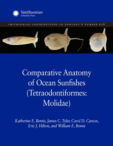
ABSTRACT Bemis, Katherine E., James C. Tyler, Carol D. Carson, Eric J. Hilton, and William E. Bemis. Comparative Anatomy of Ocean Sunfishes (Tetraodontiformes: Molidae). Smithsonian Contributions to Zoology, number 658, viii + 81 pages, 43 figures, 2026. — We review nearly 500 years of literature on the anatomy of ocean sunfishes (Molidae) and present new findings based on our extensive anatomical studies of specimens. We document similarities and differences among the three living genera, Ranzania, Masturus, and Mola, using the organ-system approach to examine general body form and external anatomy, skeleton, muscles, integument, brain and sense organs, digestive organs, heart and circulation, respiratory system, excretory system, reproductive system, and endocrine organs. Known for centuries for basking and seemingly sluggish behavior at the water’s surface, recent tagging studies on locomotion and diving behavior show that ocean sunfishes swim over great horizontal and vertical distances in the water column at higher speeds than previously thought. We interpret the evolution of many of their unusual anatomical features in light of this new information about their natural history and behavioral ecology, which helps to explain the evolution of such an extreme body form. Recommended citation: Bemis, Katherine E., James C. Tyler, Carol D. Carson, Eric J. Hilton, and William E. Bemis. 2026. Comparative Anatomy of Ocean Sunfishes (Tetraodontiformes: Molidae). Smithsonian Contributions to Zoology, No. 658, Washington, D.C.: Smithsonian Institution Scholarly Press. Cover images, from left to right: Ranzania laevis, CAS 99414, 128 mm TL. Masturus lanceolatus, UF 136518, 164 mm TL (length includes claval extension; bend in the extension is artificial). Mola sp., AMS I.27082-001, 74 mm TL.
Published by SMITHSONIAN INSTITUTION SCHOLARLY PRESS P.O. Box 37012, MRC 957 Washington, D.C. 20013-7012 https://scholarlypress.si.edu Compilation copyright © 2026 Smithsonian Institution The rights to all text and images in this publication, including cover and interior designs, are owned either by the Smithsonian Institution, by contributing authors, or by third parties.
Excluding content credited to rightsholders and third parties, this work is licensed under a Creative Commons Attribution-NonCommercial 4.0 International (CC BY-NC 4.0) License. For permission to reproduce credited materials, users are responsible for contacting rightsholders directly.
Library of Congress Control Number: 2026938462 ISSNs: 1943-6696 (online); 0081-0282 (print) Publication date (online): 10 August 2026 Ó The paper used in this publication meets the minimum requirements of the American National Standard for Permanence of Paper for Printed Library Materials Z39.48–1992.
Contents
LIST OF FIGURES
vii
INTRODUCTION
1
METHODS
5
NOTES ON TERMINOLOGY
6
ANATOMICAL ABBREVIATIONS
6
GENERAL BODY FORM AND EXTERNAL ANATOMY
8
SKELETON
12
MUSCLES
34
INTEGUMENT AND HYPODERMIS
40
BRAIN, NERVES, AND SENSE ORGANS
43
DIGESTIVE SYSTEM AND SWIM BLADDER
50
HEART AND GILL-ARCH CIRCULATION
53
RESPIRATION
56
URINARY SYSTEM
60
REPRODUCTIVE SYSTEM
61
ENDOCRINE ORGANS
62
DISCUSSION
62
SPECIMENS EXAMINED
67
ACKNOWLEDGMENTS
69
REFERENCES
71
INDEX
79
Figures 1. Early interpretations of body form 2. Phylogenetic relationships of ocean sunfishes 3. External anatomy of adults 4. Mouth, lips, and nares of adults 5. External anatomy of larvae 6. Micro-CT scans of larval Mola mola 7. External anatomy of juveniles 8. Bone microstructure 9. Dermal plates 10. Skeleton of Ranzania laevis 11. Skeleton of Masturus lanceolatus 12. Skeleton of Mola mola 13. Fused jaws, beak, and triturating teeth of Mola mola 14. Mineralized dental tissues and surrounding soft tissues of Mola mola 15. Dental ontogeny of Mola mola 16. Pharyngeal teeth of Mola mola 17. Natural dissections of median fins of Mola mola 18. Clavus of Masturus lanceolatus 19. Anatomy of dorsal and anal fins of Mola mola and Masturus lanceolatus 20. Dorsal and anal fin movements of swimming Mola mola 21. Schematic interpretation of tendons and coalesced cartilage 22. Dorsal and anal fin movements during swimming of Ranzania laevis 23. Deformed coordinate interpretations of Diodon and Mola 24. Cranial muscles of Ranzania laevis and Mola mola 25. Cranial anatomy of Mola mola 26. Body musculature 27. Epidermis, dermis, and hypodermis of Mola mola 28. Scales and scale microstructure 29. Brain of Mola mola 30. Lateral line of Mola mola 31. Olfactory system of Mola mola 32. Eye, orbit, and conjunctiva of Mola mola 33. Ear and vestibular system of Mola mola 34. Viscera of adults
3 4 9 11 13 14 15 17 18 20 21 22 23 24 24 25 28 29 30 32 33 33 34 35 36 38 39 41 44 46 47 48 49 51
viii
•
SMITHSONIAN CONTRIBUTIONS TO ZOOLOGY
35. Detailed visceral anatomy of Mola mola 36. Esophagus and stomach of Masturus lanceolatus 37. Swim bladder of larval Masturus lanceolatus and Mola mola 38. Heart of Masturus lanceolatus and Mola mola 39. Schematic interpretation of coronary arteries and gill circulation 40. Gill sac and gill-arch circulation of Mola mola 41. Gills and gill arches of Mola mola 42. CT scan of gills of Mola mola 43. Kidney of Mola mola
52 53 54 55 57 58 59 60 61
Comparative Anatomy of Ocean Sunfishes (Tetraodontiformes: Molidae) Katherine E. Bemis,1,2,3* James C. Tyler,2 Carol D. Carson,4 Eric J. Hilton,3 and William E. Bemis5
Est un grand poisson qui pese jusqu’a trois cents livres; sa forme bizzare, sa peau argentée, ses yeux grand et brillans, le rendent remarquable. (Cuvier 1798: 324) [It is a large fish that weighs up to 300 pounds; his bizarre shape, his silvery skin, his large and bright eyes, make him remarkable.] The fishes were more active than their ungainly form would seem to permit . . . (Myers and Wales 1930: 11).
1
National Systematics Laboratory, Office of Science and Technology, National Oceanic and Atmospheric Administration, Washington, D.C., USA. 2 Department of Vertebrate Zoology, National Museum of Natural History, Smithsonian Institution, Washington, D.C., USA. 3 Section for Natural Resources, Virginia Institute of Marine Science, Batten School of Coastal & Marine Science,” William & Mary, Gloucester Point, Virginia, USA. 4 New England Coastal Wildlife Alliance, 11 Clarence Soule Drive, Middleboro, Massachusetts, USA. 5 Department of Ecology and Evolutionary Biology and Cornell University Museum of Vertebrates, Cornell University, Ithaca, New York, USA. * Correspondence: bemisk@si.edu Bemis, K., https://orcid.org/0000-0002-7471-9283 Tyler, https://orcid.org/0000-0003-3202-080X Hilton, https://orcid.org/0000-0003-1742-3467 Bemis, W., https://orcid.org/0000-0002-5669-2793 Manuscript received 6 January 2025; accepted 5 September 2025.
INTRODUCTION This contribution, which includes reviews of prior work and original research toward a synthesis on the anatomy of Molidae, is the outgrowth of our paper “Overview of the Anatomy of Ocean Sunfishes (Molidae: Tetraodontiformes)” in the book The Ocean Sunfishes: Evolution, Biology and Conservation (Bemis et al. 2021 in Thys et al. 2021a; see also Hays et al. 2021). We follow the same format as in Bemis et al. (2021), beginning with external anatomy and general body form followed by sections on the ten organ systems of vertebrates (modified from Liem et al. 2001: tab. 1.1), in order of treatment here: 1. Skeletal system 2. Muscular system 3. Integumentary system 4. Nervous system (including sense organs) 5. Digestive system 6. Circulatory system 7. Respiratory system 8. Excretory system 9. Reproductive system 10. Endocrine system The Molidae are commonly known as molas, ocean sunfishes, or head-fishes. Linnaeus (1758: 334) originally described Mola mola as Tetraodon mola, but Koelreuter
2
•
SMITHSONIAN CONTRIBUTIONS TO ZOOLOGY
(1766: 337) established the new genus Mola. The genus name is derived from the Latin word for millstones (Scharpf 2025). Because of their unusual anatomy, molids have been illustrated in general works since Rondelet (1554) and Gessner (1558; Figure 1A). Early works by Planci Ariminensis (1755; Figure 1B) and Duhamel (1777; Figure 1C) captured the essence of molid body form and aspects of their anatomy. Cuvier included many comments on molid anatomy in his textbook (Cuvier 1805a–e) and firmly placed molids within Tetraodontiformes (Cuvier 1817). One of the broadest early works on molid anatomy is by Harting (1865; Figure 1D), who described and illustrated external features as well as anatomical and histological aspects of bones, integument, brain, eyes, ears, and gills. Steenstrup and Lütken (1898) summarized early works on molids and provided new information on their general osteology and ontogeny (Figure 1E). A series of papers by Rosén (1912; 1913a–c; 1916a,b) offered the first comparative organ-system approach to the anatomy of tetraodontiforms, including observations on molids. Widely cited accounts by Gregory and Raven (1934) and Raven (1939a,b) provided an anatomical framework and easily interpreted illustrations that incorporated skeletal and soft tissue anatomy (e.g., bones, muscles, and gastrointestinal tract) for each of the three extant molid genera, Ranzania, Masturus, and Mola. Fraser-Brunner (1951) synthesized anatomical information with systematic interpretations for Molidae. In 1980, Tyler published a monograph on the skeletal anatomy of tetraodontiforms in which he provided osteological descriptions and illustrations of representatives of each of the extant molid genera, and he placed them in the context of the anatomy of other Tetraodontiformes and their systematic relationships. In addition to these larger works, many specialized reports on the unusual anatomy of molids also informed our synthesis. Studies of muscles (e.g., Winterbottom 1974), nerves (e.g., Nakae and Sasaki 2006), larvae (e.g., Leis 1984), fossils (e.g., Santini and Tyler 2003; Arcila and Tyler 2017), and detailed comparative ontogenetic studies of early developmental stages (e.g., Britz and Johnson 2005a,b; Johnson and Britz 2005; Konstantinidis and Johnson 2012) yielded insights into many questions in molid evolution, such as the identity of elements in the claval skeleton. Genetic studies (e.g., Yamanoue et al. 2004, 2008; Bass et al. 2005; Yoshita et al. 2009; Caldera et al. 2021) contributed to interpreting relationships among the three living genera. Advances in behavioral and tracking studies led to a reconsideration of older ideas about molids and their functional biology and ecology, because molids are not sluggish, helpless, inactive, or “drifting at the mercy of the currents” (Bigelow and Schroeder [1953: 530], who also noted that “unlucky vagrants . . . sighted in our cool northern waters . . . have usually been chilled into partial insensibility”) but instead are visual, rapidly diving, migratory fishes of the pelagic realm (e.g., Watanabe and Sato 2008; Houghton et al. 2009; Dewar et al. 2010; Nakamura et al. 2015; Sousa et al. 2016a, 2021; Hays et al. 2021; Chang et al. 2021; Nyegaard et al. 2023a, 2023c; Rouyer et al. 2024). Molids also exhibit social behaviors, such as schooling and symbiotic cleaning
associations with fishes and albatrosses (e.g., Konow et al. 2006; Abe and Sekiguchi 2012; Abe et al. 2012; Hegde et al. 2021; Nyegaard et al. 2023b). Recent taxonomic studies of molids, the redescription of a species, and the description of a new species (Nyegaard and Sawai 2018; Nyegaard et al. 2018; Sawai et al. 2021) as well as a review of unresolved questions (Hays et al. 2021) are encouraging researchers to revisit and study anatomical differences within Molidae. Molidae is a monophyletic group (Figure 2) supported by both morphological (e.g., Tyler 1980; Santini and Tyler 2002) and molecular data (e.g., Betancur-R et al. 2017) and by combined analyses (e.g., Arcila and Tyler 2017). Prior research on the evolutionary relationships of Molidae with other Tetraodontiformes often resolved them as the sister group of Diodontidae + Tetraodontidae (Figure 2A,C) but other analyses recovered them as sister to Balistidae + Monacanthidae (e.g., Figure 2B). Figure 2D summarizes current taxonomic and phylogenetic information for the five extant species of Molidae. The monotypic Ranzania laevis is sister to the clade containing Masturus lanceolatus and the three valid species of Mola recognized by Nyegaard et al. (2018) and Sawai et al. (2021). This general topology of Ranzania + (Masturus + Mola) is recovered in studies based on anatomical character data (Fraser-Brunner 1951; Tyler 1980; Santini and Tyler 2002, 2003) and molecular data (e.g., Yamanoue et al. 2004; Bass et al. 2005; Alfaro et al. 2007). Although there has been interest in molid anatomy for centuries, comparative anatomical information for the three extant genera is incomplete. This may be because specimens occur unpredictably in many parts of the world, making it difficult to reliably obtain fresh material for dissection and study. Also, researchers tend to study and only retain smaller specimens because of the challenges of handling, transporting, dissecting, and storing complete specimens of large adults. Researchers often did not retain dissections of large specimens for logistical reasons, making it difficult to corroborate published observations. Collections often store large specimens as dry skeletons or preserved dissections of parts of specimens, which does not facilitate comparisons between individuals or taxa. Ranzani (1839) contributed early anatomical descriptions, figures, and information about the family Molidae, but most anatomical descriptions since Cuvier (1805a–e) have been based on specimens interpreted by the authors as Mola mola. Progress in understanding diversity within the genus Mola (e.g., Nyegaard et al. 2018; Sawai et al. 2021) calls into question whether some early accounts are actually based on studies of M. mola, because voucher specimens were rarely deposited in natural history collections. Taxonomic confusion in the nineteenth century about the diagnosis and limits of the genus Orthagoriscus, which has been used at various times primarily for Mola although also for both Masturus and Mola (some early authors also included Ran zania in Orthagoriscus; there is also a variant spelling, Orthrago riscus), means that it is often impossible to know with certainty even the generic identities of some specimens studied long ago (Parenti 2003; Fricke et al. 2025).
A Gessner 1558
B Planci Ariminensis 1755
C Duhamel 1777
D Harting 1865
E Steenstrup and Lütken 1898
FIGURE 1. Illustrations showing early interpretations of the body form of molids. A. From Gessner (1558). B. From Planci Ariminensis (1755). C. From Duhamel (1777). D. From Harting (1865). E. From Steenstrup and Lütken (1898: tab. 1).
4
•
SMITHSONIAN CONTRIBUTIONS TO ZOOLOGY
A Arcila and Tyler (2017)
B Betancur-R. et al. (2017)
C Ghezelayagh et al. (2022)
Lophiiformes
Lophiiformes
Lophiiformes
Triacanthidae
Triodontidae
Triodontidae
Triacanthodidae
Triacanthidae
Aracanidae
Balistidae
Triacanthodidae
Ostraciidae
Monacanthidae
Aracanidae
Triacanthidae
Aracanidae
Ostraciidae
Triacanthodidae
Ostraciidae
Diodontidae
Balistidae
Triodontidae
Tetraodontidae
Monacanthidae
Molidae
Molidae
Molidae
Diodontidae
Balistidae
Diodontidae
Tetraodontidae
Monacanthidae
Tetraodontidae
D Nyegaard et al. (2018); Sawai, Nyegaard, and Yamanoue (2021) Slender Sunfish Ranzania laevis (Pennant 1776)
Sharptail Mola Masturus lanceolatus (Liénard 1840)
Molidae
Ocean Sunfish Mola mola (Linnaeus 1758)
Bumphead Ocean Sunfish Mola alexandrini (Ranzani 1834)
Hoodwinker Ocean Sunfish Mola tecta Nyegaard et al. 2017
FIGURE 2. Phylogenetic relationships of ocean sunfishes. A. Tree recovered by Arcila and Tyler (2017; 16 loci and morphology) showing Molidae as sister to Diodontidae + Tetraodontidae. B. Tree recovered by Betancur-R. et al. (2017; nuclear and mitochondrial loci) showing Molidae as sister to Balistidae + Monacanthidae. C. Tree recovered by Ghezelayagh et al. (2022; ultraconserved elements) showing Molidae as sister to Diodontidae + Tetraodontidae. D. Relationships of extant species of Molidae recognized by Nyegaard et al. (2018) and Sawai et al. (2021). The three species of Mola form a clade with Masturus lanceolatus; this clade is sister to Ranzania laevis.
NUMBER 658
Molids are clearly a specialized extreme within Acanthomorpha (sensu Ghezelayagh et al. 2022) and even within Tetra odontiformes (sensu Tyler 1980). To interpret the functional morphology of molids, particularly the large-bodied Masturus and Mola, we found it useful to make comparisons with other pelagic fishes that are fast swimming, deep diving, migratory, and have high metabolic rates, such as tunas. The similarities and differences between these fishes help us interpret aspects of molid biology in the context of the newly reported information on the behavioral ecology and pelagic habitat of molids. We combine our new data from dissections, micro-computed tomography (CT) scans, and histological studies with information and figures from the literature to develop a comprehensive overview of the comparative anatomy of Molidae. We follow a specimen-based approach using vouchered museum specimens of ontogenetic stages, from larvae to adults, for all three extant genera; and we report on stranded individuals of Mola mola studied in the field that could not be retained in collections. We follow the current standard in ichthyology to capitalize the common names of species of fishes (Page et al. 2023). Institutional abbreviations follow Sabaj (2023).
INSTITUTIONAL AND OTHER NONANATOMICAL ABBREVIATIONS ANSP Academy of Natural Sciences, Philadelphia, Penn. AMS Australia Museum, Sydney CAS California Academy of Sciences, San Francisco c&s cleared and stained CT computed tomography CUMV Cornell University Museum of Vertebrates, Ithaca, N.Y. EtOH ethanol H&E hematoxylin and eosin MCZ Museum of Comparative Zoology, Harvard University, Cambridge, Mass. MPR multiplanar reformatting MRI magnetic resonance imaging NL notochord length NECWA New England Coastal Wildlife Alliance, Mass. NOAA National Oceanic and Atmospheric Administration OS Oregon State University, Corvallis PCBL preclaval band length SEM scanning electron microscopy SL standard length TL total length UF University of Florida, Florida Museum of Natural History, Gainesville USNM Smithsonian Institution, National Museum of Natural History, Washington, D.C. VIMS Virginia Institute of Marine Science, Gloucester Point
•
5
METHODS New specimens collected by the Northeast Fisheries Science Center, NOAA Fisheries (Politis et al. 2014), and the Pacific Islands Fisheries Science Center, NOAA Fisheries, were fixed in 10% formalin (n = 8) or frozen for fresh dissection (n = 3) and subsequently fixed after dissection. A clavus of Masturus lanceo latus collected from a stranded specimen in Florida was frozen; we thawed and dissected this specimen and subsequently fixed it in 10% formalin (VIMS 42354). We deposited new specimens in the Nunnally Ichthyology Collection at the Virginia Institute of Marine Science (VIMS) and Cornell University Museum of Vertebrates (CUMV). We also studied specimens from collections at ANSP, AMS, CAS, MCZ, OS, UF, USNM, and VIMS, including previously prepared c&s specimens (see Specimens Examined). We report the lengths of specimens that we examined as total lengths (TL); we include lengths from the literature as originally reported, including notochord length (NL) and “standard length” (SL). Because molids do not have hypurals, the “SL” measurement is preferably called PCBL, meaning pre-claval band length, but because most measurements were made before this clarification, we do not use it here (Sawai et al. 2021: 24, fig. 3). Between 2008 and 2024, one of us (CDC) also examined and documented several hundred stranded individual Mola mola under the auspices of the nonprofit New England Coastal Wildlife Alliance (NECWA). The NECWA responds to reports of stranded ocean sunfishes and works to rescue those that are still alive. Mola mola that do not survive are necropsied, photographed, and measured by NECWA. Some anatomical material from stranded specimens was retained for further study; however, most individuals could not be retained because of their large size. Here we refer to individuals that could not be retained using their field numbers, which include the year, stranding number, and other unique identifiers (e.g., Mm2021-S40-031, 1,364 mm TL). Some of the most useful stranded materials were natural disarticulations as specimens decomposed on the beach, for these exposed details of key skeletal features. Stranded individuals described herein were examined from the western North Atlantic Ocean, with almost all records coming from Massachusetts, USA. All observations reported for Ranzania laevis and Masturus lanceolatus are based on the interpretation that these two genera are monotypic. We report original observations for both of these species as well as for Mola mola, which is the only species of Mola that we examined. Nyegaard et al. (2018) described Mola tecta, and Sawai et al. (2018) clarified the taxonomic status of M. alexandrini. Britz (2022) called into question the validity of M. alexandrini, but we follow Sawai and Nyegaard (2023) in recognizing this species. The early life stages of the three species of Mola are indistinguishable (Thys et al. 2021b); therefore, we relied upon collection locality to identify larval and juvenile specimens of Mola to species. For example, MCZ 61454, MCZ 41675, MCZ 87065, MCZ 41503, and VIMS 40710 were collected in the western North Atlantic, where only M. mola is known to occur; two larval specimens that we studied from
6
•
SMITHSONIAN CONTRIBUTIONS TO ZOOLOGY
coastal Australia could only be identified as Mola sp. because all three species of Mola occur in those waters. Our identifications should be reviewed when new systematic studies of larval Mola are conducted. We performed micro-CT scanning on specimens by using either the GE 120 micro-CT or the Xradia Versa XRM-500 nanoCT in the Biotechnology Resource Center Multiscale Imaging Facility at Cornell University. Reconstructions prepared from these datasets have a resolution of 7–100 μm voxels. We studied 2D orthogonal MPR and 3D-volume reconstructions using Horos DICOM imaging software on Apple Mac computers and with Dragonfly or Avizo on a Windows Workstation. To view internal anatomy of teeth and bones, we digitally segmented reconstructions within Horos, Dragonfly, or Aviso. For histological studies of molid bone as a tissue (see Figure 8 in Skeleton), we decalcified portions of two formalin-fixed specimens (Mola mola CUMV 98740; Masturus lanceolatus VIMS 42354), washed and dehydrated them for paraffin embedding, and cut sections at 6 μm. We then stained them with either hematoxylin and eosin (H&E; see, e.g., Humason 1972) or modified tetrachrome stain for osteoid and bone (Ralis and Watkins 1992) at the Cornell Veterinary College Diagnostic Laboratory. We prepared ground sections of the lower jaw and beak of one specimen of Mola mola (see Figure 13E in Skeleton) by embedding the tissue in a low-viscosity epoxy resin (Spurr 1969), sectioning the blocks into slices 100–250 µm thick using a Buehler IsoMet Saw, and then thinning them by grinding with 1,200-grit carborundum sandpaper (Bemis 1984; Bemis and Bemis 2015). To prepare a combination plate of the beak and triturating teeth of M. mola (see Figure 14 in Skeleton), we first photographed the lower jaw of the specimen, made and analyzed a micro-CT scan, and then cut a parasagittal section through the jaw using a band saw. The tissue was decalcified using ethylenediaminetetraacetic acid (EDTA), embedded in paraffin, sectioned at 10 µm, and stained with H&E at the Cornell Veterinary College Diagnostic Laboratory. We also sectioned the skin and hypodermis of one specimen of Mola mola (VIMS 35803) and portions of the heart and bulbus arteriosus of one specimen of Masturus lanceolatus (VIMS 8120) for staining with Masson trichrome and H&E (see Figure 27 in Muscles). We photographed sections using an Olympus SZX12 microscope equipped with an Olympus DP70 digital camera or a Zeiss SteREO DiscoveryV20 microscope. We used a Canon 5D Mark II digital camera to record color macrophotographs. We adjusted images for color balance and contrast using Adobe Photoshop CC, prepared plates and line drawings in Adobe Illustrator CC, and organized the plates using Adobe InDesign.
NOTES ON TERMINOLOGY We distinguish between the terms individual and specimen, the difference being that individuals were not retained in a collection for future study. The term individual may refer to, for example, a
stranded individual studied on a beach or videos of a swimming fish in the wild or in an aquarium. We include observations about individuals that help us to interpret molid anatomy. In contrast, specimens are in collections where they are available for future study. The term specimen may refer to an entire preserved fish, partial dissections, dry skeletal materials, tissue samples, or special preparations such as histological materials or cleared and stained specimens, as well as photographs and collection data. We indicate the original sources of names of anatomical structures in the text where first mentioned. If there are synonymous terms, we explain why we chose a particular term. In some figures we give the names of features in full, to facilitate rapid orientation to the figure and its parts. In figures where many structures are indicated, such as those treating the skeleton or the muscular system, we use abbreviations with a mix of capital and lowercase letters. This mixture reflects different abbreviation standards used for different organ systems. For example, in neuroanatomical studies, many major structures, such as brain regions or nerve roots, are indicated by abbreviations that start with capital letters. Similarly, we use Roman numerals to indicate the cranial nerves. In the text and in the list below, we use Latin for muscle names by convention. Because of this, we italicize muscle abbreviations and names in the text; we use abbreviations for the muscle names in figures for clarity. Finally, we indicate structures that we named in our descriptions—for example, tendon tunnels that pass through the band of coalesced cartilage of the pterygiophores.
ANATOMICAL ABBREVIATIONS aa aap ABA
af afr ALL AM AMα′ AMα″ AMα‴ AMβ′ AMβ″ ap-mr arrv AVSD
anguloarticular m. adductor arcus palatini afferent branchial artery; series of ABA1–ABA4 m. abductor profundus m. abductor superficialis anal distal radial; series numbered in some figures anal fin anal-fin ray anterior lateral-line nerve m. adductor mandibulae complex m. adductor mandibulae α′ m. adductor mandibulae α″ m. adductor mandibulae α‴ m. adductor mandibulae β′ m. adductor mandibulae β″ anal proximal-middle radial m. arrector ventralis anterior vertical semicircular duct
bb bh
basibranchial; series of bb1 and bb2 basihyal
abdp abds adr
NUMBER 658
boc br
bsp C cart ap-mr + hyp cart clp-mr + hyp cart dp-mr + hyp cb cfr Ch cha chp cl clav edac clav eddc clav cldr clfr clp-mr co d ddr df dfr do dp-mr eb EBA
basioccipital branchiostegal ray; series of br1–br5 in Ranzania and br1–br6 in Mola and Masturus basisphenoid cerebellar lobes coalesced cartilage of anal proximal- middle radials plus hypodermis coalesced cartilage of claval proximal- middle radial plus hypodermis coalesced cartilage of dorsal proximal- middle radials plus hypodermis ceratobranchial; series of cb1–cb5 caudal-fin ray cerebral hemisphere anterior ceratohyal posterior ceratohyal cleithrum clavicular portion of the fused m. erector et depressor analis clavicular portion of the fused m. erector et depressor dosalis clavus claval distal radial claval-fin ray claval proximal-middle radial coracoid dentary dorsal distal radial; series numbered in some figures dorsal fin dorsal-fin ray; series numbered in some figures m. dilatator operculi dorsal proximal-middle radial
ecp edac eddc enp Ep epax epo exo
epibranchial; series of eb1–eb4 efferent branchial artery; series of EBA1–EBA5 ectopterygoid fused m. erector et depressor analis fused m. erector et depressor dorsalis endopterygoid epiphysis m. epaxialis epioccipital exoccipital
fr fz
frontal flexure zone
gr
gill raker
•
h H ha hab had hb hhd hhv HSD hyin
hyomandibula hypophysis hemal arch m. hyohyoidei abductores m. hyohyoidei adductores hypobranchial; series of hb1–hb3 dorsal hypohyal ventral hypohyal horizontal semicircular duct m. hyohyoideus inferioris
I ih II III incla incld iop IV IX
olfactory nerve interhyal optic nerve oculomotor nerve m. inclinatores analis m. inclinatores dorsalis interopercle trochlear nerve glossopharyngeal nerve
k
ventral keel
L lap let lo
lateral-line lobe m. levator arcus palatini lateral ethmoid m. levator operculi
m IX mV m VII mX mdr V met mpt mx
motor root of glossopharyngeal nerve motor root of trigeminal nerve motor root of facial nerve motor root of vagal nerve foramen for the mandibular ramus of the trigeminal nerve (V) mesethmoid metapterygoid maxilla
na
neural arch
obli Olf op Ot Ov
m. obliquus inferioris olfactory bulb opercle optic tectum optic ventricle
Pa pal pas pb pcl pf pfr phce
paraphysis palatine parasphenoid pharyngobranchial; series of pb2–pb4 postcleithrum pectoral fin pectoral-fin ray m. pharyngoclavicularis
7
8
•
SMITHSONIAN CONTRIBUTIONS TO ZOOLOGY
PLL pmx pop pp prhy Pro pro prof V pto pts PVSD
posterior lateral-line nerve premaxilla preopercle m. protractor pectoralis m. protractor hyoidei preoptic recess prootic profundal branch of trigeminal nerve pterotic pterosphenoid posterior vertical semicircular duct
q
quadrate
r rar
radial; series of r1–r3 retroarticular
s IX sV s VII sX s+m X sc Sc scl soc sop Sp Sp1 Sp2 spo sth sym
sensory root of glossopharyngeal nerve sensory root of trigeminal nerve sensory root of facial nerve sensory root of vagal nerve combined sensory and motor roots of vagal nerve scapula spinal cord supracleithrum supraoccipital subopercle spinal nerve first spinal nerve second spinal nerve sphenotic m. sternohyoideus symplectic
T tt
tela choroidea tendon tunnel
V v
VI VII VIII
trigeminal nerve vertebra; series of v1–v18 (Ranzania), series of v1–v16 (Masturus), series of v1–v17 (Mola) abducent nerve facial nerve octaval nerve
X
vagal nerve
GENERAL BODY FORM AND EXTERNAL ANATOMY The most distinctive external morphological feature of molids is the clavus, the structure at the posterior part of the body, also known as a gephyrocercal fin and sometimes called a pseudocaudal
fin (e.g., Raven 1939a; Tyler 1980). Authors and illustrators have long wrestled with interpretations of the clavus (Figure 1), but the definition and our modern understanding of the term come from Fraser-Brunner (1951: 90). The word derives from the Latin word clavus, for rudder, and anatomically it refers to the lobe consisting of modified dorsal and anal fins that functions as a rudder during swimming. Figure 3 shows the external appearance of the clavus in all three genera. The clavus of Ranzania laevis has 19 claval-fin rays (Figure 3A; arrows indicate the 9 claval-fin rays derived from the dorsal fin). The claval lobe in Masturus lanceolatus (Figure 3B) is the source of both its species epithet and its common name, Sharptail Mola. The clavus of Mola can vary greatly within the genus and between some distinct populations. The specimen shown in Figure 3C is from the western North Atlantic; it has a wavy or scalloped clavus (indicated by arrows), which is thought to be characteristic at the species level for Mola mola (see Caldera et al. 2021: fig. 2). Also indicated in the figure are the dorsal, anal, and pectoral fins, the position of the anus, and the distal edge of the coalesced cartilage, which marks the proximal edge of the flexure zone. The coalesced cartilage is a distinctive anatomical feature of molids that is discussed in detail in later sections. A prevailing functional anatomical aspect of Molidae concerns the limited lateral undulation possible with this body form. Fraser-Brunner (1951: 89) noted that changes in the trunk muscles mean that “. . . body flexion is lost but the dorsal and anal fins gain in power, and the latter are therefore the principal means of locomotion.” Also relevant is a study by Brainerd and Patek (1998). They discussed the fact that most extant tetraodontiforms have fewer than 21 vertebrae, which are among the lowest, if not the lowest, numbers of vertebrae known among teleosts (see Tyler, 1980: tab. 2 for vertebral counts of tetraodontiforms). The tetraodontiforms Brainerd and Patek studied (Monacanthus hispi dus, Canthigaster rostrata, and Diodon holocanthus) had smaller C-start curvatures than outgroup forms (Acanthurus chirurgus, Abudefduf saxatilis, and Scarus coeruleus; C-start curvature relates to bending the body into a C-shape for fast start performance, and larger C-start values are associated with faster escape performance). They suggested that this performance difference might impair escape performance, increase vulnerability to predators, and in turn drive the evolution of mechanical defenses, such as body inflation, spines, or bodies encased by a bony carapace. Because molids apply force to the water column using the dorsal and anal fins (with smaller contributions from the clavus and pectoral fins), many other modifications of the post-cranial skeleton, integument, hypodermis, and musculature are present, often representing morphological extremes not only within Tetraodontiformes but also among teleosts more generally. Throughout this work, we explain how organ-system modification in Molidae relates to their unique swimming mode, termed moliform locomotion (Webb 1994: 54; Pough et al. 2022: fig. 7.31). Moliform locomotion is a type of median-and-paired-fins swimming (MPF) as opposed to body-and-caudal-fin swimming (BCF; terminology sensu Friedman et al., 2021). Houghton et al.
Distal edge of coalesced cartilage marks flexure zone
NUMBER 658
Dorsal fin
Opercular opening
Clavus Falcate pectoral fin
A
Anus Anal fin
10 cm Dorsal fin
Distal edge of coalesced cartilage marks flexure zone Opercular opening
Clavus
Claval lobe
Rounded pectoral fin with horizontal insertion
Anus Anal fin
B
10 cm
Distal edge of coalesced cartilage marks flexure zone
Dorsal fin
Opercular opening
Rounded pectoral fin with horizontal insertion
Clavus
Anus
Anal fin C
10 cm
•
9
(2009) described swimming behavior of molids as unusual compared with typical teleosts because body roll, in addition to pitch and sway amplitude, are linked with vertical velocity. It is distinct from other unique swimming modes of tetraodontiforms, such as balistiform (e.g., George and Westneat 2019), ostraciiform (Hove et al. 2001), aracaniform (Gordon et al. 2020) or the multiple- fin swimming of burrfish (Arreola and Westneat 1996) because, among other reasons, molids lack a caudal fin. Watanabe and Davenport (2021: 78) termed molid swimming “synchronized vertical fin flapping,” and Watanabe and Sato (2008) interpreted the symmetrical dorsal and anal fins of Mola mola as paired vertical wings used to generate lift for swimming. They compared the vertical fin-flapping of molids with dorsoventral flapping used in the subaqueous flight of penguins (Watanabe and Davenport, 2021: 73). It is an apt comparison because molids generate lift on both the flapping and recovery strokes, as do penguins during down and recovery strokes of their wings (e.g., Clark and Bemis 1979). One of the most important aspects of moliform swimming relates to the stiffness of the median fins. Unlike most other tetraodontiforms and teleosts in general, molids have very limited mobility of the fin rays in the dorsal and anal fins, lacking fin webs between the fin rays, and thus cannot spread or collapse fin rays to change the surface area of their fins. There is flexibility of the fin rays along the thinner trailing edge of the fins, which we document below (see leading and trailing edges, Figure 20). Gregory and Raven (1934: 147) considered that undulatory movements of the fins might be possible, but we cannot confirm this and think it unlikely (i.e., we do not think that the fin rays are flexible enough to create undulatory waves along the edges of the dorsal and anal fins as in, e.g., filefishes; see George and Westneat 2019). This should be examined in future functional anatomical studies of moliform locomotion. Powerful movements of the fins of molids are typically symmetrical when swimming straight ahead, that is, both the dorsal and anal fins stroke to the same side of the body and then toward the opposite side. The clavus is important as a rudder to control swimming direction but does not in itself contribute much (if any) forward thrust to swimming. Despite these seeming limitations, molids swim rapidly and can make agile movements to evade predators, such as turning upside down, rolling backward, pivoting, spinning, and breaching (Myers and Wales 1930; Konow
FIGURE 3. Left views of the external anatomy of adult molid specimens, showing features of the fins, opercular opening, and position of the coalesced cartilage beneath the skin. A. Ranzania laevis, ANSP 103501, 492 mm TL. Arrows indicate the nine claval fin rays in the dorsal part of the clavus. B. Masturus lanceolatus, VIMS 8120, 1,175 mm TL. C. Mola mola, VIMS 35803, 1,180 mm TL. Note the wavy clavus characteristic of M. mola and the location of claval ossicles along its edge, indicated by arrows.
10
•
SMITHSONIAN CONTRIBUTIONS TO ZOOLOGY
et al. 2006; Hays et al. 2021; Nyegaard et al. 2023c). Like many other large pelagic fishes, molids can make extensive seasonal migrations (e.g., Potter et al. 2011; Chang et al. 2021, 2025).
ADULTS Figure 2D shows the general body form and external appearance of the five extant species of Molidae. Masturus lanceolatus and the three species of Mola are among the heaviest teleosts, with the largest record for the family Molidae being for Mola alexandrini at 2,744 kg and 325 cm TL (Sawai et al. 2018, 2021; Sawai and Nyegaard 2022; Gomes-Pereira et al. 2023). Ranza nia laevis is smaller than the other molids with a maximum confirmed size of 74 cm TL (Sawai et al. 2021). Reported sizes of 90 cm TL (Schmidt 1921a:1), 800 mm (Fraser-Brunner 1951: 98), or 1 m (Kamohara 1950) cannot be confirmed. Sawai et al. (2021: 21, tab. 2) provided an excellent summary of the morphological characters of the five species of Molidae. The body is laterally compressed in all three genera, but it is distinctly more elongate, streamlined, and laterally compressed in Ranzania laevis than in Masturus lanceolatus or the three species of Mola (Figure 3). The dorsal part of the body of R. laevis is rounded and widens up to the level of the pectoral girdle, where the body tapers ventrally to end in a ventral keel. The bodies of M. lanceolatus and Mola mola are less laterally compressed and do not taper so distinctly in the specimens we examined; however, Gregory and Raven (1934: 145) reported that “the body is deep and keel-like below with a knifelike lower edge in front of the vent” in the fresh 47 inch (= 119 cm) TL specimen of M. mola that they dissected. All molids are streamlined when viewed from above. The lips and mouth of Ranzania laevis form an oval funnel (Figure 4A,B) that is vertically oriented; the beak is set inside the tubular mouth. Some authors have reported that the mouth can be closed to form a vertical slit (Fraser-Brunner 1951: fig. 4; Ebenezer and Joel 1984), whereas other observers have reported that the lips are relatively inflexible and remain permanently open (Fitch 1969; Robinson 1975; Smith et al. 2010; Nyegaard et al. 2017). Smith et al. (2010) reported that both living and unpreserved specimens have inflexible and permanently open mouths; Nyegaard et al. (2017: fig. 2) confirmed this in live stranded individuals. We examined only preserved specimens of R. laevis, but we agree that the lips are inflexible. Whether the funnel-like mouth functions primarily in feeding or respiration is unknown. In Masturus lanceolatus the lips are not as extended; instead, they form an inflexible opening with a horizontal orientation (Figure 4C,D). In comparison, the lips of Mola are flexible and somewhat horizontally oriented (Figure 4E,F). In contrast to R. laevis, Masturus lanceolatus can close its mouth (Brimley 1939: 296), as can Mola mola and M. ramsayi (Marianne Nyegaard, Auckland War Memorial Museum, New Zealand, pers. comm. June 2025). The paired incurrent and excurrent openings of the nostrils of all three genera of molids are minute and flush with the surrounding surface rather than raised (nares, Figure 4A,C,E). In the specimens we examined, the narial openings are largest and
closest to the eye in Mola mola (Figure 4E). Ranzania laevis and Masturus lanceolatus have such small openings that it is difficult to locate them with the naked eye (Figure 4A,C). The eyes are large relative to body size in all three genera. The lateral line is inconspicuously present, as elegantly demonstrated by Nakae and Sasaki (2006). The pectoral fin in Ranzania laevis is relatively long and falcate (Figure 3A) and fits into a shallow depression in the body when adducted, like the pectoral fin of a tuna. This detail suggests that R. laevis can move quickly in the water column, which we can confirm from the videos described below (see subsection Postcranial Axial and Appendicular Skeleton: Dorsal and Anal Fins, Clavus, Skeletal Supports, Coalesced Cartilage, and Fin Rays). In Masturus lanceolatus and Mola mola, the pectoral fin is shorter and more rounded and inserts horizontally (Figure 3B,C); it is larger in M. mola than in M. lanceolatus. Watanabe and Davenport (2021: 74) noted that M. mola oscillates the pectoral fins to balance the body in the water column. Externally, the clavus of Ranzania laevis is truncate and slightly oblique and does not have typical body scales on the surface (Figure 3A; Tyler 1970; Sawai et al. 2021: 20, 21). In contrast, Masturus lanceolatus and Mola mola have a rounded or slightly scalloped clavus (Figure 3B,C) and scales continue to the clavus margin. The central region of the clavus of M. lanceo latus has a claval lobe of varying length (see Caldera et al. 2021: fig. 1; note that they termed the claval lobe a peduncle, a term that we reject because of possible confusion with the caudal peduncle of other fishes). The claval lobe has an elongate extension during early ontogeny (Figures 5F, 7B) and bears fin rays that we interpret as remnant caudal-fin rays (Tyler, 1980: 391). The anterior portions of the dorsal and anal fins in molids (i.e., the portions of the fins anterior to the clavus) are approximately mirror images of each other: tall, stiff, and set far posteriorly on the body (Winterbottom 1974; Tyler 1980; Nyegaard et al. 2018). Watanabe and Sato (2008: fig. 3a,b) found that the dorsal and anal fins of Mola mola change shape during ontogeny, causing a decrease in the aspect ratio of those fins with growth. Despite these changes, the dorsal and anal fins are symmetrical with each other throughout ontogeny (i.e., the shapes of the dorsal and anal fins are very nearly the same). A horizontal section through the dorsal fin of M. mola illustrates its streamlined shape (Watanabe and Davenport 2021: fig. 1a). As with other symmetrical hydrofoils used for lift-based swimming (e.g., penguin wings; Clark and Bemis 1979), this shape creates lift and reduces pressure drag created by the fin as it sweeps through the water. Perhaps unique to all vertebrates that use lift-based swimming or flying, ocean sunfishes use median rather than paired appendages to power their locomotion. The dorsal-, anal-, and pectoral-fin rays extensively branch in all three genera of molids but there are few segmentations, and these are confined to the distal ends of the rays (Tyler 1980). In Ranzania laevis, the claval rays are highly branched distally, whereas in Masturus lanceolatus and Mola mola these claval rays branch only in single to triple dichotomies.
NUMBER 658
•
11
Lip Nares
Beak of premaxilla
Beak of dentary A
20 mm
B
10 mm
Nares Lip
Beak of dentary C
20 mm
D
10 mm
Nares Lip
Beak of dentary E
20 mm
F
10 mm
FIGURE 4. Adult molid specimens showing mouth, lips, and minute nares. A, B. Ranzania laevis, ANSP 103501, 492 mm TL. The stiffened lips form a tube that extends beyond the beak. C, D. Masturus lanceola tus, VIMS 8120, 1,175 mm TL. The lips form a horizontal, somewhat stiff opening. E, F. Mola mola, VIMS 35803, 1,180 mm TL. Lips are more flexible than in either R. laevis or M. lanceolatus and follow the contour of the beak.
12
•
SMITHSONIAN CONTRIBUTIONS TO ZOOLOGY
The species of Mola have prominent dermal ossicles along the edge of the clavus near the distal ends of the claval-fin rays; neither Masturus lanceolatus nor Ranzania laevis have large dermal ossicles in the clavus. Differences in claval ossicles are diagnostic at the species level within the genus Mola (Sawai et al. 2021: tab. 2). A pleated band of smaller scales that we term the “flexure zone” lies between the coalesced cartilage and the bases of the clavus and the dorsal and anal fins, which allows them to flex (see Sawai et al. 2018 and Nyegaard et al. 2018 for discussions of this area, which they term “clavus band” as a species- level diagnostic feature).
LARVAE All molids undergo a remarkable metamorphosis from larvae to adults (Figures 5–7). Since the eighteenth century (e.g., Koelreuter 1766: pl. VIII, figs. 2, 3; Bemis et al. 2021: fig. 2) zoologists have studied small, spiny fishes thought to be related to molids. Various names have been applied to larval molids (e.g., Mola, Diodon, Orthagoriscus, Ostracion, Pallasia, Molacan thus). Putnam (1871) thought that the small molids he studied belonged in their own genus, Molacanthus, a view supported by Gill (1884: 426), who elevated Molacanthus to its own subfamily and described them as “pelagic fishes of very small size.” Knowledge of molid larvae improved and collections of additional specimens filled in gaps in ontogenetic series (Steenstrup and Lütken 1898; Schmidt 1921a). Schmidt (1921a) reviewed the history of larval and juvenile molid identification and assigned the spiny larvae to the genus Mola. Morphological diversity of larvae and juveniles of Mola has yet to be linked to each of the three valid species within Mola (Nyegaard et al. 2018; Sawai et al. 2018; Thys et al. 2021b). The early development of Ranzania laevis is better known than that of other species of molids because of the availability of larval specimens (Figure 5A–C; Leis 1977). Larval stages of Masturus lanceolatus (Figure 5D–F) are rare, as are, especially, those of the species of Mola, (Figure 5G–I; Thys et al. 2021b). All molid larvae are initially round (Figures 5, 6). The notochord does not flex during development. Instead, the notochord, the well-developed median-fin folds, and the post-anal myomeres atrophy, resulting in the formation of the clavus (Leis 1977; Johnson and Britz 2005). At about 2 mm NL, large pyramidal spines develop in all three genera (Lyczkowski-Shultz 2005). In R. laevis, these spines decrease in size shortly after the dorsal-and anal-fin rays ossify (Leis 1977). Leis (1977: 456) interpreted that ossification of the fin rays allowed the larva to swim, thereby decreasing the protective value of spines. Spines persist in larval M. lanceolatus and species of Mola during the “Molacanthus” stage, during which the body is deep and laterally compressed and has a ventral keel (Figures 5, 6; Leis 1984; Lyczkowski-Shultz 2005). As development proceeds, the distinctive body shapes of the three genera become apparent (Figure 7). For example, the elongate body of Ranzania laevis (Figure 7A), with its tall dorsal and
anal fins, short clavus, and elongated pectoral fin, is different from the deeper-bodied Masturus lanceolatus (Figure 7B) and Mola sp. (Figure 7C). The claval lobe and claval extension, which are diagnostic for the genus Masturus, are especially apparent in Figure 7B.
SEXUAL DIMORPHISM Fraser- Brunner (1951: 117) reported that sexual dimorphism was apparent when Mola mola exceed about 2 ft (= ~60 cm) in length. He described males as having a “bony tubercle” that produced a pronounced, forward projecting snout, whereas he described females as having an upward projecting snout that resulted in their having a deeper head, with the front of the snout nearly vertical. He also described sex-related differences in the claval lobes and the claval length (Fraser-Brunner 1951: 117; see side-by-side illustrations of male and female specimens of similar length in van Roon and ter Pelkwijk 1939: fig. 1). With advances in understanding species diversity in Mola, most authorities do not think it is possible to determine sex based on external anatomy (e.g., Sawai et al. 2018; Sawai et al. 2021). Sexual dimorphism has not been described for Ranzania laevis or Masturus lanceolatus.
SKELETON Extensive studies of the skeleton of molids provide a rich source of characters for systematic analyses, both within Molidae and among families of Tetraodontiformes. Tetraodontiforms have reduced features relative to outgroup taxa, such as the number of vertebrae, but molid skeletons are further reduced in two distinct ways. First, the skeleton has limited ossification and the bones have an unusual texture. Second, molids have lost individual skeletal elements beyond those already absent in many tetraodontiforms (e.g., ribs, pelvic girdle, pelvic fins; see Tyler 1980 for comments on reductive characters). For example, all molids lack the coronomeckelian (= sesamoid articular of Tyler 1980) and their exoccipitals are small, do not form condyles, and are excluded from the foramen magnum by a dorsal extension of the basioccipital. The interopercle is absent in Masturus lanceolatus and greatly reduced in Mola mola (“long delicate needle of bone entirely embedded within the ligament”; Tyler 1980: 371) and Ranzania laevis; indeed, all opercular elements except the preopercle are relatively small. The most comprehensive analysis of the skeleton of molids was presented by Tyler (1980), although aspects of skeletal anatomy were treated in many historical papers including Quekett (1855), Leydig (1857), Kölliker (1859), Harting (1865), Goette (1879), Trois (1884a,b), Steenstrup and Lütken (1898), Stephan (1900), Supino (1904), Nowikoff (1910), Kaschkaroff (1914a,b, 1916), and Studnicka (1916). The following overview is not a comprehensive osteology of the family. Rather, we briefly describe molid skeletons, based primarily upon Tyler (1980) but also on
A
0.5 mm
B
2 mm
C
2.5 mm
Claval lobe
Claval extension
D
2.5 mm
E
2.5 mm
F
5 mm
G
2.5 mm
H
2.5 mm
I
5 mm
FIGURE 5. External anatomy of larval molid specimens. A. Ranzania laevis, VIMS 32389, 2.3 mm TL. B. Ranzania laevis, USNM 385363, 6.1 mm TL. C. Ranzania laevis, USNM 385363, 12.4 mm TL. D. Masturus lanceolatus, UF 234607, 9.3 mm TL (not including claval extension). E. Masturus lanceolatus, UF 234607, 13.9 mm TL (not including claval extension). F. Masturus lanceolatus, AMS I.43072-003, 32 mm TL (length includes claval extension, which is artificially bent downward). G. Mola mola, MCZ 61454, 7.4 mm TL. H. Mola mola, MCZ 41675, 13.6 mm TL. I. Mola mola, VIMS 40710, 22 mm TL.
14
A
E
•
SMITHSONIAN CONTRIBUTIONS TO ZOOLOGY
2 mm
2 mm
B
F
2 mm
2 mm
C
2 mm
D
2 mm
G
2 mm
H
2 mm
FIGURE 6. Micro-CT scans of larval Mola mola to show larval scales. A–D: Mola mola, MCZ 61454, 7.4 mm TL. A. Lateral view, anterior left. B. Anterior view. C. Dorsal view, anterior left. D. Ventral view, anterior left. E–H: Mola mola, MCZ 41675, 13.6 mm TL. E. Lateral view, anterior left. F. Anterior view. G. Dorsal view, anterior left. H. Ventral view, anterior left.
NUMBER 658
•
15
Dorsal fin
Clavus
A
Anal fin 10 mm Dorsal fin
Clavus
Claval lobe
B
10 mm
Claval extension
Anal fin
Dorsal fin
Clavus
Anal fin
C 10 mm
FIGURE 7. External anatomy of juvenile molid specimens. A. Ranzania laevis, CAS 99414, 128 mm TL. B. Masturus lanceo latus, UF 136518, 164 mm TL (length includes claval extension; bend in the extension is artificial). C. Mola sp., AMS I.27082-001, 74 mm TL.
16
•
SMITHSONIAN CONTRIBUTIONS TO ZOOLOGY
more recent studies by Britz and Johnson (2005a,b), Johnson and Britz (2005), Konstantinidis and Johnson (2012), and on our own observations, to highlight features that are unique to Molidae as well as key variations within the family. Our treatment of the skeletal system begins with comments on molid bone as a tissue followed by the regional organization used in our previous osteological studies of actinopterygian fishes (e.g., Grande and Bemis 1998; Hilton 2002; Hilton et al. 2011) and herein adapted to the peculiar morphology of molids.
OBSERVATIONS AND COMMENTS ON MOLID BONE AS A TISSUE Bones of fresh or frozen and thawed specimens of Masturus and Mola feel firm but can be easily broken or cut with a scalpel. If a bone is dried, it shrivels into a lightweight mass that does not keep its original shape. This was described by Gregory (1933: 294) as “a sorry mess of distorted fragments.” Such fragmentation occurs because only thin struts of ossified bone lamellae surround a clear, gelatinous material termed hyaline matter (sensu Harting 1865; Figure 8A). The peculiar structure of molid bones has been the subject of investigation since the 1850s, beginning with Quekett (1855: 40), who recognized that the bone of Mola mola is acellular like that of other “higher teleosts” (see Moss 1963; Parenti 1986; and Davesne et al. 2019 for general accounts of the discovery and characterization of acellular bone in teleosts). Leydig (1857: 157, 158, fig. 84) referred to molid bones as a “eine gallertig- knorpelige Masse durchsetzte [gelatinous cartilaginous mass].” Kölliker (1859: 72) described the bones as having a “peculiar fibrous appearance,” and that they consisted “of a singular mixture of cartilage and osteoid structures, as Quekett first showed in the genera Orthragoriscus [= Mola] and Lophius.” Soon after those early descriptions, Harting (1865: 27–44, pls. 4–8) made a detailed histological study of the bone of Mola mola (= Orthragoriscus). A microscopist who employed the most advanced techniques of his time, including chemical tests, Harting described the bone lamellae as surrounding open pyramidal areas that were filled with hyaline matter, which we also observed in our own sections of the bone of Masturus lanceolatus (Figure 8A–C). Harting (1865: 31) described the extent of the hyaline matter: “La matière hyaline à elle seule constitue au moins 90 à 93 proc. de l’os entier [The hyaline matter alone constitutes at least 90 to 93% of the whole bone].” He showed that the thin bone lamellae and hyaline matter occur in both dermal and endochondral bones, for example, the frontal bone (Harting 1865: pl. 6, fig. 2) and the sphenoid bone (Harting 1865: pl. 6, fig. 3). If Harting had found the hyaline matter only in endochondral elements, then it might be possible to interpret it as a form of cartilage, but he further showed that the hyaline matter lacks chondrocytes; again, our observations of M. lanceolatus support his finding (Figure 8A–C). The absence of chondrocytes in the hyaline matter means that the osteocytes must secrete the hyaline matter (Figure 8C). Finally, Harting (1865: 34) showed that the hyaline matter of the bones
differs chemically from cartilage by boiling samples of the bone and cartilage of M. mola and comparing the resulting solutions: “. . . on voit de suile que la matière hyaline des os diffère chimiquement de celle du cartilage. Elle s’éloigne par ses réactions de la chondrine pour s’approcher do la gélatine ou plutôt de l’osteine [. . . we see immediately that the hyaline matter of the bones differs chemically from that of the cartilage. It differs by its reactions from chondrin to resemble gelatin or rather osteine].” Thus, Harting’s interpretation of the hyaline matter in the bones of Mola is that it is not hyaline cartilage but rather unmineralized bone matrix, which we interpret as homologous to osteoid. We interpret the bone microstructure of Mola and Masturus as a type of intermediate bone (sensu Hall 2015: 4; also see Witten and Hall 2015) and not cartilage. Harting’s (1865) observations and interpretations about bone microstructure in Mola, with which we concur and can extend to Masturus, have not been integrated into modern studies of molid skeletal biology. Figure 8D,E shows micro-CT reconstructions of a neural spine of Mola mola in which the bone lamellae form a honeycomb-like structure with pyramids of hyaline matter running along the axis of the neural spine. We interpret that this honeycomb structure with its enclosed hyaline matter allows the bone to keep its shape with a minimal amount of mineralized tissue, thereby yielding a lightweight skeleton that performs well in compression, like other hydrostatic skeletons, such as the notochord (e.g., Wainwright et al. 1976). Because of its high water content relative to seawater, the bone also contributes to buoyancy. Such a honeycomb bone structure might also enable rapid growth (e.g., Nakatsubo and Hirose 2007; Pan et al. 2016). Although we confirm herein that the bones of Masturus lanceolatus are histologically like those of Mola mola, bone microstructure in Ranzania laevis has yet to be studied. The bones of R. laevis are more heavily ossified than those of M. lanceo latus and M. mola according to Raven (1939a), Tyler (1980), Carnevale and Santini (2007), and our own observations. Raven (1939a: 4) speculated that R. laevis might retain stronger ossification of its skeleton because of its smaller size; we interpret this stronger ossification as a plesiomorphy of Molidae. The evolutionary origins of the highly unusual bone tissue in the clade Masturus + Mola is an important area for future research.
COMMENTS ON DERMAL PLATES AND OSSICLES Ossified plates form in the dermis of molids in several sites. Steenstrup and Lütken (1898: tab. 4) illustrated locations where ossified plates form, which we relabeled in Figure 9A. Carnevale et al. (2021: 6, fig. 3) recognized three types of plates: jugular plate, nasal plate, and claval ossicles. These bones are lightweight and highly variable in shape, size, and appearance among individuals and during ontogeny. The plates co-ossify with scales (Figure 9B), which causes them to have an externally rugose texture. The presence of claval ossicles (Figure 9C–G) is diagnostic for the genus Mola, and their arrangement is species specific (Nyegaard et al. 2018; Sawai et al. 2021). The claval ossicles develop at the tips
A
Bone lamellae
Hyaline matter
Anterior
Blood vessel Cartilage
Area shown in B Anal proximalmiddle radial 4
Anal proximalmiddle radial 5
Hemal arch and anal proximal-middle radial articulation Hemal spine
5 mm Area shown in C
B
D
Blood vessel
Hyaline matter
Bone lamellae
Anterior
Cartilage
Bone lamellae oriented along length of neural spine
50 µm
C
Hyaline matter
Osteocyte nucleus
Hemal arch and anal proximal-middle radial articulation
Chondrocyte in lacuna
Bone lamellae
Chondrocyte nuclei
E
10 mm
Bone lamellae define honeycomb structure
Capillary
5 µm
Collagen fibers in hyaline matter
10 mm
Hyaline matter fills honeycomb structure
FIGURE 8. Microstructure of molid bones. A–C: Masturus lanceolatus, VIMS 42354, estimated 1,500 mm TL. A. Section stained with hematoxylin and eosin through the hemal spine of vertebra 12 and through anal proximal-middle radials 4 and 5. Thin bone lamellae surround pyramidal areas filled with hyaline matter that we interpret as homologous to osteoid. B. Close-up of hyaline matter and adjacent cartilage in same section as part A. C. Further detail of portion of section shown in part B to indicate osteocytes adjacent to bone lamellae, capillaries passing through the hyaline matter, and chondrocytes in lacunae in the adjacent cartilage. D, E: Mola mola, CUMV 98740, estimated 1,500 mm TL. D. CT reconstruction of a portion of a neural spine of M. mola showing arrangement of bone lamellae along the axis of the neural spine. E. Oblique section through same neural spine in part D showing the bone lamellae that surround pyramids of hyaline matter to form a honeycomb-like structure.
Anterior
Claval ossicles
Nasal plate
Dermal bone of ossicle
Jugular plate(s)
Scales co-ossified with dermal bone 10 cm
A
B
1 cm
part D Clavalfin ray
Dermal bone of ossicle
Scales co-ossified with dermal bone
C Adjacent coossifications
Paired clavalfin rays
Claval ossicle Adjacent coossifications
Clavalfin ray E
D
1 cm
Clavalfin rays
Clavalfin rays Clavalfin rays
Dermal bone of claval ossicle
part G
Scales co-ossified with dermal bone of claval ossicle
Claval ossicles
F
G
NUMBER 658
•
19
FIGURE 9. (Opposite) Dermal plates. A. Illustration from Steenstrup and Lütken (1898: tab. 4) showing positions of three types of dermal plates in Mola mola: nasal plate, jugular plate, and claval ossicles. In this illustrated specimen, the clavus edge is entirely ossified and connects the individual ossicles, which are not delineated. B. Dried jugular plate from a stranded individual of M. mola showing co-ossified dermal bone and scales. CUMV 100289, collected from Massachusetts, USA. C. Clavus of M. mola to show locations of claval ossicles between the wavy edges of the clavus. CUMV 98740, estimated 1,500 mm TL. Photograph by Jan Factor. D. Reconstructed micro-CT dataset of a pair of claval-fin rays that lead to a claval ossicle that terminates between the scalloped edges of the clavus. The claval ossicle is surrounded by co-ossified scales that connect along the posterior margin of the clavus. Same specimen as C. E. Claval ossicle from a stranded individual M. mola showing fin rays partially enclosed within the ossicle. CUMV 100289 (same specimen as part B). F. Severely decomposed M. mola showing two ventral claval ossicles attached to their fin rays. White box indicates the ossicle shown in part G. CUMV 100289, est. 1590 mm TL. G. Reconstruction of a micro-CT dataset of claval ossicle shown in part F composed of co-ossified dermal bone and scales.
of paired claval-fin rays (Figure 9D). Adjacent but separate co- ossifications of dermal bone and scales connect the ossicles in M. mola along the scalloped posterior edge of the clavus. These co- ossifications are absent in smaller individuals, even when the ossicles have formed, and develop only with growth. Dried specimens (Figure 9E,G) show the arrangement of co-ossified dermal bone with the overlying scales. As a specimen decays (Figure 9F), the claval ossicles remain associated with their claval-fin rays.
CRANIAL SKELETON Skull Roof and Neurocranium A large pair of frontal bones forms the skull roof in all three genera (Figures 10–12). These bones extend for much of the length of the braincase and they serve as a dorsal complement to the parasphenoid, which lies along most of the ventral length of the neurocranium. The pterotics are also relatively large in all three genera and are greatly expanded posteriorly in Ranza nia laevis (pto, Figure 10). The shape of the supraoccipital varies within the family. In Masturus lanceolatus and Mola mola the supraoccipital bears a prominent, dorsally directed, rounded crest, whereas in R. laevis the supraoccipital extends more anteriorly on the skull roof and bears a greatly exaggerated posterior extension. There is also a posteriorly directed elongation on the epioccipital in all genera (epo, Figures 10–12). As with the supraoccipital crest, the epioccipital elongation in R. laevis is thin and extends posteriorly to the level of the articulation of the second and third vertebrae. Although prominent, the epioccipital apices in M. lanceolatus (epo, Figure 11) and M. mola are shorter and rounded; in M. mola it is distinctly bent and hornlike (epo, Figure 12). In all molids, the enlarged basioccipital extends dorsally as the only bone to surround the foramen magnum, thereby excluding the exoccipitals from this position. Tyler (1980) identified the median element in the orbit of molids as the basisphenoid, but Britz and Johnson (2012) considered it to be a modified pterosphenoid because they interpreted it as being associated with marginal portions of the neurocranium. Given that it is a median element and is separate from the
paired pterosphenoids (although in close proximity, separated by a thin cartilage in Masturus lanceolatus and Mola mola and widely separated from the pterosphenoids in Ranzania laevis), we support Tyler’s 1980 interpretation that it is a basisphenoid (Figures 10–12). The ethmoid region of molids comprises the following: a mesethmoid, dorsally positioned anterior and dorsal to the parasphenoid; paired lateral ethmoids (labeled as prefrontal in Tyler 1980), which define the anterior margin of the orbit; and the median vomer, which is anteroventral to the parasphenoid. Tyler (1980: 384) noted that the vomer is not present in Ranzania laevis, although Konstantinidis and Johnson (2012) reported an autogenous, ossified vomer in a 2.5 mm NL specimen of R. lae vis. In the next stage available to those authors (22 mm SL), the vomer is expanded anterodorsally. The vomer is present in Mola and Masturus, and Tyler (1980: 370) described it as ventrally “articulated by fibrous tissue” with the ethmoid, palatine, and parasphenoid.
Infraorbital Bones and Sclera of the Eye Molids lack infraorbital bones. The sclera of the eye consists of stout connective tissue that does not ossify.
Oral Jaws and Dentition The oral jaws of all three genera of molids bear a beak composed of modified dental tissues. The coronomeckelian (= sesamoid articular in Tyler 1980) is present in most tetraodontiforms but is absent in molids and many tetraodontids (Tyler 1980). The anguloarticular (= articular in Tyler 1980) is much smaller than the dentary and is restricted to the dorsal margin of the lower jaw in lateral view, although it spreads medially from the point where it contributes to the articular surface with the quadrate (i.e., the dentary forms the entire ventral margin of the lower jaw, from the tip of the jaw to its point of contact with the retroarticular). The retroarticular (= angular in Tyler 1980) is small in molids and restricted to the posteroventral corner of the lower jaw (Figures 10–12).
20
SMITHSONIAN CONTRIBUTIONS TO ZOOLOGY
•
Figure 10 Anatomy of Molidae August 2025 Neurocranium
Jaws
Hyoid arch and suspensorium
Opercular series and branchiostegals
Gill arches
Vertebrae
Pectoral girdle
Fin rays
Median fin radials (pterygiophores) dfr dfr
dp-mr
epo
let met pmx
bsp
enp
pas
cha
v5
v6
v7
v8
pfr
v10
v9
v11
v12
v13
v14
v15
v17
v16
v18
ha sc
pcl
sop br5
co
pop hhv
v4
h
pro
mpt
q
aa rar iop
v3 scl
op ecp
hhd
v2
pto
mx d
na
boc v1 spo
pts
pal
exo
soc
fr
clfr clp-mr
ap-mr
r1-3
afr
cl
sym
br1
A
10 mm
eb1 eb2
eb3 eb4
cb1 hb1
br5 chp
hb3
ih bh
hhd
cb2
hb2
bb1
bb2
cb3 cb4
cb5
br4
cha hhv
br3 gr
B
br2
5 mm
br1
pb4
C
5 mm
pb2
pb3
FIGURE 10. Skeleton of Ranzania laevis, ANSP 109435, 65.1 mm SL; anterior to left. Redrawn based on Tyler (1980: figs. 316, 320, and 321). A. Left lateral view. B. Branchiostegals in relation to hyoid arch. C. Gill arches. Only functional vertebrae are numbered; 8 precaudal vertebrae, 10 caudal vertebrae. Abbreviations: aa, anguloarticular; afr, anal-fin ray; ap-mr, anal proximal-middle radial; bb, basibranchial; bh, basihyal; boc, basioccipital; br, branchiostegal ray; bsp, basisphenoid; cb, ceratobranchial; cha, anterior ceratohyal; chp, posterior ceratohyal; cl, cleithrum; clfr, claval-fin ray; clp-mr, claval proximal-middle radial; co, coracoid; d, dentary; dfr, dorsal-fin ray; dp-mr, dorsal proximal-middle radial; eb, epibranchial; ecp, ectopterygoid; enp, endopterygoid; epo, epioccipital; exo, exoccipital; fr, frontal; gr, gill raker; h, hyomandibula; ha, hemal arch; hb, hypobranchial; hhd, dorsal hypohyal; hhv, ventral hypohyal; ih, interhyal; iop, interopercle (indicated with dashed lines because the element is behind the preopercle); let, lateral ethmoid; met, mesethmoid; mpt, metapterygoid; mx, maxilla; na, neural arch; op, opercle; pal, palatine; pas, parasphenoid; pb, pharyngobranchial; pcl, postcleithrum; pfr, pectoral-fin ray; pmx, premaxilla; pop, preopercle; pro, prootic; pto, pterotic; pts, pterosphenoid; q, quadrate; r, radial; rar, retroarticular; sc, scapula; scl, supracleithrum; soc, supraoccipital; sop, subopercle; spo, sphenotic; sym, symplectic; and v, vertebra.
Figure 11 Anatomy of Molidae August 2025 Neurocranium
Jaws
Hyoid arch and suspensorium
Opercular series and branchiostegals
Gill arches
Vertebrae
Pectoral girdle
Fin rays
Median fin radials (pterygiophores)
dfr clfr grey zone dp-mr
soc epo fr
pts bsp
met
cfr exo boc v1
enp ecp sym
d
q aa rar
v3
v4
v5
v6
v7
v8
v9
scl sc
h op
pas
pmx mx
v2
pto
pro
let pal
hhv
spo
clp-mr
na
v14
v15
v16
r1-3
cl
cha
20 mm
v13
pcl co
ap-mr
br6
A
v12
ha
sop
pop
v11
pfr
br6
mpt
v10
br1
br5 clfr br4
ih
afr chp
hhd br3
cha hhv
eb1 br2
eb2 eb3
B
10 mm
eb4
cb1 br1
cb2
hb1 hb2 bh
bb1
cb3 cb4 cb5
hb3
bb2
gr pb4 pb3 pb2
C
10 mm
FIGURE 11. Skeleton of Masturus lanceolatus, USNM 117330, 127 mm SL; anterior to left. Redrawn based on Tyler (1980: figs. 315, 319, 321). A. Left lateral view. B. Branchiostegals in relation to hyoid arch. C. Gill arches. The gray zone between the proximal-middle radials and the distal radials could not be illustrated in the original specimen because of its small size, but it corresponds to the area occupied by the coalesced cartilage and the flexure zones (see Figures 17–20). Abbreviations: aa, anguloarticular; afr, anal-fin ray; ap-mr, anal proximal-middle radial; bb, basibranchial; bh, basihyal; boc, basioccipital; br, branchiostegal ray; bsp, basisphenoid; cb, ceratobranchial; cfr, caudal-fin ray; cha, anterior ceratohyal; chp, posterior ceratohyal; cl, cleithrum; clfr, claval-fin ray; clp-mr, claval proximal-middle radial; co, coracoid; d, dentary; dfr, dorsal-fin ray; dp-mr, dorsal proximal-middle radial; eb, epibranchial; ecp, ectopterygoid; enp, endopterygoid; epo, epioccipital; exo, exoccipital; fr, frontal; gr, gill raker; h, hyomandibula; ha, hemal arch; hb, hypobranchial; hhd, dorsal hypohyal; hhv, ventral hypohyal; ih, interhyal; let, lateral ethmoid; met, mesethmoid; mpt, metapterygoid; mx, maxilla; na, neural arch; op, opercle; pal, palatine; pas, parasphenoid; pb, pharyngobranchial; pcl, postcleithrum; pfr, pectoral-fin ray; pmx, premaxilla; pop, preopercle; pro, prootic; pto, pterotic; pts, pterosphenoid; q, quadrate; r, radial; rar, retroarticular; sc, scapula; scl, supracleithrum; soc, supraoccipital; sop, subopercle; spo, sphenotic; sym, symplectic; and v, vertebra.
Figure 12 Anatomy of Molidae August 2025 Neurocranium
Jaws
Hyoid arch and suspensorium
Opercular series and branchiostegals
Gill arches
Vertebrae
Pectoral girdle
Fin rays
Median fin radials (pterygiophores) br6
br5
ih
br4
chp br3
dfr
cha
hhd
ddr
dp-mr hhv
br2
B 10 mm
br1
na
epo exo boc
soc fr
let
met
pmx
v4
v5
v6
scl
v8
v9
v10
v11
h op sop
ha
v12
v13
v14
v15 v16
v17
br6 pcl
pop
iop
v7 pfr
mpt q
aa rar hhd
v3
v2
sym
enp
ecp
d
v1 pto
pas
pal
mx
spo pts pro bsp
sc cl
cha
r1-3
clp-mr cldr
co
clfr
hhv br1
A 20 mm eb1
ap-mr
eb2 eb3
cb1
eb4
cb2 hb1
bh
bb1
hb2
bb2
cb3 hb3
afr
cb4 cb5
gr
pb4
C 10 mm
pb3 pb2
FIGURE 12. Composite skeleton of Mola mola, based on two specimens, SU 16438, 306 mm SL and SU 16441, 310 mm SL; anterior to left. Redrawn based on Tyler (1980: figs. 306, 307, 310–312). A. Left lateral view. B. Branchiostegals in relation to hyoid arch. C. Gill arches. Abbreviations: aa, anguloarticular; afr, anal-fin ray; ap-mr, anal proximal-middle radial; bb, basibranchial; bh, basihyal; boc, basioccipital; br, branchiostegal ray; bsp, basisphenoid; cb, ceratobranchial; cha, anterior ceratohyal; chp, posterior ceratohyal; cl, cleithrum; cldr, claval distal radial; clfr, claval-fin ray; clp-mr, claval proximal-middle radial; co, coracoid; d, dentary; ddr, dorsal distal radial; dfr, dorsal-fin ray; dp-mr, dorsal proximal-middle radial; eb, epibranchial; ecp, ectopterygoid; enp, endopterygoid; epo, epioccipital; exo, exoccipital; fr, frontal; gr, gill raker; h, hyomandibula; ha, hemal arch; hb, hypobranchial; hhd, dorsal hypohyal; hhv, ventral hypohyal; ih, interhyal; iop, interopercle; let, lateral ethmoid; met, mesethmoid; mpt, metapterygoid; mx, maxilla; na, neural arch; op, opercle; pal, palatine; pas, parasphenoid; pb, pharyngobranchial; pcl, postcleithrum; pfr, pectoral-fin ray; pmx, premaxilla; pop, preopercle; pro, prootic; pto, pterotic; pts, pterosphenoid; q, quadrate; r, radial; rar, retroarticular; sc, scapula; scl, supracleithrum; soc, supraoccipital; sop, subopercle; spo, sphenotic; sym, symplectic; and v, vertebra.
NUMBER 658
Beak of premaxilla
Beak of dentary A
1 mm
•
23
FIGURE 13. Overview of jaws that are fused across the midline and beak in Mola mola, based on micro-CT scans. Light colors indicate higher density; darker colors indicate lower density. A. Anterior view of beak and fused jaws of MCZ 41503, 55 mm TL. B. Posterior view of beak and fused jaws of MCZ 41503, 55 mm TL. C. Anterior view of dentary and beak of CUMV 98740, estimated 1,500 mm TL, showing the relatively denser tissue of the beak in comparison with the bone of the dentary. Note the paired foramina for the mandibular ramus V (mdr V). D. Left lateral view of dentary of CUMV 98740, estimated 1,500 mm TL. E. Digital sagittal section of dentary of CUMV 98740, estimated 1,500 mm TL, showing a large pulp cavity beneath the beak.
Triturating teeth
Triturating teeth B
1 mm Beak
mdr V
mdr V
Dentaries fused across midline C
10 mm Beak
Beak
Dentary
D
10 mm
Pulp cavity
Dentary
E
10 mm
The premaxillae and the dentaries indistinguishably fuse at their midlines in extant molids and no sutures are visible (Figure 13A–C; for discussions of this condition in fossil molids see Tyler and Bannikov 1992; Bemis et al. 2017; and Carnevale et al. 2021). Bundles of more densely ossified tissue lie more or less perpendicular to the beak but do not cross the symphysis (Figure 13A,C). The rest of the jaw is composed of poorly mineralized material, similar to other cranial bones of molids. The fused premaxillae support a single beak composed of dental tissues spanning the left and right sides of the jaw (Figure 13A); the fused dentaries likewise support a single beak (Figures 13–15). In both the upper and lower jaws, the posterior margins of the beak continue laterally along the crests of the bones (Figure 13D). Relative to the dentigerous bones, the tissue of the beak is well mineralized (Figures 13, 14B,C). The beak of Mola mola is composed of tall columns of osteodentine (Figures 13E, 14C); there is no enameloid tissue. We characterize these osteodentine columns by their long tubular canals for blood vessels. At higher magnification (not shown), individual dentine tubules radiate through the osteodentine. As the beak of M. mola wears away at its masticating surface, new tissue is added from a single, large, well-vascularized pulp cavity at its base (Figures 13E, 14C). A peculiarity of molids, in comparison with pufferfishes, is the absence of tooth germs entering the dentigerous bones to supply new material for the beak (Figure 14C). We did not identify individual tooth germs in the jaws of M. mola that contribute to the beak at any developmental stages based on our dissections, micro-CT scans, and histological studies of ground (Figure 13E) and paraffin sections (Figure 14C). There also do not appear to be separate tooth germs in specimens that we studied for Ranzania laevis; we did not study the condition in Masturus lanceolatus. Posterior to the beak in both the upper and lower jaws are mineralized dental surfaces termed triturating teeth (sensu Tyler 1980, for these enlarged and especially sturdy teeth on the lingual sides of the jaws that form grinding or crushing surfaces; Figures 13B, 14, 15). The most anterior triturating teeth are integrated with the biting edge of the beak, whereas those located
24
•
SMITHSONIAN CONTRIBUTIONS TO ZOOLOGY
Oral pad Beak
Osteodentine
Beak
= Triturating teeth
Beak
Oral pad Skin Pulp cavity Bone
A
B 2 mm
2 mm
C 1 mm
FIGURE 14. Three views of the beak and triturating teeth of Mola mola (CAS 13244, estimated 466 mm TL) demonstrating the relationship between mineralized dental tissues and their surrounding soft tissues. Circles indicate triturating teeth. A. Photograph of alcohol-preserved specimen. Note the large oral pads of soft tissue that occur between the triturating teeth. White dashed line indicates the plane of section for the histological view shown in part C. B. Micro-CT scan of the specimen shown in part A, showing mineralized tissues of the beak and triturating surface. Note that the anteriormost triturating teeth are integrated with the beak, and that there are smaller triturating teeth toward the posterior of the jaw that are covered by tissues of the oral pad. C. Sagittal histological section stained with hematoxylin and eosin through the beak and triturating teeth of the specimen shown in parts A and B. The beak consists of columns of osteodentine; the osteodentine is flanked by bone. Five triturating teeth lie posterior to the beak; the triturating teeth are continuous with the beak and are also made of osteodentine. Between the triturating teeth are soft tissues that form the pads of the oral epithelium. There is no evidence of tooth germs entering on the labial side of the beak as in pufferfishes (see Thiery et al. 2017: fig. 5).
A
7.4 mm TL
B
13.6 mm TL
C 55 mm TL
D
400 mm TL
E
466 mm TL
F
1500 mm TL
FIGURE 15. Micro-CT scans of fused dentaries of Mola mola to show changes in triturating surfaces across ontogeny. A. MCZ 61454, 7.4 mm TL. The beak is present and the dentaries are fused across the midline, but there are no triturating teeth. B. MCZ 41675, 13.6 mm TL. Four or five rows of similarly sized, paired triturating teeth are present posterior to the beak. C. MCZ 41503, 55 mm TL. Three slightly staggered rows of triturating teeth are located posterior to the beak. D. ANSP 109090, estimated 400 mm TL. There are three or four rows of irregularly sized and shaped triturating teeth posterior to the beak; the teeth are spread out along most of the jaw. E. CAS 13244, estimated 466 mm TL. Four rows of triturating teeth are located posterior to the beak; some of these teeth are incorporated into the beak. F. CUMV 98740, estimated 1,500 mm TL. The region posterior to the beak does not have distinct triturating teeth.
NUMBER 658
more posteriorly are separate at their distal points but connected at their bases to the same osteodentine base that supports the beak (Figure 14C). Folded oral pads of soft tissue surround the individual triturating teeth in Mola mola (Figure 14A,C). Many triturating teeth are visible between the pads; some triturating teeth visible in Figure 14B are completely covered by pads and thus are not visible at the surface shown in Figure 14A. The epithelium of the oral pads is folded into deep crypts (Figure 14A,C). The triturating teeth of molids change in number, size, and position on the jaws during ontogeny (Figure 15). The smallest specimen of Mola mola in the series shown in Figure 15 (7.4 mm TL, Figure 15A) has a beak but it has not yet developed the paired triturating teeth seen in slightly larger specimens (e.g., 13.6 mm TL, Figure 15B; 55 mm TL, Figure 15C). In a larger specimen (~400 mm TL, Figure 15D), there are three or four rows of triturating teeth from left to right along the jaw. An even larger specimen (466 mm TL, Figure 15E) has fewer and relatively smaller triturating teeth, and in a specimen 1,500 mm TL (Figure 15F) the triturating teeth are small, indistinct, mineralized structures that are continuous with the beak.
Pharyngeal Dentition
Pad
Pad
•
25
Pad Pad
pb2 tooth
pb3 tooth
A
All molid genera have long, recurved pharyngeal teeth on pharyngobranchials 2–4 (pb, Figure 16) but lack teeth on the ventral gill arches (Figures 10–12). Between these upper pharyngeal teeth are thick pads of tissue (Figure 16). Harting (1865: pl. 3, fig. 7), Steenstrup and Lütken (1898: 94, unnumbered fig.), Suyehiro (1942: 193, fig. 140), Berkovitz and Shellis (2017: fig. 4.114), and Flaum et al. (2024) illustrated the pharyngeal dentition of molids. Suyehiro (1942) was the first to interpret that the long, recurved pharyngeal teeth are related to a diet of jellyfishes; Flaum et al. (2024) further suggested that they function in prey retention. The CT scans in Flaum et al. (2024: fig. 2) show that tooth replacement occurs in the pharyngeal jaws of Mola mola. No ground sections of pharyngeal teeth are available, so it is unknown whether, as with the oral dentition, the pharyngeal teeth also lack enameloid (wisps of potential enameloid matrix may be seen in Flaum et al. 2024: fig 3 but are not labeled or addressed in their text).
pb 4 tooth
5 mm
pb3 tooth
pb 4 tooth
pb2 tooth
Suspensorium and Opercular Series As in many tetraodontiforms, the anterior articulation between the suspensorium and the ethmoid region of molids is immobile (Breder and Clark 1947). The large palatine of molids has a posterior extension that reaches about one-third the length of the parasphenoid (pal, pas, Figures 10A, 11A, 12A). The palatine firmly attaches to the ethmoid bones, vomer, and parasphenoid dorsally and to the more posteroventral portions of the suspensorium through “fibrous tissue” (Tyler 1980: 371); it also serves as the articulation surface for the maxilla.
B
5 mm
FIGURE 16. Long recurved pharyngeal teeth of Mola mola, VIMS 35803, 1,180 mm TL. A. Lateral view of pharyngeal teeth; anterior to left. B. Ventral view of pharyngeal teeth; anterior to left. Abbreviations: pb2, pharyngobranchial 2; pb3, pharyngobranchial 3; and pb4, pharyngobranchial 4.
26
•
SMITHSONIAN CONTRIBUTIONS TO ZOOLOGY
Posterior to the palatine, the suspensorium of molids contains typical elements of the teleostean suspensorium, including the ectopterygoid, endopterygoid, metapterygoid, and quadrate, and the bones of the hyosymplectic cartilage, which are the hyomandibula and the symplectic (contrary to Raven 1939a, who reported the symplectic to be absent). The hyomandibula is particularly large, and its broad dorsal head articulates with the sphenotic and prootic anteriorly and with the pterotic posteriorly (Tyler 1980); the anterior portion of this articulation is larger in Ranzania laevis than in Masturus lanceolatus and Mola mola, in which the pterotic dominates the articulation (pto, Figures 10A, 11A, 12A). The ventral shaft of the hyomandibula tapers strongly. Raven (1939a: 4) was impressed by the form of the hyomandibula in R. laevis, describing it as follows: “Perhaps the most striking modification of the skull bones is to be seen in the hyomandibular, which is produced backward as a long flange overlying the dorsal part of the gill chamber. . . . It appears that the flange on the hyomandibular of R. laevis, braced as it is, prevents the collapse of the branchial chamber, an office usually performed by the opercula.” An elongate symplectic, contacting the quadrate anteriorly, is present in R. laevis; the symplectic in M. lanceolatus and M. mola is relatively shorter. The opercular bones of molids are highly modified. In all three genera, the preopercle is the largest bone of the opercular series; it forms a gently curving element in close contact with the posterior margin of the suspensorium (Figures 10A, 11A, 12A). The more posterior bones of the opercular series are small relative to other bones of the skull and to the head in general. Dorsally, the opercle is a small, somewhat rectangular (op, Ranzania laevis, Figure 10A and Mola mola, Figure 12A) or nearly oval element (Masturus lanceolatus, Figure 11A). The subopercle of molids is straight (sop, R. laevis, Figure 10A) or sigmoid (M. lanceolatus, Figure 11A, and M. mola, Figure 12A); it variably extends anteriorly along the medial surface of the preopercle. The subopercle is longest in R. laevis, in which it reaches the level of the ventral tip of the hyomandibula (h, Figure 10A), and shortest in M. lanceo latus, in which it just overlaps the ventral flange of the subopercle (Figure 11A). The interopercle is absent in M. lanceolatus and present in M. mola only as a short needlelike bone that is far removed from the subopercle (iop, Figure 12A). In R. laevis, the interopercle remains in contact with the subopercle and is visible in lateral view at the anterior tip of the preopercle (Figure 10A).
ceratohyals are similar in Masturus lanceolatus and Mola mola and in both species the larger anterior ceratohyal is separated from the posterior ceratohyal (Figures 11B, 12B; Tyler 1980). In contrast, the anterior ceratohyal of Ranzania laevis tapers posteriorly as it nears the posterior ceratohyal (Figure 10B). Ranzania laevis has five branchiostegal rays, the posteriormost of which is the widest (br, Figure 10A,B). There are six branchiostegals in Masturus lanceolatus (Figure 11A,B) and Mola mola (Figure 12A,B), the posteriormost branchiostegal being relatively slender. Tyler (1980) suggested that R. laevis either lost the sixth branchiostegal or that it fused with the fifth to form the enlarged posteriormost branchiostegal.
Gill Arches The ventral components of the molid gill-arch skeleton comprise two ossified basibranchials, three pairs of hypobranchials, and five pairs of ceratobranchials (Figures 10C, 11C, 12C). In molids, the anterior basibranchial intercalates between the paired first hypobranchials. The posterior basibranchial is between the medial tips of hypobranchial 2 and extends posteriorly to beyond the medial tips of the left and right hypobranchial 3. Hypobranchial 1 is large relative to the other hypobranchials, with the third hypobranchial being much smaller than the anterior two, particularly in Ranzania laevis. Tyler (1980) showed that the ceratobranchials are the only gill arch elements that directly support the gill filaments; we discuss this topic in more detail below in the Respiration section. In Masturus lanceolatus and Mola mola, the five ceratobranchials are approximately similar in size, whereas in R. laevis the fifth ceratobranchial is slender compared with its preceding elements. Overall, the ceratobranchials of R. laevis are narrower compared with those of the other genera (cb, Figures 10C, 11C, 12C). Similarly, the epibranchials of R. laevis are slender bars, whereas the epibranchials of M. lan ceolatus and M. mola vary in size, with the triangle- shaped epibranchial 4 being the largest. The three pharyngobranchials (pb2–4) of all molids support the three rows of pharyngeal teeth (Figures 10C, 11C, 12C, 16). Gill rakers occur along the anterior and posterior edges of all gill arches except the fifth arch, for which they are present only along the anterior edge (gr, Figures 10C, 11C, 12C). There are also gill rakers along the anterior edge of the first gill slit (Tyler 1980).
Ventral Portion of the Hyoid Arch and Branchiostegals POSTCRANIAL AXIAL AND APPENDICULAR SKELETON The ventral components of the hyoid arch of molids are a median basihyal, paired dorsal and ventral hypohyals, paired anterior and posterior ceratohyals (= ceratohyal and epihyal, respectively, of Tyler 1980), and an interhyal (Figures 10B,C, 11B,C, 12B,C). The branchiostegals, which are dermal components of the opercular series, are closely associated with the ventral portion of the hyoid arch, as is typical for actinopterygians; thus, we include them in this section. The anterior and posterior
Vertebral Column The short length of the notochord during development foreshadows the short vertebral column (Leis 1977, 1984). In a study of the development of the vertebral column of early larval Ran zania laevis, Britz and Johnson (2005a) showed that the first vertebra fuses ontogenetically with the basioccipital. They suggested
NUMBER 658
that all previous counts of vertebrae are incorrect by one vertebra (i.e., they included the vertebra that is fused at early ontogenetic stages in their counts). Herein, we follow Bemis et al. (2021) in distinguishing between the fused first vertebra that Britz and Johnson (2005a) described and those that we term “functional vertebrae,” which are those vertebrae that do not fuse with the skull. This is because the fusion of the first vertebra with the basioccipital is so complete in juvenile and adult molids that it cannot be recognized in conventional dissections or radiographs. There appear to be generic-level differences in the number of functional abdominal and caudal vertebrae. Cleland (1862), Steenstrup and Lütken (1898), and Kaschkaroff (1914a) reported eight functional abdominal and eight functional caudal vertebrae in Mola mola, but Tyler (1980) reported eight functional abdominal vertebrae and nine caudal vertebrae for this species. There are, however, eight caudal vertebrae in Masturus lanceolatus, so perhaps the earlier authors misidentified their specimens at the generic level, or the number of vertebrae varies in M. mola (Britz and Johnson 2005a). Fraser-Brunner (1951: 94) provided no evidence for his report that M. lanceolatus and Mola have nine abdominal vertebrae and eight caudal vertebrae. Tyler (1980: 379) speculated that “Fraser-Brunner’s counts of nine abdominal vertebrae . . . were made from radiographs and that the dorsal prongs of the basioccipital were mistaken for the first vertebra.” We count the first functional vertebra as vertebra 1 (e.g., Figures 10–12). According to this method, Ranzania laevis has eight abdominal and 10 caudal vertebrae (Figure 10A = 18 total vertebrae). The specimen of M. lanceolatus shown in Figure 11A has eight abdominal and eight caudal vertebrae (= 16 total vertebrae). The specimen of M. mola illustrated in Figure 12A has eight abdominal vertebrae and nine caudal vertebrae (= 17 total vertebrae). Molid vertebrae are simplified relative to those of other tetraodontiforms in that the centra of the abdominal vertebrae lack parapophyses and the articulating surfaces between the centra are smooth (Tyler 1980). Like diodontids and tetraodontids, molids lack ribs (Gregory and Raven 1934; Tyler 1980). The neural and hemal spines interdigitate with the dorsal-and anal-fin pterygiophores to form a stiff body incapable of much lateral undulation. As noted by Raven (1939a: 7), the neural and hemal spines of Ranzania laevis (Figure 10A) are more sharply inclined posteriorly than are those of Masturus lanceolatus (Figure 11A) and Mola mola (Figure 12A). These angles of the neural and hemal spines relative to the vertebral centrum relate to the position of the dorsal and anal fins along the body, with the dorsal and anal fins of R. laevis positioned more posteriorly on the body (Figure 10A) than in M. lanceolatus (Figure 11A) and M. mola (Figure 12A). According to Raven (1939a: 7), “The most remarkable feature of the skeleton [of R. laevis] is the caudal inclination of the hemal and neural spines, indicating the posterior displacement of the dorsal and anal fins. This, in conjunction with the great development of the dorsal and anal fin musculature and the narrow high fins, suggests that Ranzania, contrary to earlier opinions, is a fast-swimming form.”
•
27
Dorsal and Anal Fins, Clavus, Skeletal Supports, Coalesced Cartilage, and Fin Rays Unlike Lophiiformes and more basal Tetraodontiformes, such as triplespines (Triacanthidae), spikefishes (Triacanthodidae), triggerfishes (Balistidae), and filefishes (Monacanthidae), ocean sunfishes lack a first spinous dorsal fin (Figure 2A–C). Porcupinefishes (Diodontidae) and pufferfishes (Tetraodontidae) also lack the first spinous dorsal fin. The most useful illustrations of skeletal supports for the dorsal and anal fins and clavus of Mola are by Ryder (1886: pl. 8), Steenstrup and Lütken (1898: tab. II; reproduced herein as Figure 9A), Tyler (1980: figs. 306, 312), and Nakae and Sasaki (2006: fig. 3). The number of dorsal-and anal-fin rays varies, although intraspecific (and perhaps geographic) variation in these meristic characters is unknown for any molid species. The dorsal and anal fins of Mola mola have a thick leading edge but a thinner and more flexible trailing edge (Figure 17A). The posterior fin rays in the dorsal and anal fins greatly expand toward their tips, with multiple bifurcations. In M. mola, for example, each fin ray may have up to 50 tips (Tyler 1980). The tips of the more posterior fin rays in the dorsal and anal fins curve posteriorly, giving each fin ray the appearance of a bent broom (Figures 10A, 11A, 12A, 17B). Deep to the fin rays are the teardrop-shaped distal radials (Figure 17C). Elegant studies by Johnson and Britz (2005) and Britz and Johnson (2005b) clarified aspects of the homologies of skeletal elements supporting the clavus. They compared the ontogeny of the axial skeleton of Ranzania laevis to that of a tetraodontid, Monotrete leiurus, which has typical hypurals supporting the caudal-fin rays. Prior to these studies, two hypotheses had been proposed to explain claval structure: (1) the clavus is a highly modified caudal fin (Goodsir 1841; Cleland 1862; Schmidt 1921a; Gudger 1937a,b, 1939; Winterbottom 1974); or (2) the clavus is composed of highly modified components of the dorsal and anal fins that take the place of the caudal fin to form a gephyrocercal fin (Ryder 1886; Regan 1903; Boulenger 1904; Raven 1939a; Fraser-Brunner 1951; Tyler 1970, 1980; Santini and Tyler 2002; Arcila and Tyler 2017). Johnson and Britz (2005) noted that most authors regarded the clavus as a gephyrocercal fin, but none of them explained or illustrated their reasoning. By examining ontogenetic changes from larval to juvenile stages of Ranzania laevis, Johnson and Britz (2005) showed that the clavus develops from elements of the dorsal and anal fins. Their interpretation was based on three observations: (1) the notochord (= chorda in Johnson and Britz 2005; also see Leis 1977, 1984) never flexes during development; (2) the clavus is supported by a series of skeletal elements and fin rays that close the gap between the dorsal and anal fins; and (3) the elongate cartilages that support the clavus are structurally equivalent to the dorsal-and anal-fin pterygiophores (Johnson and Britz 2005). Nakae and Sasaki (2006) showed that the innervation of the fin rays of the clavus in Mola mola resembles that
28
•
A
SMITHSONIAN CONTRIBUTIONS TO ZOOLOGY
Pleated flexure zone
C
B
Coalesced cartilage
Distal radial
Trailing edge Leading edge
Fin ray Bent broom fin rays
FIGURE 17. Natural dissections of the median fins in stranded individuals of Mola mola from Massachusetts, USA; specimens not retained. A. Anal fin of a recently stranded individual, Mm2021-S40-031, 1,364 mm TL. Note that the leading edge of the fin is thicker than the trailing edge. The fin rays have very limited ability to rotate, spread, or collapse to change the surface area of the fin, but because the trailing edge is thinner, it is much more flexible. B. Anal fin of a stranded individual that had already undergone some decomposition. The rays of the leading edge do not sweep back, but the tips of those along the trailing edge have a characteristic bent broom shape. Mm2022–S040–033, 1,150 mm TL. Photograph by Amy Rothenberg. C. A more decomposed specimen shows the relationship between the distal radials and overlying fin rays. Mm2021-S31-021, estimated 1,605 mm TL. The distal radials are not simple nodules of cartilage; instead they are shaped like teardrops, with the narrow end extending far out into the fin, indicated with a bracket. Figure 19A shows another view of this fin.
of the dorsal and anal-fin rays of typical teleosts, further supporting the conclusion of Johnson and Britz (2005) that modified elements of the dorsal and anal fins form the clavus. The terminal centrum of molids ends in a cartilaginous cap that articulates with radial elements of the claval pterygiophores, a condition unknown in other tetraodontiforms or teleosts (Johnson and Britz 2005: 18). A series of proximal, middle, and distal radials support the dorsal and anal fins as well as the clavus (Figures 10–12, 17C, 18, 19). We agree with Johnson and Britz (2005) that the proximal and middle radials of molids fuse to form proximal-middle radials, as are found in outgroups such as Monotrete leiurus (Britz and Johnson 2005b: fig. 1D) and Morone americana (Britz and Johnson 2005b: fig. 3A). An unusual feature of molids is the fusion of the proximal-middle radials in the dorsal and anal fins to form a deep band of coalesced cartilage (Figures 18, 19; Rosén 1913c: 10; Johnson and Britz 2005: 20). Herein we expand the definition of coalesced cartilage because the hypodermis also contributes to this deep band, in essence, co-chondrification of the fused proximal-middle radials with the overlying hypodermis (our parallel usage with the concept of co-ossification of the skin in other vertebrates, e.g., Seibert et al. 1974). The flexure zone
that demarcates the edge of the coalesced cartilage is visible externally and forms a continuous band from the insertion of the dorsal fin, around the posterior of the body, along the base of the clavus, and extending anteriorly to the insertion of the anal fin (Figure 20). Frames from a video of swimming Mola mola show that the coalesced band demarcated by the flexure zone remains stable during abduction and adduction of the dorsal and anal fins, essentially functioning as the pivot point for the distal radials and their attached fin rays (Figures 20, 21). Because intermediate bone characterizes so much of the molid skeleton (Figure 8), the coalesced cartilage provides the skeletal support needed for powerful swimming movements. The coalesced cartilage demarcates the edge of the flexure zones of the median fins; the zones have pleats of skin like an accordion (e.g., see Figure 3 for median fins, Figures 17A, 18A for anal fin). Grooves in the coalesced cartilage are roofed over by the co-chondrified hypodermis to form a series of tunnels for the tendons of the median-fin muscles (Figures 18, 19B–D). Tendons of the m. erector and depressor pass through these tunnels within lubricated tendon sheaths (Figures 18C, 19C; noted by Cleland 1862: 177). Cutting and reflecting the tendons reveals a deep groove distal to the coalesced cartilage separating the coalesced
B
A
Dorsal fin
Clavus Clavus Distal edge of coalesced cartilage marks flexure zone (fz)
Flexure zone (fz)
Anal fin
C
Claval lobe 5 cm
5 cm
ddr Coalesced cartilage (cart dp-mr + hyp)
Tendon tunnel through cart dp-mr + hyp
Dorsal fin
dp-mr
clp-mr
Coalesced cartilage (cart clp-mr + hyp) clfr cfr
na v5
v6
v7
v8
v9
v10
v11
v12
v13
v14
v15
v16
ha
Claval lobe
Peritoneum with hypodermis beneath Coalesced cartilage (cart ap-mr + hyp) Tendon in tunnel through cart clp-mr + hyp ap-mr adr Reflected tendons of edac
cldr
Aponeurosis of tendon insertion on base of clfr
Anal fin 5 cm
FIGURE 18. Dissection of the clavus of Masturus lanceolatus, VIMS 42354, estimated 1,500 mm TL. The head had been removed and the posterior portion of body had been cut into dorsal and ventral parts prior to our dissection. A. Ventral portion of clavus and base of anal fin showing the margins of the scaled area on the body and flexure zones at the bases of the anal fin and clavus. B. Base of dorsal fin, dorsal portion of clavus, and claval lobe. C. Dissection of caudal vertebrae, neural and hemal arches, dorsal and anal pterygiophores, and clavus showing positions of the proximal, middle, and distal radials. Abbreviations: adr, anal distal radial; ap-mr, anal proximal–middle radial; cart ap-mr + hyp, coalesced cartilage of anal proximal–middle radials plus hypodermis; cart clp-mr + hyp, coalesced cartilage of claval proximal–middle radials plus hypodermis; cart dp-mr + hyp, coalesced cartilage of dorsal proximal–middle radials plus hypodermis; cfr, caudal-fin ray; cldr, claval distal radial; clfr, claval-fin ray; clp-mr, claval proximal–middle radial; ddr, dorsal distal radial; dp-mr, dorsal proximal–middle radial; edac, fused m. erector et depressor analis; ha, hemal arch; na, neural arch; and v, vertebrae.
Coalesced cart (cart dp-mr + hyp)
A
B
Coalesced cart (cart dp-mr + hyp) tt1b
tt1a
dfr1
tt3
ddr3
ddr1
ddr2 ddr1
Groove distal to coalesced cartilage
2 cm
tt2a tt2b
Dorsal fin
1 cm
C Coalesced cart (cart ap-mr + hyp) A tendon of edac passing through tendon tunnel in coalesced cartilage
Anus
Anal fin
1 cm D Coalesced cart (cart ap-mr + hyp)
Groove distal to coalesced cartilage
adr2
adr1
adr3 adr4
1a 1b Two tendons of edac for fin rays 1 and 2 1 cm
2a
2b
3
4
Single tendons for more posterior fin rays
Anal fin
Reflected tendons of edac
NUMBER 658
•
31
FIGURE 19. (Opposite) Components of the dorsal and anal fins of Mola mola and Masturus lanceolatus. A. View of coalesced cartilage (= dorsal proximal-middle radials plus hypodermis), distal radials, and dorsal-fin rays of a stranded individual of M. mola, Mm2021-S31-021, estimated 1,605 mm TL, from Massachusetts, USA; specimen not retained. Note the deep groove between the coalesced cartilage and the distal radials. Figure 17C shows another view of this fin. B. Oblique view of coalesced cartilage and the base of dorsal fin in M. lanceolatus showing tendon tunnels and dorsal distal radials. VIMS 42354, estimated 1,500 mm TL; right side of specimen, image flipped so that anterior is to the left. C. Lateral view of tendons, coalesced cartilage, and base of anal fin of specimen in part B. D. Lateral view of tendons, coalesced cartilage, and base of anal fin with tendon bisected and reflected. Same specimen as in parts B and C. Abbreviations: adr, anal distal radial; cart ap-mr + hyp, coalesced cartilage of anal proximal-middle radials plus hypodermis; cart dp-mr + hyp, coalesced cartilage of dorsal proximal-middle radials plus hypodermis; ddr, dorsal distal radial; dfr, dorsal-fin ray; edac, fused m. erector et depressor analis; and tt, tendon tunnel.
cartilage from the distal radials (Figures 18C, 19). The groove lies beneath the pleated flexure zone visible on the skin, and a large synovial cavity occupies much of that groove, allowing the fins to flex to make the extreme bending movements used for moliform locomotion. The tendon tunnels are distant from the midline of the body, so the tendons that pass over them are like ropes passing over a pulley formed by the middle radials to increase mechanical advantage in flexing the fins (Figures 19, 21). As described by Winterbottom (1974: 71, 72), the first and second distal radials in the dorsal and anal fins of Mola have two large and distinct tendons that he interpreted as separate erector and depressor muscles. We confirm this condition also occurs in Masturus (not studied by Winterbottom 1974) and that each of the two tendons for both the first and second distal radials passes through separate tendon tunnels (e.g., for the anal fin see Figure 19D). In more posterior rays of Mola, and herein confirmed for Masturus, there is only one insertion of a tendon, which Winterbottom (1974) interpreted as fused erector and depressor muscles; for these rays, there is only a single tendon tunnel (e.g., for the anal fin see Figure 19D). Interestingly, Winterbottom (1974: 73) found the condition of these tendons in Ranzania to be different: in both the dorsal and anal fins, only the first distal radial has two tendons for separate fused erector and depressor muscles. Claval-fin rays and hypodermis provide support for the clavus (Figure 18C). This differs from the condition in the dorsal and anal fins for which most of the support is from fin rays. A taxonomically variable number of fin rays support the clavus. Tyler (1980: 391) noted that it is difficult to decide where the claval (= pseudocaudal in Tyler 1980) rays begin; here we distinguish the claval rays as having fewer distal branches than the preceding portions of the dorsal and anal fin. There are 19 claval-fin rays in the specimen of Ranzania laevis shown in Figure 10A, 21 claval-fin rays in the specimen of Masturus lanceolatus shown in Figure 11A, and 17 claval-fin rays in the specimen of Mola mola shown in Figure 12A. Only M. lanceolatus has a claval extension with putative caudal- fin rays (Figures 3B, 5F, 7B, 11A, 18B,C; Fraser-Brunner 1951; Tyler 1980). The posteriormost vertebral centrum (v16) may support these putative caudal-fin rays because radial elements do not appear to be involved (Figures 11A, 18C; this should be verified
because our observations are based only on a single dissection). Fraser-Brunner (1951: 101) noted: “It is admittedly hazardous to speak of caudal rays when the hypural bones are lost, since in normal fishes caudal rays are distinguishable only by their association with the hypurals. But I feel convinced that these central rays of the clavus in Masturus are homologous with the hypocaudal rays of the more generalized forms.” Later authors adopted this interpretation (e.g., Tyler 1980) and we follow it herein (Figures 11A, 18C), although the homology of these fin rays in M. lanceolatus warrants further study. Figure 22 shows images from a school of Ranzania laevis (also see Watanabe and Davenport 2021: fig. 9). Swimming by R. laevis is perhaps even more extreme than that of Mola or Masturus in that they very rapidly flap the dorsal and anal fins, creating an illusion of a propellor-like motion, and the long falcate pectoral fins contribute to lift. Thompson (1915, 1917) published two deformed coordinate comparisons of the body of Diodon and Mola (= Orthagoriscus in Thompson). In his little-known 1915 figure (1915: 881, fig. 38, reproduced herein as Figure 23A), Thompson deformed the orthogonal coordinate grid of Diodon using what he described as a system of approximately coaxial parabolas to transform it to Mola. He stated, “The old outline, transferred in its integrity to the new network, appears as a manifest representation of the allied, but very different-looking, Sunfish” (Thompson, 1915: 879). In his 1917 (fig. 382) reinterpretation of the 1915 figure (fig. 38), Thompson used circular and hyperbolic transformations (Figure 23B). Thompson (1917: 750) stated that “The co-ordinate system of fig. 382 [redrawn herein as Figure 23B] is somewhat different from that which I drew and published in my former paper. It is not unlikely that further investigation will further simplify the comparison and shew it to involve a still more symmetrical system.” He described the figure: “This is a particularly instructive case of deformation or transformation. It is true that, in a mathematical sense, it is not a perfectly satisfactory or perfectly regular deformation, for the system is no longer isogonal; but nevertheless, it is symmetrical to the eye and obviously approaches to an isogonal system under certain
A
B
C
0:00
0:03
0:06
D
E
F
Leading edge of dorsal fin sweeps to left
Trailing edge of dorsal fin Clavus
Horizontal insertion of pectoral fin
Flexure zone demarcates edge of coalesced cartilage
Recovery stroke of dorsal fin begins
0:09
0:12
G
H
0:18
0:21
0:15 I
0:24
Mola Video Stills.ai FIGURE 20. December 19, Nine 2023 frames from a video of juvenile Mola mola swimming near the coast of San Diego, California. A. The outline
of the coalesced cartilage spanning the bases of the dorsal, claval, and anal fins is visible through the skin. In this view, the dorsal fin is sweeping to the left with its leading edge (arrow) moving ahead of the thin trailing edge. The pectoral fin is adducted in A. B–D. Dorsal fin continues its excursion to the left. In D, the pectoral fin is fully abducted. E–I. Recovery stroke of dorsal fin toward midline. Video stills courtesy of Niklas Manger.
NUMBER 658
•
33
A pf
pf
df af df
Fin ray
Distal radial Flexure zone Skin Coalesced cartilage and hypodermis Fusion of proximalmiddle radial (schematic)
Tendon insertion on fin ray
clav af
k
df af
Synovial cavity in gap distal to coalesced cartilage
Middle radial acts as a pulley for tendons
B
Tendon passing through tunnel Tendon of eddc
Hypodermis clav af
FIGURE 21. Schematic cross section through the body at the level of the dorsal fin to show the mechanics and arrangement of tendons powering flexion of the fin. Long rope-like tendons of the fused erec tor et depressor dorsalis pass through tendon tunnels in the coalesced cartilage to connect to the distal radials and their attached fin rays. A large synovial cavity between the coalesced cartilage and the distal radials allows wide lateral excursions of the fin. Abbreviation: eddc, fused m. erector et depressor dorsalis.
conditions of friction or constraint. And as such it accounts, by one single integral transformation, for all the apparently separate and distinct external differences between the two fishes. It leaves the parts near to the origin of the system, the whole region of the head, the opercular orifice and the pectoral fin, practically unchanged in form, size and position; and it shews a greater and greater apparent modification of size and form as we pass from the origin towards the periphery of the system” (Thompson 1917: 751, 752). Bookstein (1977) regarded Thompson’s 1917 figure (Figure 23B) as the most famous illustration of deformed coordinates, which has been reproduced and interpreted many times in the last century (e.g., Berg 1969 [1926]; Ulett 2014; Kanti et al. 2017; De Robertis et al. 2017), and the figure has iconic status in areas far outside ichthyology and comparative biology. Thompson’s figures evoke notions of evolutionary changes in shape and form, but both
FIGURE 22. Frames from a video of a school of rapidly swimming juvenile Ranzania laevis. A. Ventral views of more than 20 individuals showing pectoral, dorsal, and anal fins; clavus; and the ventral keel. B. Lateral views of several individuals swimming in a school. Abbreviations: af, anal fin; clav, clavus; df, dorsal fin; k, ventral keel; and pf, pectoral fin. Photographs by Günter Baumgartel and Jutta Baumgartel. Also see related video available at https:// www.youtube.com/watch?v=uzK89mvjmog (Fandiving 2014).
the 1915 and 1917 versions are flawed for reasons both mathematical and biological (e.g., Bookstein 1977; Kanti et al. 2017; Scholtz et al. 2020). Thompson used the deformations to highlight organismal shape variation and how shape is constrained by mathematical coordinates; however, he did not describe methods for preparing deformed grids, and subsequent studies have not been able to replicate his results (e.g., Bookstein 1977; Scholtz et al. 2020; but see Toussaint et al. 2021 for a morphometric pipeline inspired by Thompson’s landmark-free approach). Also, as pointed out by Bookstein (1977), Thompson apparently did not consider the alternative interpretation that Mola should be represented by a rectilinear grid and Diodon by a deformed grid.
34
SMITHSONIAN CONTRIBUTIONS TO ZOOLOGY
•
Despite the many issues and seeming irony, we include Thompson’s figures herein because they continue to have heuristic value in causing people to think about the unusual body form of molids and what weird and remarkable fishes they are.
A Thompson 1915
Pectoral Girdle and Fin The pectoral girdle of all molids contains the same elements, but the positions, sizes, and shapes of these elements in Ranzania laevis (Figure 10A) differ from those in Masturus lanceolatus (Figure 11A) and Mola mola (Figure 12A). All molids lack a posttemporal, and the supracleithrum articulates directly with the pterotic through a broad lap suture. Anteriorly, the supracleithrum of R. lae vis is more slender than in the other two genera. The cleithrum and coracoid are similar in all three genera. A single postcleithrum occurs in all genera of Molidae, and it has an anteroventral extension that lies lateral to the pectoral radials (pcl, Figures 10A, 11A, 12A). The anterodorsal process of the postcleithrum extends anteriorly to contact the medial face of the supracleithrum. Posteriorly, there is a greatly expanded and platelike postcleithrum in R. laevis (Figure 10A), whereas it is short and ventrally curved in M. lanceolatus and M. mola (Figures 11A, 12A). All three genera of Molidae have pectoral fins inserted approximately horizontally, especially in Masturus lanceolatus and Mola spp. (Figure 3). Such an insertion allows the fins to function in dynamic lift when the fish is swimming (Figure 20). Ranzania laevis has 14 pectoral-fin rays (pfr, Figure 10A), whereas M. lan ceolatus has 10 (Figure 11A) and M. mola has 12 (Figure 12A; also see Tyler 1980). Intraspecific variation for pectoral-fin ray counts is summarized by Sawai et al. (2018: tab. 2) and Sawai et al. (2021: tab. 3).
B Thompson 1917
e d c b a 0
1
2
3
4
5
6
0
1
2
3 4 5
Pelvic Girdle and Fin 6
FIGURE 23. Deformed coordinate comparisons of Diodon and Mola. A. Interpretation by Thompson (1915: 881, redrawn from his figs. 37, 38). He described this deformation of the body of Mola as a series of coaxial parabolas. B. Interpretation by Thompson (1917: 751, redrawn from his figs. 381, 382).
Thompson seems to have assumed the direction of transformation, i.e., that Diodon represents the basal form that is the basis for transformation. Thompson’s focus was the mathematics of coordinate grids and their transformations, not homologies represented by landmarks, and thus a third problem with Thompson’s deformed grids for Mola is that he considered the posterior region as a caudal fin; however, the clavus is homologous to the dorsal and anal fins. Finally, among other errors, this figure shows the pectoral fins as having a vertical rather than a horizontal insertion.
Molids, like diodontids and tetraodontids, lack a pelvic girdle and fins (Tyler 1962; Bemis et al. 2023).
MUSCLES Winterbottom (1974) described and illustrated all the muscles (except those of the eye) of Mola mola and those of the head of Ranza nia laevis; he did not include Masturus lanceolatus. Other authors have also described and illustrated muscles of Molidae (e.g., Rosén 1913c), including detailed treatments of R. laevis (Raven 1939a), M. mola (Gregory and Raven 1934; Nakae and Sasaki 2006), and M. lanceolatus (Raven 1939b). In a detailed study of the peripheral nervous system of M. mola, Nakae and Sasaki (2006: figs. 4, 5) illustrated all cranial and pectoral muscles of M. mola, including the extrinsic eye muscles, and provided a reassessment of muscle homologies based on innervations. Friel and Wainwright (1997), Nakae and Sasaki (2007, 2008), and Konstantinidis and Harris (2011), among others, focused on aspects of the musculature. Davenport et al. (2018) and Watanabe and Davenport (2021)
NUMBER 658
emphasized the importance of integration between the muscles, the hypodermis, and the horizontal septum for molid locomotion, specifically with the potential for energy storage through elasticity (e.g., the long, thick tendons associated with the fin rays, the thick hypodermis, and the thickened horizontal septum, which is continuous with the hypodermis). By relying on these and other sources combined with observations from our own dissections, we provide herein an overview of the myology of Molidae, emphasizing variation within the family as far as it is known. The muscular system of Molidae, however, needs additional study.
do
lo
lap
35
eddc
pp
aap
arrv
abds
prhy
edac
JAW AND SUSPENSORIAL MUSCLES
sth
abdp had
The jaw and suspensorial muscles of Molidae include the m. adductor mandibulae complex, the m. levator and adductor arcus palatini, and the m. dilatator, levator, and adductor operculi (Figures 24, 25). The cranial muscles of molids are complex and the terminology has changed during the past 40 years, representing evolving views of muscle homologies (Winterbottom 1974; Friel and Wainwright 1997; Nakae and Sasaki 2006, 2008; Konstantinidis and Harris 2011). We follow the identity of the jaw muscles in Nakae and Sasaki (2006), which is based on innervation patterns, while adapting the terminology of Konstantinidis and Harris (2011) for the jaw adductor muscles, which is based on ontogenetic divisions of the muscles (Figure 24). An important next step will be to apply the terminology of the teleostean jaw adductor muscle complex by Datovo and Vari (2013) to new dissections of molids that were not possible in the context of this study. Gregory and Raven (1934: 149) suggested that “only two branches” of the m. adductor mandibulae [adm1 and adm3 in their figure] are present in Mola mola. Winterbottom (1974) identified adductor muscles A1α, A1β, A2α, A2β, and A3 based on topological connections of the muscles to skeletal elements. As shown in Figure 24, we interpret and label these divisions as AMα″, AMβ″, AMα′, AMβ′, AMα‴, respectively, based on the terminology of Konstantinidis and Harris (2011). The mandibular ramus of the trigeminal nerve innervates all portions of the m. ad ductor mandibulae (Nakae and Sasaki 2006). Nakae and Sasaki (2006) considered A2β (=AMβ′ of Konstantinidis and Harris 2011) to be absent based on innervation, and they identified this muscle as a portion of A1α (= AMα″ of Konstantinidis and Harris 2011, as adopted herein). AMα′ and AMα″ are both smaller in Ranzania laevis than are the corresponding muscles in M. mola, whereas AMα‴ is absent in R. laevis (Figure 24; see Winterbottom 1974). The comparative reduction in jaw musculature in R. laevis reported by Winterbottom (1974) may be related to its more-or- less fixed, funnel-like jaws (Figure 4B). In contrast, the m. levator arcus palatini is larger in R. laevis than in M. mola (lap, Figure 24), although M. mola lacks an anterior portion of the m. adduc tor arcus palatini that runs from the parasphenoid to the palatine. Winterbottom (1974: 69) remarked on the “striking resemblance [between this anterior portion of the m. adductor arcus palatini in M. mola and] the retractor arcus palatini of the balistoids and ostracioids.”
•
obli
hab
A
epax
lo lap pp
do arrv abds
abdp
prhy
hyin
obli had
hab
sth phce
B
FIGURE 24. Cranial muscles relabeled from Winterbottom (1974: fig. 178). A. Ranzania laevis. B. Mola mola. Abbreviations: aap, m. adductor arcus palatini; abdp, m. abductor profundus; abds, m. abductor superficialis; AMα′, m. adductor mandibulae α′; AMα″, m. adductor mandibulae α″; AMα‴, m. adductor mandibulae α‴; AMβ′, m. adductor mandibulae β′; AMβ″, m. adductor mandibulae β″; arrv, m. arrector ventralis; do, m. dilatator operculi; edac, fused m. erector et depressor analis; eddc, fused m. erector et depressor dorsalis; epax, m. epaxialis; hab, m. hyohyoidei abductores; had, m. hyohyoidei adductores; hyin, m. hyohyoideus inferioris; lap, m. le vator arcus palatini; lo, m. levator operculi; obli, m. obliquus infe rioris; phce, m. pharyngoclavicularis; pp, m. protractor pectoralis; prhy, m. protractor hyoidei; and sth, m. sternohyoideus. Muscles mentioned in text but not shown: m. arrector dorsalis, m. adductor superficialis and profundus, and m. levator pectoralis.
36
•
SMITHSONIAN CONTRIBUTIONS TO ZOOLOGY
Opercular valve
lo
Opercular opening
Pectoral fin
do
Orbit op
pmx
br6 mx
AM
br4
pop
d
br3
prhy
arrv
had
br5
had
abds
abdp
hab
br2 br1 hyin
hab phce cl
obli
Hypodermis
50 mm
FIGURE 25. Cranial dissection of a fresh specimen of Mola mola, VIMS 35803, 1,180 mm TL. Right side of specimen (photograph flipped so that anterior is to the left). Abbreviations: abdp, m. abductor profundus; abds, m. abductor superficialis; AM, m. adductor mandibulae complex; arrv, m. arrector ventralis; br, branchiostegal rays 1–6; cl, cleithrum; d, dentary; do, m. dilatator operculi; hab, m. hyohyoidei abductores; had, m. hyohyoidei adductores; hyin, m. hyohyoideus inferioris; lo, m. levator operculi; mx, maxilla; obli, m. obliquus inferioris; op, opercle; phce, m. pharyngoclavicularis; pmx, premaxilla; pop, preopercle; and prhy, m. protractor hyoidei. Muscles mentioned in text but not shown: m. arrector dorsalis, m. adductor superficialis and profundus, and m. levator pectoralis.
HYOID MUSCLES The hyoid muscles (terminology of Winterbottom 1974) include the m. valvulus, the m. protractor hyoidei (= m. genio hyoideus inferior, in part, of Gregory and Raven 1934), the m. hyohyoidei abductores and adductores, the m. hyohyoideus inferioris, and the m. sternoyoideus (Gregory and Raven 1934; Winterbottom 1974). Of these, the m. hyohyoidei adductores (= m. geniohyoideus superior of Gregory and Raven 1934) was described by Winterbottom (1974: 71) as “well developed” in Mola mola, with connections to the medial surfaces of all of the branchiostegals, the preopercle, the cleithrum, the fascia covering the m. pharyngoclavicularis, the hyomandibula, the pterotic, and the fascia lining the opercular chamber (had, Figures 24, 25). Nakae and Sasaki (2006: figs. 4a,b, 5a) found the medial
portion of this large muscle to be continuous with an enlarged, sheet-like m. levator externus 4 that is innervated by the second branchial trunk of the vagal nerve, as in other percomorphs. (There is also innervation of the entire muscle by the third branchial trunk of the vagal nerve and the opercular ramus of the facial nerve.) In Ranzania laevis, the m. hyohyoidei adducto res is a “small bundle” confined to the “dorsolateral wall of the opercular cavity above the sixth branchiostegal” (Winterbottom 1974: 72).
PHARYNGEAL MUSCLES We did not examine the pharyngeal muscles of molids, but Winterbottom (1974) described them for Ranzania and Mola. In a more recent treatment, Nakae and Sasaki (2008) described
NUMBER 658
innervation patterns for the pharyngeal muscles of M. mola. Flaum et al. (2024: 4) described a muscle as the “eversion muscle” associated with the pharyngeal jaws but did not name the muscle or assess its homology. Future work should include a comparison of these muscles in all three genera of Molidae.
EYE MUSCLES We confirmed in dissections that the extrinsic eye muscles and their innervations in Mola mola are typical for actinopterygian fishes; these include the m. rectus externus (innervated by the abducent nerve, VI), m. rectus internus, m. rectus inferior, and m. rectus superior (all three innervated by the oculomotor nerve, III), and the m. obliquus superior (innervated by the trochlear nerve, IV); see Nakae and Sasaki (2006: fig. 4a) for an illustration of these muscles.
PECTORAL GIRDLE AND FIN MUSCLES The pectoral fin musculature of Molidae comprises the m. abductor superficialis and profundus, the m. arrector dorsalis and ventralis, the m. adductor superficialis and profundus, the m. protractor pectoralis, and the m. levator pectoralis (Figures 24, 25; Winterbottom 1974). Of these, the m. abductor superficialis and profundus are the largest; these elongate muscles run from near the ventral tip of the cleithrum to insert on the bases of the pectoral-fin rays. Although not strictly pectoral girdle muscles, the m. obliquus inferioris (= m. retractor post clavicularis of Gregory and Raven 1934) attaches to the bones of the pectoral girdle (Figures 24, 25). This muscle has two components. The anteroventral portion originates from the coracoid and posteriorly attaches to the fascia of the abdominal cavity, where it fades out (Winterbottom 1974). The more substantial component of the m. obliquus inferioris runs obliquely and connects anteriorly to the ventral tip of the postcleithrum and grades out posterodorsally by the level of the anteriormost anal-fin ray (Winterbottom 1974). Raven (1939a: 2) described this larger portion of the m. obliquus inferioris (= his m. retractor postclavicularis) as a “distinct, flattened, narrow band” in Ranzania laevis, whereas in Mola mola and Masturus lanceolatus it is “small” (Gregory and Raven 1934: 147) or “vestigial” (Raven 1939a: 2).
POSTCRANIAL MUSCLES Molids lack segmented body muscles lateral to the abdominal cavity, which the thick hypodermis covers directly (Figures 26, 27). This absence of segmented axial musculature is a synapomorphy of the family Molidae. The small m. supracari nalis anterior arises from the supraoccipital and attaches to the first dorsal pterygiophore by a thin tendon (Winterbottom 1974; note that he did not illustrate this muscle and neither do we). Gregory and Raven (1934: 145) stated that “the ordinary axial musculature is absent and the body is enveloped in a very thick rubber-like skin [= hypodermis].” They went on to say that “The
•
37
atrophy of the axial musculature is reflected in the absence of true ribs. Whereas in normal fishes the axial musculature comprises perhaps 90 per cent of the musculature of the fish, in Mola we have been unable to find any axial musculature in the true sense, i.e., those muscles having both origin and insertion in the elements of the column including the ribs” (Gregory and Raven 1934: 147). In contrast, Winterbottom (1974: 72) described the m. epaxialis of Mola mola as being “much reduced” and a “very thin sheet” of muscle that originates from the epioccipital and spreads broadly superficial to the hypertrophied muscles of the dorsal fin, separated by a layer of fascia from the m. epaxi alis. Winterbottom (1974: fig. 184) described approximately 15 weakly defined myomeres between this anterior m. epaxialis and a posterior group of muscles that inserts on the dorsal claval-fin rays, mirroring the muscles he identified as the m. obliquus in ferioris (inserting on the ventral claval-fin rays). In contrast, we interpret these claval muscles as posterior portions of the erector and depressor musculature of the dorsal and anal fins because of the homology of the clavus with the dorsal and anal fins (clav eddc, clav edac, Figure 26). These relatively small claval muscles move the clavus from side to side to function as a rudder. Like other teleosts, Ranzania laevis retains inclinator muscles for the dorsal and anal fins (incld, incla, Figure 26A), but these are thin or completely absent in Mola mola and Mastu rus lanceolatus (Figure 26B,C). Raven (1939a: 2) noted that, in comparison with the condition of the m. inclinator dorsalis and analis in Diodon, “A careful examination of subcutaneous areas in Mola and Masturus revealed nothing in the way of an aponeurosis or inclinator muscles . . . though they may be represented by a thin, delicate, glistening membrane which is present on the inner surface of the skin. In Ranzania, however, the inclinators are present as a broad, very thin, and partly aponeurotic sheet of muscle that may be divided into dorsal and ventral divisions separated by the [horizontal] septum.” We follow Rosén (1913c), Gregory and Raven (1934), Raven (1939a,b), and Winterbottom (1974) in interpreting the main fin muscles as fused erector and depressor muscles that have, in the words of Rosén (1913c: 11), “coalesced with each other.” In all three genera, the dorsal-fin erectors and depressors (Figure 26, eddc, fused m. erector et m. depressor dorsalis) and the anal-fin erectors and depressors (Figure 26, edac, fused m. erector et m. depressor analis) are deeper and closely associated with the horizontal septum and axial skeleton. The eddc and edac of Mola mola and Masturus lanceolatus have stout, ropelike tendons enclosed within tunnels through the coalesced band of cartilage of the proximal-middle radials and hypodermis to insert on their respective fin rays (Figures 17–21; also see Davenport et al. 2018: 353). Because the fins themselves are so stiff, the erector and depressor muscles cannot erect or depress the fin rays; thus, the eddc and edac function instead like large inclinator muscles to move the fins laterally (for an orientation to this interpretation, see Liem et al. 2001: 359). Although the dorsal-, claval-, and anal-fin rays cannot be erected, the edge of the fin can be undulated (e.g., Figure 20), supporting the interpretation
38
•
SMITHSONIAN CONTRIBUTIONS TO ZOOLOGY
eddc
incld (cut) clav eddc
clav edac
A incla (cut)
edac
eddc
obli
clav eddc
eddc
clav eddc
clav edac edac
B
clav edac
edac
C
FIGURE 26. Body musculature of Molidae. A. Ranzania laevis from Raven (1939a: fig. 1). B. Masturus lanceolatus from Raven 1939b: pl. II, fig. F). C. Mola mola from Gregory and Raven (1934). Abbreviations: clav edac, clavicular portion of the fused m. erec tor et depressor analis; clav eddc, clavicular portion of the fused m. erector et depressor dosalis; edac, fused m. erector et depressor analis; eddc, fused m. erector et depressor dorsalis; incla, m. inclinatores analis; incld, m. inclinatores dorsalis; and obli, m. obliquus inferioris. Muscle mentioned in text but not shown: m. supracarinalis anterior. Relabeled from original illustrations by Helen Ziska.
Mucus layer
Distal tips of spinules on scale
Hypodermis Scale plate C
A
Hypodermis
1 mm Mucus layer (black) Epithelium (blue) Scales (red) embedded in stratum laxum (tan) Stratum compactum
20 cm
Epidermis Dermis
Skin
Skin ~1 mm thick
Body Wall
Hypodermis ~38 mm thick
Hypodermis
Body wall
B
D
1 cm Collagen fibers organized in lamellae of bone
Base of a scale embedded in stratum laxum
Skin Stratum compactum
Collagen fibers organized as sheets
Collagen fibers in random arrays Blood vessels Hypodermis
Collagen fibers Cytoplasm Nuclei Ground substance of hypodermis
E
100 µm
FIGURE 27. Epidermis, dermis, and hypodermis of Mola mola. A. Extensive, thick layer of white hypodermis exposed in dissection of CUMV 98740, estimated 1,500 mm TL. Photograph by Jan Factor; WEB is wielding knife. B. Thickness of body wall in specimen shown in part A. Photograph by Jan Factor. C. Hairlike spinules of the scales support a mucus layer floating above them; CUMV 98740, estimated 1,500 mm TL. D. Schematic interpretation of layers of the body wall. E. Cross section through the dermis and hypodermis of the trunk region to show the striking separation between the stratum compactum of the dermis and the underlying hypoderms. Masson trichrome stain; VIMS 35803, 1,180 mm TL.
40
•
SMITHSONIAN CONTRIBUTIONS TO ZOOLOGY
of Gregory and Raven (1934: 147), who noted the distinctness of the tendons’ insertions as well as the “fine lines on the skin parallel to the dermal rays” as being consistent with undulatory movement of the fins. Nonetheless, they concluded that they “agree rather with Grenholm [1923] that while the rays retain some independence, yet the principal movement is of the fin as a whole.” Davenport et al. (2018: 353) described the anterior dorsal-fin muscles (i.e., those that originate within their “supracranial chamber of the capsule”) as bipennate, a condition that is otherwise unknown within fishes; this will be important to confirm with additional specimens. The eddc and edac of Mola mola contain both white (superficial) and red (deep) muscle fibers (Davenport et al. 2018). Rosén (1913c: 9) had previously described this, stating that “the interior part has a reddish-grey colour, the exterior white.” According to Davenport et al. (2018), the red and white muscle fibers of the eddc show some degree of mixing and originate from the dorsal surface of the horizontal septum and its anterior continuation dorsal to the skull, as well as from the inner surface of the hypodermis, and they do not attach to the neural spines. In contrast, the red and white muscle fibers of the edac are distinct from one another (i.e., no mixing of fiber types) and attach to the ventral surface of the horizontal septum, to the inner surface of the hypodermis, and to the hemal spines. A likely functional interpretation is that red fibers power normal swimming movements and that both red and white fibers are recruited for short bursts of speed, but it remains unclear how this relates to the different arrangements of fiber types in the eddc and edac. Rosén (1913c: pl. 4, fig. 13) illustrated and described two patches of muscle fibers associated with the medial surface of the abdominal wall in Mola mola. We confirm their presence in both M. mola and Masturus lanceolatus. The more posterior of the two patches is located in the abdominal region near the posterior extent of the body cavity, and it consists of muscle fibers running in an anterodorsal to posteroventral direction; these muscles are intrinsic to the dermis in that they do not connect to any skeletal element. The more anterior of the two patches lies more ventrally, and its fibers run posterodorsal to anteroventral; Rosén (1913c) described them as originating on the body wall and inserting onto the pectoral girdle. The functions and homologies of these two muscle patches are unclear, and the condition in Ranzania laevis is unknown.
INTEGUMENT AND HYPODERMIS One of the most striking features of Mola mola is the thickness of the body wall. For example, in a dissection of CUMV 98740 (estimated 1,500 mm TL, Figure 27A), the extensive white hypodermis covers deeper tissues of the body. In that specimen, the body wall is about 39 mm thick (skin 1 mm + hypodermis 38 mm, Figure 27B) near the posteroventral corner of the peritoneal cavity. The body wall of M. mola may reach as much as 152 mm (specimen 1,727 mm TL; Goodsir 1841). The scales of M. mola have a central spine and fine distal spinules that
support a mucus layer (Figure 27C). The mucus layer is sometimes known as the cuticle. We confirm that Masturus lanceolatus shares the greatly thickened hypodermis and body wall with Mola mola and that it extends broadly over the whole body. The body wall, including the thin hypodermis of Ranzania laevis, is much thinner over most of the body, with the exception of the mid-ventral keel; in no region does it approach the relative thickness seen in M. mola or M. lanceolatus. This results in a much stiffer, cardboard-like skin in R. laevis.
COMPOSITION OF THE BODY WALL In a schematic cross section of the body wall of Mola mola (Figure 27D), we organize the terminology needed to foster broad comparisons across vertebrates. The body surface consists of a mucus layer produced by the underlying epidermis. Deep to (= below) the epidermis are the bases of the scales and two layers of dermal tissues, termed by convention in other vertebrates the stratum laxum and stratum compactum. Together, the mucus layer, epidermis, scales, stratum laxum, and stratum compactum collectively make up the skin, which lies superficial to the hypodermis.
Mucus Layer and Epidermis In Mola mola, the thin epidermal epithelium (blue, Figure 27D) produces the mucus layer covering the outermost surface of the body wall. Details of the secretion, maintenance, and functional role of the mucus need study. In skin samples studied using a dissecting microscope (Figure 27C), finely branched tips of the scales known as spinules support the mucus layer of Mola mola. (These spinules are lost during most preparations, e.g., c&s or SEM.) The mucus layer of M. mola is evident in fresh as well as preserved material (Figure 27C). Brimley (1939: 296) noted that the skin of fresh M. mola was slimy, but that the skin of Mas turus lanceolatus was less slimy, and this agrees with our own observations of stranded, frozen, or preserved specimens.
Scales Katayama and Matsuura (2016: 98) studied scale morphology of extant Molidae and interpreted that Masturus lanceolatus and Mola (represented in their study by M. mola) share a common pattern in which “scales vary in size[,] but the shape and the structures are similar between the scales: the scales are rounded or elliptical with a central upright spine [= central spinule herein]” (Figure 28A–C). The hexagonal body scales of Ranzania laevis are larger (Figure 28A), thicker (Tyler 1980: 379), and less interdigitated with each other than are those of either M. lanceo latus or M. mola (Figure 28B,C). Also, instead of a central spine, the scales of R. laevis bear one to four short and blunt spines in the central region of each scale (Katayama and Matsuura 2016: fig. 3d). Sawai et al. (2021: tab. 2) considered that scale morphology is also diagnostic at the species level within Mola. Gauldie (1992) studied the cellular basis for spine growth in Mola using
NUMBER 658
A Razania laevis
B Masturus lanceolatus
0.5 mm
D
41
C Mola mola
0.5 mm 0.5 mm
•
G
0.5 mm
I
Scale plate Hair-like scale spinules radiate from center Scale plate
Blood vessel
E
Central scale spinule
Scale plate
0.1 mm
0.1 mm
J
H
0.5 mm Hair-like scale spinules
F
Central scale spinule
Scale plate
0.5 mm
0.1 mm
Central scale spinule
Hair-like scale spinules 0.1 mm
FIGURE 28. Structure of the scale plates of Molidae. A. Scale plates of Ranzania laevis. B. Scale plates of Masturus lanceolatus. C. Scale plates of Mola mola. D. Surface view of scale plate and spinules of Mola mola radiating from center of the scale. E. Bottom view shows shapes of scale plates and interdigitations between adjacent plates. F. Lateral view shows spinules extending above the plate. G. Lateral view at higher magnification of a single scale plate with radiating spinules. H. Oblique surface view of a single scale plate with a central spinule and adjacent spinules showing fine branching tips, some of which have broken. I. Oblique view from beneath a single scale plate with a foramen for a blood vessel. J. Top view of a single scale plate with a central spinule and adjacent hairlike spinules. Parts A–C were drawn based on SEM images in Katayama and Matsuura (2016; Ranzania, fig. 3b; Masturus, fig. 1b; Mola, fig. 2b). We reconstructed images for parts D–J from a 1.77 µm voxel size micro-CT dataset of Mola mola (VIMS 35803, 1,180 mm TL).
scanning electron microscopy (SEM) and concluded that spine growth occurs by the addition of successive external layers of mineral because there are no cells in the core of the spines. To investigate details of scale morphology, we prepared micro-CT datasets for scales of Mola mola from the mid-body
region (Figure 28D–J). These reconstructions confirm our observations of scales using light microscopy (Figure 27C) and show details not visible in the SEM images of Katayama and Matsuura (2016). Specifically, the scales have fine hairlike branches of mineralized tissue that extend upward from the surface to the
42
•
SMITHSONIAN CONTRIBUTIONS TO ZOOLOGY
mucus layer (Figure 28D–J). In his key to the species of molids, Fraser-Brunner (1951: 94) described the skin of R. laevis as smooth in contrast with the rough skin of the other two extant genera, a difference likely related to the relatively simple scales of R. laevis (Katayama and Matsuura 2016) in comparison with the branched scales present in M. mola. Possible functions of the hairlike branches include attachment of the mucus layer to the underlying skin, as Wainwright and Lauder (2017: 229, 230) proposed for more familiar surface features of scales, such as circuli and cteni. This is potentially important in molid swimming because Wainwright et al. (2024) demonstrated that the mucus layer reduces drag in other fishes. The gap between the scale base and the mucus layer might create channels beneath the mucus layer, allowing it to deform in response to hydrodynamic forces. In contrast to the thick mucus layer of Masturus lanceolatus and the species of Mola, R. laevis has a comparatively thinner mucus layer, which may correspond to the smoother scales noted by Fraser-Brunner (1951).
Skin Coloration Patterns The skin of adult Masturus lanceolatus has irregular brown and silver spots, but these colors quickly fade after death (Gudger 1939; Hegde et al. 2021). Hegde et al. (2021: N104) described the skin color of a small (76 mm TL) Masturus lanceolatus as being “metallic blue dorsally, fading to a more silver color ventrally, with dark brown spots.” The boldness of skin patterns can change rapidly in all species of Mola (Sawai et al. 2018; Nyegaard et al. 2023b; Thys et al. 2021b; Kushimoto et al. 2022). For example, Kushimoto et al. (2022) studied skin patterns and color change in three individuals of M. mola in the aquarium at the Shimonoseki Marine Science Museum during a four-year period. They documented that patterns on the skin did not change with growth, and that they were different on the right and left sides. They also observed rapid physiological color changes (<1 min), leading to the disappearance or appearance of the patterns, which are unique to individuals. Nyegaard et al. (2023b) further explored the potential use of color patterns to identify individual Mola alexandrini. They found that color patterns varied dramatically from no or vague patterns to intricate black and white patterns across the body, dorsal and anal fins, and clavus. They reported that changes in color boldness happen rapidly in M. alexandrini (<10 seconds) and that color change is a result of “contrast enhancement” seemingly due to both darkening of dark areas and lightening of light areas in the pattern. The individually unique patterns have allowed researchers to recognize some of the same individual M. alexandrini returning to the same site during a seven-year period, but the functions of rapid pattern change in Mola spp. remain unclear. Among fishes generally, such pattern changes can serve both intra-and inter-specific communication (e.g., fright response, aggression, dominance) as well as crypsis and a means of predator avoidance. Nyegaard et al. (2023b) predicted that color change to bold patterns in Mola spp. is for signaling rather than camouflage
and may be associated with disturbance during cleaning interactions, whereas low-contrast displays perhaps serve to render skin parasites more visible to cleaner fishes or birds.
Dermis The skin is only about 1 mm thick in adult Mola mola (e.g., CUMV 98740, estimated 1,500 mm TL; Figure 27B). The dermis consists of the stratum laxum, in which the bases of the scales are located, and the underlying stratum compactum (Figure 27D,E). Sheets of aligned collagen fibers make up the stratum compactum, and there is a sharp delineation between this layer and the underlying tissue.
Hypodermis The tissue below the dermis is very thick. This layer, which is not part of the skin by conventional definitions because it is deep to the dermis, we term hypodermis. Many other terms have been used for molid hypodermis (see list in Bemis et al. 2021: 61, 62), and there have been many comments on this unusual tissue. For example, Storer (1867: 422) quoted from correspondence he received from his friend Dr. Leroy Yale (a physician who lived in Holmes Hole, now Vineyard Haven, on Martha’s Vineyard, Massachusetts, where he had access to many marine fishes of Massachusetts) regarding Mola mola: “It has an entire cartilaginous case of an inch and a half to two inches thick, covering the whole body, perfectly white and milky in appearance, and very elastic. A small ball of it, cut and thrown with moderate force upon the ground, will rebound from fifteen to twenty feet.” The conventional term hypodermis is the best descriptor for this thick layer of tissue because it is not cartilage, and it clarifies its position in the body wall to facilitate comparisons with other vertebrates (Bemis et al. 2021: 61, 62). Compared with the dermis, the collagen fibers of the hypodermis of Mola mola are thinner and more randomly arrayed within the pale-staining ground substance of the hypodermis (Figure 27E). There are histological differences between the hypodermis of M. mola and the hypodermis of other teleosts. For example, Phoxinus and Scomber have fat cells in the hypodermis (Whitear 1986: figs. 2, 3), as do most mammals. We did not observe fat cells in our histological materials of M. mola, nor have any been reported in the literature (e.g., Davenport et al. 2018: fig. 10). Nevertheless, the hypodermis of molids, scombrids, cyprinids, and mammals is in a similar position within the body wall and should be called by the same name. The composition of the molid hypodermis warrants further study to determine the arrangement and extent of elastin fibers reported by Davenport et al. (2018: fig. 10) and to further characterize its extracellular and cellular components. Also, Watanabe and Sato (2008) interpreted the hypodermis to be incompressible, and it would be useful to compare its mechanical properties with those of other connective tissues, such as cartilage and bone. Mola mola can dive to at least 844 m (Potter and Howell 2011) and M. alexandrini to at least 1,112 m, the deepest record
NUMBER 658
yet for Molidae (Thys et al. 2017). As more has been learned about the diving behaviors of M. mola, new interpretations about the hypodermis have been proposed (Watanabe and Sato 2008; Davenport et al. 2018; Watanabe and Davenport 2021). Specifically, the hypodermis may play a role in buoyancy because it is less dense than seawater (hypodermis is 1.015 g ml−1, whereas the average density of seawater is 1.03 g ml−1; Watanabe and Sato 2008: 3). Sawai and Nakamura (2020) found that the hypodermis was ~53% of the total weight in a 194.5 cm TL, 233 kg specimen of Mola alexandrini. Given the large volume of the hypodermis and its allometric increases in thickness as individuals grow (Watanabe and Davenport 2021), the hypodermis effectively decreases the overall density of the fish. As yet, there is no direct evidence of thermogenesis in molids, but the hypodermis assists in thermoregulation by reducing heat loss during deep dives into cold water (Cartamil and Lowe 2004; Nakamura et al. 2015; Nakamura and Yamada 2022). The hypodermis co-chondrifies with the proximal-middle radials to contribute to the coalesced band of skeletal tissue (Figures 17–19). It also supports the clavus, spanning between the fin rays to provide much of its thickness and stiffness. This is particularly noteworthy in Masturus lanceolatus, in which several thin fin rays, herein interpreted as remnant caudal-fin rays, support the claval extension (Figures 7B, 11A, 18C). In fresh dissection, the hypodermis around these caudal-fin rays is remarkably tough and resilient, and it provides much of the substance supporting the claval extension.
BRAIN, NERVES, AND SENSE ORGANS Harting (1865: pl. 3, fig. 1; reproduced in Bemis et al. 2021: fig. 6A) published an illustration of the cranial cavity of a specimen identified as Mola mola. The cranial cavity appears large relative to the size of the brain. We did not observe such a striking disparity in the size of the cranial cavity relative to the brain in our dissections (Figure 29A,B). We determined that the cranial cavity is mostly filled with tissue that suspends the brain, and we interpret this condition to be homologous to the arachnoid layer of other vertebrates (see subsections Brain and Ear); this tissue was not shown in Harting (1865: pl. 3, fig. 1). The cranial cavity in Harting’s illustration also appears large because the membranous labyrinth had been removed from the vestibular cavity; the vestibular cavity is continuous with the cranial cavity owing to the great reduction of the skeletal labyrinth.
BRAIN There are different interpretations of the structure of teleost meninges, with most accounts recognizing only outer and inner meninges as opposed to the three meninges of mammals, which are the dura mater, arachnoid, and pia mater (see e.g., Ariëns Kappers 1925; Aurboonyawat et al. 2007). We found, however, a distinct tissue that is positionally homologous to the arachnoid
•
43
layer of other vertebrates and that suspends the brain and inner ears within the cranial cavity (Mola mola, VIMS 35803, 1,180 mm TL; Figure 29A,B). A tough layer of connective tissue, about 100 µm thick, lines the cranial cavity. We interpret this tough layer as the outer meninx, or dura mater. The tissue that we interpret as arachnoid is directly beneath the dura mater, and, on the dorsal side of the cavity, it extends as much as 40 mm before reaching the brain. In dissection, the arachnoid layer of M. mola appears spongy; cerebrospinal fluid would have surrounded its fibers in life. The arachnoid is surprisingly tough, and we could not remove it as an intact tissue layer because it tore away in pieces. This specimen (VIMS 35803) had been fixed in formalin and stored in 70% EtOH; in dissections of frozen- thawed specimens of M. tecta and M. alexandrini, Marianne Nyegaard (pers. comm., June 2025) observed that this tissue is fragile and collapses when handled, and this is similar to descriptions by Thompson (1888: 94; 1889: 1) for M. mola. If this tissue in m olids is homologous to the arachnoid tissue of mammals, then it is much thicker. Regardless of its homologue in other vertebrates, we interpret that this tissue layer suspends the brain of M. mola and functions as a shock absorber. Several accounts have illustrated the brains of molids. Harting (1865: pl. 3, figs. 1–3; reproduced in Bemis et al. 2021: fig. 6) illustrated a lateral view of the brain in the cranial cavity as well as dorsal and ventral views of the brain after removal from the cavity; these figures showed the large optic nerves, large optic tectum, and large hypophysis. Vignal (1881) studied the central nervous system of an individual that he identified as Orthagoriscus mola (presumably Mola mola). He figured the dorsal and ventral surfaces of the brain, including the roots of cranial nerves (e.g., the olfactory nerves, labeled “a” in Vignal 1881: fig. 1, part 1). The roots of some cranial nerves are visible but not labeled in a macrophotograph of the dorsal aspect of the brain of M. mola by Uehara et al. (2015: fig. 2). Burr (1928: fig. 1) included lateral and dorsal views of the brain region and the roots of the cranial nerves; we redrew Burr’s figures (Figure 29C,D) because, to our knowledge, it is the only study of molids that documents and labels the cranial nerves and their roots. Kharlamova and Saveliev (2019: fig. 1H; note that their caption incorrectly labeled part G as M. mola) provided a dorsal outline of the brain of M. mola. They noted that the telencephalon of M. mola is in “an almost reduced state” compared with teleosts with well-developed olfaction (Kharlamova and Saveliev 2019: 342). Burr (1928: figs. 40, 41) also provided images of a wax-plate reconstruction of the brain and anterior portions of the spinal cord that we used to prepare Figure 29E (terminology is updated, as needed, based on Northcutt and Bemis 1993). Aspects of the external anatomy of the brain and the sizes of the roots of the cranial nerves correlate well with what we know about molid sensory systems. First, the olfactory nerves (I, Figure 29B–E) and olfactory bulb (Olf, Figure 29E) are relatively small, as is the peripheral olfactory system of molids generally. Second, each optic tectum is large, which correlates with the large eyes and large optic nerves (II, Figure 29B–E) and provides
44
SMITHSONIAN CONTRIBUTIONS TO ZOOLOGY
•
Supraoccipital
Membranous labyrinth suspended in arachnoid
Dura mater Arachnoid
Cauda equina + filum terminale
Ot
C Sc
Ot
Sc
L
PLL s IX ALL
Sp III
Sp 1, 2
II
1st free Vertebra fused vertebra with basioccipital
V VII
5 cm
A
mV
Four rectus muscles
prof V
VII
Prootic
I
IV III
s+m X VI
IX, X
sV
II H
1 cm
C
Ot
Left vestibular cavity Ot
C
Sc
Ch
C
L
Sc
Ch
L
PLL
Sp
I
s+m X
IX, X
IV
mV
V
Prootic
1 cm
I
s IX ALL
VI
II
III
B
D
VII
1 cm
sV
II
III
Ot
C
Ov Ep
T
Pa L
PLL ALL
sX mX
s VII VIII
Sp
Ch IV
s IX m IX VI Sp2
E
1 mm
Ch
I
Sp1
m VII
Pro V
III
H
II
Olf
NUMBER 658
evidence of the importance of the visual system. Ariëns Kappers et al. (1936: 168) noted that the optic decussation lies anterior to the diencephalon, a position different from that of most vertebrates. The prominent metencephalon relates to the large size of the vestibular system and its role coordinating swimming. The structure interpreted as the “lateral-line lobe” by Burr (1928; L, Figure 29B–E) warrants more study because the lateral-line system is so reduced in molids, yet the region appears large both in the brain we dissected and in Burr’s wax-plate model. Perhaps the size of this lobe relates to processing signals from the unusual ear of molids. Nakae and Sasaki (2010: tab. 8) traced the roots of the lateral-line nerves in Tetraodontiformes and found that in Mola mola they all pass through the enlarged trigeminal foramen anterior to the prootic. This is consistent with what we observed (Figure 29A,B), but we could not trace fully the roots of the cranial nerves in our dissection of M. mola (VIMS 35803) to confirm the findings of Burr (1928). It will be important to redescribe all of the roots of the cranial nerves when better-fixed material is available because aspects of Burr’s (1928) figures cannot be interpreted without new study. For example, he recognized two lateral-line nerves, which he termed anterior and posterior lateral-line nerves, but interpretation of lateral-line nerves has changed in the decades since Burr (1928; for example, see Northcutt and Bemis 1993, and Liem et al. 2001: fig. 13.18). Nakae and Sasaki (2006) described and illustrated in detail the peripheral distributions of some of the cranial nerves of Mola mola using specimens prepared by a modified Sihler method (Nakae and Sasaki 2004). Nakae and Sasaki (2006: 244; fig. 8A) reported that “the reduced number of foramina for emergence of the nerves from the cranium is a distinct feature of M. mola.” For example, they noted that the glossopharyngeal and vagal nerves (IX and X) exit the cranial cavity via a foramen in the prootic, which we can confirm (Figure 29B). The presence of the prootic foramen for these nerves differs from the arrangement they report for Triacanthodes anomalus (Triacanthodidae), in which IX exits at the junction of the prootic, basioccipital, and exoccipital bones, whereas X exits through its own foramen in the
•
45
exoccipital. They interpret that the different pattern in M. mola is a result of alterations in the posterior part of the cranium related to fusion of the first vertebra with the basioccipital (Figure 29A). Nakae and Sasaki (2006: fig. 5b) also illustrated gill-arch innervation in Mola mola for the glossopharyngeal nerve (IX) and the branchial trunks of the vagal nerve (their BX1–BX4). Their schematic diagram (Nakae and Sasaki 2006: fig. 6) is especially helpful in understanding the pharyngeal branches of these branchial nerves, and the arrangement of pretrematic and postrematic rami in relation to ceratobranchials 1–5. Northcutt and Bemis (1993) interpreted that the pattern of pharyngeal, pretrematic, and postrematic rami of IX and X in Latimeria chalumnae is plesiomorphic for gnathostomes, and M. mola retains these patterns (Nakae and Sasaki 2006: fig. 6). For additional interpretation of the branches of IX and X, see Norris (1924). Nakae and Sasaki (2006: 245, 246) found that the fin rays supporting the clavus in Mola have an innervation pattern characteristic of dorsal and anal fins rather than of a caudal fin. Specifically, the nerves innervating the clavus extend between the claval-fin rays rather than sandwiched between their left and right halves, as in the caudal rays of other tetraodontiforms.
SPINAL CORD Since the nineteenth century, molids have been recognized as having a short spinal cord (e.g., Owen 1866; Vignal 1881; Uehara et al. 2015; see Chanet et al. 2013 for general review). The spinal cord of molids defies easy characterization because the only part of the central nervous system that enters the spinal canal in the vertebrae has a small diameter filum terminale, which contains a central canal but little else (Uehara et al. 2015: 290, fig. 2d). The filum terminale does not give rise to spinal nerves from within the spinal canal, and the spinal canal is occupied primarily by dorsal and ventral roots of the cauda equina. If we define a spinal cord by its location in the spinal canal, then molids lack a spinal cord; however, we prefer the definition by Uehara et al. (2015: 291) that the spinal cord is defined by the
FIGURE 29. (Opposite) Brain of Mola mola. A. Cranial cavity of VIMS 35803, 1,180 mm TL; anterior to right. Note the thick layer of arachnoid spanning from the dura mater to the brain. B. Closer, oblique view of brain with covering tissues removed, same specimen as in A. The optic tecta form the largest brain region. C. Lateral view, anterior to right, of brain of a specimen identified as M. mola, redrawn from Burr (1928: fig. 1) to show brain regions and roots of cranial nerves. Length of specimen unknown. D. Dorsal view, anterior to right, of brain of same specimen in C. E. Wax-plate reconstruction of brain and cranial nerves adapted from Burr (1928: 66, fig. 40). Abbreviations: ALL, anterior lateral-line nerve; C, cerebellar lobe; Ch, cerebral hemisphere; Ep, epiphysis; H, hypophysis; II, optic nerve; III, oculomotor nerve; IV, trochlear nerve; IX, glossopharyngeal nerve; L, lateral-line lobe; m IX, motor root of glossopharyngeal nerve; m V, motor root of trigeminal nerve; m VII, motor root of facial nerve; m X, motor root of vagal nerve; Olf, olfactory bulb; Ot, optic tectum; Ov, optic ventricle; Pa, paraphysis; PLL, posterior lateral-line nerve; Pro, preoptic recess; prof V, profundal branch of trigeminal nerve; s IX, sensory root of glossopharyngeal nerve; s V, sensory root of trigeminal nerve; s VII, sensory root of facial nerve; s X, sensory root of vagal nerve; Sc, spinal cord; Sp, spinal nerve; Sp1, first spinal nerve; Sp2, second spinal nerve; and T, tela choroidea. The numbered cranial nerves are as follows: I, olfactory nerve; V, trigeminal nerve; VI, abducent nerve; VII, facial nerve; VIII, octaval nerve; and X, vagal nerve.
46
•
SMITHSONIAN CONTRIBUTIONS TO ZOOLOGY
location of the first spinal nerve. Thus, molids have a spinal cord, even though it is contained entirely within the cranium. In the material of Mola mola examined by Uehara et al. (2015), vertebra 13 was the last to have a spinal canal. It is tempting to correlate the shortened spinal cord of M. mola with reductions in the caudal region; however, given that a similarly short spinal cord occurs in other tetraodontiforms that do not have a clavus and instead have typical caudal regions, Uehara and Ueshima (1986: 333) suggested that it is more likely that most tetraodontiforms have an abbreviated spinal cord with a long filum terminale extending the entire length of the vertebral canal, surrounded by the cauda equina. Like other tetraodontiforms, molids lack Mauthner cells (Uehara et al. 2015: 299), which are large neurons that extend from the hindbrain into the spinal cord that in other teleosts mediate escape responses, such as C-starts. Because of the short vertebral column, limited lateral undulation, and loss of axial musculature, molids are incapable of conventional C- starts (e.g., Brainerd and Patek 1998; for examples of C-starts, see Witt et al. 2015).
A
Supratemporal line Supraorbital commissure
Trunk line
Supraorbital line
LATERAL-LINE SYSTEM The lateral-line system of molids is reduced compared with that of other teleosts, and Tyler (1980: 368) and Santini and Tyler (2002) considered it to be absent or extremely inconspicuous. This is because the lateral line of molids does not form canal structures but instead consists only of superficial neuromasts in specialized scales, as documented in the detailed study of the peripheral nerves of Mola mola by Nakae and Sasaki (2006: fig. 1; reproduced herein as Figure 30A; Nakae and Sasaki [2010] provided comparisons of the lateral-line system of tetraodontiforms with other taxa). They reported that there are relatively few neuromasts overall (e.g., 27 in the trunk line). The neuromasts are restricted to the anterior part of the body and do not extend onto the clavus. Nakae and Sasaki (2006) showed that the anterodorsal, anteroventral, and posterodorsal lateral-line nerves innervate the lines of neuromasts in M. mola, as in other gnathostomes (Figure 30B). The trunk line forms an abbreviated arch dorsal to the pectoral fin; the trunk line terminates just anterior to the insertions of the dorsal and anal fins (Figure 30B). The trunk line continues with the lines of the head, connecting to the supratemporal line across the posterior dorsal surface of the head and the postotic line, which continues anteriorly as the infraorbital line (Figure 30B). Above the eye, Nakae and Sasaki (2006) identified a line of neuromasts that form the supraorbital commissure, which connects with the supraorbital line. The supraorbital and infraorbital lines do not connect anteriorly. A separate preopercular line continues ventrally in front of the gill opening, then loops dorsally toward the eye. This portion of the preopercular neuromast line may correspond with the mandibular line of other fishes, because the mandibular ramus of the anteroventral lateral- line nerve innervates it; however, the neuromasts do not extend forward into the mandibular region of M. mola.
1.0 mm
Infraorbital line Preopercular line
B
FIGURE 30. Lateral line of Mola mola, BSKU 75162, 44 cm TL. A. Anterior part of the trunk line with two lateral-line scales indicated by circles. B. Drawing of the lateral-line system. Photograph reprinted and relabeled and line art redrawn from Nakae and Sasaki (2006: fig. 2) with permission from the Ichthyological Society of Japan.
Using a dissecting microscope, we confirmed the presence of neuromast lines in juvenile Ranzania laevis (CAS 99414, 128 mm TL), Masturus lanceolatus (UF 136518, 164 mm TL, length includes claval extension), and Mola sp. (AMS I.27082001, 74 mm TL). They are difficult to identify because the neuromasts are far apart and only faint lines connect them. New comparative anatomical studies of the sensory lines of all genera of molids are needed.
OLFACTORY SYSTEM The nares of adult molids are tiny (Figures 4, 31). In Tyler’s (1980: 368) diagnosis of the family, he stated that there are “two minute nostrils flush with the surface of the skin.” The nares of Masturus lanceolatus (VIMS 8120) are smaller and farther anterior from the eyes than in Mola mola (Figure 4C,E). In Ranzania
NUMBER 658
Anterior
Excurrent naris
•
47
olfactory bulb, it is unlikely that olfaction is an important sense for molids.
EYE Incurrent naris
FIGURE 31. Anatomy of the olfactory system in Mola mola, CUMV 100251, 1,290 mm TL. Surface view of the incurrent and excurrent nares. A scaleless area surrounds the incurrent naris.
laevis, the nares are also small but are located more dorsally than in either M. lanceolatus or M. mola (Figure 4A). We examined the olfactory organ in one specimen of M. mola (CUMV 100251, 1,290 mm TL) in which the incurrent (anterior) naris consists of a dome-like area that is 3 mm in diameter, with a 1 mm opening; the dome of the excurrent (posterior) naris is slightly larger, 3.5 mm, but the opening is 1 mm. Surrounding both, and especially anterior to the incurrent naris, is a region devoid of scales. On the underside of the incurrent naris is a small, pigmented, V-shaped lamella, approximately 0.5 mm deep by 0.3 mm wide; there is no nasal sac, and the rest of the olfactory epithelium is flat and without surface sculpturing, such as ridges, pits, or flaps, as in most other tetraodontiforms (Policarpo et al. 2022: fig. 2a). Jinxiang and Wanduan (1982) did not include Mola in their survey of the histology of tetraodontiform olfactory systems. In all three genera of molids, the canal for the olfactory nerve and vessels passes through and is surrounded by only the lateral ethmoid. This is a specialized condition of molids because for all other tetraodontiforms the canal passes variously along the interface of the lateral ethmoid, ethmoid cartilage, and frontal (see Tyler 1980 for details); this condition has been used as a diagnostic feature of the family (e.g., Santini and Tyler 2002, 2003; Arcila and Tyler 2017). Vignal (1881: fig. 1) and Burr (1928: fig. 1, redrawn herein as Figure 29C,D; also shown in Burr’s fig. 40, relabeled herein as Figure 29E) described and figured the olfactory nerves (I). Components of the olfactory system in the brain of Mola mola include a small olfactory bulb (OLF; Figure 29E) corresponding with the small size of the olfactory organ and the reduced repertoire of olfactory receptor genes, the fewest known for any actinopterygian (Policarpo et al. 2021, 2022). Based on this genetic information, the small superficial nares, which likely do not allow much water flow through the system, and the tiny
Mola mola has large eyes in proportion to its body length, although most measurements are from juveniles (Phillips et al. 2015). Mola also has extensive eye movement, particularly in the ventral direction (Kino et al. 2009: fig. 4). There are several, not mutually exclusive, interpretations for how M. mola uses its eyes during prey capture. Acceleration toward prey items in shallow water during diurnal foraging and feeding suggests that M. mola visually searches for prey (Nakamura et al. 2015). Phillips et al. (2015) interpreted that molids also visually forage at depth, and that M. mola may approach gelatinous prey from below, highlighting them against the water above. Kino et al. (2009: fig. 3c) studied visual acuity and documented blind spots directly above and below the body. They interpreted that Mola uses its large eyes to find prey during the downward portion of a dive because of the high density of photoreceptors in the dorsotemporal portion of the retina, which serve the lower frontal portion of the visual field. Harting (1865: pl. 2, fig. 1) observed left-right asymmetry in the position of the eyes of Mola mola. In the specimen he illustrated, the bottom of the left eye is more ventral than is the bottom of the right eye. Meek (1904) corroborated this observation in a single specimen (53 cm TL), but asymmetry has yet to be studied in a series of specimens representing a broad range of sizes. Externally, a strong ring of inflexible skin protects the eye and defines a deep conjunctival recess. The conjunctival recess allows the eye to move and to retract; it also provides space for the conjunctiva to cover the eye’s surface during eye retraction (Figure 32). Cuvier (1805b: 434) wrote about the ability of M. mola to fully cover its eye, noting that it was a peculiarity “not seen elsewhere”: “Le poisson – lune (Tetraodon mola) nous a présenté une particularité que nous n’avons point vue ailleurs.” Interestingly, Cuvier (1805a: 138) used the muscles that power this movement as a general example of how sphincter muscles work. Brimley (1939: 296) also noted the function of the conjunctiva in Masturus lanceolatus, stating: “A nictitating membrane [conjunctiva] enabled the creature to open and close its eyes. I saw this happen several times and the fisherman also called attention to this feature.” It is unknown whether Ranzania laevis can retract their eyes. We studied images and videos of stranded Mola mola that demonstrate eye retraction and covering (Figure 32). In response to external stimulation of the eye surface, the eye retracted into the head, causing the conjunctiva to move over the eye. The conjunctiva is continuous within the conjunctival recess and appears as a puffy, thick tissue when exposed during eye closure. In both videos, the posteroventral edge of the conjunctiva extended first. Complete closure of the eye took four to five seconds, and opening also took four to five seconds. It is unknown whether Mola mola covers both eyes at the same time or only a single eye. Other fishes cover the eye with
48
SMITHSONIAN CONTRIBUTIONS TO ZOOLOGY
•
A
Anterior naris
Posterior naris
Edge of conjunctival recess
B
Pupil
C
D
F
E
Nares Pupil
Pupil Edge of conjunctival recess 0 sec
Posterior conjunctiva
3.3 sec
Pupil
Posterior conjunctiva
5.3 sec
Posterior conjunctiva
Pupil
Posterior conjunctiva
8.1 sec
9.1 sec
FIGURE 32. Overview of eye, orbit, and conjunctiva of Mola mola; anterior to left. A. Fully opened, laterally directed eye showing the large pupil and the edge of the conjunctival recess. Mm2021-S39-030, 171.1 cm TL, stranded individual from Massachusetts, USA; specimen not retained. B–F: Five frames from a video of a stranded M. mola, Mm2018-S33-25, length unknown, individual from Massachusetts, USA; specimen not retained. B. Eye is directed ventrally and covered slightly by posterior conjunctiva. C. After stimulation, the eye retracts further into the orbit, and the puffy, thick tissue of the anterior and posterior conjunctiva moves over the eye’s surface. D. Anterior and posterior conjunctiva cover the entire eye, and the pupil is not visible. E. Eye protraction: as the conjunctival tissues recede, the surface of the eye and pupil is exposed. F. The eye’s surface and pupil become further exposed as the posterior conjunctiva recedes. These frames show a complete sequence because the conjunctiva is slightly covering the eye in the first and last frames (B and F).
a nictitating membrane, including the Whale shark (Rhincodon typus; Tomita et al. 2020), Giant Guitarfish (Rhynchobatus djiddensis; Tomita et al. 2016), and mudskippers (genera Peri ophthalmus and Periophthalmodon; Aiello et al. 2023). Within tetraodontiforms, pufferfishes in the genera Takifugu and Sphoer oides also close their eyes (Ogimoto et al. 2021; KEB pers. obs.). Ogimoto et al. (2021) studied the mechanism in Takifugu fla vipterus and showed that the eye retracts into the orbit, causing tissue surrounding the eye to rapidly converge at the center of the eye. The m. cutaneous orbicularis, which is superficial to the cephalic muscles and formed by concentric fibers around the eye, contracts to produce this movement. Ogimoto et al. (2021) documented that eye closure in T. flavipterus occurs more rapidly (mean <0.5 seconds) than we observed in M. mola.
EAR Cuvier (1805b: 469) described the membranous labyrinth and three semicircular ducts of Mola mola. Cleland (1862) incorrectly stated that M. mola (= Orthragoriscus mola) has only two semicircular ducts, an error corrected by Harting (1865), Thompson (1888, 1889 [same content as 1888 paper, republished in a different journal]), and Meek (1904) based on materials from specimens identified as junior synonyms of M. mola. The skeletal labyrinth does not closely surround the very elongated ducts of the membranous labyrinth; instead, the arachnoid layer suspends the membranous labyrinth within a cavity that is continuous with the cranial cavity (Figure 29A). For example, Thompson (1888: 94; 1889: 1) wrote that the labyrinth “hangs
NUMBER 658
suspended by webs of delicate connective tissue [that we interpret as the arachnoid layer; see above] within a wide space, continuous with the brain-cavity.” There has been confusion regarding the extent of a skeletal labyrinth and whether these tissues are fibrous or cartilaginous. The descriptions and figures that Thompson (1888, 1889) presented were based on a specimen from Dundee, Scotland, that apparently had been dissected by Alexander Meek. According to Meek (1904: 217), “In 1888, in Dundee, I dissected the ear of a Sun-fish measuring 1.35 m, and my notes and drawings were published by Professor D’Arcy W. Thompson.” Meek subsequently dissected the ear of another specimen, publishing his findings in 1904 and correcting a mistake in Thompson’s papers, stating: “I would not have referred to it again, for the specimen now under review bears out exactly the description given for the Dundee specimen, if a mistake of some morphological importance had not crept into the paper mentioned” (Meek 1904: 217). Meek went on to state that “In the Dundee example a pillar, reduced however to a distinct fibrous band, passed through the arc of the posterior vertical canal; and in the case of Harting’s specimen a cartilaginous pillar passed, not through the space enclosed by the anterior vertical canal [our emphasis], as Professor Thompson wrongly stated, and it would have been remarkable if it had, but through the arc of the posterior canal. In the present example I find the conditions to be exactly as they were in the Sun-fish I dissected at Dundee, with the exception that the horizontal pillar is cartilaginous, and in this and other respects it bears out the drawing given by Harting” (for a version of Harting’s figure with updated labeling, see Bemis et al. 2021: fig. 6B). Based on the material we studied (right side of VIMS 35803), we can confirm that two pillars pass through the horizontal and posterior vertical canals and that they “meet mutually to be attached to the outer wall of the capsule,” as stated by Meek (1904: 218). In addition to the elongated anterior, posterior, and horizontal semicircular ducts, the membranous labyrinth of Mola mola differs from other teleosts that have a distinct utriculus, a sacculus, and lagena. Instead, it is difficult to distinguish these three chambers in M. mola because they are very closely associated with each other (Figure 33). Thompson (1888: fig. 2; 1889: fig. 2) illustrated six maculae in the specimen he studied: one in each of the three ampullae for each of the three semicircular ducts and one each in the utricular recess, sacculus, and lagena. Only the sagittal otolith was distinct in the specimen we dissected (Figure 33A,B), and an additional figure from Lombarte et al. (2006) confirms that the sagittal otolith consists of poorly consolidated otoconia (Figure 33C). These poorly consolidated otoliths of the species of Mola have been variously interpreted in the literature. Cuvier (1805b: 456, 457) described the otoliths as gelatinous. Cleland (1862: 181) incorrectly stated that M. mola lacks otoliths (his error was subsequently corrected by others; see Tyler 1980: 29), but it is easy to understand Cleland’s mistake because the mineralized portions of the sagittal otolith
•
49
Horizontal semicircular duct
Lateral Anterior
Ampulla of AVSD
Posterior vertical semicircular duct
Anterior vertical semicircular duct
Ampulla of PVSD
Ampulla of HSD
A
Sagittal otolith
10 mm
Dorsal
Anterior vertical semicircular duct
Anterior
Ampulla of AVSD
Posterior vertical semicircular duct
Ampulla of PVSD
Ampulla of HSD
Utriculus Sagittal otolith
B
10 mm
Horizontal semicircular duct
Dorsal Anterior
C
1 mm
FIGURE 33. Ear and vestibular system of Mola mola. A. Dorsal view of large semicircular ducts with a small, unconsolidated sagittal otolith dissected from the right side of VIMS 35803, 1,180 mm TL. B. Same specimen in lateral view. C. Otolith showing unconsolidated otoconia. Image is flipped so that anterior faces left. Image courtesy of Antoni Lombarte and the AFORO Database; see Lombarte et al. (2006). Abbreviations: AVSD, anterior vertical semicircular duct; HSD, horizontal semicircular duct; and PVSD, posterior vertical semicircular duct.
50
•
SMITHSONIAN CONTRIBUTIONS TO ZOOLOGY
are tiny (Figure 33C). Nolf (1985) illustrated an unconsolidated otolith from M. mola that Nolf and Tyler (2006) later termed “chalk dust.” Gauldie (1990: fig. 3b) illustrated the asteriscus and sagitta in situ in the endolymphatic sac. Scanning electronic micrographs presented by Gauldie (1990: fig. 4) show individual disc-shaped otoconia in the sagitta of Mola spp. (likely Gauldie’s Mola ramsayi was a M. tecta and his Mola mola was a M. al exandrini; Marianne Nyegaard, pers. comm. June 2025). The otoconia range from 50 µm to 100 µm in length, and the separations between individual otoconia explain why dried otoliths fall apart and can appear as chalk-like calcareous granules. As to the mineral composition of the granules, there are different interpretations. For example, Carlström (1963) interpreted the mineral of the otoconia of teleosts generally as aragonite, a morph of calcium carbonate. Gauldie (1990) interpreted the mineral in the otoliths of Molidae as vaterite, another morph of calcium carbonate. More recently, Pracheil et al. (2019) reviewed the phylogenetic distribution of vaterite morphs of otoliths in actinopterygians, coelacanths, and lungfishes. They found that vaterite morphs are more common within actinopterygians than previously suggested by authors including Carlström (1963) and Gauldie (1990). Vaterite otoliths have been associated with reduced ability to perceive sound (Oxman et al. 2007; Reimer et al. 2016), and this, combined with the small size of the otoliths, suggests that hearing is not an important sense in the species of Mola. Nolf and Tyler (2006: 155, pl. 18: fig. 10a,b) described the small otolith of Ranzania laevis. In contrast to Mola mola, it has spinelike crystal extrusions and an “elongate ventral portion, a strong but structureless crista inferior accentuated by a deep ventral portion of the sulcus, and a very narrow, strongly salient dorsal area.” They noted that the morphology of the otolith in Ranzania is most similar to that of diodontids among tetraodontiforms. The mineral composition of the otoliths of R. laevis is unknown; however, Smith et al. (2010: 182) reported that otoliths were not present in the three specimens of R. laevis they examined. Instead, they found “microscopic calcareous granules” were present, which they considered to be homologous to the otoconia of M. mola. We could not evaluate the condition of the otoliths in our specimens of R. laevis. The only record we found of sound production in any molid is by Fish and Mowbray (1970: 191), who stated that “grating of teeth, in the manner of pigs or other ruminants, grunting, and groaning are reported by fishermen.” They observed a large individual Mola mola in a fish trap outside Narragansett Bay, Rhode Island, that made “many raspy piglike grunts, increasing in number and volume with handling.” A second large individual netted offshore made “high-pitched groaning sounds with sharp metallic quality when hoisted out of water.” They interpreted that the fish made these sounds by stridulation of the upper and lower beaks (= their “plates”). The biological roles of these sounds are unknown but may relate to defense. Interestingly, all but 2 of the 10 families of extant Tetraodontiformes are known to be soniferous, and Rice et al. (2022: fig. 2) found that sonifery is plesiomorphic to this clade.
TASTE, PAIN, TEMPERATURE, AND TOUCH We found no literature relevant to interpreting the sense of taste in molids. Similarly, apart from cutaneous nerves and palatal and other branches of cranial nerves VII–IX identified by Nakae and Sasaki (2006), we found no information on the senses of pain, temperature, and touch.
DIGESTIVE SYSTEM AND SWIM BLADDER A side-by-side comparative study of well-fixed specimens of all three molid genera has never been conducted, but many accounts touch on aspects of the anatomy of the digestive system (e.g., Cuvier 1805c; Agassiz 1857; Gregory and Raven 1934; Raven 1939a,b; Suyehiro 1942; Chanet et al. 2012). Figure 34 shows the viscera of Ranzania laevis, Masturus lanceolatus, and Mola mola. The gut of R. laevis is relatively short. In contrast, the elongated digestive systems of M. lanceolatus and M. mola have as many as 15 convoluted intestinal loops (Suyehiro 1942). All three genera of molids have an esophageal sphincter (also known as esophageal valve; not shown but observed in R. laevis, ANSP 103501; see also Raven 1939a: fig. 2; M. mola Figure 35C; M. lanceolatus Figure 36B;) but lack pyloric sphincters. On the basis of our dissections, we confirmed the presence of a rectal sphincter in M. mola (Figure 35E); such a sphincter is probably also present in the other two genera. In all three genera of molids, a mesenteric sac that originates from a broad connection of the mesentery along the posterodorsal wall of the peritoneal cavity surrounds the loops of the intestine (Figures 34, 35A). Cleland (1862: 182, fig. 2) briefly noted the arrangement of the peritoneum and mesenteries, but his illustration is difficult to interpret, and this structure was not further described in subsequent general accounts of molid anatomy (e.g., Gregory and Raven 1934: fig. 1). It is easiest to see the mesenteric sac in fresh dissections because its surface glistens (e.g., Figure 35A). Although we did not find a similar mesenteric sac in Chilomycterus schoepfii (Dio dontidae), we have yet to survey other tetraodontiforms for its presence so as to establish if it is a synapomorphy for molids. The peritoneum of molids is unpigmented, and the peritoneal cavity is separated from the pericardial cavity by the transverse septum. We identified two problems that affect older accounts of the anatomy of the digestive tract. First, viscera were rarely saved after dissection, which makes it difficult to confirm that descriptions of the viscera relate to a specific taxon, for example, whether the dissection was made of Masturus lanceolatus or one of the species of Mola. Second, the terminology used for the portions of the gut tube and associated organs varies between published accounts. Herein, we comment on our original observations and summarize literature accounts.
ESOPHAGUS, STOMACH, AND INTESTINE Agassiz (1857: 319) described the unusual digestive tract of Mola mola (= his Orthragoriscus mola): “The stomach . . .
NUMBER 658
Stomach Liver
Testes
A
Intestine
Body wall
5 cm
Intestine in mesenteric sac Liver
Body wall
B
Intestine in mesenteric sac
C
10 cm
Gall bladder
Body wall
10 cm
FIGURE 34. Overview of viscera of adult molids; anterior to right. A. The digestive tract of Ranzania laevis (ANSP 103501, 492 mm TL) is shorter than that of Masturus and Mola. B. Note the much longer and convoluted intestine in the mesenteric sac of Masturus lanceolatus (VIMS 8120, 1,175 mm TL). C. A fresh dissection of Mola mola (VIMS 35803, 1,180 mm TL) shows the glistening surface of the mesenteric sac.
•
51
[is] directly continuous with the intestine, without any indication of difference, either in form, or structure of the mucous membrane; the whole tract resembling a long hose from one orifice to the other.” There has been confusion about the presence of a “stomach” in molids, some of which comes from differing definitions of stomach loss. Wilson and Castro (2011: 8) used a three-part classification for gross anatomical shapes of fish stomachs: straight (I-shaped), siphonal (U-or J-shaped), or cecal (Y-shaped); M. mola has the straight or I-shaped form. Straight stomachs are relatively rare among teleosts and are often aglandular, with little specialization of the mucosa; to some authors, this equates to “stomach loss.” Another possible definition for “stomach loss” is the absence of sphincters at the esophageal and pyloric ends of the region, but this distinction is not universal. For example, Cuvier (1805c: 525, 526) identified a stomach in M. mola but noted that it did not have esophageal or pyloric sphincters. Gregory and Raven (1934: fig. 1) and Raven (1939a: fig. 2) labeled a stomach in their illustrations. According to Steenstrup and Lütken (1898), the stomach of Mola (referred to as Orthagoriscus) is not easily distinguished from the intestine. Suyehiro (1942: 192) appears to have been the first author to state that the stomach is absent in M. mola. We refer to the region posterior to the esophagus and esophageal sphincter that has distinct rugae as the stomach because regions anterior and posterior to the stomach do not have rugae (Figures 35C, 36B). Additional comparative work, particularly new histological studies, using methods such as those that Carlucci et al. (2019) applied to the study of other tetraodontiforms, needs to be conducted on molids to understand the glandular tissue of this region (e.g., to determine presence of acid-secreting cells). Figure 35B–E are photographs from a beach dissection of a large Mola mola (171.7 cm TL; Mm2021-S39-030). The viscera become very stiff and difficult to dissect in postmortem individuals, but the individual shown in Figure 35B–E was recently dead, so we were able to remove and straighten the stomach and intestines. Figure 35B shows folds in the external surface of the stomach that presumably allow the stomach to expand during feeding. The thick-walled bile duct opens directly into the stomach (Figure 35B,C). A sphincter defines the esophageal- stomach junction, but there is no pyloric sphincter (Figure 35C). The middle part of the intestines has a thick muscular wall with a smooth interior surface (Figure 35D); near the rectum the interior wall has a series of transverse folds (Figure 35E). A well-developed rectal sphincter separates the intestine from the rectum, which has a larger diameter than the intestines and a series of folds in its walls. The anus is separate from, and anterior to, the urogenital opening. We also investigated the condition of the stomach in Mastu rus lanceolatus (VIMS 8120). The esophagus is relatively thick- walled and has six longitudinal folds (Figure 36A). As with Mola mola (Figure 35C), the esophageal sphincter in M. lanceolatus is demarcated by a change in the structure of the walls. The stomach of M. lanceolatus has a series of eight or nine longitudinal rugae, and it merges directly with the intestine, that is, there is no pyloric sphincter (Figure 36B). As with M. mola, the bile duct
52
SMITHSONIAN CONTRIBUTIONS TO ZOOLOGY
•
A
B
Urinary bladder Gall bladder
External surface of stomach
Ovary Bile duct 5 cm
C
Intestines in mesenteric sac
Anus Esophagus
Opening of bile duct
1 cm
D
E
Horizontal folds Rectum
Intestine
Intestine
Rugae of the stomach 5 cm
Esophageal sphincter
Intestine Rectal sphincter
Muscular wall of intestine 5 cm
Anus 5 cm
FIGURE 35. Details of the viscera of Mola mola from dissections of fresh specimens; anterior to right. A. VIMS 35803, 1,180 mm TL. Note the urinary bladder and single ovary. B–E: Dissections of a stranded, recently dead male, Mm2021-S39-030, 171.7 cm TL; individual from Massachusetts, USA; specimen not retained. B. External surface of stomach showing its connection with the bile duct. Note the longitudinal folds on the stomach. C–E: Views of the longitudinal cut through the digestive tract. C. Esophagus, stomach, and opening of the bile duct. The esophageal sphincter separates the esophagus from the stomach. The walls of the stomach have rugae, and the bile duct opens into the stomach. There is no pyloric sphincter, so the stomach lining grades into the intestine. D. Image is midway along the intestinal tract revealing the muscular wall of the intestine. E. Posterior portion of intestine showing interior horizontal folds in the rectal wall and the rectal sphincter, which is located approximately 20 cm from the anus. Stomach length was 6.7 cm; total length of the digestive tract in this specimen was 757 cm.
opens directly into the stomach (Figure 36B). This is unlike the condition in other vertebrates, in which the bile duct opens into the small intestine. Gregory and Raven (1934) did not illustrate or mention either the esophageal or the pyloric sphincters in Mola mola. The intestine is relatively short in Ranzania laevis (Figure 34A), whereas in Masturus lanceolatus and Mola mola it is long, convoluted, and occupies much of the peritoneal cavity (Figures 34B,C, 35A; see also Chanet et al. 2012: figs. 3, 5, 6).
LIVER AND GALLBLADDER The liver of the specimen of Ranzania laevis that we dissected (Figure 34A; ANSP 103501) had two lobes and many lobules. (We were unable to make an exact count because of the poor preservation of the liver in this specimen.) In Masturus lan ceolatus (Figure 34B; VIMS 8120), the liver consists of a single large lobe, but the specimen of Mola mola (Figure 34C; VIMS 35803) we dissected has both a left and a right liver lobe, with
NUMBER 658
Opening of bile duct
•
53
Esophagus
Liver 3
Esophagus
2 1
A
5 mm
Esophageal sphincter
4 Intestine
5
Rugae of the stomach
6
B
1 cm
FIGURE 36. Structure of the esophagus and stomach of Masturus lanceolatus, VIMS 8120, 1,175 mm TL. A. Anterior view of the esophagus showing six longitudinal folds. B. Junction of esophagus with stomach demarcated by an esophageal sphincter and changes in the lining. The stomach has prominent rugae, and the bile duct opens into the stomach. The surface appearance of the small intestine differs from that of the stomach, but there is no pyloric sphincter. Anterior to right.
the left lobe larger than the right. We observed a large, spherical gallbladder in M. mola on the right side of the peritoneal cavity between the two liver lobes (Figures 34C, 35A; also see Chanet et al. 2012: fig. 7). Baptista et al. (2022) did not detect tetrodotoxin (TTX) in the liver or muscle of 13 individuals of Mola spp.
SWIM BLADDER We confirmed the presence of the swim bladder in a dissection of a juvenile Masturus lanceolatus (Figure 37A,B; MCZ 24875, 57 mm TL). We also used a micro-CT dataset to demonstrate for the first time the presence of a swim bladder in a larval Mola mola (Figure 37C,D; MCZ 61454, 7.4 mm TL). The swim bladder in the specimen of M. mola has very thin walls and is formed from a large outpocketing of the esophagus. The intestine loops dorsally on the right side of the peritoneal cavity, and the swim bladder is to the left of the intestine. We cannot be certain about the position of the swim bladder in life because its size and location in this specimen may be an artifact of fixation; for example, in life it could have expanded across the body cavity. We did not study conditions of the swim bladder in larval or juvenile Ranzania laevis. We did not find a swim bladder in any of the adult specimens of the three genera of molids. Watanabe and Sato (2008) found that adult Mola mola are neutrally buoyant in
the water column, and they attributed this to the low density of the hypodermis. Perhaps ontogenetic swim bladder loss becomes possible as the thickness and extent of the hypodermis increases during development, but we were unable to study a developmental series sufficient to determine if or when this occurs. The condition of the swim bladder has been used as a phylogenetic character. For example, Santini and Tyler (2002: 40) defined their Character 43 as “Air bladder: present and well developed (0); absent, at least in adults (1),” and they scored only Molidae among all Tetraodontiformes as state 1. We agree that this is an important diagnostic character for the family.
HEART AND GILL-ARCH CIRCULATION Molid hearts and circulation patterns are unusual compared with those of other teleosts, and they are best interpreted in the new context that molids are active, fast-swimming fishes.
HEART Based on our dissections of specimens of Ranzania laevis (ANSP 103501), Masturus lanceolatus (Figure 38A,B; VIMS 8120), and Mola mola (VIMS 35803) and on comparisons with
54
A
•
SMITHSONIAN CONTRIBUTIONS TO ZOOLOGY
1 cm Kidney Swim bladder
Intestine Liver
B
5 mm Swim bladder
Swim bladder Intestine
Esophagus
C
2 mm
D
2 mm
FIGURE 37. Swim bladder of larval molids. A, B: Masturus lanceo latus, MCZ 24875, 57 mm TL. A. Illustration from Gudger (1935: fig. 1) drawn by H. Ziska. B. Dissection of swim bladder in specimen shown in part A; anterior to left (image reflected). C, D: Mola mola, MCZ 61454, 7.4 mm TL. The swim bladder is an outpocketing of the esophagus and is located near the midline of the body (green), to the left of the intestine (yellow). C. Lateral view. D. Anterior view.
other teleosts we have dissected, the hearts of molids are large relative to body size. The tall, pyramid-shaped atrium receives blood from large hepatic and cardinal veins via a thin-walled sinus venosus with internal trabeculations (not visible in Figure 38A). From the sinus venosus, the atrium extends ventrally along the inner face of the pericardial cavity, against the transverse septum (Figure 38A). The walls of the atrium are trabeculated, with many pockets defined by these trabeculae (Figure 38B), which suggests the heart has a great capacity to receive venous blood. Ventral to the atrium is the thick-walled and strongly trabeculated ventricle (Figure 38A,B). Blood passes from the atrium through the atrioventricular valve, which has four leaflets in the specimen of Masturus lanceolatus we dissected (VIMS 8120; indicated in Figure 38B by the numbers 1–4). The leaflets are yellow and stiffer than the surrounding cardiac musculature and endothelium (Figure 38B). We confirmed the presence of four atrioventricular leaflets in a specimen of Mola mola (VIMS 35803) and tested their function in that specimen by compressing the ventricle that was filled with the preserving fluid (70% EtOH). Compression of the ventricle caused all four leaflets to close the atrioventricular opening. Rosén (1912: 7), who did not have a specimen to dissect, reported the range of atrioventricular leaflets as three to five based on the literature, and he concluded that there are four leaflets because this was the most commonly reported number. He also stated that four atrioventricular leaflets were more than what was known in any other living tetraodontiform (some actinopterygians have more, e.g., Kilarski [2019: 60] reported that Lepisosteus sp. has six “atrioventricular valves,” which we presume he equated to leaflets because he made a direct comparison with the four “valves” in Mola). More leaflets likely function to more completely close the orifice between the atrium and ventricle, limiting backflow of blood from the ventricle into the atrium during ventricular contraction. We confirmed that the well- developed ventriculo- bulbar valve has four leaflets in Masturus lanceolatus (Figure 38B: indicated i–iv; VIMS 8120) as noted by Harting (1865: pl. 3, fig. 6). Rosén (1912: 7) noted that the number of leaflets in the ventriculo-bulbar valve of Mola is greater than in other tetraodontiforms. The thick walls and well-developed leaflets separating the ventricle from the bulbus arteriosus suggest that the bulbus arteriosus plays a significant role as a capacitor maintaining blood flow through the large arrays of gill filaments. The thick walls of the bulbus arteriosus are composed of connective tissue (Figure 38C,D). The walls appeared red in dissection, but we confirmed using histology that there are a few muscle fibers present.
GILL-ARCH CIRCULATION Four large paired afferent branchial arteries arise from the stout ventral aorta to serve gills 1–4 (ABA, Figure 38E,F). The first three gills are larger than the fourth, and the afferent branchial arteries (ABA1–3) serving them are larger than the afferent branchial artery for the fourth gill (ABA4), which is about half
ABA2
ABA3
ABA1
ABA4
Atrium Ventricle Bulbus
Ventral aorta
Transverse septum Pericardial cavity
A
E
Hypodermis
5 cm
1 cm
Atrium (reflected) ABA2
1 4
i
3 1
B
1 cm
iii
iv
Bulbus
F
Lumen 1 cm
Lumen D
1 cm
1 cm
Path of blood flow from lumen to ventral aorta Anterior
C
Ventral aorta (reflected)
2
Ventricle Ventricle Dorsal
Posterior
ABA4
ii
3
ABA3
Collagenous matrix (purple)
FIGURE 38. Heart of Masturus lanceolatus (VIMS 8120, 1,175 mm TL) and bulbus and afferent branchial arteries of Mola mola (VIMS 35803, 1,180 mm TL). A. Heart of M. lan ceolatus showing its position in the pericardial cavity; anterior to right. B. Detail of the heart of M. lanceolatus showing walls of the atrium (partially reflected), leaflets (1–4) of the atrioventricular valve, the thick walls of the ventricle, and the four leaflets (i–iv) of the ventriculo-bulbar valve; anterior to right. C. Cut face of a portion of the bulbus of M. mola. D. Section through cut face of bulbus of M. mola, stained with Masson trichrome to show collagen. E. Ventral aorta and afferent branchial arteries (ABA1–ABA4) of M. mola; anterior to left. F. Ventral aorta and afferent branchial arteries (ABA2–ABA4) of M. mola with the ventral aorta reflected to show origin of ABA4 is anterior to the origin of ABA3; anterior to left.
56
•
SMITHSONIAN CONTRIBUTIONS TO ZOOLOGY
the diameter of the other three arteries. ABA4 branches from the ventral aorta anterior to the origin of ABA3 (Figure 38F). According to Milne- Edwards (1858) and confirmed by Parker (1900), the hearts of their specimens (presumably Mola mola) had three coronary arteries. We confirmed this in M. mola (VIMS 35803) and used information in Parker (1900: fig. 1) to make a schematic diagram of the coronary arteries (Figure 39A,B). On the ventral side (Figure 39A), efferent branchial arteries 1, 2, and 3 give rise to the lateral hypobranchial arteries on each side of the body; these merge to form the median hypobranchial artery (Figure 39A). Posteriorly, the median hypobranchial artery gives off a small epigastric artery (Parker 1900: 314) to supply the pericardial wall. The remaining branch continues posteriorly as the ventral coronary artery to supply the ventral surface of the ventricle. On the dorsal side, both of the paired efferent branchial arteries 4 (i.e., efferents of gill 4) give rise to vessels that become the right and left dorsal coronary arteries (Figure 39B). A double afferent system of vessels serves the gills in Mola mola (Figure 39C; Adeney and Hughes 1977: fig. 4). For example, upon entering gill 1, afferent branchial artery 1 divides into two vessels termed the lower and upper afferent branchial arteries, each of which has a dorsal branch and a ventral branch (e.g., dorsal and ventral branches of lower and upper ABA1, Figure 39C). Blood passes from these vessels through afferent filament arteries and the secondary lamellae of the gill before reaching the efferent filament arteries (Figure 39C). Each efferent filament artery joins a dorsal or ventral branch of the efferent branchial artery within the gill, eventually leading to the efferent branchial artery that exits the gill to supply the body (e.g., EBA1, Figure 39C). Upon exiting the gills, the efferent branchial arteries join to form the major arterial trunks in a circulus cephalicus (Figure 39D) that Ridewood (1899) illustrated based on a specimen of Ranzania laevis (= Orthagoriscus truncatus in his text). There is a striking difference in the position of the origin of the coeliaco- mesenteric artery, which does not have a paired origin from right and left EBA3 + EBA4 in any other tetraodontiform or teleost that Ridewood studied. Both the extent of variation within molids and the functional significance of this unusual circulatory pattern are unknown. Our dissection of Mola mola (VIMS 35803) suggests that it may have a different pattern in which the coeliaco-mesenteric arteries originate only from EBA4, but further study is needed.
RESPIRATION Alessandrini (1839) provided a beautiful color illustration of the gills of Mola alexandrini (identified as Orthragoriscus alexandrini by Alessandrini; figure reproduced by Bemis et al. 2021: fig. 8). More than 135 years passed before the next detailed treatment of molid gills, in which Adeney and Hughes (1977) estimated gill
surface area and illustrated the internal structure of the gill filaments (see also Hughes and Morgan 1973, and Hughes 1984 for reviews of gill surface area). We base our account on Alessandrini (1839), Adeney and Hughes (1977), and our new observations of Mola mola. Molids have a restricted opercular opening (sensu Farina et al. 2015). It is located just anterior to the base of the pectoral fin and is covered by a thick opercular valve formed by skin (Figures 3, 40A; = spiracular valve of Gregory and Raven 1934). The gills are within paired opercular chambers (= gill or branchial chambers of Raven 1939a: 2) that differ from those of other teleosts in that the small opercular and subopercular bones of molids provide little support for the outer wall of the opercular chamber. The branchiostegal rays (br, 5 in Ranzania laevis, 6 in Masturus lanceolatus and Mola; Figures 10–12, 24, 25) cover the opercular chamber, which is bounded by a tough membranous tissue that we term a gill sac (Figures 40A, 41A; equivalent to the branchial pouch of Cleland 1862: 183, 184, which he described but did not figure). Each gill sac contains the gills and, although it closely contacts the overlying branchiostegal rays, it easily separates from the branchiostegal rays during dissection. We interpret the gill-sac membrane to be homologous to the lining of the opercular chamber in other teleosts, based not only on the topology of the opercular chamber but also on the position of the pseudobranch attached to its anterolateral wall (Figures 40B, 41A). Water flows from the buccal cavity through internal gill openings 1–4, which are defined by the small epibranchials and ceratobranchials, into the gill sac (Figures 40B, 41B). Each internal gill opening has a series of soft, short gill rakers (Figures 40B, 41B,C,E). Water flows over the gill filaments and out through a narrow connection to the opercular opening (Figure 40A,C). Within each gill sac is the pseudobranch and four large gills partially supported by ceratobranchials 1–4 (Figure 41); ceratobranchial 5 does not support gill tissue. The epibranchials and ceratobranchials of Mola mola are surprisingly small and poorly mineralized relative to the large gills (Figures 12, 40–42), and they do not directly support most of the gill tissue. The dashed lines in Figures 40B and 41D indicate the ventral, middle, and dorsal regions of the gills as recognized by Adeney and Hughes (1977). Elements of the gill-arch skeleton (e.g., ceratobranchial 1, Figure 41D) support the middle region, but the epibranchials do not support gill filaments. The extension of the gills both dorsal and ventral to the ceratobranchials is obvious if the gill sac is cut open and the pseudobranch reflected (Figures 40B,C; 41D,E). The extension of the gills relative to the ceratobranchials is possible for three reasons. First, the gill rays supporting the gill filaments (primary lamellae) are well mineralized (Figure 42). Second, the gill rays of each hemibranch fuse at their bases (Figure 41G; Adeney and Hughes 1977: 828). Third, the gills have a series of novel cartilaginous elements, first illustrated by Alessandrini (1839: fig. XXXIV) and labeled in Figure 40D as the upper and lower cartilage blocks (terminology of Adeney and Hughes 1977). Adeney and Hughes (1977: 828) stated that “this
NUMBER 658
To gill 1
Ventral aorta
To gill 2 To gill 4 To gill 3 Bulbus
ABA1 From EBA1 ABA2 From EBA2 Lateral hypobranchial artery AB4 From EBA3 ABA3
Median hypobranchial artery Epigastric artery Ventral coronary artery
Ventricle
•
57
Dorsal branch upper ABA1 Dorsal branch lower ABA1
Afferent filament artery
A
Dorsal branch EBA1
Efferent filament artery EBA1 to body
To gill 1
Ventral aorta
ABA1
To gill 2
ABA2
To gill 4
ABA4 From EBA4 ABA3
To gill 3 Bulbus
Branch to supply coronary arteries ABA1 from heart Ventral branch lower ABA1
Right dorsal coronary artery Left dorsal coronary artery
Ventral branch EBA1
Ventral branch upper ABA1
C
Ventricle B
EBA1 EBA2 EBA3 Chamber or vessel with deoxygenated blood
EBA4
Vessel with oxygenated blood Dorsal aorta
Coeliaco-mesenteric artery
D
FIGURE 39. Schematic diagrams of coronary arteries, aortic arches, and formation of the dorsal aorta. Diagrams are based on Milne-Edwards (1858), Parker (1900), Ridewood (1899), Adeney and Hughes (1977), and our dissections. A–C: Mola mola. A. Ventral coronary artery and its origins from three efferent branchial arteries (EBA1–EBA3). B. Origin of paired dorsal coronary arteries from efferent branchial artery EBA4. C. Simplified circulation for gill 1 showing branching of afferent branchial artery ABA1 to form upper and lower branches, each of which has a dorsal and ventral branch. Afferent filament arteries arise from the upper and lower branches, pass through secondary gill lamellae (not shown), and deliver oxygenated blood via efferent filament arteries to the efferent branchial artery EBA1. D. Circulus cephalicus showing dorsal aorta and coeliaco-mesenteric artery in Ranzania laevis. All efferent branchial arteries (EBA) contribute to the dorsal aorta, but only EBA3 and EBA4 contribute to the coeliaco-mesenteric artery.
58
•
SMITHSONIAN CONTRIBUTIONS TO ZOOLOGY
Figure 40 Anatomy of Molidae August 2025 Eye
Left pseudobranch
Opercular opening
Pharynx Lower cartilage block
Basihyal
A
Opercular valve
Tissue of gill sac
3 cm Outer face of left gill 1 Gill rakers Internal gill opening 1
Ventral region
B
Left pseudobranch
Middle region
Secondary lamellae Cut and reflected tissue of gill sac
Efferent filament artery Dorsal region
2 cm
Filament adductor muscles
Primary lamellum
Eye
C
Upper cartilage block
Dorsal region
3 cm
Internal gill opening 1
Efferent branchial artery Afferent branchial artery
D
Afferent filament artery
1.0 cm
Narrow connection to opercular opening
FIGURE 40. Anatomy of the gills. A–C. Dissection of gill sac of Mola mola to show gill regions and water flow; CAS 13244, estimated 466 mm TL. A. Lateral view, anterior to left, showing intact gill sac and its relation to the pharynx, opercular opening, and opercular valve. B. Same view with gill sac cut and reflected to show gill rakers, internal opening for gill 1, inner view of left pseudobranch, and lateral view of left gill 1. The three regions of gill 1, namely the ventral, middle, and dorsal, are indicated; dashed lines demarcate the approximate boundaries of the regions. C. Rotated view to show the narrow connection from the gill sac to the opercular opening. D. Structure of gill filaments, circulation, and lower and upper cartilaginous blocks (shaded gray) that support the gill arch in its ventral and dorsal regions. Two hemibranchs, attached at their bases, are supported by cartilaginous blocks, and the circulatory pattern through the secondary lamellae is shown. Red indicates oxygenated blood; blue indicates deoxygenated blood. Illustration modified from Alessandrini (1839: fig. XXXIV) and relabeled with terminology of Adeney and Hughes (1977: fig. 2a).
Figure 41 Anatomy of Molidae August 2025 Cut and reflected right gill sac Right pseudobranch
Left pseudobranch (reflected)
Outer face of left gill 1 Ceratobranchial 1
Right gill 1
Ventral region
Dorsal region Middle region
D
10 cm Ceratobranchial 1 ABA1
EBA1
Left gill sac
Inner hemibranchs of right gill 1
10 cm
A
Gill raker Pharyngeal teeth on pharyngobranchials
E
Dashed line: hemibranchs shown in part G Dashed line: hemibranchs shown in part F
10 cm EBA1
Gill raker
Lower ABA1 Upper ABA1 Ceratobranchial 1
B
1 cm
Internal gill openings
Secondary lamellae F
Left gill 1 Gill raker
Inner hemibranch
Outer hemibranch
10 mm Inner hemibranch
Internal gill opening
Gill ray
Secondary lamellae
Left gill 2
Gill septum
Left gill 3
5 mm
Left gill 4 C
10 cm
G
Afferent filament artery Efferent filament artery
Primary lamellum Outer hemibranch
FIGURE 41. Dissection of the gill arches of Mola mola, VIMS 35803, 1,180 mm TL. Stars indicate unidentified parasites on the gills. A. Dorsal view, anterior to left, showing the right gills still partially enclosed in the gill sac. B. View from the dissected oral cavity into the pharynx showing positions of gill rakers, internal gill openings, and pharyngeal teeth. C. Ventral view of left gills removed from their gill sac to show position of gill rakers, internal gill opening, and the four gills. D. Lateral view of left gill 1 showing the ventral, middle, and dorsal regions (approximate boundaries indicated by dashed lines) in relation to the gill-arch skeleton. E. Medial view of right gill 1 to show position of ceratobranchial 1, location of afferent and efferent branchial arteries (ABA1 and EBA1, respectively), and the locations of hemibranchs, which are shown in more detail in F and G. F. Dissected inner and outer hemibranchs showing position of ceratobranchial 1, major vessels ABA1 and EBA1, and secondary lamellae. G. Transected portion of inner and outer hemibranchs to show organization of gill tissues. Primary lamellae on opposite sides of the gill septum are offset from each other.
60
•
SMITHSONIAN CONTRIBUTIONS TO ZOOLOGY
FIGURE 42. Reconstruction of left gill region of Mola mola (CUMV 98740) from a CT scan. The six branchiostegal rays and their articulations (indicated by *) are well ossified compared with the poorly ossified anterior and posterior ceratohyal elements. The pharyngeal teeth are also well mineralized, and the individual gill rays are as densely mineralized as the branchiostegal rays. Abbreviations: br, branchiostegal ray; cha, anterior ceratohyal; and pb, pharyngobranchial.
structure [the upper and lower cartilage blocks] supports the hemibranch and replaces the gill arch.” Only connective tissue attaches the skeletal elements of the arch and the gill lamellae, even in the middle of the gill closest to the gill-arch skeleton (Adeney and Hughes 1977: 829, 830, fig. 2a). As water passes through the internal gill opening into the gill sac, it flows over the primary gill lamellae. For example, in Figure 41E, which shows the medial surface of right gill 1, there are approximately 200 primary lamellae (VIMS 35803, 1,180 mm TL). Within each hemibranch are many fine secondary lamellae (Figure 41F). The hemibranchs are offset from each other on opposite sides of the gill septum (Figure 41G). A cross section of primary lamellae shown in Figure 41G shows the I-shaped structure of the lamella, with the side closest to the gill septum supported by a mineralized gill ray. Channels between adjacent primary lamellae are about 0.5 mm wide in this specimen. In contrast to Adeney and Hughes (1977: 831–833), we consider that the secondary lamellae are closely packed and numerous, but we did not make a new estimate of gill surface area, and this should be done. One consideration not addressed by Adeney and Hughes (1977) is the role of the gills in ionic regulation, specifically the elimination of excess monovalent ions, such as Na+ and Cl−. The diet of adult molids is primarily gelatinous prey, which is isotonic
with seawater, and this means that they ingest large quantities of Na+ and Cl−. The mechanism for elimination of these ions is as yet unknown, but it could be related to the large overall size of the gills. The presence and distribution of ionocytes (formerly known as chloride cells) in gill tissue should be examined using histology.
URINARY SYSTEM The kidneys are large, rounded, independent, paired retroperitoneal structures that extend anteriorly as far as the occipital region of the neurocranium (Figure 43A; Bemis et al. 2021: fig. 7b). Paired and large renal portal veins carrying blood from the posterior part of the body serve the kidneys (Figure 43B). The kidney tissue has several easily separated lobes, and there are three lobes in the right kidney in our specimen of Mola mola (VIMS 35803). Chanet et al. (2012: table 1) noted the independence of the left and right kidneys and considered this to be a possible synapomorphy of tetraodontiforms and lophiiforms. The condition in tetraodontiforms and lophiiforms contrasts with typical teleosts in which the paired elongate kidneys join to each other between the hemal arches dorsal to the peritoneal cavity (Chanet et al. 2013). Ureters extend posteriorly to reach the large, median, urinary bladder, which lies along the posterior wall of the peritoneal
NUMBER 658
•
61
REPRODUCTIVE SYSTEM Cranial cavity Right kidney
Orbit
A
Transverse septum 5 cm Right renal portal vein Left renal portal vein
Lobe of right kidney
B
1 cm
FIGURE 43. Dissection of kidney of Mola mola, VIMS 35803, 1,180 mm TL. A. Lateral view; anterior to right. Gill arches have been removed and head has been sagittally sectioned. The kidney extends forward to the occipital region of the neurocranium. Dashed line demarcates the cranial–vertebral junction; note that the kidney extends anterior to this point. B. Lateral view; anterior to right. Large renal portal veins serve the kidney.
cavity (Owen 1866: 536; Gregory and Raven 1934: fig. 1, reproduced and relabeled in Bemis et al. 2021: fig. 7b). The urinary bladder empties into the urogenital sinus, which also receives gametes from the gonads. The urogenital sinus of Mola mola has a separate urogenital opening posterior to the anus (Gregory and Raven 1934: fig. 1; reproduced and relabeled in Bemis et al. 2021: fig. 7b), and we herein confirm that this arrangement is present also in Ranzania laevis and Masturus lanceolatus. Sawai and Ikeda (2021) observed urination by a captive Mola mola. The urine exited through the urogenital opening; it was slightly cloudy, and urination lasted for at least 32 seconds. They reported that, when filled, the urinary bladder can be 12% of the total volume of the internal organs.
The sexes are separate throughout life (gonochorism; see Forsgren et al. 2021), but individuals cannot be sexed externally as suggested by some authors (e.g., Fraser-Bruner 1951; see comments about the inability to determine sex based on external characters in Sawai et al. 2021: 19, and in Nyegaard et al. 2021: 227). Mola and Masturus have a single ovary, but Ranzania has paired ovaries; the testes are paired in all three genera (Sawai et al. 2021: 19). Forsgren et al. (2021: 90) interpreted that female Mola mola larger than 1.5 m are sexually mature. Spawning behaviors have yet to be observed in any molid (Forsgren et al. 2021: 88), but all have external fertilization of small pelagic eggs (e.g., 1.8 mm in diameter at spawning for Mas turus and 1.3 to 1.4 mm in diameter at spawning for Ranzania; Forsgren et al. 2021: 92). It is unclear whether molids are synchronous or asynchronous spawners (Forsgren et al. 2021: 90). Based on gonad histology, Kang et al. (2015) estimated that the spawning season for Mola mola in Korean waters is from July to October. They also reported that spawning likely occurs in waters near Jeju Island. Forsgren et al. (2021: 97) estimated that the spawning season of M. mola off the Kanto coast of Japan (the greater Tokyo area) is August through September. Spawning in Ranzania laevis is better understood because of observations and collections of larvae off Hawaii (e.g., Fitch 1950; Sherman 1961; Leis 1977), Brazil (e.g., Nogueira et al. 2012), and in the Sargasso Sea (e.g., Hellenbrect et al. 2019) (see fig. 2 in Thys et al. 2021b). Horn et al. (2016: 2215, fig. 2) documented an aggregation of R. laevis off Brazil in which both males and females were mature; histological study confirmed that they were able to spawn at the time of capture. Smith et al. (2010: fig. 5b) regarded R. laevis as a serial batch spawner based on asynchronous vitellogenesis in the ovary. Hellenbrect et al. (2019) interpreted that R. laevis spawn in the Sargasso Sea in March and April based on the size and abundance of larvae in their samples. Schmidt (1921b) reported that the ovary of a 1,500 mm TL specimen of Mola mola contained an estimated 300,000,000 eggs. That specimen was not retained, so this cannot be verified, but the ovary of molids can be huge. For example, the ovary of a large Mola alexandrini (1,380 kg; est. 2 m TL) weighed 117 kg (Sawai and Chang 2018: fig. 2). Forsgren et al. (2021: 101) confirmed that Mola is extremely fecund and provided a new estimate of 874,000,000 ova in the ovary of a 2.2 m TL specimen of M. mola. They also noted that the number of yolked ova better predicts the functional, or realized, fecundity and that more work is needed to understand this. Our specimens of Masturus lanceolatus (VIMS 8120, 1,175 mm TL) and M. mola (VIMS 35803, 1,180 mm TL) each had a single small ovary measuring 80–100 mm long; these specimens were not much smaller than the one studied by Schmidt (1921b). Hellenbrect et al. (2019: 596) interpreted, based on egg size, that fecundity of Ranzania laevis is lower than that of Mola or Masturus; R. laevis also matures at much smaller sizes and has correspondingly smaller ovaries than either Mola or Masturus.
62
•
SMITHSONIAN CONTRIBUTIONS TO ZOOLOGY
For further description of the reproductive anatomy and biology of molids, see Forsgren et al. (2021).
ENDOCRINE ORGANS Little is known about the endocrine organs of molids. The limited available interpretations are based on anatomy, not physiology.
(see review by Rousseau et al. 2024). Fridberg and Bern (1968: 178) noted that this conclusion was based on light-microscopy and that reexamination using electron microscopy is needed. This would be interesting to restudy because if the urophysis is absent, then the loss may be related to the loss of the caudal region or to the absence of the spinal cord outside the braincase (Figure 29A,B).
HYPOPHYSIS
DISCUSSION
Harting (1865: pl. 3, fig. 1) showed a large hypophysis in Mola mola positioned beneath the brain. Burr (1928: fig. 13) illustrated a section through the hypophysis, but it only shows components of the neurohypophysis. Herein, we reproduce Burr’s wax-plate reconstruction of the brain and hypophysis of M. mola (Figure 29E). The adenohypohysis has not been studied, but we can predict that the hypophyseal portal system is not present because it was lost in the evolutionary history of teleosts (see Liem et al. 2001: fig. 15.5). Burr (1928: fig. 40; Figure 29E herein) illustrated the epiphysis and paraphysis of Mola mola in a wax-plate reconstruction of the brain. Nothing is known about their potential role in endocrine functions.
The anatomy of Molidae has been studied for nearly 500 years, yet anatomical information about these unusual fishes is scattered across many accounts. Conceptual frameworks that were historically used to interpret the anatomical peculiarities of molids, such as their supposedly sluggish behavior, limited the scope of past studies and interpretations. Information about systematics, locomotion, and behavior published during the last 20 years is powering new interest in molid anatomy (e.g., Britz and Johnson 2005a,b; Johnson and Britz 2005; Nakae and Sasaki 2006, 2010; Watanabe and Sato 2008; Dewar et al. 2010; Nakamura et al. 2015; Nyegaard et al. 2018; Nyegaard and Sawai 2018; see also papers in Thys et al. 2021a). Thus, our study is the first synthesis that examines molid anatomy for all 10 organ systems in light of new behavioral ecology information. Early accounts (e.g., Binney 1842: 93) described Mola mola as “very sluggish” and that “their slow movements and inefficient jaws prevent them from pursuing a more active prey.” Almost 100 years later, this notion persisted. For example, Burr (1928: 35) stated that “its habits are described as being lethargic, slow-moving, not infrequently found apparently asleep on the surface of the water.” Observations by Myers and Wales (1930) and recent data from tagging and other studies (e.g., Watanabe and Sato 2008; Houghton et al. 2009; Dewar et al. 2010; Nakamura et al. 2015; Thys et al. 2015; Nyegaard et al. 2023c) provide a different view, showing that molids are well-adapted, agile, and efficient swimmers that make deep foraging dives. When not basking, M. mola can swim at speeds of 0.4–0.7 m s−1 (Watanabe and Sato 2008; Watanabe and Davenport 2021: 78), even accelerating to 6 m s−1 (Thys et al. 2015: 74). These findings in behavioral ecology cause us to reframe interpretations of molid functional anatomy, including, for example, molid bone as a lightweight tissue (Figure 8), greatly thickened hypodermis (Figure 27), long vestibular ducts and a sagittal otolith composed of minute otoconia (Figure 33), ontogenetic loss of the swim bladder (Figure 37), large heart and coronary circulation from multiple vessels (Figures 38, 39), and large gills supported not just by the gill-arch skeleton but also by upper and lower cartilaginous blocks (sensu Adeney and Hughes 1977) and mineralized gill rakers (Figures 40–42). There are also many specializations of the skeleton, body musculature, and tendons related to moliform locomotion. These include the coalesced band of cartilage with its tendon tunnels for the fused erector and depressor muscles that power flapping movements of the dorsal and
THYROID AND RELATED TISSUES Chanet et al. (2012, 2013: fig. 8) reported that the compact thyroid gland of Mola mola is in a blood lacuna dorsal to the ventral aorta. They interpreted this condition to be a synapomorphy of tetraodontiforms and lophiiforms (Chanet and Meunier 2014). No published information about C-cells, parathyroid tissue, or the ultimobranchial body of molids is available.
PANCREAS The endocrine pancreas of molids consists of aggregated islets of Langerhans called Brockman bodies (Chanet et al. 2023). Diamare (1899) first described and illustrated them in Mola (= his Orthagoriscus), finding them to reach diameters of 10 mm. We found no information on the structure or histology of the exocrine pancreas in molids.
CHROMAFFIN CELLS AND CORTICAL TISSUES The chromaffin cells and islets of cortical tissues are not known in molids, but they should be a target of future research; for a phylogenetic overview on the evolution of the adrenal gland, see Liem et al. (2001: fig. 15.9).
UROPHYSIS Hamana (1962) reported that molids lack a urophysis (caudal neurosecretory system). This loss is unusual among teleosts
NUMBER 658
•
63
anal fins and clavus (Figures 17–21) and the loss of typical axial musculature (Figure 26).
it should be done as part of a broader study of the connective tissues of molids and other tetraodontiforms.
BONE MICROSTRUCTURE AND COALESCED CARTILAGE
MUSCULATURE AND SKELETAL SPECIALIZATIONS FOR MOLIFORM LOCOMOTION
There is still much to learn about the peculiar composition and microanatomy of molid bones. The widespread misconception that the skeleton of molids is mostly cartilaginous (e.g., Gregory and Raven 1934: 149; Summers 2007: 36; Pan et al. 2016: 1) has limited investigation into the functional and evolutionary significance of this type of intermediate bone. Remarkably, Harting (1865) understood and described bone microstructure of Mola better than any account since then. There are at least three functional anatomical interpretations of the reduced mineralization of molid bones. The first relates to reduction in the weight of the skeleton. Weight reduction would be possible because of the likelihood that molid bones function as hydrostats, that is, bags of fluid constrained by a minimal amount of mineralized connective tissue (Wainwright et al. 1976) as well as additional body support from the thickened hypodermis. The second interpretation is that molid bones have a high water content relative to seawater, so they may contribute to the neutral density of molids as reported by Watanabe and Sato (2008). The third interpretation is that this bone microstructure may facilitate very rapid growth rates. As noted by Pan et al. (2016: 1), a captive specimen of Mola mola gained 400 kg in 15 months, which is a much faster growth rate than that of typical teleosts (0.82 kg/day vs 0.02 to 0.49 kg/day). Based on their genomic studies of M. mola, Pan et al. (2016) interpreted that some genes associated with extracellular matrix, including some involved in regulation of bone and cartilage development, underwent rapid evolution in the group. Caldera et al. (2021: 49) discussed the secretory calcium-binding phosphoprotein (SCCP) genes, noting that M. mola lacks two of the SCPP genes found in Zebrafish (fa93e10 and scpp7) and that a third gene (scpp4) was silenced. Caldera et al. (2021: 50) also considered genes coding for components of the extracellular matrix, such as those related to type II collagens commonly found in hyaline cartilage in other vertebrates. For example, M. mola has two copies of the gene for the collagen type II alpha 1 chain (col2a1a, col2a1b) that exhibits elevated dN/dS ratios, which suggests that these genes were subjected to positive selection. It will be important to study other collagen types, such as types I, III, and IV, because they are associated with osteoid matrix in other vertebrates (e.g., Lin et al. 2020: tab. 1). Future characterization of these and other genes, molecules of the extracellular matrix, and developmental studies of molid bone microstructure may help explain how such an unusual type of intermediate bone evolved. Another peculiar aspect of the skeleton of Mola mola concerns the tissues of the coalesced cartilage that are so important in fin and claval movements (Figures 17–19). As described above, the hypodermis is continuous with the fused proximal-middle radials in the dorsal and anal fins and the claval skeleton. We were unable to study the histology of this co-chondrification, but
Many authors (e.g., Rosén 1913c; Gregory and Raven 1934; and Winterbottom 1974) have noticed the strangeness of the postcranial musculature of molids. The greatly reduced, essentially lost, epaxial and hypaxial muscles play no role in swimming, and the small number of vertebrae and thick hypodermis together limit lateral undulation of the body. Ranzania laevis retains thin inclinator muscles for the dorsal and anal fins (Figure 26A), but Mola mola and Masturus lanceolatus lack them (Figure 26B,C). The highly modified fin depressor and erector muscles sweep the stiff dorsal and anal fins from side to side for lift-based moliform locomotion (Figure 20 for Mola, Figure 22 for Ranzania). The leading edges of the fins are relatively rigid compared with the posterior edges, the basic structure for oscillating fin propulsion (Augier et al. 2014). Several other specializations relate to these motions, including the prominent grooves located distal to the coalesced cartilage at the bases of the dorsal and anal fins that are filled with synovial fluid (Figures 19A,D, 21), the striking tendon tunnels with lubricating bursae (Figure 19B,C), and the pleated flexure zones at the bases of the fins (Figures 17A, 18A, 20, 21). It will be important to study these details in R. laevis and to compare the arrangement of these muscles and connective tissues with those of other tetraodontiforms to better understand the evolution of such an extreme morphology. It is also important to consider the role of the pectoral fins in molid swimming, which may play a small role at low speeds (e.g., Davenport et al. 2018: 356) but probably function mostly for turning and stabilizing the body.
DENTITION AND DIET The beaks of molids differ from those of even closely related tetraodontiforms. For example, Hilgendorf (1893: 3, 4) briefly compared the beaks of molids, tetraodontids, and diodontids. He reported that the beak of Mola is not covered with enameloid and that this is unlike the condition in Tetraodon and Diodon. Instead, as subsequently shown by Andreucci et al. (1982), the beak is solely composed of osteodentine. Our results confirm that the beak is osteodentine and that it lacks enameloid (Figure 14C). Molids also lack tooth germs entering the base of the beak. This contrasts with the condition in pufferfishes, in which tooth germs enter the dentigerous bone at the base of the beak and develop intraosseously into hypermineralized elements within the beak (e.g., Thiery et al. 2017: fig. 5). Beaks of other tetraodontiforms, including diodontids, triodontids, and tetraodontids (Thiery et al. 2017), as well as those of scarids and oplegnathids, develop from individual tooth germs. In those groups, the tooth germs develop into more densely mineralized portions of the tooth. These fishes also feed on relatively hard prey. Although there is no direct
64
•
SMITHSONIAN CONTRIBUTIONS TO ZOOLOGY
evidence for this, perhaps the simplification of the beak of molids (loss of enameloid and loss of tooth germs) is related to a diet of gelatinous prey. We studied an ontogenetic series of triturating teeth (Figures 13–15) and interpret that these changes in dentition are associated with changes in the diet and the diversity of dietary items. Our data fit well with the results of Nakamura and Sato (2014) and Nakamura et al. (2015), who used behavioral analyses of tagged individuals, stomach contents, parasite species, and stable isotope ratios to demonstrate that dietary shifts in Mola mola occur at about 500 mm TL. They found that individuals <500 mm typically feed on benthic crustaceans whereas individuals >500 mm TL feed on gelatinous prey. Such ontogenetic changes in the diet of molids are reported in other studies (e.g., Pope et al. 2010; Syväranta et al. 2012; Harrod et al. 2013; Bakenhaster and Knight-Gray 2016; Sousa et al. 2016b). It remains unknown whether the well-developed triturating teeth in larval and juvenile M. mola (Figure 15) are lost (i.e., simply not replaced with new teeth) or are progressively incorporated into the posteroventral edge of the beak during ontogeny. As adults, Mola mola feed on soft-bodied prey, such as jelly fishes and siphonophores, typically during deep dives (Nakamura et al. 2015). When feeding on the gonads and oral arms of jellyfishes, M. mola leaves the bell intact (Nakamura et al. 2015: 595). This suggests that they bite off portions of a jellyfish with the beak. Brimley (1939) noted the resemblance of molid beaks to those of sea turtles, a resemblance close enough that beaks of some fossil molids were initially misidentified as those of sea turtles (Weems 1985). Suyehiro (1942) interpreted that the long, recurved pharyngeal teeth are related to the diet of jellyfishes. Dermochelys coriacea, the leatherback sea turtle, also feeds on jellyfishes, and, like M. mola, has long, recurved toothlike keratinized papillae in the anterior parts of the esophagus (see Magalhães et al. 2012: fig. 1). Other anatomical features may relate to the diet of Mola mola >500 mm TL, for its gelatinous prey are osmoconformers, with a water content equivalent to the surrounding seawater. Osmoregulators, such as vertebrates, evolved mechanisms to manage the ingestion of prey with higher loads of NaCl than in their body tissues. For example, leatherback sea turtles (Dermochelys coria cea; Hays et al. 2018) rely on lachrymal salt glands to excrete salt ingested from their diet of jellyfishes. The water content of molids is even higher than in other teleosts, with >90% water content for the hypodermis (Watanabe and Sato 2008) and 87.2% in the muscle tissue (Miura and Takeda 2003). The high water content and low density of the bones may also be related to water balance. No special salt excretory gland is known in molids, but M. mola has greatly enlarged kidneys and a urinary bladder as well as large gills that might play roles in salt elimination.
SENSORY SYSTEMS There are several fascinating specializations of the visual system. The large eye, optic nerve, and optic tectum of Mola
mola (Figures 3C, 29) reflect the importance of the visual system in the behavioral ecology of this species. A particularly striking feature is the ability to cover the cornea by retracting the eye in Masturus lanceolatus (see Brimley 1939) and M. mola (see Cuvier 1805a: 138; 1805b: 434); eye retraction ability may or may not be present in Ranzania laevis. Herein we provide the first images and description of how eye retraction works (Figure 32). In the context of the behavioral ecology of Mola, the ability to retract the eye may protect the cornea from jellyfish stings during feeding (e.g., Nakamura et al. 2015), from birds when basking on the surface (e.g., Abe and Sekiguchi 2012; Abe et al. 2012), or from cleaner fishes (Nyegaard et al. 2023b: fig. 6b,e). In other vertebrates, covering the eye protects it from objects in the environment, distributes tears to prevent desiccation of the eye surface, and removes foreign objects (Tomita et al. 2016). In aquatic vertebrates, desiccation of the eye surface is not a concern, which may explain the relative rarity of eye covering (Ogimoto et al. 2021). Nevertheless, eye covering does occur in aquatic species, so it is not necessarily linked to life on land, as proposed by Aiello et al. (2023). For example, strikingly similar eye retraction and covering to that of M. mola is also known in the sperm whale, Physeter macrocephalus (Bjerager et al. 2003), and the southern right whale, Eubalaena australis (Buono et al. 2012). Eye closure in Mola mola may protect the exposed eye from desiccation during basking or from birds that feed on external parasites. Alternatively, eye closure might protect the eye from jellyfishes or siphonophores encountered during feeding in the water column (e.g., Abe and Sekiguchi 2012; Abe et al. 2012) or cleaner fishes (Nyegaard et al. 2023b). It would be interesting to make broader comparative analyses to understand similarities, differences, and functions of the eye retraction mechanism. In a study of the ear of Bluefin Tuna, Thunnus thynnus, Song et al. (2006: 1772) described the unusual walls of the membranous labyrinth as “thick and cartilaginous.” Song et al. (2006: 1778) interpreted that “. . . the features of the ears reported here [for T. thynnus] may be evolutionary adaptations to the heavy body mass of Bluefin Tuna in order to protect its ear during high- speed acceleration and during dives to great depths. At the same time, it is also possible that these special characteristics would be found in any very large teleost. Or perhaps these features are characteristics of pelagic migratory species or of fishes that are very rapid swimmers and that make high acceleration turns. Only examination of other large pelagic species, and perhaps smaller Bluefin Tuna, will help resolve this issue.” By their reasoning, we might expect that fast-swimming and agile Mola mola should have similar specializations of the walls of the membranous labyrinth, but this is not the case. Instead, the tough arachnoid suspends the labyrinth of M. mola within a very large vestibular cavity (Figure 29A,B). There is no evidence of the thick and cartilaginous walls reported in T. thynnus, but we suggest that, functionally, the arachnoid that surrounds the labyrinth may perform a similar shock-absorbing role. Molids breach the surface of the water (e.g., Konow et al. 2006; Nyegaard et al. 2017, 2023c;
NUMBER 658
Hays et al. 2021; Thys et al. 2021a); perhaps the arachnoid serves to protect the brain during breaching. Another peculiarity of the vestibular system and inner ear of Mola mola is the comparatively large size of the semicircular ducts (Figure 33). Ladich and Schulz-Mirbach (2016: 4) noted that the “large semicircular canals [= semicircular ducts herein] are also present in the angler Lophius piscatorius (Lophiiformes) and the gray gurnard Eutrigla gurnardus. . . . The functional meaning of these enlarged semicircular canals remains to be studied.” Retzius (1881: taf. 16) illustrated the ear and semicircular ducts of two tetraodontiforms, namely Lactoria cornuta (= his Ostra cion cornutus) and Arothron mappa (= his Tetrodon mappa), but in neither case do they approach the size of the vestibular system and inner ear of M. mola. Among the many teleost ears illustrated by Retzius, only Lophius piscatorius, Eutrigla gurnardus (= his Trigla gurnardus), and Cyclopterus lumpus have somewhat elongated semicircular ducts (Retzius 1881: taf. 9), but not to the extreme seen in M. mola. For example, in illustrations of the ear of Lophius piscatorius by Retzius (1881: taf. 9, figs. 1–3) the anterior vertical and horizontal semicircular ducts are relatively elongated but the posterior vertical semicircular duct is not, whereas all three semicircular ducts are elongated in Mola. Also of interest is the small size of the unconsolidated otoliths of Mola mola (Figure 33; unconsolidated in the sense that mineral does not weld the individual otoconia together, which is why the dried otoliths break down to “chalk dust” according to Nolf and Tyler 2006: 158). Importantly, all of the species figured by Retzius (1881) and noted above (e.g., Lactoria cornuta, Aro thron mappa, Lophius piscatorius, Eutrigla gurnardus, and Cy clopterus lumpus) have consolidated sagittal otoliths despite their relatively elongated semicircular ducts. Liu et al. (2009: 155) were unable to use otoliths of Masturus lanceolatus for an age and growth study due to the “processing procedure at the fish market,” but we suspect that the otoliths of M. lanceolatus would not have been appropriate for such a study because of their small size and lack of consolidation. Attempts by one of us (CDC) to use otoliths of M. mola for aging were not successful, and Nyegaard et al. (2021: 230) noted that aging molids is challenging “due to the otolith size and structure.” Such small, unconsolidated otoliths are unknown in other tetraodontiforms, including Ranzania (illustrated on a tree by Nolf and Tyler 2006: fig. 10). The semicircular ducts of the inner ear of Thunnus thynnus are comparatively much smaller (Song et al. 2006: fig. 1) than those of Mola mola, but the consolidated otoliths of T. thynnus are much larger. What accounts for these seemingly opposite specializations in these large, fast-swimming pelagic fishes? Perhaps it relates to trade-offs in the roles of the semicircular ducts and the otoliths in detection of angular acceleration, roll, pitch, and sway reported by Houghton et al. (2009). The small, unconsolidated otoliths of M. mola represent a derived condition relative to Ranzania laevis and outgroup tetraodontiforms, and this reduction possibly relates to the detection of movements during high- speed accelerations and sharp turns. For example, Kasumyan (2004: S231) noted that “the larger the fish, the more precision is
•
65
required to respond to accelerations during directed movements” and that “otoliths with small mass provide for high response speed and high sensitivity to strong accelerations during sharp turns and drops when the fish is catching prey or avoiding predators” (Kasumyan 2004: S242). The comparison with T. thynnus is apt because its large sagittal otolith (sagitta) is elongate, that is, it is best suited for angular detection of yaw when the tuna is swimming forward in a straight line. But M. mola also needs to be able to detect angular accelerations during swimming, and we think that they do this using elongated semicircular ducts with ampullae that are almost the size of the sagittal chamber. This vestibular function might also explain the comparatively large size of the lateral-line lobe in M. mola (L in Figure 29B–D) in the absence of a well-developed lateral-line system. Lophius pis catorius provides an interesting contrast because its semicircular ducts are relatively elongate (Retzius 1881: taf. 9, figs. 1–3) yet the sagittal otoliths are well consolidated (e.g., Cañás et al. 2012: fig. 5; Lombarte et al. 2006). A detailed comparative study of the inner ears of M. mola, Masturus lanceolatus, and Ranzania laevis is warranted. Molids have poorly developed olfaction and lateral-line sense, and these conditions represent reductions in complexity compared with the same systems in other teleosts. For example, Policarpo et al. (2021, 2022) found that Mola mola has the fewest olfactory receptor genes of any teleost studied, and that this is correlated with the small nares and greatly simplified olfactory epithelium. Similarly, Nakae and Sasaki (2006) made a detailed study of the lateral-line system of M. mola and later (Nakae and Sasaki 2010: 563, tab. 3) compared the lateral-line system of M. mola with other Tetraodontiformes and outgroup taxa. Like other Tetraodontiformes, M. mola lacks canal neuromasts, which is a loss relative to outgroup taxa. Nakae and Sasaki (2010: 560) stated that “all neuromasts are superficial in the latter taxa [tetraodontiforms and lophiids] without supporting canal structures, such permitting relatively free arrangement of the lines on the skin.” This loss makes it challenging to identify homologues to the canal lines typical of outgroup taxa, but they accomplished this by studying patterns of innervation of the lines. Even though we now understand more about the possible homologies of the lateral-line components, it seems unlikely that this sensory system plays much of a role in the sensory biology of adult M. mola. Perhaps it is important during the larval phase, which could explain why it has not been lost entirely.
ONTOGENETIC LOSS OF SWIM BLADDER Most literature suggests that molids do not have a swim bladder (e.g., Fraser-Brunner 1943; Breder and Clark 1947; McCune and Carlson 2004; Watanabe and Sato 2008; Chanet et al. 2012); however, Fraser-Brunner (1951: 103, fig. 9B) illustrated and labeled an “air bladder” in a 21 mm (presumably TL) specimen of Masturus lanceolatus (= his Masturus oxyuropterus). He described the structure that he labeled as being “a very delicate, bubble-like structure at the center of mass, which unfortunately
66
•
SMITHSONIAN CONTRIBUTIONS TO ZOOLOGY
collapsed while I was examining it.” It is not surprising that juvenile molids have a swim bladder because all other tetraodontiforms have one (Chanet et al. 2014). No one has reported the presence of a swim bladder in adult molids, with the sole exception of Suyehiro (1942: 194), who described it as follows: “Air-bladder large, wall rather thick.” Based on our dissections, we think that Suyehiro (1942) mistakenly interpreted the large urinary bladder as a swim bladder. Fraser-Brunner (1951: 103) not only described the swim bladder in Masturus lanceolatus, but he also interpreted that the swim bladder is present in larvae and small juveniles but is later lost during ontogeny. Our observations support his finding (Figure 37A,B), but it remains unknown at what body size the swim bladder is lost. The swim bladder is likely important in the biology of larval molids, helping them maintain buoyancy during early life stages when they are essentially spherical in shape (Figures 4, 5, 37C,D). Swim bladder loss in adult molids may be related to diving and foraging behaviors. For example, McCune and Carlson (2004) interpreted that the absence of a swim bladder in molids relates directly to frequent diving to depths of several hundred meters or more (at least 1,000 m; Dewar et al. 2010; Thys et al. 2017). Watanabe and Sato (2008) proposed that adult Mola mola achieve neutral buoyancy by means of the thick, low-density hypodermis (see Figure 27). Because the hypodermis is incompressible, it is superior to a gas-filled swim bladder because it allows the fish to move rapidly between depths (Watanabe and Sato 2008). The hypodermis is thin in small specimens of Mola mola, but it grows with positive allometry during life, so perhaps there is a trade-off point during ontogeny at which the swim bladder is no longer needed.
HEART, CIRCULATION, AND GILLS In light of recent reports on the high activity of Mola mola, we can offer a new perspective on the heart and circulatory system. The large heart of molids has abundant trabeculae in both the atrium and ventricle, thick muscular walls in the ventricle, tough collagenous walls in the bulbus arteriosus, and complex valves between the chambers. In both Masturus lanceolatus and M. mola, we confirm the presence of an atrioventricular valve with four leaflets and a ventriculo-bulbar valve with four leaflets. The presence of these well-developed leaflets suggests that they can accommodate large pressures generated by the ventricle and bulbus arteriosus. We are skeptical about older reports (reviewed by Rosén 1912; Jensen and Lauridsen 2024) of fewer or more than four leaflets in the atrioventricular valve because this feature can be difficult to evaluate in poorly fixed specimens; also, we would not expect this level of variation in heart structure between specimens. Two unusual aspects of molid coronary circulation may also relate to high heart function and activity levels. First, in most teleosts, only a single vessel on each side of the body, typically a branch of efferent branchial artery 2, contributes to the formation of the coronary arteries (Rosén 1912). In contrast, the
coronary arteries of molids take blood from efferent branchial arteries 1–4 on both sides of the body (Figure 39A). Second, the presence of three distinct coronary arteries in Mola mola is atypical for teleosts, which usually have only two coronary arteries. Together, the multiple sources of oxygenated blood from the gills and the multiple vessels supplying the thick walls of the ventricle suggest that molids have a powerful circulatory system. In light of these new anatomical interpretations and new behavioral data, it would be interesting to measure heart rate and other physiological parameters during activity to better understand the overall metabolism of molids. Jensen and Lauriden (2024: 1040) studied the heart of Mola mola using magnetic resonance imaging (MRI) and dissection and concluded that the “heart exhibits an extraordinary [sic] simple topology . . .” and suggested that ventricular morphology might relate to what they describe as “comparatively low activity levels” (Jensen and Lauriden 2024: 1040). Despite this conclusion, they also pointed out that the relative cardiac mass and compact myocardium of M. mola are consistent with fishes that have high activity levels and high aortic blood pressures. We agree with the later interpretation based on our observations and dissections of both the heart and gills. Based on cardiovascular and gill anatomy, we interpret that molids pump large volumes of blood over large arrays of gill filaments. The great expansion of the gills is possible only because of the heavily mineralized gill rays and the novel cartilaginous blocks that effectively replace the conventional skeletal elements supporting the gills of other teleosts (e.g., ceratobranchials and epibranchials). Adeney and Hughes (1977: 826, 827) interpreted that the position of the buccal cavity relative to the restricted gill opening means that the main stream of water flow moves across the region of greatest filament area; this should be checked in live specimens. Adeney and Hughes (1977: 836) also concluded that “the gill area of Mola falls within the range of Gray’s (1954) intermediate activity group of fishes and this correlates reasonably well with its known habits.” At the time that they suggested this, molids were thought to be sluggish, slow-speed swimmers. It would be interesting to reexamine the gill surface area using SEM and microvascular studies because the secondary lamellae appear to us to be very thin and very closely packed together.
CONCLUSION AND NEXT STEPS Connecting direct observations of living molids with new studies of anatomical materials will continue to help us better understand the functional anatomy of feeding, respiration, and swimming. Obtaining sufficient material for detailed anatomical study of molids remains challenging, yet well-fixed specimens are essential to answer many questions about molids. We still have much to learn about these fascinating and iconic animals and hope that our synthesis will propel future research on Molidae.
NUMBER 658
SPECIMENS EXAMINED All specimens examined are single-specimen lots stored in alcohol unless otherwise indicated. Total lengths (TL) and standard lengths (SL) are provided, when available. Some lengths are estimated (est.). Institutional abbreviations are listed in the Introduction and follow Sabaj (2023).
Masturus lanceolatus AMS I.25362-019, 7.5 mm TL, fluid specimen AMS I.43072-003, 32 mm TL (length includes claval extension), fluid specimen MCZ 24875, 57 mm TL, fluid specimen UF 136518, 164 mm TL (length includes claval extension), fluid specimen UF 234607, 11 specimens, 5–20 mm TL, fluid specimen USNM 117330, 127 mm SL, c&s VIMS 8120, 1,175 mm TL, fluid specimen VIMS 40709, 690 mm TL, fluid specimen VIMS 42354, dorsal and anal fins and clavus only, estimated to have been 1,500 mm TL, frozen and thawed for dissection, fixed in formalin, fluid specimen dissected further
Mola mola ANSP 109090, est. 400 mm TL, dry skeleton ANSP 153731, 2 of 3 specimens examined, 223 mm TL, 245 mm TL CAS 13244, est. 466 mm TL, fluid specimen CAS 25828, est. 406 mm TL, dry skeleton, jaws only CAS 53080, 390 mm TL, fluid specimen CUMV 95840, est. 1,000 mm TL, dry skeleton CUMV 98740, est. 1,500 mm TL, frozen and thawed for dissection, fixed in formalin, fluid specimen dissected further CUMV 100251, 1,290 mm TL, fluid specimen
•
67
CUMV 100289, est. 1,590 mm TL, dry skeleton, claval ossicles only MCZ 41503, 55 mm TL, fluid specimen MCZ 41675, 13.6 mm TL, fluid specimen MCZ 61454, 7.4 mm TL, fluid specimen MCZ 87065, 19 mm TL, fluid specimen MCZ 154682, length unknown, dry skeleton SU 16438, 306 mm SL, c&s SU 16441, 310 mm SL, c&s USNM 102086, length unknown, jaws only VIMS 35803, 1,180 mm TL, frozen and thawed for dissection, fixed in formalin, fluid specimen dissected further VIMS 40046, 400 mm TL, jaws only VIMS 40047, 390 mm TL, jaws only VIMS 40710, 22 mm TL, fluid specimen
Mola sp. AMS I.27082-001, 74 mm TL, fluid specimen AMS IA.2423, 32 mm TL, fluid specimen
Ranzania laevis ANSP 103501, 492 mm TL, fluid specimen ANSP 109435, 65.1 mm SL, c&s CAS 99414, 3 of 4 specimens examined, 125 mm TL, 128 mm TL, 135 mm TL, fluid specimens OS 1432, 4 specimens, est. 88 mm TL, 89 mm TL, 89 mm TL, 90 mm TL, fluid specimens USNM 385363, 2 of 500+ specimens examined, 6.1 mm TL, 12.4 TL, fluid specimens VIMS 32389, 2.3 mm TL, fluid specimen VIMS 40705, 2 specimens, 80 mm TL, 85 mm TL, fluid specimens VIMS 40706, 99 mm TL, CT scan, fluid specimen VIMS 40707, 79 mm TL, fluid specimen VIMS 40708, 83 mm TL, fluid specimen
Acknowledgments
I
n fall 2018, Tierney Thys† (1966–2026) invited JCT to submit a chapter to a new book project, The Ocean Sunfishes: Evolution, Biology and Conservation (Thys et al. 2021a). Author JCT encouraged KEB to lead the effort; joined by WEB, EJH, and CDC, our work on the comparative anatomy of molids became not only a book chapter but also a chapter of KEB’s dissertation and, later, the contributions presented herein. Had it not been for Thys’ ability to bring together a global community of colleagues who worked on ocean sunfishes, none of this would have happened. The sheer joy that Thys had for ocean sunfishes and the community of scientists she brought together to study them will be a legacy for all who love the ocean. We are especially grateful to the New England Coastal Wildlife Alliance (NECWA) Ocean Sunfish Stranding Team for collecting data in the field. We thank John Galbraith, Heath Cook, and Jakub Kircun, Northeast Fisheries Science Center, NOAA Fisheries; Phoebe Woodworth-Jefcoats, Pacific Island Fisheries Science Center, NOAA Fisheries; and Nina Pruzinsky and Tracey Sutton, Nova Southeastern University, who collected fresh material that we studied and accessioned in the Nunnally Ichthyology Collection, Virginia Institute of Marine Science and Cornell University Museum of Vertebrates. We thank Sarah Huber, Nunnally Ichthyology Collection; Karsten Hartel and Andrew Williston, Harvard Museum of Comparative Zoology; Robert Robins, Larry Page, David Boyd, and Zachary Randall, Florida Museum of Natural History; Mariangeles Arce-H. and Mark Sabaj, Academy of Natural Sciences, Philadelphia; David Catania, California Academy of Science; Victor de Brito, Charles Dardia, and Casey Dillman, Cornell University Museum of Vertebrates; David Bohaska, G. David Johnson, Kris Murphy, Ai Nonaka, Diane Pitassy, and Sandra Raredon, National Museum of Natural History, Smithsonian Institution; Abigail Reft and La’Shaun Willis, National Systematics Lab, NOAA Fisheries; and Amanda Hay and Mark McGrouther, Australian Museum, Sydney, all of whom provided loans of specimens in their care to us for study. Breck Bartholomew provided the image from Gessner (1558) used in Figure 1A. Amy Rothenberg, New England Coastal Wildlife Alliance, took the photograph used in Figure 17C. Niklas Manger provided the video of Mola mola from which we extracted frames for Figure 20. Günter Baumgartel and Jutta Baumgartel kindly provided the images of swimming Ranzania laevis shown in Figure 22. Jan Factor took the photographs used in Figures 9C and 27A,B. Keichii Matsuura, National Museum of Nature and Science, Tokyo, provided SEM images that we traced for Figure 28A–C. Masanori Nakae, National Museum of Nature and Science, Tokyo, and the Ichthyological Society of Japan gave us permission to use the photograph in Figure 30A. Katsufumi Sato, University of
70
•
SMITHSONIAN CONTRIBUTIONS TO ZOOLOGY
Tokyo; Itsumi Nakamura, Nagasaki University; and Keiya Kumaki and Naohide Nakayama, Kochi University, provided access to Japanese literature. Images and CT scans of specimens from Museum of Comparative Zoology © President and Fellows of Harvard College. Hal Levine, New England Coastal Wildlife Alliance, filmed the video used in Figure 32B–F. Kim Smith, Department of Primary Industries and Regional Development, Western Australia, provided useful comments and photographs about the mouth and gonads of fresh specimens of Ranzania laevis. Teresa Porri, Cornell University Multiscale Imaging Facility, provided CT scans of specimens used in this study and helped us to visualize molid anatomy. Omaiya Damaj, Robin Pasiak, Chris Cohen, and Martin Slade, Cornell Veterinary College Diagnostic Laboratory, prepared histological materials. Virginia Institute of Marine Science Hargis Library, William & Mary Library, Mann Library Cornell, and Biodiversity Heritage Library provided many references. Betty McGuire, Bruce Collette, Nalani Schnell, and Michael Vecchione commented on
the manuscript. We benefited from careful and constructive reviews by Marianne Nyegaard and an anonymous reviewer. Diane M. Tyler skillfully edited and improved drafts of the manuscript. At Smithsonian Institution Scholarly Press, we thank Myka Bangert, Meredith McQuoid-Greason, and Ginger Minkiewicz, as well as freelance copyeditor Christine Doran, for meticulous review, copyediting, and production of our manuscript. Support for this project was provided by a National Science Foundation Graduate Research Fellowship, John E. Olney Sr. Ichthyology Award, American Museum of Natural History Lerner- Gray Fund for Marine Research, Virginia Institute of Marine Science Office of Academic Studies, and a Clyde D. and Lois W. Marlatt, Jr. Fellowship to KEB. Office of Naval Research N00014-22-1-2595 and the Tontogany Creek Fund provided additional support. Support for Keyence Box Microscope provided by NIH S10OD032251. Support for CT scanning provided by NIH S10OD012287 for the ZEISS/Xradia Versa 520 X-ray Microscope and from NIH S10OD025049 for the SkyScan 1276.
References
Abe, T., and K. Sekiguchi. 2012. Why Does the Ocean Sunfish Bask? Communicative and Integrative Biology, 5(4):395–398. https://doi.org/10.4161/cib.20376 Abe, T., K. Sekiguchi, H. Onishi, K. Muramatsu, and T. Kamito. 2012. Observations on a School of Ocean Sunfish and Evidence for a Symbiotic Cleaning Association with Albatrosses. Marine Biology, 159:1173–1176. https://doi.org/10.1007/s00227-011-1873-6 Adeney, R. J., and G. M. Hughes. 1977. Some Observations on the Gills of the Oceanic Sunfish, Mola mola. Journal of the Marine Biological Association of the United Kingdom, 57(3):825–837. Agassiz, A. 1857. Orthragoriscus mola, on Grounds of Internal Anatomy, Does Not Belong in the Same Family with Diodon and Tetraodon, but Is the Type of a Distinct Family. Proceedings of the American Academy of Arts and Sciences, 3:319. Aiello, B. R., M. Saad Bhamla, J. Gau, J. G. L. Morris, K. Bomar, S. da Cunha, H. Fu, J. Laws, H. Minoguchi, M. Sripathi, K. Washington, G. Wong, N. H. Shubin, S. Sponberg, and T. A. Stewart. 2023. The Origin of Blinking in Both Mudskippers and Tetrapods Is Linked to Life on Land. Proceedings of the National Acad emy of Sciences, 120(18):e2220404120. https://doi.org/10.1073/pnas.2220404120 Alessandrini, A. 1839. De Piscium Apparatu Respirationis tum Speciatim Orthragorisci: Orthragoriscus alexan drini Ranzani. Novi Commentarii Academiae Scientiarum Instituti Bononiensis, 3:359–382. https://www .biodiversitylibrary.org/page/6917615 Alfaro, M. E., F. Santini, and C. D. Brock. 2007. Do Reefs Drive Diversification in Marine Teleosts? Evidence from the Pufferfish and Their Allies (Order Tetraodontiformes). Evolution, 61(9):2104–2126. https://doi.org/10 .1111/j.1558-5646.2007.00182.x Andreucci, R. D., H. A. Britski, and J. Carneiro. 1982. Structure and Evolution of Tetraodontoid Teeth: An Autoradiographic Study (Pisces, Tetraodontiformes). Journal of Morphology, 171(3):283–292. https://doi.org/10 .1002/jmor.1051710304 Arcila, D., and J. C. Tyler. 2017. Mass Extinction in Tetraodontiform Fishes Linked to the Palaeocene–Eocene Thermal Maximum. Proceedings of the Royal Society B, 284:1–8. https://doi.org/10.1098/rspb.2017.1771 Ariëns Kappers, C. U. 1925. The Meninges in Cyclostomes, Selachians and Teleosts, Compared with Those in Man. Proceedings of the Royal Academy Amsterdam, 28:71–80. Arïens Kappers, C. U., G. C. Huber, and E. C. Crosby. 1936. The Comparative Anatomy of the Nervous System of Vertebrates, Including Man. Volume 1. New York: Hafner. Arreola, V. I., and M. W. Westneat. 1996. Mechanics of Propulsion by Multiple Fins: Kinematics of Aquatic Locomotion in the Burrfish (Chilomycterus schoepfi). Proceedings of the Royal Society of London B, 263:1689– 1696. https://doi.org/10.1098/rspb.1996.0247 Augier, B., J. Yan, A. Korobenko, J. Czarnowski, G. Ketterman, and Y. Bazilevs. 2014. Experimental and Numerical FSI Study of Compliant Hydrofoils. Computational Mechanics 55:1079–1090. https://doi.org/10.1007 /s00466-014-1090-5 Aurboonyawat T., S. Suthipongchai, V. Pereira, A. Ozanne, and P. Lasjaunias. 2007. Patterns of Cranial Venous System from the Comparative Anatomy in Vertebrates; Part I: Introduction and the Dorsal Venous System. Interventional Neuroradiology, 13(4):335–344. https://doi.org/10.1177/159101990701300404 Bakenhaster, M. D., and J. S. Knight-Gray. 2016. New Diet Data for Mola mola and Masturus lanceolatus (Tetraodontiformes: Molidae) off Florida’s Atlantic Coast with Discussion of Historical Context. Bulletin of Marine Science, 92(4):497–511. Baptista, M., A. C. Braga, R. Rosa, and P. R. Costa. 2022. Does Ocean Sunfish Mola spp. (Tetraodontiformes: Molidae) Represent a Risk for Tetrodotoxin Poisoning in the Portuguese Coast? Marine Drugs, 20(10):594. https://doi.org/10.3390/md20100594 Bass, A. L., H. Dewar, T. Thys, J. T. Streelman, and S. A. Karl. 2005. Evolutionary Divergence Among Lineages of the Ocean Sunfish Family, Molidae (Tetraodontiformes). Marine Biology, 148(2):405–414. https://doi.org /10.1007/s00227-005-0089-z
72
•
SMITHSONIAN CONTRIBUTIONS TO ZOOLOGY
Bemis, K. E., and W. E. Bemis. 2015. Functional and Developmental Morphology of Tooth Replacement in the Atlantic Wolffish, Anarhichas lupus (Teleostei: Zoarcoidei: Anarhichadidae). Copeia, 103(4):886–901. Bemis, K. E., J. C. Tyler, W. E. Bemis, K. Kumar, R. S. Rana, and T. Smith. 2017. A Gymnodont Fish Jaw with Remarkable Molariform Teeth from the Early Eocene of Gujarat, India (Teleostei, Tetraodontiformes). Journal of Verte brate Paleontology, 37(6):e1369422-1–e1369422-10. https://doi.org/10.1080 /02724634.2017.1369422 Bemis, K. E., J. C. Tyler, E. J. Hilton, and W. E. Bemis. 2021. Overview of the Anatomy of Ocean Sunfishes (Molidae: Tetraodontiformes). In The Ocean Sun fishes: Evolution, Biology and Conservation, ed. T. M. Thys, G. C. Hays, and J. D. R. Houghton, pp. 55–71. Boca Raton, Fla.: CRC Press. Bemis, K. E., J. C. Tyler, A. Kaneko, K. Matsuura, K. Murakumo, V. C. Espíndola, J.-L. Justine, D. M. Tyler, M. G. Girard, and W. E. Bemis. 2023. Pelvic-Fan Flaring and Inflation in the Three-Tooth Puffer, Triodon macropterus (Tetraodontiformes: Triodontidae), with Additional Observations on Their Behavior in Captivity. Ichthyology and Herpetology, 111(2):222–240. https://doi .org/10.1643/i2022022 Bemis, W. E. 1984. Morphology and Growth of Lepidosirenid Lungfish Tooth Plates (Pisces: Dipnoi). Journal of Morphology, 179(1):73–93. https://doi.org /10.1002/jmor.1051790108 Berg, L. S. (1926) 1969. Nomogenesis or Evolution Determined by Law. London: Constable and Company. Reprint, MIT Press, Cambridge, Mass. [Original introduction by D’Arcy Wentworth Thompson; new introduction by T. Dobzhansky, transl. J. N. Rostovtsow.] Berkovitz, B., and P. Shellis. 2017. The Teeth of Non-mammalian Vertebrates. Amsterdam: Elsevier. Betancur-R, R., E. O. Wiley, G. Arratia, A. Acero, N. Bailly, M. Miya, G. Lecointre, and G. Ortí. 2017. Phylogenetic Classification of Bony Fishes. BMC Evolu tionary Biology, 17(162):1–40. https://doi.org/10.1186/s12862-017-0958-3 Bigelow, H. B., and W. C. Schroeder. 1953. Fishes of the Gulf of Maine. Fishery Bulletin of the Fish and Wildlife Service, No. 53. Washington, D.C.: National Oceanographic and Atmospheric Administration. Binney, A. 1842. Observations Made during Two Successive Summers at Nahant, on the Habits of the Orthogariscus mola or Short Sunfish. Proceedings of the Boston Society of Natural History, 1:93. Bjerager, P., S. Heegaard, and J. Tougaard. 2003. Anatomy of the Eye of the Sperm Whale (Physeter macrocephalus L.). Aquatic Mammals, 29(1):31–36. https:// doi.org/10.1578/016754203101024059 Bookstein, F. L. 1977. The Study of Shape Transformation after D’Arcy Thompson. Mathematical Biosciences, 34(3, 4):177–219. https://doi.org/10.1016/0025 -5564(77)90101-8 Boulenger, G. A. 1904. Teleostei. In The Cambridge Natural History, Volume VII: Hemichordata, Ascidians and Amphioxus, Fishes, ed. S. F. Harmer and A. E. Shipley, pp 539–727. London: Macmillan. Brainerd, E. L., and S. N. Patek. 1998. Vertebral Column Morphology, C-Start Curvature, and the Evolution of Mechanical Defenses in Tetraodontiform Fishes. Copeia, 1998(4):971–984. https://doi.org/10.2307/1447344 Breder, C. M., and E. Clark. 1947. A Contribution to the Visceral Anatomy, Development, and Relationships of the Plectognathi. Bulletin of the American Museum of Natural History, 88(5):287–320. http://hdl.handle.net/2246/1184 Brimley, H. H. 1939. The Ocean Sun-Fishes on the North Carolina Coast: The Pointed-Tailed Masturus lanceolatus and the Round-Tailed Mola mola. Jour nal of the Elisha Mitchell Scientific Society, 55(2):295–303. https://www.jstor .org/stable/24332494 Britz, R. 2022. Comments on the Holotype of Orthragoriscus alexandrini, Ranzani 1839 (Teleostei: Molidae). Zootaxa, 5195(4):391–392. https://doi.org /10.11646/zootaxa.5195.4.6 Britz, R., and G. D. Johnson. 2005a. Occipito-Vertebral Fusion in Ocean Sunfishes (Teleostei: Tetraodontiformes: Molidae) and Its Phylogenetic Implications. Journal of Morphology, 266(1):74–79. https://doi.org/10.1002/jmor.10366 Britz, R., and G. D. Johnson. 2005b. Leis’ Conundrum: Homology of the Clavus of the Ocean Sunfishes; 1: Ontogeny of the Median Fins and Axial Skeleton of Monotrete leiurus (Teleostei, Tetraodontiformes, Tetraodontidae). Journal of Morphology, 266(1):1–10. https://doi.org/10.1002/jmor.10243 Britz, R., and G. D. Johnson. 2012. The Caudal Skeleton of a 20 mm Triodon and Homology of Its Components. Proceedings of the Biological Society of Wash ington, 125(1):66–73. https://doi.org/10.2988/10-24.1 Buono, M. R., M. S. Fernández, and Y. Herrera. 2012. Morphology of the Eye of the Southern Right Whales (Eubalaena australis). The Anatomical Record, 295(2):355–368. https://doi.org/10.1002/ar.21541
Burr, H. S. 1928. The Central Nervous System of Orthagoriscus mola. The Jour nal of Comparative Neurology, 45(1):33–128. https://doi.org/10.1002/cne .900450103 Caldera, E. J., J. L. Whitney, M. Nyegaard, E. Ostalé-Valriberas, L. Kubicek, and T. M. Thys. 2021. Genetic Insights Regarding the Taxonomy, Phylogeography and Evolution of Ocean Sunfishes (Molidae: Tetraodontiformes). In The Ocean Sunfishes: Evolution, Biology and Conservation, ed. T. M. Thys, G. C. Hays, and J. D. R. Houghton, pp. 37–54. Boca Raton, Fla.: CRC Press. Cañás, L., C. Stransky, J. Schlickeisen, M. P. Sampedro, and A. C. Fariña. 2012. Use of the Otolith Shape Analysis in Stock Identification of Anglerfish (Lophius piscatorius) in the Northeast Atlantic. ICES Journal of Marine Science, 69(2):250–256. https://doi.org/10.1093/icesjms/fss006 Carlström, D. 1963. A Crystallographic Study of Vertebrate Otoliths. The Biologi cal Bulletin, 125(3):441–463. https://doi.org/10.2307/1539358 Carlucci, R., D. Mentino, D. Semeraro, P. Ricci, L. Sion, and G. Scillitani. 2019. Comparative Histochemical Analysis of Intestinal Glycoconjugates in the Blunthead Pufferfish Sphoeroides pachygaster and Grey Triggerfish Balistes capriscus (Teleostei: Tetraodontiformes). Journal of Fish Biology, 94(1):122– 131. https://doi.org/10.1111/jfb.13871 Carnevale, G., L. Pellegrino, and J. C. Tyler. 2021. Evolution and Fossil Record of the Ocean Sunfishes. In The Ocean Sunfishes: Evolution, Biology and Con servation, ed. T. M. Thys, G. C. Hays, and J. D. R. Houghton, pp. 1–17. Boca Raton, Fla.: CRC Press. Carnevale, G., and F. Santini. 2007. Record of the Slender Mola, Genus Ranzania (Teleostei, Tetraodontiformes), in the Miocene of the Chelif Basin, Algeria. Systematic Paleontology (Vertebrate Paleontology), 6(2007):321–326. https:// doi.org/10.1016/j.crpv.2007.04.001 Cartamil, D. P., and C. G. Lowe. 2004. Diel Movement Patterns of Ocean Sunfish Mola mola off Southern California. Marine Ecology Progress Series, 266:245–253. https://doi.org/10.3354/meps266245 Chanet, B., C. Guintard, E. Betti, C. Gallut, A. Dettai, and G. Lecointre. 2013. Evidence for a Close Phylogenetic Relationship between the Teleost Orders Tetraodontiformes and Lophiiformes Based on an Analysis of Soft Anatomy. Cybium, 37(3):179–198. https://doi.org/10.26028/cybium/2013-373-006 Chanet, B., C. Guintard, T. Boisgard, M. Fusellier, C. Tavernier, E. Betti, S. Madec, Y. Richaudeau, C. Raphaël, A. Dettaï, and G. Lecointre. 2012. Visceral Anatomy of Ocean Sunfish (Mola mola (L., 1758), Molidae, Tetraodontiformes) and Angler (Lophius piscatorius (L., 1758), Lophiidae, Lophiiformes) Investigated by Non- invasive Imaging Techniques. Comptes Rendus Biologies, 335(12):744–752. https://doi.org/10.1016/j.crvi.2012.11.006 Chanet, B., C. Guintard, and G. Lecointre. 2014. The Gas Bladder of Puffers and Porcupinefishes (Acanthomorpha: Tetraodontiformes): Phylogenetic Interpretations. Journal of Morphology, 275(8):894–901. https://doi.org/10.1002 /jmor.20266 Chanet, B., and F. J. Meunier. 2014. The Anatomy of the Thyroid Gland Among “Fishes”: Phylogenetic Implications for the Vertebrata. Cybium, 38(2):89– 116. https://sfi-cybium.fr/sites/default/files/pdfs-cybium/04-Chanet%20873 .pdf (accessed 6 March 2026). Chanet, B., N. K. Schnell, C. Guintard, and W.-J. Chen. 2023. Anatomy of the Endocrine Pancreas in Actinopterygian Fishes and Its Phylogenetic Implications. Scientific Reports, 13:22501. https://doi.org/10.1038/s41598-023-49404-7 Chang, C.-T., W.‑C. Chiang, M. K. Musy, B. N. Popp, C. H. Lam, S.‑J. Lin, Y. Y. Watanabe, Y.‑H. Ho, and J.‑R. Chen. 2021. Water Column Structure Influences Long‑Distance Latitudinal Migration Patterns and Habitat Use of Bumphead Sunfish Mola alexandrini in the Pacific Ocean. Scientific Reports, 11:21934. https://www.nature.com/articles/s41598-021-01110-y (accessed 23 January 2026). Chang, C.-T., D. J. Madigan, A. B. Carlisle, N. Wallsgrove, I. Nakamura, M. Nye gaard, V. Allain, J. C. Drazen, W.‑C. Chiang, and B. N. Popp. 2025. Integrating Isoscapes and Amino Acid δ15N Analyses to Reveal Migration Patterns and Habitat Use of Molids in the Western Pacific Ocean. Progress in Ocean ography, 235: 103492. https://doi.org/10.1016/j.pocean.2025.103492 Clark, B. D., and W. Bemis. 1979. Kinematics of Swimming of Penguins at the Detroit Zoo. Journal of Zoology, London, 188:411–428. https://doi.org/10 .1111/j.1469-7998.1979.tb03424.x Cleland, J. 1862. On the Anatomy of the Short Sunfish (Orthagoriscus mola). Natu ral History Review, 2(6):170–185. Cuvier, G. 1798. Tableau élémentaire de l’histoire naturelle des animaux. Paris: Baudouin. Cuvier, G. 1805a. Leçons d’anatomie comparée. Tome I. Les organes du mouve ment. Paris: Baudouin.
NUMBER 658
Cuvier, G. 1805b. Leçons d’anatomie comparée; Tome II: Les organes des sensa tions. Paris: Baudouin. Cuvier, G. 1805c. Leçons d’anatomie comparée; Tome III: La première partie des organes de la digestion. Paris: Baudouin. Cuvier, G. 1805d. Leçons d’anatomie comparée; Tome IV: La suite des organes de la digestion et ceux de la circulation, de la respiration et de la voix. Paris: Baudouin. Cuvier, G. 1805e. Leçons d’anatomie comparée; Tome V: Les organes de la génération et ceux des sécrétions excrémentitielles ou des excrétions. Paris: Baudouin. Cuvier, G. 1817. Le règne animal. Tome 2. Paris: Deterville. Datovo A., and R. P. Vari. 2013. The Jaw Adductor Muscle Complex in Teleostean Fishes: Evolution, Homologies and Revised Nomenclature (Osteichthyes: Actinopterygii). PLoS ONE, 8(4):e60846. https://doi.org/10.1371/journal.pone .0060846 Davenport, J., N. D. Phillips, E. Cotter, L. E. Eagling, and J. D. R. Houghton. 2018. The Locomotor System of the Ocean Sunfish Mola mola (L.): Role of Gelatinous Exoskeleton, Horizontal Septum, Muscles and Tendons. Journal of Anatomy, 233(3):347–357. https://doi.org/10.1111/joa.12842 Davesne, D., F. J. Meunier, A. D. Schmitt, M. Friedman, O. Otero, and R. B. J. Benson. 2019. The Phylogenetic Origin and Evolution of Acellular Bone in Teleost Fishes: Insights into Osteocyte Function in Bone Metabolism. Biological Reviews, 94(4):1338–1363. https://doi.org/10.1111/brv.12505 De Robertis, E. M., Y. Moriyama, and G. Colozza. 2017. Generation of Animal Form by the Chordin/Tolloid/BMP Gradient: 100 Years After D’Arcy Thompson. Development, Growth, and Differentiation, 59(7):580–592. https://doi .org/10.1111/dgd.12388 Dewar, H., T. Thys, S. L. H. Teo, C. Farwell, J. O’Sullivan, T. Tobayama, M. Soichi, T. Nakatsubo, Y. Kondo, Y. Okada, D. J. Lindsay, G. C. Hays, A. Walli, K. Weng, J. T. Streelman, and S. A. Karl. 2010. Satellite Tracking the World’s Largest Jelly Predator, the Ocean Sunfish, Mola mola, in the Western Pacific. Journal of Experimental Marine Biology and Ecology, 393(1):32–42. https:// doi.org/10.1016/j.jembe.2010.06.023 Diamare, V. 1899. Studii Comparativi sulle Isole di Langerhans del Pancreas. Inter nationale Monatsschrift für Anatomie und Physiologie, 16:155–209. Duhamel du Monceau, M. 1777. Traité général des pesches, et histoire des poissons qu’elles fournissent, tant pour la subsistance des hommes, que pour plusieurs autres usages qui ont rapport aux arts et au commerce. Volume 3. Paris: Saillant and Nyon. Ebenezer, I. P., and J. J. Joel. 1984. On a Large Sunfish Ranzania typus from the Southwest Coast. Indian Journal of Fisheries, 31(3):360–361. Fandiving. 2014. Mondfische (Ranzania lavis?). [Video] YouTube. https://www .youtube.com/watch?v=uzK89mvjmog (accessed 9 March 2026). [Note: lavis is misspelled in the title of the video as posted; it should be laevis.] Farina, S. C., T. J. Near, and W. E. Bemis. 2015. Evolution of the Branchiostegal Membrane and Restricted Gill Openings in Actinopterygian Fishes. Journal of Morphology, 276(6):681–694. https://doi.org/10.1002/jmor.20371 Fish, M. P., and W. H. Mowbray. 1970. Sounds of Western North Atlantic Fishes, A Reference File of Biological Underwater Sounds. Baltimore, Md.: The Johns Hopkins Press. Fitch J. E. 1950. Notes on Some Pacific Fishes. California Fish and Game, 36(2): 65–73. https://www.biodiversitylibrary.org/item/342121#page/7/mode/1up (accessed 23 January 2026). Fitch, J. E. 1969. A Second Record of the Slender Sunfish Ranzania laevis (Pennant), from California. Bulletin of the Southern Californian Academy of Sciences, 68(2):115–118. https://scasbulletin.kglmeridian.com/view/journals/soca/68/2 /article-p115.xml (accessed 19 March 2026). Flaum, B., M. J. Blumer, M. N. Dean, and L. J. Ekstrom. 2024. Functional Morphology of the Pharyngeal Teeth of the Ocean Sunfish, Mola mola. The Anatomi cal Record, 2024:1–12. https://doi.org/10.1002/ar.25531 Forsgren, K., R. S. McBride, T. Nakatsubo, T. M. Thys, C. D. Carson, E. K. Tholke, L. Kubicek, and I. Potter. 2021. Reproductive Biology of the Ocean Sunfishes. In The Ocean Sunfishes: Evolution, Biology and Conservation, ed. T. M. Thys, G. C. Hays, and J. D. R. Houghton, pp. 87–104. Boca Raton, Fla.: CRC Press. Fraser-Brunner, A. 1943. Notes on the Plectognath Fishes; VIII: The Classification of the Suborder Tetraodontoidea, with a Synopsis of the Genera. The Annals and Magazine of Natural History, Series 11, 10:1–18. https://doi.org/10.1080 /03745481.1943.9727991 Fraser-Brunner, A. 1951. The Ocean Sunfishes (Family Molidae). Bulletin of the Brit ish Museum (Natural History), Zoology, 1(6):87–121. https://www.biodiversity library.org/page/2262289#page/141/mode/1up (accessed 23 January 2026). Fricke, R., W. N. Eschmeyer, and R. van der Laan, eds. Updated 13 January 2025. Eschmeyer’s Catalog of Fishes: Genera, Species, References. California
•
73
Academy of Sciences Institute for Biodiversity Science and Sustainability. http://r esearcharchive. calacademy. org/ research/ ichthyology/ catalog/ fish catmain.asp (accessed 23 January 2026). Fridberg, G., and H. A. Bern. 1968. The Urophysis and the Caudal Neurosecretory System of Fishes. Biological Reviews of the Cambridge Philosophical Society, 43(2):175–199. https://doi.org/10.1111/j.1469-185X.1968.tb00958.x Friedman, S. T., S. A. Price, and P. C. Wainwright. 2021. The Effect of Locomotion Mode on Body Shape Evolution in Teleost Fishes. Integrative Organismal Biology, 3(1):obab016:1–11. https://doi.org/10.1093/iob/obab016 Friel, J. P., and P. C. Wainwright. 1997. A Model System of Structural Duplication: Homologies of Adductor Mandibulae Muscles in Tetraodontiform Fishes. Systematic Biology, 46(3):441–463. https://doi.org/10.1093/sysbio/46.3.441 Gauldie, R. W. 1990. Vaterite Otoliths from the Opah, Lampris immaculatus, and Two Species of Sunfish, Mola mola and M. ramsayi. Acta Zoologica (Stock holm), 71(4):193–199. https://doi.org/10.1111/j.1463-6395.1990.tb01077.x Gauldie, R. W. 1992. “Plywood” Structure and Mineralization in the Scales of the Ocean Sunfishes, Mola mola and M. ramsayi. Tissue and Cell, 24(2):236–266. https://doi.org/10.1016/0040-8166(92)90099-s George, A. B., and M. W. Westneat. 2019. Functional Morphology of Endurance Swimming Performance and Gait Transition Strategies in Balistoid Fishes. Journal of Experimental Biology, 222(8):jeb194704. https://doi.org/10.1242 /jeb.194704 Gessner, C. 1558. Historiae Animalium; Liber IV: Qui est de Piscium and Aqua tilium Animantium Natura. Zurich, Switzerland: Tigurum. Ghezelayagh, A., R. C. Harrington, E. D. Burress, M. A. Campbell, J. C. Buckner, P. Chakrabarty, J. R. Glass, W. T. McCraney, P. J. Unmack, C. E. Thacker, M. E. Alfaro, S. T. Friedman, W. B. Ludt, P. F. Cowman, M. Friedman, S. A. Price, A. Dornburg, B. C. Faircloth, P. C. Wainwright, and T. J. Near. 2022. Prolonged Morphological Expansion of Spiny-Rayed Fishes Following the End-Cretaceous. Nature Ecology and Evolution, 6:1211–1220. https://doi .org/10.1038/s41559-022-01801-3 Gill, T. N. 1884. Synopsis of the Plectognath Fishes. Proceedings of the United States National Museum, 7(448):411–427. Goette, A. 1879. Beiträge zur Vergleichenden Morphologie des Skeletsystems der Wirbelthiere; II: Die Wirbelsaule und ihre Anhänge; 5: Die Teleostier. Archiv für Mikroskopische Anatomie, 16:117–142. Gomes-Pereira, J. N., C. K. Pham, J. Miodonski, M. A. R. Santos, G. Dionísio, D. Catarino, M. Nyegaard, E. Sawai, G. P. Carreira, and P. Afonso. 2023. The Heaviest Bony Fish in the World: A 2744-kg Giant Sunfish Mola alex andrini (Ranzani, 1839) from the North Atlantic. Journal of Fish Biology, 102(1):290–293. https://doi.org/10.1111/jfb.15244 Goodsir, J. 1841. On Certain Peculiarities in the Structure of the Short Sun-Fish (Orthagoriscus mola). Edinburgh New Philosophical Journal, 30:188–194. Gordon, M. S., D. V. Lauritzen, A. M. Wiktorowicz-Conroy, and K. M. Rutledge. 2020. Aracaniform Swimming: A Proposed New Category of Swimming Mode in Bony Fishes (Teleostei: Tetraodontiformes: Aracanidae). Physiologi cal and Biochemical Zoology, 93(3):235–242. Grande, L., and W. E. Bemis. 1998. A Comprehensive Phylogenetic Study of Amiid Fishes (Amiidae) Based on Comparative Skeletal Anatomy. An Empirical Search for Interconnected Patterns of Natural History. Society of Vertebrate Paleontology Memoir 4:i–x, 1–690; Journal of Vertebrate Paleontology, 18 (Supp. 1). [Published online 24 August 2010: https://doi.org/10.1080 /02724634.1998.10011114] Gray, I. E. 1954. Comparative Study of the Gill Area of Marine Fishes. Biological Bulletin, 107(2):219–225. Gregory, W. K. 1933. Fish Skulls: A Study of the Evolution of Natural Mechanisms. Transactions of the American Philosophical Society, 23(2):75–481. https:// doi.org/10.2307/3231917 Gregory, W. K., and H. C. Raven. 1934. Notes on the Anatomy and Relationships of the Ocean Sunfish (Mola mola). Copeia, 1934(4):145–151. https://doi.org /10.2307/1435841 Grenholm, A. 1923. Studien uber die Flossenmuskulatur der Teleostier. PhD thesis, University of Uppsala, Sweden. Gudger, E. W. 1935. Some Undescribed Young of the Pointed-Tailed Ocean Sunfish, Masturus lanceolatus. Copeia, 1935(1):35–38. https://doi.org/10.2307 /1436634 Gudger, E. W. 1937a. The Natural History and Geographical Distribution of the Pointed- Tailed Ocean Sunfish (Masturus lanceolatus), with Notes on the Shape of the Tail. Proceedings of the Zoological Society of London A, 107(3):353–396. https://zslpublications.onlinelibrary.wiley.com/doi/10.1111 /j.1096-3642.1937.tb00818.x
74
•
SMITHSONIAN CONTRIBUTIONS TO ZOOLOGY
Gudger, E. W. 1937b. The Structure and Development of the Pointed Tail of the Ocean Sunfish, Masturus lanceolatus. The Annals and Magazine of Natural History, Series 10, 19(109):1–46. https://doi.org/10.1080/00222933708655238 Gudger, E. W. 1939. Three Six-Inch Pointed-Tailed Ocean Sunfish, Masturus lan ceolatus, the Largest Post-larva on Record. Journal of the Elisha Mitchell Scientific Society, 55(2):305–313. https://www.jstor.org/stable/24332495 Hall, B. K. 2015. Bones and Cartilage: Developmental and Evolutionary Skeletal Biology. 2nd ed. Amsterdam: Academic Press. Hamana, A. K. 1962. Über die Neurophysis Spinalis Caudalis bei Fischen, 3. Jour nal of Kyoto Prefecture University of Medicine, 71:478–490. Harrod, C., J. Syväranta, L. Kubicek, V. Cappanera, and J. D. R. Houghton. 2013. Reply to Logan & Dodge: “Stable Isotopes Challenge the Perception of Ocean Sunfish Mola mola as Obligate Jellyfish Predators.” Journal of Fish Biology, 82:10–16. https://doi.org/10.1111/j.1095-8649.2012.03485.x Harting, P. 1865. Notices zoologiques, anatomiques et histiologiques, sur 1’Orthragoriscus ozodura: Suivies de considérations sur l’ostéogénèse des Téléostiens en général. Proceedings of the Koninklijke Nederlandse Akademie van Wetenschappen, 11(2):1–48. Hays, G. C., T. K. Doyle, and J. D. R. Houghton. 2018. A Paradigm Shift in the Trophic Importance of Jellyfish? Trends in Ecology and Evolution, 33:874–884. https://doi.org/10.1016/j.tree.2018.09.001 Hays, G. C., J. D. R. Houghton, T. M. Thys, D. H. Adams, A. E. Ahuir-Baraja, J. Alvarez, M. Baptista, H. Batista, N. Baylina, K. E. Bemis, W. E. Bemis, E. J. Caldera, G. Carnevale, C. D. Carson, J. P. Correia, P. R. Costa, O. Daly, J. Davenport, J. Dutton, L. E. Eagling, C. Figueiredo, K. Forsgren, M. Freese, S. García- Barcelona, C. Harrod, A. Hearn, L. Hellenbrecht, E. J. Hilton, M. J. Howard, R. Kelly, L. Kubicek, C. Lopes, T. Mowatt-Larssen, R. McBride, I. Nakamura, T. Nakatsubo, E. Nixon, M. Nyegaard, E. Ostalé-Valriberas, L. Pellegrino, N. D. Phillips, E. C. Pope, I. Potter, J. Raimundo, M. Riis, R. Rosa, J. P. Ryan, E. Sawai, G. Shinohara, D. W. Sims, L. L. Sousa, C. Taura, E. Tholke, K. Tsukamoto, J. C. Tyler, Y. Y. Watanabe, K. C. Weng, J. L. Whitney, Y. Yamanoue, and K. S. Ydesen. 2021. Unresolved Questions about Ocean Sunfishes, Molidae—A Family Comprising Some of the World’s Largest Teleosts. In The Ocean Sunfishes: Evolution, Biology and Conservation, ed. T. M. Thys, G. C. Hays, and J. D. R. Houghton, pp. 280–296. Boca Raton, Fla.: CRC Press. Hegde, S., J. C. Tyler, W. Stearns, and K. E. Bemis. 2021. Live Coloration and Schooling Behavior of Juvenile Sharptail Mola, Masturus lanceolatus (Tetraodontiformes: Molidae), Off South Florida, USA. Southeastern Naturalist, 20(4):N102–N107. https://doi.org/10.1656/058.020.0402 Hellenbrect, L. M., M. Freese, J.-D. Pohlmann, H. Westerberg, T. Blancke, and R. Hanel. 2019. Larval Distribution of the Ocean Sunfishes Ranzania laevis and Masturus lanceolatus (Tetraodontiformes: Molidae) in the Sargasso Sea Subtropical Convergence Zone. Journal of Plankton Research, 41(5):595– 608. https://doi.org/10.1093/plankt/fbz057 Hilgendorf, F. M. 1893. Ueber die Bezahnung der Gattung Mola (Orthagoris cus). Sitzungsberichte der Gesellschaft Naturforschender Freunde zu Berlin, 1893:3–4. Hilton, E. J. 2002. Osteology of the Extant North American Fishes of the Genus Hi odon Lesueur, 1818 (Teleostei: Osteoglossomorpha: Hiodontiformes). Fiel diana (Zoology), n.s., 100:1–142. https://www.biodiversitylibrary.org/item /20497#page/3/mode/1up Hilton, E. J., L. Grande, and W. E. Bemis. 2011. Skeletal Anatomy of the Shortnose Sturgeon, Acipenser brevirostrum Lesueur, 1818, and the Systematics of Sturgeons (Acipenseriformes, Acipenseridae). Fieldiana: Life and Earth Sciences, 3:1–168. https://doi.org/10.3158/2158-5520-3.1.1 Horn, T. S., R. Machado, C. V. C. Weiss, J. Giora, and L. R. Malabarba. 2016. Analysis of an Astounding Aggregation of Ranzania laevis (Molidae: Actinopterygii) in the Tropical South-Western Atlantic. Journal of Fish Biology, 89(4):2212–2218. https://doi.org/10.1111/jfb.13119 Houghton, J. D. R., N. Liebsch, T. K. Doyle, A. C. Gleiss, M. K. S. Lilley, R. P. Wilson, and G. C. Hays. 2009. Harnessing the Sun: Testing a Novel Attachment Method to Record Fine Scale Movements in Ocean Sunfish (Mola mola). In Tagging and Tracking of Marine Animals with Electronic Devices, ed. J. L. Nielsen, H. Arrizabalaga, N. Fragoso, A. Hobday, M. Lutcavage, and J. Sibert, pp. 229–242. https://doi.org/10.1007/978-1-4020-9640-2_14 Hove, J. R., L. M. O’Bryan, M. S. Gordon, P. W. Webb, and D. Weihs. 2001. Boxfishes (Teleostei: Ostraciidae) as a Model System for Fishes Swimming with Many Fins: Kinematics. The Journal of Experimental Biology, 204(8):1459– 1471. https://doi.org/10.1242/jeb.204.8.1459 Hughes, G. M. 1984. General Anatomy of the Gills. In Fish Physiology. Volume X: Gills: Part A: Anatomy, Gas Transfer, and Acid-Base Regulation, ed. W. S.
Hoar and D. J. Randall, pp. 1–72. Amsterdam: Elsevier Science & Technology Books. https://doi.org/10.1016/S1546-5098(08)60317-9 Hughes, G. M., and M. Morgan. 1973. The Structure of Fish Gills in Relation to Their Respiratory Function. Biological Reviews, 48(3):419–475. https://doi .org/10.1111/j.1469-185X.1973.tb01009.x Humason, G. L. 1972. Animal Tissue Techniques. San Francisco: W. H. Freeman. Jensen, B., and H. Lauridsen. 2024. The Simple Morphology of the Sunfish Heart. Journal of Anatomy, 246(6):1034–1041. https://doi.org/10.1111/joa.14198 Jinxiang, S., and L. Wanduan. 1982. A Study of Patterns of the Olfactory Organ of Tetra odontiform Fishes in China. Acta Zoologica Sinica, 28:389–398. [In Chinese.] Johnson, G. D., and R. Britz. 2005. Leis’ Conundrum: Homology of the Clavus of the Ocean Sunfishes; 2: Ontogeny of the Median Fins and Axial Skeleton of Ranzania laevis (Teleostei, Tetraodontiformes, Molidae). Journal of Morphol ogy, 266(1):11–21. https://doi.org/10.1002/jmor.10242 Kamohara, T. 1950. Tosa oyobi Kishû no gyorui [Description of the Fishes from the Provinces of Tosa and Kishu, Japan]. Kôchi, Japan: Zaidan Hôjin Kôchi Ken Bunkyô Kyôkai. [Text in Japanese and English.] Kang, M. J., H. J. Baek, D. W. Lee, and J. H. Choi. 2015. Sexual Maturity and Spawning of Ocean Sunfish Mola mola in Korean Waters. Korean Journal of Fisheries and Aquatic Sciences, 48(5):739–744. https://doi.org/10.5657/KFAS.2015.0739 Kanti, V. M., F. L. Bookstein, B. S. Khambay, and J. T. Kent. 2017. Deformations and Smile: 100 years of D’Arcy Thompson’s On Growth and Form. Significance, 15(3):20–25. https://doi.org/10.1111/j.1740-9713.2018.01145.x Kaschkaroff, D. N. 1914a. Materialien zur vergleichenden Morphologie der Fische. Vergleichendes Studium der Organisation von Plectognathi. Bulletin de la Société Impériale des Naturalistes de Moscou, 27:263–370. https://www .biodiversitylibrary.org/page/34239577 Kaschkaroff, D. N. 1914b. Zur Kenntnis des feineren Baues und der Entwickelung des Knochens bei Teleostiem; I: Die Knochenentwickelung bei Orthagoriscus mola. Anatomischer Anzeiger, 47:113–138. https://www.biodiversitylibrary .org/page/13742137 Kaschkaroff, D. N. 1916. La structure et le developpement de l’os d’Orthagoriscus mola. Revue Zoologique Russe, 1:136–138. Kasumyan, A. O. 2004. The Vestibular System and Sense of Equilibrium in Fish. Journal of Ichthyology, 44(Supp. 2):S224–S268. Original in Russian; transl. MAIK “Nauka/Interperiodica” [Russia]. Katayama, E., and K. Matsuura. 2016. Fine Structures of Scales of Ocean Sunfishes (Actinopterygii, Tetraodontiformes, Molidae): Another Morphological Character Supporting Phylogenetic Relationships of the Molid Genera. Bulletin of the National Museum of Nature and Science, Series A, 42:95–98. https://www .kahaku.go.jp/albums/abm.php?d=4643&f=abm00000695.pdf&n=BNMNS _A42-2_95.pdf (accessed 7 March 2026). Kharlamova, A. S., and S. V. Saveliev. 2019. Morphology and Ecomorphology of the Fish Brain: The Rhombencephalon of Actinopterygians. In The Histology of Fishes, ed. K. Formicki and F. Kirschbaum, pp. 339–372. Boca Raton, Fla.: CRC Press. Kilarski, W. 2019. Muscular System. In The Histology of Fishes, ed. K. Formicki and F. Kirschbaum, pp. 49–74. Boca Raton, Fla.: CRC Press. Kino, M., T. Miayzaki, T. Iwami, and J. Kohbara. 2009. Retinal Topography of Ganglion Cells in Immature Ocean Sunfish, Mola mola. Environmental Biol ogy of Fishes, 85:33–38. https://doi.org/10.1007/s10641-009-9453-z Koelreuter, I. T. 1766. Piscium Rariorum e Museo Petropolitano Exceptorum Descriptiones Continuatae. Novi Commentarii Academiae Scientiarum Impe rialis Petropolitanae, 10 (for 1764):329–351, Pl. 8. https://www.biodiversity library.org/page/10127242 (accessed 23 January 2026). Kölliker, R. A. 1859. On the Different Types in the Microscopic Structure of the Skeleton of Osseous Fishes. The Annals and Magazine of Natural History, Se ries 3, 4:67–77. https://www.biodiversitylibrary.org/page/2356196 (accessed 23 January 2026). Konow, N., R. Fitzpatrick, and A. Barnett. 2006. Adult Emperor Angelfish (Poma canthus imperator) Clean Giant Sunfishes (Mola mola) at Nusa Lembongan, Indonesia. Coral Reefs, 25(2):208. https:/doi.org/10.1007/s00338-006-0086-9 Konstantinidis, P., and M. P. Harris. 2011. Same but Different: Ontogeny and Evolution of the Musculus Adductor Mandibulae in the Tetraodontiformes. Jour nal of Experimental Zoology, Part B, 316(1):10–20. https://doi.org/10.1002 /jez.b.21375 Konstantinidis, P., and G. D. Johnson. 2012. Ontogeny of the Jaw Apparatus and Suspensorium of the Tetraodontiformes. Acta Zoologica, 93(3):351–366. https://doi.org/10.1111/j.1463-6395.2011.00509.x Kushimoto, T., A. Kakino, and N. Shimomura. 2022. Possible Individual Identifications by the Body Surface Marking Patterns in the Ocean Sunfish Mola Mola and
NUMBER 658
the Sharptail Sunfish Masturus lanceolatus (Molidae). Ichthy, Natural History of Fishes of Japan, 19:1–7. [In Japanese.] https://doi.org/10.34583/ichthy.19.0_1 Ladich, F., and T. Schulz-Mirbach. 2016. Diversity in Fish Auditory Systems: One of the Riddles of Sensory Biology. Frontiers in Ecology and Evolution, 4(28):1– 26. https://doi.org/10.3389/fevo.2016.00028 Leis, J. M. 1977. Development of the Eggs and Larvae of the Slender Mola, Ranza nia laevis (Pisces, Molidae). Bulletin of Marine Science, 27(3):448–466. Leis J. M. 1984. Tetraodontiformes: Relationships. In Ontogeny and Systematics of Fishes, ed. H. G. Moser, W. J. Richards, D. M. Cohen, M. P. Fahay, A. W. Kendall, and S. L. Richardson, pp. 459–463. Lawrence, Kans.: Allen Press. Leydig, F. 1857. Lehrbuch der Histologie des Menschen und der Thiere. Frankfurt, Germany: Verlag von Meidinger Sohn. https://www.biodiversitylibrary.org /item/24144#page/6/mode/1up (accessed 7 March 2026). Liem, K. F., W. E. Bemis, W. F. Walker, and L. Grande. 2001. Functional Anatomy of the Vertebrates: An Evolutionary Perspective. Orlando, Fla.: Harcourt College Publishers. Lin, X., S. Patil, Y.-G. Gao, and A. Qian. 2020. The Bone Extracellular Matrix in Bone Formation and Regeneration. Frontiers in Pharmacology, 11:757. https://doi.org/10.3389/fphar.2020.00757 Linnaeus, C., 1758. Systema Naturae per Regna Tria Naturae, Secundum Classes, Ordines, Genera, Species, cum Characteribus, Differentiis, Synonymis, Locis. Tomus I. Editio decima, reformata. Holmiae [Stockholm]: Laurentii Salvii. Liu, K.-M., M.-L. Lee, S.-J. Joung, and Y.-C. Chang. 2009. Age and Growth Estimates of the Sharptail Mola, Masturus lanceolatus, in Waters of Eastern Taiwan. Fish eries Research, 95:154–160. https://doi.org/10.1016/j.fishres.2008.08.013 Lombarte, A., Ò. Chic, V. Parisi-Baradad, R. Olivella, J. Piera, and E. Garcia-Ladona. 2006. A Web-Based Environment for Shape Analysis of Fish Otoliths; The AFORO Database. Scientia Marina, 70(1):147–152. [The AFORO database can be found at http://aforo.cmima.csic.es/ (accessed 23 January 2026).] Lyczkowski-Shultz, J. 2005. Molidae: Molas, Ocean Sunfishes. In Early Stages of Atlantic Fishes: An Identification Guide for the Western Central North Atlan tic, vol. 2, ed. W. J. Richards, pp. 2457–2466. Boca Raton, Fla.: CRC Press. https://doi.org/10.1201/9780203500217 Magalhães, M. dos S., A. J. Barsante Santos, N. B. da Silva, and C. E. B. de Moura. 2012. Anatomy of the Digestive Tube of Sea Turtles (Reptilia: Testudines). Zoologia, 29(1):70–76. https://doi.org/10.1590/S1984-46702012000100008 McCune, A. R., and R. L. Carlson. 2004. Twenty Ways to Lose Your Bladder: Common Natural Mutants in Zebrafish and Widespread Convergence of Swim Bladder Loss Among Teleost Fishes. Evolution and Development, 6(4):246– 259. https://doi.org/10.1111/j.1525-142X.2004.04030.x Meek, A. 1904. Notes on the Auditory Organ and the Orbit of Orthagoriscus mola. Anatomischer Anzeiger, 25:217–219. Milne-Edwards, H. 1858. Leçons sur la physiologie et l’anatomie compare de l’homme et des animaux. Tome 3. Paris: Masson. https://www.biodiversity library.org/page/5534064 (accessed 23 January 2026). Miura, S., and S. Takeda. 2003. 身近な地域水産物の栄養成分 [Standard Tables of Local Seafood Composition in Miyagi Prefecture]. Miyagi Prefectural Report of Fisheries Science, No. 3:67–81. [In Japanese.] Moss, M. L. 1963. The Biology of Acellular Teleost Bone. Annals of the New York Academy of Sciences, 109(1):337–350. https://doi.org/10.1111/j.1749-6632 .1963.tb13475.x Myers, G. S., and J. H. Wales. 1930. On the Occurrence and Habits of Ocean Sunfish (Mola mola) in Monterey Bay, California. Copeia, 1930(1):11. https://doi .org/10.2307/1436161 Nakae, M., and K. Sasaki. 2004. Homologies of the Adductor Mandibulae Muscles in Tetraodontiformes as Indicated by Nerve Branching Patterns. Ichthyologi cal Research, 51:327–336. https://doi.org/10.1007/s10228-004-0238-2 Nakae, M., and K. Sasaki. 2006. Peripheral Nervous System of the Ocean Sunfish Mola mola (Tetraodontiformes: Molidae). Ichthyological Research, 53:233– 246. https://doi.org/10.1007/s10228-006-0339-1 Nakae, M., and K. Sasaki. 2007. Review of Spino-Occipital and Spinal Nerves in Tetraodontiformes, with Special Reference to Pectoral and Pelvic Fin Muscle Innervation. Ichthyological Research, 54:333–349. https://doi.org/10.1007 /s10228-007-0409-z Nakae M., and K. Sasaki. 2008. Branchial Arch Muscle Innervation by the Glossopharyngeal (IX) and Vagal (X) Nerves in Tetraodontiformes, with Special Reference to Muscle Homologies. Journal of Morphology, 269(6):674–690. https://doi.org/10.1002/jmor.10611 Nakae, M., and K. Sasaki. 2010. Lateral Line System and Its Innervation in Tetraodontiformes with Outgroup Comparisons: Descriptions and Phylogenetic Implications. Journal of Morphology, 271(5):559–579. https://doi.org/10.1002/jmor.10817
•
75
Nakamura, I., Y. Goto, and K. Sato. 2015. Ocean Sunfish Rewarm at the Surface after Deep Excursions to Forage for Siphonophores. Journal of Animal Ecol ogy, 84(3):590–603. https://doi.org/10.1111/1365-2656.12346 Nakamura I., and K. Sato. 2014. Ontogenetic Shift in Foraging Habit of Ocean Sunfish Mola mola from Dietary and Behavioral Studies. Marine Biology, 161:1263–1273. https://doi.org/10.1007/s00227-014-2416-8 Nakamura, I., and M. Yamada. 2022. Thermoregulation of Ocean Sunfish in a Warmer Sea Suggests Their Ability to Prevent Heat Loss in Deep, Cold Foraging Grounds. Journal of Experimental Marine Biology and Ecology, 546:151651. https://doi.org/10.1016/j.jembe.2021.151651 Nakatsubo, T., and H. Hirose. 2007. Growth of Captive Ocean Sunfish, Mola mola. 水産増殖 [Aquaculture Science], 55:403–407. [In Japanese.] https://doi.org /10.11233/aquaculturesci1953.55.403 Nogueira, M. M., C. Sampaio de Souza, and P. Mafalda Jr. 2012. The Influence of Abiotic and Biotic Factors on the Composition of Tetraodontiforms Larvae (Teleostei) along the Brazilian Northeast Exclusive Economic Zone (1ºN–14ºS). Pan-American Journal of Aquatic Sciences (2012), 7(1):10–20. Nolf, D. 1985. Otolithi Piscium. In Handbook of Paleoichthyology, ed. H. P. Schultze, volume 10, pp. 1–145. Stuttgart, Germany: Fischer. Nolf, D., and J. C. Tyler. 2006. Otolith Evidence Concerning Interrelationships of Caproid, Zeiform and Tetraodontiform Fishes. Bulletin de l’Institut royal des sciences naturelles de Belgique, Biologie, 76:147–189. Norris, H. W. 1924. Branchial Nerve Homologies. Zeitschrift für Morphologie und Anthropologie, 24(2):211–226. Northcutt, R. G., and W. E. Bemis. 1993. Cranial Nerves of the Coelacanth, Lati meria chalumnae [Osteichthyes, Sarcopterygii, Actinistia] and Comparisons with Other Craniata. Brain Behavior and Evolution, 42(Supp. 1):1–76. https:// doi.org/10.1159/000114175 Nowikoff, M. 1910. Ueber den Bau des Knochens von Orthagoriscus mola. Anato mischer Anzeiger, 37:97–106. Nyegaard, M., C. D. Braun, M. Welly, R. Djohani, and M. C. Arostegui. 2023a. Overcoming Challenging Telemetry Data of Giant Sunfish Mola alexandrini (Molidae) in Bali, Indonesia. Marine Ecology Progress Series, 722:157–175. https://doi.org/10.3354/meps14436 Nyegaard, M., S. García-Barcelona, N. D. Phillips, and E. Sawai. 2021. Fisheries Interactions, Distribution Modelling and Conservation Issues of the Ocean Sunfishes. In The Ocean Sunfishes: Evolution, Biology and Conservation, ed. T. M. Thys, G. C. Hays, and J. D. R. Houghton, pp. 216–242. Boca Raton, Fla.: CRC Press. Nyegaard, M., J. Karmy, L. McBride, T. M. Thys, M. Welly, and R. Djohani. 2023b. Rapid Physiological Colouration Change is a Challenge—but Not a Hindrance—to Successful Photo Identification of Giant Sunfish (Mola alex andrini, Molidae). Frontiers in Marine Science, 10:1179467. https://doi.org /10.3389/fmars.2023.1179467 Nyegaard, M., N. Loneragan, and M. B. Santos. 2017. Squid Predation by Slender Sunfish Razania laevis (Molidae). Journal of Fish Biology, 90:2480–2487. https://doi.org/10.1111/jfb.13315 Nyegaard, M., and E. Sawai. 2018. Species Identification of Sunfish Specimens (Genera Mola and Masturus, Family Molidae) from Australian and New Zealand Natural History Museum Collections and Other Local Sources. Data in Brief, 19:2404–2415. https://doi.org/10.1016/j.dib.2018.07.015 Nyegaard, M., E. Sawai, N. Gemmell, J. Gillum, N. R. Loneragan, Y. Yamanoue, and A. L. Stewart. 2018. Hiding in Broad Daylight: Molecular and Morphological Data Reveal a New Ocean Sunfish Species (Tetraodontiformes: Molidae) that Has Eluded Recognition. Zoological Journal of the Linnean Society, 182(3):631–658. https://doi.org/10.1093/zoolinnean/zlx040 Nyegaard, M., I. N. Visser, and L. Fletcher. 2023c. Ocean Sunfish, Genus Mola Kölreuter, 1766 (Pisces Molidae), Exhibit Surprising Levels of Agility During Interactions with Orca, Orcinus orca (Linnaeus, 1758) (Mammalia Delphinidae). Biodiversity Journal, 14(1):3–17. https://doi.org/10.31396/Biodiv.Jour.2023.14.1.3.17 Ogimoto, K., T. Sonoyama, H. Shindo, and T. Tomita. 2021. Iris-Like Eye Closure of the Fine-Patterned Pufferfish, Takifugu flavipterus. Zoology, 145:125894. https://doi.org/10.1016/j.zool.2021.125894 Owen, R. 1866. On the Anatomy of Vertebrates, Volume 1: Fishes and Rep tiles. London: Longmans, Green. https://www.biodiversitylibrary.org/page /24272468 (accessed 26 January 2026). Oxman, D. S., R. Barnett-Johnson, M. E. Smith, A. Coffin, D. L. Miller, R. Josephson, and A. N. Popper. 2007. The Effect of Vaterite Deposition on Sound Reception, Otolith Morphology, and Inner Ear Sensory Epithelia in Hatchery-Reared Chinook Salmon (Oncorhynchus tshawytscha). Canadian Journal of Fisher ies and Aquatic Sciences, 64(11):1469–1478. https://doi.org/10.1139/f07-106
76
•
SMITHSONIAN CONTRIBUTIONS TO ZOOLOGY
Page, L. M., K. E. Bemis, T. E. Dowling, H. S. Espinosa-Pérez, L. T. Findley, C. R. Gilbert, K. E. Hartel, R. N. Lea, N. E. Mandrak, M. A. Neighbors, J. J. Schmitter-Soto, and H. J. Walker Jr. 2023. Common and Scientific Names of Fishes from the United States, Canada, and Mexico, 8th ed. American Fisheries Society Special Publication 37. Pan, H. L., H. Yu, V. Ravi, C. Li, A. P. Lee, M. M. Lian, B.-H. Tay, S. Brenner, J. Wang, H. Yang, G. Zhang, and B. Venkatesh. 2016. The Genome of the Largest Bony Fish, Ocean Sunfish (Mola mola), Provides Insights into Its Fast Growth Rate. GigaScience, 5(1):36. https://doi.org/10.1186/s13742-016-0144-3 Parenti, L. R. 1986. The Phylogenetic Significance of Bone Types in Euteleost Fishes. Zoological Journal of the Linnean Society, 87:37–51. https://doi.org/10.1111 /j.1096-3642.1986.tb01329.x Parenti, P. 2003. Family Molidae Bonaparte 1832 Molas or Ocean Sunfishes. Cali fornia Academy of Sciences Annotated Checklists of Fishes, 18:1–9. Parker, G. H. 1900. Note on the Blood Vessels of the Heart in the Sun- Fish (Orthagoriscus mola Linn.). Anatomischer Anzeiger, 17:313–316. Phillips, N. D., C. Harrod, A. R. Gates, T. M. Thys, and J. D. R. Houghton. 2015. Seeking the Sun in Deep, Dark Places: Mesopelagic Sightings of Ocean Sunfishes (Molidae). Journal of Fish Biology, 87(4):1118–1126. https://doi.org /10.1111/jfb.12769 Planci Ariminensis, J. 1755. De mola Pisce: Epistola Altera ad Josephum Montium Bononiensem. Commentarii de Bononiensi Scientiarum et Artium Instituto Atque Academia, 3:331–334. Policarpo, M., K. E. Bemis, P. Laurenti, L. Legendre, J.-C. Sandoz, S. Rétaux, and D. Casane. 2022. Coevolution of the Olfactory Organ and Its Receptor Repertoire in Ray-Finned Fishes. BMC Biology, 20:195. https://doi.org/10.1186 /s12915-022-01397-x Policarpo, M., K. E. Bemis, J. C. Tyler, C. J. Metcalfe, P. Laurenti, J. C. Sandoz, S. Rétaux, and D. Casane. 2021. Evolutionary Dynamics of the OR Gene Repertoire in Teleost Fishes: Evidence of an Association with Changes in Olfactory Epithelium Shape. Molecular Biology and Evolution, 38(9):3742–3753. https://doi.org/10.1093/molbev/msab145 Politis, P. J., J. K. Galbraith, P. Kostovick, and R. W. Brown. 2014. Northeast Fisheries Science Center Bottom Trawl Survey Protocols for the NOAA Ship Henry B. Bigelow. Northeast Fish Sciences Center Reference Document 14- 06, 1–138. Washington, D.C.: United States Department of Commerce, National Oceanic and Atmospheric Administration. https://repository.library.noaa.gov /view/noaa/4825 (accessed 26 January 2026). Pope, E. C., G. C. Hays, T. M Thys, T. K. Doyle, D. W. Sims, N. Queiroz, V. J. Hobson, L. Kubicek, and J. D. R. Houghton. 2010. The Biology and Ecology of the Ocean Sunfish Mola mola: A Review of Current Knowledge and Future Research Perspectives. Reviews in Fish Biology and Fisheries, 20(4):471–487. https://doi.org/10.1007/s11160-009-9155-9 Potter, I. F., B. Galuardi, and W. H. Howell. 2011. Horizontal Movement of Ocean Sunfish, Mola mola, in the Northwest Atlantic. Marine Biology, 158(3):531– 540. https://doi.org/10.1007/s00227-010-1578-2 Potter, I. F., and W. H. Howell. 2011. Vertical Movement and Behavior of the Ocean Sunfish, Mola mola, in the Northwest Atlantic. Journal of Experimental Ma rine Biology and Ecology, 396(2):138–146. https://doi.org/10.1016/j.jembe .2010.10.014 Pough, F. H., W. E. Bemis, B. A. McGuire, and C. M. Janis. 2022. Vertebrate Life, 11th ed. New York: Sinauer Associates/Oxford University Press. Pracheil, B. M., R. George, and B. C. Chakoumakos. 2019. Significance of Otolith Calcium Carbonate Crystal Structure Diversity to Microchemistry Studies. Reviews in Fish Biology and Fisheries, 29(3):569–588. https://doi.org/10 .1007/s11160-019-09561-3 Putnam, F. W. 1871. On the Young of Orthagoriscus mola. Proceedings of the American Association for the Advancement of Science for 1870, 19th meeting: 255–260. Quekett, J. T. 1855. Descriptive and Illustrated Catalogue of the Histological Se ries Contained in the Museum of the Royal College of Surgeons. Volume 2. London: Taylor. Ralis, Z. A., and G. Watkins. 1992. Modified Tetrachrome Method for Osteoid and Defectively Mineralized Bone in Paraffin Sections. Biotechnic & Histochemis try, 67(6):339–345. https://doi.org/10.3109/10520299209110046 Ranzani, C. 1839. Dispositio Familiae Molarum in Genera et in Species. Novi Commentarii Academiae Scientiarum Instituti Bononiensis, 3:63–82, pl. 6 + foldout table. Raven, H. C. 1939a. Notes on the Anatomy of Ranzania truncata: A Plectognath Fish. American Museum Novitates, 1038:1–7. http://hdl.handle.net/2246/4864 (accessed 26 January 2026).
Raven, H. C. 1939b. On the Anatomy and Evolution of the Locomotor Apparatus of the Nipple-Tailed Ocean Sunfish (Masturus lanceolatus). Bulletin of the American Museum of Natural History, 76(4):143–150. http://hdl.handle.net /2246/1845 (accessed 26 January 2026). Regan, C. T. 1903. On the Classification of the Fishes of the Suborder Plectognathi; with Notes and Descriptions of New Species from Specimens in the British Museum Collection. Proceedings of the Zoological Society of London, 2(2):284–303. https://www.biodiversitylibrary.org/page/31543379 (accessed 26 January 2026). Reimer T., T. Dempster, F. Warren-Myers, A. J. Jensen, and S. E. Swearer. 2016. High Prevalence of Vaterite in Sagittal Otoliths Causes Hearing Impairment in Farmed Fish. Scientific Reports, 6:25249. https://doi.org/10.1038/srep25249 Retzius, G. 1881. Das Gehörorgan der Wirbelthiere. Volumes 1–2. Stockholm: Samson and Wallin. Rice, A. N., S. C. Farina, A. J. Makowski, I. M. Kaatz, P. S. Lobel, W. E. Bemis, and A. H. Bass. 2022. Evolutionary Patterns in Sound Production across Fishes. Ichthyology and Herpetology, 110(1):1–12. https://doi.org/10.1643 /i2020172 Ridewood, W. G. 1899. On the Relations of the Efferent Branchial Blood Vessels to the Circulus Cephalicus in Teleostean Fishes. Proceedings of the Zoological Society of London, 1899:939–956, pls. LXIII–LXV. Robinson, B. H. 1975. Observations on Living Juvenile Specimens of the Slender Mola, Ranzania laevis (Pisces, Molidae). Pacific Science, 29(1):27–29. http:// hdl.handle.net/10125/907 (accessed 26 January 2026). Rondelet, G. 1554. Libri de Piscibus Marinis, in Quibus Verae Piscium Effigiens Expressae Sunt: Quae in Tota Piscium Historia Contineantur, Indicat Elen chus Pagina Nona et Decima. Postremo Accesserunt Indices Necessarii. Lyon, France: Matthias Bonhomme. Rosén, N. 1912. Studies on the Plectognaths; 1: The Blood-Vascular System. Arkiv för Zoologi, 7(25):l–24. Rosén, N. 1913a. Studies on the Plectognaths; 2: The Air-Sac, with Notes on Other Parts of the Intestines. Arkiv för Zoologi, 7(3):l–23. Rosén, N. 1913b. Studies on the Plectognaths; 3: The Integument. Arkiv för Zo ologi, 8(1):l–29. Rosén, N. 1913c. Studies on the Plectognaths; 4: The Body-Muscles. Arkiv för Zo ologi, 8(18):1–14. Rosén, N. 1916a. Studies on the Plectognaths; 5: The Skeleton. Arkiv för Zoologi, 10(8):l–28. Rosén, N. 1916b. Ueber die Homologie der Fischschuppen. Arkiv för Zoologi, 10(7):1–36. Rousseau, K., F. Girardot, C. Parmentier, and H. Tostivint. 2024. The Caudal Neurosecretory System: A Still Enigmatic Second Neuroendocrine Complex in Fish. Neuroendocrinology, 1–41. https://doi.org/10.1159/000536270 Rouyer, T., A. Landreau, O. Derridj, S. Bonhommeau, R. Frejafond, B. Wendling, and V. Kerzerho. 2024. First Long-Term Trajectory of an Ocean Sunfish (Mola mola L.) from the Northwestern Mediterranean. Journal of Fish Biol ogy, 2024:1–5. https://doi.org/10.1111/jfb.15831 Ryder, J. A. 1886. On the Origin of Heterocercy and the Evolution of the Fins and Fin-Rays of Fishes. In United States Commission of Fish and Fisheries: Report of the Commissioner for 1884, pp. 981–1085, 11 plates. Washington, D.C.: Government Printing Office. Sabaj, M. H. 2023. Codes for Natural History Collections in Ichthyology and Herpetology (Online Supplement). Version 9.5. Washington, D.C.: American Society of Ichthyologists and Herpetologists. https://asih.org (accessed 31 December 2024). Santini, F., and J. C. Tyler. 2002. Phylogeny of the Ocean Sunfishes (Molidae, Tetraodontiformes), a Highly Derived Group of Teleost Fishes. Italian Journal of Zoology, 69(1):37–43. https://doi.org/10.1080/11250000209356436 Santini, F., and J. C. Tyler. 2003. A Phylogeny of the Families of Fossil and Extant Tetraodontiform Fishes (Acanthomorpha, Tetraodontiformes), Upper Cretaceous to Recent. Zoological Journal of the Linnean Society, 139(4):565–617. https://doi.org/10.1111/j.1096-3642.2003.00088.x Sawai, E., and Y. C. Chang. 2018. A Rare Record of the Bump-Head Sunfish, Mola alexandrini (Tetraodontiformes: Molidae) with a 117 kg Ovary from Hualien, Taiwan. Biogeography, 20:73–77. https://doi.org/10.11358/biogeo.20.73 Sawai, E., and Y. Ikeda. 2021. First Video Evidence of Urination by a Captive Ocean Sunfish, Mola mola. Ichthy, Natural History of Fishes of Japan, 14:1–4. [In Japanese.] https://doi.org/10.34583/ichthy.14.0_1 Sawai E., and I. Nakamura. 2020. New Locality Record of the Bump-Head Sunfish Mola alexandrini (Tetraodontiformes: Molidae) from Nagasaki Prefecture,
NUMBER 658
Western Japan. Biogeography, 22:65–67. https://doi.org/10.11358/biogeo .22.65 Sawai, E., and M. Nyegaard. 2022. A Review of Giants: Examining the Species Identities of the World’s Heaviest Extant Bony Fishes (Ocean Sunfishes, Family Molidae). Journal of Fish Biology, 100:1345–1364. https://doi.org/10 .1111/jfb.15039 Sawai, E., and M. Nyegaard. 2023. Response to Britz (2022) Regarding the Validity of the Giant Sunfish Mola alexandrini (Ranzani, 1834) (Teleostei: Molidae). Zootaxa, 5353(4):561–574. https:/doi.org/10.11646/ZOOTAXA.5383.4.7 Sawai, E. Y., M. Nyegaard, and Y. Yamanoue. 2021. Phylogeny, Taxonomy, and Size Records of Ocean Sunfishes. In The Ocean Sunfishes: Evolution, Biology and Conservation, ed. T. M. Thys, G. C. Hays, and J. D. R. Houghton, pp. 18–36. Boca Raton, Fla.: CRC Press. Sawai, E., Y. Yamanoue, M. Nyegaard, and Y. Sakai. 2018. Redescription of the Bump-Head Sunfish Mola alexandrini (Ranzani 1839), Senior Synonym of Mola ramsayi (Giglioli 1883), with Designation of a Neotype for Mola mola (Linnaeus 1758) (Tetraodontiformes: Molidae). Ichthyological Research, 65(1):142–160. https://doi.org/10.1007/s10228-017-0603-6 Scharpf, C. 2025. The ETYFish Project: Fish Name Etymology Database. https:// etyfish.org/ (accessed 2 January 2025). Schmidt, J. 1921a. Contributions to the Knowledge of the Young of the Sunfishes (Mola and Ranzania). Meddelelser fra Kommissionen for Havundersøgelser, serien Fiskeri, 6(6):1–16. Schmidt, J. 1921b. New Studies of Sunfishes Made during the “Dana” Expedition, 1920. Nature, 107:76–79. https://doi.org/10.1038/107076a0 Scholtz, G., D. Knötel, and D. Baum. 2020. D’Arcy W. Thompson’s Cartesian Transformations: A Critical Evaluation. Zoomorphology, 139:293–308. https://doi .org/10.1007/s00435-020-00494-1 Seibert, E. A., H. B. Lillywhite, and R. J. Wassersug. 1974. Cranial Coossification in Frogs: Relationship to Rate of Evaporative Water Loss. Physiological Zool ogy, 47(4):261–265. https://doi.org/10.1086/physzool.47.4.30152529 Sherman, K. 1961. Occurrence of Early Developmental Stages of the Oblong Ocean Sunfish Ranzania laevis (Pennant) in the Central North Pacific. Copeia, 1961:467–470. Smith, K. A., M. Hammon, and P. G. Close. 2010. Aggregation and Stranding of Elongate Sunfish (Ranzania laevis) (Pisces: Molidae) (Pennant, 1776) on the Southern Coast of Western Australia. Journal of the Royal Society of Western Australia, 93(4):81–188. https://archive.org/details/biostor-256310 (accessed 26 January 2026). Song, J., A. Mathieu, R. F. Soper, and A. N. Popper. 2006. Structure of the Inner Ear of Bluefin Tuna Thunnus thynnus. Journal of Fish Biology, 68(6):1767–1781. Sousa, L. L., I. Nakamura, and D. W. Sims. 2021. Movements and Foraging Behavior of Ocean Sunfish. In The Ocean Sunfishes: Evolution, Biology and Con servation, ed. T. M. Thys, G. C. Hays, and J. D. R. Houghton, pp. 128–145. Boca Raton, Fla.: CRC Press. Sousa, L. L., N. Queiroz, G. Mucientes, N. E. Humphries, and D. W. Sims. 2016a. Environmental Influence on the Seasonal Movements of Satellite-Tracked Ocean Sunfish Mola mola in the North-East Atlantic. Animal Biotelemetry, 4(1):1–19. https://doi.org/10.1186/s40317-016-0099-2 Sousa, L. L., R. Xavier, V. Costa, N. E. Humphries, C. Trueman, R. Rose, D. W. Sims, and N. Queiroz. 2016b. DNA Barcoding Identifies a Cosmopolitan Diet in the Ocean Sunfish. Scientific Reports, 6:1–9. https://doi.org/10.1038 /srep28762 Spurr, A. R. 1969. A Low-Viscosity Epoxy Resin Embedding Medium for Electron Microscopy. Journal of Ultrastructural Research, 26(1):31–43. https://doi.org /10.1016/s0022-5320(69)90033-1 Steenstrup, J., and C. Lütken. 1898. Spolia Atlantica: Bidrag til Kundskab om Klump-eller Maanefiskene (Molidae). Kongelige Danske Videnskabernes Sel skabs Skrifter, Naturvidenskabelig og Mathematisk Afdelinge, 9(1):1–103. https://doi.org/10.5962/bhl.title.13309 Stephan, P. 1900. Recherches histologiques sur la structure du tissus osseux des poissons. Bulletin scientifique de la France et de la Belgique, série 5, 2:281– 429. https://www.biodiversitylibrary.org/item/40666#page/295/mode/1up (accessed 26 January 2026). Storer, D. H. 1867. A History of the Fishes of Massachusetts. Cambridge, Mass.: Welch, Bigelow, Dakin, and Metcalf. Studnicka, F. K. 1916. Über den Knochen von Orthagoriscus. Anatomischer An zeiger, 49:151–169, 177–194. https://www.biodiversitylibrary.org/page /13279525#page/163/mode/1up (accessed 26 January 2026). Summers, A. 2007. No Bones About ’Em. Natural History, 116(2):36–37.
•
77
Supino, F. 1904. Contributo allo Studio del Tessuto Osseo dell’Orthagoriscus. Atti Della Reale Accademia Dei Lincei Rendiconti, Classe di Scienze Fisiche, Matematiche e Naturali, serie 5, 13:118–121. Suyehiro, Y. 1942. A Study on the Digestive System and Feeding Habits of Fish. Japanese Journal of Zoology, 10:1–303. Syväranta, J., C. Harrod, L. Kubicek, V. Cappanera, and J. D. R. Houghton. 2012. Stable Isotopes Challenge the Perception of Ocean Sunfish Mola mola as Obligate Jellyfish Predators. Journal of Fish Biology, 80(1):225–231. https://doi .org/10.1111/j.1095-8649.2011.03163.x Thiery, A. P., T. Shono, D. Kurokawa, R. Britz, Z. Johanson, and G. J. Fraser. 2017. Spatially Restricted Dental Regeneration Drives Pufferfish Beak Development. Proceedings of the National Academy of Sciences, 114(22):E4425– E4434. https://doi.org/10.1073/pnas.1702909114 Thompson, D’A. W. 1888. On the Auditory Labyrinth of Orthagoriscus mola L. Anatomischer Anzeiger, 3:93–96. https://www.biodiversitylibrary.org/item /43335#page/105/mode/1up (accessed 26 January 2026). Thompson, D’A. W. 1889. On the Auditory Labyrinth of Orthagoriscus. Studies from the Museum of Zoology, 1(4):1–4. [Dundee, U.K.: University College.] Thompson, D’A. W. 1915. Morphology and Mathematics. Transactions of the Royal Society of Edinburgh, 50:857–895. https://www.biodiversitylibrary.org /page/41767138 (accessed 26 January 2026). Thompson, D’A. W. 1917. On Growth and Form. Cambridge, England: Cambridge University Press. https://www.biodiversitylibrary.org/item/43173#page/9/mode /1up (accessed 26 January 2026). Thys, T. M., G. C. Hays, and J. D. R. Houghton, eds. 2021a. The Ocean Sunfishes: Evolution, Biology and Conservation. Boca Raton, Fla.: CRC Press. Thys, T. M., A. R. Hearn, K. C. Weng, J. P. Ryan, and C. Peñaherrera-Palma. 2017. Satellite Tracking and Site Fidelity of Short Ocean Sunfish, Mola ramsayi, in the Galapagos Islands. Journal of Marine Sciences, 2017:1–10. https://doi.org /10.1155/2017/7097965 Thys, T. M., M. Nyegaard, J. L. Whitney, J. P. Ryan, I. Potter, T. Nakatsubo, M. Freese, L. M. Hellenbrecht, R. Kelly, K. Tsukamoto, G. Shinohara, T. Mowatt-Larssen, and L. Kubicek. 2021b. Ocean Sunfish Larvae: Detections, Identification and Predation. In The Ocean Sunfishes: Evolution, Biol ogy and Conservation, ed. T. M. Thys, G. C. Hays, and J. D. R. Houghton, pp. 105–128. Boca Raton, Fla.: CRC Press. Thys, T. M., J. P. Ryan, H. Dewar, C. R. Perle, K. Lyons, J. O’Sullivan, C. Farwell, M. J. Howard, K. C. Weng, B. E. Lavaniegos, G. Gaxiola-Castro, L. E. Miranda Bojorquez, E. L. Hazen, and S. J. Bograd. 2015. Ecology of the Ocean Sunfish, Mola mola, in the Southern California Current System. Journal of Experimental Marine Biology and Ecology, 471:64–76. https://doi.org/10 .1016/j.jembe.2015.05.005 Tomita, T., K. Murakumo, S. Komoto, A. Dove, M. Kino, K. Miyamoto, and M. Toda. 2020. Armored Eyes of the Whale Shark. PLoS ONE, 15(6):e0235342. https://doi.org/10.1371/journal.pone.0235342 Tomita, T., K. Murakumo, K. Miyamoto, K. Sato, S. Oka, H. Kamisako, and M. Toda. 2016. Eye Retraction in the Giant Guitarfish, Rhynchobatus djiddensis (Elasmobranchii: Batoidea): A Novel Mechanism for Eye Protection in Batoid Fishes. Zoology, 119(1):30–35. https://doi.org/10.1016/j.zool.2015.09.004 Toussaint, N., Y. Redhead, M. Vidal-García, L. Lo Vercio, W. Liu, E. M. C. Fisher, B. Hallgrímsson, V. L. J. Tybulewicz, J. A. Schnabel, and J. B. A. Green. 2021. A Landmark-Free Morphometrics Pipeline for High-Resolution Phenotyping: Application to a Mouse Model of Down Syndrome. Development, 148(18):dev188631. https://doi.org/10.1242/dev.188631 Trois, E. F. 1884a. Ricerche sulla Struttura della Ranzania truncata. Atti del Reale Istituto Veneto, serie 6, tomo 2 (1883/1884):1269–1306. Trois, E. F. 1884b. Ricerche sulla Struttura della Ranzania truncata. Atti del Reale Istituto Veneto, serie 6, tomo 2 (1883/1884):1543–1560. Tyler, J. C. 1962. The Pelvis and Pelvic Fin of Plectognath Fishes: A Study in Reduction. Proceedings of the Academy of Natural Sciences of Philadelphia, 114:207–250. https://www.jstor.org/stable/4064574 Tyler, J. C. 1970. The Progressive Reduction in Number of Elements Supporting the Caudal Fin of Fishes of the Order Plectognathi. Proceedings of the Academy of Natural Sciences of Philadelphia, 122:1–85. https://www.jstor.org/stable /4064650 Tyler, J. C. 1980. Osteology, Phylogeny, and Higher Classification of the Fishes of the Order Plectognathi (Tetraodontiformes). NOAA Technical Report, NMFS Circular 434:1–422. Washington, D.C.: U.S. Department of Commerce. https://repository.library.noaa.gov/view/noaa/5399 (accessed 26 January 2026).
78
•
SMITHSONIAN CONTRIBUTIONS TO ZOOLOGY
Tyler, J. C., and A. F. Bannikov. 1992. New Genus of Primitive Ocean Sunfish with Separate Premaxillae from the Eocene of Southwest Russia (Molidae, Tetraodontiformes). Copeia, 1992(4):1014–1023. https://doi.org/10.2307 /1446631 Uehara, M., Y. Z. Hosaka, H. Doi, and H. Sakai. 2015. The Shortened Spinal Cord in Tetraodontiform Fishes. Journal of Morphology, 276(3):290–300. https:// doi.org/10.1002/jmor.20338 Uehara, M., and T. Ueshima. 1986. Morphological Studies of the Spinal Cord in Tetraodontiformes Fishes. Journal of Morphology, 190(3):325–333. https:// doi.org/10.1002/jmor.1051900309 Ulett, M. A. 2014. Making the Case for Orthogenesis: The Popularization of Definitely Directed Evolution (1890–1926). Studies in History and Philosophy of Biological and Biomedical Sciences, 45(1):124–132. https://doi.org/10.1016 /j.shpsc.2013.11.009 van Roon, J. M., and J. J. ter Pelkwijk. 1939. Mechanism of the Jaw and Body Muscles of Orthragoriscus mola L. Zoölogische Mededelingen, 22(2):65–75. Vignal, W. 1881. Note sur l’anatomie des centres nerveux du mole, Orthagoriscus mola: Moelle et bulbe. Archives de zoologie expérimentale et générale, 9: 369– 386. https://www.biodiversitylibrary.org/page/45501256#page/445/mode/1up (accessed 26 January 2026). Wainwright, D. K., and G. V. Lauder. 2017. Mucus Matters: The Slippery and Complex Surfaces of Fish. In Functional Surfaces in Biology III, Biologically- Inspired Systems, ed. S. N. Gorb and E. V. Gorb, pp. 223–246. Cham, Switzerland: Springer International. https://doi.org/10.1007/978-3-319-74144-4_10 Wainwright, D. K., G. V. Lauder, and B. J. Gemmell. 2024. Hydrodynamic Function of the Slimy and Scaly Surfaces of Teleost Fishes. Integrative and Comparative Biology, 64: 480–495. https://doi.org/10.1093/icb/icae066 Wainwright, S. A., W. D. Biggs, J. D. Currey, and J. M. Gosline. 1976. Mechanical Design in Organisms. New York: Wiley. Watanabe, Y. Y., and J. Davenport. 2021. Locomotory Systems and Biomechanics of Ocean Sunfish. In The Ocean Sunfishes: Evolution, Biology and Conser vation, ed. T. M. Thys, G. C. Hays, and J. D. R. Houghton, pp. 72–86. Boca Raton, Fla.: CRC Press. Watanabe, Y., and K. Sato. 2008. Functional Dorsoventral Symmetry in Relation to Lift-Based Swimming in the Ocean Sunfish Mola mola. PLoS ONE, 3(10):e3446. https://doi.org/10.1371/journal.pone.0003446
Webb, P. W. 1994. The Biology of Fish Swimming. In The Mechanics and Physi ology of Animal Swimming, ed. L. Maddock, Q. Bone, and J. M. V. Rayner, pp. 45–62. Cambridge, U.K.: Cambridge University Press. Weems, R. E. 1985. Miocene and Pliocene Molidae (Ranzania, Mola) from Maryland, Virginia, and North Carolina (Pisces, Tetraodontiformes). Proceedings of the Biological Society of Washington, 98(2):422–438. https://biostor.org /reference/75267 Whitear, M. 1986. Dermis. In Biology of the Integument, ed. J. Berieter-Hahn, A. G. Matolsty, and K. S. Richards, vol. 2, pp. 39–64. Berlin: Springer Verlag. Wilson, J. M., and L. F. C. Castro. 2011. Morphological Diversity of the Gastrointestinal Tract in Fishes. In Fish Physiology 30: The Multifunctional Gut of Fish, ed. M. Grosell, A. P. Farrell, and C. J. Brauner, pp. 1–55. London: Elsevier. Winterbottom, R. 1974. The Familial Phylogeny of the Tetraodontiformes (Acan thopterygii: Pisces) as Evidenced by Their Comparative Myology. Smithsonian Contributions to Zoology, No. 155. Washington, D.C.: Smithsonian Institution Scholarly Press. https://doi.org/10.5479/si.00810282.155 Witt, W. C., L. Wen, and G. V. Lauder. 2015. Hydrodynamics of C-Start Escape Responses of Fish as Studied with Simple Physical Models. Integrative and Comparative Biology, 55(4):728–739. https://doi.org/10.1093/icb/icv016 Witten, P. E., and B. K. Hall. 2015. Teleost Skeletal Plasticity: Modulation, Adaptation, and Remodelling. Copeia, 103(4):727–739. https://doi.org/10.1643/CG -14-140https://doi.org/10.1643/CG-14-140 Yamanoue, Y., M. Miya, K. Matsuura, M. Katoh, H. Sakai, and M. Nishida. 2004. Mitochondrial Genomes and Phylogeny of the Ocean Sunfishes (Tetraodontiformes: Molidae). Ichthyological Research, 51(3):269–273. Yamanoue, Y., M. Miya, K. Matsuura, M. Katoh, H. Sakai, and M. Nishida. 2008. A New Perspective on Phylogeny and Evolution of Tetraodontiform Fishes (Pisces: Acanthopterygii) Based on Whole Mitochondrial Genome Sequences: Basal Ecological Diversification? BMC Evolutionary Biology, 8:212. https:// doi.org/10.1186/1471-2148-8-212 Yoshita, Y., Y. Yamanoue, K. Sagara, M. Nishibori, H. Kuniyoshi, T. Umino, Y. Sakai, H. Hashimoto, and K. Gushima. 2009. Phylogenetic Relationship of Two Mola Sunfishes (Tetraodontiformes: Molidae) Occurring around the Coast of Japan, with Notes on Their Geographical Distribution and Morphological Characteristics. Ichthyological Research, 56:232–244. https://doi.org /10.1007/s10228-008-0089-3
Index
T
he index list below is a combination of subject index and index of scientific names. Common names are included as main entries when they occur in text without the context of their scientific name.
abbreviations: anatomical abbreviations and terminology, 6–8, 67; institutional and nonanatomical, 5 Abudefduf saxatilis, 8 Acanthurus chirurgus, 8 air bladder, 53, 65–66 anal and dorsal fins and fin rays, 8, 9, 27–34, 45, 62–63 Aracanidae, 4 arachnoid/arachnoid layer, 43, 44–45, 48–49, 64–65 Arothron mappa, 65 arteries, 54–56, 57, 59, 66
Balistidae (triggerfishes), 2, 4, 27 beaks, 6, 10, 11, 19, 23–25, 50, 63–64 behavioral ecology, 2, 5, 62, 64, 66 behaviors: advances in studies of, 2; diving behaviors, 42–43, 62, 66; observation of and new studies about, 62–63, 66; reconsideration of older ideas about, 2, 62, 66; social behaviors, 2; sound production, 50; spawning behaviors, 61; swimming/moliform locomotion, 8–10, 12, 31, 32, 33, 34, 45, 62, 63, 65 body form and external anatomy: adults, 10–12; features and functions of, 8–12; illustrations of, 2, 3; juvenile molids, 12, 15; larvae, 12, 13–14; organization of text, 1; reconsideration of older ideas about, 62–63; sex determination through, 12, 61; studies, papers, and publications about, 2–5 body wall, 39, 40–43
bone microstructure and bones as tissue, 6, 16, 17, 63 brain, 2, 6, 19, 43–46, 47, 49, 62, 65. See also cranial cavity branchiostegals and branchiostegal rays, 20–22, 26, 36, 56, 60 Bumphead Ocean Sunfish, 4. See also Mola alexandrini burrfish, 9
Canthigaster rostrata, 8 cartilage and coalesced cartilage, 8, 9, 16, 17, 27–31, 32, 33, 62–63 cartilage blocks (gills), 56, 58, 60, 62, 66 caudal fin and caudal-fin rays, 27, 29, 31, 34, 43, 45 Chilomycterus schoepfii (Diodontidae), 50 chromaffin cells and cortical tissues, 62 circulatory system and heart, 53–56, 57, 62, 66 clavus: additional names for, 8; characteristics and appearance of, 8, 9, 10, 12; claval lobe, 10; claval ossicles, 9, 12, 16, 18–19; dorsal and anal fins and skeletal supports for, 27–34, 63 collagen, 42, 55, 56, 63 cranial cavity, 19, 43–45, 48, 49. See also brain cranial muscles, 34, 35, 36 cranial nerves, 6, 43–45, 50 cranial skeleton, 19–26 Cyclopterus lumpus, 65
deformed coordinate comparisons, 31, 33–34 dentition and oral jaws (skeleton), 19, 23–25
80
•
SMITHSONIAN CONTRIBUTIONS TO ZOOLOGY
dermal plates and ossicles, 12, 16, 18–19 dermis, 16, 39, 40, 42 Dermochelys coriacea (leatherback sea turtle), 64 diet, 25, 60, 63–64 digestive system and digestive tract, 50–53 Diodon, 12, 31, 33–34, 37, 63 Diodon holocanthus, 8 Diodontidae (porcupinefishes), 2, 4, 27, 50 diving behaviors, 42–43, 62, 66 dorsal and anal fins and fin rays, 8, 9, 27–34, 45, 62–63
ear and vestibular system, 2, 43, 45, 48–50, 64–65 ecology, 2, 5, 62, 64 endocrine system, 62 epidermis, 39, 40 esophagus and esophageal sphincter, 50, 51–52, 53, 54 Eubalaena australis (southern right whale), 64 Eutrigla gurnardus, 65 evolution/evolutionary, 2, 8, 16, 33, 62, 63, 64 excretory system, 60–61, 64 eye muscles, 37 eyes, eye retraction, and eye covering, 2, 10, 19, 47–48, 64
filefishes, 9, 27. See also Monacanthidae fins: characteristics and appearance of, 8–10, 12; dorsal and anal fins and fin rays, 8, 9, 27–34, 45, 62–63; swimming/ moliform locomotion and, 8–10, 31, 32, 33, 34, 63 fossils, 2, 23, 64
gallbladder, 51, 52–53 genes, research on and future studies, 2, 63 gill-arch circulation and heart, 53–56, 57, 62, 66 gill arches, 20–22, 26, 56, 58, 59, 60, 62 gills and respiration, 2, 56, 58–60, 62, 66
hearing, 50 heart and gill-arch circulation, 6, 53–56, 57, 62, 66 Hoodwinker Ocean Sunfish, 4. See also Mola tecta hyoid arch, 20–22, 26 hyoid muscles, 36 hypodermis, 6, 7, 8, 28, 29–31, 35, 37, 39, 40, 42–43, 53, 62, 63, 64, 66 hypophysis, 43, 44–45, 62
integumentary system, 39, 40–43 intestine, 50–52, 53, 54
ions and ionic regulation/salt elimination, 60, 64
jaw and suspensorial muscles, 35–36 jaws and dentition (skeleton), 19, 23–25 jellyfishes and gelatinous prey, 25, 47, 60, 64
kidneys, 60, 61, 64
labyrinth, 43, 48–49, 64–65 Lactoria cornuta, 65 larvae: characteristics and appearance of, 12, 13–14; names for, 12; studies about, 2 lateral-line system, 44–45, 46, 65 Latimeria chalumnae, 45 leatherback sea turtle, 64 lips and mouth, 10, 11 liver, 51, 52–53 Lophiiformes, 4, 27, 65 Lophius piscatorius, 65
Masturus, 2, 5, 7, 8, 12, 16, 19, 31, 37, 41, 51, 61 Masturus lanceolatus (Sharptail Mola), 8, 9, 10, 11, 12, 13, 15, 16, 17, 19, 21, 23, 26, 27, 29, 31, 34, 37, 38, 40, 41, 42, 43, 46, 47, 50, 51, 52, 53, 54, 55, 56, 61, 63, 64, 65, 66. Also: basis for early anatomical descriptions of, 2; characteristics of specimens examined, 5–6, 67; methods used and documentation of examination of specimens, 5–6; phylogenetic relationships, 2, 4 Masturus oxyuropterus, 65 mesenteric sac, 50, 51, 52 Mola, 2, 4, 5, 6, 7, 8, 10, 12, 16, 19, 27, 31, 33, 34, 36, 37, 40, 41, 42, 45, 47, 49, 50, 51, 54, 56, 61, 62, 63, 64, 65, 66 Mola alexandrini (Bumphead Ocean Sunfish), 4, 5–6, 10, 42–43, 50, 56, 61 Molacanthus, 12 Mola mola (Ocean Sunfish), 8, 10, 11, 12, 13, 14, 16, 17, 19, 22, 23, 24, 25, 26, 27, 28, 31, 32, 34, 35, 36, 37, 38, 39, 40, 41, 42, 43–45, 46, 47, 48, 49, 50, 51, 52, 53, 54, 55, 56, 58, 59, 60, 61, 62, 63, 64, 65, 66. Also: basis for early anatomical descriptions of, 2; characteristics of specimens examined, 5–6, 67; external anatomy, 9; genus establishment, 1–2; methods used and documentation of examination of specimens, 5–6 Mola ramsayi, 10, 50 Mola sp., 12, 15, 46. Also: basis for early anatomical descriptions of, 2; characteristics of specimens examined, 5–6, 67; methods used and documentation of examination of specimens, 5–6; phylogenetic relationships, 2, 4 Mola spp., 34, 42, 50, 53
Mola tecta (Hoodwinker Ocean Sunfish), 4, 5–6, 43, 50 Molidae, 8, 10, 12, 16, 34, 35, 37, 38, 40, 41, 43, 50, 53. Also: common names of, 1; data and methods used for comparative anatomy overview of, 5–6; evolutionary relationships of, 2, 4; evolution of, 2; first comparative organ-system approach to study of, 2; future studies, research, and observation of, 62–66; growth rate of and bone and cartilage development, 63; redescription of species and description of new species, 2; studies, papers, and publications about, 2–5, 62 moliform locomotion. See swimming/moliform locomotion Monacanthidae (filefishes), 2, 4, 27 Monacanthus hispidus, 8 mucus layer, 39, 40, 42 muscular system, 34–40, 62–63; cranial muscles, 34, 35, 36; eye muscles, 37; hyoid muscles, 36; jaw and suspensorial muscles, 35–36; pectoral girdle and fin muscles, 35, 36, 37; pharyngeal muscles, 36–37; postcranial muscles, 37–40, 62; studies and publications about, 2, 34–35; swimming/moliform locomotion and, 63
nares (nostrils), 10, 11, 46–47, 65 nervous system and nerves, 2, 43–47 neurocranium and skull roof, 19, 20–22, 60, 61
Ocean Sunfish. See Mola mola olfactory system, 43, 46–47, 65. Also: olfactory bulb, 7, 43–45, 47; olfactory nerves, 7, 43–45, 47 ontogeny/ontogenetic, 2, 5, 10, 12, 16, 24, 25, 26, 27, 35, 53, 62, 64, 65–66 opercula and opercular opening and chamber, 9, 56, 58 opercular bones, 20–22, 26 organ systems, 1, 6, 62 Orthagoriscus, 2, 12, 31, 51, 62 Orthagoriscus mola, 43 Orthagoriscus truncatus, 56 Orthragoriscus, 2, 16 Orthragoriscus alexandrini, 56 Orthragoriscus mola, 48, 50 Ostraciidae, 4 Ostracion, 12 Ostracion cornutus, 65 otoliths and otoconia, 49–50, 62, 65 ovaries, 22, 52, 61
pain sense, 50 Pallasia, 12 pancreas, 62 pectoral girdle and fin, 8, 9, 10, 20–22, 31, 32, 33, 34, 40, 63
NUMBER 658
pectoral girdle and fin muscles, 35, 36, 37 pelvic girdle and fin, 34 peritoneal cavity, 50, 52, 53, 60–61 pharyngeal muscles, 36–37 pharyngeal teeth and pharyngobranchials, 20–22, 25, 26, 59, 60, 64 phylogenetic relationships, 2, 4 Physeter macrocephalus (sperm whale), 64 porcupinefishes, 27. See also Diodontidae postcranial axial and appendicular skeleton, 26–34 postcranial muscles, 37–40, 62 pufferfishes, 23, 24, 27, 48, 63. See also Tetraodontidae
Ranzania, 2, 7, 8, 27, 31, 36, 37, 41, 50, 61, 63, 65 Ranzania laevis (Slender Sunfish), 8, 9, 10, 11, 12, 13, 15, 16, 19, 20, 23, 26, 27, 31, 33, 34, 35, 36, 37, 38, 40, 41, 42, 46–47, 50, 51, 52, 53, 56, 57, 61, 63, 64, 65. Also: basis for early anatomical descriptions of, 2; characteristics of specimens examined, 5–6, 67; methods used and documentation of examination of specimens, 5–6; phylogenetic relationships, 2, 4 reproductive system, 61–62 respiratory system and gills, 56, 58–60, 62, 66
salt ingestion and salt elimination/ionic regulation, 60, 64 scales and scale plates, 39, 40–42 Scarus coeruleus, 8 sea turtles, 64 seawater and diet, 60, 64 semicircular ducts and canals, 48, 49, 65
sensory systems and organs, 46–50, 64–65 sexual dimorphism, 12, 61 Sharptail Mola, 8. See also Masturus lanceolatus skeletal system: bone microstructure and bones as tissue, 6, 16, 17, 63; characteristics and appearance of, 12, 16–34; cranial skeleton, 19–26; future studies and research on, 63, 66; growth rate of molids and bone and cartilage development, 63; methods used and documentation of examination of, 6; misconceptions about, 63; postcranial axial and appendicular skeleton, 26–34; reconsideration of older ideas about, 62–63; studies and publications about, 2, 12, 16 skin and skin coloration patterns, 6, 39, 40, 42 skull roof and neurocranium, 19, 20–22 Slender Sunfish. See Ranzania laevis social behaviors, 2 sound production, 50 spawning behaviors, 61 specimens: characteristics of specimens examined, 5–6, 67; collection of, 5; limitations of comparative anatomical information related to, 2, 5; methods used and documentation of examination of, 5–6; next steps and future research on, 66; retention and storage of, 2, 5 sphincters, esophageal, pyloric, and rectal, 50, 51–52, 53 spikefishes, 27. See also Triacanthodidae spinal cord, 43, 44–46 stomach, 50–52, 53 suspensorial and jaw muscles, 35–36 suspensorium, 25–26 swim bladder, 53, 54, 62, 65–66 swimming/moliform locomotion, 8–10, 12, 31, 32, 33, 34, 45, 62, 63, 65
•
81
taste sense, 50 teeth: diet and, 63–64; methods used and documentation of examination of, 6; pharyngeal teeth and pharyngo branchials, 20–22, 25, 26, 59, 60, 64; tooth germs, 63–64; triturating teeth, 6, 23–25, 64 temperature sense, 50 tendons and tendon tunnels, 28–31, 33, 35, 37, 40, 62–63 terminology and anatomical abbreviations, 6–8, 67 testes, 61 Tetraodon, 63 Tetraodon mola, 1, 47 Tetraodontidae, 2, 4, 27. See also pufferfishes Tetraodontiformes, 1, 2, 5, 8, 12, 27, 45, 50, 53, 65 Tetrodon mappa, 65 Thunnus thynnus (Bluefin Tuna), 64, 65 thyroid and related tissues, 62 touch sense, 50 Triacanthidae (triplespines), 4, 27 Triacanthodes anomalus, 45 Triacanthodidae (spikefishes), 4, 27, 45 triggerfishes, 27. See also Balistidae Trigla gurnardus, 65 Triodontidae, 4 triplespines, 27. See also Triacanthidae
urinary system, 60–61, 64 urophysis, 62
vertebrae: cranial skeleton, 19, 20–22; microstructure of, 17; number of, 8, 12, 27; vertebral column, 26–27 vestibular system, 43, 45, 48–50, 62, 64–65