WELCOME TO:SIIORA SURGICAL


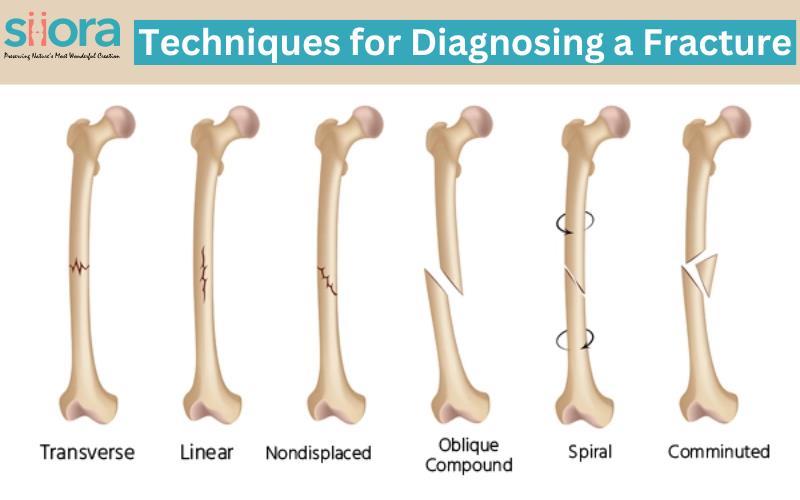
Techniques for Diagnosing a Fracture
Fractures, commonly known as broken bones, are injuries that can occur to anyone at any age. Whether it's a minor hairline fracture or a complex compound fracture, accurate and timely diagnosis is crucial for effective treatment and optimal recovery. Sometimes, a mild fracture becomes severe in the case of delayed diagnosis, and one might require stabilization using bone plates and Bone screws. In this blog, we will delve into the various techniques used by medical professionals to diagnose fractures.
How to Diagnose a Fracture?
Clinical Evaluation
The first step in diagnosing a fracture is often a thorough clinical evaluation by a healthcare provider. They will begin by taking a detailed medical history, inquiring about the circumstances of the injury, and assessing the patient's symptoms. Common signs of a fracture include localized pain, swelling, deformity, and loss of function in the injured area.

X-rays
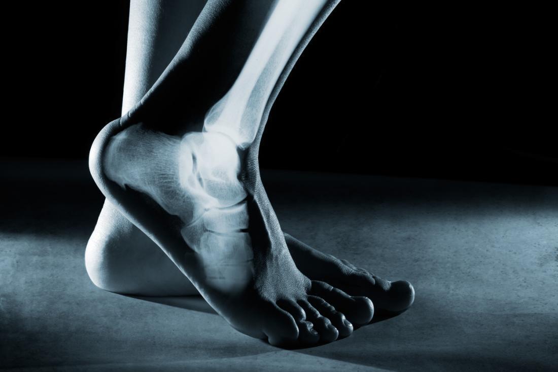
X-ray imaging is one of the most widely used and effective diagnostic tools for identifying fractures. X-rays use electromagnetic radiation to create detailed images of the bones. Fractures appear as breaks or discontinuities in the normal bone structure on the X-ray images. They can help determine the type and extent of the fracture, aiding in treatment decisions.
Computed Tomography (CT) Scans
In cases where X-rays may not provide enough information, a CT scan may be recommended. CT scans use a series of X-ray images taken from different angles to create detailed cross-sectional images of the bones. This can be especially useful for detecting complex fractures, assessing joint involvement, and planning surgical interventions.
Magnetic Resonance Imaging (MRI)
MRI is another imaging technique used to diagnose fractures, particularly those involving soft tissues like ligaments and tendons. While it may not be the first choice for routine fracture diagnosis, MRI can offer valuable insights into the extent of damage, making it useful in cases of stress fractures or suspected injuries to surrounding structures.
Bone Scans
Bone scans involve injecting a radioactive tracer into the bloodstream, which is then absorbed by bone tissue. The scanner can detect areas with increased bone activity, which may indicate a fracture. Bone scans are often used for stress fractures or to identify fractures in areas with poor blood supply, such as the scaphoid bone in the wrist.
Ultrasound
Ultrasound is commonly used for diagnosing fractures in pediatric patients, as it does not involve ionizing radiation. It can visualize fractures by detecting changes in the alignment and movement of bone fragments. However, its utility is limited in cases where fractures are not easily accessible with ultrasound, such as deep within the body.
Bone Biopsy
In rare cases, when other diagnostic methods are inconclusive, a bone biopsy may be performed. This involves removing a small sample of bone tissue for laboratory analysis to confirm the presence of a fracture or assess for underlying conditions like infections or tumors.

✔Orthopedic Trauma Implants
✔Orthopaedic Manufacturers
✔Trauma Implants Manufacturer
✔Surgical Implants Manufacturers ✔Orthopaedic Trauma Implants ✔Orthopedic Implants Suppliers