We are very excited for you to read the second volume of Endeavors, Mississippi State University’s (MSU) peer-reviewed undergraduate research journal. MSU is an R1 institution where students and faculty conduct rigorous and pioneering research Endeavors provides a platform for undergraduates to showcase their work, reinforcing the essential role that these students play in the research process.
We are honored to have been entrusted with the responsibility of Editors-in-Chief of Endeavors by the journal’s founders. Our aim this year was to advance the journal while taking steps to ensure its rigor and accessibility to the public. We collaborated with a wonderful team of students, faculty, and staff to redesign the Endeavors website, register the journal with an International Standard Serial Number (ISSN) and Digital Object Identifiers (DOIs), and make refinements to the peer review process for manuscripts in each academic field.
The second year of Endeavors was made possible with tremendous support from faculty and staff both within and outside of the MSU community. We wish to show our gratitude to:
● The Office of Undergraduate Research and Creative Discovery
● Dr Anastasia Elder, Associate Dean of Shackouls Honors College and Director of Undergraduate Research and Creative Discovery
● University Libraries, especially Julie D. Shedd, Assistant Professor of Practice and Director of Open Scholarship Initiatives
● Mae Switzer, Digital Commons
● The Shackouls Honors College
● The members of our Editorial Board
In this second volume, we proudly present four outstanding articles that represent diverse fields of scholarship at MSU. These works offer insights into topics from engineering, social sciences, and life sciences. Each article underwent a double-blind peer review process conducted by a team of Assistant (undergraduate) and Associate (graduate) reviewers as well as faculty approval This process ensures that all published work meets the journal’s standards for scope and quality.
In addition to publishing undergraduate research, we continued to provide opportunities for reviewers to learn how to conduct peer review and held training sessions for each subfield, organized by our Managing Editors. We want to thank the faculty members who participated in and led these sessions, including: Dr. Amy Dapper, Associate Professor of Biological Sciences; Dr Leslee Strawderman, International Paper Chair, Professor, and Associate Director of the Athlete Engineering Institute; and Dr. Cliff McKinney, Professor of Clinical Psychology.
Finally, we want to thank our readers for showing support to the journal Readership is a vital aspect of the research process, and we appreciate you spending your time engaging with the outstanding scholarship presented in this volume.
Sincerely,
Lydia Bailey (Master of Arts in Applied Anthropology, 2026)
Co-Editor-in-Chief
Lydia will be continuing her education at the University of Colorado Boulder to earn a PhD in Anthropology.
Alexis Cutshall (Master of Arts in Applied Anthropology, 2026; Master of Science in Health Promotion, 2026)
Co-Editor-in-Chief
Alexis is currently finishing her thesis with the goal of completing two master’s degrees in the summer of 2026.
About the Cover
This year’s cover artist is Morgan Elmore, a Master of Arts student in the Department of Anthropology and Middle Eastern Cultures. Morgan writes:
“This year's cover was influenced by the breadth of contributions made to the journal: four pieces of scholarship from four different colleges at MSU. The scrapbook style was intended to reflect the diversity of research happening among undergraduate students around campus. Endeavors represents a unique opportunity to see the product of such disparate disciplines celebrated in one place, and this cover was one attempt to encapsulate that.”
2025-2026 Editorial Board
Editors-in-Chief
Lydia Bailey
Alexis Cutshall
Managing Editors by Discipline
Engineering: Evan Garrison
Life Sciences: Sadie Bryan
Social Sciences: Lexi Holdiness
Arts/Humanities: Claire Anne Boudreaux
Reviewers by Discipline
Associate Reviewers: Current MSU graduate students or MSU alumni currently enrolled in graduate or professional programs.
Assistant Reviewers: Current MSU undergraduate students.
Engineering
Associate
Horace Adams
Shanna Ajayi
Laura Amato-Yarbrough
Melissa Diaz Delgado
Nicholas Esposito
Joshua Freeman
Bruce Hicks
Marionei Fomaca de Sousa Junior
Thinh Nguyen
Vaghawan Ojha
Jesus Ortiz
Marcos Pinares
Asifur Rahman
Ellen Reaux
Layla Stevens
David Williams
Yue Zhao
Assistant
Jordan Amacher
Emmanuel Braimah
Dylan Branca
Annemarie Coatney
Emma Cook
Lauren Edwards
Evan Garrison
Paul Gramelspacher
Katie Hardwick
Andrei Martin
Shriya Rajan
Aayam Raj Shakya
Camila Da Silva
Vitor Da Silva
Ella Storey
Thomas Thurner
Flora Yang
Life Sciences
Associate
Laura Amato-Yarbrough
Rachel Coppock
Alexis Cutshall
Morgan Elmore
Joshua Freeman
Yasas Gamagedera
Joshi Himani
Lexi Holdiness
Chloe Hudson
Madisen James
Marionei Fomaca de Sousa Junior
Madeline McKnight
Vaghawan Ojha
Jesus Ortiz
Yue Zhao
Assistant
Jordan Amacher
Dylan Branca
Sadie Bryan
Annemarie Coatney
Emma Cook
Je’Kayla Crawford
Jeremy Dawe
Lauren Edwards
Paul Gramelspacher
Katie Hardwick
Abigail Pugh
Shriya Rajan
Vitor Da Silva
Ella Storey
Riley Thompson
Physical Sciences
Associate
Laura Amato-Yarbrough
Melissa Diaz Delgado
David Heson
Chloe Hudson
James Jolley
Marionei Fomaca de Sousa Junior
Vaghawan Ojha
David Williams
Assistant
Jordan Amacher
Emmanuel Braimah
Dylan Branca
Sadie Bryan
Jeremy Dawe
Gabriel DuPree
Paul Gramelspacher
Katie Hardwick
Andrei Martin
Shriya Rajan
Camila Da Silva
Ella Storey
Thomas Thurner
Flora Yang
Social Sciences
Associate
David Ademule
Laura Amato-Yarbrough
Lydia Bailey
Madeline Brock
Madeline Burdine
Mary Catington
Rachel Coppock
Alexis Cutshall
Morgan Elmore
Gbenga I Elufisan
Nicholas Esposito
Elizabeth Fox
Joshua Freeman
Tanner Grubbs
David Heson
Lexi Holdiness
Kierstynn Hunter
Madisen James
James Jolley
Haomiao Li
Madeline McKnight
Zackary Murphy
Layla Stevens
David Williams
Yue Zhao
Assistant
Jordan Amacher
Claire Anne Boudreaux
Dylan Branca
Sadie Bryan
Je’Kayla Crawford
Rose Doyle
Paul Gramelspacher
Katie Hardwick
Christopher Jolivette
Shriya Rajan
Aayam Raj Shakya
Vitor Da Silva
Ella Storey
Thomas Thurner
Flora Yang
Arts/Humanities
Associate
Laura Amato-Yarbrough
Lydia Bailey
Rachel Coppock
Alexis Cutshall
Morgan Elmore
Elizabeth Fox
Kierstynn Hunter
Madisen James
James Jolley
Audrie Sifford
Layla Stevens
Assistant
Jordan Amacher
Claire Anne Boudreaux
Dylan Branca
Rose Doyle
Rowan Feasel
Katie Hardwick
Christopher Jolivette
Thomas Thurner
Flora Yang
Health Sciences
Associate
Oluwabori Adekanye
Laura Amato-Yarbrough
Lydia Bailey
Rachel Coppock
Morgan Elmore
Madisen James
James Jolley
Madeline McKnight
Zackary Murphy
Layla Stevens
Assistant
Jordan Amacher
Claire Anne Boudreaux
Annemarie Coatney
Emma Cook
Lauren Edwards
Paul Gramelspacher
Katie Hardwick
Vitor Da Silva
Riley Thompson
Designer
Morgan Elmore
Webmaster
Anoop Mishra
Outreach
Claire Anne Boudreaux
Endeavors: Mississippi State Undergraduate Endeavors: Mississippi Undergraduate Research Journal Research Journal Volume 2 Issue 1
How Mindfulness Training Influences Attention: A Systematic How Mindfulness Training Attention: Systematic Review Review
Ingrid E. O'Brien
MississippiStateUniversity, ieo14@msstate.edu
Kent W. Barrett
MississippiStateUniversity, kwb158@msstate.edu
Berna Altunisik Ms MississippiStateUniversity, ba925@msstate.edu
Follow this and additional works at: https://scholarsjunction.msstate.edu/endeavors
Part of the Cognitive Psychology Commons
Recommended Citation
Recommended O'Brien, Ingrid E.; Barrett, Kent W.; and Altunisik, Berna Ms (2026) "How Mindfulness Training Influences
Available at: https://scholarsjunction.msstate.edu/endeavors/vol2/iss1/2
This Social Sciences is brought to you for free and open access by the Hosted Journals at Scholars Junction. It has been accepted for inclusion in Endeavors: Mississippi State Undergraduate Research Journal by an authorized editor of Scholars Junction. For more information, please contact sct@library.msstate.edu
Social Sciences
How Mindfulness Training InfluencesAttention:ASystematic Review
Ingrid E. O’Brien1 , Kent W. Barrett
1, BernaAltunisik1
1. Mississippi State University, Starkville, MS
Abstract
The aim of this literature review is to understand the effectiveness of mindfulness meditation on training attention control skills. Relevant resources were identified through academic databases by entering keywords such as “attention” and “mindfulness training”. Preference was given to recent publications to ensure the inclusion of up-to-date research findings. Three main factors that influence training efficacy emerged from the literature: Reducing mind wandering, increasing acceptance, and lifting mood and affect. Review of empirical findings suggested that mindfulness does contribute to enhancing attentional abilities, although findings are somewhat inconsistent. Mind wandering and acceptance were important moderators in this effect, while no strong evidence was found in regard to mood and affect. Current gaps, limitations of the findings, and recommendations have also been discussed. Ultimately, this review contributes to the understanding of the current state of research on the link between attention and mindfulness.
Introduction
Attention is one of the most valuable resources we have. Throughout daily life, whether we are learning something new or evenholdingaconversationwithafriend,we are constantly using attention. Considering
that attention is impactful in many areas of life, the ability to improve this skill can be very beneficial. Therefore, a large body of research is dedicated to understanding whetherattentioncanbeimprovedortrained. Mindfulness is defined as bringing about enhanced awareness by paying attention to
the present moment and allowing each moment to pass without judgement (KabatZinn, 2003). An emerging field of research has surrounded the topic of mindfulness training and its impact on attentional ability. Given that mindfulness training involves garnering one’s attention on the present moment, this review seeks to answer the question of whether mindfulness training improves the ability of attentional control. This literature review will particularly analyze the research investigating the effects of mindfulness meditation on attention control skills, as well as the mechanisms through which mindfulness achieves these effects. Given that mindfulness is a relatively new topic of research, and with the possible benefits of its effects on attention, we seek to answer the question, “Can practicing mindfulness improve attention?”
What is Mindfulness?
Mindfulness is the non-judgmental state of awareness of the present moment (KabatZinn, 2003). The term "non-judgmental" refers to the act of not passing judgment while observing passing thoughts or experiences. This state can be reached through the practice of mindfulness meditation, in which practitioners bring their attention to the present moment by following theirbreathorotherpresentbodilysensations like sounds in the room or their sense of touch Intheliterature,mindfulnessisstudied as either state or trait mindfulness. State mindfulness refers to the transient, present state of mindfulness in an individual. Trait mindfulness, on the other hand, refers to the general dispositionofmindfulnessregardless of situation (Warren et al., 2023). State mindfulness can be measured through emerging techniques, such as breath counting, which constantly monitors the present state of mindfulness (Clapper et al., 2024).Traitmindfulnessismeasuredthrough surveys and questionnaires in order to
understand the disposition of mindfulness over time (Bravo et al., 2017).
The Broad Benefits of Mindfulness
Mindfulness is linked to a wide range of mental and physical benefits. Due to its ability to reduce stress, mindfulness has been found to be effective in improving health across many physical disorders such as fibromyalgia, coronary artery disease, and chronic pain (Grossman et al., 2003). By bringing awareness to the present moment in a non-judgmental way, mindfulness reduces rumination and obsessive thinking (ChemsMaarif et al., 2025). It has also been found to cause significant decreases in symptoms of anxiety and depression (Vollestad et al., 2011). Mindfulness seems to serve some cognitive benefits as well, such as reducing mind wandering (Rahl et al., 2016) and positively affecting attention (Sumantry et al., 2021).
Methods
The research papers used in this literature review were found through searching the keywords “mindfulness”, “attention”, and “mindfulness training” on the PsycARTICLES database. The search included peer-reviewed, fully published journal articles between January 2005 and September2025andyielded264articles.The listed articles included irrelevant outcomes (e.g. well-being, memory, meditation practices unrelated to mindfulness) and reliabilitystudies.Therefore, tonarrowdown our search to the most relevant articles, we have selected the 20 most relevant journal articles by reviewing the abstracts and prioritizing the recency of the articles. Additional articles were found during the writing of the paper to provide expert definitions for concepts (mind wandering, acceptance, mood) that emerged from analysis of the articles.
Attention Control
Attentioncontrolreferstoacognitiveprocess that filters out unnecessary or irrelevant information and allows one to focus on relevant information for extended periods of time (Chambers et al., 2007).Attention plays an important role in many aspects of life. It enables learning, problem solving, and decision making, which impact academic performance as well as solving trivial (and non-trivial) problems in daily life (Lindsay, 2020). Attentionisalsoassociatedwithsome mental health outcomes, such as emotionregulation skills (O’Bryan et al., 2017).
Mindfulness andAttention
Mindfulness practices have continually been examined on whether they may positively impact cognition (Gill et al., 2020). This literature review explores how mindfulness techniques specifically affect attention, and how future research can improve the investigation of this topic. For example, researchers in one study compared how a mindfulness intervention affects conflict monitoring skills and attentional selfregulating skills (Felver et al., 2014). It was found that mindfulness training had a significant effect on improving conflict monitoring skills, although no effect was found on attentional self-regulating skills.
Ainsworth et al. (2013) conducted a study examining the effects of two different types of mindfulness training on attention with healthy and novice meditation practitioners. Participants practiced either focused-attention meditation or openmonitoring focused meditation, and they were compared to a control group who did not practice meditation. It was hypothesized that the focused-attention group would show greaterimprovementsthanthoseoftheopenmonitoring group. Analysis of preintervention and post-intervention attention scores showed that participants in the intervention groups exhibited greater
improvements in attentional performance compared to the control group. While this studybroadlydemonstratedhowmindfulness practices may improve attention, it failed to find the expected advantage in the focusedattention intervention group. Thus, it is possible that mindfulness training benefits attention regardless of whether an individual purposefullyfocusestheirattentionorsimply practices self-awareness. This implies that the mechanism through which mindfulness improves attention can be general to various mindfulness-based interventions.
The distribution of mindfulness training over time has been considered as another factor that may impact mindfulness’s effectiveness. Krumholz et al. (2022) compared a condensed format to a spaced formatofmindfulnesstrainingtosee.Similar to Ainsworth et al. (2013), this study sought to determine whether these varying mindfulness training approaches differed in the enhancement of attention and mindfulnessscores.Inthisstudy,participants wereassignedto either fourconsecutivedays of mindfulness training (condensed format) or four days of mindfulness training spaced over four weeks (spaced format). Results showed increased attention skills in both formats. However, participants’ trait mindfulness was improved only in the spaced-interval group. Although this study gives implications for how training can impact trait mindfulness and attention, there are some important limitations to note. The study did not utilize random assignment to experimentalgroups,whichmakesitdifficult to interpret whether the results stem from the true effect of training or baseline differences betweengroups.Moreover,thestudyalsodid not implement a control group, which further weakens the results, as it is unclear whether similar gains would have occurred in a control group from pre- to post-test. Therefore, it cannot be inferred whether the observed improvements in participant
performance are truly due to the mindfulness intervention.
Studies have also been performed to further understand the relationship between mindfulness and somatosensory attention. Bokk and Forster (2022) examined somatosensory attention to assess whether brief mindfulness training mitigates the loss of attentional resources directed towards a task due to repetition of the task. This was examined by utilizing the P300 Event Related Potential (ERP), a neurophysiological marker of attention. The P300 is most pronounced when subjects attend to target stimuli, as shown on a given task. After an intervention of either mindfulness meditation or a control intervention, the test was repeated. Results showed a significant decrease in the P300 frompre-topost-testinthecontrolgroup.On the other hand, no significant difference in P300 amplitude was found from the first to the second task for the meditation group. Results are interpreted such that brief mindfulness training may prevent the depletion of attentional resources.
Research has also examined whether the relationship between mindfulness and attentionisapplicabletochildren.Wilsonand Dixon (2010) sought to investigate this in their study, where they found that the implementation of mindfulness exercises for a sample of first and second grade students improved participants’attention. To measure attention in the classroom, researchers observed and noted indices of inattentive behavior through subjective observation. Results showed that students had an 18% increase in attention compared to baseline. While it is important to note the increase in attention that was observed, there are various reasons why this study is problematic. Participants consisted of students at a private school, with a sample size of only 12. This is a very small sample that prevents the results from being generalized to the population.
Also, unlike other studies that utilize more objective and empirically valid methods of measuring attention (Semple, 2010; Clapper et al., 2024; Mrazek et al., 2012), this study’s use of subjective means of observation may notbetheoptimalassessmentofparticipants’ attention. Future studies should utilize more objective measures of attention. Overall, numerous studies have exemplified that mindfulness training can enhance attentional control. It should be considered, though, that many of these studies suffer limitations, and some have found inconsistent effects These inconsistencies could be corrected by further understanding the causal pathways through which mindfulness impacts attention. It is possible that mindfulness affects attention through multiple mechanisms. This must be assessed to broaden the depth of our understanding of the benefits of mindfulness on attention.
Why Does Mindfulness Improve Attention?
Studiesnotonlyexamine thedirectimpactof mindfulness on attention but also consider the mechanisms through which mindfulness may be able to affect attention. Mind wandering, acceptance, and mood will be discussed as three main mechanisms.
Reduced Mind Wandering
Mind wandering is operationalized as the instancesinwhich ourmindsdriftawayfrom a task to internal thoughts, feelings, and fantasies (Smallwood and Schooler, 2006). This experience stands in contrast to mindfulness, in which the mind is brought to the present. Mind wandering has been investigated as part of the research on the effects of mindfulness on attention (Turkelson and Mano, 2021). Considering that mind wandering is disruptive to attention, it may be that mindfulness improves attention by reducing mind
wandering. Mrazek et al. (2012) attempted to clarify the relationship between mindfulness and mind wandering using two studies. The first studymeasuredthe relationshipbetween trait mindfulness and mind wandering. Participants reported mind wandering by being interrupted and asked about their mind wandering (probe-caught method), or by spontaneously reporting when they notice thattheirmindhasshiftedfromthetask(selfcaught method). Trait mindfulness predicted less mind wandering when the experiment asked them about their mind wandering. However, it was associated with an increased self-caught mind wandering. This paradoxical difference between self-reported and self-caught mind wandering related to trait mindfulness may be explained in multiple ways. Participants with high trait mindfulness may be less likely to spend time mind wandering and therefore show less mind wandering when the experiment interrupts them. However, these participants may also be more likely to notice when their minds wander, so that the mind is immediately brought back to the present moment.Ontheotherhand,participantswith low trait mindfulness may experience more mind wandering but could be less aware of it and less likely to catch it.
After establishing the relationship between mind wandering and mindfulness, a second study by Mrazek et al. (2012) aimed to determine whether mindfulness can attenuate mind wandering. After completing a Sustained Attention to Response Task to measure mind wandering, participants were randomly assigned to one of three groups: eight minutes of reading, passive relaxation, ormindfulbreathing.Theywerethenretested on the SustainedAttention to ResponseTask. The mindful breathing group was found to have significantly less mind wandering. The paper theorizes that mindfulness leads to an increased awareness of mind wandering, allowingittobestopped.Thiseffectexplains
why individuals with high trait mindfulness hadhigherself-caughtmindwanderinginthe first study.
Zanesco et al. (2024) also investigated the association between mind wandering, mindfulness, and attentional vigilance (the ability to sustain attention on a task for a long period of time). In the two studies performed, a negative relationship was found between attentional vigilance and mind wandering. There was also a negative relationship found between mind wandering and trait mindfulness. However, the direction of the relationship between attention and mindfulness is unclear: does mindfulness make one’s mind wander less, or do people with strong attention skills tend to be more mindful? Nevertheless, it seems that mind wandering is one of the mechanisms that moderates the relationship between mindfulness and attention.
Acceptance
Acceptanceistheactofexperiencinginternal thoughts and feelings without judgement or without trying to change them (Wolfe and Isaacowitz,2022).Thisconcepthasalsobeen examined concerning its relationship with mindfulness. Mindfulness and acceptancebased interventions have been used to treat a wide range of disorders, including anxiety (Vollestad et al., 2011), psychosis (Cramer et al., 2016), and chronic pain (Veehof et al., 2016). Considering that mindfulness interventions that include acceptance have demonstrated wide-ranging effects, it is likely that acceptance influences the relationship between mindfulness and attention.
Rahl et al. (2016) proposed that acceptance plays a critical role in reducing mind wandering. This study compared participants randomly assigned to four groups: attention monitoring only mindfulness intervention, attentionmonitoring-and-acceptance-combined mindfulness intervention, relaxationtraining,
or active reading control group. The attention-monitoring-only group showed significant reductions in mind wandering. However, when attention-monitoring-only mindfulness was combined with acceptance in the attention-monitoring and-acceptancecombined mindfulness group, even greater reductionsinmindwanderingwereobserved. Therefore,thestudydemonstratedthecritical role of acceptance in mindfulness training and improvements in attention skills.
Mindfulness is tied to acceptance, as many mindfulness-based interventions include the prompt to be non-judgmental and accepting of present thoughts, emotions, and sensations. Chin et al. (2021) found that a monitoring and accepting mindfulness group showed the largest improvement in daily life non-judgement perceptions compared to the monitor-only control groups. Considering this, including acceptance prompts in a mindfulness-based intervention could serve toimproveacceptance,which will strengthen the effectiveness of said mindfulness-based intervention for enhancing attention.
Mood and Affect
Many studies have also investigated the influence of mindfulness on overall mood or affect as another mechanism through which attention is improved by mindfulness. For example, decreasing anxiety may allow for improvement in attention due to its negative effect on goal-directed attentional functioning (Eysenck et al., 2007).
Chambersetal.(2007)examinedhow a 10-day mindfulness training course would impact participants’ sustained attention and attention switching abilities, as well as their overall emotional state. Their results showed that trait mindfulness was negatively correlated with depressive and anxiety symptoms. Also, the mindfulness intervention significantly improved sustaining attention, along with depressive symptoms. These findings imply that mindfulness training optimizes mood, which
might facilitate attention. While the results found a beneficial effect on sustained attention, there was no significant impact on attention switching.
Another study examining mindfulness's impact on anxiety and attention found significant improvement in anxietysymptoms(Semple,2010).Thestudy included a mindfulness group, a progressive musclerelaxationgroup, andacontrolgroup. The Continuous Performance Test, Digit Symbol Substitution, and Stroop Task were administered to measure attention. Results showed that the mindfulness group significantly improved only on the Continuous Performance Test, a measure of sustained attention. This improvement in sustained attention is consistent with the findingsofChambers etal.(2007).Scoreson anxiety symptoms after the interventions revealed a significant decline for both intervention groups compared to the control group. Given that the mindfulness group did notdifferfromthemusclerelaxationgroupin reducing anxiety, there might be some mechanism other than mood impacting attention.
It has been proposed that attention may be inhibited by negative mood because anxiety consumes cognitive resources and decreases accuracy on effortful tasks (Eysenck et al., 2007).Astudy by Jankowski and Holas (2020) tested the effects of brief mindfulness meditation on attention, particularly in those whose mood is diminished by negative emotion and stress. Three groups were implemented: a mindfulness training condition, a mindfulness training and induced worry state condition, and a free mind wandering (control) condition. Each group completed a pre- and post-intervention attention task and watched a movie scene that induced negative affect. Those in the worry state condition were instructed to interpret this scene as if it were real-life in order to induce a state of
worry. Results showed that the mindfulnessonly group was faster at responding to the attention task compared to other groups, but not more accurate. Therefore, mindfulness’s impact on the efficiency of executive function was not sufficiently supported.
Astudy conducted by Zylowska et al. (2007) tested the effects of mindfulness on mood and attention in a sample of participants diagnosed with ADHD. Their study sought to determine whether mindfulness helped to decrease anxiety, depression, and ADHD symptoms. A group of adolescents and a group of adults were given surveys to assess the relevant symptoms and multiple attention tests. After an 8-week mindfulness training intervention, both groups showed a significant improvementintheattentiontests.Moreover, significant improvements were found in depressionandanxietyintheadultgroup,but notintheadolescentgroup.Alargelimitation of this study was the utilization of a small sample size. The authors note that the size of the sample within the adolescent group precluded statistical testing in regard to measuring change in symptoms. These studies, among many others, that have begun to investigate how mindfulness enhances attention through its impact on mood, are importantstartingpointsfortheimprovement of research to come.
Conclusion
In summary, mindfulness, which involves bringing awareness to the present moment, has been shown to benefit some attentional abilities Mainly, sustained attention and somatosensory attention can benefit from these interventions. Sustained attention refers to maintaining focus over a period of time. The practice of mindfulness can improve our ability to listen, process relevant information, and stay on task over longer amounts of time. Therefore, benefits to this type of attention may have a more
robust impact on educational outcomes. The other type of attention that seems to benefit from mindfulness training was somatosensory attention, or reaction times. These abilities reflect more on our perceptual skills. Mindfulness training seems to be effective in enhancing our motor reactions to target information. Benefits to this type of attention can enhance capabilities related to athletic activities, handling tools and equipment, playing musical instruments, and other tasks requiring good motor skills. Continuing to understand the aspects of attention that mindfulness reliably assists could promote the advancement of many important aspects of life.
While the benefits of mindfulness training on attention are backed by several empirical studies, unfortunately, many studies suffer limitations, including small sample sizes, unclear causality, and lack of control measures. The use of small sample sizes impairs the generalizability of significant findings. The lack of necessary control measures, such as the random assignment of participants to conditions and the inclusion of a control group, hinders experimenters’ abilities to confidently infer that a mindfulness intervention caused any observed improvement in attention. Along with these limitations, the absence of valid and reliable methods to measure attention impedes the clarity of researchers’ findings. Using empirically validated measures of attention, such as the Sustained Attention to Response Task (SART), Continuous Performance Test (CPT), and the Stroop Task, will allow for more reliable and valid inferences of results. Therefore, future studies must address these issues to produce more conclusive and trustworthy findings. The research on this topic also seeks more refined explanations as to why mindfulness may improve attention. Prominent factors researched as potential
explanations include reducing mind wandering, increasing acceptance, and improving mood or affect. While reduced mind wandering and acceptance seem to play a significant role, no strong evidence was found regarding the role of mood and affect on the relationship between mindfulness and attention. By addressing possible mechanisms, these studies provided more fine-grained explanations for which contexts of mindfulness training can be effective. With this consideration, future studies should be aware that mindfulness has a broad range of effects, and there is still much to learn about its potential benefits. The durability of these benefits should also be considered. Research must seek to understand whether mindfulness has a lasting effect on attention, or whether its effects are fleeting.
With diligent use of reliable and valid methods, in combination with better operational definitions and sample sizes, researchers can progress our understanding of how mindfulness meditation works to enhance attention control.
References
Ainsworth, B., Eddershaw, R., Meron, D., Baldwin, D. S., & Garner, M. (2013). The effect of focused attention and open monitoring meditation on attention network function in healthy volunteers. Psychiatry Research, 210(3), 1226–1231.
https://doi.org/10.1016/j.psychres.201 3.09.002
Bokk, O., & Forster, B. (2022). The effect of a short mindfulness meditation on somatosensory attention. Mindfulness, 13(8), 2022–2030.
https://doi.org/10.1007/s12671-02201938-z
Bravo,A. J., Pearson, M. R., Wilson,A. D., & Witkiewitz, K. (2017). When traits match states: Examining the
associations between self-report trait and state mindfulness following a state mindfulness induction. Mindfulness, 9(1), 199–211.
https://doi.org/10.1007/s12671-0170763-5
Chambers, R., Lo, B. C., &Allen, N. B. (2007). The impact of intensive mindfulness training on attentional control, cognitive style, and affect. Cognitive Therapy and Research, 32(3), 303–322. https://doi.org/10.1007/s10608-0079119-0
Chems-Maarif, R., Cavanagh, K., Baer, R., Gu, J., & Strauss, C. (2025). Defining mindfulness:Areview of existing definitions and suggested refinements. Mindfulness, 16(1), 1–20.
https://doi.org/10.1007/s12671-02402507-2
Chin, B., Lindsay, E. K., Greco, C. M., Brown, K. W., Smyth, J. M., Wright, A. G., & Creswell, J. D. (2021).
Mindfulness interventions improve momentary and trait measures of attentional control: Evidence from a randomized controlled trial. Journal of Experimental Psychology: General, 150(4), 686–699.
https://doi.org/10.1037/xge0000969
Clapper, J., Ware, S., Martinez, F. J., Benitez, K., & Koshino, H. (2024). Breath counting as a measure of sustained attention in mindfulness meditation and its effect on mood. Psychology of Consciousness: Theory, Research, and Practice, 11(3), 348–374.
https://doi.org/10.1037/cns0000311
Cramer, H., Lauche, R., Haller, H., Langhorst, J., & Dobos, G. (2016). Mindfulness- and acceptance-based interventions for psychosis:A systematic review and meta-analysis. Global Advances in Health and
Medicine, 5(1), 30–43.
https://doi.org/10.7453/gahmj.2015.08 3
Eberth, J., & Sedlmeier, P. (2012). The effects of mindfulness meditation:A Meta-analysis. Mindfulness, 3(3), 174–189.
https://doi.org/10.1007/s12671-0120101-x
Felver, J. C., Tipsord, J. M., Morris, M. J., Racer, K. H., & Dishion, T. J. (2014). The effects of mindfulness-based intervention on Children’sAttention
Regulation. Journal of Attention Disorders, 21(10), 872–881.
https://doi.org/10.1177/108705471454 8032
Gill, L.-N., Renault, R., Campbell, E., Rainville, P., & Khoury, B. (2020).
Mindfulness Induction and Cognition: Asystematic review and metaanalysis. Consciousness and Cognition, 84, 102991.
https://doi.org/10.1016/j.concog.2020. 102991
Grossman, P., Niemann, L., Schmidt, S., & Walach, H. (2003). Mindfulness‐based stress reduction and health benefits:A meta‐analysis. Focus on Alternative and Complementary Therapies, 8(4), 500–500.
https://doi.org/10.1111/j.20427166.2003.tb04008.x
Jankowski, T., & Holas, P. (2020). Effects of brief mindfulness meditation on attention switching. Mindfulness, 11(5), 1150–1158.
https://doi.org/10.1007/s12671-02001314-9
Krumholz, M. F., Pinnell, C., & Sullivan, D. (2022). Effectiveness of brief mindfulness practice for enhancing graduate students’attention. Psychology & Neuroscience, 15(2), 177–185.
https://doi.org/10.1037/pne0000268
Kabat-Zinn, Jon. “Mindfulness-Based Interventions in Context: Past, Present, and Future.” Clinical Psychology: Science and Practice, vol. 10, no. 2, 2003, p. 145, onlinelibrary.wiley.com/doi/full/10.1 093/clipsy.bpg016, https://doi.org/10.1093/clipsy.bpg01 6.
Lindsay, G. W. (2020).Attention in psychology, neuroscience, and machine learning. Frontiers in Computational Neuroscience, 14. https://doi.org/10.3389/fncom.2020.00 029
Mrazek, M. D., Smallwood, J., & Schooler, J. W. (2012). Mindfulness and mindwandering: Finding convergence through opposing constructs. Emotion, 12(3), 442–448. https://doi.org/10.1037/a0026678
O’Bryan, E. M., Kraemer, K. M., Johnson, A. L., McLeish,A. C., & McLaughlin, L. E. (2017). Examining the role of attentional control in terms of specific emotion regulation difficulties. Personality and Individual Differences, 108, 158–163. https://doi.org/10.1016/j.paid.2016.12. 015
Rahl, H.A., Lindsay, E. K., Pacilio, L. E., Brown, K. W., & Creswell, J. D. (2017). Brief mindfulness meditation training reduces mind wandering: The critical role of acceptance. Emotion, 17(2), 224–230. https://doi.org/10.1037/emo0000250
Semple, R. J. (2010). Does mindfulness meditation enhance attention?A randomized controlled trial. Mindfulness, 1(2), 121–130. https://doi.org/10.1007/s12671-0100017-2
Smallwood, J., & Schooler, J. W. (2006). The Restless Mind. Psychological Bulletin, 132(6), 946–958.
https://doi.org/10.1037/00332909.132.6.946
Sumantry, D., & Stewart, K. E. (2021). Meditation, mindfulness, and attention:Ameta-analysis. Mindfulness, 12(6), 1332–1349. https://doi.org/10.1007/s12671-02101593-w
Turkelson, L., & Mano, Q. (2021). The Current State of Mind:Asystematic review of the relationship between mindfulness and mind-wandering. Journal of Cognitive Enhancement, 6(2), 272–294.
https://doi.org/10.1007/s41465-02100231-6
Veehof, M. M., Trompetter, H. R., Bohlmeijer, E. T., & Schreurs, K. M. (2016).Acceptance- and mindfulnessbased interventions for the treatment of chronic pain:Ameta-analytic review. Cognitive Behaviour Therapy, 45(1), 5–31.
https://doi.org/10.1080/16506073.201 5.1098724
Vøllestad, J., Nielsen, M. B., & Nielsen, G. H. (2011). Mindfulness‐ and acceptance‐based interventions for anxiety disorders:Asystematic review and meta‐analysis. British Journal of Clinical Psychology, 51(3), 239–260. https://doi.org/10.1111/j.20448260.2011.02024.x
Warren, M. T., Galla, B. M., & Grund,A. (2023). Using whole trait theory to unite trait and state mindfulness. Journal of Research in Personality, 104, 104372.
https://doi.org/10.1016/j.jrp.2023.1043 72
Wilson,A. N., & Dixon, M. R. (2010).A mindfulness approach to improving classroom attention. Journal of Behavioral Health and Medicine, 1(2), 137–142.
https://doi.org/10.1037/h0100547
Wolfe, H. E., & Isaacowitz, D. M. (2022). Acceptance as a cognitive emotion regulation strategy across the lifespan. Psychology of Learning and Motivation, 291–312. https://doi.org/10.1016/bs.plm.2022.07 .002
Zanesco,A. P., Denkova, E., Barry, J., & Jha,A. P. (2024). Mind wandering is associated with worsening attentional vigilance. Journal of Experimental Psychology: Human Perception and Performance, 50(11), 1049–1066. https://doi.org/10.1037/xhp0001233
Zylowska, L.,Ackerman, D. L.,Yang, M. H., Futrell, J. L., Horton, N. L., Hale, T. S., Pataki, C., & Smalley, S. L. (2007). Mindfulness meditation training in adults and adolescents with ADHD. Journal of Attention Disorders, 11(6), 737–746. https://doi.org/10.1177/108705470730 8502
Volume 2 Issue 1
June 2026
Endeavors: Mississippi State Undergraduate Endeavors: Mississippi Undergraduate Research Journal Research Journal
Near-Infrared Spectroscopy for Monitoring Bovine Respiratory Near-Infrared for Monitoring Bovine
Syncytial Virus Infection in Experimentally Challenged Calves
Available at: https://scholarsjunction.msstate.edu/endeavors/vol2/iss1/3
This Life Sciences is brought to you for free and open access by the Hosted Journals at Scholars Junction. It has been accepted for inclusion in Endeavors: Mississippi State Undergraduate Research Journal by an authorized editor of Scholars Junction. For more information, please contact sct@library.msstate.edu
Near-Infrared Spectroscopy for Monitoring Bovine Respiratory Syncytial Virus
Near-Infrared for Monitoring Syncytial Virus
Infection in Experimentally Challenged Calves
Infection in Experimentally Challenged Calves
Cover Page Footnote Cover Page Footnote
This project was supported by a United States Department of Agriculture – Agriculture and Food Research Initiative Grant (Award #2022-67016-36978), and the U.S. Department of Agriculture, Agricultural Research Service, Biophotonics project 6066-31000-015-00D. Additionally, this project was supported by the National Institute of Food and Agriculture, USDA (#MIS001), as well as Mississippi State University’s College of Agriculture and Life Sciences/Mississippi Agricultural and Forestry Experiment Station Undergraduate Research Scholars Program (URSP).
Authors Authors
Erin Blanchard, Jelena Muncan, Grace Beddingfield, Cassandra Barber, Florencia Meyer, Santiago Cornejo, Merrilee Thoresen, Amelia Woolums, and Carrie Vance
This life sciences is available in Endeavors: Mississippi State Undergraduate Research Journal: https://scholarsjunction.msstate.edu/endeavors/vol2/iss1/3
Life Sciences
Near-Infrared Spectroscopy for Monitoring Bovine Respiratory
Syncytial Virus Infection in Experimentally Challenged Calves
Erin Blanchard1 , Jelena Muncan1 , Grace Beddingfield1 ,
1. Department of Biochemistry, Nutrition, and Health Promotion, Mississippi State University
2. Department of Pathobiology and Population Medicine, Mississippi State University
3. Department ofAnimal and Dairy Science, Mississippi State University
Abstract
Bovine respiratory disease (BRD) is a multifactorial disease complex that causes significant economic loss in the cattle industry. Bovine respiratory syncytial virus (BRSV), one of the primary viruses included in BRD, primarily targets the lower respiratory tract and causes a wide variety of subclinical and clinical signs. Current diagnosis techniques require off-site laboratory testing, creating delays that allow BRSV to spread through herds and highlighting the need for rapid diagnostic methods. The objective of the study was to evaluate near-infrared spectroscopy as a diagnostic tool for BRSV detection across varying infection severities. Twenty-two Holstein bull calves were reared in research pens at Mississippi State University from 1 week old until 12 weeks of age when the study began.At 12 weeks old, the calves were randomly divided into 4 groups and challenged via aerosol with 103, 104, and 105 TCID50 units of BRSV, creating low,
medium, and high dose groups respectively. Negative control was nebulized with cell culture media. Biofluid samples, including serum, plasma, nasal secretions, saliva, and breath condensate, were collected daily for 14 days following infection. The presence and severity of infection was confirmed by quantitative real-time PCR (qPCR) threshold (Ct) values. Nearinfrared spectra of all sample types were acquired using anASD FieldSpec 4 NIR spectrometer (350–2500 nm) and analyzed within the 1300–1600 nm region (first overtone of water). Data was pre-processed using different techniques and classified using Soft Modeling of Class Analogies (SIMCA) to distinguish BRSV-positive (Ct < 35) from negative (Ct > 35) samples. Preliminary results showed variation in accuracy, sensitivity, and specificity across biofluids, with overall classification accuracy ranging from 42.8% to 76.4%. Nasal secretions produced the highest accuracy, while plasma demonstrated a more balanced sensitivity–specificity relationship among blood-derived samples. This approach demonstrates potential for a rapid alternative to conventional diagnostic methods for BRSV, suggesting that NIRS can detect biochemical changes in biofluids in cattle.
Introduction
Bovine respiratory syncytial virus (BRSV) is amajorviralpathogencontributingtoBovine Respiratory Disease (BRD), a multifactorial disease complex responsible for significant morbidity and mortality in cattle, leading to economic losses through increased treatment costs and reduced herd productivity (Snowder et al., 2006). Of the many viral agents that contribute to BRD, BRSVplays a central roleby infectingthelower respiratory tract placing cattle at high risk for secondary bacterial infections (Sarmiento-Silva et al., 2012). Clinical signs of BRSV include fever, nasal and ocular discharge, coughing, and labored breathing. However, symptoms present differently based on each individual animal and can appear much later following infection, causing a delay between infection and detection (Larsen, 2000).
EarlyandaccuratedetectionofBRSV remains a significant challenge in livestock production systems. Clinical signs alone are insufficient for diagnosis, as these symptoms canbecausedbymultipledifferentpathogens or environmental factors (Makoschey & Berge, 2021). Molecular diagnostics such as quantitative polymerase chain reaction (qPCR) are commonly used for viral detection. However, such methods typically
require off-site sample processing and extended analysis times before results are available, limiting timely intervention and allowing continued viral transmission within the herd.
Near-infrared spectroscopy (NIRS) has emerged as a promising tool for fast and accurate quantification of viruses across diverse biological samples and experimental systems (Premasinghe et al., 2025; De Oliveira et al., 2025; Gomez et al., 2024). It is a nondestructive optical approach that measures the absorbance of light in the NIR region 700–2500 nm, and the subfield of aquaphotomics examines how water absorbs lightthroughchangesinmagnitudeandshifts in absorbance bands within the 1300–1600 nm range, corresponding to the first overtone of OH stretching vibrations (Muncan & Tsenkova, 2019). Infection can alter the biochemical composition of biological fluids through immune systemactivation, and these changes can influence the surrounding hydrogen-bonding structure of water. Aquaphotomics is particularly applicable to biological fluids as water molecules tend to form unique and complex hydrogen-bonding networks around biomolecules. Infection and inflammation caused by viruses lead to detectable alterations in the biochemical compositionofbiofluids,whichinturnaffect
water structure fingerprinting of these fluid systems.
Previous studies have demonstrated that NIRS can detect biochemical changes associated with BRSV in livestock (SantosRivera et al., 2022). While these previous studiesprovidedproofof concept,theyrelied on small sample sizes leading to simplified experimental designs whereby infection is treated as a binary variable, despite the variability in magnitude of infection that can arise across animals. Furthermore, viral progression occurs over time with varying viral loads impacting the degree of clinical severity in each infected individual. Thus, evaluating the effectiveness of new diagnostic models such as NIRS under more realistic conditions can provide an assessment of both model performance and practicality.
Unlikepreviousstudiesthatprimarily evaluated binary infection classification undersimplifiedexperimentalconditions,the present study examined NIRS performance across multiple biofluids within a biologically realistic infection gradient and longitudinal sampling design. This approach was intended to better reflect the variability associated with naturally occurring respiratory disease progression.
In past experimental BRSVchallenge studies,viralloadwasquantifiedusingqPCR cycle threshold (Ct) values, which provides a measure of infection status (Santos-Rivera et al., 2022). Ct values can therefore be incorporated into spectroscopic models to evaluate whether spectral data could discriminate between infected and noninfected cattle. The development and assessment of a new diagnostic model under these conditions is an important step to determine the feasibility of NIRS as a detection tool for respiratory disease management.
The objective of this study was to develop and evaluate an NIRS-based detection method for BRSV using serum, plasma, saliva, and nasal secretions collected from experimentally challenged calves.After creating an infection gradient ranging from subclinical to clinical symptoms, NIRS was utilized to differentiate between healthy and infected calves with measurable accuracy, sensitivity, and specificity while using qPCR criteria to confirm the degree of infection. This study specifically examined potential infection-associated biochemical shifts in the spectral profile of water in the range 1300–1600 nm. It was hypothesized that NIRS would differentiate infected and healthy calves across multiple biofluids, with respiratory-origin samples expected to demonstrate stronger classification performance than other sample types.
Materials and Methods
Animals and Experimental Challenge
Twenty-two 1-week-old Holstein bull calves were raised at Mississippi State University College of Veterinary Medicine research pens. At 3 months old, the calves were admitted to a controlled BRSV challenge study. Calves were housed in covered individual pens under standard management conditions that were cleaned daily and bedded with sand. All calves had unlimited water access, powdered colostrum (150 g daily) and 20% fat and 20% protein milk replacer.Weaningcalvesstartedat6weeksof age, where they decreased to 4 pints twice daily and a week later to 4 pints once daily. As calves matured, they were fed medicated feed increasing as intake increased until 5 lb twice daily per calf. Bermuda grass hay started at 5 weeks of age and was given ad libitum. All procedures were conducted in accordancewithinstitutionalanimalcareand use guidelines and approved by the
Institutional Animal Care and Use Committee (IACUC # 22-223).
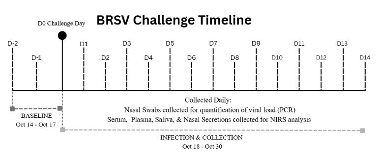
Experimental infection was performed mid-October, approximately three months after the calves’ arrival in July, as shown in Figure 1. Calves were randomly assignedtooneoffourtreatmentgroupsprior totheviralchallenge:control(n=6),lowdose (n=5), medium dose (n=5), and high dose (n=6).Groupsizesweredeterminedbasedon animal availability and practical limitations associated with the design of the BRSV challengestudy.OnDay0ofthestudy,calves were administered the virus via aerosol using a Pulmo-Aide nebulizer. To generate an infection gradient, the control group was nebulized with cell culture media, and the
low, medium and high groups with 103,104 , and 105 TCID50 units/mL of virus, respectively. This gradient in infection range was created to generate a biologically relevant range of disease severity, to produce a variety of subclinical indicators and observable clinical symptoms.
Clinical health assessments were acquired daily throughout the 17-day collection period prior to and following viral administration. Clinical respiratory scoring and thoracic ultrasonography were performed daily to assess disease severity, but these data points were not incorporated into the spectroscopic modeling or statistical analyses presented here.
1. Experimental timeline of BRSV challenge and daily sample collection from Day -2 through Day 14 post-challenge.
Sample Collection
Biofluid samples were collected daily from each calf, beginning two days prior to infection (Day -2) and continuing until 14 days after infection (Day 14). Sample types collected included serum, plasma, nasal secretion, saliva, breath condensate, and nasal swabs. Although breath condensate samples were collected, they were reserved for future analysis and were not included in the spectroscopic modeling or results presented in this manuscript.
Blood samples were collected daily, alternating from the left and right jugular veins using vacutainers and immediately placed in an ice chest while in the field to maintain sample integrity. Once transported to the laboratory, blood samples were separated by centrifugation at 1,800 x g for 20 minutes at room temperature for serum and 4°C for plasma.
Figure
Nasal secretions were collected using absorbent foam sponges inserted into the nostril and allowed to absorb secretions (~3 minutes). Nostrils were alternated between collection days to minimize irritation. The sponge was placed into a sterile 20 mL syringe, and collected fluid was expressed into a 1.5 mLcentrifuge tube.
To collect saliva, residual feed materialwasmanuallyremovedfromtheoral cavity prior to sample collection. Similar to nasal secretions, an absorbent foam sponge was placed in the oral cavity until saturated, transferred to a sterile syringe, and the fluid expressed into a 1.5 mLcentrifuge tube.
Nasal swabs were collected for molecular detection and quantification of BRSV by quantitative real-time polymerase chain reaction (qPCR). Sterile 12-inch swabs were inserted into the nostril opposite the nasal secretion sponge and rotated to collect epithelial secretions. Following collection, swabs were placed in 1.5 mL centrifuge tubesandstoredoniceduringfieldcollection until laboratory processing.
Following processing of the blood samples, all samples were stored at a -80 °C until required for NIRS analysis. Consistent handling, cooling, and storage procedures were maintained across all sampling days to reduce variation.
Infection Confirmation by qPCR
To confirm the presence of BRSV and quantifyviralload,RNAextractedfromnasal swab samples were analyzed by qPCR following complementary DNA (cDNA). Swabs were processed using Quick-RNA Viral Kit (Zymo Research).
All swabs were placed in tube containing 400 µL of DNA/RNA Shield and vortexedtofacilitatesamplerelease.Samples were centrifuged at 12,000 RPM for 2
minutes, releasing all liquid from the swab. The mixture containing DNA/RNA Shield and swab material was transferred to a clean, microcentrifugetubeandcentrifugedagainat 12,000 RPM for 2 minutes before RNA purification.
Viral RNA purification was performed using silica spin-column technology, where a lysis buffer was used to disruptviralparticlesfromwithinthesample. The lysate was applied to a silica membrane column under high salt conditions, allowing RNAto bind to the membrane and isolate the viral contents. Any contaminants were removed through washing steps, and purified RNA was eluted using DNase/RNase-Free Water to continue further analysis. Purified viral RNA was reverse transcribed into cDNA prior to quantitative real-time PCR analysis.
Quantitative real-time PCR was performed using BRSV-specific primers under standardized thermal cycling conditions. Cycle threshold (Ct) values were recorded for each calf, representing the number of amplification cycles required for fluorescence to exceed background levels. Ct values are inversely proportional to viral RNAconcentration,meaninglowerCtvalues correspond to higher viral loads within the sample.
Forthisstudy,sampleswithCtvalues below 35 were operationally designated as BRSV positive to indicate detectable viral RNA amplification, while samples with Ct values greater than or equal to 35 were considered BRSV negative for spectroscopic classification modeling. Lower Ct values correspond to higher viral RNA concentration, while higher Ct values reflect lower viral RNA abundance. Ct values approaching 35 are commonly associated with low-level viral RNA detection near the analytical detection limit of qPCR assays (Ruiz-Villalba et al., 2021). These Ct values
were used both as a molecular reference for infection and for future spectroscopic model development.
Near-Infrared Spectral Acquisition
Near-infrared spectra (NIRS) were collected on serum, plasma, saliva, and nasal secretion samples using a FieldSpec 4 spectrometer (Malvern Panalytical, ASD Analytical Spectral Devices Inc. Boulder, CO. USA). Spectra were collected across the 350–2500 nm range, and the 1300–1600 nm region was later selected foranalysis. Room temperature was recorded before each sample measurement to account for environmental conditions.
All samples were stored at −80 °C until analysis. Prior to spectral acquisition, samples were thawed in a room-temperature water bath for approximately 15 minutes to allow for equilibration to room temperature. To minimize degradation, samples were kept at room temperature for only 20 minutes and were returned to ice immediately after spectral acquisition.
Prior to sample scanning, a baseline spectrum was collected using an empty quartz cuvette, which served as the reference for subsequent measurements. For spectral measurements, 100 µL of sample was pipetted into a 1 mm quartz cuvette. For each sample, two independent pipetting replicates were prepared from the original sample tube. Each pipetting replicate was measured three times, with the cuvette removed from the instrument, reloaded, and reinserted between measurements. During each measurement, fiveconsecutivescanswereacquiredwithout removing the cuvette, and these scans were later averaged to produce a single spectrum. This produced six spectra per sample (two pipetting replicates × three measurements).
After each scan, the sample was removed from the cell and placed back into
the original centrifuge tube and placed back on ice. The cuvette was rinsed with distilled water for 10 seconds, followed by a minimum 15-minute 2% Cell Cleaning Solvent bath while in an ultrasonic cleaning to ensure all remnants from the sample were removed from the quartz cuvette.
Spectral Preprocessing and Diagnostic Modeling
Spectral analysis focused on the 1300–1600 nm wavelength region, which corresponds to the first overtone of O–H stretching vibrations and contains absorbance bands associated with water structure that are evaluated in aquaphotomics studies.
All spectra were preprocessed to reduce noise, correct baseline variation, and minimize scattering effects commonly observed in biological samples. The preprocessing techniques chosen were selected to improve detection of subtle infection-associated spectral differences commonly masked by biological variability and scattering effects in complex biofluids. Several preprocessing approaches were evaluated to improve spectral comparability and model performance. Standard Normal Variate (SNV) transformation (Barnes et al. 1989) was applied to correct for multiplicative scatter effects common in biofluids and baseline variability across samples. Spectral smoothing was performed using the Savitzky–Golay algorithm (Savitzky & Golay, 1964) with a 21-point window,whichreduceshigh-frequencynoise within the spectra while preserving the overall spectral shape. In addition, secondderivative transformation was applied to reveal and enhance subtle spectral features and improve the resolution of overlapping absorption bands (Rieppo et al., 2012). All derivative calculations were performed using a 21-point window.
In addition to different preprocessing combinations, two different approaches were tested regarding the selection of data used in analysis: using the spectra from all days of the trial (D-2 and D-1, D1 to D14) and using only a subset of days (D-2, D-1, and D5–D10).Thesecondapproach,usingasubsetof days, was chosen to maximize the difference between the spectra of healthy animals during the baseline period versus animals during the peak of infection.
For spectroscopic classification modeling, samples were assigned to either infected or healthy classes according to qPCR Ct values collected on the corresponding sampling day. Samples with Ct values below 35 were classified as infected regardless of original treatment group assignment. This allowed classification status to reflect viral shedding throughout the trial.
Following spectral preprocessing, multivariate classification modeling was performed using Soft Independent Modeling of Class Analogy (SIMCA) (Wold, 1976). SIMCAis a supervised classification method used for developing models that analyze separate predefined classes within spectra to independently construct an identification diagnosis model. Each group within this study (infected and healthy) had Principal Component Analysis (PCA) applied to reduce the number of dataset features while retaining any unique variation within the class. The PCA defines a model to represent the normal variability and average spectra within each group (Viviani et al., 2004).
Following the establishment of class models, individual spectra were evaluated based on their similarity to each class to determine the accuracy of classification.
Each unknown spectrum was assigned to the class model to which it was the closest fit, based on principal component analysis (Wold, 1976). Model performance was assessed using accuracy, sensitivity (true positive rate), and specificity (true negative rate) (Monaghan et al. 2021). Validation was performed using the built-in functionality of the aquap2 R package, which evaluates the model on a held-out subset of data (50% of spectra not used for training), providing unbiased estimates of predictive performance.All analyses were conducted in R software for statistical computing (R Core Team, 2025).
Results
qPCR Confirmation of Infection
Quantitative real-time PCR analysis confirmed both the infection of each individualcalfaswellas theestablishmentof an infection gradient. Ct values to identify positive or negative results for the presence of BRSV varied across experimental groups and over time, reflecting the differences in exposure as well as progression.
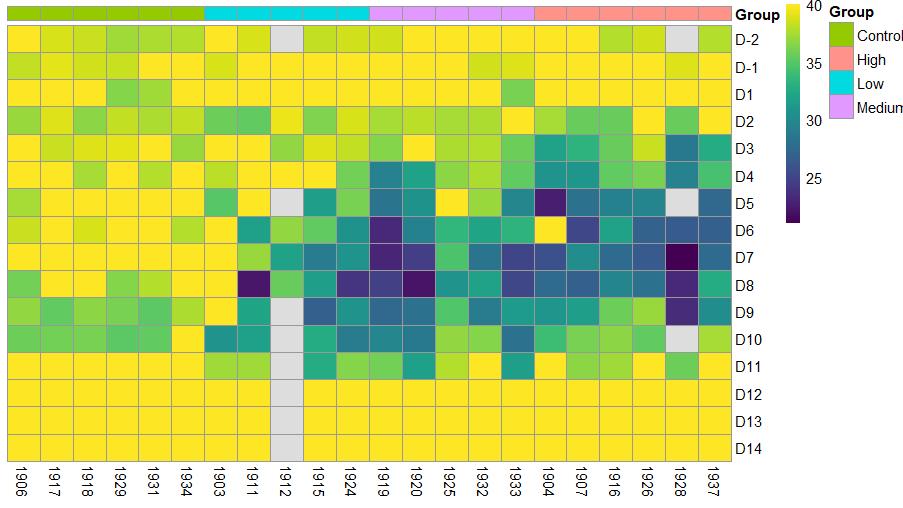
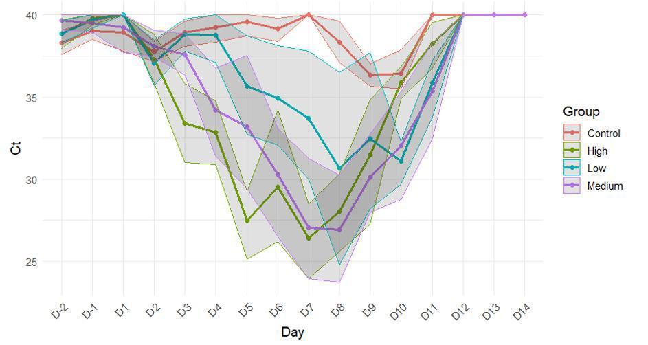
Figure 2 presents a heat map of Ct values across all calves and time points, highlighting lower Ct values primarily in the medium- and high-dose groups during peak infection. Individual calf identification numbers displayed in Figure 2 correspond to experimental animals monitored throughout the challenge study. Mean Ct values with standard deviations by experimental group are shown in Figure 3. Figures 2 and 3 demonstrate viral replication peaked within the infected groups during the mid-stage infection, while overlap between the treatment groups reflected individual variation in viral response.
Figure 2. Heat map of qPCR Ct values across all calves and time points. Lower Ct values indicate higher viral load.
Figure 3. Mean qPCR Ct values ± standard deviation per experimental group over the trial.
Control calves maintained high Ct values, while infected groups exhibited decreased Ct values during peak viral replication.
SIMCA Classification Performance
Classification models were developed using SIMCAbased on qPCR-confirmed infection. Model performance metrics are summarized in Table 1.Across the four sample types and
preprocessing techniques, the accuracy of classification ranged from 42.8% to 76.4%, depending on biofluid type and dataset configuration. Sensitivity ranged from 11%–95.3%, and specificity ranged from 6.5%–95.4%.
When analyzing data across all days (D-2 to D14), nasal secretion and plasma models demonstrated the most accurate classification performance relative to the othersample types. Subset analysis restricted D-2, D-1, and D5-D10 showed variable performance depending on the matrix type.
Table 1. Classification performance of SIMCAmodels developed using qPCR-confirmed BRSV infection status (Ct < 35), including accuracy, sensitivity, and specificity Matrix
Sensitivity and Specificity Analysis
Across all biofluids and preprocessing techniques, nasal secretions and plasma demonstrated the best balance between sensitivity and specificity. Some models showed high sensitivity but low specificity, while others had the opposite trend, indicating a trade-off between correctly
identifying infected versus healthy calves. These differences in true positive and true negative rates reflect both the biofluid type andthepreprocessingapplied(Figure4).The relationship between sensitivity and specificity suggests the sample matrix and type of applied preprocessing influences the detection of infected animals while minimizing false positive classifications.
Sensitivity (True Positive Rate, %)
Specificity (True Negative Rate, %) Serum
Figure 4. SensitivityversusspecificityofSIMCAclassificationmodelsbasedonqPCRconfirmed BRSV infection status. Each point represents a classification model developed from a specific biofluid sample and preprocessing techniques within the 1300–1600 nm spectral range.
Model Accuracy Comparison
Accuracy across biofluids is summarized in Figure 5. Overall, accuracy ranged from 42.8% to 76.4%. Nasal secretions with second-derivative preprocessing produced thehighestaccuracy.Serummodelsachieved 71.9–73.7%, saliva ranged 58.3–73.1%, and plasma had moderate accuracy ranging from
42.8 to 63.5%, with the subset model performing substantially worse than the alldays approach.
Comparing accuracy with sensitivity and specificity highlighted differences in diagnostic performance. While serum achieved relatively high accuracy, plasma providedthebestbalancebetweensensitivity and specificity.
Serum; SNV, smooth 21 pts
Serum; 2nd deriv, smooth 21 pts
Serum; SNV, subset D-2, D-1, D5-10
Plasma; SNV, smooth 21
Plasma; 2nd deriv, smooth 21 pts
Plasma; SNV, subset D-2, D-1, D5-10
Saliva; SNV, smooth 21
Saliva; 2nd deriv, smooth 21 pts
Saliva; SNV, subset D-2, D-1, D5-10
Matrix and Preprocessing Technique
Nasal; SNV, smooth 21
Nasal; 2nd deriv, smooth 21 pts
Nasal; SNV, subset D-2, D-1, D5-10
Figure 5. Classification accuracy (%) of SIMCA models developed from serum, plasma, saliva, and nasal secretions. Bars represent models generated using SNV transformation, secondderivative preprocessing with Savitzky-Golay smoothing.
Discussion
This study evaluated the feasibility of using NIRS as a rapid diagnostic tool for detection of BRSV infection in experimentally challenged calves. By creating an infection gradient and confirming infection status using qPCR Ct values as a reference, this study assessed NIRS-based SIMCA classification performance across multiple biofluids and preprocessing approaches. Overall, classification accuracy varied by sample type and preprocessing approach, demonstratingboththepotentialofNIRSfor respiratory disease monitoring and the limitations of the current modeling framework.
Quantitative real-time PCR confirmed the presence of BRSV across treatment groups, with Ct values below the threshold of 35 observed primarily in
medium and high dose calves around Day 5 post-challenge. However, despite the fourlevel dose design, Ct values showed considerable overlap between the low, medium, and high dose groups throughout the trial, indicating substantial individual variation in viral replication kinetics. This overlap reflects the biological reality that host factors, including innate immune response timing, mucosal barrier integrity, andpriorexposurehistory,stronglyinfluence viral replication. As a result, the infection gradient did not produce cleanly separable molecularphenotypesbetweendosegroups.
This finding underscores a fundamental challenge in supervised spectroscopic modeling: the quality of the classificationmodelisinherentlyconstrained by the resolution of the reference standard usedtodefineclasses.
Serum Plasma Saliva Nasal
Afurtherandimportantconsideration is that qPCR itself, while widely used as the referencestandardforBRSVdetection,isnot a perfect diagnostic tool. A recent Bayesian latent class modeling study by Donbraye et al.(2026)estimatedthediagnosticsensitivity of qPCR for BRSV in nasal swab samples at only 32%, meaning that under field conditions, qPCR fails to detect approximately two-thirds of truly infected animals. While the specificity of qPCR for BRSV is high (98%), the low sensitivity impliesthataproportionofanimalsclassified asBRSV-negativeinthe presentstudybased on Ct > 35 may in fact have carried active infections that qPCR failed to detect. Consequently, some apparent NIRS misclassifications may not represent true model errors but rather reflect the detection of real biochemical changes in animals whoseinfectionwentunconfirmedbyqPCR. This is a field-wide limitation of supervised spectroscopic approaches: the ceiling on model performance is set not only by the spectral signal itself, but by the accuracy of the labels used to train and validate the model.
In BRSV infections, viral detection does not always align with the period of greatest clinical disease severity. This dissociation between virological and clinical status is consistent with the well-established immunopathological component of BRD, in which tissue damage and inflammatory cascades initiated by infection continue, and often intensify, after viral clearance (Valarcher & Taylor, 2007; Brodersen, 2010). This raises an important interpretive point:NIRSdoesnotdetectvirusdirectlybut ratherdetectschangesinthebiochemicaland water-structural composition of biofluids. During the later stages of disease, the biochemical signal captured by NIRS may therefore reflect ongoing inflammatory and immune responses rather than active viral replication. Rather than being a limitation,
this may represent a clinically meaningful advantage,asNIRScouldpotentiallyserveas a marker of disease state and host response instead of only infection status, which more directly reflects the physiological basis of BRD morbidity and treatment decisions (Guzman&Taylor,2015).
Theclassificationmethodusedinthis study, SIMCA, is a well-established chemometric approach that constructs independent PCA-based class models and assigns samples based on their proximity to each class boundary. While SIMCA is appropriate as a foundational assessment of spectroscopic class separability, it has inherent limitations when applied to biologically complex, high-variance data. SIMCA assumes that each class can be adequately represented by a linear subspace, which may not capture the full nonlinear structure of infection-associated spectral variation, particularly when class boundaries are blurred by individual biological variability and an imperfect reference standard. The modest classification accuracies observed, and the consistent sensitivity-specificity trade-offs across biofluids, are consistent with the limitations of a linear, disjoint modeling framework applied to overlapping class distributions. Machine learning approaches, including supportvectormachines,randomforests,and deep neural networks, offer greater capacity to model nonlinear decision boundaries and may better exploit the subtle, distributed spectralinformationpresentintheNIRwater matrix. Support vector machines maximize separation between overlapping classes, random forests identify important spectral patterns using collections of decision trees, and deep neural networks can learn subtle biochemical features directly from the data itself (Cortes & Vapnik, 1995; Breiman, 2001;LeCunetal.,2015).Thepresentresults provide a meaningful baseline against which
Among the four biofluids, nasal secretions produced the highest overall classification accuracy, consistent with BRSV's primary infection of respiratory epithelium and the resulting concentration of infection-related biochemical changes in respiratory-origin samples (Valarcher & Taylor, 2007; Brodersen, 2010). Plasma demonstrated a more balanced sensitivityspecificityrelationshipthanserum,makingit the most practically informative blood-based matrix, though both blood-derived samples showed reduced sensitivity relative to nasal secretions, likely reflecting the delayed and attenuated biochemical signal in circulation comparedtotheprimaryinfectionsite.Saliva exhibited the greatest variability across preprocessing approaches, consistent with the compositional unpredictability of oral fluids and susceptibility to environmental contamination. The physical basis for NIRS discrimination across all matrices lies in the sensitivity of water hydrogen-bonding networkstoinfection-inducedproteomic and inflammatory changes, detectable within the 1300–1600 nm first O–H overtone region (Muncan & Tsenkova, 2019; Barcelos et al., 2024).
The subset analysis, restricted to baseline (D-2, D-1) and peak infection days (D5–D10), consistently underperformed the full-dataset models across all four biofluids. Althoughthissubsetapproachwasdeveloped under the hypothesis that peak infection periodswouldmaximizeclassseparation,the resultsdidnotsupportthisassumptionwithin this biologically variable infection model. Rather than sharpening class separation, removing intermediate days may have eliminated transitional spectral information important for SIMCA class definition. Individual variability in viral replication timinglikelycompoundedthiseffect,aspeak infectiondidnotoccursimultaneouslyacross
all animals. These results suggest that capturing the full temporal trajectory of the spectral response is more informative than isolatingspecificdisease phases.
Several limitations of this study should be acknowledged. The use of only hand-raised Holstein bull calves within a controlled research environment limits generalizability to commercial production settings where environmental variation, management differences, and co-infections are common. More broadly, the supervised modeling framework used here is inherently constrained by the quality of the reference labels, a challenge shared by all supervised NIRS approaches, and one that will require either improved reference diagnostics or label-freestrategiestofullyaddress.
Futureworkshouldprioritizeanalysis of breath condensate samples collected during this trial, which hold particular promise given their direct respiratory origin and likely higher concentration of infectionand inflammation-related biomarkers. Systematic comparison of machine learning classifiers using these data would also substantially advance understanding of the upperperformanceboundaryachievablewith NIRS-basedBRSVdetection.
Conclusions & Future Perspectives
This study evaluated NIRS as a rapid diagnostic tool for BRSV detection in experimentally challenged calves, using SIMCA classification across four biofluids and multiple preprocessing approaches. Nasal secretions produced the strongest classification performance, followed by plasma, while serum and saliva showed greater variability and lower sensitivity consistent with the biochemical differences ofeachmatrix.
Beyond classification performance, the findings highlight broader considerations
for NIRS-based disease diagnostics. Overlap in Ct values between treatment groups and the imperfect sensitivity of qPCR likely contributed to classification challenges, while the biochemical changes detected by NIRS may reflect broader inflammatory and physiological responses rather than direct viralpresencealone.
The results of this study position SIMCA as an informative but limited baseline, with the modest accuracies observed reflecting the constraints of linear class-boundary modelingappliedtocomplex biological datasets. Future work should prioritize nonlinear machine learning approaches and analysis of the remaining breath condensate, to improve diagnostic performance, and advance NIRS toward practical livestock respiratory disease monitoring.
Acknowledgements
ThisprojectwassupportedbyaUnitedStates Department of Agriculture – Agriculture and Food Research Initiative Grant (Award #2022-67016-36978), and the U.S. Department of Agriculture, Agricultural Research Service, Biophotonics project 6066-31000-015-00D. Additionally, this project was supported by the National Institute of Food and Agriculture, USDA (#MIS001), as well as Mississippi State University’s College of Agriculture and Life Sciences/Mississippi Agricultural and Forestry Experiment Station Undergraduate ResearchScholarsProgram(URSP).
Parikh,R.,Mathai,A.,Parikh,S.,Chandra Sekhar,G.,&Thomas,R.(2008). UnderstandingandUsing sensitivity, SpecificityandPredictiveValues. Indian Journal of Ophthalmology, 56(1),45.
Endeavors: Mississippi State Undergraduate Endeavors: Mississippi Undergraduate Research Journal Research Journal
Distribution and impact of cogongrass (Imperata cylindrica) in Distribution and cogongrass (Imperata in northern Mississippi northern Mississippi
Carl R. Pulver V
MississippiStateUniversity, Crp479@msstate.edu
Ryan E. Burke
MississippiStateUniversity, reb599@msstate.edu
Elizabeth Esser
MississippiStateUniversity, eae197@msstate.edu
Evan Moore
MississippiStateUniversity, egm193@msstate.edu
Ashley Schulz
MississippiStateUniversity, as5112@msstate.edu
Follow this and additional works at: https://scholarsjunction.msstate.edu/endeavors
Part of the Forest Management Commons, and the Other Forestry and Forest Sciences Commons
Recommended Citation
Recommended
Pulver, Carl R. V; Burke, Ryan E.; Esser, Elizabeth; Moore, Evan; and Schulz, Ashley (2026) "Distribution and impact of cogongrass (Imperata cylindrica) in northern Mississippi," Endeavors:MississippiState UndergraduateResearchJournal: Vol. 2: Iss. 1, Article 4.
DOI: https://doi.org/10.55533/3071-012X.1019
Available at: https://scholarsjunction.msstate.edu/endeavors/vol2/iss1/4
This Life Sciences is brought to you for free and open access by the Hosted Journals at Scholars Junction. It has been accepted for inclusion in Endeavors: Mississippi State Undergraduate Research Journal by an authorized editor of Scholars Junction. For more information, please contact sct@library.msstate.edu
Distribution and impact of cogongrass (Imperata cylindrica) in northern
Distribution and impact cogongrass (Imperata cylindrica) in northern Mississippi Mississippi
Cover Page Footnote Cover Page Footnote
We thank Dr. A. Brady Self, Dr. Gary Ervin, and Baylor Doughty for help locating cogongrass for this project. This study is a contribution of the Forest and Wildlife Research Center, Mississippi State University, and was conducted with funding from the Bipartisan Infrastructure Law of the U.S. Department of Agriculture (USDA) Forest Service (23-DG-11083150-102) and USDA Forest Service, Southern Research Station (24JV11330101010) to the Mississippi State University Department of Forestry, as well as the USDA National Institute of Food and Agriculture McIntire-Stennis project MISZ-069550. Any opinions, findings, conclusions, or recommendations expressed in this publication are those of the authors and do not necessarily reflect the view of the U.S. Department of Agriculture.
This life sciences is available in Endeavors: Mississippi State Undergraduate Research Journal: https://scholarsjunction.msstate.edu/endeavors/vol2/iss1/4
Discipline: Life Sciences
Distribution and impact of cogongrass (Imperata cylindrica) in northern Mississippi
C.
Raymond Pulver1, Ryan E. Burke1, ElizabethA. Esser1 , Evan G. Moore1, andAshley N. Schulz1
1. Mississippi State University, Department of Forestry, Starkville, MS
Abstract
Cogongrass (Imperata cylindrica) is a non-native, rhizomatous grass that has invaded the southeastern United States. It forms dense mats that outcompete native plants, alters decompositionandmicrobialassemblages,andhashighlyflammablelitterthataltersfirebehavior, affecting native plant communities. Though surveys for cogongrass have been conducted in southern Mississippi, the degree of cogongrass infestations in northern Mississippi has remained unclear and concerning. This study aimed to update cogongrass location data for the state and identify problem areas to guide state invasive plant management efforts. To provide a starting point, previously documented cogongrass locations were synthesized from natural history collections. From January to December 2025, roadside and ground surveys of cogongrass were conducted and resulted in over 75 patches identified in 31 counties in northern Mississippi. All cogongrass patches were less than 0.04 hectares in size and near heavily disturbed areas. Two cogongrass patches were found in and along streams, including one along Sand Creek on MississippiStateUniversityNorthFarm.Additionalsurveyswereconductedalong4.7kmofSand Creek and located another 38 disjunct patches, ranging from a few individuals to 0.02 hectares in size. This study demonstrates that cogongrass has established and spread in northern Mississippi, butmostpatchesarestillsmallandtreatable.Futureresearchshouldcontinuetomonitorthespread of cogongrass in northern Mississippi, as well as mechanisms of cogongrass spread through riparian areas.
Introduction
Cogongrass (Imperata cylindrica) is a nonnative grass that has spread aggressively through the southeastern United States (MacDonald, 2004). It is considered a rhizomatous (i.e., produces underground stems that store energy and develop new roots and shoots from their nodes) and perennial C4 (i.e., warm-season plant that thrives in hot, arid, and/or high-light environments due to specialized photosynthetic pathways that allow for efficient fixation of carbon dioxide) species. Originally native to southeastern Asia (Dozier et al., 1998), it is now found across the globe and has become one of the top ten most invasive species in the world (MacDonald, 2004). Cogongrass was introduced to Alabama and Mississippi through ports-of-entry and intentional plantings in the early 1900s (Hubbard et al., 1944; Tabor, 1949). It has spread through southern Mississippi, Alabama, Florida, and Georgia, though populations can also be found in Louisiana, North Carolina, South Carolina, and Texas (EDDMapS, 2026), which has driven its designation as a federally regulated noxious weed.
Cogongrass can commonly be identifiedbythesheathedandsharprhizomes (i.e., belowground plant stem that stores nutrients and enables propagation), fluffy seed heads present from March to June or, periodically, the off-center midrib on its leaves (Fig. 1) (Dozier et al., 1998; MacDonald, 2004). Whereas the winddispersed seed helps cogongrass reproduce and spread sexually (MacDonald, 2004), the belowground rhizome helps it spread asexually and form dense mats that outcompete native plants, alter microbial assemblages and decomposition, and alter fire behavior due to its highly flammable leaf litter(Lippincott,2000;Holly&Ervin,2006; Holly et al., 2009). Because of these negative ecological impacts, many methods have been
tested for the control of cogongrass, including burning, herbicide, mowing, tilling, and combinations of these methods (Dozier et al., 1998; MacDonald, 2004). Burning and mowing often only remove the leaves, leaving the belowground rhizome to quickly regenerate new leaves (Holzmueller & Jose, 2011), while tilling can suppress cogongrass when done repeatedly, but can also aid in further dispersal of cogongrass rhizome and seed (Ramsey et al., 2003). Herbicide is the only method that has demonstrated the ability to eradicate a patch of cogongrass, but it requires combination treatments of glyphosate and imazapyr repeated over at least two seasons within a year and over many years to achieve elimination (Aulakh et al., 2014). Thus, the time and cost of herbicide treatments, in additiontotheriskofnon-targeteffects, have made cogongrass eradication and control efforts prohibitive (Annett et al., 2014). Without control, existing cogongrass populationscontinuetoreproduceandspread viaseedthatcanbewind-dispersedorcarried in a vehicle grille, or rhizome that can be transported in soil stuck in vehicle tires (Dozier et al., 1998). Within Mississippi, cogongrasshasspreadthroughoutmostofthe southernpartofthestate,rangingfromBiloxi northtoMeridian,withmost recordsbeingin Wayne County and the DeSoto National Forest area (EDDMapS, 2026). In contrast, the northern part of Mississippi has very few up-to-date records of cogongrass, with most reports being just one recorded specimen or patch per county (EDDMapS, 2026) or records from over 15 years ago (SERNEC, 2024), which has limited the ability of the Mississippi Forestry Commission to locate and treat cogongrass patches in this region. Sincethedegreeofcogongrassinfestationsin northern Mississippi remains understudied, this study aimed to: (1) locate cogongrass patches in northeastern Mississippi, (2) assess the size of the cogongrass patches to
Figure 1. Identifying features of cogongrass include (A) the seed heads on long, slender stalks, (B) off-center mid-rib on leaves, (C) segmented rhizomes with sharp protrusions, (D) straw-colored leaves in the winter, and (E) a hairy leaf collar with overlapping sheaths. Photos B, C, and E by Chris Evans, University of Illinois, Bugwood.org.
inform future management of these patches by Mississippi Forestry Commission, and (3) investigate cogongrass patch distribution along Sand Creek on Mississippi State University property
Materials and Methods StudyArea
The study area included 31 counties in northern Mississippi (Fig. 2), ranging from Attala,Noxubee,andWinstonCountiesnorth to the counties bordering Tennessee. The landscape of this region is diverse, ranging from rolling hills to plains, farmland to forest and wetlands. Historically, the forests in this region have been predominantly oak-hickory forests with mixtures of shortleaf pine (Pinus echinata) and loblolly pine (Pinus taeda) (MFC, 2020). The major soil regions of this area are UpperThick/Thin Loess (i.e., fertile, deep, silty soils from wind-blown glacial outwash), Upper Coastal Plain and Interior Flatwoods (i.e., less fertile, older, weathered soil with well-defined topsoil and horizons), and Blackland Prairie (i.e., chalky, alkaline soils) (Self, 2024). Some areas, such as Grenada County, the GoldenTriangle region, andtheOxford/Tupeloregions,arebecoming more urbanized, with major interstates and highways (e.g., I-55, I-22, Highway 45) increasing the amount of travel through these regions, which can aid in the spread of cogongrass via seed and dislodged rhizome Counties in the Mississippi Delta were excluded from this study, as that area has widespread row crop agriculture with regular tillage and herbicide treatments, which likely limits cogongrass establishment.
Sand Creek is an important stream in Starkville, Mississippi (Oktibbeha County, Fig. 2) that flows through Mississippi State University’s R.R. Foil Plant Science Research Center (hereafter, North Farm) and W.B. Andrews Agricultural Systems Research Farm. Mississippi State University North Farm is a 304 hectare property on the
north side of Highway 182 and the W.B. Andrews Agricultural Systems Research Farm is a 90 hectare property on the south side of Highway 182 that were both established in 1964 for research on commercial row crops, such as soybeans (Glycine max),cotton(Gossypium hirsutum), and rice (Oryza sativa) (MSU, 2026a,b). These properties commonly have active farm equipment, including combines, plows, tractors, center pivot irrigation systems, lab trucks, and other types of machinery. North Farm also houses the Veterans Memorial Rose Garden and gravel roads that pedestrians walk for exercise and recreation, often with pets on or off a leash.
Roadside and Field Surveys
Previously documented cogongrass patches inthestudyareawerelocatedthroughnatural history collections (SERNEC, 2024) and Mississippi State University Extension professionals. These locations served as a starting point for roadside and ground surveys, which took place from January to December 2025. The surveyor(s) would leave Mississippi State University campus and drive to the pre-determined locations, searching for patches of cogongrass on the way to the location. Upon identifying a potentialpatchofcogongrass,thesurveyor(s) ensured that the patch was not comprised of species similar to cogongrass (Loewenstein et al., 2022; Schulz & Self, 2024), such as Johnsongrass (Sorghum halepense), silver beardgrass (Bothriochloa saccharoides), tall fescue(Lolium arundinaceum),Pampasgrass (Cortaderia selloana), or Vasey grass (Paspalum urvillei). From spring to early summer, cogongrass was identified by the rhizome and fluffy seed heads (Fig. 1A), and from mid-summer to late fall the mid-rib on the leaves (Fig. 1B) and the distinct rhizomes (Fig.1C)wereusedforidentification.During the winter roadside surveys, cogongrass specimens were identified by the rhizome and the straw-colored leaves (Fig. 1D) (MacDonald, 2004). At least one specimen wascollectedfromeachpatchtoconfirmthat the specimen had rhizome, a hairy leaf collar (i.e., ligule; Fig. 1E), and a singular stem as opposedtoadenseclumpofstems(Schulz& Self, 2024). These collected specimens were accessioned in the College of Forest Resources Herbarium. Coordinates for the confirmed cogongrass populations were noted using a Garmin eTrex 22x GPS, then mapped in Google Earth.
For the Sand Creek survey, ground surveys were conducted on foot along and within a 4.7 km stretch of Sand Creek on the Mississippi State University North Farm and W.B. Andrews Agricultural Systems
ResearchFarmproperties.Beforethesurveys were conducted,Google Earthaerialimagery software was used to identify areas with a high potential for cogongrass and determine where road infrastructure bisects the creek. Sand Creek was separated by Highway 182 (Fig. 2) and sampled over two days. The portion of Sand Creek on the south side of Highway 182 was surveyed in October 2025, and followed the flow of the water from Highway 182 southeast along the creek around 1.6 km towards more rural areas of Mississippi. Sand Creek on the north side of Highway 182 was surveyed approximately three weeks after the first section, in November 2025, following the creek northwest for approximately 3 km until stopping just northwest of Highway 12. Both sections of Sand Creek were surveyed using a “search and find” method to denote the presence of cogongrass. This method involvedtwopeoplewalkinginthecreekbed and visually scanning the creek banks for cogongrass patches. If a patch extended past the top of the bank, the surveyors climbed to the top of the bank to measure the entire extent of the patch. If grass appearing to be cogongrass was found, it was examined to determine whether it was cogongrass or a commonlookalike(Loewensteinet al.,2022; Schulz & Self, 2024). Surveyors identified the grass by looking for sheathed rhizomes, serrated leaf edges, offset white midribs, and a hairy leaf collar (Fig. 1; Schulz & Self, 2024). If the patch was determined to be cogongrass, surveyors marked the start and end of the patch using GPS points mapped in Google Earth and included notes about the patch size and other common flora within the area. Upon reaching each end of the creek, patches were rechecked while returning to the start of the section. Each section of Sand Creek required approximately five hours to fully survey, with some portions of the creek requiring waders to navigate the deep water.
Approximately 97.8% of the cogongrass SERNEC (2024) records for the northern Mississippi areawerefrom1986 -2010,with 59.1%documentedbyDr.CharlesT.Bryson, the Research Botanist with the USDA Crop Production Research Unit in Stoneville, MS. The roadside and field surveys of the 31 counties in northern Mississippi (Fig. 3) and the Sand Creek area of Mississippi State University (Fig. 4) located 76 patches of
cogongrass.At least one patch of cogongrass wasfoundinAlcorn,Attala,Carrol,Choctaw, Clay, Grenada, Lee, Leflore, Lowndes, Montgomery, Noxubee, Oktibbeha, Pontotoc, Prentiss, Quitman, Tippah, Tishomingo, Webster, Winston, and Yalobusha Counties. Due to the extra sampling along Sand Creek, Oktibbeha County had the most identified patches of cogongrass (n = 47), while 13 counties had justoneidentifiedpatchofcogongrass.Allof
4. Distribution of cogongrass along Sand Creek on Mississippi State University North Farm and W.BAndrewsAgricultural Systems Research Farm, split by Highway 182.
the cogongrass patches ranged from a few individual plants to approximately 0.02 hectare in size (Fig. 5). Other invasive plant species, such as Chinese privet (Ligustrum sinense), Johnsongrass, and kudzu (Pueraria montana var. lobata), and common native plant species, such as eastern red cedar (Juniperus virginiana), loblolly pine, and sweetgum (Liquidambar styraciflua), were commonly found in the environment around the cogongrass patches. The patches were all in close proximity to heavily disturbed areas, such as forest edges, highways, logging roads, mowed or grazed pastures, or yards. Cogongrass patches were also found in and along waterways, including one along the Natchez-Trace Parkway in Pontotoc County and along Sand Creek on Mississippi State University campus (Fig. 6).
The cogongrass in and along Sand Creek was found to frequently have rhizome emerging from the soil and in the water. The cogongrass patches were often long and narrow transects along the creek, spanning from6.4to38.4malongthecreekbed.Some of these patches also covered up to the top of the bank and spread into the neighboring understory. Johnsongrass and Vasey grass were sometimes found intermixed with cogongrass along the creek, in addition to woody invasive species like mimosa (Albizia julibrissin), and native species like boxelder (Acer negundo), water oak (Quercus nigra), and brambles (Rubus sp.). Cogongrass patches were present along the entire extent of the southern section of Sand Creek, which was forested for approximately half of the section. On the north side of Sand Creek, cogongrass patches only extended 430 m
Figure
Figure 5. Range of cogongrass patch sizes found in the study, including (A) small patches with few individual cogongrass interspersed with other herbaceous plants, (B & C) small- to medium-sized patches with cogongrass clustered together, and (D) medium- to large-sized patches with clustered cogongrass.
northwest from the start at Highway 182. Thisareawasforested foraround onequarter of the section. No cogongrass patches were identified upstream past this point on University property.
Discussion
The roadside and ground surveys of cogongrass identified 76 cogongrass patches
in 31 counties in northern Mississippi, with most of the patches ranging from a few individual plants up to around 0.02 hectare, and occurring in disturbed areas, such as riparianareas.Overall,cogongrasswasnotas abundant or in as large of patches as in southern Mississippi, where cogongrass patches are found in correlation with tree canopy cover and in high proximity to roads, with patches typically no more than 30 m
from roadsides, though patches can extend for long distances along roads (Ervin and Holly, 2011). Cogongrass was not confirmed in Benton, Calhoun, Chickasaw, Itawamba, Lafayette, Marshall, Monroe, Panola, Tallahatchie, Tate, and Union Counties. This doesnotmean thatcogongrassis absentfrom these areas, only that it was not located throughthisstudy.Over15yearsago,Bryson et al. (2010) located cogongrass in Calhoun, Monroe, and Union Counties, though the same patches could not be re-located for this study. Since most of the patches identified through this study were very small, with only a few individual cogongrass plants, and/or were broadly mixed with other grass species, thesearenotlikelytrueabsenceswithinthese counties. Further coordination and monitoring with state extension agents and landowners will be vital to identify possible locations of cogongrass in these areas.
The surveys also found that many of the cogongrass patches were found in disturbed areas, which is a factor that is often associated with highly invasible (i.e., vulnerability of a habitat and the associated biological community to invasion; Catford et al., 2012) ecosystems. Other invasive plant species, such as Chinese privet, Johnsongrass, kudzu, or mimosa, were frequently found in and around the cogongrass patches, which is possibly due to mutualisms between invasive plants that lead to invasional meltdowns (Simberloff & Von Holle, 1999). The finding that most of the patches of cogongrass are small is promising for long-term treatment and control of the patches. Since herbicide is the only method that can effectively control cogongrass, having smaller patches will mean less herbicide is needed for control compared to patches in southern Mississippi. However,
the northern Mississippi patches will still require treatments of glyphosate and imazapyr twice a year for at least three years to possibly achieve control of cogongrass (Aulakh et al., 2014). Applications of herbicides to control cogongrass could also help control some of these other invasive plant species, though control of Chinese privet and mimosa may require applications to cut stumps for larger specimens (Self, 2020) and there are some populations of Johnsongrass that are resistant to glyphosate (Byrd & Maddox, 2024).
One unexpected finding from this study is the prevalence of cogongrass in streams and riparian areas. Past research has demonstrated that cogongrass does not germinate and grow well in saturated soils (King & Grace, 2000). However, dense patches of cogongrass were found in and around two creek systems: one just off the Natchez-Trace Parkway in Pontotoc County and another set of patches along Sand Creek in Oktibbeha County (Fig. 6). The creek along the Natchez-Trace Parkway was not explored further due to the risk of entering private property, so this study focused on the cogongrass patch distribution along Sand Creek. This survey revealed that cogongrass is outcompeting native grasses and taking over the edges and creek bed of Sand Creek. Cogongrasspatcheswere foundalong the entire downstream portion of Sand Creek south of Highway 182, but abruptly stopped near the mesocosm facility on North Farm (33.474880, -88.772089) and were not found further upstream.
This observed cogongrass distribution pattern could be evidence that cogongrass was initially accidentally deposited closer to the agriculture fields and mesocosm facility on North Farm, possibly byfarming equipment(Schulz&Self,2024), though more investigation would be necessary to confirm. Cogongrass that established in the North Farm agricultural
fields may have spread into Sand Creek, which flows southeast. Based on all of the exposed rhizome that was observed, cogongrass could be spreading through dislodged rhizome that travels downstream during flooding events and then lodges into the soil somewhere else along the creek. Some patches of cogongrass along Sand Creek are also mowed, which would help seed become windborne and spread via mowing equipment or farm trucks. People who walk the North Farm roads with pets could also potentially disperse cogongrass if theywalkthroughthecogongrassandpickup seeds or rhizome on clothing or fur. Though accidental anthropogenic dispersal likely contributed to the initial establishment of cogongrass on North Farm, further anthropogenic spread is less probable, as it would likely have resulted in more cogongrassestablishmentonthenorthsideof SandCreekonNorthFarm.Sincecogongrass appears to be absent from the north side of Sand Creek, it seems most likely that cogongrass is primarily spreading through the flow of water down Sand Creek during flooding events. Abiotic disturbances (e.g., flooding) paired with anthropogenic disturbances (e.g., mowing, tilling, deforestation) around Sand Creek can encouragesoilerosionandcreateopenniches forintroducedspecies,suchascogongrass,to invade (MacDonald, 2004)
Patches of cogongrass have similarly beenfoundalongtheYazooRiverinHolmes, Carroll, and Leflore Counties by Dr. Charles T. Bryson (Bennett, 2006), though he documented that cogongrass in these areas does not thrive in soils persistently saturated with water (Bryson et al., 2010). There are additional obstacles with treating the cogongrass in and around streams and riparian areas, such as Sand Creek and the YazooRiver.Typically,chemicalcontrols are more expensive in aquatic areas, as pesticide applicators must use herbicides specially
formulated for aquatic use (Madsen, 2006). Glyphosate and imazapyr have products that have been approved for aquatic use and have labels with generic directions for perennial weeds, including cogongrass. However, further research will be necessary to assess the effectiveness of these products on cogongrass, and potential for non-target effects, in riparian areas in Mississippi.
Conclusions
Cogongrass remains a major invasive plant speciesinthesoutheasternUnitedStates,and appears to be spreading northward in Mississippi. Though northern Mississippi terrestrial patches remain fairly small compared to cogongrass patches in southern Mississippi and would be suitable for herbicide treatments, cogongrass patches in and along streams present obstacles for control. Future research should continue surveys in northern Mississippi, southern Tennessee,andalongstreamstohaveabetter understanding of the prevalence of cogongrassintheregionandhowcogongrass isbeingdispersed.
Acknowledgements
Wethank Dr. A.BradySelf,Dr. GaryErvin, and Baylor Doughty for help locating cogongrass for this project. This study is a contribution of the Forest and Wildlife Research Center, Mississippi State University, and was conducted with funding fromtheBipartisanInfrastructureLawofthe U.S. Department of Agriculture (USDA) Forest Service (23-DG-11083150-102) and USDA Forest Service, Southern Research Station (24JV11330101010) to the Mississippi State University Department of Forestry, as well as the USDA National Institute of Food and Agriculture McIntireStennisprojectMISZ-069550.Anyopinions, findings, conclusions, or recommendations expressed in this publication are those of the
authors and do not necessarily reflect the viewoftheU.S.DepartmentofAgriculture.
References
Annett, R., Habibi, H.R., & Hontela, A. (2014). Impact of glyphosate and glyphosate-based herbicides on the freshwater environment. Journal of AppliedToxicology, 34(5),458-479.
Aulakh, J.S., Enloe, S.F., Loewenstein, N.J., Price, A.J., Wehtje, G., & Miller, J.H. (2014). Pushing toward cogongrass (Imperatacylindrica) patch eradication: the influence of herbicide treatment and application timing on cogongrass rhizome elimination. Invasive Plant Science and Management, 7(3), 398407.
Bennett,D.(2006).Cogongrass,deep-rooted sedge found in Mississippi. FarmProgress. Accessible online at: https://www.farmprogress.com/weeds/c ogongrass-deep-rooted-sedge-found-inmississippi
Bryson, C.T., Krutz, L.J., Ervin, G.N., Reddy, K.N. and Byrd Jr, J.D. (2010). Ecotype variability and edaphic characteristics for cogongrass (Imperata cylindrica) populations in Mississippi. Invasive Plant Science and Management,3(3),199-207.
Byrd,J.&Maddox,V.(2024).Johnsongrass. Mississippi State University Extension Publication3205(POD-04-24).4p.
Catford, J.A., Vesk, P.A., Richardson, D.M., & Pyšek, P. (2012). Quantifying levels of biological invasion: towards the objective classification of invaded and invasible ecosystems. Global Change Biology,18(1),44-62.
EDDMapS. (2026). Early Detection & Distribution Mapping System. The University of Georgia - Center for Invasive Species and Ecosystem Health. http://www.eddmaps.org/
Ervin, G. and Holly, D.C. (2011). Anthropogenic dispersal corridors override large-scale natural disturbance in determining distribution of a widespread invasive grass (Imperata cylindrica). Plant Invasions: Policies, Politics and Practices - Proceedings of the 5th Biennial Weeds Across Borders Conference (Rindos, E. ed.), Bozeman, Montana: Montana State University, Center for Invasive Plant Management. pp.60-69.
Holly, D.C., & Ervin, G.N. (2006). Characterization and quantitative assessment of interspecific and intraspecific penetration of below‐ground vegetation by cogongrass (Imperata cylindrica (L.) Beauv.) rhizomes. Weed Biology and Management,6(2),120-123.
Holly, D.C., Ervin, G.N., Jackson, C.R., Diehl, S.V., & Kirker, G.T. (2009). Effect of an invasive grass on ambient rates of decomposition and microbial community structure: a search for causality. Biological Invasions, 11(8), 1855-1868.
Holzmueller, E.J., & Jose, S. (2011). Invasion success of cogongrass, an alien C4 perennial grass, in the southeastern United States: exploration of the ecological basis. Biological Invasions, 13,435–442.
Hubbard, C.E., Whyte, R.O., Brown, D., & Gray, A.P. (1944). Imperatacylindrica Taxonomy, distribution, economic significance and control. Commonwealth Agricultural Bureau Joint Publication,7,1-66.
King, S.E. & Grace, J.B. (2000). The effects of soil flooding on the establishment of
cogongrass (Imperata cylindrica), a nonindigenous invader of the southeastern United States. Wetlands, 20(2),300–306.
Lippincott, C.L. (2000). Effects of Imperata cylindrica (L.) Beauv. (Cogongrass) Invasion on Fire Regime in Florida Sandhill (USA). Natural Areas Journal, 20(2),140–149.
Loewenstein,N.J.,Evans,C.W.,&Bargeron, C.T. (2022). Field guide to the identification of cogongrass: With comparisonstootherlook-a-likespecies. AlabamaCooperativeExtensionSystem PublicationANR-1349.13p.
Madsen, J.D. (2006). Techniques for managing invasive aquatic plants in Mississippi water resources. Proceedings of the 36th Annual Mississippi Water Resources Conf., 1, 42-51.
MSU (2026a). R.R. Foil Plant Science Research Center - About. Mississippi State University. Available online at: https://www.mafes.msstate.edu/branche s/mainstation.php?location=foil
MSU (2026b). W. B. Andrews Agricultural Systems Research Farm - About Us. Mississippi State University. Available online at: https://www.mafes.msstate.edu/branche s/mainstation.php?location=andrews
Ramsey, C.L., Jose, S., Miller, D.L., Cox, J., Portier, K.M., Shilling, D.G., & Merritt, S. (2003). Cogongrass (Imperata cylindrica (L.) Beauv.) response to herbicides and disking on a cutover site and in a mid-rotation pine plantation in
southern USA. Forest Ecology and Management,179(1–3), 195–207.
Schulz, A.N. & Self, A.B. (2024). Cogongrass (Imperata cylindrica). Mississippi State University Extension Publication4027(08-24).2p.
Self, A.B. (2020). Herbicidal Control of Invasive Tree, Shrub, and Vine Species in Mississippi. Mississippi State University Extension Publication 3474 (POD-06-23).4p.
Self,A.B.(2024).ForestSoilsofMississippi. Mississippi State University Extension Publication2822(POD-04-24).8p.
SERNEC. (2024). SouthEast Regional Network of Expertise and Collections Data Portal. Accessed on 12/1/2024. https://sernecportal.org/portal/collection s/index.php
Simberloff, D. & Von Holle, B. (1999). Positive interactions of nonindigenous species: invasional meltdown? BiologicalInvasions,1(1),21-32.
Tabor, P. (1949). Cogon grass, Imperata cylindrica (L.) Beauv., in the Southeastern United States. Agronomy Journal,41,270.
Volume 2 Issue 1
June 2026
Endeavors: Mississippi State Undergraduate Endeavors: Mississippi Undergraduate Research Journal Research Journal
A Scoping Review of Sycophancy in Large Language Models: A Review of in Large Language Models:
Operational
Operational
Kallen Zhou
and Theoretical Recognition
and Theoretical Recognition
MississippiStateUniversity, kz185@msstate.edu
Manning Littlejohn
MississippiStateUniversity, mwl141@msstate.edu
Isabella Garrard
MississippiStateUniversity, idg36@msstate.edu
Follow this and additional works at: https://scholarsjunction.msstate.edu/endeavors
Part of the Artificial Intelligence and Robotics Commons, and the Communication Technology and New Media Commons
Recommended Citation
Recommended Zhou, Kallen; Littlejohn, Manning; and Garrard, Isabella (2026) "A Scoping Review of Sycophancy in Large Language Models: Operational and Theoretical Recognition," Endeavors:MississippiStateUndergraduate ResearchJournal: Vol. 2: Iss. 1, Article 5. DOI: https://doi.org/10.55533/3071-012X.1017
Available at: https://scholarsjunction.msstate.edu/endeavors/vol2/iss1/5
This Engineering is brought to you for free and open access by the Hosted Journals at Scholars Junction. It has been accepted for inclusion in Endeavors: Mississippi State Undergraduate Research Journal by an authorized editor of Scholars Junction. For more information, please contact sct@library.msstate.edu
A Scoping Review of Sycophancy in Large Language Models: Operational and A Scoping of in Large Models: Operational
Theoretical Recognition Theoretical Recognition
Cover Page Footnote Cover Page Footnote
This is Group for Undergraduate AI Research and Development (G.U.A.R.D)’s inaugural publication, representing the work of Mississippi State University (MSU) undergraduates dedicated to AI research. We would like to express gratitude to Dr. Jonathan Barlow for his mentorship throughout this project. Special thanks are extended to GUARD’s research assistants: Benjamin Dahle, Madalyn Fairley, Olivia Palmer, and Carter Scaggs. Additional thanks to the Shackouls Honors College for providing support through the Shackouls Research Fellowship. Finally, an acknowledgment to Bear Bear for his single important insight that helped refine the direction of our project.
This engineering is available in Endeavors: Mississippi State Undergraduate Research Journal: https://scholarsjunction.msstate.edu/endeavors/vol2/iss1/5
Arvin,C.(2025). “Check my work?”: Measuring sycophancy in a simulated educational context.arXiv. https://doi.org/10.48550/arXiv.2506. 10297*
Atwell,K.,Heydari,P.,Sicilia,A.,& Alikhani,M.(2025). BASIL: Bayesian assessment of sycophancy in LLMs arXiv https://arxiv.org/abs/2508.16846*
Beigi,M.,Shen,Y.,Shojaee,P.,Wang,Q., Wang,Z.,Reddy,C.K.,Jin,M.,& Huang,L.(2025). Sycophancy mitigation through reinforcement learning with uncertainty-aware adaptive reasoning trajectories.In Proceedings of the 2025 Conference on Empirical Methods in Natural Language Processing (pp. 13079–13092).
https://doi.org/10.18653/v1/2025.em nlp-main.661*
Brown,T.B.,etal.(2020). Language models are few-shot learners Advances in Neural Information Processing Systems, 33,1877–1901. https://doi.org/10.48550/arXiv.2005. 14165
Bleick,M.,Feldhus,N.,Burchardt,A.,& Möller,S.(2024). German voter personas can radicalize LLM chatbots via the echo chamber effect. InS.Mahamood,N.L.Minh,&D. Ippolito(Eds.), Proceedings of the 17th International Natural Language Generation Conference (pp. 153–164).Associationfor ComputationalLinguistics. https://doi.org/10.18653/v1/2024.inl g-main.13*
Cahyono,J.A.,&Subramanian,S.(2025). Can you trust an LLM with your life-changing decision? An investigation into AI high-stakes responses.arXiv. https://doi.org/10.48550/arXiv.2507. 21132*
Carro,M.V.,Mester,D.A.,Nieto,F., Stanchi,O.A.,Bergman,G.E., Leiva,M.A.,Sprejer,E.,Forziati Gangi,L.N.,GaunaSelasco,F., Corvalán,J.G.,Simari,G.I.,& Martinez,M.V.(2025). AI debaters
are more persuasive when arguing in alignment with their own beliefs CoRR,abs/2510.13912. https://doi.org/10.48550/arXiv.2510. 13912*
Chen,S.,Gao,M.,Sasse,K.,Hartvigsen,T., Anthony,B.,Fan,L.,Aerts,H., Gallifant,J.,&Bitterman,D.S. (2025). When helpfulness backfires: LLMs and the risk of false medical information due to sycophantic behavior. npj Digital Medicine, 8, Article605.
https://doi.org/10.1038/s41746-025-0 2008-z*
Cheng,M.,Yu,S.,Lee,C.,Khadpe,P., Ibrahim,L.,&Jurafsky,D.(2025). ELEPHANT: Measuring and understanding social sycophancy in LLMs.arXiv. https://doi.org/10.48550/arXiv.2505. 13995*
CreditKarma.(2024,June11). The rise of fin-AI: Why Americans are trusting generative AI with their wallets https://www.creditkarma.com/about/ commentary/the-rise-of-fin-ai-why-a mericans-are-trusting-generative-aiwith-their-wallets
Du,L.,Lyu,X.,Xie,L.,&Feng,B.(2025). Alignment without understanding: A message- and conversation-centered approach to understanding AI sycophancy [Preprint].arXiv https://doi.org/10.48550/arXiv.2509. 21665
Eshuijs,L.,Chaudhury,A.,McBeth,A.,& Nguyen,E.(2025). But what is your honest answer? Aiding LLM-judges with honest alternatives using steering vectors arXiv
Hong,J.,Byun,G.,Kim,S.,&Shu,K. (2025). Measuring sycophancy of language models in multi-turn dialogues.In Findings of the Association for Computational Linguistics: EMNLP 2025 (pp. 2239–2259).Associationfor ComputationalLinguistics.
DeceptionBench: A comprehensive benchmark for AI deception behaviors in real-world scenarios. arXiv
https://doi.org/10.48550/arXiv.2510. 15501*
Jin,Y.,Liu,J.,Li,P.,Wang,B.,Yan,Y., Zhang,H.,Ni,C.,Wang,J.,Li,Y., Bu,Y.,&Wang,Y.(2025). The
applications of large language models in mental health: Scoping review.JournalofMedicalInternet Research,27,e69284. https://doi.org/10.2196/69284
Kim,S.,&Khashabi,D.(2025). Challengingtheevaluator:LLM sycophancyunderuserrebuttal. Findings of the Association for Computational Linguistics: EMNLP 2025,22461–22478. https://doi.org/10.18653/v1/2025.fin dings-emnlp.1222*
KFF.(2024,August15). KFF health misinformation tracking poll: Artificial intelligence and health information. https://www.kff.org/public-opinion/k ff-health-misinformation-tracking-po ll-artificial-intelligence-and-health-in formation/
Laban,P.,Murakhovs’ka,L.,Xiong,C.,& Wu,C.-S.(2023). Are you sure? Challenging LLMs leads to performance drops in The FlipFlop experiment.arXiv. https://doi.org/10.48550/arXiv.2311. 08596*
Mak,S.,&Thomas,A.(2022). Steps for conducting a scoping review.Journal ofGraduateMedicalEducation, 14(5),565–567. https://doi.org/10.4300/JGME-D-2200621.1
Marioriyad,A.,Adim,S.,Alighardashi,N., SoleymaniBaghshah,M.,&Rohban, M.H.(2025). Unspoken hints: Accuracy without acknowledgement in LLM reasoning.CoRR, abs/2509.26041. https://doi.org/10.48550/arXiv.2509. 26041*
Moore,J.,Grabb,D.,Agnew,W.,Klyman, K.,Chancellor,S.,Ong,D.C.,& Haber,N.(2025). Expressing stigma and inappropriate responses prevents LLMs from safely replacing mental health providers.In Proceedings of the 2025 ACM Conference on Fairness, Accountability, and Transparency. https://doi.org/10.1145/3715275.373 2039*
OpenAI.(2025,April29). Sycophancy in GPT-4o: What happened and what we're doing about it
https://openai.com/index/sycophancy -in-gpt-4o/
Ouyang,L.,etal.(2022). Training language models to follow instructions with human feedback. Advances in Neural Information Processing Systems, 35, 27730–27744.
https://doi.org/10.48550/arXiv.2203. 02155
Pandey,S.,Chopra,R.,Puniya,A.,&Pal,S. (2025). Beacon: Single-turn diagnosis and mitigation of latent sycophancy in large language models.arXiv. https://doi.org/10.48550/arXiv.2510. 16727*
Petrov,I.,Dekoninck,J.,&Vechev,M. (2025). BROKENMATH: A benchmark for sycophancy in theorem proving with LLMs arXiv https://doi.org/10.48550/arXiv.2510. 04721*
Ranaldi,L.,&Pucci,G.(2023). When large language models contradict humans? Large language models’ sycophantic behaviour.arXiv. https://doi.org/10.48550/arXiv.2311. 09410*
Ray,P.P.(2025). A Bayesian-latent model of large language model sycophancy International Journal of Information Technology,17(8),4829–4842. https://doi.org/10.1007/s41870-025-0 2718-3*
Sharma,M.,Tong,M.,Korbak,T., Duvenaud,D.,Askell,A.,Bowman, S.R.,Cheng,N.,Durmus,E., Hatfield-Dodds,Z.,Johnston,S.R., Kravec,S.,Maxwell,T., McCandlish,S.,Ndousse,K., Rausch,O.,Schiefer,N.,Yan,D., Zhang,M.,&Perez,E.(2023). Towards understanding sycophancy in language models [Preprint].arXiv https://doi.org/10.48550/arXiv.2310. 13548
Sicilia,A.,Inan,M.,&Alikhani,M.(2025). Accountingforsycophancyin languagemodeluncertainty estimation. Findings of the Association for Computational Linguistics: NAACL 2025, 7851–7866. https://doi.org/10.18653/v1/2025.fin dings-naacl.438*
Simmonds,M.,Elliott,J.H.,Synnot,A.,& Turner,T.(2022).Livingsystematic reviews. Methods in Molecular Biology,2345,121–134. https://doi.org/10.1007/978-1-0716-1 566-9_7
Vennemeyer,D.,Duong,P.A.,Zhan,T.,& Jiang,T.(2025). Sycophancy is not one thing: Causal separation of sycophantic behaviors in LLMs. arXiv
https://doi.org/10.48550/arXiv.2509. 21305*
Wang,K.,Li,J.,Yang,S.,Zhang,Z.,& Wang,D.(2025). When truth is overridden: Uncovering the internal origins of sycophancy in large language models CoRR, abs/2508.02087. https://doi.org/10.48550/arXiv.2508. 02087*
Wei,J.W.,Huang,D.,Lu,Y.,Zhou,D.,& Le,Q.V.(2023). Simple synthetic data reduces sycophancy in large language models.arXiv. https://doi.org/10.48550/arXiv.2308. 03958*
Zappavigna,M.,&Doran,Y.J.(2025). Emoji as interpersonal resources in LLM chatbot conversations: A social semiotic analysis of tenor and affiliation in human–AI interaction. Social Semiotics.
https://doi.org/10.1080/10350330.20 25.2570317*
Zhang,K.,Jia,Q.,Chen,Z.,Sun,W.,Zhu, X.,Li,C.,Zhu,D.,&Zhai,G. (2025). Sycophancy under pressure: Evaluating and mitigating sycophantic bias via adversarial dialogues in scientific QA.CoRR, abs/2508.13743.
https://doi.org/10.48550/arXiv.2508. 13743*
Zhao,W.X.,etal.(2023). A survey of large language models. arXiv. https://doi.org/10.48550/arXiv.2303. 18223