Surgical & Cosmetic Dermatology is an official publication of the Brazilian Society of Dermatology (SBD) in partnership with the Brazilian Society of Dermatological Surgery. The technical and scientific content presented in this publication is co-owned by the Brazilian Society of Dermatology.
The advertisements published in this issue are the sole responsibility of the advertisers, just as the concepts expressed in signed articles are the sole responsibility of their authors, and do not necessarily reflect the opinion of the SBD.
All rights reserved and protected by law 9.610 of 02/19/98. No part of this publication may be reproduced without prior written authorization from the Brazilian Society of Dermatology, regardless of the means employed: electronic, mechanical, photographic, recording or any other. Material for distribution to the medical profession. The magazine is in the Legal Deposit, at the National Library, in accordance with Decree No. 1.825, of December 20, 1907.
Licença Creative Commons
Indexed
n Sumár ios. org (www.sumarios.org/)
n DOAJ (https://doaj.org/)
n Latindex (www.latindex.org)
n Lilacs (https://bases.bireme.br/)
n SCOPUS (http://www.scopus.com/home.url)
n Periódica (http://periodica.unam.mx)
n Redalyc (http://www.redalyc.org)
PUBLISHED QUARTERLY
Technical Team
Nazareno Nogueira de Souza
Librarian Vanessa Mafra
Subscription information on the website: www.surgicalcosmetic.org.br
SURGICAL & COSMETIC DERMATOLOGY / INSTRUCTIONS TO AUTHORS
GUIDE FOR AUTHORS
Surgical & Cosmetic Dermatology is an interdisciplinary open-access journal dedicated to publishing research on all aspects of Dermatologic Surgery. It also welcomes articles in Cutaneous Oncology, Cosmetic Surgery and Dermatology, Laser, and other Therapy Technologies.
Please see Editorial Policies to:
General information
Language
Open access
Authorship
Peer-review policy
Copyright and licensing
Ethical guidelines and research integrity
PREPRINT
We accept the submission of articles deposited in preprints repositories. Authors can share their preprint anywhere at any time.
If a preprint is accepted for publication, we encourage authors to link it to their formal publication through their Digital Object Identifier (DOI).
Authors can update their preprints on bioRxiv (https:// www.biorxiv.org/), medRxiv (https://www.medrxiv.org/) or others with their accepted manuscript.
DISCLAIMER
Statements and opinions expressed in the articles and communications published on Surgical & Cosmetic Dermatology are those of the author(s) and not necessarily those of the Editor(s), Publisher, or Society.
SUBMISSION GUIDELINES
Proper preparation of the manuscript makes the review and publication processes more efficient. Thus, we recommend some precautions that can significantly facilitate the preparation of papers.
Articles must be unpublished and written in the author’s original language (Portuguese or English): the editorial team will provide the necessary versions. The font must be Times New Roman or Arial, 12 points.
The study's title must be concise, informed in Portuguese and English, containing up to 150 characters without spaces, accompanied by a short title.
Abstracts in Portuguese and English must follow the appropriate format for the type of article.
4. Authors must inform full name, abbreviations, and their institutional affiliations, followed by city, state, and country. Links to institutions must be listed in hierarchical order (e.g., first the
Department, second the University). The inclusion of minicurriculums is not allowed.When an author is affiliated with more than one institution, each must be identified separately.When two or more authors are affiliated with the same institution, it should be identified only once. Authors belonging to Private Clinics must cite them as an institution. It is mandatory to mention the ORCID number used to identify researchers for the first and last authors. The author must assume at least one responsibility in the elaboration of the study and must inform the contribution of each one in the submission. One of the authors must be designated as the corresponding author, providing an email address. Authors must also mention the place where the work was conducted. Authors must clearly state any conflicts of interest and financial support.
The keywords must be cited in Portuguese and English, totaling 3 to 10 per language, and they must be included in all types of articles. We recommend that the keywords are obtained from DeCS (Health Science Descriptors) or MeSH (Medical Subject Headings), which can be accessed on the internet.
The limit number of words for texts must follow the type of article and calculated excluding references and abstracts in Portuguese and English.
Extensive and repetitive introductory information should be avoided, giving preference to the most recent, not yet published. Avoid repeating the same information in the abstract, introduction, and discussion.
Weights and measurements must be expressed in the decimal metric system and temperatures in degrees centigrade. Drugs must be mentioned by their generic names, followed by the dose and dosage used, avoiding commercial terms or brands. Descriptions of any equipment, instruments, tests, and reagents must contain the name of the manufacturer and the place of manufacture.
According to the ICMJE, only those who actively participated in the study can be designated as authors, thus assuming public responsibility for its content. Authorship credits must be based solely on substantial contributions to: discussion and planning of the topic and protocol, analysis or interpretation of data; writing of the article or its review; responsibility for final approval for publication.
Other minor contributions such as literature suggestions, data collection and analysis, funding obtaining, technical assistance in conducting routines, patients referral, routine test interpretation, and head of service or department that are not involved in the study do not constitute criteria for authorship. They can be recognized separately in the “Acknowledgments” session, according to the authors’ decision.
ARTICLE TYPES
A Surgical & Cosmetic Dermatology publishes the following article types:
REVIEW ARTICLES
Review articles deepen specific themes in the areas of interest of S&CD, algorithms, compilations, statistics, consensus, and guidelines.These papers have a free format; however, they must contain an unstructured abstract of up to 100 words and conclusions or final considerations. Limit: text up to 6000 words, 10 illustrations, and 60 references. Systematic review articles or meta-analyses must follow relevant guidelines (http://cochrane.org.br).
ORIGINAL ARTICLE
Original articles report original investigative research in Dermatological Surgery, Cutaneous Oncology, Imaging Diagnosis, Dermatology Technology, and Cosmetic Dermatology. Examples: experimental studies, clinical studies, comparisons and descriptions of techniques, or evaluation methods.The text should contain up to 4000 words, 10 illustrations, and 35 references and follow the IMRDC format (Introduction and objective, Methods, Results, Discussion, Conclusion).
Abstract: The abstract must contain up to 250 words and be structured, comprising: Introduction, Objective, Methods, Results, and Conclusions. It is not allowed to state that results or other data will be presented or discussed.
Introduction: State the reasons that motivated the study, describing the current state of knowledge on the subject. Use the last paragraph to specify the central question or objective of the research and the primary hypothesis tested, if any.
Methods: Explain how the study was conducted:
Study type: describe its design specifying the temporal direction (retrospective or prospective), the type of randomization, if any (pairing, drawing, sequencing, etc.), if the study was blind, comparative, placebo-controlled, etc.
Location: indicate where the study was conducted (private or public institution), mentioning the research’s approval by the Research Ethics Committee, the selection procedures, the inclusion and exclusion criteria, and the initial number of patients.
Procedures: describe the main characteristics of the interventions performed, detailing the technique and considering that the investigation study should be reproducible.
Methodology: Description of the methods used to evaluate the results.
Statistical analysis: Inclusion of descriptive and/or comparative statistical analysis describing the sample planning (representative of the universe to be studied), analysis and statistical tests, and presenting the adopted significance levels.We encourage using unusual statistical analyses, but a more detailed description should be made in this case.
Results: Report the main results that point estimates and measures of dispersion should accompany (e.g., mean and standard error) or interval estimates (e.g., confidence intervals), as well as the descriptive levels of the tests statistics used (e.g., “p-value”). Findings must also be interpreted from a clinical point of view.
Discussion: Emphasize the new and essential results found by the study, which will be part of the conclusion. Also, report observations from other relevant studies, mentioning the limitations of the findings and implications for future research.
Conclusions: Clearly and concisely answer the proposed objectives of the study. The same emphasis must be given to studies with positive or negative results.
DIAGNOSTIC IMAGING
Diagnostic Imaging addresses topics or clinical cases where imaging exams (dermoscopy, confocal microscopy, ultrasound, and other methods) are essential for diagnosis or treatment. It must contain unstructured abstract of up to 100 words, text up to 1200 words, 6 illustrations, and 5 references.
HOW I DO IT?
How do I do it? describes new techniques or details of procedures. It must contain unstructured abstract of up to 100 words, an introduction with a literature review, methods, results, discussion, and conclusion. Limit: 1200 words, 8 illustrations, and 30 references.
CASE REPORT
Case report is the description of cases or series of relevant cases in the areas of interest of S&CD, depicting treatments, complications, etc. It must contain unstructured abstract of up to 100 words, an introduction with a literature review, methods, results, discussion, and conclusion, whenever pertinent. Limit: text up to 1200 words, 8 illustrations and 30 references.
LETTERS TO THE EDITOR
Letters to the editor are objective, brief, and constructive comments on previously published studies or research. The text should be up to 600 words, with a maximum of 5 references.
ETHICS COMMITTEE
The authorization certificate by an Ethics Committee is only required for the Original Articles of prospective research.
REFERENCES
Bibliographic references must be listed on the last pages of the article and numbered according to the citation in the text (in sequential numerical order), following the Vancouver style, as indicated by the International Committee of Medical Journal Editors (ICMJE). References cited in table and figure legends must
keep the sequence with the citations in the text. If a document has six or more authors, provide the name of the first six authors followed by "et al".
Below are examples of the most common types of references taken from ICMJE:
Journal articles:
Hallal AH, Amortegui JD, Jeroukhimov IM, Casillas J, Schulman CI, Manning RJ, et al. Magnetic resonance cholangiopancreatography accurately detects common bile duct stones in resolving gallstone pancreatitis. J Am Coll Surg. 2005;200(6):869-75.
Book chapter:
Reppert SM. Circadian rhythms: basic aspects and pediatric implications. In: Styne DM, Brook CGD, editors. Current concepts in pediatric endocrinology. New York: Elsevier; 1987. p .91-125.
Website and online content:
With author: Fugh-Berman A. Pharmed OUT [Internet]. Washington: Georgetown University, Department of Physiology and Biophysics; c2006 [cited 2007 Mar 23]. Available from: http:// www.pharmedout.org/.
When the author is an organization: International Union of Biochemistry and Molecular Biology. Recommendations on Biochemical & Organic Nomenclature, Symbols & Terminology etc. [Internet]. London: University of London, Queen Mary, Department of Chemistry; [updated 2006 Jul 24; cited 2007 Feb 22]. Available from: http://www.chem.qmul.ac.uk/iubmb/
Previous presentation at events:
Bruhat M, Silva Carvalho JL, Campo R, Fradique A, Dequesne J, Setubal A, editors. Proceedings of the 10th Congress of the European Society for Gynaecological Endoscopy; 2001 Nov 22-24; Lisbon, Portugal. Bologna (Italy): Monduzzi Editore, International Proceedings Division; c2001. 474 p.
FIGURES
Figures, charts, and tables must be cited in sequential numerical order in the text in Arabic numerals (example: Figure 3, Chart 7), and the Editor is responsible for suppressing the redundant ones. Figure and charts legends and table titles and footnotes must accurately describe their content in short sentences, but sufficient for understanding, even if the article is not fully read. They must be uploaded in the system in the step corresponding to illustrations, avoiding using the field for the text, so the words within the figures are not counted.
Figures must have a minimum resolution of 300 DPI, minimum width of 1,200 pixels with proportional height, and JPG or TIF formats. The photographs must be in focus, allowing the visualization of the details. Arrows or lines can be used to highlight areas of interest.The legends of histological images must specify staining and magnification. If a figure has been previously published, its source should be cited and included in the references.
The copyright holder's permission for its reproduction must be sent to the journal. The use of pictures that identify patients’ faces requires a written authorization (see the document Consent of Publishing Patient Photographs on the journal’s website).
Regarding videos, it is necessary to insert subtitles containing information such as the title of the manuscript, authorship, institution, and other relevant comments. When using patient images, their identity must be preserved; otherwise, written permission for disclosure must be attached.
Charts must be prepared in Microsoft Excel. Tables do not need to be described in the text because their objective is to supplement it and not augment it. The units used to express the results (m, g, g/100, mL, etc.) should appear at the top of each column. Patients must be identified by numbers or letters, and never by names, initials, or hospital registration numbers.
COVER LETTER
The cover letter must include the following information: An explanation of why your manuscript is suitable for publishing in Surgical & Cosmetic Dermatology.
Confirmation that the manuscript's content is original and has not been published nor is being considered for publication elsewhere.
If the manuscript is being submitted for a particular special issue, its specific name must be mentioned in the cover letter.
If the article reports the results of a health intervention in human participants, Surgical & Cosmetic Dermatology strongly recommends its record in an appropriate registry. The registration number and date must be indicated in this letter.
AUTHORS’ DECLARATIONS
The following final statements must be included in the final version of the manuscript. These declarations are expected to be submitted along with the cover letter since Surgical & Cosmetic Dermatology adopts a double-blind peer review.
‘Acknowledgments’ – list of people who contributed to the article (and does not meet the criteria for authorship), including contributors who provided professional writing services or materials.
‘Availability of data and materials’ – availability statements contain information on where data supporting the results reported in the article can be found, such as hyperlinks to publicly archived datasets. Example sentences are: ‘All data generated or analyzed during this study are included in this article’; ‘The datasets generated and/or analyzed during the current study are available in the [NAME] repository’, ‘The datasets generated during and/ or analyzed during the current study are available from the corresponding author on reasonable request’ or ‘Not applicable’ if no new data were created or analyzed in the study.
‘Financial support’ - all sources of funding of the study
should be disclosed; it is necessary to indicate grants that authors have received in support of the research.
‘Conflict of interest’ – all financial and non-financial competing interests must be declared in this section. If the authors do not have any competing interests, it is necessary to state ‘The authors declare that they have no conflict of interest’.
‘Authors’ contributions’ – Each author is expected to have made substantial contributions to the manuscript, which should be specified in this section. Please use initials to refer to each authors’ contribution. For example: ‘AB, CD, and EF conceived this research and designed experiments. GH participated in the design and interpretation of the data. IJ performed experiments and analysis. KL and MN wrote the paper and participated in the revisions of it. All authors read and approved the final manuscript’.
The maximum acceptable limit of authors is 5 (five); there will only be an exception for more complex works (ex. Original Article, Review) upon justification and approval by the editors.
‘Ethics approval/Ethics approval and consent to participate’: manuscripts reporting studies involving human participants or human data must include a statement on ethics approval and consent and include the name of the ethics committee that approved the study and the committee’s reference number. Studies involving animals must include a statement on ethics approval. If the manuscript does not report on or involve the use of any animal or human data, it is necessary to state ‘Not applicable’.
‘Consent for publication’if the manuscript contains any individual's personal data in any form, consent for publication must be obtained from that person, or in the case of children, their parent or legal guardian. All presentations of case reports must have consent for publication.
SUBMISSION
Manuscripts should be submitted through our online submission system (https://www.gnpapers.com.br/scd/default. asp?lang=en). The submitting author is responsible for the manuscript during the submission and peer-review process. They must ensure that all eligible co-authors have been included in the author list and have all read and approved the submitted manuscript. The following files are required to submit a manuscript:
The main manuscript file, which must not include the names of authors or co-authors (an anonymous file since the journal uses double-blind peer review). References and smaller tables should be included in this file.
A title page, which must contain the names of all authors and co-authors, their affiliations, emails, and all authors’ declarations.
A cover letter.
Figure files.
Any additional files as supplemental material to the manuscript.
All documents such as Copyright, Conflict of Interest, and Consent for Publishing Photographs are available on the journal’s website and in the online submission system. These documents must be signed by all participating authors and attached to the system when submitting the manuscript. Consent for Publishing Patient Photographs is only necessary when the patient’s face is fully identified. The editors will only request the Paper Participation document if necessary.
Table of contents
Official publication of the Brazilian Society of Dermatology
April - June - 2026 l Volume 18 l Number 2
Online ISSN: 1984-8773
REVIEW ARTICLE
Treatment modalities for exogenous ochronosis: a narrative review of the current and emerging therapeutic approaches.
98 (Treatment of Exogenous Ochronosis) Emmanuela Dapomaa Asante, Hsuan-Hsiang Chen
ORIGINAL ARTICLE
Assessing Acne Treatment information on TikTok: A Cross-Sectional Pilot Study of Brazilian Portuguese Content
109 Vitor Costa Fabris, Inae Paula Battisti, Maria Luísa Rocha Dadalt
Complications in dermatologic surgery: "a case series study"
116 Luiz Roberto Dal Bem Pires Júnior, Maira Mitsue Mukai
LETTER TO THE EDITOR
A Comment on: "Lip reconstruction using superficial flaps: functional and aesthetic integration"
123 Muhammed Said Aydin, Gökay Özler
Table of contents
CASE REPORT
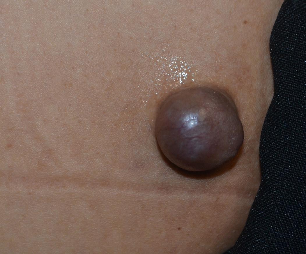
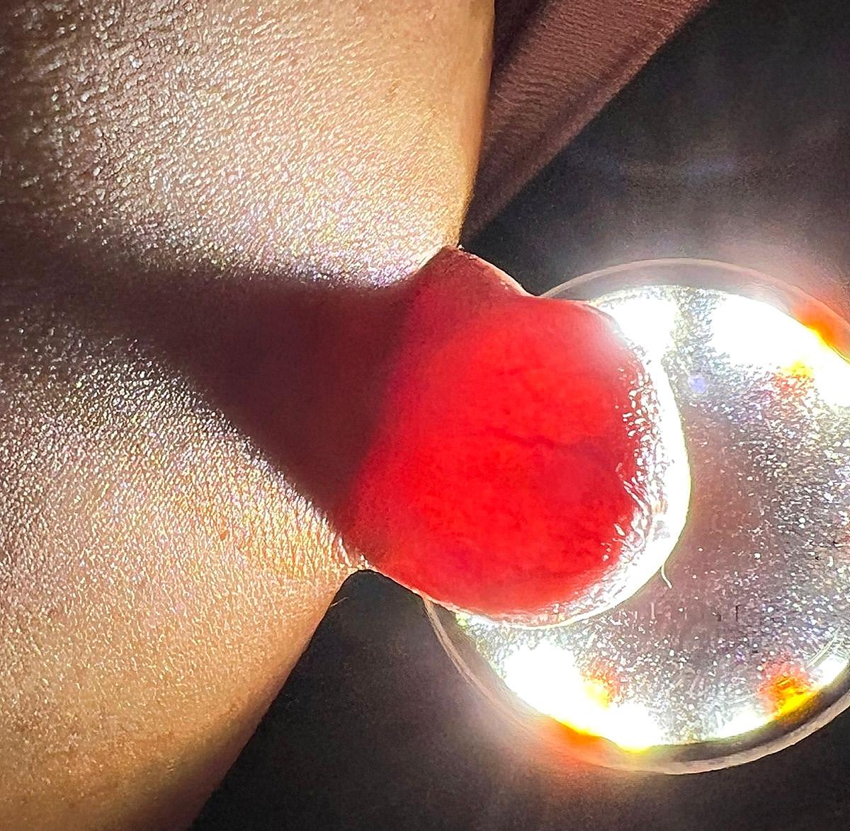
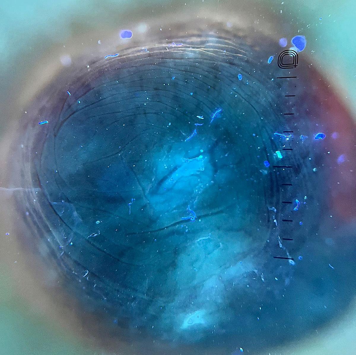
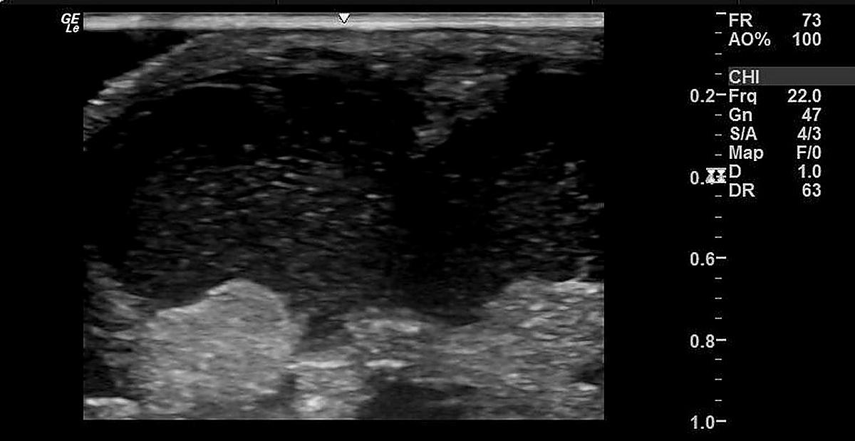
Nodular hidradenoma: diagnostic innovation in a rare case
Ana Paula Weber, Giulia Maria dos Santos Goedert, Barbara Brunca, Hamilton Ometto Stolf, Paula Tavares Colpas
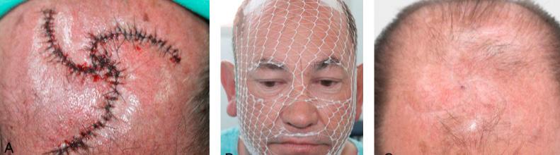
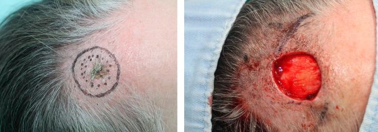
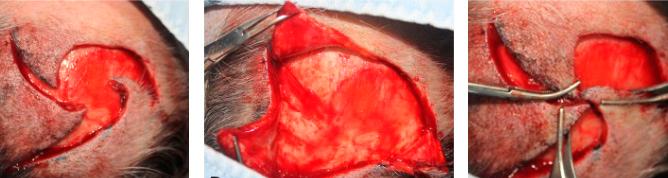
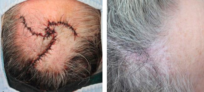
Pinwheel flap: series of two cases
126
130 Joao Marcos Franco de Souza, Barbara Messias Pereira, Bianca Miyazawa, Carlos Borges-Junior, Rogerio Nabor Kondo
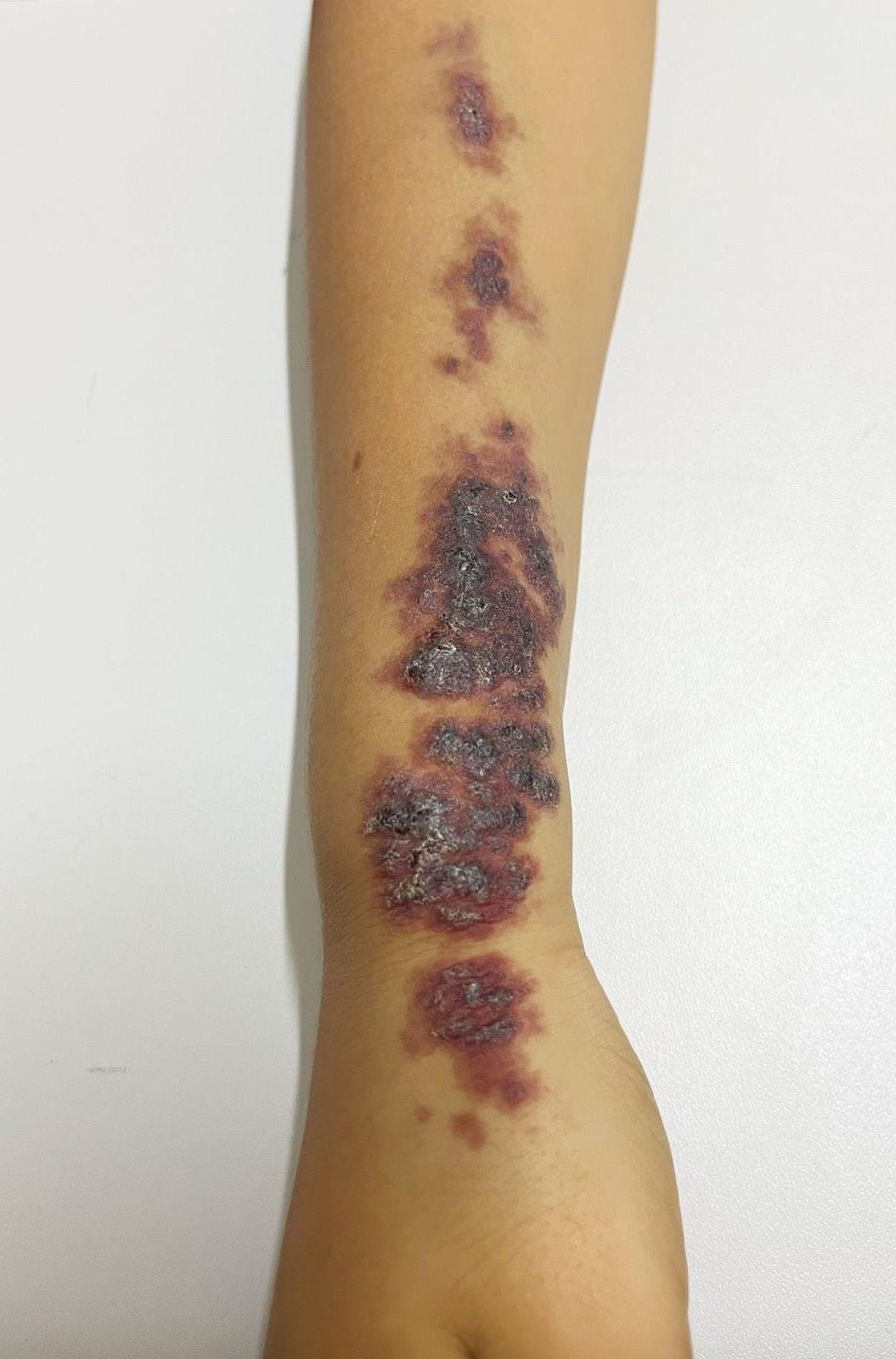
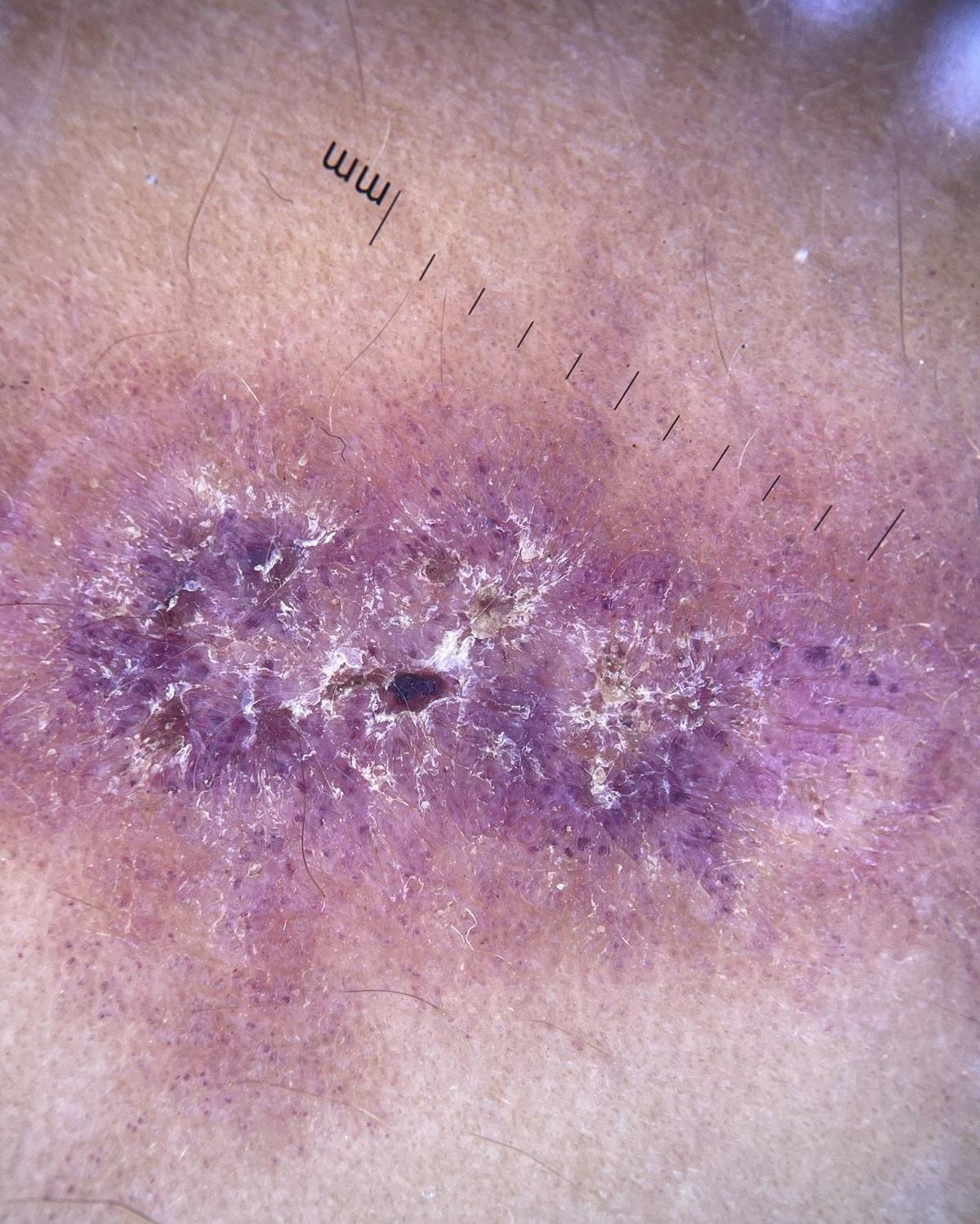
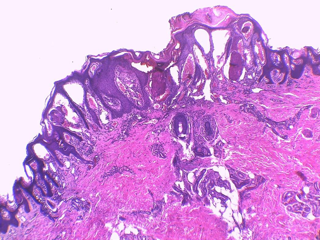
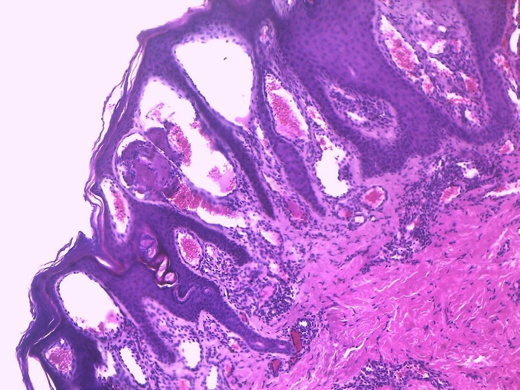
Use of Cryotherapy in the Treatment of Circumscribed Angiokeratoma: A Case Report
134 Malu de Aquino Almeida Garcia, Louise Santos Abdulmassih, Maria Amelia Lopes dos Santos, Renata Mie Oyama Okajima, Josie Eiras Bisi dos Santos
Odontogenic cutaneous fistula mimicking skin neoplasia: case report
139 Clarissa Brito Farias, Ana Vitória Lins Paiva Antunes, Aretuza Iolanda Pimentel Torres Portella, Antônio Carlos Evangelista de Araújo Bonfim, Luciana Cavalcanti Trindade
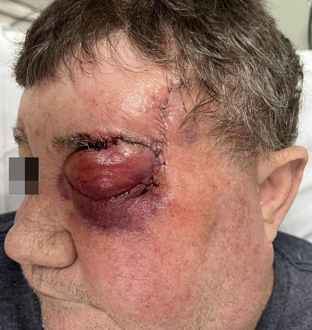
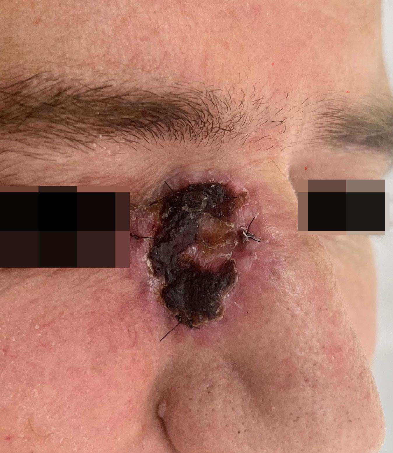
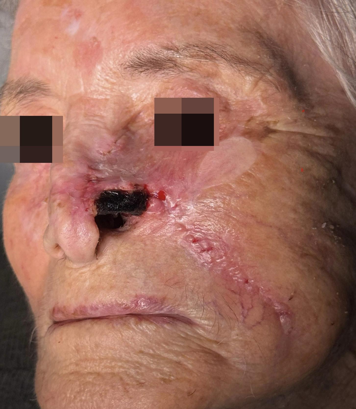
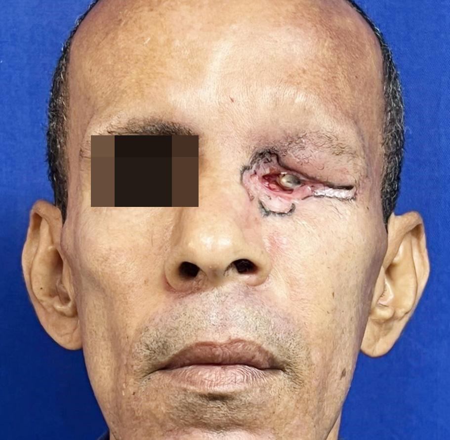
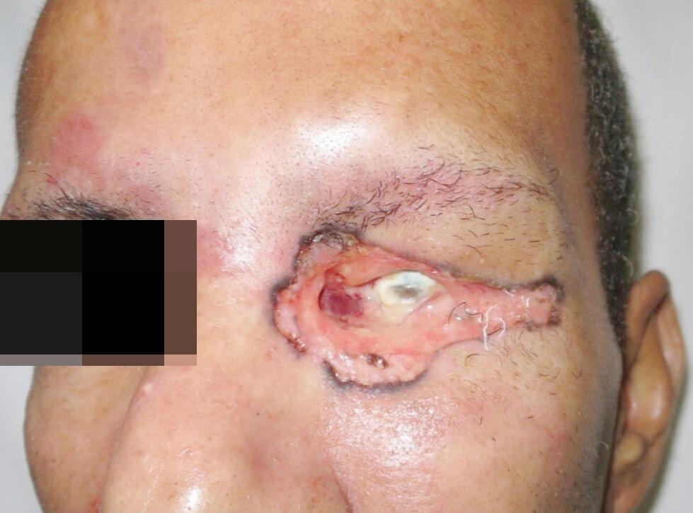
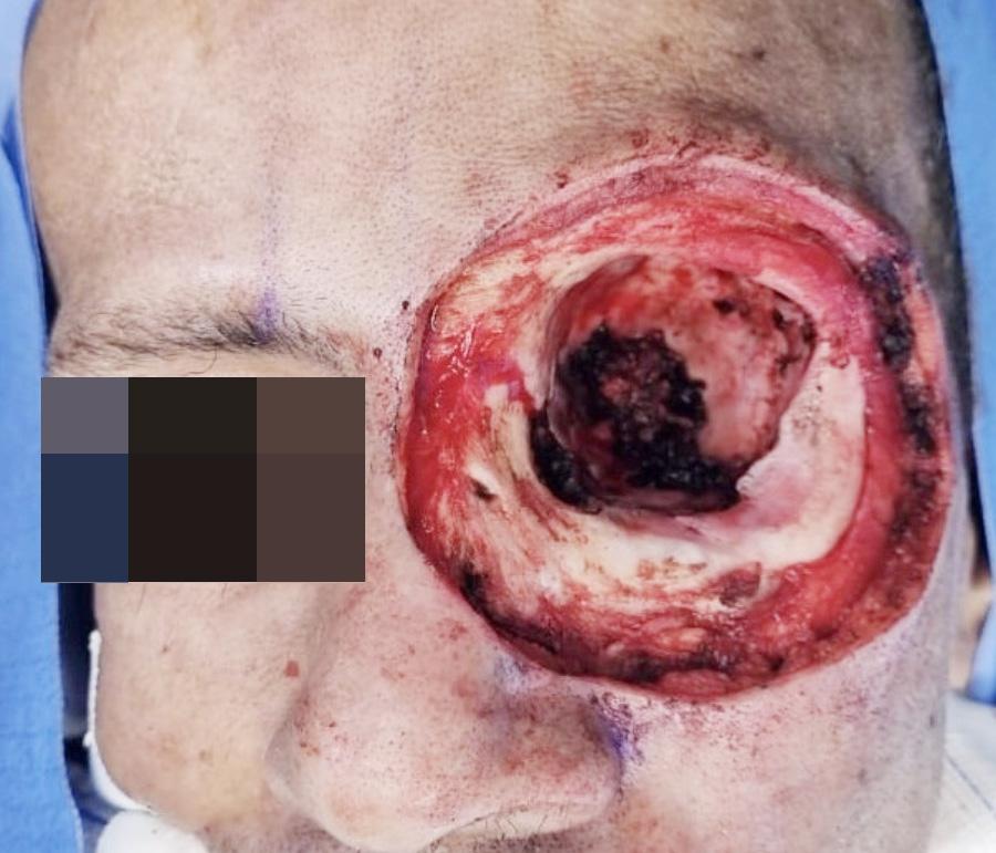
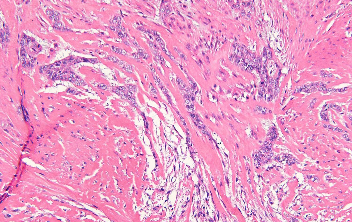
Periocular basal cell carcinoma with orbital invasion: a case report
144 Mariana Sofia Monteiro Silva, Luiz Fernando Eller Riscaroli, Eduardo Garbin Rosset, Ricardo Barbosa Lima, Luciana Ferreira de Araújo, Ruy Gomes Neto
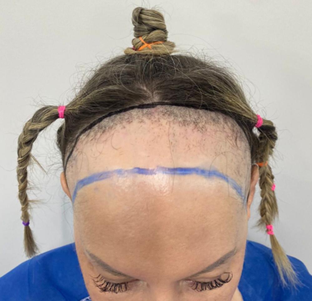
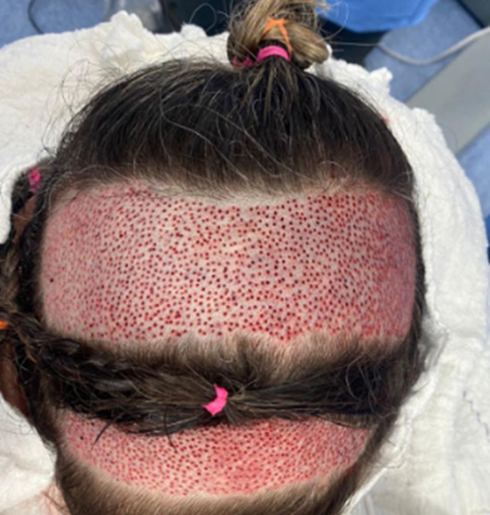
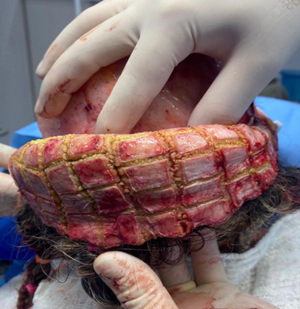
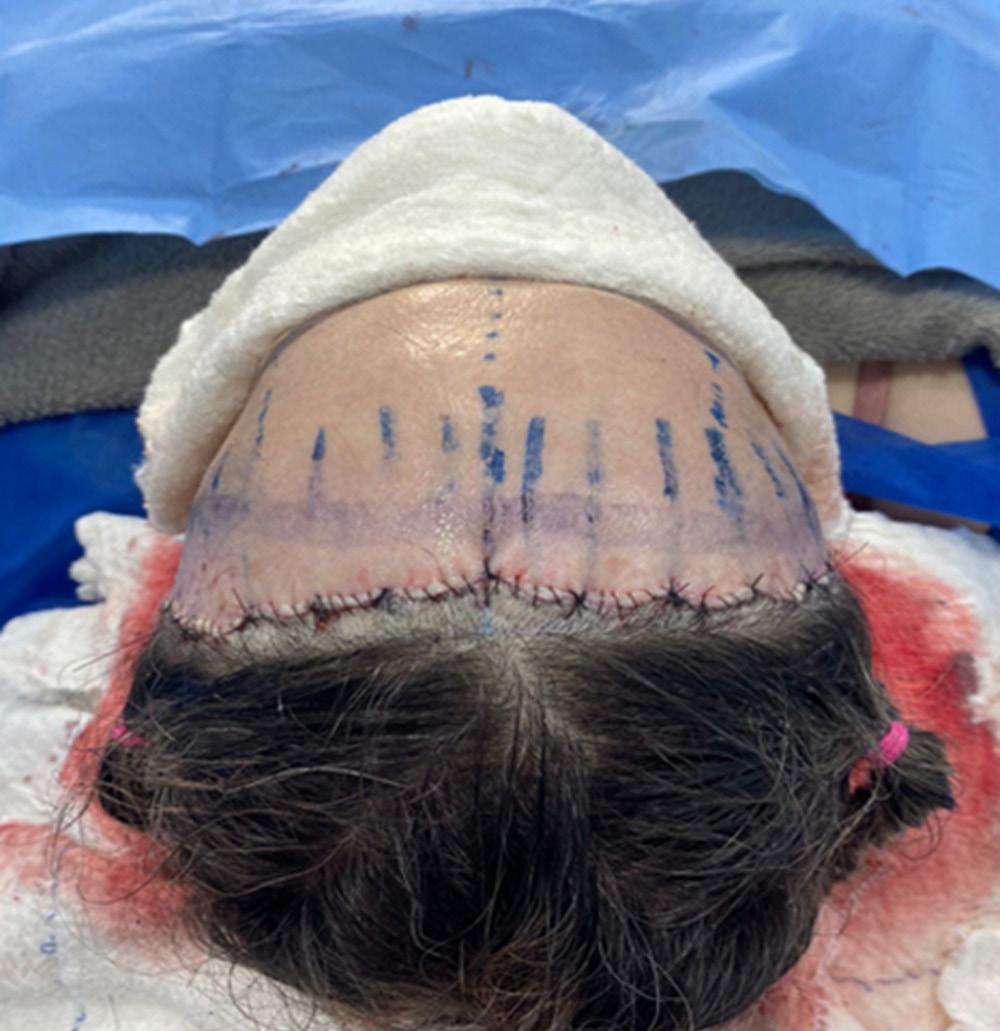
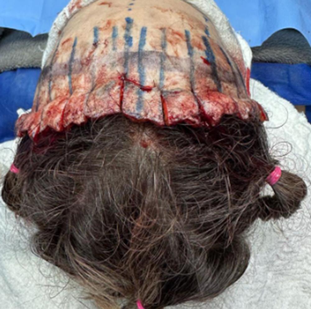
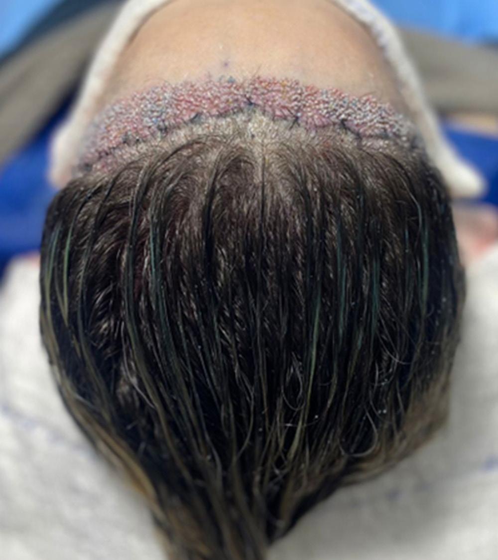
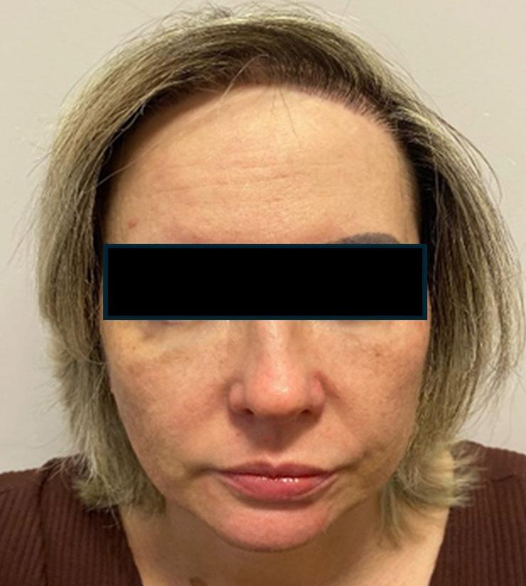
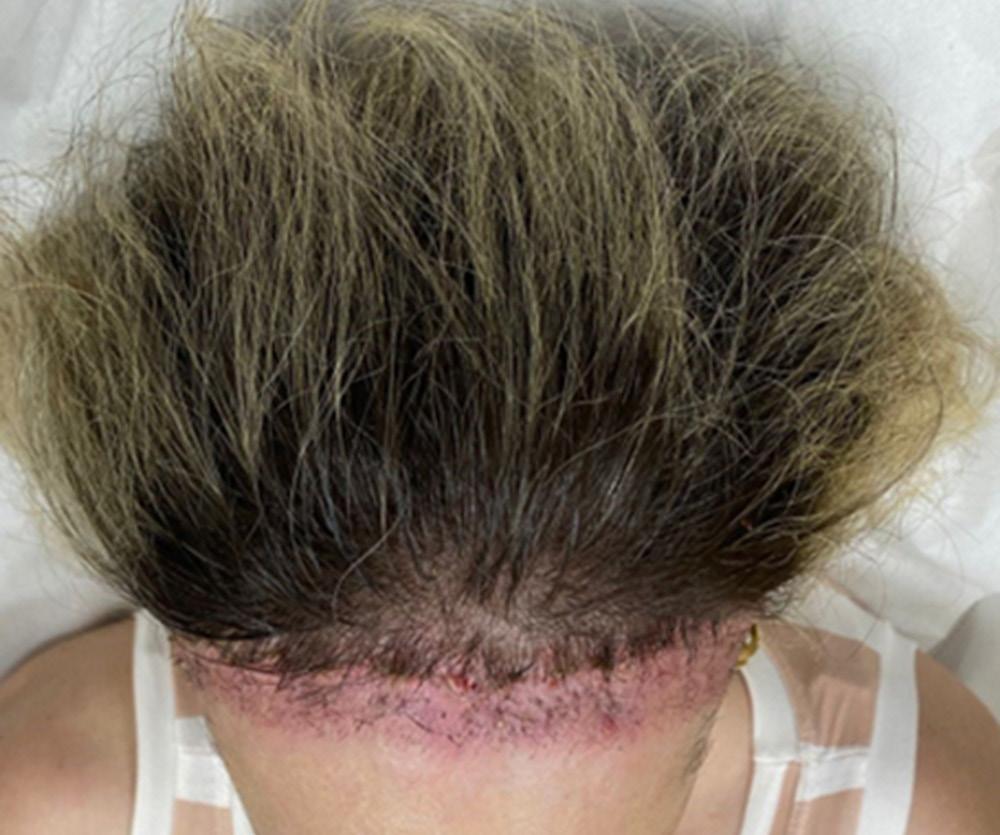
Frontoplasty and hair transplant fue technique in the same surgical time
149 Rafael Reinert, André Felipe Kroenke, Emanoelle Machado, Marcos Felippe Takano-de-Saidneuy, Lauren Menna Marcondes, Murilo Gamba Beduschi
Therapeutic refractoriness of multiple pyogenic granulomas on nail folds as an adverse effect of amivantamab: a case report
154 Ana Vitoria Lins de Paiva Antunes, Clarissa Brito Farias, Luciana Cavalcanti Trindade, Renata Monteiro de Lacerda
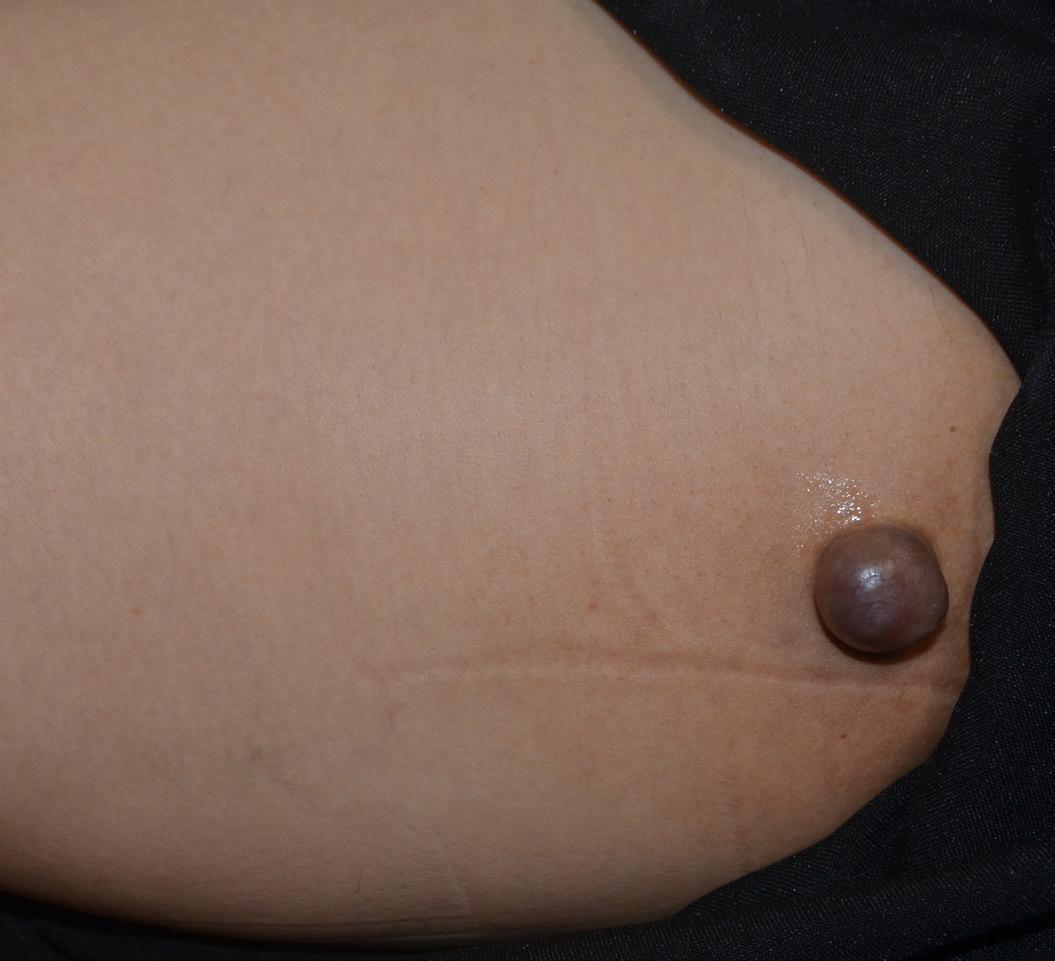
Atypical location of granular cell tumor in a female patient: a rare case report
158 Nivin Mazen Said, Gisele Alves Morikawa Caldeira, Airton Kenji Motizuki, Mariana Bastos Amanajás, Maraya de Jesus Semblano Bittencourt Silvia Fer reira Rodrigues Müller, Franklin de Souza Rocha
Amelanotic Melanoma within a Tattoo: A Greater Masquerader
162 João Alfredo Monte Melo de-Barros, Rafael Freire de Lira Sobral, Ana Beatriz Albuquerque Nunes, Maria Tereza de Medeiros Leite Espínola Jader Freire Sobral Filho
www.surgicalcosmetic.org.br/
Treatment modalities for exogenous ochronosis: A narrative review of current and emerging therapeutic approaches. (Treatment of exogenous ochronosis)
Modalidades de tratamento para ocronose exógena: Uma revisão narrativa das abordagens terapêuticas atuais e emergentes. (Tratamento para ocronose exógena)
Exogenous ochronosis is a chronic skin disorder characterized by dermal hyperpigmentation, often resulting from prolonged use of hydroquinone. This review analyzed 29 studies (2000-2025) assessing treatments for exogenous ochronosis. Laser and light-based therapies, particularly in combination regimens, showed the greatest efficacy, while microneedling and chemical peels provided moderate improvement. Topical agents alone offered limited benefit. Post-inflammatory hyperpigmentation was the most frequent adverse event. Emerging modalities, including intradermal vitamin C, exosome therapy, and platelet-rich fibrin lysate, demonstrated promising early results. Overall, multimodal and light-based therapies appear to be the most effective; however, further controlled studies are essential to confirm their safety and long-term outcomes.
A ocronose exógena é uma doença cutânea crônica caracterizada por hiperpigmentação dérmica, geralmente causada pelo uso prolongado de hidroquinona. Esta revisão analisou 29 estudos (2000-2025) sobre tratamentos para a ocronose exógena. As terapias baseadas em laser e luz, especialmente em combinações, apresentaram maior eficácia, enquanto o microagulhamento e peelings químicos proporcionaram melhoria moderada. Os agentes tópicos isolados tiveram benefício limitado. A hiperpigmentação pós-inflamatória foi o evento adverso mais comum. Modalidades emergentes, como vitamina C intradérmica, terapia com exossomos e lisado de fibrina rica em plaquetas, tiveram resultados promissores. Terapias multimodais parecem mais eficazes, exigindo estudos controlados adicionais para confirmar sua segurança e eficácia.
How to cite this article: Asante ED, Chen HH. Treatment modalities for exogenous ochronosis: A narrative review of current and emerging therapeutic approaches. (Treatment of exogenous ochronosis). Surg Cosmet Dermatol. 2026;18(2):e20260541.
ED, Chen HH
INTRODUCTION
The term “ochronosis” is derived from the Greek word ochre, meaning yellow discoloration, and was first described in the 18th century by Virchow, who noted the ochre-like pigmentation in tissues.1-3
Ochronosis refers to a bluish-gray discoloration of connective tissues caused by pigment deposition and is classified into endogenous and exogenous forms.4 Endogenous ochronosis, or alkaptonuria, is a rare autosomal recessive disorder resulting from homogentisic acid oxidase deficiency,5-10 which leads to systemic manifestations such as arthropathy, pigmentation, and organ calcification.11-19
Exogenous ochronosis (EO), by contrast, is an acquired disorder characterized by blue-black pigmentation of the skin, typically induced by chronic application of topical skin-lightening agents. Hydroquinone is the most commonly implicated compound, though resorcinol, phenol, quinine, picric acid, and mercury have also been linked to the condition.3 First described by Pick in 1906 and later associated with hydroquinone use by Findlay and De Beer in 1975, EO has since been recognized as an important adverse effect of long-term skin-lightening practices.3
EO is increasingly prevalent, particularly in populations with darker skin phototypes, where demand for skin-lightening is high and products are often unregulated. Notably, the risk of EO is not solely determined by hydroquinone concentration, as cases have been reported with low-strength formulations (2%), implicating cumulative dose and duration of exposure as central risk factors.14
Alcohol-based formulations may further heighten risk by enhancing percutaneous absorption.3 Additional predisposing factors include extensive or prolonged sun exposure, application over large body surface areas, and the presence of active melanocytes that interact with hydroquinone-induced metabolic pathways.15-17
The pathogenesis of EO remains incompletely understood. Several mechanisms have been proposed, including hydroquinone-induced stimulation of melanogenesis, its oxidation to quinones and melanin-like intermediates, and its inhibition of homogentisic acid oxidase leading to pigment polymerization in the dermis.20-23
Clinically, EO presents as bilaterally symmetric blue-black macules, predominantly over the malar regions, temples, and cheeks, progressing through 3 stages: mild erythema and pigmentation (stage I), hyperpigmentation with colloid milia (stage II), and papulonodular lesions (stage III).24-30
Diagnosis can be challenging, particularly in the early stages, when EO may mimic melasma. Dermoscopy may reveal gray-blue amorphous areas with obliteration of follicular openings, while reflectance confocal microscopy has found characteristic dermal structures.
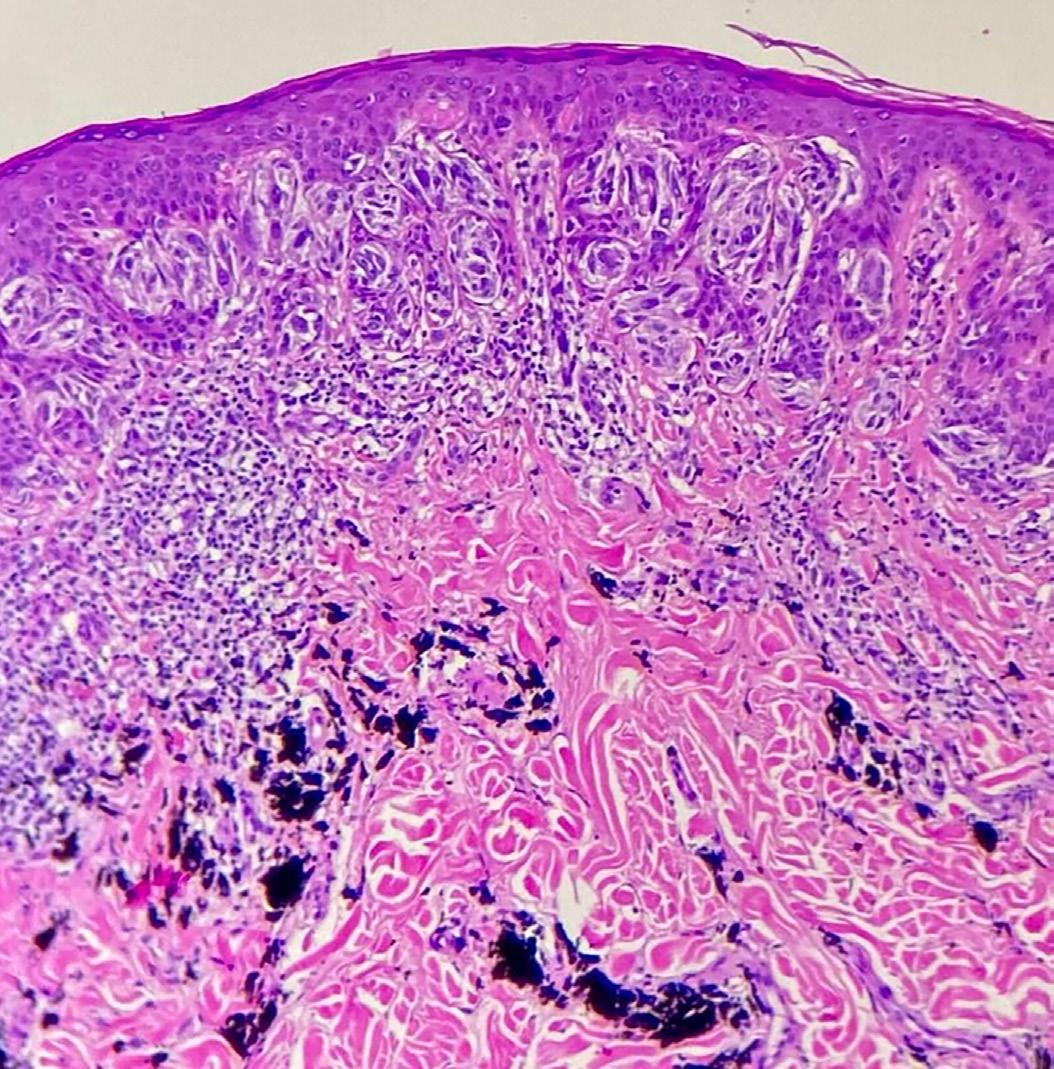
Histopathology remains the gold standard for diagnosis, with hallmark ochre-colored, banana-shaped fibers in the der-
mis.3
Epidemiological data are limited due to underrecognition and misdiagnosis. Early reports suggested prevalence rates of up to 35% among Black populations in South Africa, but more recent evidence shows that EO also affects lighter skin phototypes, including Hispanic, European, and Asian populations.31-34
In the United States, a dermatology survey identified 512 suspected cases, of which 130 were biopsy-confirmed.30
Management of EO is difficult, as pigmentation is often persistent and sometimes irreversible. Discontinuation of hydroquinone and strict photoprotection are essential first steps, but improvement may take years. Pharmacological interventions such as retinoids, corticosteroids, antioxidants, and systemic agents yield variable results, while procedural approaches—including chemical peels, dermabrasion, microneedling, and energy-based devices such as Q-switched and Nd:YAG lasers—have shown promise but lack standardization.24,30
Given its growing prevalence, diagnostic challenges, and limited treatment success, EO represents an important dermatological concern. This review critically evaluates current pharmacological and non-pharmacological treatment modalities for EO, with emphasis on evidence from clinical case series, procedural outcomes, and emerging therapies, to guide future practice and research.
METHODS
Study Design
This study employed a narrative literature review to explore and evaluate treatment modalities for EO, with emphasis on effectiveness, safety, and emerging therapies. The review was guided by 3 objectives: (1) to identify the most effective treatment modalities, (2) to evaluate safety concerns and adverse effects, and (3) to highlight promising novel interventions.
The narrative review approach was chosen because it allows the integration of diverse sources of evidence, including case reports, case series, and retrospective studies, which constitute the majority of the EO literature. Unlike systematic reviews, which require homogeneous data, this method facilitates a broad overview of treatment trends, knowledge gaps, and potential innovations, making it particularly valuable for under-researched conditions.35-37
Search Strategy
An extensive search was performed across PubMed, Scopus, Google Scholar, ScienceDirect, JAMA, the University of South Wales FINDit tool, and Surgical & Cosmetic Dermatology. The search covered publications from January 2000 to May 2025 using keywords and combinations such as “Exogenous Ochronosis,” “Hydroquinone-induced ochronosis,” “Treatment,” “Laser therapy,” “Microneedling,” “Dermabrasion,” and “Oral medication.” Boolean operators (“AND,” “OR”) were used to refine the search, and reference lists of key articles were
screened to identify additional studies.
Inclusion and Exclusion Criteria
Included studies comprised peer-reviewed case reports, case series, retrospective studies, or systematic reviews reporting treatment outcomes in patients with clinically or histopathologically confirmed EO. Only English-language publications involving human subjects were considered. Studies exclusively on endogenous ochronosis, animal or in vitro research, studies not focusing primarily on treatment modalities, and non-English works without usable abstracts were excluded.
Data Extraction and Synthesis
Twenty-nine articles met the inclusion criteria: 2 retrospective studies, 8 case series, and 19 case reports. Data were extracted into tables capturing author and year, study design, treatment modality, outcomes, and adverse effects. Findings were synthesized qualitatively, with particular attention to combination therapies and emerging approaches. A systematic review or meta-analysis was not feasible due to the heterogeneity in study designs and outcome measures.
RESULTS
The treatment of EO encompasses non-pharmacological measures, pharmacological approaches such as topical and oral medications, and procedural interventions like dermabrasion, microneedling, and energy-based device therapies.
Non-Pharmacological Approaches
A key step in the management of EO is the immediate discontinuation of the offending agent, although visible improvement may take years.19
Strict photoprotection, including broad-spectrum sunscreens, hats, UV-protective sunglasses and protective clothing, is also critical to prevent further pigmentation.3
Several case reports highlight the benefit of sunscreen as an adjunctive therapy. For example, Tekgöz et al.7 reported reduced hyperpigmentation after 7 months of sunscreen use and discontinuation of hydroxychloroquine. Similarly, Ceglio et al.,5 Bellew and Alster,2 and Gulwani et al.28 emphasized the use of broad-spectrum sunscreen during EO treatment. A narrative review by Simmons et al.30 also reported improved outcomes with sunscreen use in some cases. These findings underscore the importance of photoprotection in preventing disease progression and for protecting the skin during treatment.
Pharmacological Approaches
Topical Medications
Topical treatments alone have shown limited effectiveness in the management of EO. However, when combined with other therapies, they may provide clinical improvement. Commonly used topical agents include corticosteroids, retinoic acid, azelaic acid, kojic acid, topical calcineurin inhibitors, and tyro-
sinase inhibitors. A 2007 case series by Charlín et al.6 described 4 women (skin phototypes IV–V) with long-standing melasma who developed progressive facial hyperpigmentation after 10–25 years of hydroquinone-based cream use (2%–6%). Clinical signs included blue-brown macules, caviar-like papules, and confetti-like depigmentation, consistent with Dogliotti Stage II EO. Histopathology confirmed pigment incontinence and yellow-brown banana-shaped fibers in the dermis.
Treatment with combinations of 0.05% retinoic acid, 20% azelaic acid, and kojic acid led to only partial or minimal improvement.6
Singh and Ramesh31 reported the case of a 44-year-old woman who developed bluish-black pigmentation after 1 year of skin-lightening cream use. Histopathology confirmed EO. Despite discontinuing the cream and using 0.05% topical retinoic acid with strict photoprotection, there was no notable improvement after 3 months.
Similarly, Qorbani et al.27 described 2 patients with EO secondary to hydroquinone and tretinoin use. In one case, desonide 0.5% lotion with photoprotection reduced inflammation but did not improve pigmentation. The second patient was treated with tazarotene 0.1% gel and pimecrolimus cream in conjunction with photoprotection, but pigmentation persisted for more than 1 year.
Patel and Shah24 reported the case of a 55-year-old woman with EO following 2 years of daily use of hydroquinone-containing cream. Clinical examination showed bluish-brown patches with erythema and telangiectasia. Despite discontinuing the cream, applying topical emollients and retinoid gel, and practicing strict photoprotection, only minimal improvement was observed after 2 months.
A recent retrospective study by Keshavamurthy Vinay et al.12 evaluated 29 patients with EO, predominantly women (93%) with skin phototypes III–VI. Most had a history of hydroquinone use for melasma, averaging 16 months. The malar region was the most commonly affected, with 41% of cases classified as Dogliotti stage I and 59% as stage II. Among 11 patients for whom treatment data were available, combination therapies demonstrated variable outcomes. Chemical peels with topical tacrolimus 0.1% led to moderate improvement over 6 months in 6 patients. A regimen of kojic acid, arbutin, and glycolic acid resulted in minimal improvement, while 1 patient receiving topical tacrolimus combined with oral medication achieved moderate improvement over 6 months.
Overall, topical pharmacological treatments for EO are largely adjunctive. Monotherapy rarely produces substantial improvements, and outcomes are generally enhanced when topical agents are combined with photoprotection, chemical peels, or systemic treatments. Despite limited efficacy, topical agents remain a cornerstone of EO management, particularly for patients with mild disease or as part of multimodal therapeutic strategies.
ED, Chen HH
Oral Medications
Oral medications are infrequently used in EO, generally alongside other therapies. Agents include tranexamic acid, corticosteroids, glutathione, and systemic retinoids.
Moche et al.22 reported 2 Black female patients with EO from prolonged hydroquinone use who developed cutaneous and systemic sarcoidosis. Treatment with oral prednisone (30 mg/day) and chloroquine (200 mg/day) led to improvement in both systemic and skin symptoms.
Pirrone et al.25 described a 51-year-old Filipino woman with histologically confirmed EO who received oral isotretinoin 20 mg 4 times daily, resulting in mild improvement.
Sunkara et al.32 reported the case of a 39-year-old woman with EO after 3 months of using a 4% hydroquinone cream. Oral glutathione was administered, leading to a 50% reduction in pigmentation after 4 months.
In a retrospective study by Keshavamurthy Vinay et al.,12 one patient received oral tranexamic acid as part of the treatment, achieving moderate improvement.
Procedural Treatments
Procedural therapies for EO are typically considered when topical or oral medications provide limited benefit. These include dermabrasion, microneedling, and chemical peels, all of which aim to reduce dermal pigmentation through controlled skin injury or tissue remodeling.The available evidence is largely based on case reports, case series, and small retrospective studies, reflecting both the rarity and treatment-resistant nature of EO.
Dermabrasion
Dermabrasion induces superficial skin injury, triggering wound healing and dermal remodeling, which can improve skin appearance. It has demonstrated moderate to substantial success in selected EO cases, particularly when topical therapies fail. Sunkara et al.32 reported the case of a 39-year-old woman with EO from 3 months of 4% hydroquinone use who underwent microdermabrasion alongside photoprotection, achieving a 50% reduction in pigmentation after 4 months. Similarly, Lamas et al.15 described a 70-year-old woman with a 10-year history of EO who underwent facial dermabrasion with water sandpaper, combined with a nighttime topical regimen (0.05% desonide, 0.01% tretinoin, 2% alpha-bisabolol) and strict photoprotection. After 3 months, the patient exhibited a marked reduction in hyperpigmentation, highlighting the role of dermabrasion in achieving substantial pigment clearance when combined with adjunctive measures.
Microneedling
Microneedling involves the creation of micro-injuries within the dermis to stimulate collagen production and facilitate pigment reduction. Moerbono Mochtar et al.23 reported the cases of 3 women aged 45–50 years with long-standing EO who underwent biweekly microneedling using a 1-mm derma-
roller over an 8-week period, associated with daily SPF 30 use. Improvements in pigmentation and vascular changes were noted by week 4, accompanied by increased patient satisfaction and physician-rated appearance scores. A larger 10-year retrospective study by Lazar et al.16 involving 25 patients with EO demonstrated that microneedling promoted dermal remodeling and achieved visible pigment reduction without substantial adverse effects, particularly in patients with darker skin types, indicating its safety and effectiveness as a procedural option.
Chemical Peels
Chemical peels employ caustic agents such as glycolic acid, salicylic acid, and trichloroacetic acid (TCA) to remove the epidermal and superficial dermal layers, promoting pigment reduction.
Franca et al.8 reported the case of a 40-year-old woman with EO who initially underwent a combination peel consisting of 5% hydroquinone, 5% retinoic acid, and 14% salicylic acid, which resulted in minimal improvement. A subsequent 20% TCA peel in combination with other treatment modalities led to complete resolution of the lesions.
Similarly, Keshavamurthy Vinay et al.12 reported moderate improvement in patients with EO treated with 30% glycolic acid peels. When glycolic acid peels were combined with topical medications, 6 patients experienced moderate pigment reduction over a 6-month period. These findings underscore that chemical peels may be more effective when used in combination with other treatment modalities rather than as monotherapy.
Overall, procedural treatments for EO provide valuable therapeutic options when conventional pharmacological approaches fail. Dermabrasion, microneedling, and chemical peels promote dermal remodeling and pigment reduction, with outcomes often enhanced when combined with topical agents and strict photoprotection. Although the available evidence is limited, these interventions have consistently demonstrated the potential to achieve partial to substantial improvement, particularly in long-standing or treatment-resistant cases. Further studies are needed to standardize protocols, optimize treatment combinations, and determine long-term efficacy and safety across diverse skin phototypes.
Energy-Based Device Therapy
Kramer et al.14 described a case of a Hispanic woman with EO unresponsive to topical treatments who underwent treatment with a Q-switched ruby laser (694 nm, 7 J/cm² fluence, 5-mm spot size). The patient had noticeable clinical improvement, suggesting that Q-switched ruby lasers may represent a promising option for refractory EO.
Alster and Bellew2 reported the use of a Q-switched 755nm alexandrite laser in 2 patients with hydroquinone-induced EO. The first patient underwent 6 sessions at 2-month intervals with a mean fluence of 7.8 J/cm², while the second patient un-
derwent 4 sessions at 4-month intervals with a mean fluence of 6.9 J/cm². Both exhibited substantial lightening of pigmented areas without scarring or textural changes, and histopathological examination confirmed a reduction in dermal pigmentation.
Gil et al.9 assessed the use of intense pulsed light (IPL) therapy in a 63-year-old woman with Fitzpatrick skin phototype V diagnosed with EO. She underwent 6 IPL sessions (645 nm, pulse duration 6 ms, fluence 20–22 J/cm²), alongside topical therapy with 4% kojic acid and 0.2% salicylic acid and strict photoprotection. After 2 months, only mild clinical improvement had been achieved. Dermoscopic features of EO persisted, and reflectance confocal microscopy found unchanged subepidermal banana-shaped bodies, indicating IPL efficacy was limited.
França et al.8 reported the case of a 40-year-old woman with EO after prolonged hydroquinone use who underwent multiple treatment modalities. Four sessions of Q-switched Nd:YAG laser (1064 nm, 2.9–3.05 J/cm², 4-mm spot size) resulted in limited response. Subsequent treatment with ultrapulse CO₂ laser (5 W, 30-day intervals) resulted in partial improvement, particularly in the left malar region. IPL therapy (36 J, 10 ms) was then administered, followed by microdermabrasion and a chemical peel combining 5% hydroquinone, 5% retinoic acid, and 14% salicylic acid. Complete lesion clearance was achieved after 3 additional IPL sessions (36 J, 10 ms) in conjunction with 20% TCA peels.
Kanechorn-Na-Ayuthaya et al.11 described 3 patients with EO treated with Q-switched Nd:YAG (1064 nm) and fractional CO₂ lasers. In the first case, a 67-year-old Thai woman (skin phototype V) underwent 4 Q-switched Nd:YAG laser sessions (1.9–2.2 J/cm²) and 1 fractional CO₂ laser session, achieving progressive fading and smoother skin texture by the third session, despite transient post-inflammatory hyperpigmentation (PIH). The second case, that of a 58-year-old woman (skin phototype III) who underwent 4 Nd:YAG sessions and one CO₂ laser session, achieved noticeable improvement in pigmentation and skin tone after the third session, accompanied by temporary PIH. In the third case, a 66-year-old woman (skin phototype IV) underwent 2 Nd:YAG laser sessions followed by fractional CO₂ laser treatment, with minimal PIH and visible skin lightening and mild rejuvenation at 5 months. Overall, combination laser therapy resulted in gradual improvement of lesions and high patient satisfaction.
Tan33 evaluated 6 ethnic Chinese patients with histologically confirmed EO and concomitant melasma, classified according to Dogliotti staging: 4 with Stage I disease, 1 with Stage II, and 1 with Stage III. Treatment consisted of fortnightly Q-switched Nd:YAG (1064 nm) laser sessions using a low-fluence first pass (1.2 J/cm², 8- mm spot size) and targeted high-fluence passes (4–6 J/cm², 4-mm spot size) until petechiae or erythema appeared. Topical EMLA cream was applied before treatment, and post-treatment care included topical tetracycline and continuation of non-hydroquinone lightening agents. Patients with stage II and III disease showed marked improvement after 8 and
16 sessions, respectively, while patients with stage I disease demonstrated mild-to-moderate improvement. Pigment clearance was maintained, and no serious adverse events occurred, supporting the use of Q-switched Nd:YAG lasers, particularly with a multi-pass low-fluence and targeted high-fluence technique, for moderate-to-severe EO in darker skin phototypes.
Lee and Weiss17 reported the case of a 48-year-old woman with EO treated with IPL (570 nm, 12 J/cm², 15 ms) at 6-week intervals, with lightening of pigmentation after the initial session.
Liu et al.21 described a 50-year-old Chinese man (skin phototype IV) with Stage II EO who underwent 6 sessions of Q-switched Nd:YAG laser (1064 nm, 6–9 J/cm²) with no improvement, despite treatment adherence and discontinuation of hydroquinone.
Ko and Wang13 reported the case of a 50-year-old Chinese woman with EO treated with Q-switched Nd:YAG (1064 nm, 5.3 J/cm²) and Q-switched alexandrite (755 nm, 8.5 J/cm²) lasers. Pigmentation worsened after 2 sessions; the use of fractional CO₂ laser was recommended, but declined. Carvalho et al.4 described a 46-year-old woman (skin phototype V) treated with monthly fractional CO₂ laser sessions for 12 months, achieving substantial improvement. Pirrone et al.25 reported the case of a 51-year-old Filipino woman who experienced mild improvement with oral isotretinoin and topical corticosteroids, but subsequent fractional laser therapy over 3 sessions achieved marked lightening of the hyperpigmented lesions. Baca et al.1 described a 55-year-old woman (skin phototype IV) with long-standing EO refractory to multiple prior therapies, who underwent 9 sessions of fractional nonablative picosecond laser (1064/532 nm), achieving clear improvement in lesion color and texture.
Qian26 presented 2 cases of EO treated with microneedle pulsed radiofrequency adjunctive to laser therapy, with satisfactory outcomes.
Lee et al.18 reported the case of a 66-year-old woman treated with fractional CO₂ and Q-switched Nd:YAG lasers, with no clinical improvement after 3 sessions. Ceglio et al.5 described a 52-year-old man (skin phototype V) treated sequentially with Q-switched Nd:YAG laser (12 sessions), IPL (7 sessions), and fractional CO₂ laser (5 sessions), with the most notable improvement achieved after fractional CO₂ laser therapy. Lazar et al.16 conducted a 10-year review of 25 patients with EO (88% women, skin phototypes IV–VI) and found Q-switched alexandrite (755 nm) laser effective, with marked improvement in observed in 3 patients after a mean of 4.66 sessions.
Li and Zhang20 reported the case of a 28-year-old woman with concomitant EO and vitiligo who achieved improvement after 4 chemical peels and one IPL session.
Almutairi et al.38 reported the case of a 31-year-old man with refractory EO who responded to 3 sessions of picosecond alexandrite laser (755 nm) following prior unsuccessful treatment with Q-switched and 1064-nm picosecond laser therapy. The man achieved marked lesion improvement after 6 months with no complications.
NEW AND EMERGING TREATMENT MODALITIES
Emerging treatment modalities for EO aim to improve therapeutic outcomes and patient satisfaction. One such novel approach involves intradermal vitamin C injections, which act as antioxidants and may reduce pigmentation. Another innovative modality is platelet-rich fibrin lysate (PRF-L), an autologous derivative of platelet-rich fibrin that is rich in growth factors and cytokines and may promote skin rejuvenation and pigment regulation.
A case series involving 3 healthy Javanese women aged 45–50 years explored the potential of PRF-L in the management of EO. Each patient had progressive facial hyperpigmentation previously diagnosed as melasma, with disease durations of 2, 5, and 6 years, a history of prolonged use of dermatologist-prescribed skin-lightening creams without follow-up or knowledge of their ingredients. Clinical examination revealed patchy hyperpigmentation, facial erythema, hypopigmented macules, and telangiectasia.
The diagnosis of EO was supported by skin stretching tests, Wood’s lamp, and dermoscopy. After informed consent was obtained, autologous PRF-L was prepared by collecting peripheral blood in 10-mL sterile tubes without anticoagulant, centrifugation at 2700 rpm (~400 g) for 12 minutes, and incubation of the isolated PRF gel at 4 °C for 24 hours to obtain the PRF-L supernatant. Facial skin was cleansed and anesthetized topically for 1 hour. PRF-L was administered using 2 methods: topical application followed by dermarolling (1-mm depth) on the right side of the face and intradermal injection with a 1-mL syringe on the left. Patients were instructed to apply SPF 30 sunscreen daily. The 2-month follow-up period included biweekly clinical photography, with 2 blinded physicians assessing improvement using a 10-point scale. Early improvements were noted on dermoscopic examination, and 1 patient demonstrated visible clinical improvement.23
Rajesh Gulwani et al.28 described a 45-year-old Indian man with bilateral cheek hyperpigmentation following intermittent use of multiple skin-lightening agents over a 9-year period, including a triple-combination cream (2% hydroquinone, 0.05% tretinoin, 0.1% mometasone) over 3 years. Clinical examination revealed blue-black macules on an erythematous background, and dermoscopy found dark globules in annular, arciform, and curvilinear patterns, follicular obliteration, and telangiectasia. Histopathological examination confirmed EO, revealing epidermal thinning, elastotic degeneration, and banana-shaped ochronotic bodies within the dermis. The patient underwent intradermal vitamin C (ascorbic acid) injections: 1 mL (100 mg) at 1-cm intervals across affected areas, repeated every 4 weeks for 3 sessions. Sunscreen to protect against ultraviolet, infrared, and visible light and a barrier-repair moisturizer containing hyaluronic acid, ceramide, squalene, and lecithin were prescribed. The treatment resulted in complete resolution of erythema within 1 month and approximately 80% improvement in hyperpigmentation after 3 months. The treatment regimen was well tolerated, with mild-to-moderate discomfort at injection sites.28
Plant-based exosome therapy represents another emerging therapeutic option. Widjaja et al.35 reported the case of a 39-year-old Indonesian woman with widespread facial hyperpigmentation following 8 months of using a 4% hydroquinone cream. She developed erythema and patchy pigmentation and had never used sunscreen. Clinical examination revealed dry, sallow skin with hyperpigmented macules, hypopigmented areas, and telangiectasia, supporting the diagnosis of EO. Treatment consisted of intradermal injections of 6 mL of a plant-derived exosome skin booster across facial zones, followed by topical application of 1.2 mL of exosome serum and dual-light photodynamic therapy (PDT) for 10 minutes per light source. A comprehensive skincare regimen was instituted, including a morning routine consisting of facial cleanser, toner, serum, and cream containing niacinamide, alpha-arbutin, 3-O-ethyl ascorbic acid, and tranexamic acid, as well as nighttime cream containing retinol. An SPF 50 hybrid sunscreen was introduced for photoprotection. At the 36-day follow-up, improvements in skin texture and tone were evident, with brighter, better-hydrated skin and a notable reduction in hyperpigmented patches, particularly on the cheeks.
These emerging therapies reflect a trend toward minimally invasive, regenerative, and targeted approaches for EO. PRF-L offers autologous growth factor-mediated modulation of pigmentation and dermal remodeling, demonstrating early dermoscopic and clinical improvements. Intradermal vitamin C provides antioxidant-mediated pigment reduction and skin repair, achieving substantial clinical improvement in severe EO. Plant-based exosome therapy, combined with topical agents and PDT, represents a multimodal approach that addresses pigmentation, inflammation, and skin barrier restoration.
Across these modalities, photoprotection remains a critical adjunctive measure, emphasizing the importance of using sunscreen to prevent further pigmentary changes and protect against ultraviolet, infrared, and visible light-induced skin damage. In addition, supportive skincare with barrier repair and antioxidant components may enhance overall treatment outcomes. Case reports and small case series suggest that these therapies are generally well tolerated, with minimal adverse effects such as mild injection-site pain and transient erythema, while resulting in measurable improvements in hyperpigmentation, erythema, telangiectasia, and skin texture.
Collectively, early evidence suggests that these innovative approaches can complement conventional EO management strategies, offering the potential for faster clinical improvement, greater patient satisfaction, and expanded therapeutic options for individuals with treatment-resistant cases. Long-term follow-up and larger studies are needed to establish standardized protocols, determine optimal dosing schedules, and compare efficacy with established procedural and pharmacological therapies. Nonetheless, PRF-L, intradermal vitamin C, and plant-based exosome therapy represent promising additions to the evolving therapeutic armamentarium for EO.
AUTHORS
1. Charlín et al., 2007 Case series
2. Singh and Ramesh, 2014 Case report
3. Qorbani et al., 2020 Case series
4. Lazar et al., 2023
Retrospective study
5. Patel and Shah, 2024 Case report
6 Keshavamurthy Vinay et al., 2025)
Retrospective study
Table 1: TOPICAL TREATMENT
Retinoic acid + azelaic acid + kojic acid
Retinoic acid + azelaic acid
Retinoic acid + sunscreen
Desonide lotion + sunscreen
Tazarotene 0.1% gel + pimecrolimus cream +photoprotection
Triluma
Tretinoin
Partial improvement No improvement Not documented Not documented
No improvement Not documented
Minimal improvement
Minimal to no improvement Not documented Not documented
Ammonium lactate Not helpful Not documented
Emollient + retinoid gel
Kojic acid+ arbutin + glycolic acid + sunscreen
Table 2: ORAL TREATMENT
AUTHORS STUDY DESIGN TREATMENT
1. Moche et al., 2010 Case series Tab prednisone 30 mg + Tab chloroquine 200 mg
Table 3: PROCEDURAL TREATMENT
AUTHORS STUDY DESIGN TREATMENT
1. Lamas et al., 2023 Case report Dermabrasion
2. Lazar et al., 2023 Retrospective Study Microneedling
3. Keshavamurthy Vinay et al., 2025 Retrospective study Chemical peel with 30% glycolic acid
Marked improvement Not documented Post inflammatory hyperpigmentation
9 Carvalho et al., 2016 Case report Fractional CO2 laser (120 mm tip, 120 mJ, 150 points stitches per cm2) No improvement Not documented
10 Baca et al., 2018 Case report
Fractional picosecond laser (1064 nm and 532 nm, initial fluence 1.30 and 0.18 J/cm2, increased by 0.20/0.02 J/cm2 per application up to a maximum fluences of 2.9/0.30 J/ cm2)
Photoprotection is widely recommended, as EO is exacerbated by exposure to ultraviolet radiation. Bhattar et al.3 highlight its role in limiting disease progression, while Simmons et al.30 caution that sunscreen alone does not result in substantial cosmetic improvement. Across the literature, photoprotection is mostly used as an adjunctive therapy, suggesting that optimal outcomes are achieved when combining it with active therapies.
Topical agents, particularly in early-stage EO, demonstrate modest efficacy. Studies by Charlín et al.,6 Singh and Ramesh,31 and Qorbani et al.27 report limited response to retinoic acid, azelaic acid, corticosteroids, and calcineurin inhibitors. Although these agents theoretically target melanin production and inflammation, poor dermal penetration likely limits their effectiveness given the deep pigment deposits found in EO. Keshavamurthy Vinay et al.12 further demonstrated minimal improvement with topical monotherapy unless it was combined with peels or systemic agents. Thus, topicals should generally be considered adjunctive rather than definitive treatments.
A consistent observation across multiple studies4-6,9,12,13,15,24,27,31,33 is the near-universal history of hydroquinone use in patients who develop EO. This highlights a potentially underrecognized association between melasma management and the onset of EO, raising concerns about long-term safety, particularly in individuals with darker skin phototypes or prolonged hydroquinone use. Reevaluation of melasma guidelines and development of safer alternatives or stricter protocols may help reduce the incidence of EO.
Oral medications are used sporadically, primarily for atypical presentations or experimental purposes. Moche et al.22 reported corticosteroids and chloroquine for sarcoid-like EO, suggesting a possible role for systemic immunomodulation in inflammatory variants. More conventional agents, such as isotretinoin25 and glutathione,32 yield mild-to-moderate improvements in pigmentation, likely through cell turnover and reduction of oxidative stress. Oral tranexamic acid, traditionally prescribed for melasma, was modestly effective in one case,12 possibly due to anti-inflammatory effects. However, these findings are isolated, highlighting the need for controlled studies to assess safety and dosing.
Visible clinical improvement
80% reduction in hyperpigmentation
Visible improvement in pigmentation
None reported
Mild to moderate injection pain
None reported
Procedural interventions appear more promising, particularly for patients who do not respond to pharmacologic therapy. Dermabrasion has demonstrated notable reduction in pigmentation, especially when combined with topical agents and photoprotection.15 Microdermabrasion32 and microneedling16,23 have also shown favorable outcomes, particularly in individuals with darker skin phototypes, in whom dermal pigmentation is more pronounced. Microneedling offers the advantages of promoting dermal remodeling and enhancing topical drug absorption.
Chemical peels, particularly TCA, seem to outperform superficial peels such as glycolic acid. França et al.8 reported complete lesion resolution with 20% TCA after failure of treatment with superficial agents, whereas Vinay et al.12 observed only moderate improvement with 30% glycolic acid, underscoring the importance of peel depth. Sequential or combination approaches may further enhance efficacy, although variability in formulations, techniques, and patient factors complicate direct comparisons.
Laser and light-based therapies provide compelling but variable results. Q-switched lasers (ruby, Nd:YAG, alexandrite) have shown efficacy but may induce PIH or even worsen pigmentation, particularly in patients with darker skin phototypes.13 Fractional CO₂ lasers, alone or combined with Q-switched devices, have resulted in more consistent outcomes.4,5 IPL appears to be the most effective when added to multimodal regimens. Picosecond lasers have shown encouraging preliminary results with fewer adverse effects,38 but further validation is required. Emerging regenerative therapies, including intradermal vitamin C injections, plant-based exosomes, and PRF-L, aim to restore dermal integrity while reducing pigmentation. Vitamin C injections achieved reductions in pigmentation of up to 80%,28 while plant exosomes and PRF-L were associated with improvements in skin quality and lightening.23,35 While promising, these early-stage studies have small sample sizes and limited long-term safety data.
Few studies report adverse effects. PIH is the most frequent, especially after laser-based therapies, posing particular risk for patients with darker phototypes, who represent a large share of EO cases. Reports of worsening pigmentation with Q-swit-
ched and alexandrite lasers13 underscore the need for caution. Microneedling, picosecond lasers, and select chemical peels, on the other hand, are generally well tolerated.
In conclusion, the management of EO requires a multimodal, individualized approach. Non-pharmacological measures remain foundational, topicals therapies provide adjunctive benefits, and procedural or energy-based interventions achieve the most consistent reduction in pigmentation. Emerging regenerative strategies may further expand treatment options, but high-quality studies are needed to optimize treatment protocols, minimize adverse events, and identify predictive factors for treatment response, particularly in individuals with darker skin phototypes.
CONCLUSION
This review underscores the complexity of managing EO, a challenging pigmentary disorder. Among available therapeutic options, laser and light-based therapies—particularly when used in combination with other modalities—demonstrate the most consistent clinical efficacy, supporting a multimodal approach over monotherapy. Procedural treatments such as microneedling and chemical peels provide moderate improvement, whereas topical agents alone are generally ineffective, especially in long-standing disease or cases involving deep dermal pigmentation.
Regarding safety, PIH is the most frequently reported complication, particularly following laser-based therapies. Microneedling and picosecond lasers are generally well tolerated and may represent safer options for patients with darker skin
phototypes, who are more susceptible to pigmentary rebound and adverse effects.
Emerging interventions, including intradermal vitamin C, plant-derived exosomes, and PRF-L, have shown promising preliminary results, potentially combining pigment reduction with dermal remodeling, barrier restoration, and local antioxidant and anti-inflammatory effects.
Overall, the management of EO remains multifaceted and requires individualized treatment plans based on pigmentation depth, histopathological severity, skin phototype, and treatment tolerance. Combination therapies appear to be the most effective, but further controlled studies are required to establish standardized protocols, confirm long-term safety, and validate emerging approaches for broader clinical application.
ACKNOWLEDGEMENTS
I would like to express my deepest gratitude to the Almighty God for the gift of life, strength, and wisdom that enabled me to undertake and complete this project.
My sincere thanks also go to my supervisor, Dr. Hsuan-Hsiang Chen, for his guidance, support, and encouragement throughout this work.
Finally, I would like to extend my heartfelt appreciation to the Merck Foundation for sponsoring my studies and making this academic journey possible.
This work was completed in partial fulfillment of the requirements for the Master of Science (MSc) in Dermatology in Clinical Practice at the University of South Wales. l
REFERENCES:
1. Baca IM, Firas Al-Niaimi, Colina C, Anuzita A. A Case of Ochronosis Successfully Treated with the Picosecond Laser. J Cosmet Dermatol. 2018;18(5):1322–5.
2. Bellew SG, Alster TS. Treatment of Exogenous Ochronosis with a Q-Switched Alexandrite (755 nm) Laser. Dermatol Surg. 2004;30(4):555–8.
3. Bhattar P, Zawar V, Godse K, Patil S, Nadkarni N, Gautam M. Exogenous Ochronosis. Indian J Dermatol. 2015;60(6):537.
4. Carvalho CGS, Vilela VN, Rocha AEA, Carvalho GSM, França ER, Rodrigues AGA. Exogenous Ochronosis Treated with CO2 Laser. Surg Cosmet Dermatol. 2016;8(4).
5. Ceglio WW, Careta MF, Regia Patriota, Luís Antônio Torezan. Exogenous Ochronosis Successfully Treated with the Combination of Intense Pulsed Light and Fractional CO2 Laser. Ann Bras Dermatol. 2022;98(1):138–40.
6. Charlín R, Barcaui CB, Kac BK, Soares DB, Rabello-Fonseca R, Azulay-Abulafia L. Hydroquinone-induced Exogenous ochronosis: a Report of Four Cases and Usefulness of Dermoscopy. Int J Dermatol. 2007;47(1):19–23.
7. Emre T, Egemen A, Muhammet Ç, Sedat Y. A Case of Exogenous Ochronosis Associated with Hydroxychloroquine. European Journal of Rheumatology. 2018;5(3):206–8.
8. França ER, Paiva V, Toscano LPN, Nunes GJB, Rodrigues TFA. Exogenous ochronosis: a Case Report. Surg Cosmet Dermatol. 2010;2(4):319–21.
9. Gil I, Segura S, Martínez-Escala E, Lloreta J, Puig S, Vélez M, et al. Dermoscopic and Reflectance Confocal Microscopic Features of Exogenous Ochronosis. Arch Dermatol. 2010;146(9).
10. Hull PR, Procter PR. The melanocyte: an Essential Link in hydroquinone-induced Ochronosis. J Am Acad Dermatol. 1990;22(3):529–31.
11. Kanechorn NAP, Niumphradit N, Aunhachoke K, Nakakes A, Sittiwangkul R, Srisuttiyakorn C. Effect of Combination of 1064 Nm Q-switched Nd:YAG and Fractional Carbon Dioxide Lasers for Treating Exogenous Ochronosis. J Cosmet Laser Ther. 2013;15(1):42–5.
12. Keshavamurthy V, Mehta H, Bhat YJ, Khare S, Nkechi AE, Chauhan P, et al. Clinical and Dermoscopic Characteristics of Exogenous ochronosis: Results of a Multicenter Study by the International Dermoscopy Society Task Force on “Imaging in Skin of Color.” Clin Exp Dermatol. 2025.
13. Ko WL, Wang KH. Exogenous Ochronosis. Dermatol Sinica. 2015;33(1):29–30.
14. Kramer KE, Lopez A, Stefanato CM, Phillips TJ. Exogenous Ochronosis. J Am Acad Dermatol. 2000;42(5):869–71.
15. Lamas CN, Maria F, Manfredini E, Regina Ferreira F, Santos P, Gonçalves F. Dermabrasion in Exogenous ochronosis: a Therapeutic Option. Surg Cosmet Dermatol. 2023;15.
16. Lazar M, Garza H, Vashi NA. Exogenous Ochronosis: Characterizing a Rare Disorder in Skin of Color. J Clin Med. 2023;12(13):4341.
17. Lee MD, Weiss E. Treatment of Exogenous Ochronosis with Advanced Fluorescence Technology. Dermatol Surg [Internet]. 2014;40(9):1046–8.
18. Lee MY, Yun SJ, Lee SC, Won YH, Lee JB. A Case of Ochronosis with Atypical Manifestations Involving the Perioral Area and Sclera. Ann Dermatol. 2019;31(1):106–6.
19. Levin CY, Maibach H. Exogenous Ochronosis. Am J Clin Dermatol. 2001;2(4):213–7.
20. Li ZZ, Zhang C. Exogenous Ochronosis with Vitiligo: a Therapeutic Challenge. Skin Res Technol. 2024;30(10).
21. Liu W, Tey H, Lee S, Goh B. Exogenous Ochronosis in a Chinese patient: Use of Dermoscopy Aids Early Diagnosis and Selection of Biopsy Site. Singapore Med J. 2014;55(1).
22. Moche MJ, Glassman SJ, Modi D, Grayson W. Cutaneous Annular Sarcoidosis Developing on a Background of Exogenous ochronosis: a Report of Two Cases and Review of the Literature. Clin Exp Dermatol. 2010;35(4):399–402.
23. Moerbono M, Sri EI, Dendy Z, Ammarilis M, Suci W. A split-face of Dermaroller and Intradermal Injection with the Autologous Platelet Rich Fibrin Lysate in the Treatment of Exogenous ocronosis: a Case Series. Dermatol Rep. 2019.
24. Patel N, Shah H. Exogenous Ochronosis from Skin-Lightening Cream. New England journal of medicine/ New Engl J Med. 2024;390(3):254–4.
25. Pirrone J, Broyer J, Otte N, Reich K, Almut BA. Extensive Hyperpigmentation of the Face in a Filipina. JDDG J Deutschen Dermatol Gesell. 2017;15(11):1167–8.
26. Qian YH. Micro-needling Pulsed Radiofrequency as an Adjuvant to Lasers for Exogenous Ochronosis. J Clin Exp Dermatol Res. 201809.
27. Qorbani A, Mubasher A, Sarantopoulos GP, Nelson S, Fung MA. Exogenous Ochronosis (EO): Skin Lightening Cream Causing Rare caviar-like Lesion with banana-like pigments; Review of Literature and Histological Comparison with Endogenous Counterpart. Autopsy Case Rep. 2020;10(4):e2020197.
28. Rajesh Gulwani P, Parekh AR, Shah HA. Intradermal Vitamin C: a New Paradigm in the Treatment of Exogenous Ochronosis. Indian J Dermatol, Venereol Leprol. 2024;90:378–80.
29. Rocha MDN, Aguiar C, Saraiva MJ. Exogenous ochronosis: Case Report and Literature Review. Ann Bras Dermatol. 2012;87(4):633–6.
30. Simmons BJ, Griffith RD, Bray FN, Falto-Aizpurua LA, Nouri K. Exogenous Ochronosis: a Comprehensive Review of the Diagnosis, Epidemiology, Causes, and Treatments. Am J Clin Dermatol. 2015;16(3):205–12.
31. Singh A, Ramesh V. Exogenous Ochronosis. Indian J Med Res. 2014;139(2):327.
32. Sunkara HP, Kilaru KR, Kumar AP, Ramineni HB, Krishna PR. A Case Report on Hydroquinone Induced Exogenous Ochronosis. Int J Adv Med. 2020;7(2):337.
33. Tan SK (SK). Exogenous Ochronosis – Successful Outcome after Treatment with Q-switched Nd:YAG Laser. J Cosmet Laser Ther. 2013;15(5):274–8.
34. Tan SKS. Exogenous Ochronosis - a Diagnostic Challenge. J Dermatol. 20109(4):313–7.
35. Widjaja DK, Ruma IMW, Widianti IGA, Kwanda V. Administration of Exosome-based Treatment for Exogenous Ochronosis Caused by Prolonged Use of Topical Hydroquinone. J Adv Med Pharm Sci. 2025;27(2):1–7.
Background: TikTok is a short-video platform popular among the young, the age group most affected by acne. Health-related content is often produced by creators without academic backgrounds in health care, raising concerns about information quality and reliability.
Objective: To evaluate the educational quality, reliability, and evidence-based accuracy of TikTok videos on acne treatment in Brazilian Portuguese.
Methods: The top 30 videos for “tratamento da acne” (acne treatment) were selected using a new TikTok account. Educational quality was assessed using the Patient Education Materials Assessment Tool for Audiovisual Materials (PEMAT-A/V), accuracy using the 2024 American Academy of Dermatology (AAD) Guidelines of Care for the Management of Acne Vulgaris, and reliability using the Journal of the American Medical Association (JAMA) Benchmark Criteria.
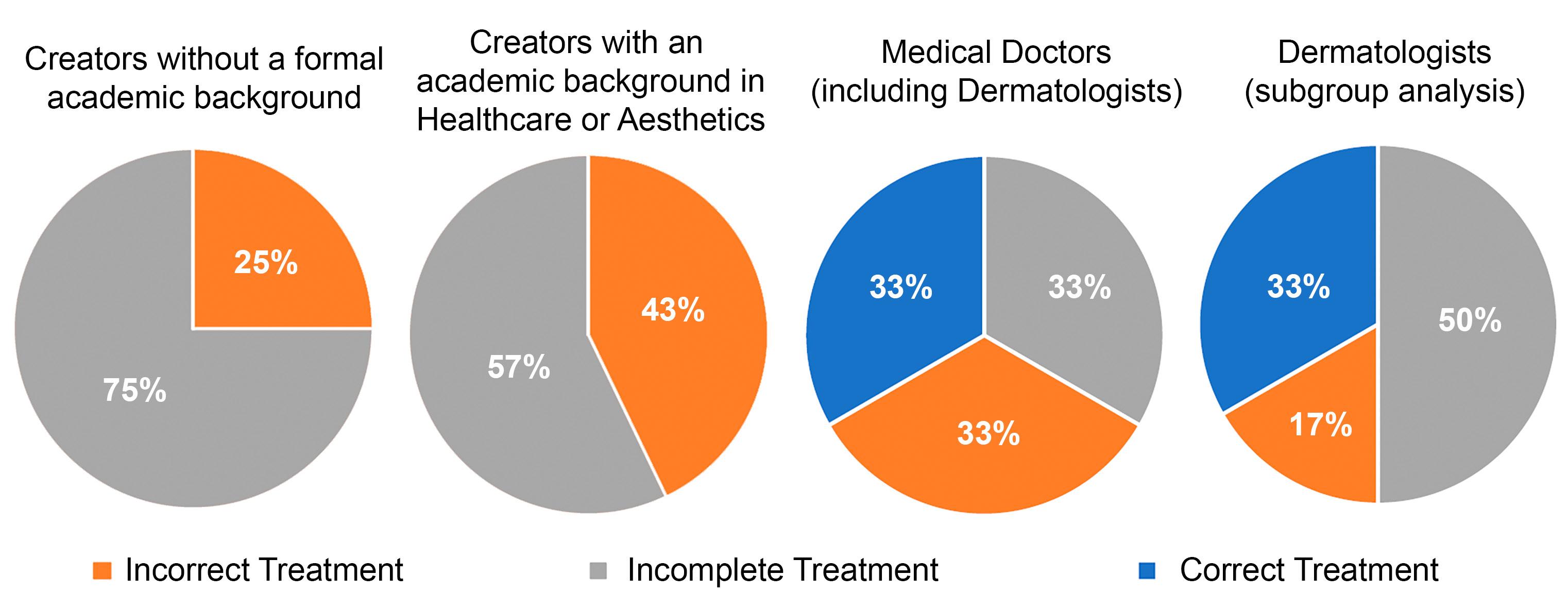
Results: Only 10% of videos recommended treatments consistent with AAD guidelines. No non-medical creator provided correct recommendations, and only 33% of physician-created videos were correct. PEMAT-A/V scores were high, indicating well edited and understandable content, whereas JAMA scores were uniformly low, reflecting poor reliability.
Conclusions: Caution is warranted when using TikTok as a source of information on acne treatment in Brazilian Portuguese, as most videos analyzed were incorrect or imprecise.
Keywords: Acne Vulgaris; Cross-sectional studies; Social Media; Health Communication
Original Article
Authors:
Vitor Costa Fabris1,2
Inae Paula Battisti1 Maria Luísa Rocha Dadalt1
1 UNIARP, Faculdade de Medicina, Caçador (SC), Brazil
2 Centro de Estudos em Dermatologia Estética, Caçador (SC), Brazil
RESUMO
Introdução: O TikTok é uma plataforma de vídeos curtos popular entre jovens, grupo etário mais afetado pela acne. O conteúdo de saúde nessa rede frequentemente é produzido por criadores sem formação acadêmica em saúde, gerando preocupações quanto à qualidade e confiabilidade das informações.
Objetivo: Avaliar a qualidade educacional, a confiabilidade e o embasamento científico de vídeos do TikTok em português brasileiro sobre tratamento da acne.
Métodos: Os 30 “top videos” sobre “tratamento da acne” foram selecionados usando um novo perfil do TikTok. A qualidade educacional foi avaliada usando a Patient Education Materials Assessment Tool for Audiovisual Materials (PEMAT-A/V), o embasamento científico usando as Diretrizes de 2024 para o Tratamento da Acne Vulgar da American Academy of Dermatology (AAD) e a confiabilidade usando o Journal of the American Medical Association (JAMA) Benchmark Criteria.
Resultados: Apenas 10% dos vídeos recomendaram tratamentos corretos segundo as diretrizes da AAD. Nenhum criador não médico indicou condutas adequadas. A pontuação do PEMAT-A/V foi alta, indicando fácil compreensão; a pontuação no JAMA Benchmark Criteria foi baixa.
Conclusão: Recomenda-se cautela ao usar o TikTok como fonte de informação sobre tratamento da acne em português brasileiro, pois a maioria dos vídeos apresentou recomendações incorretas ou imprecisas.
Palavras-chave: Acne Vulgar; Comunicação em saúde; Dermatologia; Análise de Rede Social
How to cite this article: Fabris VC, Battisti IP, Dadalt MLR. Assessing acne treatment information on TikTok: A cross-sectional pilot study of Brazilian Portuguese content. Surg Cosmetc Dermatol. 2026;18(2):e20260544.
INTRODUCTION
TikTok is an online social network and short-video platform that has experienced rapid growth since its launch in 2017. It is characterized by short, engaging, user-generated videos and is particularly popular among individuals under 29 years of age, who account for approximately 60% of its audience.1 As the internet has become an important medium through which patients seek health information, there has been a notable increase in the creation of health-related videos on online social networks, including TikTok. Such content is produced not only by dermatologists, physicians, and health educators, but also by creators without formal academic training in health sciences, raising concerns about the quality and reliability of the information available on the platform.2
Most TikTok users fall within the age group most affected by acne.1,3 Due to their young age, insecurity about their looks, and high internet usage, this population is particularly vulnerable to online misinformation.4 Moreover, studies have shown that patients with acne actively seek therapeutic options online and may adopt treatments found on the internet without consulting a dermatologist or physician to determine whether the intervention is appropriate for their specific case.5
Considering that, if left untreated or if treatment is delayed, moderate and severe forms of acne can lead to scarring and long-term disfigurement,6 it is important to assess the quality of acne-related content to which patients are exposed on TikTok, as such content may negatively influence patient behavior. Studies have shown that videos about acne produced in English have low informational quality, even when created by health educators,7 but there is a lack of scientific literature addressing the Brazilian population. As internet usage trends vary across cultures,8 data from other populations may not accurately reflect the Brazilian context. Therefore, this study was designed to evaluate the educational quality, reliability, and evidence-based accuracy of TikTok videos on acne treatment in Brazilian Portuguese.
METHODS
This was a cross-sectional study, with data collection conducted in March 2025 in the state of Santa Catarina, Brazil. A new user profile was created on TikTok to avoid algorithm training bias, and a search for “tratamento da acne” (Brazilian Portuguese term for “acne treatment”) was performed on the platform. The recommended Top Videos were screened individually, starting from the top, until 30 videos were identified that met the inclusion criteria: videos in Brazilian Portuguese recommending treatments for acne vulgaris. The videos were then saved for data extraction at a later date.
Two independent investigators extracted both quantitative (number of views, likes, shares, and video length) and qualitative (creator academic background and gender) data from the videos. The educational quality of the videos was assessed using the Patient Education Materials Assessment Tool for Audiovisual
Materials (PEMAT-A/V),9 a validated instrument designed to measure how understandable and actionable health education materials are for patients. PEMAT-A/V scores range from 0% to 100%, with higher scores indicating better educational quality. Two investigators independently studied the PEMAT-A/V scale and underwent joint training to maximize consistency in their assessments. Given that the scale includes over 20 items, complete agreement was unlikely; therefore, the mean of their scores was used as the final value. For all other variables assessed in the study, a third researcher was consulted to resolve any disagreements.
It is important to note that the PEMAT-A/V does not evaluate the scientific accuracy of the content—a video may achieve a perfect score while presenting information that is not evidence-based.Taking this into account, and to allow for a more robust evaluation of evidence-based accuracy and reliability, the videos were also assessed using the Journal of the American Medical Association (JAMA) Benchmark Criteria10 and the 2024 Guidelines of Care for the Management of Acne Vulgaris of the American Academy of Dermatology11 (hereafter referred to as the 2024 AAD guidelines for the management of acne).
The JAMA Benchmark Criteria assess the quality and reliability of online health information across four domains: authorship, attribution, disclosure, and currency. Scores of 3 or 4 are generally considered indicative of high reliability, whereas scores of 2 or less suggest low reliability.
The 2024 AAD guidelines for the management of acne were used to assess whether treatment recommendations in the videos were correct, incomplete, or incorrect. Treatments for mild acne were considered correct if they included multimodal topical therapy comprising a retinoid (adapalene, tazarotene, tretinoin, or trifarotene), salicylic acid, or azelaic acid, combined with benzoyl peroxide (alone or with other topical antibiotics) and/or clascoterone. Treatments for moderate to severe acne were considered correct if they combined a systemic treatment with topical therapy or prescribed isotretinoin. Systemic treatments included systemic antibiotics (doxycycline, minocycline, or sarecycline), hormonal agents (combined oral contraceptives or spironolactone), and isotretinoin administered as a daily regimen rather than intermittently. Treatments were classified as incomplete if they recommended products included in the 2024 AAD guidelines for the management of acne but did not constitute a multimodal regimen for mild acne or failed to include systemic therapy for moderate-to-severe acne. The recommendation of any active ingredient not included in the guidelines was considered an incorrect treatment. All incorrect treatments were recorded and subsequently compiled for qualitative analysis. In addition, the investigators documented whether a video functioned as promotion for a single product, either through explicit company sponsorship or through self-reported financial benefit from product sales, and recorded the corresponding brand when applicable.
The initial study design did not include any statistical analysis, as it was not possible to anticipate whether the resulting videos would exhibit characteristics allowing for quantitative comparisons among pre-specified groups— for example, the study could have identified 30 videos created by the same category of creator, such as individuals without formal academic training in health sciences. For this reason, all statistical analyses were performed post hoc and should be regarded as exploratory, as no prior sample-size calculation was conducted.
For statistical analysis, the videos were divided into three groups: creators without a formal academic background in health care or aesthetics, creators with an academic background in health care or aesthetics, and medical doctors (including dermatologists).
Dermatologists were not classified separately from other medical doctors because the resulting number of videos per group would have been too low, reducing the sensitivity of the statistical power of the analysis. However, the quantitative and qualitative characteristics of videos produced by dermatologists were recorded in a subgroup and reported, even though they were not included in the formal statistical comparisons. Likes and shares of the videos were reported as percentages to serve as a measure of user engagement. While videos with more views tend to accumulate a higher absolute number of likes and shares, less popular videos may have a higher proportion of these interactions, indicating greater relative user engagement.
Normality was assessed using the Shapiro-Wilk test, with a p-value of 0.05 as the threshold for normality. Parametric data were reported as mean (standard deviation) and analyzed using one-way ANOVA, whereas non-parametric data were reported as median and interquartile range and analyzed using the Kruskal-Wallis test. Categorical variables were analyzed using the chi-square test. For all analyses, an alpha level of 0.05 was considered the threshold for statistical significance. Data analysis was performed using GNU PSPP software, version 2.0.0.
RESULTS
A total of 197 videos were screened, of which 30 met the study’s inclusion criteria.Videos were excluded if they were not in Brazilian Portuguese or did not address acne treatments (eg, those focusing on acne pathophysiology or personal patient experiences). Table 1 presents the data for each group, while Table 2 provides the results for the dermatologist subgroup.The group of video creators with an academic background in health care or aesthetics consisted of three pharmacists, three biomedical professionals, and one aesthetician.
Figure 1 illustrates the rates of correct, incorrect, and incomplete treatment recommendations in each group, including the subgroup analysis of dermatologist-created videos. Table 3 summarizes all incorrect treatments recommended in the videos. Seven of the 30 videos were promotional in nature, corresponding to 23% of the sample. Three were from Creamy, 2 from Visara, 1 from Beiersdorf, and 1 from TheraSkin.
DISCUSSION
These results provide insights into the current landscape of TikTok videos on acne treatment. First, the quality of treatments recommended in the most-viewed videos is very low— only 10% of the videos recommended treatments in accordance with the 2024 AAD guidelines for the management of acne. Notably, none of the creators without a medical degree recommended correct acne treatments, and even among physicians, the rate of correct recommendations was low (33%). This finding is in line with studies assessing the quality of videos about acne in American English7 and is consistent with research in other areas of medicine, where TikTok videos have been shown to provide low-quality information on topics such as oncology,12-14 urology,15-17 and cardiology.18 It is likely that this trend of online misinformation on TikTok extends across all areas of medicine, and one potential explanation may be reflected in the PEMAT-A/V scores observed in the videos analyzed in this study.
PEMAT-A/V scores were high for all videos included in this study, regardless of creator background. This suggests that, on TikTok, video formatting may be more important than content for achieving high view counts and engagement. High PEMAT-A/V scores indicate that the videos were well edited and easily understandable to the audience, even when conveying health misinformation. Research in education has shown that student perceptions of lecture quality are often influenced more by lecturer charisma than by the content of the lecture itself.19 A similar phenomenon may occur with audiovisual content on TikTok: the manner in which the message is delivered may be more influential than the message itself in reaching wide audiences. This does not imply that evidence-based, high-quality videos on acne treatment do not exist on TikTok, but the results of this study suggest that such videos are not reaching broader audiences effectively.
One of the primary barriers to effective, evidence-based science communication on TikTok is its short-video format. Median video length in this study was approximately 1 minute, often too short to convey the complexity required for prescribing evidence-based acne treatments. This structural limitation likely contributes to the poor quality of medical content disseminated. Notably, none of the 30 videos assessed achieved a score higher than 1 on the JAMA Benchmark Criteria, with that single point awarded stemming from the platform’s automatic disclosure of the publication date rather than from any deliberate effort by content creators to ensure transparency or reliability. Most videos lasted less than 90 seconds, and creators carefully optimize the content presented within this brief time frame. Consequently, creators may omit information on potential conflicts of interest or the sources used to support their content, as such details are unlikely to interest the average viewer. The result is that the most-viewed videos on acne treatment frequently rely on a fallacy known as the argument from authority, or appeal to authority20—the perceived correctness of a treatment is judged based on the status of the person recommending it rather than
Table 1: Qualitative and quantitative characteristics of the study groups
n: number; IQR: interquartile range; SD: standard deviation. *All videos in the groups scored 1 on the JAMA Benchmark Criteria, all of them in the “currency” domain.
Table 2. Qualitative and quantitative characteristics of the dermatologist subgroup
n: number; IQR: interquartile range; SD: standard deviation.
* All videos in the group scored 1 on the JAMA Benchmark Criteria, all of them in the “currency” domain.
on an evidence-based approach. Without disclosure of conflicts of interest or sources, viewers have no means of verifying the claims presented. This is particularly concerning given that only 33% of videos produced by dermatologists recommended correct acne treatments, as the audience is likely to perceive these creators as authoritative figures in acne management.
The skincare market is closely linked to acne treatment and is projected to generate revenues of US$198.35 billion worldwide in 2025.21 Our study suggests that this industry is successfully leveraging online social network marketing, as 7 of the 30 most-viewed videos on acne treatment were promotional in nature. This may further degrade the health care informa-
tion consumed by TikTok users, as marketing on the platform is poorly regulated, raising concerns regarding transparency, disclosure, and potentially misleading content.22
Another notable finding of our study is that approximately half of the incorrect treatments recommended in the videos (Table 3) involved home-based remedies for acne, including teas, supplements, and lifestyle changes. This is consistent with studies showing that up to 80% of patients use non-pharmacological, home-based remedies for their illnesses.23,24 Specifically for acne, it has been estimated that up to 80% of female patients who follow acne advice online adopt either home-based remedies or dietary changes.25 This behavior is concerning, as acne is a
potentially scarring disease, and delays in effective treatment may result in poorer cosmetic outcomes.6 The incorrect treatments portrayed in these videos are also consistent with key themes of acne misinformation identified in previous studies.4 They include the pursuit of natural remedies, the belief that dietary changes can cure the disease, and the search for “miracle cures”—the idea that a single treatment can resolve acne.4 The pursuit of a miracle cure may also explain why some patients cycle through different topical treatments, even for moderate to severe acne, before seeking specialized care, failing to recognize that the issue lies not with the products used but with the need for systemic treatment.
The present study has some limitations, however. The constant influx of new videos on TikTok, as well as the undisclosed variables the platform uses to recommend content,26 li-
mits the replicability of our findings. Even though our sample of videos was relatively small, the small sample size was intentional, as the study focused exclusively on the best-performing videos on the platform.
CONCLUSIONS
These results provide insights into the current landscape of TikTok videos on acne treatment in Brazilian Portuguese.The quality of treatments recommended in the most-viewed videos was very low: only 10% provided treatment recommendations fully in line with the 2024 AAD guidelines for acne management. None of the creators without a medical degree recommended correct treatments, and even among physicians, the rate of correct recommendations was low (33%). On the other hand,
Figure 1: Analysis of treatments according to the 2024 Guidelines of Care for the Management of Acne Vulgaris of the American Academy of Dermatology.
Table 3: Incorrect acne treatments recommended in the videos
the videos were generally well edited, engaging, and easy to understand, as reflected by their high PEMAT-A/V scores. Given these characteristics, coupled with low JAMA Benchmark Criteria scores—which indicate that viewers cannot reliably assess whether the proposed treatments are supported by legitimate sources—caution is warranted regarding the use of TikTok as a source of information on acne treatment. Most treatments recommended in the most-viewed videos in Brazilian Portuguese analyzed in this study were either incorrect or incomplete.
Our findings also highlight the importance of physician participation—particularly that of dermatologists—in the production of health education content about acne on social networks. In our dataset, none of the videos published by creators without a medical degree fully adhered to the treatment recommendations of the 2024 AAD guidelines for the management of acne, while the subgroup of videos created by dermatologists showed the highest level of adherence to evidence-based recommendations. l
REFERENCES:
1. Jain L, Velez L, Karlapati S, Forand M, Kannali R, Yousaf RA, et al. Exploring Problematic TikTok Use and Mental Health Issues: A Systematic Review of Empirical Studies. J Prim Care Community Health. 2025;16:21501319251327303.
2. Campbell J, Williams K, Woolery-Lloyd H. DermTok: How TikTok Is Changing the Landscape of Dermatology Patient Education. J Drugs Dermatol. 2023;22(3):302-4.
3. Kutlu Ö, Karadağ AS, Wollina U. Adult acne versus adolescent acne: a narrative review with a focus on epidemiology to treatment. An Bras Dermatol. 2023;98(1):75-83.
4. O'Connor C, O'Grady C, Murphy M. Spotting fake news: a qualitative review of misinformation and conspiracy theories in acne vulgaris. Clin Exp Dermatol. 2022;47(9):1707-11.
5. Ertekin SS, Salici NS, Manav Bas V, Karali MG, Ergün EZ, Avcı EB, et al. Influence of Social Media and Internet on Treatment Decisions in Adult Female Acne Patients: A Cross-Sectional Survey Study. Dermatol Pract Concept. 2024;14(3):e2024156.
6. Rocha M, Barnes F, Calderón J, Fierro-Arias L, Gomez CEM, Munoz C, et al. Acne treatment challenges - Recommendations of Latin American expert consensus. An Bras Dermatol. 2024;99(3):414-24.
7. Irfan B, Yasin I, Yaqoob A. Navigating Digital Dermatology: An Analysis of Acne-Related Content on TikTok. Cureus. 2023;15(9):e45226.
8. Hermeking, Marc. Culture and Internet Consumption: Contributions from Cross-Cultural Marketing and Advertising Research. J. Computer-Mediated Communication. 2005;11:192-216.
9. Shoemaker SJ, Wolf MS, Brach C. Patient Education Materials Assessment Tool for Audiovisual Materials (PEMAT-A/V). Rockville, MD: Agency for Healthcare Research and Quality; 2013. Report No.: AHRQ Publication No. 14-0002-EF.
10. Silberg WM, Lundberg GD, Musacchio RA. Assessing, controlling, and assuring the quality of medical information on the Internet: Caveant lector et viewor—Let the reader and viewer beware. JAMA. 1997;277(15):1244–5.
11. Reynolds RV, Yeung H, Cheng CE, Cook-Bolden F, Desai SR, Druby KM, et al. Guidelines of care for the management of acne vulgaris. J Am Acad Dermatol. 2024;90(5):1006.e1–1006.e30.
12. Ayer D, Jain A, Singh M, Tawfik A, Tadros M. Understanding TikTok's Role in Young-Onset Colorectal Cancer Awareness and Education. J Cancer Educ. 2025.
13. Duggal S, Mahapatra S, Garrison K, Robles A. Assessing Gastrointestinal Awareness on TikTok: A Content Analysis of Colorectal Cancer, IBS, and IBD Narratives. Am J Gastroenterol. 2025;120(8):1870-3.
14. Zhu W, He B, Wang X, Du Y, Young K, Jiang S. Information quality of videos related to esophageal cancer on tiktok, kwai, and bilibili: a cross-sectional study. BMC Public Health. 2025;25(1):2245.
15. O'Sullivan NJ, Nason G, Manecksha RP, O'Kelly F. The unintentional spread of misinformation on 'TikTok'; A paediatric urological perspective. J Pediatr Urol. 2022;18(3):371-5.
16. Salka B, Aljamal M, Almsaddi F, Kaakarli H, Nesi L, Lim K. TikTok as an Educational Tool for Kidney Stone Prevention. Cureus. 2023;15(11):e48789.
17. Wong HPN, So WZ, Senthamil Selvan V, Lee JY, Ho CERH, Tiong HY. A cross-sectional quality assessment of TikTok content on benign prostatic hyperplasia. World J Urol. 2023;41(11):3051-7.
18. Gong X, Zhang Z, Dong B, Pan H. TikTok's cardiopulmonary exercise testing videos: A content analysis of quality and misinformation. Digit Health. 2025;11:20552076251341090.
19. Ware JE Jr, Williams RG. The Dr. Fox effect: a study of lecturer effectiveness and ratings of instruction. J Med Educ. 1975;50(2):149-56.
20. Logically Fallacious: The Ultimate Collection of Over 300 Logical Fallacies (Academic Edition). 1st ed. San Francisco, CA: Academic Edition; 2012.
21. Statista [Internet]. Skin Care – Worldwide. Statista. [Cited September 10, 2025]. Available from: https://www.statista.com/outlook/cmo/ beauty-personal-care/skin-care/worldwide
22. Maharany, Sylvana. Consumer Protection in the Digital Economy Era: Tiktok Shop Case Study. JISIP (Jurnal Ilmu Sosial dan Pendidikan). 2024;8:1652.
23. Parisius LM, Stock-Schröer B, Berger S, Hermann K, Joos S. Use of home remedies: a cross-sectional survey of patients in Germany. BMC Fam Pract. 2014;15:116.
24. Winkler NE, Sebo P, Haller DM, Maisonneuve H. Primary care patients' perspectives on the use of non-pharmacological home remedies in Geneva: a cross-sectional study. BMC Complement Med Ther. 2022;22(1):126.
25. Ertekin SS, Salici NS, Manav Bas V, Karali MG, Ergün EZ, Avcı EB, et al. Influence of Social Media and Internet on Treatment Decisions in Adult Female Acne Patients: A Cross-Sectional Survey Study. Dermatol Pract Concept. 2024;14(3):e2024156.
26. Zhou, Ren. Understanding the Impact of TikTok's Recommendation Algorithm on User Engagement. International Journal of Computer Science and Information Technology. 2024;3:201-8.
AUTHOR’S CONTRIBUTION:
Vitor Costa Fabris
0000-0002-3540-2769
Statistical analysis, Approval of the final version of the manuscript, Conception and design of the study, Preparation and writing of the manuscript, Acquisition, analysis and interpretation of data, Effective participation in the conduct of the study, Critical review of the literature, Critical revision of the manuscript.
Inae Paula Battisti
0009-0008-8182-5835
Conception and design of the study, Preparation and writing of the manuscript, Acquisition, analysis and interpretation of data, Critical review of the literature, Critical revision of the manuscript.
Maria Luísa Rocha Dadalt
0000-0001-8497-0546
Approval of the final version of the manuscript, Conception and design of the study, Preparation and writing of the manuscript, Effective participation in the conduct of the study, Critical review of the literature, Critical revision of the manuscript.
www.surgicalcosmetic.org.br/
Complications in Dermatologic Surgery: A Case Series Study
Complicações na Cirurgia Dermatológica: Estudo de Série de Casos
Introduction: Dermatologic surgery is widely used in the treatment of cutaneous neoplasms, but early and late complications remain underreported despite their functional and aesthetic impact.
Objective: To analyze complications in dermatologic surgery and their correlation with the repair technique, anatomical location, and tumor subtype.
Methods: A retrospective study of 107 lesions from 86 patients who underwent resection of skin tumors under local anesthesia between May 2023 and March 2024. Clinical, histological, and technical variables were assessed. Early complications (≤ 4 weeks) and late complications (1-6 months) were analyzed using SPSS v.29 (p < 0.05).
Results: Mean patient age was 73.3 years, and 57% were women. Most lesions were located on the face (69.2%), and basal cell carcinoma was the most frequent subtype (64.5%). Primary closure was performed in 49.5% of cases, and flaps in 44.9%. Early complications occurred in 42.1%, mainly dehiscence; late complications occurred in 13.1%, predominantly unaesthetic scars. Flaps had higher rates of early (53.7% vs. 30.2%) and late complications (25.9% vs. 1.9%). Lower-limb lesions showed a high complication rate. Early complications strongly correlated with late events (33.3% vs. 2.1%).
Conclusions: Complications are frequent and influenced by technique and location. Complex techniques and early events increase the risk of late outcomes.
Keywords: Dermatology; Skin Neoplasms; Surgical Flaps; Medical Oncology; Postoperative Complications
Original Article
Authors:
Luiz Roberto Dal Bem Pires Júnior1,2 Maira Mitsue Mukai1
1 Hospital de Clínicas, Universidade Federal do Paraná (UFPR), Dermatology, Curitiba (PR), Brazil
2 Hospital de Clínicas da Unicamp, Dermatology, Campinas (SP), Brazil
RESUMO
Introdução: A cirurgia dermatológica é amplamente utilizada no tratamento de neoplasias cutâneas, mas complicações precoces e tardias seguem pouco relatadas, apesar do impacto funcional e estético.
Objetivo: Analisar complicações na cirurgia dermatológica, correlacionando-as com a técnica utilizada no reparo, a localização anatômica e o subtipo do tumor.
Métodos: Estudo retrospectivo com 107 lesões de 86 pacientes submetidos à exérese de tumores cutâneos sob anestesia local entre maio de 2023 e março de 2024. Variáveis clínicas, histológicas e técnicas foram avaliadas. Complicações precoces (≤ 4 semanas) e tardias (1-6 meses) foram analisadas (SPSS v.29; p < 0,05).
Resultados: A média de idade foi 73,3 anos, com 57% de mulheres. A face concentrou a maioria das lesões (69,2%) e o carcinoma basocelular foi o subtipo mais frequente (64,5%). O fechamento primário foi utilizado em 49,5% dos casos e retalhos em 44,9%. Complicações precoces ocorreram em 42,1%, sobretudo deiscências, e tardias em 13,1%, principalmente cicatrizes inestéticas. Retalhos apresentaram mais complicações precoces (53,7% vs. 30,2%) e tardias (25,9% vs. 1,9%). Lesões em membros inferiores tiveram alta taxa de eventos. Complicações precoces mostraram forte associação com intercorrências tardias (33,3% vs. 2,1%).
Conclusões: Complicações são frequentes e influenciadas pela técnica e localização. Técnicas complexas e eventos precoces aumentam o risco de desfechos tardios.
Luiz Roberto Dal Bem Pires Júnior E-mail: lrdalbem@gmail.com
Funding: None
Conflict of interest: None
Submitted on: 11/19/2025
Final decision: 4/9/2026
How to cite this article: Pires Júnior LRDB, Mukai MM. Complications in Dermatologic Surgery: A Case Series Study. Surg Cosmet Dermatol. 2026;18(2):e20260525.
INTRODUCTION
Skin cancer is the most common malignant neoplasm worldwide, arising primarily from cumulative exposure to ultraviolet (UV) radiation. 1-3
Skin cancers are classified into melanoma and non-melanoma skin cancer (NMSC), the latter consisting primarily of basal cell carcinoma (BCC) and cutaneous squamous cell carcinoma (SCC), which together account for the majority of cases.4
Outpatient dermatologic surgery performed under local anesthesia is widely adopted and recognized for its safety and efficacy. 5-8
Multicenter studies have reported complication rates ranging from 1.6% to 12.7%, depending on procedure complexity, anatomical location, and patient risk factors.5-6,9
The most frequent complications include wound dehiscence (3.3%), surgical site infection (2.9%–8.7%), hematoma, and bleeding.5-6,10
Wound dehiscence represents an important complication resulting from the interaction between mechanical factors (such as excessive tension, inadequate closure technique, and inappropriate undermining) and physiological factors, including local vascularization, comorbidities, and characteristics of the excised tumor.6,11,12
Anatomical location plays a key role in determining the risk of complications. The risk of infection and overall complications in the lower limbs is up to three times higher due to reduced tissue perfusion, increased skin tension, and impaired lymphatic drainage.13-15 In contrast, the trunk exhibits the lowest complication rates, benefiting from better vascularization and reduced mobility.5,14 The face, although frequently subjected to complex reconstructions, has intermediate outcomes.10,16
The size of the surgical defect constitutes an independent risk factor for complications. Defects larger than 10 mm are associated with a 1.52-fold higher risk of complications, whereas defects exceeding 10 cm² increase the risk of infection by 3.64-fold.5,13,15
Tumor type also influences the risk of complications, with invasive and in situ SCC carrying a 2.96-fold greater risk of infection compared with other skin neoplasms.13,17,18
Appropriate surgical techniques are essential for minimizing complications. Proper undermining can reduce closure tension by as much as 83%–92%, making it essential in closure under tension.19-22 Deep-layer suturing is responsible for most of the approximation and eversion of wound edges, significantly reducing tension on the epidermal suture and the risk of wound dehiscence.23-26
Patient-related factors also influence surgical outcomes. Advanced age is associated with impaired wound healing due to changes in skin composition and thickness, reduced microcirculation, and compromised immune response.27-32 Immunosuppression and male sex, in turn, are risk factors for surgical site infection.13,33,34
Prolonged operative time (≥ 38 minutes) is also associated with an increased risk of complications, possibly reflecting procedural complexity.5
There is a strong correlation between early and late complications. Patients who develop complications in the immediate postoperative period are significantly more likely to develop long-term sequelae, such as hypertrophic scars, retractions, and functional impairment, highlighting the importance of rigorous early management of these complications to prevent their progression into chronic conditions.10,35
Given this context, the present study aimed to analyze postoperative complications, tumor control, functional preservation, and aesthetic outcomes in patients undergoing skin cancer resection, considering different reconstructive techniques and anatomical regions, based on data obtained from the outpatient Dermatology Department of a university hospital.
METHODS
A retrospective observational study was conducted using medical record data and image analyses from the database of the Dermatology Department of a university hospital. Patients who underwent skin cancer resection followed by reconstruction in a single surgical time, on an outpatient basis and under local anesthesia, between May 10, 2023, and March 27, 2024, were selected.
Patients of all age groups and both sexes were included, provided they had complete surgical records, including details regarding the surgical techniques used and the body areas affected, and attended follow-up evaluations during the 6 months following surgery.
Early complications, such as wound dehiscence, hematoma, infection, necrosis, and bleeding, were monitored during the first 4 weeks after the procedure. Late complications, assessed during the 6-month follow-up period, included abnormal wound healing (hypertrophic, incomplete, or keloid scars), aesthetic alterations (facial or body asymmetry, changes in skin texture, or loss of function), and altered sensitivity.
The data were organized in an Excel® spreadsheet and analyzed using IBM SPSS Statistics v.29.0 (Armonk, NY: IBM Corp.). Age data were described using means, standard deviations, medians, minimums, and maximums. Frequencies and percentages were presented for categorical variables. Associations between two categorical variables were assessed using Fisher’s exact test or the chi-square test. A p-value < 0.05 was considered statistically significant (Figure 1).
RESULTS
A total of 107 lesions from 86 patients who underwent outpatient dermatologic surgery for resection of skin neoplasms were analyzed. Most participants (81.4%) had only one lesion, while a single patient had four lesions treated during the study period.
Mean age at the time of surgery was 73.3 years (range: 40–94 years), with a predominance of individuals older than 60 years (91.9%). There was a slight predominance of female patients, accounting for 57% of participants.
Sousa LMG, Barros JAMM, Sobral RFL, Nunes ABA.
Regarding the reconstructive techniques employed, primary closure was the most commonly used, being performed in 49.5% of lesions (53 cases). Skin flaps were used in 44.9% of cases (48 lesions), whereas skin grafts were employed in 3.7%
(4 lesions). Two reconstructions involved a combination of techniques, including a flap associated with a graft or healing by secondary intention.
The anatomical distribution of lesions showed a predominance of facial involvement, with 74 cases (69.2%), followed by the trunk (10.3%), hands (5.6%), and lower limbs (4.7%). Other locations included the scalp, upper limbs, and cervical region, each accounting for less than 4% of cases.
From a histopathological perspective, BCC was the most prevalent tumor type, diagnosed in 69 cases (64.5%), followed by SCC in 26 cases (24.3%). Melanoma was identified in 6 cases (5.6%). Collision tumors (BCC + SCC) accounted for 2.8% of cases, while basosquamous carcinoma, sebaceous carcinoma, and eccrine spiradenoma accounted for one case each.
Positive surgical margins were observed in 7.5% of lesions. Among these, two lesions showed simultaneous involvement of both deep and lateral margins, whereas three had isolated lateral margin involvement and three had isolated deep margin involvement.
Early complications
Early complications occurred in 42.1% of lesions (45 cases). Wound dehiscence was the most frequent complication, occurring in 25.2% of cases, followed by hematomas (16.8%) and infections (9.3%). Bleeding was the least frequent complication, occurring in only 3.7% of lesions (Figure 2).
Early complications showed a variable distribution, depending on the surgical technique employed. Flaps presented the highest rate of complications (54.2%), followed by grafts (50.0%), whereas primary closure had the lowest rate (30.2%). When techniques were grouped into primary closures and complex closures (flaps, grafts, and combined techniques), the rate of early complications was 30.2% for primary closures compared with 53.7% for complex closures. This difference was statistically significant (p = 0.019) (Table 1).
Regarding anatomical location, lower limbs exhibited complications in 100% of treated lesions, whereas the trunk showed the lowest rate of early complications (18.2%) (Table 1). Among tumor subtypes, BCC was most frequently associated with early complications (44.9%), followed by SCC (38.5%) and melanoma (16.7%). However, comparison among these variables did not find a statistically significant association (p = 0.376) (Table 1).
Late complications
Late complications were less frequent, occurring in 14.0% of lesions (15 cases). Among these, abnormal scarring was the most common outcome (46.7%), followed by aesthetic complications (40.0%) and altered sensitivity (6.7%).
Late complications also showed statistically significant differences among the techniques used (p < 0.001), with rates of 25.9% for complex closures compared with 1.9% for primary closures. Among reconstructive techniques, flaps continued to
Figure 1: Postoperative infection and necrosis of scalp flap
Figure 2: Postoperative hematoma of periocular flap
Table 1: Lesion with early complication (n = 107)
*Fisher’s exact test or chi-square test, p < 0.05
exhibit the highest prevalence of late complications (27.1%), followed by grafts (25.0%) (Table 2).
Regarding anatomical location, the lower limbs again stood out, with a 100% rate of late complications. In contrast, the scalp, upper limbs, and cervical region showed no late complications (Table 2).
Among tumor subtypes, although the difference was not statistically significant (p = 0.396), melanoma had the highest rate of late complications (33.3%), followed by BCC (14.5%) and SCC (11.5%) (Table 2).
Furthermore, patients who experienced early complications were more likely to develop late complications (33.3%) than those without early complications (2.1%), and this difference was statistically significant (p < 0.001) (Table 3).
DISCUSSION
This study reinforces the safety and effectiveness of outpatient dermatological procedures for the treatment of skin neoplasms while identifying critical factors associated with the development of postoperative complications.
The demographic profile observed, with 91.9% of patients older than 60 years and a slight female predominance (57%), is consistent with the global epidemiological pattern of skin cancers, which predominantly affect older individuals due to the cumulative effects of sun exposure and the physiological changes associated with aging.1-6
Profile and magnitude of early complications
The incidence of immediate postoperative complications was 42.1%, a value considerably higher than the 1.6%–12.7% range reported in large multicenter series.6-8 Wound dehiscence
(25.2%) was the most common complication and substantially exceeded the previously reported rates of 0.5%–3.3%.5-7 This discrepancy may be attributed to the greater complexity of the reconstructions performed, the unfavorable anatomical distribution of the lesions, and the specific clinical characteristics of the study population, including advanced age, multiple comorbidities, and limited access to health care.
This complication occurs when there is an imbalance between the mechanical forces applied to the tissue—including excessive tension, suboptimal undermining technique, and inadequate suture method—and the physiological capacity for wound healing, which is influenced by tissue perfusion, associated clinical conditions, and the nature of the excised lesion.4,7,9,10 Layered closure transfers most of the mechanical load to the subdermal layers, significantly relieving tension on the superficial suture line and reducing the risk of wound edge separation.8,10,13,14
Other early complications included hematomas (16.8%), infections (9.3%), and bleeding (3.7%), all occurring at frequencies higher than those usually reported in the literature.5-7,12,14,15
Impact of reconstructive complexity and anatomical location
Comparative analysis revealed that complex reconstructions (flaps, grafts, and combined techniques) resulted in a 53.7% rate of early complications, compared with 30.2% for primary closures (p = 0.019).5-7 This disparity reflects the greater tissue trauma, longer operative times, and more demanding technical requirements needed to ensure adequate perfusion and satisfactory functional outcomes (Figure 3).5,6
Lesion topography had a substantial influence on outcomes. The lower limbs exhibited complications in 100% of cases,
Pathology Basal cell carcinoma (n = 107)
Squamous cell carcinoma (3 most frequent classifications) Melanoma
*Fisher’s exact test or chi-square test, p < 0.05
Table 2: Lesion with late complication (n = 107)
Patient has early complication in a lesion (n = 86)
3: Association between early and late complications
confirming previous reports indicating the risk of infection and other complications is three times higher in this region, attributed to limited blood supply, increased skin tension, and impaired lymphatic drainage.14-16 In contrast, the trunk had the lowest rate of early complications (18.2%).6,15 The facial region exhibited intermediate outcomes, likely benefiting from its rich vascular network (Figure 4).5,17
Late complications and their relationship with early events
Late complications occurred in 14% of postoperative cases, predominantly abnormal scarring (46.7%) and aesthetic impairment (40%). Analysis by reconstructive technique revealed a statistically significant difference, with late complication rates of 25.9% in complex reconstructions versus 1.9% in primary closures.5
Table
The robust association between early and late complications represents a finding of particular clinical relevance. Patients who developed early postoperative complications were markedly more likely to experience persistent sequelae (33.3% vs. 2.1%; p < 0.001), underscoring the importance of prompt and rigorous intervention to prevent their progression into chronic conditions.4,5 Events such as wound dehiscence, blood pooling, and infection during the early postoperative period may compromise the healing process, ultimately resulting in permanent sequelae such as hypertrophic scars, tissue contractures, and functional deficits.4,9,10
Once again, the lower limbs ha a 100% rate of late complications, reinforcing the particular vulnerability of this anatomical region.14-16
Influence of histological type and other risk determinants
Although no statistically significant association was identified between tumor subtype and complications, melanoma presents unique challenges. Its wide resections, often performed across skin tension lines, increase the risk of late complications, present in 33.3% of melanoma cases, compared with BCC (14.5% late complications; 44.9% early complications) and SCC (11.5% late complications; 38.5% early complications).
Study limitations
This study has limitations inherent to its retrospective design, single-center setting, and potential variability in technical expertise among surgeons. The limited number of cases involving rare neoplasms or severe complications restricted statistical analyses in specific subgroups. Future prospective multicenter studies with larger sample sizes will be necessary to validate and expand upon these findings.
CONCLUSION
The outcomes of dermatologic surgery for skin cancer result from the interaction of anatomical, technical, and oncological factors. Differences between anatomical sites and the use of complex reconstructions were associated with increased complication rates, whereas early postoperative events were predictive of late complications. Differences among tumor subtypes imposed distinct technical demands and risk profiles. Therefore, the choice of reconstructive technique should consider not only tumor removal but also anatomical factors, tumor subtype, and functional and aesthetic impact. l
Figure 3: Postoperative graft necrosis of the medial canthus
Figure 4: Postoperative partial necrosis of nasal wing flap.
REFERENCES:
1. Zhou L, Zhong Y, Han L, Xie Y, Wan M. Global, regional, and national trends in the burden of melanoma and non-melanoma skin cancer: insights from the Global Burden of Disease Study 1990–2021. Sci Rep. 2025;15.
2. Wang R, Chen Y, Shao X, et al. Burden of skin cancer in older adults from 1990 to 2021 and modelled projection to 2050. JAMA Dermatol. 2025.
3. Arnold M, Singh D, Laversanne M, et al. Global burden of cutaneous melanoma in 2020 and projections to 2040. JAMA Dermatol. 2022;158(5):495–503.
4. Long GV, Swetter SM, Menzies AM, Gershenwald JE, Scolyer RA. Cutaneous melanoma. Lancet. 2023;402(10399):485–502.
5. Kato K, Kato H, Magara T, Morita A. Postoperative complications and risk factors in skin tumor excision with simple suturing under local anesthesia: a multicenter retrospective study in Japan. J Dermatol. 2025;52.
6. Amici JM, Rogues AM, Lasheras A, et al. A prospective study of the incidence of complications associated with dermatological surgery. Br J Dermatol. 2005;153(5):967–71.
7. Alam M, Schaeffer MR, Geisler A, et al. Safety of local intracutaneous lidocaine anesthesia used by dermatologic surgeons for skin cancer excision and postcancer reconstruction: quantification of standard injection volumes and adverse event rates. Dermatol Surg. 2016;42(12):1320–4.
8. Locke MC, Davis JC, Brothers RJ, Love WE. Assessing the outcomes, risks, and costs of local versus general anesthesia: a review with implications for cutaneous surgery. J Am Acad Dermatol. 2018;78(5):983–8.
9. O’Neill JL, Lee YS, Solomon JA, et al. Quantifying and characterizing adverse events in dermatologic surgery. Dermatol Surg. 2013;39(6):872–8.
10. Bordeaux JS, Martires KJ, Goldberg D, et al. Prospective evaluation of dermatologic surgery complications including patients on multiple antiplatelet and anticoagulant medications. J Am Acad Dermatol. 2011;65(3):576–83.
11. Sandy-Hodgetts K, Carville K, Santamaria N, Parsons R, Leslie GD. The Perth Surgical Wound Dehiscence Risk Assessment Tool (PSWDRAT): development and prospective validation in the clinical setting. J Wound Care. 2019;28(6):374–84.
12. Sandy-Hodgetts K, Carville K, Leslie GD. Surgical wound dehiscence: a conceptual framework for patient assessment. J Wound Care. 2018;27(3):119–26.
13. Schlager JG, Patzer K, Wallmichrath J, et al. Surgical site infection in skin surgery: an observational study. Int Wound J. 2023;20(1):85–94.
14. Schlager JG, Ruiz San Jose V, Patzer K, et al. Are specific body sites prone for wound infection after skin surgery? A systematic review and meta-analysis. Dermatol Surg. 2022;48(6):680–9.
15. Liu X, Sprengers M, Nelemans PJ, Mosterd K, Kelleners-Smeets NWJ. Risk factors for surgical site infections in dermatological surgery. Acta Derm Venereol. 2018;98(2):246–50.
16. Mori WS, Demer AM, Mattox AR, Maher IA. Mohs micrographic surgery at challenging anatomical sites. Dermatol Surg. 2019;45(3):401–12.
17. Nakamura Y, Sasaki K, Ishizuki S, et al. Invasive and in situ lesions of squamous cell carcinoma are independent factors for postoperative surgical-site infection after outpatient skin tumor surgery: a retrospective study of 512 patients. J Dermatol. 2021;48(8):1162–9.
AUTHOR’S CONTRIBUTION:
18. Heal CF, Buettner PG, Drobetz H. Risk factors for surgical site infection after dermatological surgery. Int J Dermatol. 2012;51(7):796–803.
20. Chen DL, Carlson EO, Fathi R, Brown MR. Undermining and hemostasis. Dermatol Surg. 2015;41(3):e1–e7.
21. Melis P, Noorlander ML, Bos KE. Tension decrease during skin stretching in undermined versus not undermined skin: an experimental study in piglets. Plast Reconstr Surg. 2001;107(5):1201–5.
22. Huang L, Wenzhi L. The role of the undermining during circular excision of secondary intention healing. Am Surg. 2014;80(9):E246–8.
23. Joo JS, Zhuang AR, Tchanque-Fossuo C, et al. Dermal suture only versus layered closure: a randomized, split wound comparative effectiveness trial. J Am Acad Dermatol. 2019;81(3):721–7.
24. Gurusamy KS, Toon CD, Allen VB, Davidson BR. Continuous versus interrupted skin sutures for non-obstetric surgery. Cochrane Database Syst Rev. 2014;(2):CD010365.
25. Luo W, Tao Y, Wang Y, et al. Comparing running vs interrupted sutures for skin closure: a systematic review and meta-analysis. Int Wound J. 2022;19(5):1198–1208.
26. Yang CS, Yeh CH, Chen MY, et al. Mechanical evaluation of the influence of different suture methods on temporal skin healing. Dermatol Surg. 2009;35(2):227–33.
27. Li J, Zhu D, Zhang M, et al. Deciphering age-related differences in wound healing: insights from the interaction between endothelial cells and fibroblasts. Mol Med Rep. 2025.
28. Bentov I, Reed MJ. Anesthesia, microcirculation, and wound repair in aging. Anesthesiology. 2014;120(3):760–72.
29. Brubaker AL, Rendon JL, Ramirez L, Choudhry MA, Kovacs EJ. Reduced neutrophil chemotaxis and infiltration contributes to delayed resolution of cutaneous wound infection with advanced age. J Immunol. 2013;190(4):1746–57.
30. Sgonc R, Gruber J. Age-related aspects of cutaneous wound healing: a mini-review. Gerontology. 2012;59(2):159–64.
31. Dube CT, Ong YHB, Wemyss K, et al. Age-related alterations in macrophage distribution and function are associated with delayed cutaneous wound healing. Front Immunol. 2022;13:821143.
32. Greenhalgh DG. Management of the skin and soft tissue in the geriatric surgical patient. Surg Clin North Am. 2015;95(1):103–14.
33. Schlager JG, Hartmann D, Wallmichrath J, et al. Patient-dependent risk factors for wound infection after skin surgery: a systematic review and meta-analysis. Int Wound J. 2022;19(4):899–909.
34. Schwartzman G, Khachemoune A. Surgical site infection after dermatologic procedures: critical reassessment of risk factors and reappraisal of rates and causes. Am J Clin Dermatol. 2021;22(4):503–13.
35. Sandy-Hodgetts K, Carville K, Leslie GD. Determining risk factors for surgical wound dehiscence: a literature review. Int Wound J. 2015;12(3):265–75.
Luiz Roberto Dal Bem Pires Júnior 0009-0001-6728-420X
Statistical analysis, Approval of the final version of the manuscript, Conception and design of the study, Preparation and writing of the manuscript, Acquisition, analysis and interpretation of data, Effective participation in the conduct of the study, Intellectual participation in the propaedeutic and/or therapeutic approach to the cases studied, Critical review of the literature, Critical revision of the manuscript
Maira Mitsue Mukai 0000-0002-4591-8451
Conception and design of the study, Critical review of the literature
www.surgicalcosmetic.org.br/
A Comment on: “Lip reconstruction using superficial flaps: functional and aesthetic integration”
Comentário sobre: “Reconstrução labial com retalhos superficiais: integração funcional e estética”
Letter to the Editor
Authors: Muhammed Said Aydin1 Gökay Özler1
1 Faculty of Medicine, Mugla Sitki Koçman University, Department of Plastic, Reconstructive and Aesthetic Surgery, Mugla, Mentese, Turkey
This Letter to the Editor comments on the article “Lip reconstruction using superficial flaps: functional and aesthetic integration.” The reported case series provides valuable clinical examples of individualized superficial flap selection for upper and lower lip reconstruction following excision of non-melanoma skin cancer. We emphasize the importance of tailored reconstructive planning while suggesting that objective functional outcome measures and clearer flap-selection criteria could further strengthen the translational impact of the study.
Keywords: Plastic Surgery Procedures; Surgical Flaps; Dermatologic Surgical Procedures
RESUMO
Esta Carta ao Editor comenta o artigo “Lip reconstruction using superficial flaps: functional and aesthetic integration”. A série de casos apresentada oferece exemplos clínicos valiosos da seleção individualizada de retalhos superficiais para reconstrução do lábio superior e inferior após excisão de câncer de pele não melanoma. Enfatizamos o valor do planejamento reconstrutivo personalizado, sugerindo que medidas funcionais objetivas e critérios mais claros de seleção dos retalhos poderiam fortalecer ainda mais o impacto translacional do estudo. Palavras-chave: Procedimentos de Cirurgia Plástica; Retalhos Cirúrgicos; Procedimentos Cirúrgico Dermatológicos
Correspondence: Muhammed Said Aydin E-mail: muhammedsaid.aydin@ gmail.com
Funding: None
Conflict of interest: None
Submitted on: 05/24/2026
Final decision: 05/27/2026
How to cite this article: Aydin MS, Özler G. A Comment on: “Lip reconstruction using superficial flaps: functional and aesthetic integration.” Surg Cosmet Dermatol. 2026;18(2):e20260586.
Aydin MS, Özler G.
We read with great interest the article by Aita et al. entitled “Lip reconstruction using superficial flaps: functional and aesthetic integration.” The authors are to be commended for presenting a well-structured case series demonstrating the successful use of multiple superficial flap techniques for reconstruction of upper and lower lip defects following surgical excision of non-melanoma skin cancers.
The main strength of the manuscript lies in its clinically oriented and individualized approach to reconstructive planning. The selection of mucosal rotation flaps, double V-Y advancement flaps, and O-Z rotation flaps according to defect characteristics reflects sound surgical reasoning and adherence to established principles of lip reconstruction. The emphasis on preserving oral sphincter function and maintaining aesthetic subunits is particularly relevant in perioral reconstruction, where both functional and cosmetic outcomes are critical.
The variety of techniques presented offers useful practical guidance for surgeons managing similar defects in routi-
ne practice. In particular, the successful application of double V-Y advancement flaps for central lip defects and O-Z rotation flaps for more complex reconstructions highlights the versatility of superficial flap techniques in achieving both symmetry and functional preservation.
However, the manuscript could be strengthened by the inclusion of more objective functional outcome measures. Although photographic documentation provides valuable evidence of aesthetic success, standardized assessments of oral competence, speech articulation, and sensory recovery would enhance the clinical robustness of the reported outcomes. In addition, a more detailed discussion of the indications, limitations, and selection criteria for each flap type would further improve the translational value of the study and assist surgeons in preoperative decision-making.
In conclusion, this case series makes a meaningful contribution to the existing literature on lip reconstruction and reinforces the importance of tailoring reconstructive strategies to defect characteristics and functional requirements. l
Cosmet Dermatol. 2026;18:e20260586.
REFERENCES:
1. Aita VH, Prócel C, Jacques NS, Cafrune FE. Lip reconstruction using superficial flaps: functional and aesthetic integration. Surg Cosmet Dermatol. 2026;18(1):e20260480.
2. Chellappan B, Obanigba G, Hoyer P, Ross L. Lip reconstruction after Mohs micrographic surgery: a guide on flaps. Cutis. 2023;111(4):205-9.
3. Shim GJ, Yoon HW, Kim D, Kwon TG. Reconstruction of an upper lip vermilion defect with a mucosal V-Y advancement flap: a case report. J Korean Assoc Oral Maxillofac Surg. 2024;50(4):222-6.
4. Lee TY, Shin YH, Choi DI. Reconstruction of medial epicanthal fold using V-Y advancement and turnover flap. J Plast Reconstr Aesthet Surg. 2020;73(2):363-8.
AUTHOR’S CONTRIBUTION:
Muhammed Said Aydin 0009-0001-9374-3131
Approval of the final version of the manuscript, Critical review of the literature, Acquisition, analysis and interpretation of data, Critical revision of the manuscript, Preparation and writing of the manuscript, Conception and design of the study.
Gökay Özler
0009-0002-7217-3894
Approval of the final version of the manuscript, Critical review of the literature, Critical revision of the manuscript, Conception and design of the study.
www.surgicalcosmetic.org.br/
Nodular hidradenoma: diagnostic innovation in a rare case
Hidradenoma nodular: inovação diagnóstica em um caso raro
Case Report
Authors:
Ana Paula Weber1
Giulia Maria dos Santos Goedert1
Barbara Brunca1
Hamilton Ometto Stolf1
Paula Tavares Colpas1
1 Hospital PUC Campinas, Dermatology, Campinas (SP), Brazil
Nodular hidradenoma is a rare benign adnexal tumor originating from the sweat glands, which clinically presents as a tumor mimicking primary or metastatic malignant lesions. Dermoscopy, as well as clinical characteristics, reveals structural patterns that allow for broad differential diagnoses. Dermatological ultrasonography has become a scientifically recognized diagnostic tool to enable early and accurate diagnosis of hidradenoma, supporting surgical treatment and histopathological analysis. This study reports a rare case of nodular hidradenoma, highlighting the diagnostic innovation achieved through low-frequency ultrasonography, which includes clinical, radiological, and histopathological characteristics. Keywords: Skin neoplasms; Ultrasonography; Diagnostic imaging; Clinical diagnosis
RESUMO
O hidradenoma nodular é um tumor anexial benigno raro, originado das glândulas sudoríparas, que, clinicamente, apresenta-se como uma tumoração que mimetiza lesões malignas primárias ou metastáticas. A dermatoscopia, assim como as características clínicas, revela padrões estruturais que possibilitam amplos diagnósticos diferenciais. A ultrassonografia dermatológica tornou-se um método diagnóstico reconhecido cientificamente para possibilitar o diagnóstico precoce e preciso do hidradenoma, contribuindo para a terapêutica cirúrgica e a análise histopatológica. Este estudo relata um caso raro de hidradenoma nodular com inovação diagnóstica por meio da ultrassonografia de baixa frequência que inclui características clínicas, radiológicas e histopatológicas. Palavras-chave: Neoplasias cutâneas; Ultrassonografia; Diagnóstico por imagem; Diagnóstico clínico
Correspondence:
Ana Paula Weber E-mail: anapaulaweber21@gmail. com
Funding: None
Conflict of interest: None
Submitted on: 04/02/2026
Final decision: 05/23/2026
How to cite this article: Weber AP, Goedert GMS, Brunca B, Stolf HO, Colpas PT. Nodular hidradenoma: diagnostic innovation in a rare case. Surg Cosmet Dermatol. 2026;18(2):e20260571.
INTRODUCTION
Nodular hidradenoma is a rare benign adnexal tumor originating from the sweat glands and represents a diagnostic challenge because it may clinically mimic malignant lesions, including basal cell carcinoma and melanoma. It affects predominantly women, with a mean age at presentation of 37 years. It is more commonly found on the scalp, face, and upper limbs and is rare on the lower limbs. It has a low potential for malignant transformation; however, diagnosis may be delayed because of its variety in clinical and morphological patterns, including diverse histopathological and clinical presentations.1,2 Currently, apocrine hidradenomas are the most common subtype and are defined by histopathological examination, since no confirmatory radiologic criteria have been described, contributing to inappropriate therapeutic approaches and a higher likelihood of local recurrence.2 In this context, dermatological ultrasonography, described in a limited number of prospective studies, has emerged as an essential tool to assist in early and accurate diagnosis, enabling appropriate surgical management.3 The present case demonstrates a rare clinical presentation of nodular hidradenoma on the lower limb (thigh), associated with diagnostic investigation using high-frequency cutaneous ultrasonography, identifying the two characteristic ultrasonographic signs first described in 2018, thereby facilitating appropriate surgical treatment and corresponding histopathological evaluation. It is important to report diagnostic innovations confirmed in a rare clinical and surgical dermatologic case.
CASE REPORT
A 50-year-old female patient presented with a violaceous mass located on the medial aspect of the right thigh, first noticed 7 months earlier, with progressive enlargement and impairment of quality of life. The patient denied any previous cutaneous disorders. Dermatological examination revealed a cystic tumor (Figures 1 and 2), slightly infiltrated, with positive transillumination (Figure 3). Ultraviolet dermoscopy showed a central homogeneous pink area and arborizing vessels (Figure 4). Diagnostic investigation was performed using dermatological ultrasonography with a 22-MHz high-frequency linear transducer, revealing a hypoechoic nodular lesion in the dermis and subcutaneous tissue, with the presence of the snow-globe sign, also known as the “snowfall sign” (Figure 5), and a fluid-fluid level (Figure 6), measuring 2.23 × 2.34 cm. The ultrasonographic findings were suggestive of nodular hidradenoma. After combined assessment of the clinical, dermoscopic, and ultrasonographic features, complete excision of the lesion was performed, given its presumed benignancy and low malignant potential. Histopathological examination described a dermal epithelial lesion composed of a densely cellular basaloid proliferation surrounded by fibrosis, with apocrine ductal differentiation, cystic spaces, and pinkish hyaline fluid content (Figure 7), consistent with nodular hidradenoma. The patient remains under follow-up with the clinical and surgical dermatology team, with no signs of local recurrence and satisfied with the treatment provided.
DISCUSSION
Nodular hidradenoma belongs to the group of benign skin adnexal tumors with a broad range of morphologic differential diagnoses, clinically simulating various primary and metastatic tumors. Clinically, it presents as a solitary nodule with cystic and serous areas, showing a predilection for the scalp and upper limbs. It is exceedingly rare on the lower limbs, as in the present case. Growth is slow and generally asymptomatic. Ma-
AP, Goedert GMS, Brunca B, Stolf HO, Colpas PT.
Figure 1: Clinical examination: tumor on the medial aspect of the right lower limb
Figure 2: Clinical examination: erythematous-violaceous cystic mass
4:
croscopically, positive transillumination is explained by serous areas containing fluid illuminated by the polarized light of the dermoscope.1 Dermoscopic findings described in the literature include a homogeneous pinkish area, whitish structures, and a
Figure 7: Hematoxylin-eosin histopathology (40× amplification): ductal structures consisting of basaloid cells with prominent apocrine differentiation
variable vascular pattern, including arborizing and atypical polymorphous vessels, most often mimicking basal cell carcinoma and melanoma.4
Given the clinical diagnostic challenge, high-frequency
Figure 3: Positive transillumination in dermoscopy
Figure
Ultraviolet dermoscopy
Figure
AP, Goedert GMS, Brunca B, Stolf HO, Colpas PT.
dermatological ultrasonography, considered an advantageous noninvasive method, was important in establishing the diagnosis through the characteristic findings of nodular hidradenoma, including the “snowfall sign” and the “fluid-fluid level,” both identified in the present case. These findings were first described in 2018 in a case series, highlighting the importance of this imaging method. It is also important to emphasize that there is no consensus regarding diagnostic and therapeutic criteria, further underscoring the importance of ultrasonography.3 Accordingly, diagnostic investigation was essential in determining the surgical approach, with juxtalesional excision representing one of the available options given the low probability of progression to hidradenocarcinoma. In this case, surgery is considered
curative, although recurrence following incomplete excision has been reported. Histopathological examination remains the gold standard for diagnostic confirmation, described by a well-circumscribed lesion composed of solid and cystic spaces, allowing identification of ductal structures with basaloid cells.2 Therefore, an accurate diagnosis in this rare case is essential for follow-up and surgical management.
CONCLUSION
We report a rare benign neoplasm in an unusual location, for which ultrasonography was fundamental to diagnosis and surgical planning. However, further scientific studies are still needed to establish ultrasonographic diagnostic criteria. l
REFERENCES:
1. Arthi M, Joseph LD, Arun Kumar K. Clinicopathological profile of nodular hidradenoma: A ten year study in a tertiary care center. J Cutan Aesth Surg. 2023;16(1):34.
2. Nandeesh BN, Rajalakshmi T. A study of histopathologic spectrum of nodular hidradenoma. Am J Dermatopathol. 2012;34(5):461-70.
3. Wortsman X, Reyes C, Ferreira-Wortsman C, Uribe A, Misad C, Gonzalez S. Sonographic characteristics of apocrine nodular hidradenoma of the skin. J Ultra Med. 2017;37(3):793-801.
4. Serrano P, Lallas A, Del Pozo LJ, Karaarslan I, Medina C, Thomas L, et al. Dermoscopy of Nodular Hidradenoma, a Great Masquerader: a morphological study of 28 cases. Dermatol. 2016;232(1):78-82.
AUTHOR’S CONTRIBUTION:
Ana Paula Weber 0000-0002-8841-7817
Statistical analysis, Approval of the final version of the manuscript, Conception and design of the study, Preparation and writing of the manuscript, Acquisition, analysis and interpretation of data, Effective participation in the conduct of the study, Intellectual participation in the propaedeutic and/or therapeutic approach to the cases studied, Critical review of the literature, Critical revision of the manuscript.
Giulia Maria dos Santos Goedert 000.0002-6672-0276
Statistical analysis, Approval of the final version of the manuscript, Conception and design of the study, Preparation and writing of the manuscript, Acquisition, analysis and interpretation of data, Effective participation in the conduct of the study, Intellectual participation in the propaedeutic and/or therapeutic approach to the cases studied, Critical review of the literature, Critical revision of the manuscript.
Barbara Brunca 0000-0002-2492-8474
Statistical analysis, Approval of the final version of the manuscript, Conception and design of the study, Preparation and writing of the manuscript, Acquisition, analysis and interpretation of data, Effective participation in the conduct of the study, Intellectual participation in the propaedeutic and/or therapeutic approach to the cases studied, Critical review of the literature, Critical revision of the manuscript.
Hamilton Ometto Stolf 0000-0003-4867-0276
Statistical analysis, Approval of the final version of the manuscript, Conception and design of the study, Preparation and writing of the manuscript, Acquisition, analysis and interpretation of data, Effective participation in the conduct of the study, Intellectual participation in the propaedeutic and/or therapeutic approach to the cases studied, Critical review of the literature, Critical revision of the manuscript.
Paula Tavares Colpas 0000-0002-1389-0749
Statistical analysis, Approval of the final version of the manuscript, Conception and design of the study, Preparation and writing of the manuscript, Acquisition, analysis and interpretation of data, Effective participation in the conduct of the study, Intellectual participation in the propaedeutic and/or therapeutic approach to the cases studied, Critical review of the literature, Critical revision of the manuscript.
Scalp reconstructions can be challenging for dermatologic surgeons because the site has little elasticity and is located over a convex, rather than flat, area. The pinwheel flap is a type of rotational flap used for the vertex region of the scalp. We describe two cases, one male and one female, using the technique to reconstruct defects after skin cancer excisions in the vertex and the frontal region of the scalp.
1 Universidade Estadual de Londrina (UEL), Dermatology, Londrina (PR), Brazil
RESUMO
Reconstruções em couro cabeludo podem ser desafiadoras para o cirurgião dermatológico, por ser um local de pouca elasticidade e estar sobre uma área convexa e não plana. O retalho em cata-vento é um tipo de retalho em rotação utilizado para a região do vértex do couro cabeludo. Relatamos dois casos, um masculino e um feminino, utilizando a técnica para reconstruir defeitos após excisões de câncer de pele, no vértex e na região frontal do couro cabeludo. Palavras-chave: Couro Cabeludo; Carcinoma Basocelular; Carcinoma de Células Escamosas; Retalhos Cirúrgicos
Correspondence:
João Marcos Franco de Souza E-mail: jmfs.md@gmail.com
Funding: None
Conflict of interest: None
Submitted on: 2/6/2026
Final decision: 4/9/2026
How to cite this article: Souza JMF, Pereira BM, Miyazawa B, Borges Junior C, Kondo RN. Pinwheel flap: a series of two cases. Surg Cosmet Dermatol. 2026;18(2):e20260556.
INTRODUCTION
Basal cell carcinoma (BCC) and squamous cell carcinoma (SCC) are the two most common types of skin cancer.1 When located on the scalp, surgical treatment can be challenging because the scalp is a convex, non-flat surface, and even relatively small defects may require flaps or grafts due to poor local tissue elasticity.2
The pinwheel flap was originally developed to correct circular defects on the scalp, specifically to overcome the limited mobility of the scalp skin, but it has also been used in other regions of the face. It consists of three or more flaps that, through rotational movement, allow local defect closure.3,4
We report two cases of patients with SCC and BCC of the scalp for whom the pinwheel flap was selected for reconstructive surgery, achieving satisfactory aesthetic outcomes.
Case reports
Case 1
A 57-year-old male patient presented with a 19-mm plaque in the central region of the scalp. A previous incisional biopsy found SCC on histopathological examination. Excision with 5-mm margins was performed, resulting in a 29-mm defect, followed by reconstruction with a pinwheel flap (Figures 1, 2 and 3).
Case 2
B - Flap elevation.
C - Flap positioned
A 47-year-old female patient presented with a 16-mm plaque in the right frontal scalp region. A previous incisional biopsy found BCC on histopathological examination. Excision with 5-mm margins was performed, resulting in a 26-mm defect, followed by reconstruction with a pinwheel flap (Figures 4, 5 and 6). A A A B B B C C
The patients were followed at 1, 3, 6, and 12 months. Histopathological examination found tumor-free margins for SCC and BCC, respectively. Both patients showed good wound healing, with no signs of recurrence and satisfactory aesthetic outcomes (Figures 3C and 6B).
DISCUSSION
Skin flaps or grafts may be necessary for closure following excision of scalp skin tumors.2 Because they preserve adjacent skin with similar characteristics,3 flaps are generally preferred over grafts, particularly to avoid alopecia in female patients, as in Case 2.2
In 1978,Vecchione and Griffith used three-part flaps rotated in the same direction to close a circular scalp defect, creating a pinwheel-like design (Figures 3A and 6A). It became a classic for use on the scalp vertex and was later extended to other scalp regions. Currently, the pinwheel flap is also used beyond the scalp, including non-hair-bearing nasal and temporal regions.3,4
One advantage of the pinwheel flap is that it can be performed in a single surgical time, unlike interpolation flaps. Because it does not require tie-over pressure dressing, as skin grafts do, it is also more comfortable for the patient.2
REFERENCES:
1. Kondo RN, Gon AS, Pontello Junior R. Recurrence rate of basal cell carcinoma in patients submitted to skin flaps or grafts. 2019;94(4):442-5.
2. Raposio G, Gualdi A, Baldelli I, Raposio E. Basal cell carcinoma of the scalp: surgical approach and reconstructive options. Ital J Dermatol Venerol. 2024;159(4):412-6.
3. Kondo RN, Cestari AI, Soares BM, Scalone FM Aybar SAI. Pinwheel flap as an option to reconstruct a nasal defect: a series of two cases. Dermatol Arch. 2021;5(1):122-6.
AUTHOR’S CONTRIBUTION:
João Marcos Franco de Souza 0000-0002-9297-7841
One option for maintaining compressive dressing for 24 hours, while preventing hemorrhage or hematoma without the need for technically demanding elastic bandages, is the Kondo compressive scalp dressing (Figure 3B).5
In Case 2, unlike Case 1, reconstruction was somewhat more challenging because the patient was female, had no androgenetic alopecia, and the lesion was located in the frontal scalp region. The flaps were designed to allow adequate rotation and defect closure while preventing the scars from extending beyond the hairline.
Single- or double-rotation flaps are also reconstructive options; however, the pinwheel flap design was chosen because it produces less cutaneous redundancy (dog-ears). This technique avoids additional tissue excision and allows application in smallto medium-sized circular defects, with excellent adaptability to the recipient area.6
Both cases progressed to favorable outcomes, with no signs of recurrence and satisfactory aesthetic and functional results.
CONCLUSION
The pinwheel flap remains a valuable surgical option for reconstruction of scalp defects, regardless of lesion location or patient sex. l
4. Vecchione TR, Griffith L. Closure of scalp defects by using multiple flaps in a pinwheel design. Plast Reconstr Surg. 1978;62(1):74–7.
5. Kondo RN, Ortega FT. Compressive dressing for the scalp. Surg Cosmet Dermatol 2016;8(3):260-1.
6. Simsek T, Eroglu L. Versatility of the pinwheel flap to reconstruct circular defects in the temporal and scalp region. J Plast Surg Hand Surg. 2013;47(2):97-101.
Approval of the final version of the manuscript, Preparation and writing of the manuscript, Critical review of the literature, Critical revision of the manuscript.
Barbara Messias Pereira 0000-0002-1180-7438
Approval of the final version of the manuscript, Preparation and writing of the manuscript, Critical review of the literature, Critical revision of the manuscript.
Bianca Miyazawa 0009-0003-0561-8504
Approval of the final version of the manuscript, Preparation and writing of the manuscript, Critical revision of the manuscript.
Carlos Borges Junior 0009-0005-2561-3879
Author’s contribution: Conception and design of the study, Acquisition, analysis and interpretation of data, Intellectual participation in the propaedeutic and/or therapeutic approach to the cases studied.
Rogerio Nabor Kondo 0000-0003-1848-3314
Statistical analysis, Approval of the final version of the manuscript, Conception and design of the study, Preparation and writing of the manuscript, Acquisition, analysis and interpretation of data, Effective participation in the conduct of the study, Intellectual participation in the propaedeutic and/or therapeutic approach to the cases studied, Critical review of the literature, Critical revision of the manuscript.
www.surgicalcosmetic.org.br/
Use of Cryotherapy in the Treatment of Angiokeratoma Circumscriptum: A Case Report
Uso da Crioterapia no Tratamento do Angioqueratoma Circunscrito: Relato de Caso
Case Report
Authors:
Malu de Aquino Almeida Garcia1
Louise Santos Abdulmassih1
Maria Amelia Lopes dos Santos1
Renata Mie Oyama Okajima1
Josie Eiras Bisi dos Santos1
1 Universidade do Estado do Pará (UEPA), Dermatology Service, Belém (PA), Brazil
Angiokeratoma circumscriptum is a rare vascular malformation characterized by dilated papillary dermal vessels associated with epidermal hyperkeratosis. This study reports the case of a 10-year-old male patient presenting violaceous, verrucous plaques on the right forearm since birth, with recurrent bleeding after minor trauma. Diagnosis was established through clinical, dermoscopic, and histopathological findings. Cryotherapy with liquid nitrogen was chosen as treatment using freeze-thaw cycles. The procedure led to a marked reduction in bleeding episodes and improvement in functional and psychosocial aspects. Cryotherapy proved to be a safe, effective, and minimally invasive therapeutic option for angiokeratoma circumscriptum.
O angioqueratoma circunscrito é uma malformação vascular rara, caracterizada por ectasia de capilares dérmicos associada a alterações epidérmicas hiperqueratóticas. Relata-se o caso de um paciente do sexo masculino, de 10 anos, com lesões violáceas verrucosas no antebraço direito desde o nascimento, com episódios recorrentes de sangramento. O diagnóstico foi estabelecido por achados clínicos, dermatoscópicos e histopatológicos. Optou-se por tratamento com crioterapia, utilizando ciclos de congelamento com nitrogênio líquido. Observou-se redução significativa dos sangramentos e melhora funcional e psicossocial. A crioterapia mostrou-se alternativa eficaz, segura e minimamente invasiva para o manejo do angioqueratoma circunscrito.
Malu de Aquino Almeida Garcia Email: malualmeida98@hotmail. com
Funding: None
Conflict of interest: None
Submitted on: 02/02/2026
Submitted on: 03/19/2026
How to cite this article: Garcia MAA, Abdulmassih LS, Santos MAL, Okajima RMO, Santos JEB. Use of Cryotherapy in the Treatment of Angiokeratoma Circumscriptum: A Case Report. Surg Cosmet Dermatol. 2026;18(2):e20260554.
INTRODUCTION
Angiokeratomas are rare vascular malformations, defined as a group of disorders characterized histologically by ectasia of blood vessels in the dermis, associated with epidermal changes such as hyperkeratosis, papillomatosis, and acanthosis.1,3 These are telangiectasias of preexisting vessels and therefore are not classified as angiomas.1,2 Five subtypes of angiokeratomas have been described: solitary or multiple angiokeratomas, Mibelli angiokeratoma, Fordyce angiokeratoma, angiokeratoma corporis diffusum, and angiokeratoma circumscriptum.1,3
Angiokeratoma circumscriptum is the rarest subtype, accounting for approximately 13% of cases, found predominantly in females and usually being present at birth.1,3,5 Clinically, it initially presents as reddish macules that evolve into verrucous and/or hyperkeratotic violaceous plaques with a linear or zosteriform distribution, typically unilateral on the lower limbs, although it may also occur on the trunk.1,4,5
The etiology of angiokeratoma circumscriptum remains unknown.1,3,5 The epidermal changes are progressive and accompany the vascular ectasia.1 It is a benign condition with no systemic manifestations and, in rare cases, may be associated with Klippel-Trenaunay syndrome and Cobb syndrome.1,2,5
Histopathologically, the lesion is characterized by dilated papillary dermal capillaries, partially surrounded by the epidermis, which exhibits acanthosis and hyperkeratosis with elongated papillary crests.1,3,5 Unlike verrucous hemangioma, which affects the deep dermis and subcutaneous tissue, the vascular alterations of angiokeratoma circumscriptum are limited to the superficial dermis, making it the main differential diagnosis.1,3,5 The primary indications for treatment include aesthetic concerns and recurrent bleeding.1,3,7 Small lesions may be managed with diathermy, curettage with electrocautery, cryosurgery, or surgical excision.1,7,8,9 Larger plaques are more appropriately treated with laser ablation, since surgical excision may result in substantial disfigurement.1,5,8
The present study reports a rare case of angiokeratoma circumscriptum with atypical epidemiological characteristics, treated with cryotherapy, highlighting the efficacy of this method as a minimally invasive therapeutic option.
CASE REPORT
A 10-year-old male patient, born and residing in Abaetetuba, Pará, Brazil, was admitted to the Dermatology Service of Universidade Estadual do Pará with red-purplish patches on the right forearm that had been present since birth. The lesions progressively thickened, with the formation of blood-filled blisters and episodes of bleeding after minor trauma, negatively affecting the patient’s quality of life, and particularly his mental health.
Dermatological examination revealed violaceous plaques with a verrucous surface, irregular borders, coalescent, and with a tendency toward confluence, and a linear distribution, the largest measuring 8 × 2 cm on the lateral aspect of the right forearm (Figure 1). Dermoscopy revealed a subtle whitish veil,
1: Violaceous plaques with a verrucous surface. Irregular borders, coalescent and with a tendency toward confluence, and linear distribution; the largest lesion measures 8 × 2 cm and is located on the lateral aspect of the right forearm
dark red vascular lagoons, a small hemorrhagic crust, and peripheral erythema (Figure 2).
A biopsy of the lesion was performed, revealing an epidermis with areas of hyperkeratosis and papillomatosis, associated with markedly ectatic capillary vessels in the dermis, lined by flattened endothelium and congested with erythrocytes (Figures 3 and 4).
Based on clinical and histopathological findings, a diagnosis of angiokeratoma circumscriptum was established, and initial treatment with cryotherapy was indicated.
For cryotherapy, local infiltration anesthesia was performed using 2% lidocaine diluted in 0.9% saline. The procedure consisted of two freezing cycles of 30 seconds each, interspersed
Garcia MAA, Abdulmassih LS, Santos MAL, Okajima RMO, Santos JEB
Figure
2: Dermoscopy of the lesion.
A subtle whitish veil, dark red vascular lagoons, a small hemorrhagic crust, and peripheral erythema are observed
Areas of hyperkeratosis, papillomatosis, and acanthosis with thin, elongated papillary crests are observed; markedly ectatic capillary vessels are present in the dermis. The deep dermis is preserved, with no evidence of atypia
with spontaneous thawing periods of approximately 2 minutes, encompassing the lower third of the largest lesion. Following healing, a residual scarred area with a fibrotic and mildly erythematous appearance was observed (Figure 5), and both the patient and his legal guardian reported a high degree of satisfaction. Given the favorable therapeutic response, additional sessions were scheduled, during which small areas of the lesion were treated gradually in order to minimize procedural pain. Treatment resulted in a substantial reduction in recurrent bleeding episodes, providing the patient with greater freedom to perform daily activities.
DISCUSSION
Angiokeratoma is a rare vascular malformation of still incompletely understood etiology, with an estimated prevalence of approximately 0.16% in the general population.1,2 According to the literature, the circumscriptum subtype accounts for approximately 13% of cases, predominantly affecting females and preferentially involving the distal segments of the lower limbs.1,3,5 In contrast, the present report describes a young male patient with involvement of an upper limb, representing an atypical clinical presentation.4-6
Angiokeratoma circumscriptum is clinically characterized by erythematous-violaceous, verrucous, and hyperkeratotic plaques that are generally asymptomatic; however, episodes of bleeding following trauma may occur in approximately 25% of cases.1,2,5 Although most cases are observed at birth, there have also been reports of late development during childhood or adolescence.2,4,6
100×
Areas of hyperkeratosis, papillomatosis, and acanthosis with thin, elongated papillary crests are observed; markedly ectatic capillary vessels are present in the dermis
Figure 3: Histological section of skin with angiokeratoma. Hematoxylin-eosin (H&E) staining, 40× magnification.
Figure 4: Histological section of skin with angiokeratoma. Hematoxylin-eosin (H&E) staining,
magnification.
Figure
Figure 5: After the first cryotherapy session.
A residual scarred area with a fibrotic and mildly erythematous appearance is observed
Diagnosis is predominantly clinical, aided by dermoscopy and confirmed by histopathological examination.1,2,5 The main differential diagnosis is verrucous hemangioma, which differs from angiokeratoma by exhibiting vascular ectasia involving not only the epidermis but also the dermis and hypodermis.1,3,5 Other differential diagnoses include malignant melanoma, Spitz nevus, deep mycoses, and viral warts.1,2,5
The main indications for treatment of angiokeratoma circumscriptum are recurrent bleeding and aesthetic discomfort.1,3,7 Therapeutic options include cryotherapy, surgical excision, and curettage associated with electrocautery for smaller lesions.1,7,8 For more extensive lesions, laser ablation is preferred in order to reduce the risk of substantial disfigurement.1,5,8
Cryosurgery consists of the application of liquid nitrogen, using either a spray device or a cotton-tipped dipstick, promoting cellular destruction through freezing.8,9 Liquid nitrogen has an approximate temperature of −195 °C, resulting in tissue temperatures between −50 °C and −60 °C, which are considered ideal for cellular destruction.9 It is a quick, simple, low-cost, and widely available technique.8,9
In the present case, therapeutic intervention was chosen because of the frequent episodes of bleeding following minor trauma.1,3 Cryotherapy was selected based on lesion size, the availability of the technique at the service, and patient cooperation,7-9 providing progressively satisfactory results throughout the treatment sessions. Although complete remission of the lesion was not achieved, there was a substantial reduction in the frequency of bleeding episodes, with a consequent improvement in the patient’s quality of life.
CONCLUSION
Angiokeratoma circumscriptum is a rare vascular malformation with variable clinical presentations. This report describes a case with atypical features in which cryotherapy proved to be a safe and effective therapeutic alternative, promoting bleeding control and functional improvement, with a positive impact on the patient’s quality of life. Therefore, cryotherapy may be considered a viable and minimally invasive option for the management of selected cases of angiokeratoma circumscriptum. l
Garcia MAA, Abdulmassih LS, Santos MAL, Okajima RMO, Santos JEB
Surg Cosmet Dermatol. 2026;18:e20260554.
REFERENCES:
1. Papaiordanou F, Loda G, Benez M, Quintella L. Angioqueratoma circunscrito: clínica, dermatoscopia e tratamento cirúrgico. Surg Cosmet Dermatol. 2020;12(1):70-3.
2. Ivy H, Julian CA. Angiokeratoma circumscriptum [Internet]. In: StatPearls. Treasure Island (FL): StatPearls Publishing; 2025 [citado 6 jan 2025]. Disponível em: https://www.ncbi.nlm.nih.gov/books/ NBK549769/
3. Massucatti K, Villa RT, Bedin V. Angioqueratoma circunscrito: uma entidade subdiagnosticada. Med Cutan Ibero Lat Am. 2011;39(1):19-22.
4. Grewal AK, Chambers CJ, Silverstein M. Relato de caso de múltiplos angioqueratomas de início na idade adulta com distribuição zosteriforme. Dermatol Online J. 2018;24(10):13030/qt9644290r.
5. Guillen-Climent S, García-Vázquez A, Silva Díaz E, Pinazo I. Angiokeratoma circumscriptum. Dermatol Online J. 2020;26(11).
6. Yunoki M, Kondo S, Otsuka M, Tokura Y. Angioqueratoma circunscrito adquirido com configuração em aranha e regressão espontânea. JAAD Case Rep. 2025;63:84-6.
7. Bayraktar N. Angioqueratoma de Fordyce: comparação entre crioterapia e eletrocauterização. Dermatol Res Pract. 2022;2022:2223602.
8. Yang S, Kampp J. Procedimentos dermatológicos comuns. Med Clin North Am. 2015;99(6):1305-21.
9. Prohaska J, Jan AH. Crioterapia em dermatologia [Internet]. In: StatPearls. Treasure Island (FL): StatPearls Publishing; 2025 [atualizado 15 set 2023].
AUTHOR’S CONTRIBUTION:
Malu de Aquino Almeida Garcia
0009-0003-7107-2888
Approval of the final version of the manuscript, Critical review of the literature, Acquisition, analysis and interpretation of data, Intellectual participation in the propaedeutic and/or therapeutic approach to the cases studied, Critical revision of the manuscript, Preparation and writing of the manuscript, Statistical analysis, Conception and design of the study
Louise Santos Abdulmassih
0009-0006-3221-913X
Approval of the final version of the manuscript, Critical review of the literature, Acquisition, analysis and interpretation of data, Intellectual participation in the propaedeutic and/or therapeutic approach to the cases studied, Critical revision of the manuscript, Statistical analysis, Conception and design of the study
Maria Amelia Lopes dos Santos
0000-0001-8606-6948
Critical review of the literature, Effective participation in the conduct of the study, Intellectual participation in the propaedeutic and/or therapeutic approach to the cases studied, Critical revision of the manuscript, Statistical analysis, Conception and design of the study
1Universidade do Estado do Pará (UEPA), Dermatology Service, Belém (PA), Brazil
Renata Mie Oyama Okajima
0000-0003-2168-5515
Approval of the final version of the manuscript, Critical review of the literature, Intellectual participation in the propaedeutic and/or therapeutic approach to the cases studied, Critical revision of the manuscript, Statistical analysis, Conception and design of the study
Josie Eiras Bisi dos Santos
0000-0001-8512-3920
Acquisition, analysis and interpretation of data, Intellectual participation in the propaedeutic and/or therapeutic approach to the cases studied, Critical revision of the manuscript, Preparation and writing of the manuscript, Statistical analysis
www.surgicalcosmetic.org.br/
Odontogenic cutaneous fistula mimicking skin neoplasia: case report
Fístula cutânea odontogênica mimetizando neoplasia cutânea: relato de caso
Odontogenic cutaneous fistulas represent a tract between the oral cavity and the skin. They are manifestations of pulpal necrosis and chronic apical periodontitis, caused by caries, infections, or trauma. Cutaneous manifestations include nodules and cysts and may present with perilesional retraction. They pose a diagnostic challenge, as most patients do not report toothache. We report a case with delayed diagnosis due to lack of clinical suspicion where the patient underwent multiple treatments, including surgery, because of clinical resemblance to cutaneous neoplasia. The lesion resolved completely after dental treatment.
As fístulas cutâneas odontogênicas representam um trajeto entre a cavidade oral e a pele e constituem manifestações de necrose pulpar e periodontite apical crônica, causadas por cárie, infecções ou traumas. As manifestações cutâneas incluem lesões como nódulos e cistos e podem apresentar retração perilesional. São um desafio diagnóstico, pois, na maioria dos casos, os pacientes não referem odontalgia. Neste estudo, relatamos o caso de uma paciente cujo diagnóstico foi retardado pela ausência de suspeita clínica, tendo sido submetida a diversos tratamentos, inclusive cirúrgicos, devido à semelhança clínica da lesão com uma neoplasia cutânea. Após o tratamento odontológico, houve resolução completa da lesão. Palavras-chave: Fístula Bucal; Fístula; Carcinoma Basocelular
1 Faculdade de Medicina Nova Esperança, Dermatology Service, João Pessoa (PB), Brazil
How to cite this article: Farias CB, Antunes AVLP, Portella AIPT, Bonfim ACEA, Trindade LC. Odontogenic cutaneous fistula mimicking skin neoplasia: case report. Surg Cosmet Dermatol. 2026;18(2):e20260547.
INTRODUCTION
Odontogenic cutaneous fistulas (OCFs) represent a communication between the oral cavity and the skin and constitute cutaneous manifestations secondary to pulp necrosis and chronic apical periodontitis.1 The causes include dental caries, pulpal or periapical infection, periodontal diseases, root fractures, and mechanical problems resulting from trauma, with no predilection for sex or age group.2,3
Cutaneous manifestations include lesions such as nodules, cysts, and areas of solution of continuity and may be associated with retraction of the perilesional region.The fistulous tracts most commonly reported are related to involvement of the mandibular dental arch, with drainage to the mandible and chin.4,5
Although well documented, OCFs remain a diagnostic challenge because, in most cases, patients do not report toothache. Definitive diagnosis is usually established through imaging studies, such as panoramic radiography and computed tomography, which find abnormalities of the alveolar process.5
It is estimated that half of all patients with OCFs undergo unnecessary treatments, including multiple dermatologic surgical procedures and prolonged antibiotic therapy, until the correct diagnosis is established.6
In this context, the objective of the present study is to report a case of OCF with delayed diagnosis due to the absence of diagnostic suspicion, underscoring the importance of awareness of this condition and its relationship to oral cavity examination in order to enable appropriate and timely management.
CASE REPORT
A 43-year-old female patient with a light-brown skin phototype reported the appearance of a painless lesion in the right mandibular region, with progressive growth over the previous 6 months. Physical examination found an erythematous nodule with yellowish areas and slight shininess, of fibroelastic consistency and painless on palpation (Figure 1). Dermoscopy revealed a nodular lesion with a pink background, covered by small adherent yellowish crusts, with homogeneous yellow areas and arborizing telangiectasias crossing the lesion (Figure 2), suggestive of basal cell carcinoma.
Fusiform resection of the lesion was performed, and histopathological examination revealed skin with a dense and diffuse mixed lymphoplasmacytic and neutrophilic inflammatory infiltrate involving both the superficial and deep dermis, findings that did not support the initial hypothesis of a skin neoplasm (Figure 3).
The patient was subsequently lost to follow-up and returned 2 years after the excision, reporting recurrence of the lesion at the same site. On examination, she presented a pink, lobulated nodule with a central yellowish spot draining exudate and retraction of the surrounding skin (Figure 4). At that point, the hypothesis of an OCF was first considered. Examination of the oral cavity revealed right molars and premolars in very poor condition, with the presence of retained root fragments (Figure 5).
nodule
yellowish areas and slight shininess located on the chin
showing a nodule with a pink background, with yellowish areas and arborizing vessels crossing the lesion
Figure 1: Erythematous
with
Figure 2: Dermoscopy
Figure 3: Histopathological examination showing skin with a dense and diffuse mixed lymphoplasmacytic and neutrophilic inflammatory infiltrate involving the superficial and deep dermis (hematoxylin and eosin, 4×)
Figure 4: Nodule with a yellowish central spot draining exudate and retraction of surrounding skin, observed 2 years after the initial excision of the lesion at the same site
Figure 5: Complete coronal destruction of tooth 46, with an evident retained root fragment and absence of tooth 44 on clinical examination
A panoramic radiograph of the jaws and periapical radiographs of the right mandibular premolars and molars were requested, revealing residual roots of teeth 44 and 46, with a radiolucent area at the root apex of tooth 44, extending into the periapical region of tooth 45 (Figure 6).
The patient was referred to a dental surgeon, who opted for extraction of the aforementioned root remnants with alveolar curettage and antibiotic therapy with amoxicillin and clavulanate. Tooth 45 was managed conservatively given that pulp vitality was preserved. The patient returned to the outpatient clinic 6 months after completion of dental treatment, showing complete closure of the fistula and a slightly atrophic scar at the site (Figure 7).
DISCUSSION
OCFs represent a tract between the oral cavity and the skin. They are chronic infections, manifestations of pulp necrosis and chronic apical periodontitis, caused by dental caries, pulpal or periapical infection, periodontal diseases, root fractures, or mechanical problems resulting from trauma. They induce an osteoclastic inflammatory process with abscess formation, which gradually progresses through the alveolar bone, creating a tract toward the area of least resistance.7 Drainage may occur intrao-
6: Presence of retained root fragments of teeth 44 and 46, suggesting remnants resulting from advanced coronal destruction (red arrows). Periapical radiolucent areas associated with teeth 44 and 46 are observed, with contiguous involvement of tooth 45, suggesting chronic apical periodontitis (dashed outline)
rally or extraorally. When not detected early, OCFs may cause aesthetic alterations and psychological distress, compromising patient self-esteem, in addition to complications and secondary infections.
A high degree of diagnostic suspicion is required for OCFs because patients do not always report toothache, and the lesion may be located at a distance from the infectious focus, as in the case of fistulization to the face and neck.8 Lee et al.7 found that, among 33 patients with fistulas of odontogenic origin, 27 were initially misdiagnosed (81.8%). Misdiagnoses included epidermal cyst (24.2%), furuncle (21.2%), subcutaneous mycosis (15.2%), squamous cell carcinoma (9.1%), basal cell carcinoma (6.1%), and foreign body granuloma (6.1%).
Cutaneous manifestations show considerable morphological variability and may present as skin retractions, nodules, abscesses, cysts, ulcers, or draining lesions at different sites, depending on the affected teeth.4 Fistulous tracts are most frequently associated with infections of the mandibular teeth, with preferential drainage to the mandible and chin.5
Diagnosis is based on clinical suspicion and confirmed by panoramic radiography of the face, periapical radiography, and computed tomography.9 Cutaneous ultrasonography is a noninvasive and emerging technique with proven utility in localized lesions,10 representing a safe and accessible tool for the diagnosis of OCFs. The presence of a hypoechoic fistulous tract on color Doppler is a characteristic finding that aids in diagnosis.5
Treatment consists of eliminating the primary cause, resulting in healing and regression of the skin lesion within 1 to 2 weeks. The residual scar may be surgically revised if aesthetically unsatisfactory for the patient.6
Though rare, OCFs should always be considered in the differential diagnosis. Physicians should be familiar with this condition and pay close attention to the oral cavity, particularly when assessing perioral and cervical lesions. When this condition develops and patients are unaware of the underlying dental pathology, they often seek dermatological care before being referred to dental surgeons. l
Figure 7: Slightly atrophic scar and complete closure of the fistula 6 months after dental treatment
Figure
REFERENCES:
1. Giménez-García R, Martinez-Vera F, Fuentes-Vera L. Cutaneous sinus tracts of odontogenic origin: two case reports. J Am Board Fam Med. 2015;28(6):838-40.
2. Sheehan DJ, Potter BJ, Davis LS. Cutaneous draining sinus tract of odontogenic origin: unusual presentation of a challenging diagnosis. South Med J. 2005;98(2):250-2.
3. Barbosa CAM, Tancredo F, Fonseca CF, Pinho MAB. Diagnosis of cutaneous sinus tract in association with traumatic injuries to the teeth. Braz J Dent Traumatol. 2011;2:75-9.
4. Chhabra A, Chhabra N. Dental infection mimicking dermatological lesion: three case reports of cutaneous fistulae and sinus tracts on face. Indian Dermatol Online J. 2018;9:441-4.
5. Altemir-Vidal A, Iglesias-Sancho M, Quintana-Codina M. Usefulness of high-frequency ultrasonography in the diagnosis of odontogenic cutaneous fistula. An Bras Dermatol. 2021;96:259-60.
6. Cantatore JL, Klein PA, Lieblich LM. Cutaneous dental sinus tract, a common misdiagnosis: a case report and review of the literature. Cutis. 2002;70:264-7.
7. Lee EY, Kang JY, Kim KW, Choi KH, Yoon TY, Lee JY. Clinical characteristics of odontogenic cutaneous fistulas. Ann Dermatol. 2016;28(4):417.
8. Moreira C, Medeiros N, Silva IV, Vaz P, Corrales T, Viegas S. Fístula cutânea odontogénica no pescoço: a importância do exame intraoral. Rev Port Estomatol Med Dent Cir Maxilofac. 2018;59(S1):3.
9. Amorim GM, Paul IR, Molozzi B, Steckert SEV, Biazussi B, Amorim-Filho RM. Fístula cutânea odontogénica. Rev SPDV. 2020;78(3).
10. Wortsman X, Wortsman J. Clinical usefulness of variable-frequency ultrasound in localized lesions of the skin. J Am Acad Dermatol. 2010;62:247-56.
AUTHOR’S CONTRIBUTION:
Clarissa Brito Farias 0009-0009-5395-045X
Statistical analysis, Approval of the final version of the manuscript, Conception and design of the study, Preparation and writing of the manuscript, Acquisition, analysis and interpretation of data, Intellectual participation in the propaedeutic and/or therapeutic approach to the cases studied, Critical review of the literature.
Ana Vitória Lins Paiva Antunes 0000-0002-5171-245x
Conception and design of the study, Preparation and writing of the manuscript, Acquisition, analysis and interpretation of data.
Conception and design of the study, Preparation and writing of the manuscript, Acquisition, analysis and interpretation of data, Intellectual participation in the propaedeutic and/or therapeutic approach to the cases studied.
Antônio Carlos Evangelista de Araújo Bonfim 0000-0001-8739-365X
Approval of the final version of the manuscript, Conception and design of the study, Preparation and writing of the manuscript, Acquisition, analysis and interpretation of data, Effective participation in the conduct of the study, Intellectual participation in the propaedeutic and/or therapeutic approach to the cases studied, Critical review of the literature, Critical revision of the manuscript
Luciana Cavalcanti Trindade 0000-0002-0643-1093
Conception and design of the study, Effective participation in the conduct of the study, Critical review of the literature, Critical revision of the manuscript.
www.surgicalcosmetic.org.br/
Periocular basal cell carcinoma with orbital invasion: a case report
Carcinoma basocelular periocular com invasão orbitária: relato de caso
Case Report
Authors:
Mariana Sofia Monteiro Silva1
Luiz Fernando Eller Riscaroli1
Eduardo Garbin Rosset2
Ricardo Barbosa Lima1
Luciana Ferreira de Araújo1
1 Hospital Universitário Gaffrée e Guinle, Dermatology, Rio de Janeiro (RJ), Brazil
Basal cell carcinoma is the most frequent periocular neoplasm. Although mortality is low, orbital invasion represents a severe complication associated with high morbidity.We report the case of a 49-year-old male smoker with an aggressively evolving periocular lesion culminating in amaurosis. Treatment included orbital exenteration and complex reconstruction using local and regional flaps, complicated by partial necrosis and bone exposure, requiring multiple debridements. Histopathological examination revealed a sclerodermiform pattern with squamous differentiation. This case highlights the devastating consequences of locally advanced basal cell carcinoma as well as the importance of early diagnosis and a multidisciplinary approach to prevent deformity and loss of function.
Keywords: Carcinoma, Basal Cell; Orbital Exenteration; Skin Neoplasms
RESUMO
O carcinoma basocelular é a neoplasia periocular mais frequente. Embora a mortalidade seja baixa, a invasão orbitária representa uma complicação grave associada a alta morbidade. Relata-se o caso de um paciente do sexo masculino, 49 anos, tabagista, com lesão periocular de evolução agressiva, culminando em amaurose. O tratamento incluiu exenteração orbitária e reconstrução complexa com retalhos locais e regionais, complicada por necrose parcial e exposição óssea, exigindo múltiplos debridamentos. A histopatologia evidenciou padrão esclerodermiforme e diferenciação escamosa. O caso destaca as consequências devastadoras do carcinoma basocelular localmente avançado e a importância do diagnóstico precoce e da abordagem multidisciplinar para evitar deformidades e perda funcional. Palavras-chave: Carcinoma Basocelular; Exenteração Orbitária; Neoplasias Cutâneas
2 Hospital Universitário Gaffrée e Guinle, Plastic Surgery, Rio de Janeiro (RJ), Brazil
3 Hospital Universitário Gaffrée e Guinle, Otorhinolaryngology, Rio de Janeiro (RJ), Brazil
Correspondence:
Mariana Sofia Monteiro Silva E-mail: marianasofiams@gmail.com / mariana.sofia.1@ebserh.gov.br
Funding source: None
Conflict of interest: None
Submitted on: 12/10/2025
Final decision: 02/16/2026
How to cite this article: Silva MSM, Riscaroli LFE, Rosset EG, Lima RB, Araújo LF, Gomes Neto R. Periocular basal cell carcinoma with orbital invasion: a case report. Surg Cosmet Dermatol. 2026;18(2):e20260542.
INTRODUCTION
Basal cell carcinoma (BCC) is the most common malignant neoplasm of the periocular region, accounting for approximately 80% to 96% of tumors in this location. Although often considered a tumor with indolent behavior and low metastatic potential, periocular BCC may exhibit locally aggressive behavior. Orbital invasion is an uncommon event, with an estimated incidence ranging from 0.8% to 5.5% in developed countries, reaching alarming rates of up to 17% in developing countries, often due to delays in diagnosis and treatment.1
Clinical progression may be silent, requiring constant vigilance for signs of high-risk tumors.The main factors associated with orbital invasion include multiple recurrences, large tumor size, positive surgical margins, location in the canthal region (medial or lateral), and aggressive histological subtypes, such as morpheaform/sclerodermiform, infiltrative, and basosquamous variants. Perineural invasion, although present in less than 1% of cases, is a marker of poorer prognosis. 2
Although the mortality rate associated with periocular BCC remains low, morbidity is high. Tumor progression may result in severe facial disfigurement and vision loss, dramatically affecting patient quality of life.3
The objective of this study is to report a case of periocular BCC with aggressive progression, resulting in visual loss and the need for orbital exenteration, highlighting the serious consequences of delayed specialized care and the importance of early treatment.
CASE REPORT
A 49-year-old male patient, smoker, with no comorbidities, was referred to a specialized service with a history of a periocular lesion that had first appeared in 2021. The condition began as a small papule on the lower eyelid and progressed over two years with ulceration, intense pain, and expansive growth, culminating in ipsilateral loss of visual acuity (Figure 1).
Dermatological examination revealed extensive ulceration in the left orbital region, with pearly borders and large-scale tissue destruction. Ophthalmological examination found ocular globe atrophy and corneal ulceration (Figure 2).
An incisional biopsy was performed, confirming the diagnosis of BCC. Imaging studies found orbital involvement without intracranial invasion. Given the extent of the tumor and the functional loss, orbital exenteration was performed with negative frozen-section margins (Figure 3).
Histopathological examination revealed sclerodermiform BCC with squamous differentiation (Figures 4 and 5).There was no optic nerve invasion.
Reconstruction included a mid-forehead flap associated with a cervicofacial advancement flap (Figure 6). The postoperative course was marked by partial necrosis (Figure 7), orbital bone exposure, serial debridements, and subsequent reconstruction using a temporalis muscle flap. Conservative management was adopted, allowing healing by secondary intention
2: Lesion with pearly borders and hyperpigmentation, ocular globe atrophy, corneal leukoma, and corneal ulceration
(Figure 8). Following these interventions, the patient was lost to clinical follow-up, and resuming it was not possible despite repeated attempts at contact by the medical team.
DISCUSSION
This case illustrates the concept of locally advanced BCC, a challenging condition in which conventional surgery or radiotherapy alone may not be curative or may result in ma-
Silva MSM, Riscaroli LFE, Rosset EG, Lima RB, Araújo LF, Gomes Neto R.
Figure 1: Ulceration in the left orbital region
Figure
3: Intraoperative view of orbital exenteration and tumor resection with frozen-section histological margin control.
Figure 4: Histology showing basal cell carcinoma composed of thin cords of basaloid cells embedded in a desmoplastic stroma, characteristic of the sclerodermiform pattern
6: Immediate postoperative appearance showing the mid-forehead flap based on the supratrochlear artery and a cervicofacial advancement flap
7: Partial flap necrosis with bone exposure, requiring serial debridements
jor deformities. The 2023 European consensus-based guideline emphasizes that BCCs should not be underestimated; neglected or recurrent tumors may invade deep tissues, making treatment complex.7
The classification proposed by the European Association of Dermato-Oncology (EADO) and the guidelines of the National Comprehensive Cancer Network (NCCN) (version 2.2024) recommend a multidisciplinary approach for these cases. The first line of treatment for locally advanced BCC remains surgery (with strict margin control) and/or radiotherapy.4
In the present case, radical surgery (orbital exenteration) was required because of orbital invasion and prior visual loss. Histopathological examination revealed aggressive subtypes (sclerodermiform and squamous differentiation), corroborating
Figure
Figure
Figure
Figure 5: Evidence of squamous differentiation.
the rapid and destructive clinical course observed.5,6 The sclerodermiform pattern is known for its poorly defined margins and high recurrence rate, whereas squamous differentiation (basosquamous pattern) confers a more aggressive biological behavior, approaching that of squamous cell carcinoma. 8 When surgery and radiotherapy are contraindicated or unsuccessful, current guidelines recommend systemic therapy. Hedgehog pathway inhibitors (such as vismodegib and sonidegib) and immunotherapy with anti-PD-1 checkpoint inhibitors (such as cemiplimab) have emerged as valuable treatment options.4,7 For locally advanced periocular BCCs and metastatic orbital disease, vismodegib (150 mg/day) has demonstrated efficacy and may even be used as neoadjuvant therapy to reduce tumor mass and potentially decrease the rate of orbital exenteration. 5
Regarding prognosis, periocular BCC with negative surgical margins presents recurrence rates ranging from 1.5% to 5.6%, depending on the anatomical location.The medial canthus is the site most prone to recurrence and deep invasion because of embryologic cleavage planes that facilitate tumor spread.4 The literature documents that recurrence rates are significantly higher for periorbital malignancies than for BCCs arising at other cutaneous sites.9
CONCLUSION
This case report highlights the importance of early diagnosis and regular follow-up for periocular lesions. Although BCC is often curable, it may progress to locally advanced forms with orbital invasion, resulting in blindness and mutilation, including the need for orbital exenteration. A multidisciplinary approach involving dermatologists, ophthalmologists, head and neck surgeons, and plastic surgeons is essential for appropriate management, aiming to achieve oncologic cure while minimizing functional and aesthetic sequelae. l
Silva MSM, Riscaroli LFE, Rosset EG, Lima RB, Araújo LF, Gomes Neto R.
Figure 8: Wound closure by secondary intention after partial flap necrosis, 10 months after the initial tumor surgical procedure.
REFERENCES:
1. Bengoa-González A, Mencía-Gutiérrez E, Garrido M, Salvador E, Lago-Llinás MD. Advanced periocular basal cell carcinoma with orbital invasion: update on management and treatment advances. J Ophthalmol [Internet]. 2024;2024:1-15.
2. Madge SN, Khine AA, Thaller VT, Davis G, Malhotra R, Selva D. Globe-sparing surgery for medial canthal basal cell carcinoma with anterior orbital invasion. Ophthalmology. 2010;117(11):2222–8.
3. Seth R. Periocular basal cell carcinoma: a review of atypical presentations, high-risk features, and management. Ophthalmic Plast Reconstr Surg. 2020;36(4):415–23.
4. Schmults CD. Basal cell skin cancer, version 2.2024, nccn clinical practice guidelines in oncology. J National Compr Cancer Netw [Internet]. 2023;21(11):1181-203.
5. Mathis J, Doerr T, Lin E, Ibrahim SF. Oral hedgehog pathway inhibition as a means for ocular salvage in locally advanced intraorbital basal cell carcinoma. Dermatol Surg [Internet]. 2019;45(1):17-25.
6. Hooper J, Shao K, Feng PW, Falcone M, Feng H. Periocular and ocular surface non-melanoma skin cancer. Clin Dermatol [Internet]. 2023.
7. Peris K. European consensus-based interdisciplinary guideline for diagnosis and treatment of basal cell carcinoma – Update 2023. Eur J Cancer [Internet]. 2023:113254.
8. Crowson AN. Basal cell carcinoma: biology, morphology and clinical implications. Mod Pathol. 2006;19(S2):S127–47.
9. Cook BE Jr, Bartley GB. Treatment options and future prospects for the management of eyelid malignancies: an evidence-based update. Ophthalmology. 2001;108(11):2088–98.
AUTHOR’S CONTRIBUTION:
Mariana Sofia Monteiro Silva
0009-0007-9928-7337
Approval of the final version of the manuscript, Conception and design of the study, Preparation and writing of the manuscript, Acquisition, analysis and interpretation of data, Critical review of the literature, Critical revision of the manuscript.
Luiz Fernando Eller Riscaroli
0009-0006-6665-0710
Intellectual participation in the propaedeutic and/or therapeutic approach to the cases studied.
Eduardo Garbin Rosset
0009-0002-5743-8743
Intellectual participation in the propaedeutic and/or therapeutic approach to the cases studied
Ricardo Barbosa Lima 0000-0002-4297-3236
Approval of the final version of the manuscript, Effective participation in the conduct of the study, Intellectual participation in the propaedeutic and/or therapeutic approach to the cases studied, Critical review of the literature, Critical revision of the manuscript.
Luciana Ferreira de Araújo 0000-0001-6323-4961
Acquisition, analysis and interpretation of data, Critical revision of the manuscript.
Ruy Gomes Neto
0009-0006-6676-2173
Author’s contribution: Intellectual participation in the propaedeutic and/or therapeutic approach to the cases studied.
www.surgicalcosmetic.org.br/
Frontoplasty and hair transplantation with FUE technique in the same surgical time
Frontoplastia e transplante capilar pela técnica FUE em um mesmo tempo cirúrgico
Frontal fibrosing alopecia (FFA) is a condition characterized by cicatricial alopecia affecting the frontal hairline, causing aesthetic and psychosocial repercussions. This case report describes a 47-year-old patient with no comorbidities and stable FFA. After two years of clinical treatment, she underwent frontoplasty combined with Follicular Unit Extraction (FUE) hair transplantation, with implantation of 2,100 follicular units. Combining the two surgical techniques in a single procedure proved safe and effective, with good aesthetic results after 15 months. Disease stabilization and a multidisciplinary intervention were key. A combined approach of medical and surgical treatment is feasible and enhances results in selected cases.
Keywords: Alopecia; Hair; Atrophy; Lichen planus
RESUMO
A alopecia frontal fibrosante (AFF) é uma condição caracterizada por alopecia cicatricial que acomete a linha capilar frontal e ocasiona repercussões estéticas e psicossociais. O presente relato descreve uma paciente de 47 anos, sem comorbidades, com AFF estabilizada. Após 2 anos de tratamento clínico, foi submetida a frontoplastia associada a transplante capilar pela técnica Follicular Unit Extraction (FUE), com implantação de 2.100 unidades foliculares. A associação das técnicas cirúrgicas em um único tempo cirúrgico mostrou-se segura e eficaz e obteve bons resultados estéticos após 15 meses. A estabilização da doença e a atuação multidisciplinar foram fundamentais. A abordagem combinada de tratamento medicamentoso e cirúrgico é viável e potencializa os resultados em casos selecionados. Palavras-chave: Alopecia; Cabelo; Atrofia; Líquen Plano
1 Clínica Visaderm, Dermatology Clinic, Blumenau (SC), Brazil
2 Universidade Federal do Rio Grande (FURG), Rio Grande (RS), Brazil
3 Fundação Universitária Regional de Blumenau, Blumenau (SC), Brazil
4 Universidade Federal de Santa Catarina (UFSC), Florianópolis (SC), Brazil
5 BML Patologia, Blumenau (SC), Brazil
Correspondence: André Felipe Kroenke E-mail: kandrefk@gmail.com
Funding source: None
Conflict of interest: None
Submitted on: 11/11/2025
Final decision: 03/10/2026
How to cite this article: Reinert R, Kroenke AF, Machado E, Saidneuy MFT, Marcondes LM, Beduschi MG. Frontoplasty and hair transplantation with FUE technique in the same surgical time. Surg Cosmet Dermatol. 2026;18(1):e20260539.
INTRODUCTION
Frontoplasty and hair transplantation techniques emerged in the 1990s.1,2 Both procedures aim to improve quality of life and restore self-esteem through the treatment of dermatologic and morphologic conditions.3
Frontal fibrosing alopecia (FFA) is one of the classic forms of cicatricial alopecia. This pathology requires prolonged pharmacological treatment because it causes progressive scarring damage to the skin, compromising the viability of local skin appendages.1,3 In conjunction with pharmacological treatment, an invasive approach may be chosen to restore hair growth in areas affected by the disease.1,3
OBJECTIVE
To demonstrate the feasibility of performing frontoplasty and hair transplantation using the Follicular Unit Extraction (FUE) technique at the same surgical time.
METHODS
This is an observational, descriptive case report addressing the treatment of FFA.
CASE REPORT
A 47-year-old woman with no comorbidities and an 8-year history of FFA underwent medical treatment for 2 years with hydroxychloroquine sulfate 400 mg/day, intralesional injections of triamcinolone, and clobetasol shampoo twice weekly, achieving remission of disease activity.
After 12 months of disease stabilization under continuous treatment, hair transplantation using the FUE technique was planned in association with frontoplasty to lower the anterior hairline. The shave FUE technique was selected based on the technological context available at the time, when it represented the most predictable, safe, and reproducible method for follicular unit extraction, with a low transection rate, making it the best technical option available.
The procedure was performed in an operating room under local anesthesia with sedation by a multidisciplinary team consisting of a dermatologist, a plastic surgeon, and an anesthesiologist. To maximize patient safety and comfort during the procedure, perioperative corticosteroid therapy, analgesia, and antibiotic therapy were administered.
Initially, surgical planning and marking of the donor area were performed to determine the number of follicular units (FUs) to be harvested (Figure 1). A total of 2,188 FUs were harvested from the occipital region (Figure 2) using a motorized Trivellini® system with a 0.9-mm punch, a diameter reported in the literature to minimize follicular trauma and preserve graft integrity. During graft preparation, a scalp flap was elevated in the subgaleal plane, with relaxing incisions performed to increase tissue mobility and elasticity (Figure 3).
After measurement of the advancement achieved through detachment, an elliptical segment of skin was excised from the
frontal region, measuring approximately 4 cm in width, corresponding to the advancement obtained, and 20 cm in length (Figure 4). Simultaneously, a scalp biopsy was performed to monitor the underlying disease. Histopathological examination
Reinert R, Kroenke AF, Machado E, Saidneuy MFT, Marcondes LM, Beduschi MG.
Figure 1: Preoperative surgical markings
Figure 2: Follicular unit extraction
individualized aesthetic parameters, taking into account the anticipated advancement obtained through frontoplasty and resection of scarred tissue. The recipient area was prepared using 0.8-mm premade incisions, respecting the natural angulation and direction of hair growth. Implantation density was adjusted according to the recipient site, using 50 follicular units/cm² in well-vascularized scarred areas and 40 follicular units/cm² in regions with fibrosis or atrophy, seeking to optimize graft survival. The follicular units were implanted using blunt implanters, a technique that provides greater control of implantation depth, reduces graft trauma, and minimizes risks (Figure 6). The design and orientation of the grafts followed anatomical and aesthetic principles, respecting the natural pattern of female frontal hair growth, with special attention given to the transition between the scarred area and non-scarred scalp. Figure 7 documents the outcome 30 days after the procedure. Following surgery, the patient continued using clobetasol shampoo twice weekly until the 15-month postoperative follow-up visit (Figure 8), reducing use to once weekly afterward. During longitudinal follow-up, a multidisciplinary approach was adopted, including psychological follow-up.
DISCUSSION
found mild perivascular and perifollicular inflammatory infiltrates, followed by closure in two layers (Figure 5).
Positioning of the new hairline was based on facial proportions, the anatomy and dynamics of the frontalis muscle, and
FFA was first identified and described by Kossarden in 1994 as a primary lymphocytic cicatricial alopecia characterized by irreversible destruction of hair follicles. Clinically, the disorder affects the frontotemporal hairline and is characterized by perifollicular erythema, follicular hyperkeratosis, and scarring with
Figure 3: Incisions in the subdermal aponeurosis
Figure 5: Two-layer closure following frontoplasty
Figure 4: Resection of scarred tissue
hairline recession, features that aid in the clinical diagnosis.4,5
The absence of a standardized treatment protocol for alopecia limits the treatment; however, studies have shown that the combination of antimalarial agents and intralesional corticosteroid therapy yields a positive response in up to 60% of
cases, whereas oral corticosteroid therapy is ineffective. The use of 5-alpha-reductase inhibitors, within the recommended maximum daily dosage, leads to disease stabilization.6,7
After disease stabilization has been achieved through medical treatment for a minimum period of 12 months, aesthetic reconstructive surgery may be performed to improve the affected area. Accordingly, hair transplantation can be performed as an isolated procedure in areas affected by cicatricial alopecia.8,9
A multicenter study involving 52 patients reported satisfaction rates exceeding 80% when hair transplantation was performed in areas with stabilized disease following pharmacological treatment.9 A different study found improvements in both patient satisfaction and self-esteem following frontal hairline-lowering surgery among women.10 Therefore, in collaboration with the multidisciplinary team and to achieve a superior aesthetic outcome, excision of scarred tissue by means of frontoplasty combined with advancement of the hairline through individual follicular unit transplantation was selected.11
FU extraction was performed using the shave FUE technique, chosen to minimize the transection rate, as the literature reports damage rates below 4% with this approach.12 Hair density planning was designed to preserve a natural appearance while respecting aesthetic standards and the average follicular unit density recommended for the patient’s ethnicity, resulting in a final density of approximately 40 to 45 FUs/cm².13
Reinert R, Kroenke AF, Machado E, Saidneuy MFT, Marcondes LM, Beduschi MG.
Figure 6: Frontal hairline lowering
Figure 8: 15 months after the procedure
Figure 7: Postoperative day 30
The ultimate aesthetic outcome and the true survival of the transplanted follicular units can be adequately observed 12 months postoperatively. However, the possibility of loss of transplanted FUs and recurrence of inflammatory activity in the treated area cannot be ruled out.7,9,10 For this reason, continued pharmacological treatment and regular follow-up with a dermatologist are necessary for adjustment and maintenance of therapy with clobetasol shampoo.14
CONCLUSION
This study shows that pharmacological treatment is crucial for stabilizing and promoting regression of FFA, thereby allowing appropriate planning for aesthetic correction of the af-
REFERENCES:
1. Graça Neto L, Araujo LRR, Graça ACM. Frontoplastia endoscópica subperiostal com fixação temporal por fios de sutura. Rev Bras Cirurgia Plástica, v. 37, p. 3-8, 2022.
2. Cintra GS, Cristovão AX, Silva MVA, Corazza AV. Técnicas de Transplante Capilar e suas Principais Complicações: uma Revisão Integrativa. Arch Health Invest. 2022;11(5):827–31.
3. Schmidt M, Ramelli E, Atlan M, Cristofari S. Étude anatomique scanographique du sinus frontal des patients transgenres en vue d’une chirurgie de frontoorbitoplastie de féminisation [Frontal sinus anatomical scanographic study of transgender patients for feminization frontoorbitoplasty surgery]. Ann Chir Plast Esthet. 2023;68(2):93-8.
4. Kossard S. Postmenopausal frontal fibrosing alopecia: scarring alopecia in a pattern distribution. Arch Dermatol. 1994;130(6):770–4.
5. Jouanique C, Reygagne P. L'alopécie frontale fibrosante [Frontal fibrosing alopecia]. Ann Dermatol Venereol. 2014;141(4):272-8.
6. Marmor MF, Kellner U, Lai TY, Melles RB, Mieler WF, American Academy of Ophthalmology. Recommendations on screening for chloroquine and hydroxychloroquine retinopathy. Ophthalmology. 2016;123(6):1386-94.
fected area. Hair transplantation can be performed over scarred tissue, although complete graft survival cannot be guaranteed. Combining this procedure with surgical excision of the affected region may result in superior aesthetic and functional outcomes. This case report contributes to the literature by highlighting the feasibility of performing both procedures in the same surgical time, promoting improved aesthetic results and enhancing patient self-esteem.
This treatment strategy emphasizes the importance of initial pharmacological treatment followed by the potential benefits of combining hair transplantation with surgical resection of the fibrotic area, providing both aesthetic and psychological benefits for patients. l
S, Rodrigues-Barata AR, Garnacho-Saucedo G. Frontal fibrosing alopecia: a multicenter review of 355 patients. J Am Acad Dermatol. 2014;70(4):670-8.
8. Unger W, Unger R, Wesley C. The surgical treatment of cicatricial alopecia. Dermatol Ther. 2008;21(4):295-311.
9. Vañó-Galván S, Villodres E, Pigem R, Navarro-Belmonte MR, Asín-Llorca M, Meyer-González T. Hair t.ransplant in frontal fibrosing alopecia: A multicenter review of 51 patients. J Am Acad Dermatol. 2019;81(3):865-6.
10. Park JH, You SH, Kim N. Frontal hairline lowering with hair transplantation in Asian women with high foreheads. Int J Dermatol. 2019;58(3):360-4.
11. Jimenez F, Alam M, Vogel JE, Avram M. Hair transplantation: Basic overview. J Am Acad Dermatol. 2021;85(4):803-14.
12. Avram MR, Watkins S. Robotic Hair Transplantation. Facial Plast Surg Clin North Am. 2020;28(2):189-96.
13. Epstein GK, Epstein J, Nikolic J. Follicular Unit Excision: Current Practice and Future Developments. Facial Plast Surg Clin North Am. 2020;28(2):169-76.
14. Mendes-Bastos P, Camps-Fresneda A. Hair Transplantation for Frontal Fibrosing Alopecia: Part of the Solution? Actas Dermosifiliogr. 2016;107(1):3-4.
AUTHOR’S CONTRIBUTION:
Rafael Reinert 0009-0002-2357-435X
Approval of the final version of the manuscript.
André Felipe Kroenke 0009-0004-7581-9368
Effective participation in the conduct of the study.
Emanoelle Machado 0009-0008-7433-3517
Critical review of the literature.
Marcos Felippe Takano de Saidneuy 0009-0009-3815-4439
Author’s contribution: Approval of the final version of the manuscript, Preparation and writing of the manuscript.
Lauren Menna Marcondes 0009-0009-5124-0470
Acquisition, analysis and interpretation of data.
Murilo Gamba Beduschi 0000-0002-5218-9509
Critical revision of the manuscript.
www.surgicalcosmetic.org.br/
Therapeutic refractoriness of multiple pyogenic granulomas on nail folds as an adverse effect of amivantamab: a case report
Refratariedade terapêutica de múltiplos granulomas piogênicos em dobras ungueais como efeito adverso do uso do amivantamabe: relato de caso
Amivantamab is a bispecific anti-EGFR and anti-MET monoclonal antibody indicated for non-small cell lung cancer harboring EGFR exon 20 insertion mutations. Although effective, it may cause cutaneous adverse events such as paronychia and pyogenic granulomas.We report a case of periungual pyogenic granuloma in a patient undergoing amivantamab therapy, emphasizing the causal relationship, clinical evolution, and surgical management. Early recognition and a multidisciplinary approach are crucial to maintain oncological treatment while ensuring adequate control of dermatologic manifestations. Keywords: Granuloma, Pyogenic. Drug-Related Side Effects and Adverse Reactions; Molecular Targeted Therapy
Case Report
Authors:
Ana Vitoria Lins de Paiva Antunes1
Clarissa Brito de Farias1
Luciana Cavalcanti Trindade1
Renata Monteiro de Lacerda1
1 Faculdade de Medicina Nova Esperança, Dermatology Service, João Pessoa (PB), Brazil
RESUMO
O amivantamabe é um anticorpo monoclonal biespecífico anti-EGFR e anti-MET indicado para câncer de pulmão de não pequenas células com mutações de inserção no éxon 20 do EGFR. Embora eficaz, está associado a efeitos cutâneos adversos, como paroníquia e granulomas piogênicos. Relata-se um caso de granuloma piogênico periungueal em paciente sob uso de amivantamabe, destacando a relação causal, a evolução clínica e o manejo cirúrgico. A identificação precoce e a abordagem multidisciplinar são fundamentais para manter a terapia oncológica e garantir o controle das lesões cutâneas. Palavras-chave: Granuloma Piogênico; Efeitos Colaterais e Reações Adversas Relacionados a Medicamentos; Terapia de Alvo Molecular
Correspondence:
Ana Vitoria Lins de Paiva Antunes E-mail: vitorialpaiva@outlook.com
Funding source: None
Conflict of interest: None
Submitted on: 11/09/2025
Final decision: 03/22/2026
How to cite this article: Antunes AVLP, Farias CB, Trindade LC, Lacerda RM. Therapeutic refractoriness of multiple pyogenic granulomas on nail folds as an adverse effect of amivantamab: a case report. Surg Cosmet Dermatol. 2026;18(2):e20260538.
INTRODUCTION
Amivantamab is a fully human immunoglobulin G1 (IgG1)-based bispecific monoclonal antibody produced in a mammalian cell line using recombinant DNA technology.¹ As monotherapy or in combination with other agents, it is indicated for the treatment of adults with metastatic or inoperable non-small cell lung cancer (NSCLC).²
The drug received accelerated Food and Drug Administration (FDA) approval in May 2021, demonstrating a substantial overall response rate and durable responses.¹ However, adverse events may occur in patients receiving amivantamab, including infusion-related reactions, skin rashes, paronychia, musculoskeletal pain, dyspnea, nausea, and fatigue.²
Cutaneous adverse events are common and are characterized by acneiform eruptions, xerosis, fissures, nail changes, and periungual inflammatory lesions. Pyogenic granuloma is a relevant lesion in this context, as it may arise from the progression of chronic paronychia, an inflammation of the nail folds frequently observed during amivantamab therapy. These effects are attributed to chronic inflammatory processes and impairment of skin integrity.7,10
Pyogenic granulomas are benign vascular proliferations of the skin or subcutaneous tissue.³ The development of these lesions is associated with triggering factors such as trauma,
infection, foreign-body reactions, delayed wound healing, and, in cases of multiple lesions, the use of oncologic medications. Although multiple therapeutic options are available for this type of lesion, persistence of the causal factor results in refractoriness.4
Given the above, this study aims to demonstrate the persistence of periungual manifestations as an adverse effect of amivantamab therapy despite multiple therapeutic interventions.
CASE REPORT
A 45-year-old female patient was diagnosed with a pulmonary malignancy with brain metastases in May 2023. Amivantamab was initiated in October of the same year because the neoplasm was inoperable and metastatic. She presented for dermatologic evaluation in June 2024 due to the onset of lesions affecting the fingernails and toenails that had been present for 6 months.
Physical examination revealed erythematous-violaceous papules measuring 4–8 mm in diameter, rounded and friable, with seropurulent discharge involving the lateral and medial nail folds of all fingernails and toenails, without signs of onychocryptosis, consistent with paronychia and pyogenic granuloma (Figure 1).
Figure 1:
Erythematous-violaceous papules measuring 4–8 mm in diameter, rounded and friable, with seropurulent discharge involving the lateral and medial nail folds of all fingernails and toenails, without signs of onychocryptosis
Given this diagnosis, treatment was initiated with cefadroxil, trimethoprim-sulfamethoxazole, and amoxicillin-clavulanate. In addition, topical treatments with 65% nitric acid, 50% trichloroacetic acid (TCA), liquid nitrogen cryosurgery, topical timolol, and silver nitrate were administered. These therapies resulted in temporary improvement of the infectious process but failed to induce regression of the lesions, thus characterizing the condition as an adverse effect of systemic amivantamab therapy.
The persistence of the lesions despite treatment led to the decision to initiate an off-label therapy for lesions such as those observed in this patient, which has shown promising results in similar cases: low-dose oral isotretinoin. However, this treatment was contraindicated by the medical oncologist because the patient had hepatic metastases.
DISCUSSION
Amivantamab is a bispecific monoclonal antibody designed to simultaneously target the epidermal growth factor receptor (EGFR) and the mesenchymal-epithelial transition factor receptor (MET), representing a major therapeutic advance in the management of NSCLC.1,3 The EGFR exon 20 insertion mutation confers resistance to conventional tyrosine kinase inhibitors, which is why amivantamab monotherapy was approved for use in adult patients with advanced disease after treatment failure.2,5 Despite promising results in terms of efficacy and durability of response, the safety profile and adverse events associated with this therapy are still undergoing more comprehensive characterization in real-world clinical settings.3
Among the reported adverse events are infusion-related reactions, skin conditions, infections, and respiratory complications.2,3 Cutaneous manifestations are particularly frequent and include acneiform eruptions, xerosis, fissures, and paronychia, the latter frequently accompanied by the development of pyogenic granulomas (PGs).3,6 Paronychia induced by anti-EGFR therapies results from chronic inflammation of the nail folds, leading to disruption of the skin barrier and favoring secondary vascular proliferative processes.4
PG is a benign reactive vascular tumor that may occur at any age and typically presents clinically as a rapidly growing erythematous nodular lesion with a lobulated surface and a tendency to bleed.4,7 In recent years, there has been a marked increase in the occurrence of PGs associated with targeted antineoplastic therapies, particularly those that inhibit the EGFR pathway, such as amivantamab.6,7 Inhibition of this pathway promotes changes in epidermal repair and inflammatory mechanisms, predisposing patients to the development of multiple recurrent periungual lesions.7
Therapeutic management of these lesions requires an individualized approach that takes into account both local control of the granuloma and maintenance of anticancer therapy.4 Treatment options include topical or systemic agents, chemical cauterization with silver nitrate, cryosurgery, laser therapy, sclerotherapy, and surgical procedures.4,8 Surgical excision is considered the gold standard because of its lower recurrence rate, although less invasive interventions are preferred in cases involving multiple lesions or impaired wound healing.8 More recently, topical beta-blockers such as timolol and betaxolol have emerged as promising alternatives for the treatment of PGs associated with EGFR inhibitors, possibly through modulation of angiogenesis and reduction of local blood flow.9,10
Despite the various therapeutic options available, continued exposure to the causal agent—in this case, amivantamab— represents an important therapeutic challenge.3,6 Discontinuing the medication is rarely feasible because of its essential role in tumor control, which limits complete resolution of the skin lesions. Therefore, management should prioritize a balance between continuation of anticancer treatment and control of dermatologic manifestations, through a multidisciplinary approach involving both oncologists and dermatologists. This integration is essential to optimize treatment adherence, minimize the impact on quality of life, and contribute to the safer use of emerging targeted therapies. l
REFERENCES:
1. Neijssen J, Cardoso RMF, Chevalier KM, Wiegman L, Valerius T, Anderson GM, et al. Discovery of amivantamab (JNJ-61186372), a bispecific antibody targeting EGFR and MET. J Biol Chem. 2021;296:100641.
2. Vijayaraghavan S, Lipfert L, Chevalier K, Bushey BS, Henley B, Lenhart R, et al. Amivantamab (JNJ-61186372), an Fc Enhanced EGFR/cMet Bispecific Antibody, Induces Receptor Downmodulation and Antitumor Activity by Monocyte/Macrophage Trogocytosis. Mol Cancer Ther. 2020;19(10):2044-56.
3. Zhang J, Li W. Real-world pharmacovigilance analysis unveils the toxicity profile of amivantamab targeting EGFR exon 20 insertion mutations in non-small cell lung cancer. BMC Pulm Med. 2025;25(1):63.
4. Wollina U, Langner D, França K, Gianfaldoni S, Lotti T, Tchernev G. Pyogenic Granuloma – A Common Benign Vascular Tumor with Variable Clinical Presentation: New Findings and Treatment Options. Open Access Maced J Med Sci. 2017;5(4):423-6.
5. Moores SL, Laquerre S, Lorenzi MV. The user’s guide to amivantamab: A bispecific EGFR-MET antibody in EGFR exon 20 insertion-mutated NSCLC. Curr Treat Options Oncol. 2025;26(2):67.
6. Harrington S, Straka J, Aguh C. Erosive pustular dermatosis–like eruption of the scalp from amivantamab: A series of 3 cases. JAAD Case Rep. 2024;52:116-9.
7. Lim SY, Seet JE, Wong EPY, Chow PKH. Pyogenic granuloma as a dermatological adverse event induced by epidermal growth factor receptor inhibitors. Oncologist. 2025;30(1):36-44.
8. Lee EJ, Park JH, Kim JW. Nd:YAG laser versus sclerotherapy for treatment of pyogenic granuloma in children: a retrospective study. Sci Rep. 2025;15:1021.
9. Chen Y, Pradhan S, Xue S. Topical brimonidine for pyogenic granuloma after paronychia surgery. JAAD Case Rep. 2020;7:41-3.
10. Fabbrocini G, Marasca C, Ruggiero A, Cacciapuoti S. Topical betaxolol for the treatment of paronychia and granulation tissue-like lesions induced by epidermal growth factor receptor inhibitors. J Eur Acad Dermatol Venereol. 2021;35(6):e403-5.
AUTHOR’S CONTRIBUTION:
Ana Vitoria Lins de Paiva Antunes 0000-0002-5171-245X
Conception and design of the study, Preparation and writing of the manuscript, Intellectual participation in the propaedeutic and/or therapeutic approach to the cases studied, Critical review of the literature, Critical revision of the manuscript.
Clarissa Brito de Farias 0009-0009-5395-045X
Acquisition, analysis and interpretation of data, Intellectual participation in the propaedeutic and/or therapeutic approach to the cases studied, Critical review of the literature.
Luciana Cavalcanti Trindade 0000-0002-0643-1093
Effective participation in the conduct of the study, Critical review of the literature, Critical revision of the manuscript.
Renata Monteiro de Lacerda 0000-0003-4661-3745
Approval of the final version of the manuscript, Conception and design of the study, Effective participation in the conduct of the study, Critical revision of the manuscript.
www.surgicalcosmetic.org.br/
Atypical
location of granular cell tumor in a female patient: a rare case report
Localização atípica de tumor de células granulares em paciente do sexo
Granular cell tumors are rare neurogenic neoplasms, usually benign, with a nonspecific clinical presentation. We report the case of a 70-year-old woman with an ulcerated nodular lesion on the arm, showing progressive growth, pain, and pruritus. Findings from complementary examinations, including magnetic resonance imaging, histopathological examination, and immunohistochemistry, confirmed, respectively, an expansive subcutaneous mass, a proliferation of polyhedral cells with granular cytoplasm, and positivity for the S100 protein, consistent with a diagnosis of granular cell tumor. The report discusses the importance of early diagnosis, complete surgical excision, and clinical follow-up to prevent recurrences and to monitor the rare risk of malignancy.
Keywords: Granular Cell Tumor; Schwann Cells; Neoplasms by Site
RESUMO
Tumores de células granulares são neoplasias neurogênicas raras, geralmente benignas, com apresentação clínica inespecífica. Relatamos o caso de uma mulher de 70 anos com lesão nodular ulcerada no braço, de crescimento progressivo, com dor e prurido. Os achados descritos nos exames complementares, como a ressonância magnética, o exame histopatológico e a imunohistoquímica, confirmaram, respectivamente, a presença de uma massa subcutânea expansiva, uma proliferação de células poliédricas com citoplasma granulado e positividade para proteína S100, compatível com o diagnóstico de tumor de células granulares. Discutem-se a importância do diagnóstico precoce, da ressecção completa e do acompanhamento clínico do paciente para prevenir recidivas e monitorar o risco raro de malignidade. Palavras-chave: Tumor de Células Granulares; Células de Schwann; Neoplasias por Localização
Case Report
Authors: Nivin Mazen Said1
Gisele Alves Morikawa Caldeira1
Airton Kenji Motizuki2
Maraya de Jesus Semblano Bittencourt1
Mariana Bastos Amanajás1
Silvia Ferreira Rodrigues Müller1 Franklin de Souza Rocha1
1 Universidade Federal do Pará (UFPA), Dermatology Service, Belém (PA), Brazil
2 Universidade Federal do Pará (UFPA), Faculdade de MedicinaBelém - PA - Brazil
Correspondence: Nivin Mazen Said E-mail: nivinmsaid@gmail.com
Funding source: None
Conflict of interest: None
Clinical trial? No
Submitted on: 11/08/2025
Final decision: 05/14/2026
How to cite this article: Said NM, Caldeira GAM, Motizuki AK, Amanajás MB, Bittencourt MJS, Müller SFR, Rocha FS. Atypical location of granular cell tumor in a female patient: a rare case report. Surg Cosmet Dermatol. 2026;18(2):e20260537.
INTRODUCTION
Granular cell tumors (GCTs) are neurogenic neoplasms, typically benign and derived from Schwann cells.1-4 Clinically, they usually present as generally asymptomatic, slow-growing subcutaneous nodules that have nonspecific clinical features, which may hinder their initial diagnosis.1-4 GCTs are found predominantly in adults, especially women and black individuals, and may arise in various anatomical sites, such as the skin, tongue, gastrointestinal tract, peripheral nervous system, and subcutaneous tissue.1-4
METHODS
This study consists of a case report of a 70-year-old woman diagnosed with a granular cell tumor in the right arm region. The case is described from diagnosis through treatment of the neoplasm, supported by a literature review conducted using the PubMed, Scientific Electronic Library Online (SciELO), and Latin American and Caribbean Health Science Literature (LILACS) databases. In addition, the study followed the CARE checklist guidelines for case reports and case series. The case report was based on the patient’s medical records and complementary examinations that assisted in diagnosis and clinical management, observing all ethical procedures and after having obtained informed consent from the patient.
CASE REPORT
A 70-year-old woman was referred to a dermatologist because of the appearance of a nodular lesion on her left arm approximately 2 years earlier, with progressive growth, pain, and local pruritus, without prior treatment (Figure 1).
At the initial dermatologic examination, she presented with an infiltrated violaceous plaque with a central ulceration, measuring 2 cm in diameter, and located on the anterolateral aspect of the left arm (Figure 2).
Magnetic resonance imaging (MRI), histopathological examination (HPE), and immunohistochemical analysis (IHC) of the skin lesion were requested. The MRI revealed a heterogeneous expansive mass with lobulated contours permeating the subcutaneous
Figure 2: Dermoscopic image of the lesion showing an infiltrated violaceous plaque with a central ulceration, measuring 2 cm in diameter
tissue of the left axillary region, measuring approximately 3.8 × 3.3 × 2.5 cm, extending to the adjacent skin and in close contact with the biceps brachii muscle, but without signs of invasion or other abnormalities. Correlation with histopathological findings was considered necessary to establish the diagnosis.The HPE revealed a specimen superficially covered by epidermis without atypia, overlying the mid and deep dermis expanded by a compact proliferation of juxtaposed polygonal cells with abundant amphophilic granular cytoplasm and small hyperchromatic central nuclei (Figure 3A, 3B, and 3C).
The IHC found diffuse positivity for S100 protein and NKI-C3, without evidence of malignancy. Correlation of the IHC with the clinical findings confirmed the diagnosis of granular cell tumor (Figure 3D).
Initial management consisted of requesting preoperative testing for surgical resection and continuing clinical follow-up. The tumor was surgically resected with primary closure (Figure 4). Three months after the procedure, only a residual surgical scar was observed at the lesion site (Figure 5).
DISCUSSION
Granular cell tumors (GCTs), also known as Abrikossoff’s tumors, are rare neoplasms of controversial origin, predominantly benign, with only a small proportion of malignant cases reported in the literature.1,2 Initially believed to be derived from myoblasts, evidence from immunohistochemical and electron microscopy studies indicates that their true lineage is neurogenic, more specifically originating from Schwann cells, as suggested by the strong expression of the S100 marker.3-5
GCTs may occur in various anatomical sites and are most commonly found in the tongue, skin, and subcutaneous tissues, although they can arise in virtually any part of the body, including the respiratory tract, gastrointestinal tract, breast, and central nervous system. However, skin involvement remains poorly documented in the literature.1,3,4 The clinical presentation is often
Figure 1: Clinical presentation of the lesion in the left axillary region
A C B D
Figure 3: Histopathological examination with hematoxylin and eosin staining at 10× (A), 20× (B), and 40× (C) magnification, demonstrating a compact proliferation of juxtaposed polygonal cells with abundant amphophilic granular cytoplasm and small hyperchromatic central nuclei. Immunohistochemistry revealed positivity for S100 protein (D)
A C D B
Figure 4: Stages of subcutaneous lesion resection. (A) Preoperative marking of the lesion. (B) Exposure of the lesion after dissection of subcutaneous tissue planes. (C) Resection of the lesion. (D) Primary wound closure with cutaneous sutures.
Figure 5: Late postoperative appearance of the lesion, 3 months after the surgical procedure
nonspecific, with slow and painless growth, which may lead to delays in diagnosis.1,4,5 Although infrequent, GCTs have been reported in association with genetic syndromes such as Noonan syndrome and neurofibromatosis. 4-6
They predominantly affect adults between the third and sixth decades of life, with a slight predominance in women and individuals of African descent.2-4 Most tumors are benign, but fewer than 2% do exhibit malignant behavior, with histopathological findings such as necrosis, nuclear pleomorphism, and increased mitotic activity, carrying a risk of metastasis.2
Given their rarity and their potential to mimic other benign or malignant lesions, definitive diagnosis of GCT requires histopathological examination and may be supplemented by immunohistochemistry.2,4 Histopathologically, GCTs are characterized by a nonencapsulated tumor with large polygonal cells, small hyperchromatic central nuclei, and cytoplasm containing abundant eosinophilic granules, extending into the hypodermis.2 Tumor cells frequently display large eosinophilic granules surrounded by a clear halo, known as pustulo-ovoid bodies of Milian.2
The treatment of choice is complete surgical excision with tumor-free margins, and clinical follow-up is essential for the early detection of recurrences, particularly in cases presenting histopathological parameters suggestive of aggressiveness.2
CONCLUSION
In conclusion, this case documents a rare neoplasm with both a challenging diagnosis and an unusual anatomical site, a presentation that is seldom described in the scientific literature when occurring in the upper limbs. This report also reinforces the importance of early diagnosis of this tumor, given its potential for malignant behavior. l
1. Marcoval J, Bauer-Alonso A, Llobera-Ris C, Moreno-Vilchez C, Penín RM, Bermejo J. Granular Cell Tumor: A Clinical Study of 81 Patients. Actas Dermosifiliogr (Engl Ed). 2021;112(5):441-6.
2. Fahim S, Aryanian Z, Ebrahimi Z, Kamyab-Hesari K, Mahmoudi H, Alizadeh N, et al. Cutaneous granular cell tumor: A case series, review, and update. J Family Med Prim Care. 2022;11(11):6955-8.
3. Nasser KR, Cuce LC, Vasconcelos RF, Macedo AC, Rodriguez JGK, Arruda RG, et al. Apresentação atípica de tumor de células granulares. Surg Cosmet Dermatol. 2015;7(1):72-4.
4. Neelon D, Lannan F, Childs J. Tumor de células granulares. Em: StatPearls [Internet]. Treasure Island (FL): StatPearls Publishing; jan. de 2025.
5. Ardeleanu V, Jecan RC, Moroianu M, Teodoreanu RN, Tebeica T, Moroianu LA, et al. Case report: Abrikossoff's tumor of the facial skin. Front Med (Lausanne). 2023;10:1149735.
6. Paul SP, Osipov V. An unusual granular cell tumour of the buttock and a review of granular cell tumours. Case Rep Dermatol Med. 2013;2013:109308.
AUTHOR’S CONTRIBUTION:
Nivin Mazen Said 0000-0002-0611-5672
Author’s contribution: Approval of the final version of the manuscript, Conception and design of the study, Preparation and writing of the manuscript, Acquisition, analysis and interpretation of data, Effective participation in the conduct of the study, Intellectual participation in the propaedeutic and/or therapeutic approach to the cases studied, Critical review of the literature, Critical revision of the manuscript.
Gisele Alves Morikawa Caldeira
0000-0001-7655-1173
Author’s contribution: Approval of the final version of the manuscript, Conception and design of the study, Preparation and writing of the manuscript, Acquisition, analysis and interpretation of data, Effective participation in the conduct of the study, Intellectual participation in the propaedeutic and/or therapeutic approach to the cases studied, Critical review of the literature, Critical revision of the manuscript.
Airton Kenji Motizuki
0009-0002-7188-0984
Approval of the final version of the manuscript, Conception and design of the study, Preparation and writing of the manuscript, Acquisition, analysis and interpretation of data, Effective participation in the conduct of the study, Critical review of the literature, Critical revision of the manuscript.
Maraya de Jesus Semblano Bittencourt
0000-0002-7297-0749
Author’s contribution: Approval of the final version of the manuscript, Conception and design of the study, Effective participation in the conduct of the study, Intellectual participation in the propaedeutic and/or therapeutic approach to the cases studied, Critical revision of the manuscript.
Silvia Ferreira Rodrigues Müller
0000-0002-5714-8466
Author’s contribution: Approval of the final version of the manuscript, Conception and design of the study, Effective participation in the conduct of the study, Intellectual participation in the propaedeutic and/or therapeutic approach to the cases studied, Critical revision of the manuscript.
Franklin de Souza Rocha
0000-0002-7386-1616
Author’s contribution: Approval of the final version of the manuscript, Conception and design of the study, Effective participation in the conduct of the study, Intellectual participation in the propaedeutic and/or therapeutic approach to the cases studied, Critical revision of the manuscript.
www.surgicalcosmetic.org.br/
Amelanotic melanoma in a tattoo: A great masquerader
Melanoma amelanótico em uma tatuagem: Um grande mascarado
Amelanotic malignant melanoma (AMM) is a rare and diagnostically challenging variant, especially when located on tattooed skin, where visual clues may be obscured. We report the case of a young male patient with AMM over a deltoid tattoo, initially misdiagnosed as a benign lesion. Histopathology revealed melanoma in situ with a Breslow thickness of 0.7 mm. This case deviates from the typical AMM profile and raises questions about the potential role of tattoos in skin carcinogenesis. Although a causal link is unproven, the rising prevalence of tattoos highlights the need for clinical vigilance and further research into this possible association.
O melanoma maligno amelanótico (MMA) é uma variante rara e de difícil diagnóstico, especialmente em áreas tatuadas, onde sinais visuais podem ser mascarados. Relatamos o caso de um paciente jovem com MMA sobre uma tatuagem na região deltoide, inicialmente diagnosticado como lesão benigna. A biópsia revelou lentigo maligno in situ, com Breslow de 0,7 mm. O caso desafia o perfil epidemiológico típico do MMA e levanta questionamentos sobre a possível influência das tatuagens na carcinogênese cutânea. Embora a associação não esteja comprovada, a crescente prevalência das tatuagens reforça a necessidade de vigilância clínica e investigações adicionais sobre essa possível relação. Palavras-chave: Melanoma; Melanoma Amelanótico; Tatuagem
2 Universidade Federal da Paraíba (UFPB), João Pessoa (PB), Brazil
3 Hospital Universitário Lauro Wanderley, João Pessoa (PB), Brazil
4 Faculdade de Medicina Nova Esperança, João Pessoa (PB), Brazil
5 Unipê - Centro Universitário de João Pessoa, João Pessoa (PB), Brazil
Correspondence: Luíza Maria Grangeiro de Sousa E-mail: luizamaria.academica@ gmail.com
Funding source: None
Conflict of interest: None
Submitted on: 08/02/2025
Final decision: 02/09/2026
How to cite this article: How to cite this article: Sousa LMG, Barros JAMM, Sobral RFL, Nunes ABA, Espínola MTML, Sobral Filho JF. Amelanotic melanoma in a tattoo: A great masquerader. Surg Cosmet Dermatol. 2026;18(2):e20260504.
INTRODUCTION
Amelanotic malignant melanoma (AMM) presents a particular challenge in the diagnosis of cutaneous melanoma due to its lack of pigmentation,1 accounting for 2% to 20% of all melanomas.2 The growing popularity of tattoos adds complexity to dermatologic diagnosis because of their association with various skin lesions, both benign and malignant. Identification of AMM can be challenging given that it does not exhibit the typical features of melanoma, such as asymmetry, irregular borders, and color variation. This challenge is even greater when AMM occurs in tattooed areas, where the visual clues used to identify skin abnormalities may be obscured.3 In this report, we present a case that illustrates AMM arising in the context of a tattoo, emphasizing the importance of clinicians remaining vigilant for melanomas associated with tattoos. Early recognition and diagnosis are essential, since diagnostic delays have been associated with adverse outcomes.
CASE REPORT
A 27-year-old male patient with Fitzpatrick skin phototype II sought care at our service for evaluation of a red patch with occasional pruritus on a tattoo located over the left deltoid region, which had appeared approximately four months earlier. The tattoo had been performed eight months prior, without immediate complications or reactions, and the lesion was not present at that time (Figura 1). The patient reported that previous dermatologists had suspected either an allergic reaction or a bacterial infection and had prescribed treatment with betamethasone and amoxicillin for 7 days. Despite treatment, the lesion persisted.
Clinical examination revealed an erythematous lesion 1.5 cm in diameter on the left deltoid region, overlying a black-pigmented tattoo and exhibiting regular oval borders. The appearance of the lesion was consistent with amelanotic melanoma (Figura 2). Histopathological analysis revealed lentigo maligna in situ with an epithelioid cell pattern. In addition, a mild inflammatory process was observed surrounding the tumor, as well as absence of mitoses (Figura 3). Breslow thickness was 0.7 mm, classifying the lesion as Clark level III. No lymphatic invasion, perineural invasion, or regression was detected. A second excision was performed to widen the safety margins. No evidence of residual melanoma was identified in the scar tissue.
The patient remains under clinical follow-up, with quarterly visits scheduled for five years to ensure adequate monitoring. As part of follow-up care, the patient was also instructed to perform regular self-examinations of all skin lesions, assessing them for asymmetry, borders, color, diameter, and progression.
DISCUSSION
AMM is a subtype of cutaneous melanoma characterized by little to no clinically perceptible pigmentation.1 Its diagnosis is complicated by the absence of the cardinal warning signs of melanoma, which may cause it to resemble less severe skin lesions, including inflammatory disorders and benign tu-
LMG, Barros JAMM, Sobral RFL, Nunes ABA.
Figure 1: Tattoo performed eight months earlier
Figure 2: Amelanotic melanoma lesion
mors (benign melanocytic lesions, pyogenic granulomas, nevi, and seborrheic keratoses), as well as malignant neoplasms (basal cell carcinoma, Bowen’s disease, and squamous cell carcinoma).1,6 This complexity may result in inappropriate treatment. In the present case, the patient had previously been treated for other conditions, resulting in delays in biopsy and subsequent treatment. The impact of this diagnostic challenge on melanoma mortality is a key factor to consider. AMMs are frequently diagnosed at advanced stages, resulting in lower overall survival rates than those of pigmented malignant melanomas (PMMs).4 However, when Breslow thickness is taken into account, AMM becomes comparable to PMM.5 This suggests that, once Breslow thickness is considered, the mortality risk becomes more closely aligned between the two subtypes, highlighting the importance of this specific parameter in assessing the prognosis of melanomas with varying degrees of pigmentation.
It remains unclear whether the advanced stage is primarily due to delayed diagnosis or whether amelanotic melanoma inherently exhibits features associated with greater Breslow thickness, mitotic activity, ulceration, and the nodular subtype.2,4,5 This case adds an intriguing dimension, as the lesion presented with a Breslow stage I, without mitoses or ulceration, challenging conventional expectations. Typically, AMM is associated with older age and is most often found in chronically sun-exposed areas.2,5,7 The present case diverges from this pattern, as the patient was only 27 years old and the lesion was located in a sun-protected site. This departure from the typical AMM characteristics raises the possibility that tattooing may play a role in carcinogenesis. However, it is important to note that this relationship has not been conclusively demonstrated.
Despite the increasing prevalence of tattoos, only a limited number of reported cases have involved melanoma in tattoos. The mechanisms underlying malignant transformation in tattoos remain unclear, giving rise to several hypotheses. Pigment toxicity, chronic inflammatory responses, and trauma may trigger malignant transformations, whereas exposure to ultraviolet (UV) radiation may induce oxidative stress. In addition, melanoma cells may be disseminated during the tattooing process.8 Dark tattoos may not exert a direct carcinogenic effect but still interfere with the early detection of malignant transformations, making it more difficult to identify the “ugly duckling” sign. Although increased UV light absorption by dark tattoo pigments has been proposed as a potential factor in carcinogenesis, the present case introduces uncertainty, given that the tumor site was not exposed to sunlight.10
The findings of this case are consistent with the clinical patterns reported in the literature, showing that malignancies in tattoos tend to occur at a younger age compared to typical epidemiological data for melanoma diagnosis in the general population.9,10
In conclusion, this case of AMM deviates from the conventional pattern and underscores the importance of early and accurate diagnosis given its deceptive presentation. As tattoos continue to increase in popularity, investigating possible associations with melanoma becomes critical, reinforcing the existing clinical uncertainty and the need for ongoing vigilance. l
Figure 3: Histopathological analysis with hematoxylin and eosin (H&E) staining, 500×
REFERENCES:
1. Pizzichetta MA, Talamini R, Stanganelli I, Puddu P, Bono R, Argenziano G, et al. Amelanotic/hypomelanotic melanoma: clinical and dermoscopic features. Br J Dermatol. 2004;150(6):1117–24.
2. Thomas NE, Kricker A, Waxweiler WT, Dillon PM, Busam KJ, From L. Comparison of Clinicopathologic Features and Survival of Histopathologically Amelanotic and Pigmented Melanomas: A Population-Based Study. JAMA Dermatol. 2014;150(12):1306–14.
3. Jaimes N, Braun RP, Thomas L; Marghoob AA. Clinical and dermoscopic characteristics of amelanotic melanomas that are not of the nodular subtype. J Eur Acad Dermatol Venereol. 2011;26(5):591–6.
4. Strazzulla LC, Li X, Zhu K, Okhovat JP, Lee SJ; Kim CC. Clinicopathologic, misdiagnosis, and survival differences between clinically amelanotic melanomas and pigmented melanomas. J Am Acad Dermatol. 2019;80(5):1292–8.
5. Wee E, Wolfe R, Mclean C, Kelly JW, Pan Y. Clinically amelanotic or hypomelanotic melanoma: Anatomic distribution, risk factors, and survival. J Am Acad Dermatol. 2018;79(4):645-51.
6. Jaimes N, Braun RP, Thomas L, Marghoob AA. Clinical and dermoscopic characteristics of amelanotic melanomas that are not of the nodular subtype. J Eur Acad Dermatol Venereol. 2011;26(5):591–6.
7. Gualandri L, Betti R, Crosti C. Clinical features of 36 cases of amelanotic melanomas and considerations about the relationship between histologic subtypes and diagnostic delay. J Eur Acad Dermatol Venereol. 2009;23(3):283–7.
8. Tchernev G, Chokoeva AA. Melanoma in a Chinese dragon tattoo. The Lancet. 2015.
9. Leijs M, Schaefer H, Rübben A, Cacchi C, Rustemeyer T. Cutaneous Malignancies in Tattoos, a Case Series of Six Patients. Curr Oncol. 2021;28(6):4721–37.
10. Melanoma Arising in a Tattoo: Case Report and Review of the Literature. [online] Available at: https://www.mdedge.com/dermatology/ article/78662/melanoma/melanoma-arising-tattoo-case-report-and-review-literature.
AUTHOR’S CONTRIBUTION:
Luíza Maria Grangeiro de Sousa
0000-0002-0067-959X
Approval of the final version of the manuscript, Conception and design of the study, Preparation and writing of the manuscript, Acquisition, analysis and interpretation of data, Effective participation in the conduct of the study, Intellectual participation in the propaedeutic and/or therapeutic approach to the cases studied, Critical review of the literature, Critical revision of the manuscript.
João Alfredo Monte Melo de Barros 0000-0002-4681-0311
Approval of the final version of the manuscript, Conception and design of the study, Preparation and writing of the manuscript, Critical revision of the manuscript.
Rafael Freire de Lira Sobral 0009-0006-5732-9968
Author’s contribution: Critical revision of the manuscript.
Ana Beatriz Albuquerque Nunes 0009-0005-3551-0670
Critical revision of the manuscript.
Maria Tereza de Medeiros Leite Espínola 0009-0004-2990-1125
Author’s contribution: Critical revision of the manuscript.
Jader Freire Sobral Filho
0000-0001-7822-8994
Approval of the final version of the manuscript, Acquisition, analysis and interpretation of data, Effective participation in the conduct of the study, Intellectual participation in the propaedeutic and/or therapeutic approach to the cases studied, Critical revision of the manuscript.