Surgical & Cosmetic Dermatology is an official publication of the Brazilian Society of Dermatology (SBD) in partnership with the Brazilian Society of Dermatological Surgery. The technical and scientific content presented in this publication is co-owned by the Brazilian Society of Dermatology.
The advertisements published in this issue are the sole responsibility of the advertisers, just as the concepts expressed in signed articles are the sole responsibility of their authors, and do not necessarily reflect the opinion of the SBD.
All rights reserved and protected by law 9.610 of 02/19/98. No part of this publication may be reproduced without prior written authorization from the Brazilian Society of Dermatology, regardless of the means employed: electronic, mechanical, photographic, recording or any other. Material for distribution to the medical profession. The magazine is in the Legal Deposit, at the National Library, in accordance with Decree No. 1.825, of December 20, 1907.
Licença Creative Commons
Indexed
n Sumários. org (www.sumarios.org/)
n DOAJ (https://doaj.org/)
n Latindex (www.latindex.org)
n Lilacs (https://bases.bireme.br/)
n SCOPUS (http://www.scopus.com/home.url)
n Periódica (http://periodica.unam.mx)
n Redalyc (http://www.redalyc.org)
PUBLISHED QUARTERLY
Technical Team
Nazareno Nogueira de Souza
Librarian Vanessa Mafra
Subscription information on the website: www.surgicalcosmetic.org.br
SURGICAL & COSMETIC DERMATOLOGY / INSTRUCTIONS TO AUTHORS
GUIDE FOR AUTHORS
Surgical & Cosmetic Dermatology is an interdisciplinary open-access journal dedicated to publishing research on all aspects of Dermatologic Surgery. It also welcomes articles in Cutaneous Oncology, Cosmetic Surgery and Dermatology, Laser, and other Therapy Technologies.
Please see Editorial Policies to:
General information
Language
Open access
Authorship
Peer-review policy
Copyright and licensing
Ethical guidelines and research integrity
PREPRINT
We accept the submission of articles deposited in preprints repositories. Authors can share their preprint anywhere at any time.
If a preprint is accepted for publication, we encourage authors to link it to their formal publication through their Digital Object Identifier (DOI).
Authors can update their preprints on bioRxiv (https:// www.biorxiv.org/), medRxiv (https://www.medrxiv.org/) or others with their accepted manuscript.
DISCLAIMER
Statements and opinions expressed in the articles and communications published on Surgical & Cosmetic Dermatology are those of the author(s) and not necessarily those of the Editor(s), Publisher, or Society.
SUBMISSION GUIDELINES
Proper preparation of the manuscript makes the review and publication processes more efficient. Thus, we recommend some precautions that can significantly facilitate the preparation of papers.
Articles must be unpublished and written in the author’s original language (Portuguese or English): the editorial team will provide the necessary versions. The font must be Times New Roman or Arial, 12 points.
The study's title must be concise, informed in Portuguese and English, containing up to 150 characters without spaces, accompanied by a short title.
Abstracts in Portuguese and English must follow the appropriate format for the type of article.
4. Authors must inform full name, abbreviations, and their institutional affiliations, followed by city, state, and country. Links to institutions must be listed in hierarchical order (e.g., first the
Department, second the University). The inclusion of minicurriculums is not allowed.When an author is affiliated with more than one institution, each must be identified separately.When two or more authors are affiliated with the same institution, it should be identified only once. Authors belonging to Private Clinics must cite them as an institution. It is mandatory to mention the ORCID number used to identify researchers for the first and last authors. The author must assume at least one responsibility in the elaboration of the study and must inform the contribution of each one in the submission. One of the authors must be designated as the corresponding author, providing an email address. Authors must also mention the place where the work was conducted. Authors must clearly state any conflicts of interest and financial support.
The keywords must be cited in Portuguese and English, totaling 3 to 10 per language, and they must be included in all types of articles. We recommend that the keywords are obtained from DeCS (Health Science Descriptors) or MeSH (Medical Subject Headings), which can be accessed on the internet.
The limit number of words for texts must follow the type of article and calculated excluding references and abstracts in Portuguese and English.
Extensive and repetitive introductory information should be avoided, giving preference to the most recent, not yet published. Avoid repeating the same information in the abstract, introduction, and discussion.
Weights and measurements must be expressed in the decimal metric system and temperatures in degrees centigrade. Drugs must be mentioned by their generic names, followed by the dose and dosage used, avoiding commercial terms or brands. Descriptions of any equipment, instruments, tests, and reagents must contain the name of the manufacturer and the place of manufacture.
According to the ICMJE, only those who actively participated in the study can be designated as authors, thus assuming public responsibility for its content. Authorship credits must be based solely on substantial contributions to: discussion and planning of the topic and protocol, analysis or interpretation of data; writing of the article or its review; responsibility for final approval for publication.
Other minor contributions such as literature suggestions, data collection and analysis, funding obtaining, technical assistance in conducting routines, patients referral, routine test interpretation, and head of service or department that are not involved in the study do not constitute criteria for authorship. They can be recognized separately in the “Acknowledgments” session, according to the authors’ decision.
ARTICLE TYPES
A Surgical & Cosmetic Dermatology publishes the following article types:
REVIEW ARTICLES
Review articles deepen specific themes in the areas of interest of S&CD, algorithms, compilations, statistics, consensus, and guidelines.These papers have a free format; however, they must contain an unstructured abstract of up to 100 words and conclusions or final considerations. Limit: text up to 6000 words, 10 illustrations, and 60 references. Systematic review articles or meta-analyses must follow relevant guidelines (http://cochrane.org.br).
ORIGINAL ARTICLE
Original articles report original investigative research in Dermatological Surgery, Cutaneous Oncology, Imaging Diagnosis, Dermatology Technology, and Cosmetic Dermatology. Examples: experimental studies, clinical studies, comparisons and descriptions of techniques, or evaluation methods.The text should contain up to 4000 words, 10 illustrations, and 35 references and follow the IMRDC format (Introduction and objective, Methods, Results, Discussion, Conclusion).
Abstract: The abstract must contain up to 250 words and be structured, comprising: Introduction, Objective, Methods, Results, and Conclusions. It is not allowed to state that results or other data will be presented or discussed.
Introduction: State the reasons that motivated the study, describing the current state of knowledge on the subject. Use the last paragraph to specify the central question or objective of the research and the primary hypothesis tested, if any.
Methods: Explain how the study was conducted:
Study type: describe its design specifying the temporal direction (retrospective or prospective), the type of randomization, if any (pairing, drawing, sequencing, etc.), if the study was blind, comparative, placebo-controlled, etc.
Location: indicate where the study was conducted (private or public institution), mentioning the research’s approval by the Research Ethics Committee, the selection procedures, the inclusion and exclusion criteria, and the initial number of patients.
Procedures: describe the main characteristics of the interventions performed, detailing the technique and considering that the investigation study should be reproducible.
Methodology: Description of the methods used to evaluate the results.
Statistical analysis: Inclusion of descriptive and/or comparative statistical analysis describing the sample planning (representative of the universe to be studied), analysis and statistical tests, and presenting the adopted significance levels.We encourage using unusual statistical analyses, but a more detailed description should be made in this case.
Results: Report the main results that point estimates and measures of dispersion should accompany (e.g., mean and standard error) or interval estimates (e.g., confidence intervals), as well as the descriptive levels of the tests statistics used (e.g., “p-value”). Findings must also be interpreted from a clinical point of view.
Discussion: Emphasize the new and essential results found by the study, which will be part of the conclusion. Also, report observations from other relevant studies, mentioning the limitations of the findings and implications for future research.
Conclusions: Clearly and concisely answer the proposed objectives of the study. The same emphasis must be given to studies with positive or negative results.
DIAGNOSTIC IMAGING
Diagnostic Imaging addresses topics or clinical cases where imaging exams (dermoscopy, confocal microscopy, ultrasound, and other methods) are essential for diagnosis or treatment. It must contain unstructured abstract of up to 100 words, text up to 1200 words, 6 illustrations, and 5 references.
HOW I DO IT?
How do I do it? describes new techniques or details of procedures. It must contain unstructured abstract of up to 100 words, an introduction with a literature review, methods, results, discussion, and conclusion. Limit: 1200 words, 8 illustrations, and 30 references.
CASE REPORT
Case report is the description of cases or series of relevant cases in the areas of interest of S&CD, depicting treatments, complications, etc. It must contain unstructured abstract of up to 100 words, an introduction with a literature review, methods, results, discussion, and conclusion, whenever pertinent. Limit: text up to 1200 words, 8 illustrations and 30 references.
LETTERS TO THE EDITOR
Letters to the editor are objective, brief, and constructive comments on previously published studies or research. The text should be up to 600 words, with a maximum of 5 references.
ETHICS COMMITTEE
The authorization certificate by an Ethics Committee is only required for the Original Articles of prospective research.
REFERENCES
Bibliographic references must be listed on the last pages of the article and numbered according to the citation in the text (in sequential numerical order), following the Vancouver style, as indicated by the International Committee of Medical Journal Editors (ICMJE). References cited in table and figure legends must
keep the sequence with the citations in the text. If a document has six or more authors, provide the name of the first six authors followed by "et al".
Below are examples of the most common types of references taken from ICMJE:
Journal articles:
Hallal AH, Amortegui JD, Jeroukhimov IM, Casillas J, Schulman CI, Manning RJ, et al. Magnetic resonance cholangiopancreatography accurately detects common bile duct stones in resolving gallstone pancreatitis. J Am Coll Surg. 2005;200(6):869-75.
Book chapter:
Reppert SM. Circadian rhythms: basic aspects and pediatric implications. In: Styne DM, Brook CGD, editors. Current concepts in pediatric endocrinology. New York: Elsevier; 1987. p .91-125.
Website and online content:
With author: Fugh-Berman A. Pharmed OUT [Internet]. Washington: Georgetown University, Department of Physiology and Biophysics; c2006 [cited 2007 Mar 23]. Available from: http:// www.pharmedout.org/.
When the author is an organization: International Union of Biochemistry and Molecular Biology. Recommendations on Biochemical & Organic Nomenclature, Symbols & Terminology etc. [Internet]. London: University of London, Queen Mary, Department of Chemistry; [updated 2006 Jul 24; cited 2007 Feb 22]. Available from: http://www.chem.qmul.ac.uk/iubmb/
Previous presentation at events:
Bruhat M, Silva Carvalho JL, Campo R, Fradique A, Dequesne J, Setubal A, editors. Proceedings of the 10th Congress of the European Society for Gynaecological Endoscopy; 2001 Nov 22-24; Lisbon, Portugal. Bologna (Italy): Monduzzi Editore, International Proceedings Division; c2001. 474 p.
FIGURES
Figures, charts, and tables must be cited in sequential numerical order in the text in Arabic numerals (example: Figure 3, Chart 7), and the Editor is responsible for suppressing the redundant ones. Figure and charts legends and table titles and footnotes must accurately describe their content in short sentences, but sufficient for understanding, even if the article is not fully read. They must be uploaded in the system in the step corresponding to illustrations, avoiding using the field for the text, so the words within the figures are not counted.
Figures must have a minimum resolution of 300 DPI, minimum width of 1,200 pixels with proportional height, and JPG or TIF formats. The photographs must be in focus, allowing the visualization of the details. Arrows or lines can be used to highlight areas of interest.The legends of histological images must specify staining and magnification. If a figure has been previously published, its source should be cited and included in the references.
The copyright holder's permission for its reproduction must be sent to the journal. The use of pictures that identify patients’ faces requires a written authorization (see the document Consent of Publishing Patient Photographs on the journal’s website).
Regarding videos, it is necessary to insert subtitles containing information such as the title of the manuscript, authorship, institution, and other relevant comments. When using patient images, their identity must be preserved; otherwise, written permission for disclosure must be attached.
Charts must be prepared in Microsoft Excel. Tables do not need to be described in the text because their objective is to supplement it and not augment it. The units used to express the results (m, g, g/100, mL, etc.) should appear at the top of each column. Patients must be identified by numbers or letters, and never by names, initials, or hospital registration numbers.
COVER LETTER
The cover letter must include the following information: An explanation of why your manuscript is suitable for publishing in Surgical & Cosmetic Dermatology.
Confirmation that the manuscript's content is original and has not been published nor is being considered for publication elsewhere.
If the manuscript is being submitted for a particular special issue, its specific name must be mentioned in the cover letter.
If the article reports the results of a health intervention in human participants, Surgical & Cosmetic Dermatology strongly recommends its record in an appropriate registry. The registration number and date must be indicated in this letter.
AUTHORS’ DECLARATIONS
The following final statements must be included in the final version of the manuscript. These declarations are expected to be submitted along with the cover letter since Surgical & Cosmetic Dermatology adopts a double-blind peer review.
‘Acknowledgments’ – list of people who contributed to the article (and does not meet the criteria for authorship), including contributors who provided professional writing services or materials.
‘Availability of data and materials’ – availability statements contain information on where data supporting the results reported in the article can be found, such as hyperlinks to publicly archived datasets. Example sentences are: ‘All data generated or analyzed during this study are included in this article’; ‘The datasets generated and/or analyzed during the current study are available in the [NAME] repository’, ‘The datasets generated during and/ or analyzed during the current study are available from the corresponding author on reasonable request’ or ‘Not applicable’ if no new data were created or analyzed in the study.
‘Financial support’ - all sources of funding of the study
should be disclosed; it is necessary to indicate grants that authors have received in support of the research.
‘Conflict of interest’ – all financial and non-financial competing interests must be declared in this section. If the authors do not have any competing interests, it is necessary to state ‘The authors declare that they have no conflict of interest’.
‘Authors’ contributions’ – Each author is expected to have made substantial contributions to the manuscript, which should be specified in this section. Please use initials to refer to each authors’ contribution. For example: ‘AB, CD, and EF conceived this research and designed experiments. GH participated in the design and interpretation of the data. IJ performed experiments and analysis. KL and MN wrote the paper and participated in the revisions of it. All authors read and approved the final manuscript’.
The maximum acceptable limit of authors is 5 (five); there will only be an exception for more complex works (ex. Original Article, Review) upon justification and approval by the editors.
‘Ethics approval/Ethics approval and consent to participate’: manuscripts reporting studies involving human participants or human data must include a statement on ethics approval and consent and include the name of the ethics committee that approved the study and the committee’s reference number. Studies involving animals must include a statement on ethics approval. If the manuscript does not report on or involve the use of any animal or human data, it is necessary to state ‘Not applicable’.
‘Consent for publication’if the manuscript contains any individual's personal data in any form, consent for publication must be obtained from that person, or in the case of children, their parent or legal guardian. All presentations of case reports must have consent for publication.
SUBMISSION
Manuscripts should be submitted through our online submission system (https://www.gnpapers.com.br/scd/default. asp?lang=en). The submitting author is responsible for the manuscript during the submission and peer-review process. They must ensure that all eligible co-authors have been included in the author list and have all read and approved the submitted manuscript. The following files are required to submit a manuscript:
The main manuscript file, which must not include the names of authors or co-authors (an anonymous file since the journal uses double-blind peer review). References and smaller tables should be included in this file.
A title page, which must contain the names of all authors and co-authors, their affiliations, emails, and all authors’ declarations.
A cover letter.
Figure files.
Any additional files as supplemental material to the manuscript.
All documents such as Copyright, Conflict of Interest, and Consent for Publishing Photographs are available on the journal’s website and in the online submission system. These documents must be signed by all participating authors and attached to the system when submitting the manuscript. Consent for Publishing Patient Photographs is only necessary when the patient’s face is fully identified. The editors will only request the Paper Participation document if necessary.
Table of contents
Official publication of the Brazilian Society of Dermatology
January - March 2026 l Volume 18 l Number 1
Online ISSN: 1984-8773
REVIEW ARTICLE
Rosacea: pathophysiology and therapeutics 011 Célia Luiza Petersen Vitello Kalil, Matheus Silva Melo, Giulia Dambrós Malacarne, Isabela de Liz Pereira
Clean beauty: definitions, distinctions among natural, organic, and vegan cosmetics, and experimental evidence
384 on endocrine disruptors in cosmetics Pedro Vargas,Valéria Barreto Campos, Flávia Pereira Reginatto, Christine Chaves Prato, Célia Luiza Petersen Vitello Kalil
ORIGINAL ARTICLE
Safety and efficacy of intense pulsed light therapy combined with fractional laser therapy for scar management: a systematic review
A Comment on: Transfollicular subcutaneous forehead and brow lift
410 Ana Paula de Araújo Raimundo Hudson Dutra Rezende, Sandra Lopes Mattos Dinato, Bruna Marques Bilia
DIAGNOSTIC IMAGING
The role of high-frequency ultrasound in the evaluation of an acral subcutaneous nodule: a case report and literature review
413 Elisandra Barbara Pontes Carlos, Paulo Roberto Gorgatti-Filho, Ruan Giorgenon, Hélio Amante Miot, Anna Carolina Miola
Lip reconstruction using super ficial flaps: functional and aesthetic integration
Verônica Hamann Aita, Nicoly de Souza Jacques, Claudia Prócel, Fernando Eibs Cafrune
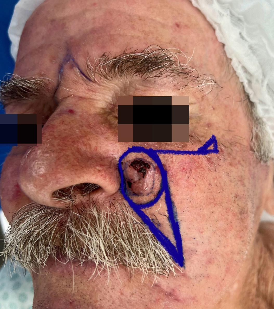
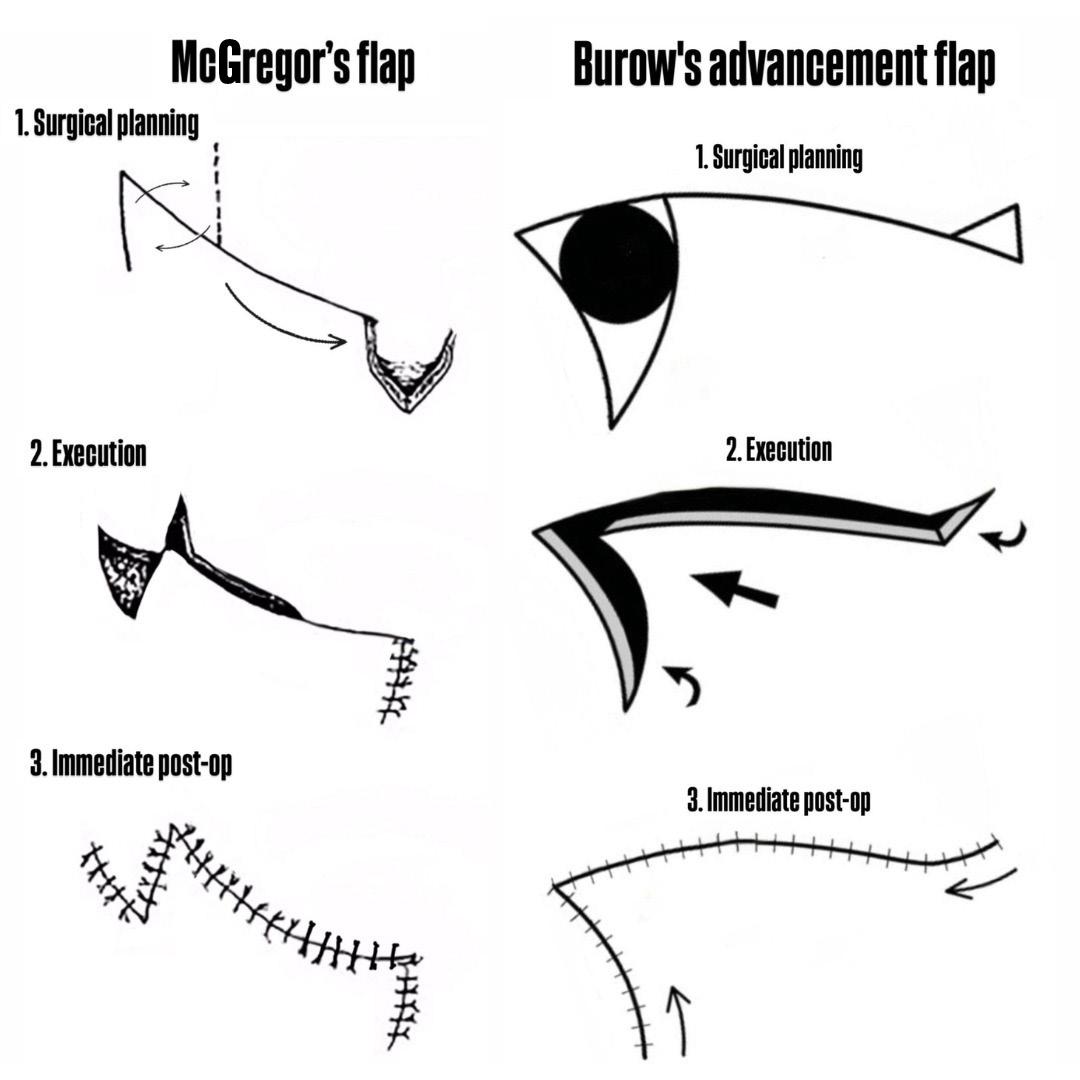
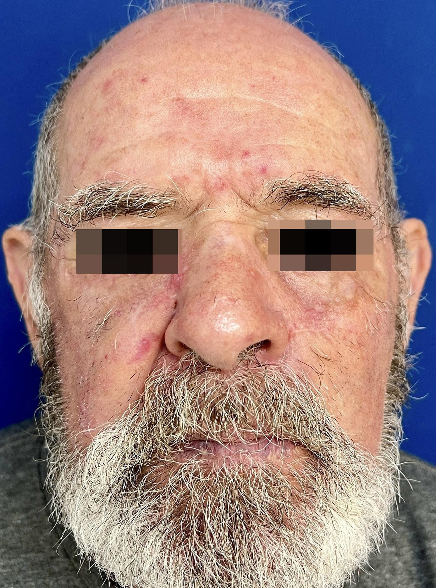
Reconstructive strategy using advancement flaps for bilateral medial malar defects: application of the McGregor and Burow flaps 427 Luiz Roberto Dal Bem Pires Júnior, Carlos Augusto Silva Bastos
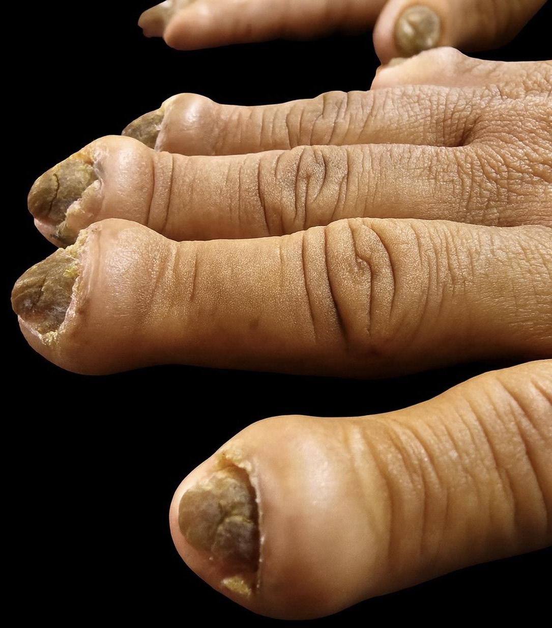
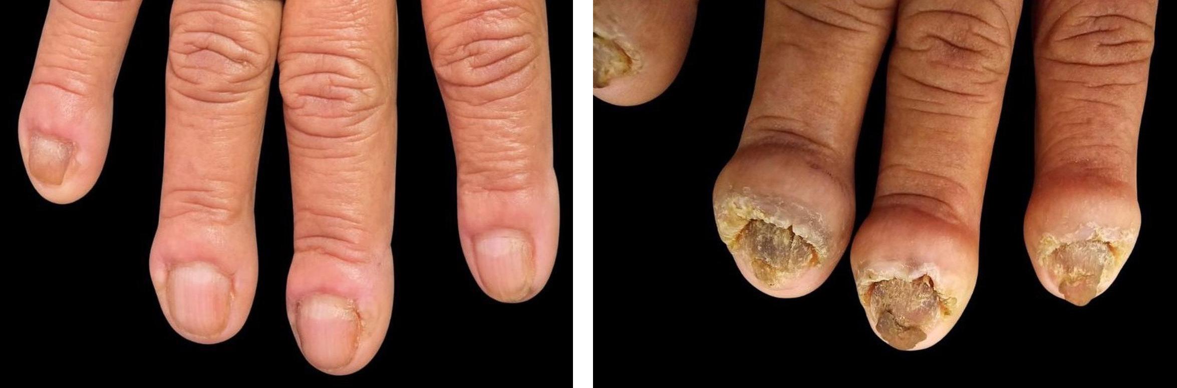
Exuberant chronic paronychia: a surgical approach using the square flap technique
Clarissa Brito Farias, André Martins Ornelas, Flauberto de Sousa Marinho
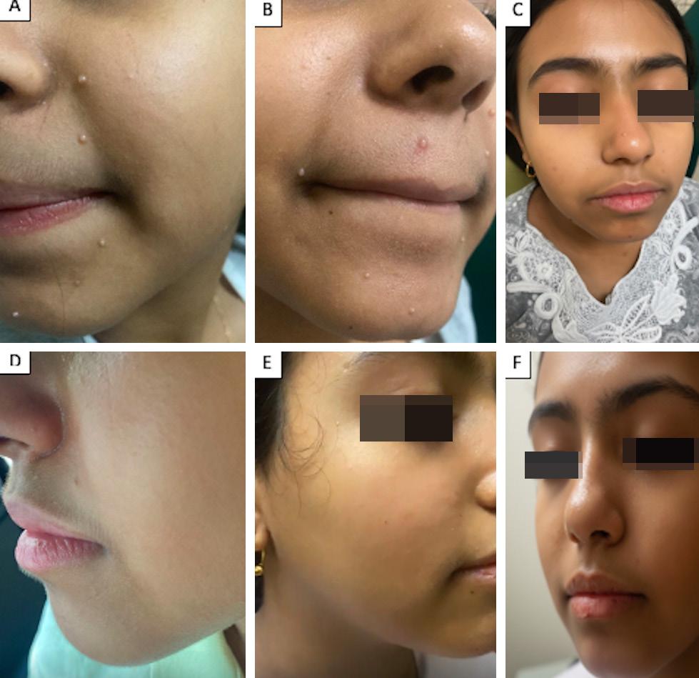
Oral isotretinoin as a promising treatment for refractory molluscum contagiosum in a child: a case report
Shaimaa Farouk
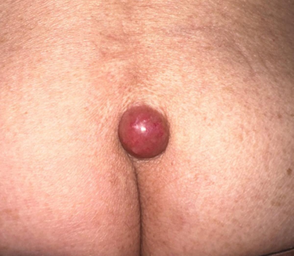
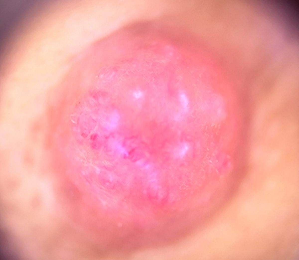
Distant cutaneous metastasis from solid neoplasm: a case report
Ana Vitoria Lins de Paiva Antunes Clarissa Brito Farias, Luciana Cavalcante Trindade, Kamila Pascoal Magno do Nascimento
Bednar tumor: a case report
Fernanda Guimarães Lopes, Juliana Cristina Silva Fraga, Mariana Campos Souza Menezes Ana Karen Alves, André Rossetti Portela
Negative pigment network and its importance in the diagnosis of melanoma: a case report
Mariana Freitas de Assis Pereira Rosa, Carlos Arthur de Figueiredo, Athayde Gustavo Costa Verardino, Ricardo Barbosa Lima
Cemiplimab Therapy for Marjolin Ulcer Arising from Folliculitis Decalvans: A Case Report
Leticia Midori Kondo Iwamoto, Flavia Rodrigues Dias, Otavio Augusto Noschang Moreira, Helio Amante Miot, Ana Claudia Athanasio Shwetz
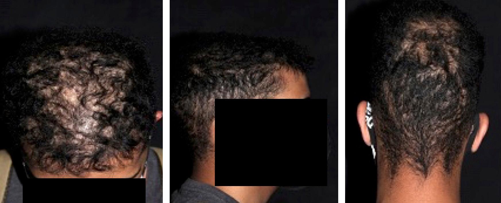
Trichotemnomania in androgenetic alopecia: a case report
Diana Coello-Bonilla, Giovanna Gelli-Carrascoza, Gabrielle Cassulo-Franciscatti, Maria Victória Quaresma
Rosacea is a chronic, multifactorial inflammatory dermatosis involving immune dysregulation, neurovascular alterations, and microbial imbalance. Overexpression of TLR2, activation of TRP channels, and increased LL-37 cathelicidin are central to its pathophysiology Therapeutic management includes behavioral strategies, topical treatments (metronidazole, ivermectin, azelaic acid, brimonidine), systemic agents (doxycycline, isotretinoin), and procedures such as laser and intense pulsed light. A personalized, multifaceted approach combining trigger control, pharmacologic therapy, and physical interventions is crucial for reducing symptoms and improving patient quality of life.
Keywords: Rosacea; Innate immunity; Cathelicidins; Botulinum toxin type A;Tetracyclines;Technology
Review Article
Authors:
Célia Luiza Petersen Vitello Kalil1
Matheus Silva Melo1
Giulia Dambrós Malacarne1 Isabela de Liz PereiraI
1 Universidade Federal da Fronteira Sul (UFFS), Dermatology Service, Passo Fundo (RS), Brazil
2 Universidade do Planalto Catarinense (UNIPLAC), School of Medicine, Lages (SC), Brazil
RESUMO
A rosácea é uma dermatose inflamatória crônica e multifatorial que envolve disfunção imunológica, alterações neurovasculares e desequilíbrio microbiano. A superexpressão do TLR2, a ativação de canais TRP e o aumento da catelicidina LL-37 são elementos centrais na sua fisiopatologia. O manejo terapêutico combina medidas comportamentais, cuidados tópicos (metronidazol, ivermectina, ácido azelaico, brimonidina), terapias sistêmicas (doxiciclina, isotretinoína) e procedimentos como laser e luz intensa pulsada. A abordagem personalizada, que alia controle de gatilhos, farmacoterapia e intervenções físicas, é essencial para reduzir os sintomas e melhorar a qualidade de vida dos pacientes. Palavras-chave: Rosácea; Imunidade inata; Catelicidinas; Toxina botulínica tipo A; Tetraciclinas; Tecnologia
Correspondence:
Matheus Silva Melo
E-mail: matheus.melo.sc@gmail. com / celia@celiakalil.com.br
Funding source: None
Conflict of interest: None
Submitted on: 10/22/2025
Final decision: 12/19/2025
How to cite this article: Kalil CLPV, Melo MS, Malacarne GD, Pereira IL. Rosacea: pathophysiology and therapeutics. Surg Cosmet Dermatol. 2026;18:e20260526.
INTRODUCTION
Rosacea is a common chronic inflammatory dermatosis that preferentially affects the centrofacial region, characterized by persistent or episodic erythema, telangiectasias, papules, and pustules. It may also present with phymatous and ocular manifestations. Its epidemiology shows considerable variability: global prevalence is estimated to range between 2% and 10% of the adult population, but higher rates, of up to 18%, have been reported in Northern European populations, where lighter skin phototypes (I–II) predominate.1,2 In Asian and Latin American countries, reported prevalence is lower, although cultural factors, lower physician familiarity with the disease, and atypical clinical presentations are known to contribute to underdiagnosis.3
Rosacea typically manifests between the fourth and sixth decades of life, although cases in young adults are not rare. It affects both sexes but shows striking differences in presentation: the erythematotelangiectatic and papulopustular forms occur more frequently in women, whereas the phymatous form is more prevalent in men, particularly at older ages. Involvement in children is rare, with ocular disease being the primary manifestation, frequently underestimated or mistaken for recurrent conjunctivitis.4 Known predisposing factors include light skin phototype, positive family history, and cumulative sun exposure, as well as environmental triggers such as extreme temperatures, alcohol consumption, spicy foods, and emotional stress, all associated with the vascular hyperreactivity characteristic of the disease.5,6
From a clinical standpoint, rosacea is notoriously heterogeneous. In addition to persistent centrofacial erythema and episodes of flushing, often exacerbated by environmental stimuli, visible telangiectasias are common, as are inflammatory flares of papules and pustules, without the presence of comedones, which helps differentiate rosacea from acne vulgaris. Up to 90% of patients report subjective symptoms such as stinging and burning sensation of the skin, pruritus, and skin hypersensitivity, which explains the frequent perception of “sensitive skin.”3,6
The clinical spectrum also includes less common variants, such as granulomatous rosacea, characterized by firm, monomorphic erythematous or yellowish papules, and persistent solid edema (lymphedematous rosacea), which is difficult to manage and associated with chronic facial lymphedema.3,4 Ocular involvement occurs in up to 50% of cases and ranges from mild blepharitis to more severe complications such as keratitis, episcleritis, and uveitis, with a potential risk of visual loss if not diagnosed and treated promptly.6,7 The most common symptoms include foreign-body sensation, pruritus, photophobia, and conjunctival hyperemia.The lack of correlation between cutaneous and ocular severity makes active screening for these manifestations essential.6
The clinical significance of rosacea transcends its cutaneous and ocular manifestations. Several studies have documented its psychosocial impact, with frequent reports of social stigmatization, low self-esteem, and impaired interpersonal interactions. In quality-of-life assessments, patients with rosacea score comparably to those with chronic diseases such as psoriasis
and atopic dermatitis.2,3 In addition, increasing evidence suggests an association between rosacea and systemic comorbidities, including gastrointestinal disorders (Helicobacter pylori infection, irritable bowel syndrome, and inflammatory bowel disease), neurological conditions (migraine and Parkinson’s disease), and psychiatric disorders (anxiety and depression), suggesting that the disease may reflect a systemic inflammatory state.3,4
Historically classified into 4 clinical subtypes (erythematotelangiectatic, papulopustular, phymatous, and ocular), this categorization has run into limitations in clinical practice due to frequent overlap of findings. In 2017, the international ROSCO panel (Rosacea Consensus) proposed a phenotype approach based on primary (persistent erythema, papules/pustules, telangiectasias, phymatous changes, and ocular signs) and secondary features (flushing, stinging, burning, edema, and extracutaneous manifestations). This approach improves diagnostic accuracy and facilitates the development of individualized therapeutic strategies.3,6
Regarding therapeutic management, rosacea encompasses a spectrum of options ranging from general measures, such as identification and avoidance of triggers, strict photoprotection, and skin barrier care, to pharmacological interventions and specific procedures. Topical treatments include brimonidine and oxymetazoline (vasoconstrictors for erythema), metronidazole, ivermectin, and azelaic acid (anti-inflammatory and antiparasitic agents). Low-dose oral antibiotics (such as subantimicrobial doxycycline), isotretinoin in refractory cases, and more recently investigations involving immunomodulatory agents stand out among systemic therapies. Physical modalities such as laser therapy and intense pulsed light (IPL) play an important role, particularly in controlling telangiectasias and persistent erythema. Treatment should be guided not only by the predominant phenotype but also by symptom severity and the impact on the patient’s quality of life.5,6
Given its high prevalence, diversity of clinical manifestations, psychosocial impact, and potential association with comorbidities, rosacea represents a frequent and relevant challenge in dermatological practice. Therefore, this study aimed to review the therapeutic approach to erythematotelangiectatic and papulopustular rosacea in light of the most recent evidence, without addressing in detail the phymatous and ocular forms, which fall outside the scope of this study.
PATHOPHYSIOLOGY
The pathophysiology of rosacea is multifactorial and complex, resulting from the dynamic interaction between genetic predisposition, immune dysfunction, neurovascular alterations, skin dysbiosis, and environmental factors. Although it is not yet fully understood, this network of mechanisms contributes to the development and perpetuation of the chronic skin inflammation characteristic of the disease.7 To facilitate understanding of this process, the main pathophysiological elements can be divided into the following topics:
CLPV, Melo MS, Malacarne GD, Pereira IL.
exacerbated innate immune response and production of pro-inflammatory cathelicidin; neurovascular dysfunction and hypersensitivity to stimuli;and various etiopathogenic mechanisms, particularly the role of microorganisms (Figure 1).
Each of these mechanisms will be discussed below in light of current evidence, with the aim of integrating pathophysiological findings with clinical practice and therapeutic management of rosacea.
• Microorganisms (D. folliculorum, C. acne, H. pylori)
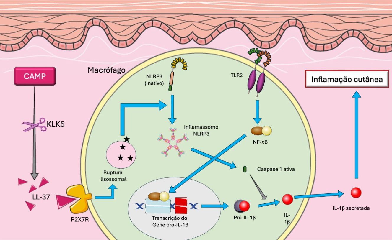
Exacerbated innate immune response Although its pathogenesis is not fully understood, the role of innate immunity as a central factor in the initiation and perpetuation of skin inflammation is increasingly recognized. This means that receptors such as Toll-like receptors (TLRs) and NOD-like receptors (NLRs) have emerged as key elements in the activation of inflammatory pathways in the skin.7 Understanding the mecha-nisms involving these receptors helps clarify the molecular processes underlying the clinical manifestations of the disease (Figure 2)
Innate immune system dysregulation:
- TLR2 activation
- NLRP3 activation
- LL-37 production via KLK5 -Pro-inflammatory cytokine production (IL-1β, IL-8, etc.)
Neurovascular dysregulation:
- TRPV activation -Vasodilation
Clinical manifestations:
• Erythema
• Papules
• Pustules
• Telangiectasias
• Flushing
• Phymatous changes
Figure 1: Interaction between etiopathogenic mechanisms, innate immune system dysregulation, neurovascular dysfunction, and clinical manifestations
Figure 2: CAMP: cathelicidin antimicrobial peptide. KLK: kallikrein. IL: interleukin. NLR: NOD-like receptor. TLR: Toll-like receptor. Image by the author, modified from Servier Medical Art
TLRs constitute a class of transmembrane proteins expressed mainly in cells of the innate immune system, such as keratinocytes, macrophages, and dendritic cells. They act as molecular sensors capable of recognizing pathogen-associated molecular patterns (PAMPs) and damage-associated molecular patterns (DAMPs), triggering pro-inflammatory signaling cascades. These receptors may be found both in the plasma membrane (such as TLR2 and TLR4), where they recognize bacterial surface components, or in endosomal compartments (such as TLR7 and TLR9), where they detect viral and bacterial nucleic acids.7,8
At least 10 functional TLRs have been identified in humans (TLR1 to TLR10), each with specificity for different classes of ligands. For example, TLR2 recognizes lipoproteins and peptidoglycans from gram-positive bacteria, TLR4 recognizes lipopolysaccharides (LPS) from gram-negative bacteria, and TLR7 and TLR9 recognize viral nucleic acids.7,8
In rosacea, there is particularly noteworthy overexpression of TLR2 in the epidermis, sebaceous glands, and adnexal structures. This increase lowers the threshold for skin immune activation, making it hyperresponsive to normally innocuous stimuli, such as temperature changes, topical products, or even the resident microbiota.5,8
NLRs, in turn, are intracellular receptors. Unlike TLRs, which detect threats in extracellular space or endosomal compartments, NLRs recognize microbial components and signs of cellular stress. Activation of NOD receptors leads to signaling through pathways such as nuclear factor kappa B (NF-κB), resulting in the production of pro-inflammatory cytokines such as interleukin (IL)-1β, IL-6, and tumor necrosis factor-α (TNF-α). In addition, NLRs participate in the formation of inflammasomes, intracellular protein complexes that activate caspase-1 and promote the release of IL-1β, a cytokine widely associated with inflammatory lesions in rosacea.7,8
Both TLR2 overexpression and exacerbated activation of the NOD2 intracellular receptor contribute to the amplification of the inflammatory response in rosacea through increased production of cathelicidins, a family of small antimicrobial peptides present in humans and animals that play an important role in the innate immune system.9 A particularly noteworthy member of this group is LL-37, which is generated through cleavage of ca-
thelicidin antimicrobial peptide (CAMP), its inactive precursor This cleavage is performed by the enzyme kallikrein 5 (KLK5), whose expression is also stimulated byTLR2.5,8,10 KLK5 is a serine protease that plays an important role in the skin, more specifically in the stratum corneum of the epidermis Encoded by the KLK5 gene, it is involved in the regulation of skin desquamation and may also contribute to skin inflammation because it can cleave protease-activated receptors (PARs) in keratinocytes, leading to the release of inflammatory cytokines.11
Once formed, LL-37 acts on different types of receptors, triggering intracellular signaling cascades (such as activation of NF-κB) that result in the production of inflammatory cytokines (IL-1β, IL-6, and TNF-α), induction of vasodilation, recruitment of immune cells, and vascular remodeling The abnormal and persistent accumulation of LL-37 in the skin of patients with rosacea therefore not only reflects a dysfunction in the regulation of innate immunity but also represents a point of convergence between different inflammatory pathways involved in the pathogenesis of the disease.10
Neurovasculardysfunction
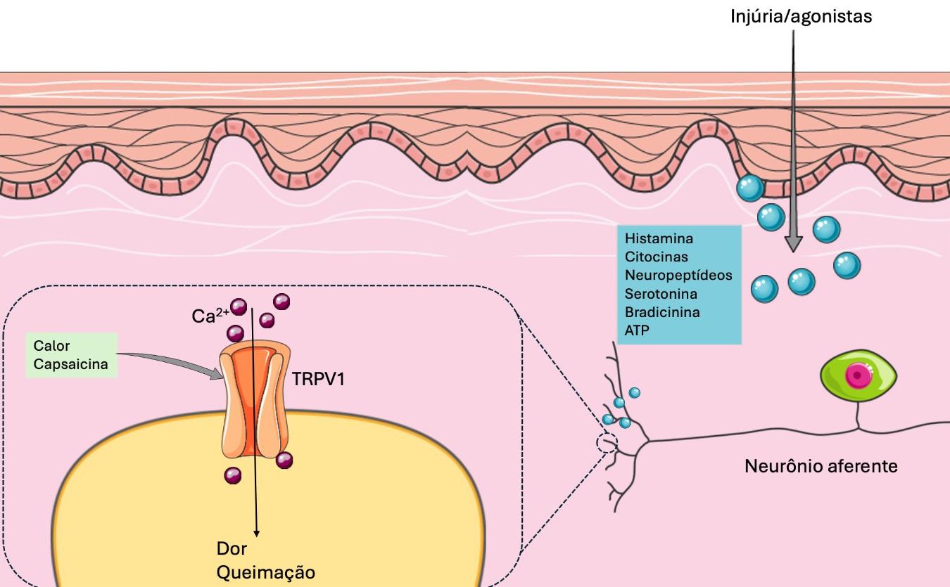
Neurovascular dysregulation constitutes one of the central pillars of rosacea pathophysiology, particularly in clinical forms characterized by persistent erythema, flushing, and telangiectasias. One of the molecular mechanisms that explains this hyperreactivity involves the overexpression and abnormal activation of transient receptor potential (TRP) channels.TRP channels are a superfamily of nonselective ion channels that regulate the influx of cations, especially calcium (Ca²+) and sodium (Na+), in response to various environmental and endogenous stimuli. Present in multiple tissues,including the skin,TRP channels act as molecular sensors that translate thermal,mechanical,chemical, and osmotic stimuli into electrical and biochemical signals.They are particularly abundant in sensory nerve endings, where they contribute to the perception of pain, pruritus, heat, cold, and inflammation.12
The TRP family is subdivided into 6 main subgroups based on sequence homology and functional characteristics Table 1 summarizes these proteins The overexpression and abnormal activation of specific TRP subtypes have been observed in the
TRPC Canonical Response to hormonal signals and neurotransmitters
TRPV Vanilloid Detection of heat, pain, and osmolarity
TRPM Melastatin Regulation of temperature and ions such as Mg²+
TRPA Ankyrin Sensation of extreme cold and chemical irritants
TRPP Polycystin Renal function and ciliary mechanisms
TRPML Mucolipin Lysosomal homeostasis and autophagy
TRP, transient receptor potential.
Kalil CLPV, Melo MS, Malacarne GD, Pereira IL.
Table 1: Classification of TRP subfamilies
skin of individuals with rosacea, particularly TRPV1, TRPA1,andTRPV412 (Figure 3).
Among external factors, microorganisms have been implicated as important triggers or modulators of skin inflammation. Although the presence of these organisms is not pathognomonic for the disease, increasing evidence suggests that they play an important role in perpetuating the inflammatory response.7,10,13
Themite Demodex folliculorum isacommensalectoparasite of human skin with a predilection for the pilosebaceous follicles of the face Studies have shown a significant increase in Demodex density in patients with rosacea, particularly in papulopustular and granulomatous forms A high parasite load may disrupt the local immune balance and trigger an exacerbated inflammatory response It is believed that antigenic products from Demodex, as well as symbiotic microorganisms living within the mite (such as Bacillus oleronius), stimulate overexpression of TLR-2 and promote activation of the LL-37 cathelicidin pathway, favoring
neutrophil chemotaxis and the release of metalloproteinases. In addition, follicular degradation caused by the parasite may act as a DAMP, amplifying the immune response.7,8,14
GENERAL
THERAPEUTIC MEASURES
The management of rosacea is based on the adoption of behavioral measures, which include patient education about the disease, identification of triggering factors, and the implementation of specific skin care practices.13
Identification and prevention of aggravating agents, such as sun exposure, temperature variations, psychosocial stress, alcohol consumption, diet, caffeine intake, strenuous exercise, and the use of potentially irritating cosmetics, are essential elements for understanding and individualizing disease management. Evidence indicates that more than 90% of patients are able to associate worsening of the clinical condition with triggering factors, reinforcing the importance of educational strategies aimed at reducing flare-ups.15 In addition, the adoption of personal records, such as diaries, helps identify individual patterns of exacerbation and promotes more effective disease control.15,16
Injury/agonists
Figure 3: Mechanism of action of TRPV. Following triggers of tissue injury, TRPV1, present in the free nerve endings of sensory neurons, allows the influx of calcium ions. The increase in intracellular calcium ions depolarizes the axon, which transmits sensations of pain and burning to the cerebral cortex. Image adapted from Zhang et al. (2013), modified from Servier Medical Art
Among aggravating factors, the consumption of alcohol (especially wine and spirits), hot drinks, and foods containing capsaicin and cinnamaldehyde is correlated with worsening of the clinical condition. Exposure to heat and the consumption of foods containing capsaicin may lead to activation of TRPV vanilloid channels, particularly subtypes 1 and 6, which are involved in vasodilation and increased skin sensitivity, thereby contributing to flushing and burning sensations in rosacea.17
Exposure to UV radiation, in turn, induces an innate immune inflammatory response mediated by antimicrobial peptides such as LL-37 and pro-inflammatory cytokines such as IL-33, which promote angiogenesis and vascular hyperreactivity.10
The relationship between smoking and rosacea shows a protective association. In a large population-based analysis conducted in the United Kingdom, a lower risk of rosacea was observed in smokers.18 Furthermore, there was also a significant reduction in the risk of developing rosacea among smokers. However, the nicotine from tobacco has angiogenic effects that may trigger rosacea symptoms and aggravate pustular lesions and rhinophyma.Therefore, given the negative impact of tobacco on overall and skin health, patients with rosacea should be counseled on the importance of smoking cessation despite the paradoxical associations described.17
In addition to the management of aggravating factors, rosacea substantially compromises the integrity of the skin barrier, leading to dehydration, irritability, and increased sensitivity.10 In this context, specific and individualized skin care becomes essential for optimizing therapeutic response. For cleaning, the use of mild, soap-free cleansers with a pH around 5.5 is recommended once or twice daily. In addition to cleaning, moisturization should also be incorporated into the daily skin care routine, as it is essential for restoring barrier function and relieving symptoms such as burning, pruritus, and xerosis. Formulations should be gentle, contain soothing and reparative agents, and be free of potentially irritating or sensitizing substances.19
Complementarily, broad-spectrum photoprotection should be incorporated into rosacea treatment. Daily use of water-based sunscreens with a sun protection factor (SPF) of 30 or higher is recommended, with frequent reapplication. Sunscreens formulated with vehicles containing dimethicone and cyclomethicone are preferable to minimize skin irritation.18,20 In addition, formulations containing physical filters such as zinc oxide and titanium dioxide have good tolerability profiles.20
Beyond these measures, the use of corrective cosmetics to camouflage erythema may be a useful strategy to mitigate the psychosocial impact of the disease. Among available options, mineral makeup is one of the most recommended because of its good tolerability, photoprotective properties, anti-inflammatory effect, and low comedogenic potential.17,20
TOPICAL THERAPIES
Topical alpha-adrenergic receptor agonists
Brimonidine and oxymetazoline act as topical α-2 and α-1 adrenergic receptor agonists, respectively.7 Topical use of
these agents promotes transient vasoconstriction of dermal vessels, with a rapid onset of action, within about 30 minutes. Their effects may last between 3 and 12 hours.10,15
Brimonidine 0.33% gel was approved in 2013 by the U.S. Food and Drug Administration (FDA) for symptomatic relief of persistent facial erythema. Its main mechanism of action involves highly selective vasoconstriction of dilated superficial vessels, in addition to exerting anti-inflammatory effects, mainly through the reduction of mast cells and neutrophilic infiltrate.21 It has demonstrated efficacy with once-daily application, both in clinical assessments of erythema and patient self-assessment, and a favorable safety profile when associated with other topical and physical therapies, such as IPL.17,21
Oxymetazoline is an imidazole derivative approved in 2017 by the FDA in a 1% cream formulation for the treatment of moderate to severe facial erythema in adults.19,21,22 In addition to the vasoconstrictive effect described above, oxymetazoline cream exhibits anti-inflammatory properties by reducing the release of inflammatory cytokines and inhibiting neutrophil phagocytosis.21 It also exerts immunomodulatory effects by reducing the stimulatory capacity of dendritic cells on T cells and modulates arachidonic acid activity.23
Metronidazole
Metronidazole (MTZ) is an antibiotic derived from nitroimidazoles that exhibits anti-inflammatory and antioxidant activities and is widely used in the management of papulopustular rosacea. The primary mechanism of action of MTZ involves modulation of neutrophil activity, with reduction of reactive oxygen species (ROS) and inhibition of T cells.7,13,21,22 It also acts on reducing inflammation and combating bacteria that may be involved in the development of rosacea.24 MTZ is available in different formulations, including gel, cream, or lotion at 0.75% (applied twice daily) or 1% gel (applied once daily). Comparative studies have not found significant differences in clinical efficacy between the different vehicles or concentrations.13,15 Adverse effects associated with MTZ use are generally mild and self-limited, including pruritus, burning sensation, desquamation, dryness, or local irritation.15 In mature and sensitive skin, the use of cream or lotion formulations is recommended, as they have better tolerability profiles compared with gel vehicles.22
Azelaic acid
Azelaic acid (AA) is a dicarboxylic acid indicated for the treatment of mild to moderate inflammatory rosacea. It is available in topical formulations at concentrations of 15% (gel and foam) and 20% (cream), all with comparable efficacy.21 Its mechanism of action integrates antimicrobial, anti-inflammatory, and antioxidant activities.13,21 It acts by reducing the expression and secretion of pro-inflammatory cytokines, such as IL-1β and TNF-α, by keratinocytes. In addition, it protects cells from oxidative damage by reducing ROS production by neutrophils and promoting their elimination. It also modulates cathelicidin acti-
Kalil CLPV, Melo MS, Malacarne GD, Pereira IL.
vity by inhibiting KLK5 and regulates gene expression, promoting reduction of CAMP and KLK5 mRNA levels in the skin of patients with rosacea.7,13,21 Adverse effects include burning sensation, pain, stinging, pruritus, xerosis, and local irritation, usually mild to moderate in intensity and transient.13,21,22
Ivermectin
Ivermectin 1% cream is widely used in the treatment of moderate to severe papulopustular rosacea due to its antiparasitic and anti-inflammatory properties. Its antiparasitic action is related to its ability to act as a ligand-gated ion channel agonist, resulting in paralysis and subsequent elimination of Demodex mites.7,22 In addition, it exerts anti-inflammatory effects by modulating innate and adaptive immune responses, inhibiting the secretion of pro-inflammatory cytokines, such as IL-1β and TNF-α, and increasing the expression of anti-inflammatory cytokines, such as IL-10. It also reduces neutrophil phagocytosis and chemotaxis.13,17,21
In addition to its therapeutic efficacy, ivermectin 1% cream offers the convenience of once-daily application, making it more practical for patients. Adverse effects are mostly mild and localized and it has high tolerability, potentially making it safer and more tolerable than other available topical agents.21
Ivermectin 1% cream has demonstrated superiority in reducing the social burden associated with rosacea when compared with other topical agents,21 although caution is recommended during pregnancy and lactation due to potential risks to fetuses and infants.14
Minocycline
Minocycline is a semi-synthetic antibiotic belonging to the tetracycline class, notable not only for its bacteriostatic activity but also for its anti-inflammatory, immunoregulatory, and anti-angiogenic properties.6,25 Its anti-inflammatory activity occurs through suppression of neutrophil recruitment and pro-inflammatory cytokines, such as TNF-α, IL-6, and IL-1β 6 An experimental study, conducted both in vitro and in vivo, helped elucidate the anti-inflammatory mechanism of minocycline by exposing HaCaT cells and BALB/c mice to inflammatory induction with LL-37 and subsequently treating them with the medication. Treatment significantly reduced the expression of TLR4, NF-κB, inflammatory cytokines, and ROS, suggesting an effect mediated through the TLR4/NF-κB signaling pathway.25 The topical formulation of minocycline 1.5% foam was approved in the United States in 2020, making it the first topical minocycline therapy indicated for treatment of moderate to severe papulopustular rosacea.21 In 2 identical phase III randomized clinical trials, patients who applied minocycline 1.5% foam once daily for 12 weeks showed a reduction in inflammatory lesions compared with the group using vehicle alone. At the end of the study, more than 80% of patients had only residual telangiectasias or mild erythema at the application site. In addition, improvement was observed in Investigator Global Assessment
(IGA) success rates, with at least a 2-grade improvement from baseline at week 12.6,25,26 The foam formulation demonstrated good tolerability and may have a lower incidence of systemic adverse effects compared with oral administration.26
Calcineurin inhibitors
Calcineurin inhibitors, such as cyclosporine, pimecrolimus, and tacrolimus, are used in the management of rosacea due to their immunomodulatory and anti-inflammatory properties.
Cyclosporine, in a 0.05% ophthalmic emulsion formulation, is widely used for ocular manifestations of the disease and has been shown to be more effective than artificial tears in improving symptoms of ocular rosacea. Its effect results from the inhibition of pro-inflammatory cytokine production by T lymphocytes in the conjunctiva, in addition to reducing the release of IL-1, IL-8, and TNF-α by monocytes, thereby contributing to the control of local inflammation.7,13
In the skin, pimecrolimus (1% cream) and tacrolimus (0.03%-0.1% ointment) act primarily by modulating T-lymphocyte activation and inhibiting IL-2 transcription. In ocular rosacea, these agents have also been used to treat the local inflammatory process of the eyelids, promoting restoration of the epidermal barrier and control of local inflammation.7,21
ORAL THERAPIES
Tetracyclines
Doxycycline is a second-generation tetracycline used in the treatment of papules and pustules due to its anti-inflammatory, rather than antimicrobial, effects. It is approved by the FDA at a dose of 40 mg/day for the management of papulopustular rosacea, and this dosage has been shown to be as effective as antimicrobial doses of 100–200 mg/day but with fewer adverse effects. It should be administered at the end of the day to reduce photosensitivity and taken away from meals containing dairy products, as these reduce its absorption. It is also effective in the treatment of ocular rosacea symptoms.7
Tetracycline is a first-generation antibiotic of the tetracycline family, with an effect similar to that of doxycycline. Subantimicrobial doses are also recommended in order to reduce bacterial resistance. Adverse effects may include nausea, diarrhea, and constipation. All tetracyclines are teratogenic.7
Oral minocycline may also be used off-label for the treatment of rosacea at a dose of 100 mg/day. A new modified low-dose formulation of minocycline is under investigation. Known as DFD-29 (minocycline hydrochloride capsules, 40 mg), the formulation includes 10 mg of immediate-release minocycline and 30 mg of extended-release minocycline. Two studies demonstrated a reduction in inflammatory lesions and improved IGA success rates compared with placebo and doxycycline. Regarding safety, adverse events were similar between groups and generally mild, with upper respiratory tract infection being the most common. No relevant differences were observed in laboratory tests, and no serious events attributable to the medication were reported.27
Kalil CLPV, Melo MS, Malacarne GD, Pereira IL.
Macrolides
Macrolides constitute a class of antibiotics that may be used in patients with rosacea who have hypersensitivity or contraindications to tetracyclines, such as pregnant patients. These medications have anti-inflammatory and antioxidant activity. The main adverse effects are gastrointestinal, although they are less frequent with second-generation macrolides such as azithromycin and clarithromycin. Azithromycin, when used at a dose of 250 mg 3 times per week, has shown improvement in papulopustular lesions.7
Gabapentin
A multicenter study evaluating gabapentin at a dose of 900 mg/day found improvement in flushing and redness compared with placebo. However, the therapy was ineffective for other rosacea symptoms, such as burning and tingling sensations and psychological effects.28 Another study compared gabapentin 900 mg/day with carvedilol 10 mg/day for the treatment of erythematotelangiectatic rosacea, and both therapies showed similar results in controlling facial flushing, although gabapentin produced a faster response. In addition, gabapentin may be better tolerated and provide greater benefit in patients with other comorbidities, such as migraine and sleep disorders. Mild adverse effects, such as drowsiness and nausea, were observed in 13% of patients.29
Isotretinoin
Isotretinoin, a retinoid derivative of vitamin A, may be used in cases of persistent rosacea that are resistant to other treatments. Its mechanism of action involves inhibition of pilosebaceous glands and downregulation of TLR-2.22 Although this treatment is off-label, the medication has demonstrated efficacy in patients who do not respond to topical therapies and oral antibiotics. The dose used in most studies is 0.2 mg/kg/day for 6 months. In addition, isotretinoin is effective in the treatment of phymatous rosacea and is indicated in early cases.7,26 Isotretinoin has also shown positive effects in edematous rosacea, known as Morbihan disease, suggesting that its mechanism of action extends beyond effects on sebaceous glands.26
Even at low doses, isotretinoin may cause dryness of mucous membranes and skin, which are its main adverse effects. Laboratory monitoring with liver enzyme and lipid profile measurements should be performed during treatment.22 The medication is a known teratogen and must not be used by patients attempting conception, pregnant individuals, or those who are breastfeeding.7
Antihypertensive agents
Antihypertensive agents may be effective in the treatment of flushing caused by rosacea. Among these, beta-blockers are the most commonly used medications, particularly propranolol and carvedilol. The use of these medications requires a prior electrocardiogram as well as consultation with and follow-up by a cardiologist, especially in patients over 50 years of age.10
NON-PHARMACOLOGICAL MEASURES
Intense pulsed light (IPL)
IPL consists of a device in which light is filtered through specific wavelengths selected according to the absorption characteristics of the target tissues. In rosacea, the target is oxyhemoglobin. IPL acts by reducing inflammation and increasing the upregulation of anti-inflammatory cytokines while downregulating pro-inflammatory cytokines. It may also stimulate collagen synthesis and increase cellular metabolism by affecting the mitochondrial electron transport chain. In addition, it contributes to the destruction of telangiectasias, since it promotes the formation of intravascular thrombi, leading to coagulation.30
One advantage of IPL in the treatment of rosacea is the flexibility of its parameters. In other words, depending on the parameters selected, it is possible to target superficial or deep vessels as well as to focus on the caliber of the vessels to be treated. Tip size is also a favorable aspect: because it is larger than those used in lasers, it allows treatment of broader areas with fewer pulses, making the procedure faster.
IPL is the treatment of choice for the erythematotelangiectatic stage, as it acts on the vascular hyperreactivity associated with rosacea. Treatment allows reduction of blood flow, superficial telangiectasias, and erythema intensity. The therapeutic response is effective and, given the chronic nature of the disease, periodic maintenance is recommended. The generally mild adverse-effect profile also favors IPL treatment. Combining IPL with topical and systemic treatments may enable synergistic therapeutic effects, optimizing outcomes.31 The use of skincare products containing soothing and antioxidant active ingredients may reduce procedural downtime and enhance results.30
Lasers
Lasers may reduce erythema and promote coagulation of rosacea-associated telangiectasias. The most commonly used are the pulsed dye laser (PDL) and the long-pulsed neodymium-doped yttrium aluminum garnet (Nd:YAG) laser. Studies have compared the efficacy of IPL and PDL and failed to find statistically significant differences between the two methods. IPL appears to be more effective in the treatment of erythema than telangiectasias in severe forms of rosacea. Both techniques are effective when repeated sessions are performed.10 In addition to these options, both fractional and non-fractional ablative lasers, including carbon dioxide lasers (CO2, 10,600 nm) and erbium-doped yttrium aluminum garnet lasers (Er:YAG, 2940 nm), have been used in the treatment of rhinophyma and will be discussed later in this article.
Botulinum toxin
Intradermal injection of botulinum toxin diluted to 10 IU/mL may reduce erythema and facial flushing associated with rosacea. It may also reduce pore size and some papular lesions. Applications may be performed at a dose of 0.05 mL per point, spaced 0.5 cm apart.17,32 It acts by blocking mast cell activity,
which reduces erythema induced by LL-37.17 In addition, it blocks the release of acetylcholine in autonomic nerves, reducing cutaneous vasodilation.33 Other neurotransmitters have also been suggested as potentially amenable to reduction by botulinum toxin type A (BoNT-A) injection, including substance P, calcitonin gene-related peptide, and glutamate 34 The effect often lasts 3-4 months, and reapplication after 4-5 months is recommended to maintain symptom remission.32 Some studies have shown that the effect of botulinum toxin in rosacea, when associated with thermal ablation of the stratum corneum using technologies such as laser and fractional thermal systems, may be prolonged.17
Bloom et al. evaluated the safety and efficacy of intradermal abobotulinumtoxinA (15–45 IU) for the treatment of facial erythema of rosacea and reported visible improvement in 93% of patients. No worsening, severe adverse events (such as anaphylaxis), or rebound effect were observed.33 AbobotulinumtoxinA has high molecular weight; therefore, its penetration into the intact stratum corneum is low and it is typically administered via intradermal injection. Methods that produce stratum corneum ablation (such as ablative lasers, microneedle radiofrequency, and electroporation) combined with botulinum toxin application have demonstrated results similar to those of intradermal injection in addition to reducing procedural pain and the risk of muscle paralysis. The primary adverse effect is erythema at the injection site, observed in approximately one-quarter of patients. Ecchymoses and facial muscle involvement were less frequent, occurring in fewer than 5% of cases.34 However, the use of botulinum toxin for the treatment of rosacea is still considered off-label, and additional randomized clinical trials are needed.
Photobiomodulation (PBM)
Photobiomodulation (PBM) is a therapeutic technique that uses low-intensity light to modulate cellular responses. This approach has emerged as a promising alternative in the treatment of rosacea, mainly due to its anti-inflammatory and tissue-regenerative properties.35
Although the mechanism of action of PBM is not yet fully understood, its action is known to occur largely through light absorption by mitochondrial chromophores, such as cytochrome c oxidase. This interaction stimulates adenosine triphosphate (ATP) production, improves tissue oxygenation, and modulates the inflammatory pathways involved in the pathophysiology of rosacea. Wavelengths in the red spectrum (630–660 nm) and near-infrared range (800–850 nm), commonly emitted by LEDs and low-power lasers, are among the most effective. These spectra have been associated with reduced expression of pro-inflammatory cytokines, such as IL-1β, IL-6, and TNF-α, as well as reduction of immune-cell infiltrates in tissues.35
Despite positive results and the favorable safety profile of PBM, variability among protocols and parameters, such as session frequency, dosage, and treatment duration, remains considerable. In addition, the scarcity of controlled clinical trials as-
sessing PBM in the treatment of rosacea highlights the need for further studies to better understand this therapeutic approach and the cellular responses involved.35
Peculiarities in the treatment of rhinophyma
Rhinophyma is a chronic and progressive condition characterized by proliferation of sebaceous glands, thickening of connective tissue, changes in skin coloration, local deformity, irregular exophytic growth, and visible telangiectasias. Although nasal presentation is the most common, similar swellings may affect other regions of the face.36
This form of rosacea responds poorly to conventional treatments, and ablative therapies are often required to reduce lesions.36
The first surgical techniques were described in the seventeenth century, and over time several approaches have been reported, including those of Von Langenbeck (healing by secondary intention), Ollier (decortication), Wood (partial skin graft), Adamopoulos (dermabrasion), Nolan (cryosurgery), and Bohigian (CO2 laser).37
Among ablative techniques, cryosurgery and electrosurgery stand out, as they allow excision of the phyma with excellent hemostasis and at a lower cost. However, they carry risks of necrosis, dyschromia, and unsightly scarring, which may be minimized with appropriate energy settings. Coblation, in turn, uses radiofrequency in a conductive medium at temperatures below 90 °C, decreasing thermal damage risk.36
Excisional techniques involve total or partial removal of the affected tissue. Full-thickness resection eliminates the risk of recurrence but is associated with a greater likelihood of graft or flap failure, less favorable aesthetic outcomes, and a potential need for corrective procedures; consequently, its use has declined in contemporary practice. Partial excision preserves pilosebaceous units and promotes spontaneous re-epithelialization at a lower cost, although strict hemostatic control is required. Dermabrasion may be used as an adjuvant procedure for aesthetic refinement.36
The use of ablative technologies has also increased in the treatment of rhinophyma. Because it has been widely studied, the CO2 laser has become one of the most effective options, producing highly satisfactory aesthetic results. Due to its specific wavelength (10,600 nm), the laser is primarily absorbed by water, enabling precise cutting and vaporization with lower depth of penetration, intraoperative hemostasis, reduced thermal damage, and low risk of scarring, while also allowing rapid re-epithelialization.36
Other ablative lasers, such as Er:YAG and diode lasers (1450–1470 nm), may also be used, providing faster recovery and fewer adverse effects, although they generally produce milder results because they are more superficial than the CO2 laser. PDL may be used as an adjuvant for treatment of vascularization.37,38
A more recent combination of helium plasma with radiofrequency has shown promising results. This technique promotes
Kalil CLPV, Melo MS, Malacarne GD, Pereira IL.
nearly instantaneous heating and cooling, allowing sequential ablation with minimal bleeding during the procedure and recovery similar to that observed with the CO2 laser, although residual erythema may persist for a longer period.36
FUTURE THERAPIES
The monoclonal antibody that inhibits the calcitonin gene-related peptide (CGRP) has been investigated as a therapeutic option for the treatment of acne and rosacea. CGRP is a peptide associated with the pathogenesis of migraine and is involved in processes of vasodilation and neurogenic inflammation. More recent studies have shown that it is also present in sebaceous glands, and its inhibition may reduce the risk of rosacea development by up to 47%.39
Zeolites, microporous aluminosilicates with tunable physicochemical properties, have the ability to adsorb toxins, modulate local inflammation, and act as carriers for antimicrobial or therapeutic agents.They may also improve dermal absorption of challenging hydrophilic compounds. One example is tranexamic acid, used in the treatment of rosacea and melasma, when combined with zeolitic imidazolate frameworks (ZIF-8). This system facilitates the expression of aquaporin-3 (AQP-3) in keratinocytes, enhancing transepidermal water and solute transport, with decreased erythema and visible skin brightening without adverse effects. Zeolite microneedles represent an evolution in transdermal drug-delivery systems, combining the mechanical integrity, porosity, and bioinertness of zeolites to achieve minimally invasive, long-acting dermal administration of both small molecules and macromolecules.40
CONCLUSION
Rosacea is a multifactorial dermatosis, and our pathophysiological understanding of the condition has advanced significantly in recent decades. Current evidence points to a complex interaction among immunological, neurovascular, microbial, and
REFERENCES:
1. Gether L, Overgaard LK, Egeberg A, Thyssen JP. Incidence and prevalence of rosacea: a systematic review and meta-analysis. Br J Dermatol. 2018;179(2):282–9.
2. Abram K, Silm H, Oona M. Prevalence of rosacea in an Estonian working population using a standard classification. Acta Derm Venereol. 2010;90(3):269–73.
3. Tan J, Almeida LMC, Bewley A, Cribier B, Dlova NC, Gallo R, et al. Updating the diagnosis, classification and assessment of rosacea: recommendations from the global ROSacea COnsensus (ROSCO) panel. Br J Dermatol. 2017;176(2):431–8.
4. Two AM, Wu W, Gallo RL, Hata TR. Rosacea: part I. Introduction, categorization, histology, pathogenesis, and risk factors. J Am Acad Dermatol. 2015;72(5):749–58; quiz 759–60.
5. Clanner-Engelshofen BM, Bernhard D, Dargatz S, Flaig MJ, Gieler U, Kinberger M, et al. S2k guideline: Rosacea. JDDG - Journal of the German Society of Dermatology. 2022;20(8):1147–65.
6. van Zuuren EJ, Arents BWM, van der Linden MMD, Vermeulen S, Fedorowicz Z, Tan J. Rosacea: New concepts in classification and treatment. Am J Clin Dermatol. 2021;22(4):457–65.
environmental factors as the main axes in the development and perpetuation of the clinical condition. Identification of elements such as TLR2 overexpression, the role of LL-37 (an antimicrobial peptide produced by the human body that is part of the innate immune system), activation of TRP channels, and the increased presence of Demodex folliculorum not only clarifies key aspects of the disease but also opens the way for more targeted therapeutic interventions.
Topical treatments represent the first-line therapy for mild to moderate cases, including substances such as metronidazole, azelaic acid, ivermectin, topical minocycline, encapsulated benzoyl peroxide, brimonidine, oxymetazoline, sulfacetamide/ sulfur, and, in some cases, off-label retinoids. When necessary, these therapies may be combined with oral medications, notably doxycycline, minocycline, tetracycline, isotretinoin, azithromycin, beta-blockers, and even Janus kinase (JAK) inhibitors, a class of medications that block JAK enzymes to modulate the inflammatory response of the immune system.41
In more severe cases, particularly those with phymatous changes or those refractory to conventional treatment, procedural interventions may be required. These include different laser modalities such as PDL, IPL, potassium titanyl phosphate (KTP), Nd:YAG, CO₂, and Er:YAG, as well as approaches such as cryotherapy, electrosurgery, dermabrasion, and other surgical interventions.41
In addition to pharmacological and procedural measures, changes in lifestyle play a central role in the prevention and control of exacerbations. Avoiding known triggers, adopting rigorous photoprotection, using gentle skin care practices, and investing in patient education are essential strategies to minimize flare-ups.
Thus, a combined and personalized approach integrating topical and systemic therapies, procedures, and behavioral changes is fundamental for optimizing symptom control and improving the quality of life of patients with rosacea. l
7. Geng RSQ, Bourkas AN, Mufti A, Sibbald RG. Rosacea: Pathogenesis and therapeutic correlates. Vol. 28, J Cutan Med Surg. 2024;28(2):178–89.
8. Yoon SH, Hwang I, Lee E, Cho HJ, Ryu JH, Kim TG, et al. Antimicrobial Peptide LL-37 Drives Rosacea-Like Skin Inflammation in an NLRP3-Dependent Manner. J Invest Dermatol. 2021;141(12):2885-94.e5.
9. Kościuczuk EM, Lisowski P, Jarczak J, Strzałkowska N, Jóźwik A, Horbańczuk J, et al. Cathelicidins: family of antimicrobial peptides. a review. Mol Biol Rep. 2012;39(12):10957–70.
10. Cribier B. Rosacea: Treatment targets based on new physiopathology data. Ann Dermatol Venereol. 2022;149(2):99–107.
11. Nauroy P, Nyström A. Kallikreins: essential epidermal messengers for regulation of the skin microenvironment during homeostasis, repair and disease. Matrix Biol Plus. 2019;6–7:100019.
12. Zhang M, Ma Y, Ye X, Zhang N, Pan L, Wang B. TRP (transient receptor potential) ion channel family: structures, biological functions and therapeutic interventions for diseases. Signal Transduct Target Ther. 2023;8(1):261.
13. Okońska A, Kozakiewicz J, Ziemba M, Kałuska J, Suprun T, Sokół A, et al. Rosacea pathogenesis and topical treatment options – a review. Quality in Sport. 2025;39:58360.
14. Geng R, Bourkas AN, Sibbald RG. Rosacea: Clinical aspects and treatments. Adv Skin Wound Care. 2023;36(12):626–34.
15. Frazier W, Zemtsov RK, Ge Y. Rosacea: common questions and answers. Am Fam Physician. 2024;109(6):533–42.
16. Cribier B. Rosacea: new data for better care. Ann Dermatol Venereol. 2017;144(8-9):508–17.
17. Paiva-Santos AC, Gonçalves T, Peixoto D, Pires PC, Velsankar K, Jha NK, et al. Rosacea topical treatment and care: from traditional to new drug delivery systems. Mol Pharm. 2023;20(8):3804–28.
18. Mikkelsen CS, Holmgren HR, Kjellman P, Heidenheim M, Kappinnen A, Bjerring P, et al. Rosacea: a clinical review. Dermatol Rep. 2016;8(1):6387.
19. Oliveira CMM, Almeida LMC, Bonamigo RR, Lima CWG, Bagatin E. Consensus on the therapeutic management of rosacea – Brazilian Society of Dermatology. An Bras Dermatol. 2020;95(Suppl 1):53–69.
20. Sobkowska D, Szałapska A, Pawlaczyk M, Urbańska M, Micek I, Wróblewska-Kończalik K, et al. The role of cosmetology in an effective treatment of rosacea: a narrative review. Clin Cosmet Investig Dermatol. 2023;16:1419–30.
21. Dall’Oglio F, Nasca MR, Micali G. Emerging topical drugs for the treatment of rosacea. Expert Opin Emerg Drugs. 2021;26(1):27–38.
22. Lee JJ, Chien AL. Rosacea in older adults and pharmacologic treatments. Drugs Aging. 2024;41(5):407–21.
23. Del Rosso JQ, Tanghetti E. Topical Oxymetazoline Hydrochloride Cream 1% for the treatment of persistent facial erythema of rosacea in adults: a comprehensive review of current evidence. J Clin Aesthet Dermatol. 2021;14(3):32–7.
24. McClellan KJ, Noble S. Topical metronidazole. A review of its use in rosacea. Am J Clin Dermatol. 2000;1(3):191–9.
25. Hua P, Tu Y, Yang Z, He Y, He L, Yao Q, et al. Minocycline inhibits rosacea-like inflammation through the TLR4-mediated NF-κB signaling pathway in vivo and in vitro. PLoS One. 2025;20(5):e0323598.
26. Gold LS, Del Rosso JQ, Kircik L, Bhatia ND, Hooper D, Nahm WK, et al. Minocycline 1.5% foam for the topical treatment of moderate to severe papulopustular rosacea: Results of 2 phase 3, randomized, clinical trials. J Am Acad Dermatol. 2020;82(5):1166–73.
27. Bhatia N, Del Rosso J, Stein Gold L, Lain E, Draelos ZD, Sidgiddi S, et al. Efficacy, safety, and tolerability of oral dfd-29, a low-dose formulation of minocycline, in rosacea. JAMA Dermatol. 2025;161(5):499.
28. Ma G, Zhang Y, Gao Q, Yuan X, Zhou Z, Jian D, et al. Gabapentin improves the flushing of rosacea, but not other rosacea symptoms or quality of life: results from a multicenter, randomized, double-blind, placebo-controlled pilot study. J Am Acad Dermatol. 2025;92(4):920–2.
AUTHOR’S CONTRIBUTION:
Célia Luiza Petersen Vitello Kalil
0000-0002-1294-547X
29. Wei J, Wan M, Chen Q, Fu J, Yang L, Ni R, et al. Gabapentin is an efficacy treatment for facial flushing and erythema of erythematotelangiectatic rosacea: a randomized clinical noninferiority trial. J Am Acad Dermatol. 2025;92(4):927–30.
30. Barbarino SC, Bucay VW, Cohen JL, Gold MH. Integrative skincare trial of intense pulsed light followed by the phyto‐corrective mask, phyto‐corrective gel, and resveratrol <scp>BE</scp> for decreasing post‐procedure downtime and improving procedure outcomes in patients with rosacea. J Cosmet Dermatol. 2022;21(9):3759–67.
32. Bharti J, Sonthalia S, Jakhar D. Mesotherapy with botulinum toxin for the treatment of refractory vascular and papulopustular rosacea. J Am Acad Dermatol. 2023;88(6):e295–6.
33. Bloom BS, Payongayong L, Mourin A, Goldberg DJ. Impact of intradermal AbobotulinumtoxinA on facial erythema of rosacea. Dermatol Surg. 2015;41(Suppl 1):S9–16.
34. Alsaati AA, Alsaadoun D, Kinkar LI, Alkhamis RS, Ahmed WA, Almathami AH. The efficacy and safety of Botulinum Toxin A for the treatment of rosacea: a systematic review. Cureus. 2023;15(12):e51304.
35. Boaretto Netto J, Borges AC, Farias, TG. Could photobiomodulation be used for treatment of rosacea?. Lasers Med Sci. 2025;40(1):250.
36. Dick MK, Patel BC. Rhinophyma. 2023 Aug 8. In: StatPearls. Treasure Island (FL): StatPearls Publishing; 2025.
37. Costa TC, Firme WAA, Brito LMR, Vieira MBG, Leite LA de S. Rinofima: opções cirúrgicas utilizadas no serviço de cirurgia Plástica do hospital Agamenon Magalhães - PE. Rev Bras Cir Plást. 2010;25(4):633–6.
38. Kamrani P, Boen M, Fabi SG, Goldman MP. From CO2 to Er:YAG: a comprehensive review of laser treatments for rhinophyma. J Drugs Dermatol. 2024;23(11):932-6.
39. Thang CJ, Lai J, Garate D, Golovko G, Wilkerson MG, Loder EW, et al. Calcitonin gene-related peptide inhibition and development of acne and rosacea. JAMA Dermatol. 2024;160(8):895–8.
40. Dring JC, Kaczynski M, Zureikat RM, Kaczynski M, Forma A, Baj J. Review of applications of zeolites in dermatology: molecular perspectives and translational potentials. Int J Mol Sci. 2025;26(14):6821.
41. Volk K, Ulfers A, Yi RC, Feldman S, Taylor SL. Treatment management for rosacea: current pharmacological and non-pharmacological options. Expert Rev Clin Pharmacol. 2025;18(8):589-605.
Approval of the final version of the manuscript, Conception and design of the study, Effective participation in the conduct of the study
Matheus Silva Melo
0000-0003-3027-8719
Conception and design of the study, Preparation and writing of the manuscript, Acquisition, analysis and interpretation of data, Critical review of the literature, Critical revision of the manuscript
Giulia Dambrós Malacarne
0000-0002-0313-3840
Conception and design of the study, Preparation and writing of the manuscript, Acquisition, analysis and interpretation of data, Intellectual participation in the propaedeutic and/or therapeutic approach to the cases studied
Isabela de Liz Pereira
0009-0001-4727-8400
Preparation and writing of the manuscript, Acquisition, analysis and interpretation of data, Intellectual participation in the propaedeutic and/or therapeutic approach to the cases studied, Critical review of the literature
www.surgicalcosmetic.org.br/
Clean beauty: definitions, distinctions among natural, organic, and vegan cosmetics, and experimental evidence on endocrine disruptors in cosmetics
Clean beauty: definições, diferenciações entre cosméticos naturais, orgânicos e veganos, e achados experimentais sobre disruptores endócrinos em cosméticos
In recent years, the demand for eco-friendly cosmetics formulated with safer and more sustainable ingredients has driven the expansion of the global market.The concept of clean beauty has evolved into a philosophy that requires transparency, safety, and sustainability throughout the production chain.This review article updates the definitions of clean beauty, differentiates natural, organic, and vegan cosmetics, and details experimental findings on endocrine disruptors present in cosmetic formulations, including in vitro and in vivo evidence.
Nos últimos anos, a busca por cosméticos que sejam "amigos da natureza", formulados com ingredientes mais seguros e sustentáveis, impulsionou o crescimento do mercado global. O conceito de clean beauty tornou-se uma filosofia que exige transparência, segurança e sustentabilidade em toda a cadeia de produção. Este artigo de revisão atualiza as definições de clean beauty, diferencia cosméticos naturais, orgânicos e veganos, e detalha achados experimentais sobre os disruptores endócrinos presentes em formulações cosméticas, incluindo evidências in vitro e in vivo.
1 Farmatec RS, Pesquisa e Desenvolvimento, Porto Alegre (RS), Brazil
2 Faculdade de Medicina de Jundiaí, Dermatologia, Jundiaí (SP), Brazil
3 Universidade Federal da Fronteira Sul, Dermatologia, Passo Fundo (RS), Brazil
Correspondence:
Pedro Vargas E-mail: pedro@farmatecrs.com.br / pedro.vargas@acad.pucrs.br
Funding: None
Conflict of interest: None
Submitted on: 11/18/2025
Final decision: 02/01/2026
How to cite this article:
Vargas P, Campos VB, Reginatto FP, Prato CC, Kalil CLPV. Clean beauty: definitions, distinctions among natural, organic, and vegan cosmetics, and experimental evidence on endocrine disruptors in cosmetics. Surg Cosmet Dermatol. 2026;18(1):e20260540.
INTRODUCTION
Market trends
The demand for skincare routines using natural products free of controversial ingredients has become a well-established trend and continues to drive global sales.1–3 The global market for natural cosmetics is projected to grow at an annual rate of 6.75% between 2024 and 2028, reaching USD 18.01 billion.3 Brazil remains the fourth largest beauty market worldwide, generating BRL 173.4 billion in 2024, with a growth rate of 10.3% and projections to reach USD 40 billion by 2027.4 Clean beauty products are expected to outpace the traditional cosmetics industry, with an average annual growth of 12%.2
This study is a narrative literature review based on the critical analysis of relevant scientific publications addressing the concept of clean beauty, distinctions among natural, organic, and vegan cosmetics, and experimental evidence related to endocrine disruptors (EDs) in cosmetic products. Databases including PubMed, Scopus, and Google Scholar were searched, prioritizing experimental studies, narrative reviews, and regulatory documents.
Minimalist skincare
Associated with clean beauty, the minimalist skincare approach emphasizes simplified routines, avoiding excessive active ingredients while prioritizing skin barrier health.2 This trend reflects a shift in consumer behavior toward more streamlined, safer formulations with a lower risk of sensitization.
CONCEPTS IN COSMETICS
Clean beauty
Clean beauty refers to products that are considered safe, transparent, and free of controversial ingredients such as parabens, sulfates, phthalates, petrolatum, and synthetic fragrances.1,5 Although there is no universally accepted regulatory definition, the term is widely used to describe formulations developed under more conservative safety standards.
Natural cosmetics
Natural cosmetics are primarily composed of raw materials derived from animal, plant, or mineral sources, provided they are minimally processed. The maximum allowable percentage of synthetic ingredients varies across certification bodies but generally does not exceed 5% and is restricted to substances considered low risk.5
Organic
cosmetics
Organic cosmetics are derived from raw materials cultivated without synthetic pesticides and fertilizers, using crop rotation and biological pest control methods. Certification requires ≥ 95% organic ingredients. An example of organic certification in cosmetics is Ecocert, internationally recognized for ensuring that products are formulated with natural and organically sourced ingredients. This certification guarantees the absence of
synthetic components, such as parabens, silicones, and artificial fragrances, and requires that production processes adhere to environmental sustainability, animal welfare, and sustainable practices, including eco-friendly packaging.5
Vegan cosmetics
Vegan cosmetics align with the cruelty-free philosophy, meaning they do not contain ingredients of animal origin and are not tested on animals at any stage of production.Vegan products may be synthetic and are not necessarily natural or organic.5 Importantly, the term “vegan” refers exclusively to the origin of ingredients and does not inherently imply lower toxicological risk or greater sustainability.
REGULATIONS
Brazilian regulations
The Brazilian Health Regulatory Agency (Anvisa) does not yet provide formal definitions for “natural,”“organic,” or “vegan.” As a result, these definitions remain under the purview of private certification bodies and international standards.6 Recent regulatory updates — Normative Instruction No. 280/2024 and Collegiate Board Resolutions No. 951/2024 and No. 894/2024 — have brought light to substance lists, risk classification, mandatory labeling in Portuguese, and the reporting of serious adverse events, including suspected endocrine disruption.6
A bill currently under consideration proposes the inclusion of warning labels addressing the risk of endocrine disruption in cosmetic products.6 Despite advances, Brazil still lacks unified and specific regulatory criteria for natural, organic, or vegan formulations.
International regulations
In the United States, cosmetics are regulated by the Food and Drug Administration (FDA) under the Federal Food, Drug, and Cosmetic Act (FD&C Act), which historically did not require premarket approval for most cosmetic ingredients, except for color additives. With the enactment of the Modernization of Cosmetics Regulation Act of 2022 (MoCRA), the FDA’s authority has been expanded to include mandatory facility registration, product listing, reporting of serious adverse events, scientific substantiation of ingredient safety, and the ability to mandate product recalls.7,8
In the European Union (EU), regulation — coordinated by the European Medicines Agency (EMA) — is more stringent and is governed by Regulation (EC) No. 1223/2009. This regulation determines that the safety of any cosmetic product must be proven prior to marketing. It constitutes the primary legal framework governing cosmetic products marketed in the EU, establishing strict safety requirements to ensure a high level of protection of human health and the free movement of cosmetic products in the internal market. The European Commission (executive arm of the EU) maintains a priority list of substances
under evaluation for their potential endocrine-disrupting properties and applies harmonized criteria for their restriction based on the precautionary principle.6,9
ENDOCRINE DISRUPTORS IN COSMETICS
Concept
EDs are chemical substances capable of interfering with the synthesis, transport, metabolism, and action of endogenous hormones, even at low concentrations.10,11 These effects may occur through multiple mechanisms, including hormone mimicry, receptor blockade, enzymatic changes, and modulation of nuclear signaling pathways. Examples of EDs in cosmetics include:
Parabens (methyl-, propyl-, butylparaben): weak estrogenic activity and potential risk of breast carcinogenesis10;
Phthalates: antiandrogenic effects and disruption of steroidogenesis11;
Chemical UV filters (benzophenone-3, octocrylene): bioaccumulation and toxicity12,13;
Butylated hydroxytoluene (BHT): antioxidant associated with aquatic toxicity14;
Triclosan and bisphenol A (BPA — a chemical widely used in plastics and resins and associated with health concerns): modulation of hormone receptors and adverse metabolic effects.15
Male reproductive system: reduced sperm count and motility, infertility, and risk of testicular cancer11,15;
Metabolic system: obesogenic effects, insulin resistance, and transgenerational inheritance of obesity15;
Immune system: potential autoimmune effects.10
EDs may also affect vulnerable populations, such as pregnant and lactating individuals, as well as those with chronic conditions, including malignancies.16 Increased susceptibility in these groups results from physiological changes, higher rates of skin absorption, or exacerbated systemic effects.
Experimental evidence on endocrine disruptors
In vitro studies
Parabens: Increased proliferation of MCF-7 cells, a human breast cancer cell line originally developed at the Michigan Cancer Foundation (now the Barbara Ann Karmanos Cancer Institute, Detroit, USA). This cell line, derived from an adenocarcinoma, is widely used in research as a model of hormone (estrogen and progesterone) receptor-positive breast cancer and demonstrates activation of estrogen-responsive elements.10
Phthalates: Reduced testosterone production and changes in steroidogenic enzymes.12
Chemical UV filters: Estrogenic/antiandrogenic activity and changes in thyroid gene expression.12,13
Triclosan and BPA: Modulation of nuclear receptors and hormonal signaling pathways.15
In vivo studies
Parabens: Hormonal alterations in pregnant rodents and breast tissue.10
Phthalates: Antiandrogenic effects and genital malformations in fetuses.11
UV filters: Bioaccumulation and transgenerational effects in fish and vertebrates.12,13
Triclosan and BPA: Reproductive and metabolic dysfunctions in animal models.15
The consistency of these findings supports the biological plausibility of adverse effects associated with chronic exposure; however, extrapolation to humans requires caution.
Accumulation of mineral oil hydrocarbons in cosmetics
In recent years, concern has also emerged regarding the accumulation of mineral oil saturated hydrocarbons (MOSH) and mineral oil aromatic hydrocarbons (MOAH) in cosmetic products, particularly lipsticks, creams, and lip products that may be inadvertently ingested. These compounds, derived from incompletely refined mineral oils, can bioaccumulate in human tissues and induce chronic inflammatory responses, such as MOSH syndrome, characterized by the formation of hepatic and splenic microgranulomas in both animal and human studies.18, Autores: a lista de referências termina na ref 18, favor verificar a que se refere esta citação de número.
Studies indicate that some cosmetic products contain elevated levels (>5%) of MOSH and polyolefin oligomeric saturated hydrocarbons (POSH), which are derived from plastics such as polyethylene and polypropylene (polyolefins) and are often associated with migration from packaging materials. Chronic exposure to these compounds may be associated with endocrine disruption and hepatotoxic effects.17 Table 1 summarizes the main in vitro and in vivo experimental findings related to EDs in cosmetics, including study models, assay types, and concentration ranges evaluated.
The EU considers the presence of MOAH, particularly those containing 3 or more aromatic rings, a potential concern due to their possible genotoxic and carcinogenic properties. Therefore, regulatory authorities recommend their substitution with highly refined raw materials and continuous monitoring of residual contamination.18
International regulatory agencies have intensified surveillance of MOSH/MOAH in cosmetics; however, a global consensus has yet to be established.
CONCLUSION
The clean beauty movement has emerged as a new pa-
Table 1: Experimental findings of EDs in cosmetics
Compound Model Assay type
Parabens (methyl-, propyl-, butylparaben)
MCF-7 cells / Pregnant rodents In vitro / In vivo
Phthalates (DBP and DEHP) Leydig cells / Rodent fetuses In vitro / In vivo
Benzophenone-3
HepG2 cells / Zebrafish In vitro / In vivo
Octocrylene Larvae / Fish In vivo
BPA
Triclosan
HepG2, embryonic cells / Rodents In vitro / In vivo
radigm in the cosmeceutical industry, integrating safety, transparency, and sustainability as essential pillars. A clear distinction among natural, organic, and vegan cosmetics is critical to avoid misinterpretation and to ensure that consumers and professionals — especially dermatologists — understand the true scope and limitations of each category, beyond the marketing appeal often associated with the topic.
Concurrently, a growing body of scientific evidence indicates that several EDs present in cosmetic formulations may interfere with hormonal systems through multiple mechanisms, as demonstrated in both in vitro and in vivo studies. These findings highlight the clinical relevance of the topic and the need for continuous surveillance regarding cumulative exposure to these compounds.
More robust regulatory policies are essential to establish exposure limits, standardized evaluation criteria, and specific guidelines for substances of concern. In this context, coordinated efforts involving regulatory advancements, toxicological
surveillance, and educational strategies directed at health care professionals, researchers, industry stakeholders, and consumers are necessary. Only through such an integrated approach will it be possible to promote safer cosmetic practices aligned with the principles of clean beauty, while safeguarding both human health and environmental integrity.
This review has limitations inherent in its design, including the absence of systematic search criteria and formal assessment of the methodological quality of the included studies. In addition, much of the available evidence on EDs in cosmetics is derived from in vitro studies and animal models, which requires caution when extrapolating findings directly to clinical practice in humans. The heterogeneity of experimental models, substances evaluated, doses employed, and outcomes assessed also limits direct comparisons across studies. Nevertheless, the compiled data provide consistent biological plausibility and reinforce the relevance of the topic, highlighting the need for well-designed clinical studies and continued monitoring of exposure to these compounds. l
REFERENCES:
1. Kalil CLPV, Vargas AS, Grazziotin FPR, Campos VB, Chaves CRP. Clean beauty -literature review of new trends in cosmetics. Surg Cosm et Dermatol. 2022;14:e20220137.
2. Rubin CB, Brod B. Natural does not mean safe: the dirt on clean beauty products. JAMA Dermatol. 2019;155(12):1344–5.
3. All Sensez. Mercado global de cosméticos naturais deve crescer com uma taxa anual de 6,75% entre 2024 e 2028 [Internet]. 2024 Jul 18 [cited 2024 Oct 21]. Available from: https://www.allsensez.com.br/post/ mercado-global-de-cosm%C3%A9ticos-naturais-deve-crescer-com-uma-taxa-anual-de-6-75-entre-2024-e-2028
4. ABRE. Mercado brasileiro de higiene e beleza movimenta R$ 173,4 bilhões em 2024 [Internet]. 2025 May 26 [cited 2024 Oct 21]. Available from: https://www.abre.org.br/inovacao/comunicacao/mercado-brasileiro-de-higiene-e-beleza-movimenta-r-1734-bilhoes-em-2024
5. Chagas NAL, Silva EC. Cosméticos naturais, orgânicos e veganos: uma tendência de mercado. Brasília: Universidade de Brasília; 2024. Available from: https://bdm.unb.br/handle/10483/38642
6. Kalofiri P, Biskanaki F, Kefala V, Tertipi N, Sfyri E, Rallis E. Endocrine disruptors in cosmetic products and the regulatory framework: public health implications. Cosmetics. 2023;10(6):160.
7. Vieira D, Duarte J, Vieira P, Gonçalves MBS, Figueiras A, Lohani A, et al. Regulation and safety of cosmetics: pre- and post-market considerations for adverse events and environmental impacts. Cosmetics. 2024;11(6):184.
8. Wells N. FDA regulation of cosmetics and personal care products under the Modernization of Cosmetics Regulation Act of 2022 (MoCRA). Congr Res Serv Rep. 2023;NA:NA.
9. European Commission. Regulation (EC) No 1223/2009 of the European Parliament and of the Council on cosmetic products [Internet]. Brussels: Official Journal of the European Union; 2009 [cited 2024 Oct 21]. Available from: https://eur-lex.europa.eu/legal-content/EN/TXT/?uri=CELEX:32009R1223
10. Gruber GA. A influência dos disruptores endócrinos na saúde feminina. Res Soc Dev.2024;13(12):e37131247579.
11. Souza LDSR, Oliveira Neta AI, Gomes JS, et al. Cosméticos com presença de disruptores endócrinos e gestação: percepção de risco pelos profissionais de saúde. Rev Rene. 2024;25:377.
12. Carve M, Nugegoda D, Allinson G, Shimeta J. A systematic review and ecological risk assessment for organic ultraviolet filters in aquatic environments. Environ Pollut. 2021;268:115894.
13. Pinasseau L, Wiest L, Fildier A, Volatier L, Fones GR, Mills GA, et al. Passive sampling and high-resolution mass spectrometry using suspect screening to characterise emerging pollutants in contaminated groundwater and runoff. Sci Total Environ. 2019;672:253–63.
14. Costa FG. Avaliação da toxicidade dos subprodutos de degradação por cloração do filtro solar benzofenona-3. 2025.
15. Huang Y, Law JCF, Lam TK, Leung KSY. Risks of organic UV filters: a review of environmental and human health concern studies. Sci Total Environ. 2021;755:142486.
16. Bikiaris N, Nikolaidis NF, Barmpalexis P. Microplastics in cosmetics: a review on their presence in PCCPs and sustainable alternatives from biobased and biodegradable polymers. Cosmetics. 2024;11(5):145.
17. Martínez-Rodríguez Y, Acosta-Muñiz C, Olivas GI, Guerrero-Beltrán J, Rodrigo- Aliaga D, Sepúlveda DR. High hydrostatic pressure processing of cheese. Compr Rev Food Sci Food Saf. 2012;11(4):399–416.
18. EFSA Panel on Contaminants in the Food Chain (CONTAM). Scientific opinion on mineral oil hydrocarbons in food. EFSA J. 2012;10(6):2704.
AUTHOR’S CONTRIBUTION:
Pedro Vargas 0000-0002-5376-2696
Approval of the final version of the manuscript, Preparation and writing of the manuscript, Acquisition, analysis and interpretation of data, Effective participation in the conduct of the study, Intellectual participation in the propaedeutic and/or therapeutic approach to the cases studied, Critical review of the literature, Critical revision of the manuscript.
Valéria Barreto Campos 0000-0002-3350-8586
Approval of the final version of the manuscript, Effective participation in the conduct of the study, Critical revision of the manuscript.
Flávia Pereira Reginatto 0000-0002-8189-2375
Approval of the final version of the manuscript, Effective participation in the conduct of the study.
Christine Chaves Prato 0000-0001-8861-6499
Approval of the final version of the manuscript, Effective participation in the conduct of the study, Critical review of the literature, Critical revision of the manuscript.
Approval of the final version of the manuscript, Acquisition, analysis and interpretation of data, Effective participation in the conduct of the study, Intellectual participation in the propaedeutic and/or therapeutic approach to the cases studied, Critical review of the literature, Critical revision of the manuscript.
www.surgicalcosmetic.org.br/
Safety and efficacy of intense pulsed light therapy combined with fractional laser therapy for scar management: a systematic review
Segurança e eficácia da terapia com luz intensa pulsada combinada com laser fracionado para o tratamento de cicatrizes: uma revisão sistemática
Background: Wound healing is a complex process, and various factors can disrupt this process, leading to non-healing wounds or scarring. Conventional treatments for scar management often have low efficacy and high recurrence rates.The combination of intense pulsed light (IPL) and fractional laser therapy (FLT) has emerged as a minimally invasive alternative with promising results.The goal of this study was to evaluate the safety and efficacy of combined IPL + FLT therapy and to manage pathological scars.
Methods: This study followed PRISMA guidelines. Systematic searches were performed in PubMed, ProQuest, Scopus, and the Cochrane Library up to December 2025. Studies involving patients with pathological scars treated using combination IPL and FLT were included.
Results: Five studies met the eligibility criteria,comprising 3 randomized controlled trials,1 retrospective cohort study, and 1 case series, totaling 288 patients.Across all studies, combination therapy produced significant reductions in POSAS, MSS, and VSS scores in hypertrophic and postoperative scars. One study reported significantly higher patient satisfaction rates (p<0.05). Adverse effects were minimal, primarily mild transient erythema.
Conclusion: Combined FLT and IPL safely enhance scar appearance, with low adverse effect rates, offering an effective and minimally invasive therapeutic option.
1 Faculty of Medicine, Universitas Indonesia, Jakarta, Indonesia
2 Cipto Mangunkusumo General Hospital, Faculty of Medicine, Universitas Indonesia, Department of Dermatology and Venereology, Jakarta, Indonesia
3 Cipto Mangunkusumo General Hospital, Faculty of Medicine, Universitas Indonesia, Division of Plastic, Reconstructive and Aesthetic Surgery, Department of Surgery, Jakarta, Indonesia
RESUMO
Introdução: A cicatrização é um processo complexo cuja interrupção pode resultar em cicatrizes patológicas. Os tratamentos convencionais muitas vezes apresentam eficácia limitada e altas taxas de recorrência. A combinação de luz intensa pulsada (LIP) com laser fracionado (FLT) surge como alternativa minimamente invasiva com resultados promissores. Este estudo avaliou a segurança e a eficácia do uso combinado de LIP e FLT no tratamento de cicatrizes patológicas.
Métodos: Esta revisão foi conduzida conforme as diretrizes PRISMA. Realizou-se uma busca sistemática nas bases PubMed, ProQuest, Scopus e Cochrane Library até dezembro de 2025, incluindo estudos com pacientes com cicatrizes patológicas tratados com a terapia combinada LIP + FLT.
Resultados: Cinco estudos preencheram os critérios de inclusão (3 ensaios clínicos randomizados, 1 estudo de coorte retrospectivo e 1 série de casos), totalizando 288 pacientes. Em todos os estudos, a terapia combinada resultou em redução significativa dos escores POSAS, MSS e VSS para cicatrizes hipertróficas e pós-operatórias. Um estudo observou taxas de satisfação significativamente mais altas entre os pacientes tratados com a combinação (p < 0,05). Os efeitos adversos foram mínimos, principalmente eritema leve e transitório.
Conclusão: A terapia combinada com LIP e FLT melhora a aparência das cicatrizes de forma segura e com baixa taxa de efeitos adversos, configurando uma opção eficaz e minimamente invasiva. Palavras-chave: Queloide; Cicatriz Hipertrófica; Terapia a Laser; Terapia de Luz Intensa Pulsada
Correspondence:
Indira Saraswati Sanjaya E-mail: indirasanjaya78@gmail. com / indirasanjayaa@gmail.com
Funding: The authors received no financial support for the research, authorship, and/or publication of this article.
Submitted on: 09/01/2025
Final decision: 12/17/2025
How to cite this article: Sanjaya IS, Kohar K, Harlim C, Legiawati L, Putri MN. Safety and efficacy of intense pulsed light therapy combined with fractional laser therapy for scar management: a systematic review. Surg Cosmet Dermatol. 2026;18(1):e20260513.
INTRODUCTION
Pathological scars, particularly hypertrophic scars and keloids, result from abnormal wound healing and are characterized by excessive collagen deposition and persistent fibroblast activity. These scars may cause pain, pruritus, contractures, and cosmetic disfigurement, leading to functional impairment and a serious psychosocial burden for affected patients. Because their pathogenesis is multifactorial and chronic, achieving durable and satisfactory scar improvement remains clinically challenging.1–3
Conventional non-laser treatments, including surgical excision, pressure garments, silicone sheeting, intralesional corticosteroids or other pharmacologic agents, cryotherapy, and radiotherapy, have shown variable and often limited efficacy.3,4 Recurrence rates, especially after excision of keloids and hypertrophic scars, remain high, and many modalities are associated with discomfort, prolonged downtime, or adverse effects that may further compromise cosmetic outcomes. These limitations have driven the search for alternative, minimally invasive approaches that can target multiple components of scar pathology simultaneously.5,6
Laser and light-based therapies have emerged as important options for scar management. Fractional laser therapy (FLT) delivers energy in a pixelated pattern to generate microthermal zones within the dermis, promoting collagen remodeling, softening scar texture, and improving thickness, pliability, and pigmentation.7,8 Intense pulsed light (IPL), a noncoherent, polychromatic light source, acts through selective photothermolysis of hemoglobin and melanin to reduce erythema and dyschromia while exerting modest effects on dermal remodeling.9 Given the complementary mechanisms of the two modalities (vascular and pigment targeting by IPL and deep structural remodeling by FLT), their combination has been proposed as a rational strategy to address the multifaceted nature of pathological scars. Early clinical studies suggest that IPL-FLT combinations can enhance overall scar improvement and patient satisfaction without substantially increasing adverse events.10,11
With growing interest in scar management and more studies on the use of combined IPL-FLT, synthesizing high-quality evidence is warranted. Accordingly, we conducted a systematic review on the safety and efficacy of IPL-FLT combination therapy for scar management. The goal was to provide clinicians with insight into an alternative therapeutic modality for managing scars.
METHODS
This systematic review was conducted in accordance with the Preferred Reporting Items for Systematic Review and Meta-Analyses (PRISMA) guidelines. The protocol was registered in the International Prospective Register of Systematic Reviews (PROSPERO), registration number CRD420251012803.
Search Strategy and Study Selection
A systematic search of 4 large scientific and medical databases, PubMed, ProQuest, Scopus, and Cochrane, was performed to identify all relevant studies published up to December 2025. In addition, reference lists from all eligible articles were screened for additional studies. The inclusion criteria were studies in which patients had pathological scars (keloids or hypertrophic scars) and treated with a combination of FLT and IPL. Primary outcomes were assessed using standardized scar evaluation tools, such as the Patient and Observer Scar Assessment Scale (POSAS), Manchester Scar Scale (MSS), Vancouver Scar Scale (VSS), and other validated scar assessment tools. Incomplete studies, studies lacking an available full-text versions, studies in languages other than English, and those published more than 15 years prior to the search period were excluded.
Data Extraction and Analysis
Data extraction was conducted in accordance with the Cochrane Handbook for Systematic Reviews of Interventions. Three independent reviewers screened and extracted data using Google Sheets and Microsoft Excel for systematic organization and analysis. Discrepancies arising from the extraction process were resolved through discussion and consensus. The extracted data included study (authors, year of publication, level of evidence, country, study duration, study design, and follow-up period) and patient characteristics (sample size, sex distribution, age range, Fitzpatrick skin type, pathological scar type, anatomical site, and etiology). Intervention details were also documented, including type of fractional laser, type of IPL parameters, number of treatment sessions, energy settings, pulse duration, and any adjunctive treatment protocols. Outcomes were measured using standardized scar scoring tools such as POSAS, MSS, and VSS. Complications and both physician- and patient-reported adverse events were also noted. The level of evidence for each study was assessed according to Oxford Centre for Evidence-Based Medicine criteria.
Quality Assessment
Studies were systematically assessed using tools appropriate to each study design. Three independent reviewers assessed study quality using the Cochrane Risk of Bias 2 (RoB 2) tool for randomized controlled trials (RCTs) and the Joanna Briggs Institute (JBI) checklist for case series and cohort studies. Disagreements were resolved through discussion to achieve consensus.
RESULTS
Search Results
The literature search was limited to English-language publications and over a 15-year period. Database-specific keywords, along with the corresponding search results, are presented in table 1. The study selection process is depicted in figure 1.
IS, Kohar K, Harlim C, Legiawati L, Putri MN.
Database
Table 1. Database-specific keywords and number of records retrieved.
Keywords Hits
PubMed ("intense light therapy" OR "IPL") AND ("laser therapy") AND ("scar" OR "hypertrophic scar" OR "keloid") 6
ProQuest ("intense light therapy" OR "IPL") AND ("laser therapy") AND ("scar" OR "hypertrophic scar" OR "keloid")
437
Scopus ("intense light therapy" OR "IPL") AND ("laser therapy") AND ("scar" OR "hypertrophic scar" OR "keloid") 71
Cochrane ("intense light therapy" OR "IPL") AND ("laser therapy") AND ("scar" OR "hypertrophic scar" OR "keloid") 9
Figure 1: PRISMA 2020 flow diagram for study selection. Flow of information through the phases of the systematic review of intense pulsed light (IPL) combined with fractional laser therapy (FLT) for pathological scars. Records were identified from 4 electronic databases (PubMed, Scopus, ProQuest, and Cochrane Library) up to December 2025. After removal of duplicate records (n = 27), 496 records were screened by title and abstract. Seven full-text reports were sought for retrieval; 1 could not be retrieved. Six full-text articles were assessed for eligibility, of which 2 were excluded due to different PICO criteria. One additional eligible study was identified through hand searching, resulting in a total of 5 included studies.
Included Studies
The initial search yielded 523 records. After removal of duplicates, 496 studies underwent title and abstract screening. Six were assessed for full-text eligibility. Two studies were excluded due to a lack of relevant outcome and population data, leaving 4 studies from the database search for inclusion. One additional eligible retrospective comparative study was identified through
manual searching, resulting in a total of 5 studies. The studies included in this review consisted of 3 RCTs, 1 retrospective cohort study, and 1 case series. Due to heterogeneity in study design and outcome measures, no meta-analysis was performed; instead, a qualitative synthesis was conducted.
Participants
A total of 288 patients were included across the studies, which were conducted in the United States, China, South Korea, and Australia. Participant ages ranged from 16 to 65 years, and follow-up intervals ranged from 6 weeks to 12 months. The studies primarily included pathological scars (hypertrophic and postoperative scars). One study evaluated early post-traumatic facial wounds following cosmetic suturing, focusing on preventing hypertrophic scarring. Fitzpatrick skin types I–IV were most prevalent, with few studies involving individuals with darker skin type (III–IV). Scar locations varied, including the face, neck, trunk, extremities, and limbs. Etiologies included burns (flame, scald, hot oil, contact, flash burns), surgery, and trauma. An in-depth description of participant characteristics can be found in table 2.
Outcomes: Scar Assessment and Aesthetic Outcome
Scar outcomes were assessed using standardized instruments such as POSAS, MSS, and VSS. Across all studies, combination therapy with IPL and FLT consistently outperformed monotherapy. In a study by Dahm et al.,11 a single treatment pass of combined IPL and CO₂ laser significantly improved POSAS scores, with a mean reduction of 1.93 for the overall score (p < 0.0001), and significant improvements in color (–1.97), thickness (–2.10), and stiffness (–2.47), among others. Daoud et al.12 found a statistically significant (p < 0.001) greater improvement in
scar color (–2.21) and texture (–1.97) in the combination group compared with CO₂ laser alone.
Kim et al.10 reported that early intervention with IPL and Er:YAG laser significantly lowered POSAS scores at week 8 (p = 0.001) and week 20 (p = 0.012) compared with Er:YAG monotherapy. In a retrospective cohort study by Dai et al.,13 early preventive treatment with IPL combined with fractional erbium laser resulted in significantly lower VSS scores for color, thickness, vascularity, pliability, and overall score at 12 months compared with conventional therapy (p < 0.05). Finally, Zhang et al.4 reported significantly better outcomes in color, pruritus, thickness, and irregularity in the CO₂+IPL group (p < 0.01), and 100% patient satisfaction versus 84% in the IPL-only group. Table 3 presents additional information on interventions and outcomes.
Outcomes: Adverse Effects
Adverse events were generally mild and self-limited. The most common adverse events were transient erythema, edema, and discomfort, resolving within days. Dahm et al.11 reported 2 minor infections requiring oral antibiotics, while Daoud et al.12 observed no adverse events. Kim et al.10 reported no complications, and Zhang et al.4 found no significant difference in adverse event rates between combination and monotherapy groups. Dai et al.13 reported 3 complications in the combination group, 2 cases of transient blistering and 1 case of temporary pigmenta-
Zhang et al. (2023)4
Dahm et al. (2023)11
Dai et al. (2025)13
Table 2. Study characteristics and participant demographics.
Table 3.
Author (Year)
Daoud et al. (2019)12
Interventions,
treatment parameters, and clinical outcomes
of included studies on pathological scars treated with IPL combined with fractional laser therapy
Groups Intervention Outcomes
Group 1: Control
Group 2: CO2 alone
Group 3: CO2 +IPL
Group 1: Control
Kim et al. (2021)10
Group 2: Er:YAG alone
Group 3: Er:YAG +IPL
4 sessions
IPL: 15 × 35 mm footprint, settings varied based on scar
IPL: 555–950 nm [VL] filter; double pulse; pulse width, 2.5 ms; delay time, 10 ms; energy density, 12.5 J/cm2
Er:YAG: long pulse mode, 1500 J, 15% density, 4 Hz, 6 mm spot
Group 3 demonstrated the highest average improvement in 4 of the 5 MSS categories (P < 0.001), with color being the most prominent
Group 3 demonstrated greatest improvement in the POSAS scale
At week 8, group 3 had better POSAS scores than group 2 (p = 0.001) and group 1 (p = 0.000)
Group 3 had consistent improvement in VSS scores at week 8 (p = 0.005) and week 20 (p = 0.02) compared with week 4, while other sites showed no such improvement
Group 3 showed a significantly lower melanin index value compared to Er:YAG at week 8 (32.46 [8.25] vs. 40.69 [6.26], p = 0.023)
Group 2 reported significantly lower total POSAS scores (38.58 [4.71]) compared with the IPL group (44.04 [5.07]) (p < 0.0001)
Adverse Effect
Patient satisfaction rate was significantly higher in the combined CO2-IPL therapy group compared with the IPL monotherapy group (p < 0.05).
CO2: 17.5–25 mJ/cm2 for one pass at a density of 5%
For PSAS, improvements in pruritus, color, stiffness, thickness, and irregularity were significantly greater in group 2 than group 1 (p < 0.01), except for pain (p = 0.4205)
In the OSAS, significant differences were observed in vascularization, pigmentation, thickness, relief, and pliability after CO2-IPL treatment compared with IPL (p < 0.01)
Not mentioned
No severe were observed during the application of CO2 and IPL
adverse events
Mild erythema may occur for about 3 days after CO2-IPL therapy, and post-inflammatory hyperpigmentation was uncommonº
Group 1: CO2
Dahm et al. (2023)11
Dai et al. (2025)13
Group 2: IPL
Group 3: CO2 +IPL
Group 1: Control
Group 2: IPL + Erbium laser
IPL: intense pulsed light
1 session
IPL:VL 555 nm
CO2: spot size 0.15 mm, pulse = 0.02-3 ms, energy 2-94.7 mJ, fractional density = 3%-40%
3-5 sessions (once a month)
IPL: 500 to 600 nm, pulse width of 12 to 15 ms, spot diameter of 3 cm2, and energy output of 8.5 to 12 J/cm2
Erbium: wavelength of 2940 nm, energy parameters of 800 to 1200 mJ/cm2, and repeat scanning performed 2 to 4 times
tion, all of which resolved without long-term sequelae. Overall, combination therapy was well tolerated and demonstrated a favorable safety profile.
Statistically significant difference in mean POSAS score was observed for both the CO2 monotherapy (2.45, p = 0.0007), and combined IPL and CO2 (1.84, p < 0.001) groups, while the difference in mean scores in the IPL monotherapy group was not significant (0.56, p = 0.3485)
Group 2 showed significantly lower VSS scores for color, thickness, vascularity, pliability, and overall score compared with control at 12 months (P < 0.05).
Risk of Bias
Infection (2%)
2 cases of skin blisters (3,3%) and 1 case of pigmentation (1,6%) in Group 2. No immediate skin lesions, depigmentation, infections, ulcers, or other adverse reactions were reported.
Study quality was assessed using design-appropriate critical appraisal tools. Three RCTs were evaluated using the Cochrane RoB-2 tool. All RCTs (Daoud et al.,12 Kim et al.,10 and Zhang et al.4) demonstrated low risk of bias across most do-
mains, particularly in randomization, intervention delivery, and outcome measurement. - However, each study raised concerns regarding selective reporting due to unclear pre-specified analysis plans or lack of trial registration, which may introduce reporting bias (Table 4). Nevertheless, outcome assessments were largely blinded and used validated scar assessment scales, supporting the reliability of their findings.
The case series by Dahm et al.11 was assessed using the JBI checklist and met most criteria, including clear inclusion criteria, standardized outcome measures, valid condition identification, and appropriate statistical analysis. - However, it remained unclear whether participants were included consecutively, which may introduce selection bias (Table 5). The retrospective cohort study by Dai et al.13 was assessed using the JBI Critical Appraisal Checklist for Cohort Studies. The study was judged to have moderate risk of bias, primarily due to non-random allocation, limited reporting of potential confounders, and absence of multivariable adjustment. However, outcome assessment used standardized scar scores with an adequate 12-month follow-up period (Table 6). Overall, the studies included were of moderate to high quality, supporting the validity of the synthesized findings.
DISCUSSION
Wound healing is a dynamic biological process that progresses through overlapping phases: inflammation, proliferation, and remodeling. Effective healing involves tightly regulated proand anti-inflammatory signaling, fibroblast proliferation, and extracellular matrix (ECM) remodeling. Dysregulation at any stage, most critically in the remodeling phase, can lead to pathological scarring, characterized by abnormal collagen deposition and persistent fibroblast activity.14,15
Hypertrophic scars and keloids are pathological scars characterized by fibrotic tissue overgrowth, although they vary in extent and histological features. Hypertrophic scars remain confined to the primary lesion site and may regress spontaneously, while keloids, which extend beyond wound boundaries, are permanent and symptomatic. Globally, over 100 million new scars are estimated to form annually, with approximately 11 million progressing to keloids. Their etiology is multifactorial, including chronic inflammation, dysregulated transforming growth factor-β (TGF-β) signaling, mechanical tension, hypoxia, and genetic predisposition.14–16
Current management strategies include surgical excision, intralesional corticosteroid injection, silicone therapy, pressure garments, cryotherapy, and laser therapy. However, traditional
Question
Were there clear criteria for inclusion in the case series? Yes
Was the condition measured in a standard, reliable way for all participants included in the case series? Yes
Were valid methods used for identification of the condition for all participants included in the case series? Yes
Did the case series have consecutive inclusion of participants? Unclear
Did the case series have complete inclusion of participants? Yes
Was there clear reporting of the demographics of the participants in the study? Yes
Was there clear reporting of the clinical information of the participants? Yes
Were the outcomes or follow-up results of cases clearly reported? Yes
Was there clear reporting of the presenting site(s)/clinic(s) demographic information? Yes
Was statistical analysis appropriate? Yes
Table 4: Risk of bias assessment of randomized controlled trials using the Cochrane RoB 2 tool.
Daoud et al. (2019)12
Kim et al. (2021)10
Zhang et al. (2023)4
Table 5. Critical appraisal of the case series study using the JBI checklist.
Dahm et al. (2023)11
Table 6: Critical appraisal of the retrospective cohort study using the JBI checklist.
Question Dai et al. (2025)13
Were the two groups similar and recruited from the same population?
Were the exposures measured similarly to assign people to both exposed and unexposed groups?
Was the exposure measured in a valid and reliable way?
Were confounding factors identified?
Unclear
Yes
Yes
Unclear
Were strategies to deal with confounding factors stated? No
Were the participants free of the outcome at the start of the study (or at the moment of exposure)? Yes
Were the outcomes measured in a valid and reliable way? Yes
Was the follow-up time reported and sufficient to be long enough for outcomes to occur? Yes
Was follow up complete, and if not, were the reasons for loss to follow up described and explored?
Were strategies to address incomplete follow up utilized?
Unclear
Unclear
Was appropriate statistical analysis used? Yes
treatments often yield incomplete responses and have high recurrence rates, particularly in keloids, which have post-excision recurrence rates of up to 70%. These limitations have spurred exploration of combination therapies that target multiple pathways involved in scar formation.14,17,18 One promising breakthrough is the use of IPL in combination with FLT. IPL targets vascular and pigmentary components through selective photothermolysis, while FLT induces dermal remodeling and stimulates neocollagenesis via microthermal injury. Their synergistic mechanism aligns with the multifactorial nature of pathological scar formation, offering a more comprehensive, minimally invasive, and well-tolerated therapeutic approach.4,9,19
IPL is a nonlaser technology that generates high-intensity, polychromatic light across a broad wavelength (typically 500–1200 nm). Its mechanism of operation is based on selective photothermolysis, whereby targeted chromophores in the body, primarily hemoglobin and melanin, absorb specific wavelengths, generating controlled thermal damage. For scars, IPL selectively targets hemoglobin to induce coagulation in the neovasculature, reducing vascularity and erythema in hypertrophic scars. Additionally, thermal effects can limit oxygen availability to hyperproliferative fibroblasts, indirectly reducing collagen synthesis and promoting remodeling.9,12
In addition to vascular effects, IPL influences dyschromia by targeting melanin, thereby reducing the hyperpigmentation often found in hypertrophic scars. IPL also weakly induces dermal remodeling by causing low-level thermal damage, promoting matrix metalloproteinase (MMP) expression while inhibiting TGF-β1, a key profibrotic cytokine. Clinically, IPL has been shown to reduce scar thickness, erythema, and stiffness with low downtime and a good safety profile.4,9,12
FLT, particularly ablative fractional CO₂ lasers, delivers energy in a pixelated pattern, creating microthermal zones (MTZs) that penetrate the epidermis and the dermis. The con-
trolled injury to the dermis stimulates an exaggerated healing process in the wound that includes upregulation of MMPs (eg, MMP-1 and MMP-9), dissolution of disorganized collagens, and deposition of new, organized collagens.20,21
The fractional approach offers rapid re-epithelialization while preserving surrounding untreated tissue, attaining an improved safety-efficacy balance. Histologically, FLT reduces dermal fibrosis; normalizes the distorted architecture of the hypertrophic scar, flattening it; and improves pliability, vascularity, and pigmentation through ECM remodeling and regularization of fibroblast phenotype. FLT may also control inflammatory cytokines and suppress TGF-β1 signaling, limiting collagen overproduction in scar pathogenesis. Clinically, multiple studies and meta-analyses confirm major improvements in VSS and POSAS scores following FLT, especially in thickness, pliability, and pigmentation.19–21
IPL and FLT are effective as monotherapies, but their combination offers synergistic benefits by addressing different pathophysiological aspects of scar formation. IPL primarily targets vascularity and pigmentation, contributing to early modulation of the inflammatory response and reduction of erythema and hyperpigmentation. Meanwhile, FLT helps deep dermal remodeling, targeting scar thickness, collagen disorganization, and textural irregularities.4,10,12,19
The complementary use of IPL and FLT has demonstrated enhanced efficacy in scar management by targeting multiple pathological features. In the study by Dahm et al., combined CO₂ and IPL therapy significantly improved scar parameters, including pain, pruritus, color, stiffness, and thickness, with notable improvements across all POSAS domains after a single session. Importantly, the treatment was well-tolerated, with no major adverse effects reported and only minor, self-limited infections in 2 cases.11 Similarly, Daoud et al. found that combination IPL and fractional CO₂ laser resulted in superior improvement in
scar texture and color compared with CO₂ laser alone, and no adverse events were reported during the study, supporting the safety of the combined modality.12 In a split wound trial by Kim et al., early combined IPL and Er:YAG laser treatment provided significantly better scar prevention than Er:YAG alone or control, and was well-tolerated throughout follow-up, with no serious adverse events observed.10 Zhang et al. reported that the combination of CO₂ fractional laser and narrowband IPL significantly improved pruritus, color, stiffness, and irregularity compared with IPL alone, with 100% patient satisfaction in the combination group and no reports of severe complications, indicating a favorable safety profile.4 Consistent with these interventional trials, Dai et al. demonstrated that early preventive treatment with IPL combined with fractional erbium laser resulted in significantly lower VSS scores for color, thickness, vascularity, pliability, and overall score at 12 months compared with conventional anti-scar therapy alone, suggesting that the benefits of combination therapy extend beyond established scars to the prevention of hypertrophic scar formation.13 Collectively, these findings suggest that combining IPL and fractional lasers offers synergistic benefits in scar improvement while but also maintaining a minimal and acceptable safety profile, making it a safe and comprehensive therapeutic strategy for pathological scar management. This multimodal approach is in line with current concepts of algorithmic scar therapy, in which vascular-targeting treatments such as IPL are initiated early, followed by structural remodeling with FLT. Such integrative strategies allow for individualized, comprehensive treatment plans with superior cosmetic and functional outcomes.
Strengths and Limitations
This systematic review has several strengths. First, it was conducted in accordance with PRISMA guidelines and preregistered in the PROSPERO database, supporting methodological rigor and transparent reporting.The studies included spanned multiple countries as well as diverse pathological scar types, such as postoperative and hypertrophic scars, enhancing the generalizability of the findings. Many studies employed established outcome instruments such as POSAS, MSS, and VSS, allowing treatment efficacy to be assessed in a consistent and objective manner. Combination therapy with IPL and FLT demonstrated statistically significant decreases in scar prominence in all studies
in terms of color, thickness, and stiffness, while maintaining a positive safety profile. Low-level, self-limited adverse events such as transient erythema and mild stinging were recorded, as were high levels of patient satisfaction. In addition, the inclusion of an early preventive cohort to assess combination IPL and fractional laser in a post-traumatic setting suggests that the benefits of this approach may extend beyond the management of established pathological scars to the prevention of hypertrophic scar formation, thereby broadening its potential clinical application across the scar continuum.
Despite these strengths, several limitations should be noted. Only 5 studies were included, involving a total of 288 patients, most with small sample sizes and short follow-up periods (≤6 months in most studies, with only one providing 12-month data), limiting the assessment of long-term outcomes. There was also heterogeneity in treatment parameters, including energy levels, number of sessions, and device types, which limit reproducibility and comparability. Three RCTs were included, but none had preregistered protocols or fully implemented blinding, which may be a source of bias. Moreover, direct head-to-head comparisons between combination IPL-FLT and standard non-laser therapies such as intralesional corticosteroids, pressure garments, and silicone sheeting, are lacking. Finally, none of the studies included in this review systematically evaluated cost-effectiveness, resource utilization, or workflow integration, which are critical for the widespread adoption of combined IPL-FLT therapy. Future studies should incorporate economic evaluations, include patients with a broader range of skin phototypes, and use standardized treatment protocols and outcome measures to strengthen the evidence base.
CONCLUSIONS
The combination of IPL and FLT is a promising approach in scar management. By targeting both vascular and structural components, this dual-modality therapy provides benefits in scar texture, color, thickness, and patient satisfaction. The evidence suggests combination therapy has a favorable safety profile with minimal adverse events. Based on these findings, IPL combined with FLT may represent a safe, effective, and minimally invasive therapy for managing of pathological scars. Additional large-scale, long-term studies are required to standardize treatment protocols and confirm clinical efficacy. l
REFERENCES:
1. Wang ZC, Zhao WY, Cao Y, Liu YQ, Sun Q, Shi P, et al. The roles of inflammation in keloid and hypertrophic scars. Front Immunol. 2020;11:603187.
2. Lubczyńska A, Garncarczyk A, Wcisło-Dziadecka D. Effectiveness of various methods of manual scar therapy. Skin Res Technol. 2023;29(3):e13272.
3. Atiyeh B, Ibrahim A. Nonsurgical management of hypertrophic scars: evidence-based therapies, standard practices, and emerging methods: an update. Aesthetic Plast Surg. 2020;44(4):1345–7.
4. Zhang Y, Ye R, Dong J, Bai Y, He Y, Ni W, et al. Efficacy and safety of ablative CO2 fractional laser and narrowband intense pulsed light for the treatment of hypertrophic scars: a prospective, randomized controlled trial. J Dermatol Treat. 2023;34(1).
5. Tian Q, Zhang YX, Wang JJ, Huang GB. Advances in photoelectric therapy for the early intervention and treatment of traumatic scars. Clin Cosmet Investig Dermatol. 2023;16:869–77.
6. Barone N, Safran T, Vorstenbosch J, Davison PG, Cugno S, Murphy AM. Current advances in hypertrophic scar and keloid management. Semin Plast Surg. 2021;35(3):145–52.
7. Almeghawesh Y. The efficacy of low energy fractional carbon dioxide laser therapy in management of post-surgical hypertrophic scars. Int J Health Sci (Qassim). 2023;7(S1):2794–802.
8. Simbolon Sitohang IB, Sirait SAP, Safira FD. Fractional carbon dioxide laser for treating hypertrophic scars: a systematic review of randomised trials. Australas J Dermatol. 2022;63(1):27–35.
9. Thanh LT, Quan TS, Anh LV, Hung TQ, Vuong NL. The efficacy of intense pulsed light in the treatment of keloids and hypertrophic scars. J Lasers Med Sci. 2023;14.
10. Kim JC, Kang SY, Kim HO, Park CW, Kwon O, Chung BY. Efficacy of combined treatment with intense pulsed light and fractional erbium:YAG Laser in scar prevention: A randomized split wound trial. Dermatol Ther. 2021;34(5).
11. Dahm S, Lee G, Cleland H, Menezes H, Ng S. The use of carbon dioxide and intense pulsed light laser for the treatment of hypertrophic burn scars: A case series. Scars Burn Heal. 2023 Jan;9:20595131231202104.
12. Daoud AA, Gianatasio C, Rudnick A, Michael M, Waibel JS. Efficacy of combined intense pulsed light (ipl) with fractional co2-laser ablation in the treatment of large hypertrophic scars: a prospective, randomized control trial. Lasers Surg Med. 2019;51(8):678–85.
13. Dai Q, Cheng L, Wang C. The effectiveness of early treatment with intense pulsed light combined with fractional erbium laser in preventing post-traumatic hypertrophic scar formation. J Craniofac Surg. 2025;36(3):e342–5.
14. Riedemann HI, Schmidt MF, Baron JM. Therapy of pathological scars. JDDG - J German Soc Dermatol. 2023;21(7):761–76.
15. Ma Y, Liu Z, Miao LL, Jiang X, Ruan H, Xuan R, et al. Mechanisms underlying pathological scarring by fibroblasts during wound healing. Int Wound J. 2023;20(6):2190.
16. Li S, Ding H, Yang Y, Yu B, Chen M. Global research status of pathological scar reported over the period 2001–2021: A 20-year bibliometric analysis. Int Wound J. 2023;20(5):1725–38.
17. Bronte J, Zhou C, Vempati A, Tam C, Khong J, Hazany S, et al. A comprehensive review of non-surgical treatments for hypertrophic and keloid scars in skin of color. Clin Cosmet Investig Dermatol. 2024;17:1459.
18. Ogawa R. Keloid and hypertrophic scars are the result of chronic inflammation in the reticular dermis. Int J Mol Sci. 2017;18(3).
19. Wang J, Yu Q, Zhou Q, Tao M, Cao Y, Yang X. Application of fractional carbon dioxide laser monotherapy in keloids: a meta-analysis. J Cosmet Dermatol. 2024;23(4):1178–86.
20. Peng W, Zhang X, Kong X, Shi K. The efficacy and safety of fractional CO2 laser therapy in the treatment of burn scars: a meta-analysis. Burns. 2021;47(7):1469–77.
21. Almeghawesh Y. The efficacy of low energy fractional carbon dioxide laser therapy in management of post-surgical hypertrophic scars. Int J Health Sci (Qassim). 2023;7(S1):2794–802.
AUTHOR’S CONTRIBUTION:
Indira Saraswati Sanjaya
0009-0000-6616-1657
Statistical analysis, Approval of the final version of the manuscript, Conception and design of the study, Preparation and writing of the manuscript, Acquisition, analysis and interpretation of data, Effective participation in the conduct of the study, Critical review of the literature, Critical revision of the manuscript
Kelvin Kohar 0000-0002-4789-0747
Statistical analysis, Approval of the final version of the manuscript, Preparation and writing of the manuscript, Acquisition, analysis and interpretation of data, Effective participation in the conduct of the study, Critical revision of the manuscript
Chastine Harlim
0009-0007-1979-249X
Approval of the final version of the manuscript, Conception and design of the study, Preparation and writing of the manuscript, Acquisition, analysis and interpretation of data, Critical revision of the manuscript.
Lili Legiawati 0000-0002-5793-2525
Approval of the final version of the manuscript, Conception and design of the study, Preparation and writing of the manuscript, Effective participation in the conduct of the study, Critical revision of the manuscript.
Nandita Melati Putri
0000-0003-0839-7877
Approval of the final version of the manuscript, Conception and design of the study, Preparation and writing of the manuscript, Effective participation in the conduct of the study, Critical revision of the manuscript.
www.surgicalcosmetic.org.br/
A Comment on: Transfollicular subcutaneous forehead and brow lift
Um comentário sobre: Lifting transfolicular subcutâneo do terço superior da face
Letter to the Editor
Authors:
Ana Paula de Araújo Raimundo1
Hudson Dutra Rezende1
Sandra Lopes Mattos Dinato1
1 Lusíada University Center, Department of Dermatology, Santos (SP), Brazil
This correspondence comments on the recent article describing the transfollicular subcutaneous forehead and brow lift technique.Alopecia is a potential complication of such procedures, ranging from reversible telogen effluvium to permanent scarring forms.Although rare, hair loss can significantly affect patient satisfaction. Even minimally invasive techniques carry some risk.The transfollicular approach offers facial rejuvenation with hairline lowering, improvement of rhytids, and effective scar camouflage through preservation of anterior hair follicles.We emphasize the importance of transparent preoperative counseling and aim to enrich the discussion initiated by the original article.
Esta correspondência comenta o recente artigo que descreve a técnica de lifting transfolicular subcutâneo do terço superior da face. A alopecia é uma complicação potencial desses procedimentos, variando desde eflúvio telógeno reversível até formas cicatriciais permanentes. Embora rara, a perda de cabelo pode impactar significativamente a satisfação dos pacientes. Mesmo técnicas minimamente invasivas apresentam algum risco. A abordagem transfolicular oferece rejuvenescimento facial com abaixamento da linha capilar, melhora das rítides e camuflagem eficaz da cicatriz por meio da preservação dos folículos anteriores. Enfatizamos a importância do aconselhamento pré-operatório transparente e buscamos enriquecer a discussão iniciada pelo artigo original. Palavras-chave: Alopecia. Procedimentos Cirúrgicos Dermatológicos. Complicações Pós-Operatórias. Ritidoplastia
Correspondence:
Ana Paula de Araújo Raimundo E-mail: anapaulaaraujo11@ hotmail.com
Funding: None
Conflict of interest: None Clinical trial? No
Submitted on: 09/24/2025
Final decision: 15/10/2025
How to cite this article: Raimundo APA, Rezende HD, Dinato SLM, Bilia BM. A Comment on: transfollicular subcutaneous forehead and brow lift. Surg Cosmet Dermatol. 2026;18(1):e20260516.
A Comment on: Transfollicular subcutaneous forehead and brow lift
Abstract
This correspondence comments on the recent article describing the transfollicular subcutaneous forehead and brow lift technique. Alopecia is a potential complication of such procedures, ranging from reversible telogen effluvium to permanent scarring forms.Although rare, hair loss can significantly affect patient satisfaction. Even minimally invasive techniques carry some risk. The transfollicular approach offers facial rejuvenation with hairline lowering, improvement of rhytids, and effective scar camouflage through preservation of anterior hair follicles. We emphasize the importance of transparent preoperative counseling and aim to enrich the discussion initiated by the original article.
Dear Editor,
We would like to congratulate Gouveia et al. for their elegant and well-illustrated description of the transfollicular subcutaneous forehead and brow lift, a technique that combines aesthetic rejuvenation of the upper third of the face with safe dissection and excellent scar camouflage in the scalp.1 By enabling lowering of the hairline, improvement of forehead and glabellar rhytids, and harmonious brow repositioning, this method represents a practical and innovative alternative to endoscopic approaches, particularly for patients with dense frontal hair. The emphasis on cost-effectiveness and technical reproducibility is another valuable contribution, making this procedure accessible in diverse clinical settings.
Nevertheless, we believe it is important to expand the discussion on postoperative alopecia in patients undergoing forehead and brow lifting procedures. While this complication is usually infrequent, the clinical spectrum of alopecia after surgical lifting is broader than often acknowledged, ranging from transient and self-limited forms to permanent and scarring alopecia.
In 2015, Kim et al. reported a case of localized telogen effluvium after rhytidectomy, with onset two months postoperatively and symmetric hair thinning along the incision lines. Biopsy revealed normal follicular density but an increased proportion of telogen follicles, with no peribulbar inflammation or scarring.2
The patient experienced complete spontaneous regrowth within four months, without requiring treatment. This benign and reversible presentation contrasts with more severe outcomes described by Pham et al. in 2022, who demonstrated a significant association between facial surgical procedures—including rhytidectomy, blepharoplasty, and brow lift—and frontal fibrosing alopecia.3 In that cohort, nearly half of the patients with frontal alopecia reported prior facial surgery compared with fewer than 10% of controls, with an odds ratio of 7.8. Latency between surgery and alopecia onset averaged nine years, although cases were reported as early as three months postoperatively. The pattern and histology, when available, were consistent with lymphocytic cicatricial alopecia of the lichen planopilaris spectrum.
Furthermore, Cho et al. conducted a systematic review including more than 7000 patients and identified alopecia as one of the most frequent complications of brow lift techniques, with prevalence rates of 2.8% in the endoscopic method, 2.2% in the coronal approach, and 0.8% in temporal and lateral lifts.4 Although usually localized, alopecia in this context may still represent a relevant cosmetic drawback, especially in case of scarring or permanent hair loss.
The technique presented by Gouveia et al. is noteworthy for its careful incision planning. By angling the incision to transect follicles at different heights, some hair follicles are preserved, allowing anterior hair to grow in front of the scar, thereby providing effective camouflage.This refinement not only enhances the aesthetic outcome but also minimizes the risk of visible alopecia at the incision site.
Collectively, these reports emphasize the heterogeneity of alopecia presentations following surgical lifting procedures and highlight the need for vigilance regarding novel manifestations. Given the psychosocial impact of hair loss, this possibility should be discussed in detail with patients during preoperative counseling. Even though the risk is low, transparency about the potential for transient or permanent alopecia ensures adequate patient awareness and may improve satisfaction by aligning expectations with possible outcomes.
Once again, we commend the authors for their valuable contribution and hope this additional perspective may further enrich the discussion of complications in brow lifting surgery. l
REFERENCES:
1. Gouveia AK, Loda G, Benez M, Loda AB. Transfollicular subcutaneous forehead and brow lift. Surg Cosmet Dermatol. 2025;17:e20250372.
2. Kim JH, Lew BL, Sim WY. Localized telogen effluvium after face lift surgery. Ann Dermatol. 2015;27(1):119–20.
3. Pham CT, Juhasz M, Ekelem C. The association of frontal alopecia with a history of facial and scalp surgical procedures. Skin Appendage Disord. 2022;8:13–19.
4. Cho MJ, Carboy JA, Rohrich RJ. Complications in brow lifts: a systematic review of surgical and nonsurgical brow rejuvenations. Plast Reconstr Surg Glob Open. 2018;6:e1943.
AUTHOR’S CONTRIBUTION:
Ana Paula de Araújo Raimundo 0000-0003-0883-0474
Statistical analysis, Conception and design of the study, Preparation and writing of the manuscript, Acquisition, analysis and interpretation of data.
Hudson Dutra Rezende 0000-0002-7039-790X
Statistical analysis, Conception and design of the study, Effective participation in the conduct of the study.
Sandra Lopes Mattos Dinato 0000-0002-4547-0474
Statistical analysis, Approval of the final version of the manuscript, Intellectual participation in the propaedeutic and/or therapeutic approach to the cases studied.
Bruna Marques Bilia
0009-0003-9550-5406
Statistical analysis, Acquisition, analysis and interpretation of data, Intellectual participation in the propaedeutic and/or therapeutic approach to the cases studied.
www.surgicalcosmetic.org.br/
The role of high-frequency
ultrasound in the
evaluation of an acral subcutaneous nodule: a case
report and literature review
O papel da ultrassonografia de alta frequência na avaliação de um nódulo subcutâneo acral: relato de caso e revisão da literatura
Diagnostic Imaging
Authors:
Elisandra Barbara Pontes Carlos 1
Paulo Roberto Gorgatti Filho 1
Ruan Giorgenon 1
Hélio Amante Miot 1
Anna Carolina Miola 1
1 UNESP, Infectious Diseases, Dermatology, Imaging Diagnosis and Radiotherapy, Botucatu (SP), Brazil
Acral subcutaneous nodules pose a diagnostic challenge due to their broad differential diagnoses. We report a case of a foreign body granuloma presenting as a long-standing acral subcutaneous nodule on the thumb of a 64-year-old woman. High-frequency ultrasound revealed a homogeneous hypoechoic nodule with posterior acoustic enhancement, surrounding a central linear hyperechoic structure suggestive of a foreign body reaction. Excisional biopsy confirmed a granulomatous inflammatory process surrounding a vegetal fiber. This case highlights the value of high-frequency ultrasound as a noninvasive tool to support etiological diagnosis and guide surgical planning in chronic acral subcutaneous nodules.
Os nódulos subcutâneos acrais representam um desafio diagnóstico devido ao amplo diagnóstico diferencial. Relatamos um caso de granuloma por corpo estranho que se apresentou como um nódulo subcutâneo acral de longa duração no polegar de uma mulher de 64 anos. A ultrassonografia de alta frequência revelou um nódulo hipoecoico homogêneo com reforço acústico posterior, envolvendo uma estrutura linear hiperecogênica central, sugerindo reação a corpo estranho. A biopsia excisional confirmou um processo inflamatório granulomatoso ao redor de uma fibra vegetal. Este caso destaca ovalor da ultrassonografia de alta frequência como ferramenta não invasiva para auxiliar no diagnóstico etiológico e orientar o planejamento cirúrgico em nódulos subcutâneos acrais crônicos.
Palavras-chave: Ultrassonografia; Granuloma de Corpo Estranho; Cisto Epidérmico
Correspondence: Anna Carolina Miola E-mail: anna.c.miola@unesp.br
Funding: None
Conflict of interest: None
Submitted on: 01/09/2026
Final decision: 02/12/2026
How to cite this article: Carlos EBP, Gorgatti Filho PR, Giorgenon R, Miot HA, Miola AC. The role of high-frequency ultrasound in the evaluation of an acral subcutaneous nodule: a case report and literature review. Surg Cosmet Dermatol. 2026;18(1):e20260545.
Carlos EBP, Gorgatti Filho PR, Giorgenon R, Miot HA, Miola AC.
Acral subcutaneous nodules (ASNs) pose a diagnostic challenge given the broad range of conditions that may present with this clinical appearance In this context, complementary imaging techniques, such as high-frequency ultrasound (HFUS), can aid in establishing an etiological diagnosis,1 since ASNs, although common in dermatological practice, may represent inflammatory, infectious, or neoplastic conditions that require distinct management strategies.
We report a case of a foreign body granuloma (FBG) presenting as an ASN on the thumb and discuss the clinical features and HFUS findings that helped elucidate the etiological diagnosis.
A 64-year-old White woman presented with a painless, slow-growingnodulethathadprogressively increased in size over 15 years Physical examination revealed a 1.5 × 1.0 cm no¬dular lesion located on the lateral aspect of the proximal phalanx of the thumb, which was fibroelastic in consistency, mobile, and non-tender (Figure 1). The initial clinical differential diagnoses included giant cell tumor of the tendon sheath, epidermal in-
clusion cyst, lipoma, FBG, and low-grade fibromyxoid sarcoma HFUS was performed using a 22 MHz transducer (LogicE GE® , GE Electronics®, Boston, MA, USA) and demonstrated a ho¬mogeneous hypoechoic nodule with posterior acoustic enhan¬cement, surrounding a central linear hyperechoic structure (Figure 2). No cen-tral vascular flow was detected on Doppler imaging.These fin-dings were suggestive of an FBG.
An excisional biopsy was subsequently performed. Histopathological examination revealed granulation tissue composed of aggregates of epithelioid histiocytes surrounded by a lymphocytic rim, with a central suppurative area and no cellular atypia A central foreign body compatible with a vegetal fiber was identified (Figure 3), confirming the diagnosis of FBG. Upon further questioning, the patient recalled a prior injury involving “broom fibers.”
Foreign body granulomatous reaction is defined a s a chronic inflammatory response to exogenous materials introdu-
BAFigure 3: A - Mixed granulomatous in ltrate, rich in histiocytes and lymphocytes, with a central non-human cellular structure (H&E, 400×); B - presence of a non- human cellular structure with visible cell walls, consistent with plant-derived material, forming a foreign body granuloma (H&E, 1000×)
Figure 1: Subcutaneous nodule on the lateral aspect of the right thumb, becoming more prominent with flexion of the distal phalanx
Figure 2: Homogeneous hypoechoic nodule with posterior acoustic enhancement, surrounding a central linear hyperechoic structure (yellow asterisk)
Table 1: Differential diagnoses of chronic digital subcutaneous nodules and their clinical, ultrasonographic, and histopathological findings
Differential diagnosis
Foreign body granuloma
Lipoma
GCTTS
LGFMS
Epidermoid inclusion cyst
Synovial cyst
Vascular tumors (hemangioma)
Traumatic neuroma
Deep granuloma annulare
Ultrasonographic findings
Isoechoic nodular lesion with posterior acoustic enhancement, surrounding a central linear hyperechoic structure.
Isoechoic lesion, generally homogeneous.
Generally heterogeneous lesion with increased vascularity on Doppler imaging.
Generally hyperechoic, hypoechoic, or homogeneous lesion, with increased vascularity on Doppler imaging.
Well-defined hypoechoic or anechoic lesion, possibly with echogenic lamellar content (keratin).
Anechoic, well- defined lesion with fine internal echoes, adjacent to joints or tendon sheaths.
Hypoechoic lesion with visible internal vessels on Doppler imaging; may contain anechoic spaces.
Hypoechoic solid mass contiguous with a nerve, poorly defined.
Well-defined hypoechoic dermo-hypodermal nodule.
Histopathological findings
Epithelioid histiocytes surrounded by a lymphocytic rim and a central suppurative area.
Clinical (semiological) findings
Indurated nodule, erythematous or hyperpigmented, variably painful, typically located at sites of prior trauma or inoculation.
Adipocytes without atypia, increased in size, occasionally with focal necrosis. Soft, mobile, painless subcutaneous mass with slow growth.
Multinucleated giant cells, villonodular architecture, collagenized stroma, and inflammatory infiltrate.
Alternating myxoid and fibrous areas, low to moderate cellularity, mild pleomorphism, and absence of mitotic figures or necrosis.
Cyst lined by stratified squamous epithelium containing laminated keratin.
Fibrous wall without epithelial lining, containing mucinous material.
Proliferation of vascular channels (capillary, cavernous, or mixed types).
Disorganized proliferation of axons, Schwann cells, and fibrous tissue.
Central collagen degeneration with palisading histiocytes.
GCTTS: giant cell tumor of the tendon sheath; LGFMS: low-grade fibromyxoid sarcoma.
ced into the skin, such as sand, plant fibers, glass, or injectable substances (e.g., polymethyl methacrylate [PMMA]).2 In the present case, HFUS suggested the diagnosis of FBG through the visualization of a central hyperechoic structure corresponding to the retained foreign material.
The use of HFUS has expanded across all areas of dermatology. Owing to its high spatial resolution (0.1–0.2 mm), HFUS
Firm, painless, slow-growing nodule adjacent to tendons, most commonly in the hands.
Deep mass with insidious growth, generally painless and located in proximal extremities.
Firm, mobile, painless subcutaneous nodule, sometimes with a central punctum (pore).
Rounded, fluctuating, usually painless mass, which may become painful due to local compression.
Bluish or purplish lesion, compressible, with enlargement during Valsalva maneuver.
Painful nodule along a nerve pathway after trauma or surgery, with paresthesia on palpation.
Firm, painless, asymptomatic nodule, generally located in the extremities (mainly the dorsum of the hands or feet).
enables the assessment of small structures, improving diagnostic accuracy and facilitating the evaluation of the activity of several inflammatory, infectious, neoplastic, and degenerative skin disorders.1
A previous study compared the ultrasound findings of patients with FBG secondary to injectable fillers (e.g., PMMA) with those of patients with hyaluronic acid deposits.3 In FBG,
Carlos EBP, Gorgatti Filho PR, Giorgenon R, Miot HA, Miola AC.
lesions are characteristically oval with irregular, poorly defined margins and contain hyperechoic foci representing accumulated material surrounded by granulomatous inflammation — features consistent with those observed in the present case.4
HFUS may also aid in differentiating ASNs of various etiologies. Lesions with heterogeneous central areas and increased Doppler flow may suggest a neoplastic etiology, whereas inflammatory lesions or benign tumors tend to be more homogeneous and avascular on HFUS. Cystic lesions, in turn, typically demonstrate posterior acoustic enhancement.5 Table 1 summa¬rizes the main HFUS and histopathological features of chronic ASN differential diagnoses.
In patients with chronic ASNs, obtaining an accurate clinical history may be challenging due to the often prolonged period between exposure and lesion development, hindering clinical characterization and diagnosis. In this setting, HFUS — a noninvasive, painless, rapid, and increasingly common procedure in dermatological practice — emerges as an important tool in the management of ASNs.
In conclusion, we report a case of ASN whose etiology was FBG, which could be anticipated by HFUS findings. Dermatologists should consider HFUS as part of the diagnostic workup of ASNs, thereby aiding surgical planning. l
REFERENCES:
1. Argalia G, Reginelli A, Molinelli E, Russo A, Michelucci A, Sechi A, et al. High- Frequency and Ultra-High-Frequency Ultrasound in Dermatologic Diseases and Aesthetic Medicine. Medicina (Kaunas) 2025;61(2):220.
2. Molina-Ruiz AM, Requena L. Foreign body granulomas. Dermatol Clin 2015;33:497-523.
3. Mlosek RK, Skrzypek E, Skrzypek DM, Malinowska S. High-frequency ultrasound-based differentiation between nodular dermal filler deposits and foreign body granulomas. Skin Res Technol 2018;24(3):417-22.
4. Valentim F de O, Miola AC, Miot HA. Low-grade fibromyxoid sarcoma: important differential diagnosis in acral tumor lesions. Surg Cosmet Dermatol 2022;14.
5. Kuwano Y, Ishizaki K, Watanabe R, Nanko H. Efficacy of diagnostic ultrasonography of lipomas, epidermal cysts, and ganglions. Arch Dermatol 2009;145(7):761-4.
AUTHOR’S CONTRIBUTION:
Elisandra Barbara Pontes Carlos
0009-0001-4150-2598
Approval of the final version of the manuscript, Preparation and writing of the manuscript, Acquisition, analysis and interpretation of data, Critical review of the literature.
Paulo Roberto Gorgatti Filho
0000-0003-4127-7172
Approval of the final version of the manuscript, Preparation and writing of the manuscript, Acquisition, analysis and interpretation of data, Critical review of the literature.
Ruan Giorgenon
0000-0001-7131-3280
Author’s contribution: Approval of the final version of the manuscript, Preparation and writing of the manuscript, Acquisition, analysis and interpretation of data, Critical review of the literature.
Hélio Amante Miot
0000-0002-2596-9294
Approval of the final version of the manuscript, Preparation and writing of the manuscript, Intellectual participation in the propaedeutic and/or therapeutic approach to the cases studied, Critical revision of the manuscript.
Anna Carolina Miola
0000-0001-8926-734X
Approval of the final version of the manuscript, Conception and design of the study, Effective participation in the conduct of the study, Intellectual participation in the propaedeutic and/or therapeutic approach to the cases studied, Critical review of the literature, Critical revision of the manuscript.
www.surgicalcosmetic.org.br/
Lip reconstruction using superficial flaps: functional and aesthetic integration
Reconstrução labial com retalhos superficiais: integrando funcionalidade e estética
Case report
Authors:
Verônica Hamann Aita 1
Claudia Prócel 1
Nicoly de Souza Jacques1
Fernando Eibs Cafrune 1
1 Hospital Santa Casa de Porto Alegre, Serviço de Dermatologia, Porto Alegre (RS), Brazil
Mohs micrographic surgery is the preferred technique for the treatment of skin cancers involving the lip region. Reconstruction of surgical defects in this area is particularly complex, requiring preservation of anatomical structures, respect for cosmetic subunits, and maintenance of function whenever possible. This study describes 4 cases of lip surgical defects managed with different reconstructive techniques: mucosal rotation flaps, double V-Y advancement flaps, and O-Z rotation flaps. The choice of technique was individualized according to the location and size of the defect, aiming to achieve symmetry, preserve oral sphincter function, and obtain satisfactory aesthetic outcomes.
Keywords: Surgical Flaps; Mohs Surgery; Lip
RESUMO
A cirurgia micrográfica de Mohs é a técnica mais adequada para o tratamento dos cânceres de pele na região dos lábios. A reconstrução dos defeitos cirúrgicos nessa área é bastante complexa, sendo necessário preservar as estruturas anatômicas, respeitar as subunidades cosméticas e manter a funcionalidade, sempre que possível. Este trabalho descreve quatro casos de defeitos cirúrgicos labiais tratados com diferentes técnicas reconstrutivas: retalho de rotação de mucosa,V-Y com duplo avanço e rotação em O-Z. A escolha da abordagem foi individualizada, conforme localização e tamanho do defeito, visando simetria, manutenção da função esfincteriana do lábio e resultado estético satisfatório. Palavras-chave: Retalhos Cirúrgicos; Cirurgia de Mohs; Lábio
Correspondence:
Verônica Hamann Aita E-mail: veronicahaita98@gmail. com
Funding: None
Conflict of interest: None
Submitted on: 06/15/2025
Final decision: 12/03/2025
How to cite this article:
Aita VH, Jacques NS, Prócel C, Cafrune FE. Lip reconstruction using superficial flaps: functional and aesthetic integration. Surg Cosmet Dermatol. 2026;18(1):e20260480.
INTRODUCTION
The lips are a common site for skin cancer. Basal cell carcinoma (BCC) is the most frequent subtype affecting the upper lip, whereas squamous cell carcinoma (SCC) predominates in the lower lip.1,2 Surgical treatment is typically indicated, with Mohs micrographic surgery (MMS) providing superior margin control, greater tissue preservation, and lower recurrence rates.3,4
Nevertheless, reconstruction of surgical defects in the lip region remains challenging. The lips have limited tissue availability for primary closure, and their boundaries encompass multiple cosmetic subunits, increasing the risk of substantial distortion, facial asymmetry, and functional impairment.3,4
In this study, we describe 4 cases involving defects of the upper or lower lip that achieved favorable aesthetic and functional outcomes after reconstruction using mucosal rotation flaps, double V-Y advancement flaps, and double O-Z rotation flaps.
METHODS AND RESULTS CASE 1
A 70-year-old woman was diagnosed with superficial BCC on the left upper lip and underwent MMS. The procedure resulted in a surgical defect measuring 16 x 11 mm in diameter after tumor-free margins were confirmed at the second stage. Reconstruction was performed using a rotation flap. This technique involved an incision along the medial aspect of the surgical defect, extending toward the inner labial mucosa. Submucosal undermining of the adjacent upper lip tissue was then performed to facilitate tissue mobilization and rotation. A Burow’s triangle was excised, with its lateral limit at the labial commissure. The wound edges were approximated using 5-0 Vicryl sutures, selected for their flexibility and ability to promote optimal edge coaptation (Figure 1).
Figure 1: A - Lesion marked on the upper lip.
B - Surgical defect after Mohs micrographic surgery.
C - Exposure of submucosal undermining of the upper lip after incision along the medial aspect of the wound.
D and E - Immediate postoperative result after rotation flap reconstruction.
F - Postoperative result at 30 days.
CASE 2
A 59-year-old man presented with nodular BCC located centrally along the vermilion border of the upper lip, measuring 9 x 10 mm in diameter. MMS was performed, achieving tumor-free margins at the first stage. Reconstruction was performed using a double V-Y advancement flap. A V-shaped incision was made adjacent and lateral to the defect, allowing mobilization with compatible tissue. Unlike the conventional approach, this procedure was performed bilaterally, followed by horizontal advancement of the incised areas toward the center of the initial defect while preserving subcutaneous pedicles to maintain adequate perfusion. Both advanced flaps were then sutured at their medial edges, and the margins of the donor sites were closed with linear edge-to-edge sutures, resulting in a bilateral Y-shaped scar (Figures 2 and 3).
AC B D
CASE 3
A 54-year-old man underwent MMS for invasive SCC located centrally on the vermilion border of the lower lip. After confirmation of tumor-free margins at the first stage, reconstruction was performed using a double O-Z rotation flap. An initial incision was made in the submucosal plane extending from the anterior border of the defect to the left labial commissure. A second incision was made on the contralateral side of the mucosa, extending from the posterior border of the defect to the right labial commissure. The 2 triangular flaps were then rotated over the defect and sutured in a Z configuration using 4-0 Vicryl (Figures 4 and 5).
Figure 2: A - Surgical defect of the upper lip skin at the midline after basal cell carcinoma excision using Mohs micrographic surgery.
B - Bilateral V-Y advancement flap incisions.
C - Immediate postoperative result after closure.
D - Postoperative result at 30 days.
Figure 3: V-shaped incision created lateral to the defect, allowing tissue mobilization. Bilateral advancement of the incised areas toward the center of the lesion, preserving the subcutaneous pedicles. Suturing of the medial edges of the advanced flaps, resulting in a Y-shaped scar.
CASE 4
A 68-year-old man presented with moderately differentiated SCC located in the vermilion border of the left lower lip. Tumor excision was performed using a conventional surgical approach. Reconstruction was performed using a mucosal rotation flap. An incision was made along the medial aspect of the surgical defect on the lower lip, extending toward the inner mucosa. Submucosal undermining of the perilesional tissue was performed to allow adequate coverage of the defect. To compensate for tissue rotation and prevent redundancy, a lateral triangular excision was performed up to the labial commissure. The mobilized tissue was then rotated anteriorly and sutured using 5-0 Vicryl (Figure 6).
DISCUSSION
MMS with complete margin control not only achieves significant cure rates but also allows for maximal tissue preservation for reconstruction. Reconstruction of surgical defects from skin cancer involving the lip region remains technically challenging due to the limited tissue availability and the high risk of functional impairment. Microstomia and sensory innervation may occur, leading to changes in aesthetic perception, mastication, and speech articulation.3
Some reconstructive options have been described for this anatomical site, including healing by secondary intention, primary closure, or wedge excision. Although these approaches are less complex and may yield satisfactory outcomes in selected
cases, they carry an increased risk of unpredictable results, such as scar contracture and eclabium.1
Mucosal advancement flaps are most commonly reserved for superficial reconstructions of the vermilion border.5 However, this technique has inherent limitations, including varying degrees of contracture and cosmetic distortion due to differences in color and texture between the labial mucosa and vermilion border.6 Additionally, increased dryness and occasional lip sensory loss have been reported.7
The mucosal rotation technique, as described in cases 1 and 4, tends to mitigate these drawbacks. In this approach, only the medial portion of the defect is rotated and advanced anteriorly, preserving the lateral mucosal tissue, from which only a Burow’s triangle is excised to facilitate proper edge alignment.
Another technique, less commonly described in the literature — typically used for scalp reconstruction but effective in various anatomical regions — is the double O-Z rotation flap, as detailed in case 3. This method involves incisions that create 2 triangular flaps, which are repositioned and sutured in a Z configuration. This design minimizes traction on mucosal tissue while preserving lip volume.
Bilateral advancement flaps are indicated for larger defects of the upper lip, particularly those extending beyond the boundaries of the philtrum and compromising the mucocutaneous junction, or in cases where the natural curvature of the philtrum has been lost due to age-related lip atrophy.8 Unilateral
A C B D
Figure 4: A - Surgical defect of the lower lip.
B - O-Z flap rotation.
C - Immediate postoperative result after closure.
D - Postoperative result at 30 days.
Figure 5: Submucosal incision extending from the anterior border of the defect to the left labial commissure. Contralateral mucosal incision extending from the posterior border of the defect to the right labial commissure. Rotation of the triangular flaps over the defect in preparation for closure. Z-shaped suturing using absorbable material.
A C B D
Figure 6: A - Lesion marked with 1-mm margins.
B - Surgical defect of the lower lip.
C and D - Postoperative result at 30 days, with preserved functional mobility.
V-Y advancement flaps, based on a subcutaneous pedicle, are suitable for lateral upper lip defects larger than 1 cm, whereas bilateral V-Y advancement flaps are recommended for central lip defects.¹ In case 2, a double V-Y advancement flap was selected to allow mobilization of adjacent tissue with optimal compatibility in color, texture, and thickness. The technique provided adequate defect coverage, restoring anatomical symmetry and
function while preserving the oral sphincter without the need for muscle transposition, which would be difficult to achieve with other reconstructive techniques.
CONCLUSION
Multiple techniques have been described for lip reconstruction after MMS. The choice of technique depends on the
Aita VH, Jacques NS, Prócel C, Cafrune FE.
size and depth of the defect, as well as the aggressiveness and histologic subtype of the tumor, with the overarching goal of preserving anatomical boundaries and aesthetic subunits.
Primary closure should be reserved for smaller defects in selected cases. Mucosal rotation and double O-Z rotation flaps appear more favorable than mucosal advancement techniques due to superior preservation of lip tissue, resulting in reduced
aesthetic and functional impairment. In contrast, double V-Y advancement flaps contribute to maintaining lip symmetry and preserving neural and sphincter function.
Therefore, selecting the most appropriate reconstructive technique requires comprehensive anatomical knowledge, surgical expertise, and continuous updating based on techniques described in the literature. l
REFERENCES:
1. Chellapan B; Obanigba G; Hoyer P; Ross L. Lip Reconstruction After Mohs Micrographic Surgery: A Guide on Flaps. Cutis. 2023;111(4):205-9.
2. Goldman A, Wollina U, França K, Lotti T, Tchernev G. Lip repair after Mohs surgery for squamous cell carcinoma by bilateral tissue expanding vermillion myocutaneous flap (Goldstein technique modified by Sawada). Open Access Maced J Med Sci. 2018;6(1):93–5.
3. Ubaldo HDC, Frey MN, Dellatorre G. Emprego do retalho de avanço em ilha (V-Y) na reconstrução labial após cirurgia micrográfica de Mohs: relato de três casos. Surg Cosmet Dermatol. 2019;11(3):248–51.
4. Cerci FB, Kubo EM. Retalho de rotação para reconstrução de lábio cutâneo superior após cirurgia micrográfica de Mohs. An Bras Dermatol. 2010;85(5):680–3.5. Baker SR. Retalhos Locais na Reconstrução Facial. 4th ed. São Paulo: Di Livros; 2022.
5. Eirís N, Suarez-Valladares MJ, Blanco HAC, Rodríguez-Prieto, MA. Bilateral mucosal rotation flap for repair of lower lip defect. J Am Acad Dermatol 2015;72:e81-2.
6. Rohrer TE, Cook JL, Kaufman AJ. Flaps and Grafts in Dermatologic Surgery. 2nd ed. Philadelphia: Elsevier; 2017.
7. Kaufman AJ, Grekin RC. Bilateral advancement flaps for upper lip reconstruction. In: Roenigk RK, Roenigk HH, editors. Dermatologic Surgery. Principles and Practice. 2nd ed. New York: Marcel Dekker; 1996. p. 617–22.
AUTHOR’S CONTRIBUTION:
Verônica Hamann Aita 0000-0002-5746-1970
Approval of the final version of the manuscript, Conception and design of the study, Preparation and writing of the manuscript, Acquisition, analysis and interpretation of data, Intellectual participation in the propaedeutic and/or therapeutic approach to the cases studied, Critical review of the literature, Critical revision of the manuscript
Nicoly de Souza Jacques
0009-0009-3799-1497
Approval of the final version of the manuscript, Conception and design of the study, Preparation and writing of the manuscript, Acquisition, analysis and interpretation of data, Intellectual participation in the propaedeutic and/or therapeutic approach to the cases studied, Critical review of the literature, Critical revision of the manuscript
Claudia Prócel
0009-0009-6006-8326
Approval of the final version of the manuscript, Conception and design of the study, Preparation and writing of the manuscript, Acquisition, analysis and interpretation of data, Intellectual participation in the propaedeutic and/or therapeutic approach to the cases studied, Critical review of the literature, Critical revision of the manuscript
Fernando Eibs Cafrune
0000-0002-6645-0122
Approval of the final version of the manuscript, Conception and design of the study, Preparation and writing of the manuscript, Acquisition, analysis and interpretation of data, Effective participation in the conduct of the study, Intellectual participation in the propaedeutic and/or therapeutic approach to the cases studied, Critical review of the literature, Critical revision of the manuscript
www.surgicalcosmetic.org.br/
Reconstructive strategy using advancement flaps for bilateral medial malar defects: application of the McGregor and Burow flaps
Estratégia reconstrutiva com retalhos de avanço na correção bilateral da região malar medial: aplicação dos retalhos de McGregor e Burow
Case report
Authors:
Luiz Roberto Dal Bem Pires Júnior1,2 Carlos Augusto Silva Bastos1
1 Hospital de Clínicas da Universidade Federal do Paraná, Dermatologia, Curitiba (PR), Brazil
2 Universidade Estadual de Campinas, Dermatologia, Campinas (SP), Brazil
Reconstruction of the malar region poses a surgical challenge due to its aesthetic relevance and the need to preserve function.We report the case of a 75-year-old man with bilateral basal cell carcinomas of the medial malar region.After tumor excision,reconstruction was performed using advancement flaps: a McGregor flap on the right side and a Burow flap on the left side. Advancement flaps proved effective owing to their simplicity, favorable tissue mobility, and adaptability to facial aesthetic units.The patient had an uneventful postoperative course, achieving satisfactory functional and aesthetic outcomes, highlighting the value of these flaps in facial reconstruction.
Keywords: Surgical Flaps; Basal Cell Carcinoma; Surgical Oncology; Dermatology; Face
RESUMO
A reconstrução da região malar é um desafio cirúrgico devido à importância estética e à necessidade de preservar a função. Relata-se o caso de um homem de 75 anos com carcinomas basocelulares bilaterais em malar medial. Após exérese, optou-se por técnicas com retalhos de avanço: retalho de McGregor à direita e retalho de Burow à esquerda. Os retalhos de avanço mostraram-se eficazes por sua simplicidade, boa mobilidade tecidual e capacidade de adaptação às unidades estéticas da face. O paciente evoluiu sem complicações, com bom resultado funcional e estético, reforçando ovalor desses retalhos na reconstrução da face.
Palavras-chave: Retalhos Cirúrgicos; Carcinoma Basocelular; Oncologia Cirúrgica; Dermatologia; Face
Correspondence:
Luiz Roberto Dal Bem Pires Júnior E-mail: lrdalbem@gmail.com
Funding: None
Conflict of interest: None
Submitted on: 07/12/2025
Final decision: 09/11/2025
How to cite this article: Pires Júnior LRDB, Bastos CAS. Reconstructive strategy using advancement flaps for bilateral medial malar defects: application of the McGregor and Burow flaps. Surg Cosmet Dermatol. 2026;18(1):e20260494.
INTRODUCTION
The malar region represents the largest cosmetic unit of the face, characterized by a slightly convex contour, minimal shadowing or depressions, and heterogeneous skin texture. Despite its skin reservoir, camouflaging surgical scars in this area remains challenging.1
Anatomically, the malar region is divided into 4 subunits: medial, lateral, zygomatic, and buccal. The medial portion is bounded by the lower eyelid, medial canthus, nasofacial sulcus, perialar region, and the superior portion of the nasolabial fold.2 Positioning suture lines along these peripheral boundaries improves scar concealment. When this is not feasible, incisions should follow relaxed skin tension lines (RSTLs) to reduce tension and enhance aesthetic outcomes.3
For small defects in the medial malar region, elliptical excision followed by primary closure with scars aligned along RSTLs is an excellent option. In larger defects, local flaps are preferred, including advancement flaps and their variations.
CASE REPORT
A 75-year-old man, under outpatient follow-up for pemphigus foliaceus and on continuous azathioprine therapy (100 mg/day), presented with a history of multiple basal cell carcino-
mas (BCCs) on the face. He was diagnosed with 2 new nodular BCCs (Figure 1): one located in the right medial malar region extending to the lower eyelid and lateral nasal wall (Figure 2), and another in the left medial malar region adjacent to the nasal ala (Figure 3).
After complete excision of both lesions with adequate resection margins (Figure 4), reconstruction of the right-sided defect was performed using a McGregor flap (Figure 5). This technique involves a lateral incision following the natural curvature of the lower eyelid, which may extend toward the preauricular region anterior to the hairline depending on the defect size. This curvature is essential to provide adequate flap height. At the lateral end of the incision, a Z-plasty was performed, in which the central limb corresponded to the width of the defect, while the ascending and descending limbs were of equal length and oriented at 60° relative to the central limb, facilitating tissue rotation and advancement. Dissection was performed in the subcutaneous plane, allowing appropriate mobilization and subsequent transposition of the flap in the Z-plasty. A Burow’s triangle was excised perpendicular to the main incision line to accommodate excess skin. Closure was achieved with 5-0 Vicryl for the deep layer and 6-0 nylon for the superficial layer.
Figure 1: Preoperative view
Figure 2: Dermoscopic image of basal cell carcinoma on the right malar region
On the left side, the defect was reconstructed using a Burow advancement flap (Figure 6). The defect was initially converted into a triangular shape through an incision extending its base to include the nasolabial fold, aiming to optimize tissue mobilization and improve scar camouflage. Secondary movement of the flap resulted in a skin protrusion (dog ear), which was corrected by excision of a Burow’s triangle distant from the
Figure 3: Dermoscopic image of basal cell carcinoma on the left malar region
Figure 4: Intraoperative view
Figure 5: Planning of the McGregor flap
Figure 6: Planning of the Burow flap
primary defect. Dissection was performed in the subcutaneous plane, and closure was achieved with 5-0 Vicryl for the deep layer and 5-0 nylon for the skin (Figure 7).
The patient had an uneventful postoperative course, with satisfactory functional, oncologic, and aesthetic outcomes (Figure 8).
DISCUSSION
Successful facial reconstruction requires careful consideration of multiple factors. In the present case, the patient posed a particularly challenging scenario, with bilateral involvement of the primary cosmetic unit of the face, a history of multiple prior surgical procedures, and immunosuppression — factors that required careful reconstructive planning.
Advancement flaps were selected due to their simplicity and efficacy. First described more than 2000 years ago by Aulus Cornelius Celsus in De Medicina Octo Libri,4 this type of flap is based on the sliding of adjacent tissue along a single direction vector. Among its variations, U-plasty is widely used across multiple anatomic sites.5,6 When applied to the nasal dorsum in combination with Burow’s triangles lateral to the glabellar pedicle, it is referred to as the Rintala flap, as described in 1969.7 H-plasty (or O-H flap) consists of bilateral advancement and is particularly suited to areas with parallel skin tension lines, such as the forehead and eyebrows6; however, their dissection may pose a risk of paresthesia or dysesthesia due to proximity to the supratrochlear and supraorbital nerves.8 T-plasty (or A-T flap), in turn, is useful in regions with limited tissue mobility, such as the
Figure 7: Illustration of the McGregor and Burow flaps
lip border and hairline.9,10 The O-L flap represents a unilateral advancement and is termed a Burow flap when combined with excision of Burow’s triangles. Its main advantage lies in redistributing dog ears to less conspicuous areas, thereby improving tissue accommodation,10 as demonstrated in the present case, in which lateral skin laxity was used to facilitate closure of the surgical defect.
Despite these advantages, advancement flaps have reduced tissue mobility (compared with more complex flap designs) and require larger incisions to enhance their efficacy. Since the lateral cheek rotation flap described by Mustardé in 1966, several techniques have been developed based on advancing lateral facial skin medially to reconstruct defects. Although there is relative skin laxity in this region, advancement is limited; without adjunctive measures to reduce tension on the flap, it tends to be pulled back to its original site postoperatively.11
To address these limitations, McGregor introduced, in 1973, an approach combining lateral periorbital Z-plasty with
advancement flaps, thereby increasing tissue mobility and reducing tension on the lower eyelid. After 15 years of experience with this technique, he reported favorable outcomes, including preservation of the natural appearance of the lateral canthus.12 In the present report, this approach was successfully applied to reconstruction of the lower eyelid and medial malar region, yielding a favorable outcome and supporting its role as a valuable option in the reconstructive armamentarium.
In conclusion, advancement flaps represent an effective option for reconstruction of the malar region, given the relative laxity and mobility of the skin in this area. They allow adequate coverage of skin defects with favorable alignment of incision lines along facial aesthetic units. The ability to combine these flaps with adjunctive techniques, such as Z-plasty and Burow’s triangles, further expands their applicability to more complex cases, enabling satisfactory outcomes even in challenging scenarios. l
Figure 8: Postoperative result at 60 days
REFERENCES:
1. Menick FJ. Artistry in aesthetic surgery. Aesthetic perception and the subunit principle. Clin Plast Surg. 1987;14(4):723-35.
3. Hanks JE, Moyer JS, Brenner MJ. Reconstruction of Cheek Defects Secondary to Mohs Microsurgery or Wide Local Excision. Facial Plast Surg Clin North Am. 2017;25(3):443-61.
4. Spencer W. Books VII-VIII. In: Spencer W, ed. Celsus on medicine. Cambridge: Harvard University Press; 1994.
5. Shew M, Kriet JD, Humphrey CD. Flap Basics II: Advancement Flaps. Facial Plast Surg Clin North Am. 2017;25(3):323-35.
6. Krishnan R, Garman M, Nunez-Gussman J, Orengo I. Advancement flaps: a basic theme with many variations. Dermatol Surg. 2005;31(8Pt2):986-94.
7. Rintala AE, Asko-Seljavaara S. Reconstruction of midline skin defects of the nose. Scand J Plast Reconstr Surg. 1969;3(2):105-8.
8. Wiewrodt D, Wagner W. Long-term significance of injury to the supraorbital or supratrochlear nerves during frontoorbital advancement in infancy. Childs Nerv Syst. 2009;25(12):1589-91.
9. Stevens CR, Tan L, Kassir R, Calhoun K. Biomechanics of A-to-T flap design. Laryngoscope. 1999;109(1):113-7.
10. Cerci FB, Fantini BC. Retalhos e enxertos em cirurgia micrográfica de Mohs. São Paulo: Editora Atheneu; 2022.
11. Mustardé JC. The use of flaps in the orbital region. Plast Reconstr Surg. 1970;45(2):146-50.
12. McGregor IA. Eyelid reconstruction following subtotal resection of upper or lower lid. Br J Plast Surg. 1973;26(4):346-54.
AUTHOR’S CONTRIBUTION:
Luiz Roberto Dal Bem Pires Júnior 0009-0001-6728-420X
Statistical analysis, Approval of the final version of the manuscript, Conception and design of the study, Preparation and writing of the manuscript, Acquisition, analysis and interpretation of data, Effective participation in the conduct of the study, Intellectual participation in propaedeutic and/ or therapeutic approach to the cases studied, Critical review of the literature, Critical revision of the manuscript
Carlos Augusto Silva Bastos
0000-0003-0255-2696
Approval of the final version of the manuscript, Conception and design of the study
www.surgicalcosmetic.org.br/
Exuberant chronic paronychia: a surgical approach using the square flap technique
Paroníquia crônica exuberante: abordagem cirúrgica com técnica de retalho quadrilátero (square lap)
Case report
Authors:
Clarissa Brito Farias 1
André Martins Ornelas 1 Flauberto de Sousa Marinho 1
1 Faculdade de Medicina Nova Esperança, Serviço de Dermatologia, João Pessoa (PB), Brazil
Chronic paronychia is an inflammatory condition of the proximal nail fold, often associated with prolonged exposure to moisture and irritant substances. Surgical treatment is indicated in refractory cases. We report the case of a patient with involvement of all digits and an unsatisfactory response to clinical therapies.The square flap technique was employed, yielding favorable functional and aesthetic outcomes. This case highlights the efficacy of the surgical approach and underscores the importance of dermatologists’ training in nail surgery techniques.
Keywords: Nails; Paronychia; Nail Diseases
RESUMO
A paroníquia crônica é uma inflamação da prega ungueal proximal, frequentemente relacionada à exposição crônica à umidade e a substâncias irritantes. O tratamento cirúrgico é indicado nos casos refratários. Relatamos o caso de uma paciente com envolvimento de todos os quirodáctilos e resposta insatisfatória às terapias clínicas. A técnica do retalho quadrilátero foi utilizada com bons resultados funcionais e estéticos. O caso ilustra a eficácia da abordagem cirúrgica, reforçando a importância da capacitação do dermatologista em técnicas ungueais. Palavras-chave: Unhas; Paroníquia; Doenças da Unha
How to cite this article: Farias CB, Ornelas AM, Marinho FS. Exuberant chronic paronychia: a surgical approach using the square flap technique. Surg Cosmet Dermatol. 2026;18(1):20260505.
INTRODUCTION
Chronic paronychia (CP) is an inflammatory disorder of the proximal nail fold (PNF) lasting longer than 6 weeks. It accounts for approximately 18% of nail dystrophies and most commonly affects the first, second, and third fingers of the dominant hand, particularly in women with repeated exposure to moisture, cleaning agents, and local trauma. Health care professionals, homemakers, and food industry workers are among the most affected populations.1,2
CP etiology is multifactorial and involves cuticle disruption with exposure of the eponychium, irritant contact dermatitis, and secondary fungal infection (especially with Candida albicans).1-3 Cases associated with the use of medications, such as retinoids, tyrosine kinase inhibitors, and epidermal growth factor receptor inhibitors, have also been reported.4
Clinically, CP is characterized by hypertrophy of the PNF and lateral nail folds, absence of cuticle, onychodystrophy, and pain.1-3 Initial treatment aims to eliminate triggering factors and includes topical and/or systemic medications such as antifungals, antibiotics, corticosteroids, and emollients. However, outcomes are often unsatisfactory and slow to achieve. Refractory cases, after at least 6 months of clinical treatment, may benefit from surgical intervention.5 Several techniques have been described in the past decade, among which the square flap technique, proposed by Ferreira Vieira d’Almeida et al. in 2016, stands out for providing favorable functional and aesthetic outcomes, in addition to reduced healing time.6
This study aimed to report a severe CP case treated surgically with the square flap technique.We provide a detailed description of the procedure, the outcomes obtained, and a brief literature review comparing different surgical techniques commonly employed in the treatment of CP. For this review, we conducted searches in the PubMed, SciELO, and CAPES Journals databases using the descriptors “chronic paronychia,” “surgical treatment,” and “paronychia surgeries.”
CASE REPORT
A 44-year-old previously healthy woman, a homemaker, reported progressive nail changes for more than 20 years. She denied any family history of nail disorders. She reported previous use of topical and systemic antifungals, corticosteroids, and antibacterial agents without significant improvement.
On physical examination, nail dystrophy was observed in all fingers, with absence of cuticle, retraction, and marked hypertrophy of the PNF (Figures 1 and 2). Given the lack of response to prior therapies, surgical intervention was indicated.
Description of the technique
With the patient in the supine position and the upper extremity placed on a side table, antisepsis was performed using 2% chlorhexidine. A proximal digital nerve block was administered using 2% lidocaine without vasoconstrictor, followed by placement of a tourniquet at the base of the finger. Oblique
Figure 1: Nail dystrophy with absence of cuticle, retraction, and marked hypertrophy of the proximal nail fold
Figure 2: Exuberant chronic paronychia affecting all digits
markings measuring 3-4 mm were made over the PNF, followed by corresponding incisions. An incision was then made parallel to the epidermis, below the fibrotic tissue and above the nail plate, using a dental spatula to assist and prevent damage to the nail matrix.
This approach resulted in a square flap filled with fibrosis. The flap was reflected posteriorly to allow visualization of the fibrotic tissue, which was then removed using a No. 11 scalpel blade while preserving the epidermis of the PNF (Figure 3).The detached epidermis was repositioned and secured with simple interrupted 4-0 nylon sutures (Figure 4). Fibrotic tissue present in the lateral nail folds was removed through the same access. A pressure dressing was applied at the end of the procedure.
The surgical procedures were performed with 4- to 6-week intervals between the affected digits. Postoperatively, the patient was instructed to clean the area with saline solution and to use dipyrone for analgesia, along with cefadroxil 1 g/day for 7 days. Biotin 10 mg/day and fluconazole 150 mg 3 times weekly were also prescribed during follow-up.
The patient demonstrated regression of nail fold hypertrophy, new cuticle formation, and improvement in both functional and aesthetic aspects of the nails (Figure 5). Compared with the initial presentation, a marked improvement was observed (Figure 6).
DISCUSSION
CP is an inflammatory disorder of the PNF, clinically characterized by erythema, edema, hypertrophy of periungual tissues, and absence of cuticle for at least 6 weeks. The nail plate becomes dystrophic, and patients may experience local pain,
substantially impairing quality of life. Repeated exposure to moisture and irritants damages the cuticle, facilitating the entry of allergens and pathogens into the nail folds, leading to recurrent episodes of acute inflammation and subsequent fibrosis. The nail folds become progressively more exposed, compromising the protective seal and perpetuating the continuous entry of microorganisms and irritant or allergenic substances.1,2
Clinical management of CP involves eliminating triggering factors, including excessive moisture, irritant substances (such as detergents and soaps), and repeated trauma. Topical therapies include corticosteroids, antifungals, and calcineurin inhibitors. Intralesional corticosteroid injections have also been described as an option for more severe cases. Oral antifungal agents, especially fluconazole and itraconazole, are indicated when C. albicans involvement is confirmed or in refractory cases before proceeding with surgical intervention.1,2 However, conservative treatment is often ineffective, since fibrosis and progressive impairment of the vascular supply prevent drugs from reaching the nail folds, limiting treatment success.1
Nail surgery requires specific technical expertise and careful patient selection and is generally reserved for cases refractory to clinical management. Multiple surgical techniques for CP have been described over recent decades, each differing in complexity, recovery time, and aesthetic outcomes. The primary goal of surgery is to remove the fibrosis and adjacent chronically inflamed tissue, thus enabling cuticle regeneration and improved drug penetration.1,2,5 Table 1 summarizes the main surgical techniques.
Block excision of the PNF, proposed by Baran and Bureau in 1981, involves complete removal of the inflamed tissue,
while preserving
Figure 3: The flap is reflected posteriorly to allow visualization of the fibrotic tissue, which is removed
the epidermis of the proximal nail fold
including the eponychium, through a wedge-shaped excision measuring 5-6 mm in width and extending from one lateral nail fold to the other. Although technically straightforward, this method may result in scar contracture and permanent deformities.1,5,9
Eponychial marsupialization, described by Keiser and Eaton in 1976, consists of crescent-shaped excision of the PNF while maintaining the eponychium intact. The excision area begins approximately 1 mm from the distal margin of the eponychium and extends proximally for about 6 mm. This technique facilitates drainage and reduces chronic inflammation but is associated with prolonged healing time and risk of recurrence.1,10
Another method indicated for the surgical management of CP is the “Swiss roll” technique. This approach involves partial de-epithelialization of the PNF, which is then rolled up like a “Swiss roll” over a dressing to form a “small roll” maintained in place for a few days. This maneuver promotes exudate drainage and facilitates nail bed remodeling. Despite its simplicity and relatively rapid recovery, this technique does not remove fibrotic tissue, which may predispose to recurrence in chronic cases.7
More recently, the square flap technique, developed by Ferreira Vieira d’Almeida et al. in 2016, has gained attention for enabling selective removal of fibrotic tissue while preserving the epidermis and nail matrix. This results in improved anatomical
Figure 4: Repositioning and suturing of the proximal nail fold
Figure 5: Marked regression of nail fold hypertrophy after surgery
Eponychial marsupialization10
Block excision of the PNF9
Swiss roll technique7
Square flap technique6
PNF: proximal nail fold.
Dorsal excision of the PNF, leaving the wound open to heal by secondary intention
Complete excision of the PNF, including epidermis, dermis, and underlying fibrotic tissue
Partial de-epithelialization of the PNF, followed by rolling it up like a “Swiss roll” over a dressing to form a “small roll” maintained in place for a few days to promote adequate drainage and nail bed remodeling
Oblique incisions with detachment and repositioning of the epidermis
Simple technique; preservation of part of the PNF
Curative approach; good functional outcome
Prolonged healing time; possible mild scar contracture
Slower healing; permanent shortening of the PNF
Effective exudate drainage; faster recovery
No fibrotic tissue removal; increased risk of recurrence
recovery, reduced healing time, and favorable functional and aesthetic outcomes. By preventing retraction of the PNF, the technique preserves nail plate length.1,2,6
The square flap technique allows excision of fibrotic tissue without complete removal of the PNF and lateral nail folds. Initially, oblique incisions measuring 4-5 mm in length are made on both sides of the PNF. An incision is then carefully made parallel to the epidermis, below the fibrotic tissue, to prevent damage to the nail matrix. The resulting square flap is elevated using forceps, and the fibrotic tissue is excised while preserving
Preservation of epidermis and PNF; rapid healing; lower recurrence rate
More complex technique; requires an experienced surgeon
the PNF epidermis.The square flap is subsequently repositioned, and closure is achieved with simple interrupted sutures. Nail fold skin quality is of vital importance for the success of this technique.1,6
A retrospective study by Ferreira Vieira d’Almeida et al. involving 48 patients demonstrated promising results with the square flap technique, with significant clinical improvement in 95.6% of cases and a recurrence rate of only 4.4%.6 These findings support the role of surgical intervention as an effective option in refractory CP. Therefore, when properly performed,
Table 1: Comparison of surgical techniques for the treatment of chronic paronychia
Figure 6: Comparative image with the patient’s initial presentation, highlighting signicant improvement of paronychia
this technique yields excellent functional and cosmetic outcomes with a low recurrence rate. However, its complexity and execution time require a highly experienced surgeon, underscoring the importance of surgical expertise for optimal results.6
In addition to selecting an appropriate surgical technique, an individualized treatment plan and a close postoperative follow-up are essential to achieve favorable outcomes. In this context, biotin supplementation has gained attention as an adjuvant in nail regeneration, particularly due to its role in keratinization and strengthening of the nail plate, contributing to more effective and sustained functional recovery.7,8
CONCLUSION
The square flap technique is an effective and safe option for the treatment of refractory CP, providing both functional and aesthetic improvement with a low risk of complications and recurrence. This technique should be considered among treatment options when conservative treatment fails. Although technically more demanding, it offers several advantages, such as faster postoperative recovery, enabling earlier return to daily activities. Moreover, this technique preserves nail plate length, which is a major advantage in maintaining both nail appearance and function. l
REFERENCES:
1. Relhan V, Bansal A. Acute and chronic paronychia revisited: A narrative review. J Cutan Aesthet Surg. 2022;15:1–16.
2. Srivastav Y, Shrivastava N, Kumar M. Paronychia: Its Diagnosis and Current Potential Treatment-based Management: Brief Schematic Review. Asian J Res Dermatol. 2023;6(1):112–120.
3. Davis DA, Hurt MA. Chronic paronychia: Modern diagnostic and therapeutic approaches. Clin Dermatol. 2021;39(1):53–9.
4. Yang JH, Kim MS, Shin JU. Chronic paronychia induced by epidermal growth factor receptor inhibitors. Int J Dermatol. 2020;59(2):e58–60.
5. Scher RK, Daniel CR III. Nails: Therapy, Diagnosis, Surgery. 4th ed. Elsevier Saunders; 2018.
6. Ferreira Vieira d'Almeida L, Papaiordanou F, Araújo Machado E, Loda G, Baran R, Nakamura R. Chronic paronychia treatment: Square flap technique. J Am Acad Dermatol. 2016;75(2):398–403.
7. Piraccini BM, Alessandrini A. Nutritional factors and hair loss. Clin Dermatol. 2021;39(1):52–60.
9. Baran R, Bureau H. Surgical treatment of recalcitrant chronic paronychia of fingers. J Dermatol Surg Oncol. 1981;7:106–7.
10. Keiser JJ, Eaton RG. Surgical cure of chronic paronychia by eponychial marsupialization. Plast Reconstr Surg. 1976;58:66–70.
AUTHOR’S CONTRIBUTION:
Clarissa Brito Farias
0009-0009-5395-045X
Conception and design of the study, Preparation and writing of the manuscript, Acquisition, analysis and interpretation of data, Intellectual participation in the propaedeutic and/or therapeutic approach to the cases studied.
André Martins Ornelas
0009-0002-0393-7205
Preparation and writing of the manuscript, Effective participation in the conduct of the study, Critical review of the literature, Critical revision of the manuscript.
Flauberto de Sousa Marinho
0000-0002-9337-9931
Approval of the final version of the manuscript, Conception and design of the study, Acquisition, analysis and interpretation of data, Effective participation in the conduct of the study, Intellectual participation in the propaedeutic and/or therapeutic approach to the cases studied, Critical review of the literature, Critical revision of the manuscript.
www.surgicalcosmetic.org.br/
Oral isotretinoin as a promising treatment for refractory molluscum contagiosum in a child: a case report
Isotretinoína oral como tratamento promissor para molusco contagioso refratário em uma criança: relato de caso
Case report
Authors: Shaimaa Farouk 1
1 Cairo Hospital of Dermatology and Venereology (Al Haud Al Marsoud), Egyptian Ministry of Health and Population, Dermatology Department, Cairo, Egypt
Molluscum contagiosum is a common viral infection in children, usually self-limiting, but treatment of extensive or recalcitrant cases can be challenging.We report a child with refractory MC unresponsive to conventional therapies who achieved complete clearance with low-dose oral isotretinoin.This case highlights the potential of isotretinoin as a novel therapeutic approach for refractory molluscum contagiosum in children.
O molusco contagioso é uma infecção viral comum em crianças, geralmente autolimitada, mas casos extensos ou recalcitrantes podem ser difíceis de tratar. Relatamos o caso de uma criança com MC refratário, sem resposta às terapias convencionais, que obteve cura completa com isotretinoína oral em baixas doses. Este caso destaca o potencial da isotretinoína como uma nova abordagem terapêutica para o molusco contagioso refratário em crianças. Palavras-chave: Molusco contagioso; Isotretinoína; Criança; Retinoides
Correspondence: Shaimaa Farouk E-mail: dr.shaimaafarouk@gmail. com
Funding: None
Conflict of interest: None
Submitted on: 08/26/2025
Final decision: 10/09/2025
How to cite this article: Farouk S. Oral isotretinoin as a promising treatment for refractory molluscum contagiosum in a child: a case report. Surg Cosmet Dermatol. 2026;18(1):e20260510.
INTRODUCTION
Molluscum contagiosum (MC) is a benign viral skin infection caused by a poxvirus, predominantly affecting children. Although it typically resolves spontaneously within 6–18 months, some patients develop extensive, persistent lesions that are resistant to conventional treatments such as cryotherapy, curettage, and topical keratolytics.1 In such cases, management becomes challenging and may result in significant discomfort, secondary infections, and psychosocial distress.
Oral isotretinoin, a systemic retinoid primarily used for severe acne, has demonstrated beneficial effects in certain viral dermatoses, including recalcitrant warts and MC, due to its immunomodulatory, antiproliferative, and keratinocyte-differentiating properties.2,3 Emerging reports suggest that isotretinoin may serve as a promising therapeutic modality in refractory MC, including in pediatric populations.4
Case Presentation
A 10-year-old girl presented with multiple pearly, umbilicated papules distributed across the face, trunk, axillae, and extremities for 14 months. The lesions had progressively increased in number despite several treatment modalities (Figure 1).
Previous therapies included cryotherapy, curettage, and topical keratolytics (salicylic acid), all with minimal improvement. Lesions recurred within weeks and became more widespread. The disease was associated with mild pruritus and occasional bacterial superinfection secondary to scratching.
On examination, more than 120 lesions were noted, ranging from 2 to 5 mm in size, with some showing erythematous or crusted surfaces. Routine hematologic and biochemical parameters, including complete blood count (CBC), liver function tests (ALT, AST), renal function tests, and fasting lipid profile, were within normal limits. HIV serology was negative, and there was no family history of immunodeficiency.
Given the refractory nature of the disease and its psychosocial burden, oral isotretinoin was initiated at 0.5 mg/kg/day after baseline laboratory evaluation. Laboratory monitoring was repeated every 4 weeks both during treatment and for 6 months after discontinuation, including CBC, liver enzymes, and lipid profile. All remained within normal ranges throughout therapy and follow-up. Treatment continued for 10 weeks, and isotretinoin was discontinued after complete resolution of the lesions.
The therapy was well tolerated, with only mild cheilitis reported. By 6 weeks, the lesion count had decreased by more than 70%, and complete clearance was achieved by week 10 without scarring or postinflammatory pigmentation. At 6-month follow-up after treatment discontinuation, the patient remained lesion-free, with normal laboratory findings.
DISCUSSION
Although MC is typically self-limiting, extensive or recalcitrant cases often require active treatment. Conventional destructive modalities, such as cryotherapy, curettage, and topical
Figure 1: A 10-year-old girl presenting with A, B - multiple pearly, umbilicated molluscum contagiosum papules distributed over the face, ranging from 2 to 5 mm in size; C - after 6 weeks of treatment, showing a reduction of more than 70% in lesion count; D, E - after 10 weeks of treatment, demonstrating complete clearance without scarring or postinflammatory pigmentation; F - at 6-month follow-up, the patient remained lesion-free, with no recurrence.
caustics, may be painful, cosmetically undesirable, and prone to recurrence.1 Alternative systemic options, including cimetidine and interferon, have demonstrated inconsistent efficacy.2
Isotretinoin exerts its effect through modulation of keratinocyte differentiation, inhibition of epidermal proliferation, and enhancement of cutaneous immune surveillance.3,5 Furthermore, isotretinoin downregulates epidermal growth factor receptor (EGFR) expression, which may reduce viral replication and propagation.5
Our findings are consistent with recent studies. Ramdan et al. reported a series of 20 children with refractory MC treated with isotretinoin (0.5 mg/kg/day), achieving significant improvement within a mean of 6.6 weeks, with only one recurrence at 3-month follow-up.4 Similarly, Paudel and Chudal documented rapid clearance of extensive molluscum lesions in an immunocompromised adult treated with isotretinoin, further supporting its potential role. The authors also discussed the potential role of isotretinoin in MC, particularly in immunosuppressed patients. Although an “antiviral role” has been hypothesized, current evidence favors an indirect mechanism rather than a direct antiviral effect. Isotretinoin primarily acts by normalizing keratinocyte differentiation, reducing epidermal proliferation, and altering follicular keratinization, thereby creating an unfavorable environment for viral replication and persistence. In addition, its immunomodulatory properties may enhance host defense mechanisms, contributing to lesion clearance. These combined effects likely explain the benefits observed in recalcitrant MC, although definitive mechanistic studies remain limited.6
In our case, low-dose isotretinoin proved effective and well tolerated, with only mild mucocutaneous dryness, consistent with previous reports.4,6 Sustained clearance at 6-month follow-up further supports a durable therapeutic benefit.
Oral isotretinoin, though effective, requires caution in pediatric patients under 12 years of age, as this represents an off-label indication. Adverse effects are dose-dependent and primarily include mucocutaneous dryness (cheilitis, xerosis), transient elevations in liver enzymes, and hyperlipidemia. Less often, headache, musculoskeletal discomfort,2,3 or mood changes, including depression and anxiety, have been reported during treatment; however, this association remains controversial and unconfirmed by large-scale analyses.7
In children, the safety profile appears similar to that in adolescents when low doses (≤0.5 mg/kg/day) are used with appropriate monitoring. Laboratory evaluations should include baseline and periodic assessment of hepatic function and lipid profile every 4– 6 weeks. Adequate hydration, use of emollients, and sun protection should be advised. Parental counseling regarding off-label use, potential adverse effects, and the need for regular laboratory monitoring is essential before initiation.
CONCLUSION
Oral isotretinoin represents a safe and promising therapeutic option for refractory MC in children when standard modalities fail. Its role as a systemic immunomodulator and regulator of keratinocyte biology supports its clinical efficacy. Larger randomized controlled trials are warranted to establish isotretinoin as a standard treatment for resistant pediatric MC. l
2. Hebert AA, Bhatia N, Del Rosso JQ. Molluscum contagiosum: epidemiology, considerations, treatment options, and therapeutic gaps. J Clin Aesthet Dermatol. 2023;16(8 Suppl 1):S4-S11.
3. Layton A. The use of isotretinoin in acne. Dermatoendocrinol. 2009;1(3):162-9.
4. Ramdan R, Abou Alwafa H, Elgamal EE. Oral isotretinoin in the treatment of recalcitrant molluscum contagiosum in children. Int J Med Arts. 2021;3(3):1598-1601.
5. Rubin Grandis J, Zeng Q, Tweardy DJ. Retinoic acid normalizes the increased gene transcription rate of TGF-alpha and EGFR in head and neck cancer cell lines. Nat Med. 1996;2(2):237-40.
6. Paudel V, Chudal D. Oral isotretinoin therapy in recalcitrant molluscum contagiosum in an immunocompromised patient. Case Rep Dermatol Med. 2021;14:5529382.
7. Gradwohl K, Verghese M, Rosenblatt AE. Mood changes and clinical decision making in adolescent patients on isotretinoin therapy for acne vulgaris. Pediatr Dermatol. 2023;40(3):494-6.
AUTHOR’S CONTRIBUTION:
Shaimaa Farouk 0009-0002-8704-3910
Statistical analysis, Approval of the final version of the manuscript, Conception and design of the study, Preparation and writing of the manuscript, Acquisition, analysis and interpretation of data, Effective participation in the conduct of the study, Intellectual participation in the propaedeutic and/or therapeutic approach to the cases studied, Critical review of the literature, Critical revision of the manuscript.
www.surgicalcosmetic.org.br/
Distant cutaneous metastasis from solid neoplasm: a case report
Metástase cutânea à distância de neoplasia sólida: relato de caso
Cutaneous metastases (CMs) are uncommon manifestations of systemic malignancies, usually indicating poor prognosis. Breast cancer, lung cancer, and melanoma are the main primary tumors associated with CMs. Lesions may mimic several dermatological conditions, which makes diagnosis challenging. Clinical history, histopathology, and immunohistochemistry are essential for confirmation. Management depends on the primary tumor and may include adjuvant therapies tailored to cutaneous involvement.This study reports a case of a patient with CM from a primary pulmonary carcinoma, initially suspected to be a fibroepithelioma of Pinkus, highlighting the importance of differential diagnosis and complementary examinations in clinical practice.
As metástases cutâneas (MCs) são manifestações incomuns de neoplasias sistêmicas e, em geral, indicam pior prognóstico. Os principais tumores primários associados às MCs são câncer de mama, pulmão e melanoma. As lesões podem se assemelhar a diversas doenças dermatológicas, dificultando o diagnóstico. Para confirmação, são indispensáveis a história clínica, o exame anatomopatológico e a imuno-histoquímica. O tratamento é determinado pelo tumor primário, podendo incluir terapias adjuvantes direcionadas ao comprometimento cutâneo. Este estudo relata o caso de um paciente com MC de carcinoma pulmonar primário, inicialmente suspeito de fibroepitelioma de Pinkus, destacando a relevância do diagnóstico diferencial na prática clínica. Palavras-chave: Metástase Neoplásica; Imuno-Histoquímica; Neoplasias Pulmonares
1 Faculdade de Medicina Nova Esperança, Dermatology Service, João Pessoa (PB), Brazil
Correspondence:
Ana Vitoria Lins de Paiva Antunes E-mail: vitorialpaiva@outlook.com
Funding source: None
Conflict of interest: None
Submitted on: 09/12/2025
Final decision: 10/15/2025
How to cite this article:
Antunes AVLP, Farias CB, Trindade LC, Nascimento KPM. Distant cutaneous metastasis from solid neoplasm: a case report. Surg Cosmet Dermatol. 2026;18(1):e20260515.
INTRODUCTION
Metastasis is defined as the development of a tumor at some distance from the primary site and may involve any organ in the body, including the skin.1–5 Cutaneous metastases (CMs) are uncommon, occurring in fewer than 10% of oncology patients, and generally develop months to years after the diagnosis of the primary neoplasm, being associated with a worse prognosis.5–7 The primary tumors that most commonly metastasize to the skin are breast carcinoma, lung carcinoma, and melanoma.2,11
Clinically, CMs may present in a variety of forms, mimicking numerous dermatoses, such as inflammatory diseases, benign tumors, or even primary malignant skin neoplasms, which makes diagnosis challenging for dermatologists.6 In addition, lesions may be asymptomatic or associated with pain and tenderness,3,8 making a detailed clinical history, together with histopathological examination, essential for diagnosis. In some cases, immunohistochemistry is also required for a conclusive diagnosis.
The aim of this study is to report the case of a patient with distant CM from a solid pulmonary neoplasm, initially suspected to be fibroepithelioma of Pinkus. This report emphasizes the importance of including CMs in the differential diagnosis in dermatological practice, particularly in patients with a history of prior malignancies.
CASE REPORT
An 84-year-old male patient presented to the outpatient clinic with a lesion in the lumbosacral region, with onset 6 months earlier, associated with intense pain and progressive growth. On physical examination, an erythematous, pearly, rounded, and well-demarcated tumor was observed, with fibroelastic consistency and marked tenderness on palpation (Figure 1). Dermoscopy revealed a pink background lesion with amorphous areas and whitish streaks interspersed with telangiectasias (Figure 2).
The patient had a history of pulmonary adenocarcinoma diagnosed in June 2016, treated with segmentectomy and adjuvant therapy. In addition, metastatic bone lesions were diagnosed in the thoracic spine in 2017 and in the sacrum, iliac bones, and ischium in 2022. At the time of consultation, chemotherapy was on hold due to ongoing cardiologic evaluation.
The diagnostic hypotheses included fibroepithelioma of Pinkus, CM from a primary lung tumor, pilonidal cyst, and amelanotic melanoma.
Biopsy and histopathological examination of the lesion revealed an undifferentiated neoplasm with an epithelioid pattern diffusely infiltrating the dermis, with perineural and angiolymphatic involvement (Figure 3). Immunohistochemical analysis of the skin specimen showed morphological features compatible with primary pulmonary adenocarcinoma (Figure 4).
The patient remains under follow-up in the clinical oncology service.
background lesion with amorphous areas and whitish streaks interspersed with telangiectasias, located in the lumbosacral region
Figure 1: Erythematous tumor in the lumbosacral region
Figure 2: Dermoscopy (DermLite DL5, ×10): pink
DISCUSSION
CM is a rare clinical entity in oncology patients and may represent either the first sign of a clinically silent neoplasm or even an indication of tumoral relapse.6,8 In most cases, metastatic skin lesions appear late in the course of the primary disease and are associated with a poor prognosis.7 The age range with the highest incidence of CM is that between 50 and 70 years of age and, on average, onset occurs approximately 5 years after the initial diagnosis.5
The primary tumors most frequently associated with CMs include breast and lung cancers and melanoma; breast cancer is more prevalent in women, whereas lung cancer predominates in men.10 Cutaneous dissemination may occur via hematogenous or lymphatic spread, direct extension, or following surgical procedures.7
Up to 12% of patients with lung cancer may develop skin metastases, most commonly involving the chest, abdomen, head, and neck.1,9 In the present case, the lesion was located in the lumbosacral region. The most common histological type of lung carcinoma that metastasizes to the skin is adenocarcinoma, followed by squamous cell carcinoma and then small- and largecell carcinomas.8
Clinically, CMs may mimic various dermatoses, presenting as macules, plaques, nodules, blisters, or tumors. They may
be asymptomatic or associated with pain, burning, pruritus, and local tenderness.6,8
Lesions secondary to primary lung tumors usually present as normochromic or slightly erythematous subcutaneous nodules, hardened and adhered to deep planes. Most of the time they appear as solitary lesions,10 as observed in the present case. Due to their resemblance to numerous dermatoses,10 diagnosis is challenging and relies on a complete clinical examination, detailed medical history, histopathological analysis, and immunohistochemistry to determine the subtype of the primary tumor. Histopathological features of metastases tend to resemble those of the primary tumor but are usually more anaplastic.13
In most cases, treatment is directed at the primary tumor, with systemic antineoplastic therapy as the approach of choice. However, in selected cases, adjuvant therapies such as electrochemotherapy, photodynamic therapy, radiotherapy, intralesional therapy, and topical therapy may be used, as well as surgical excision in cases of solitary lesions.10–13
Although uncommon, CMs may precede the diagnosis of an as-yet unidentified primary tumor. Accordingly, dermatologists should be attentive to this possibility, especially in patients with a prior history of malignancies. l
Figure 3: Adenocarcinoma infiltrating the dermis (H&E, 200×)
Figure4: Adenocarcinoma showing nuclear positivity for TTF-1 (immunohistochemistry, 200×)
REFERENCES:
1. Sharma G, Kumar P, Veerwal H, Singh P, Gupta S, Dhingra V. Cutaneous metastases as initial presentation of lung carcinoma. Cureus. 2021;13(5):e15344.
2. Marcoval J, Penín RM, Llatjós R, Martínez-Ballarín I. Cutaneous metastasis from lung cancer: retrospective analysis of 30 patients. Australas J Dermatol. 2012;53(4):288-90.
3. Souza BC, Miyashiro D, Pincelli MS, Sanches JA. Cutaneous metastases from solid neoplasms: literature review. An Bras Dermatol. 2023;98:571-9.
4. Molina Garrido MJ, Guillén Ponce C, Soto Martínez JL, Martínez Y Sevila C, Carrato Mena A. Cutaneous metastases of lung cancer. Clin Transl Oncol. 2006;8(5):330-3.
5. Bittencourt MJS, Fernandes NC, Cuzzi T, Pereira FB. Cutaneous metastasis of a breast cancer diagnosed 13 years before. An Bras Dermatol. 2015;90(3 Suppl 1):134-7.
6. Komurcugil I, Gurel MS, Cakir E, Cengiz FP, Erdemir AT. Cutaneous metastases with different clinical presentations: case series and review of the literature. Dermatol Rep. 2023;15(1):9568.
7. Weimann ETS, Bezerra LS, Valente DS, Kakizaki P, Oliveira Filho RS, Sanches JA. Cutaneous metastasis as the first manifestation of occult malignant breast neoplasia. An Bras Dermatol. 2016;91(5 Suppl 1):105-7.
8. Wong CYB, Helm MA, Kalb RE, Helm TN, Zeitouni NC. The presentation, pathology, and current management strategies of cutaneous metastasis. N Am J Med Sci. 2013;5(9):499-504.
9. Khaja M, Mundt D, Dudekula RA, Al-Abbadi M, Sami S, Alraiyes AH. Lung cancer presenting as skin metastasis of the back and hand: a case series and literature review. Case Rep Oncol. 2019;12(2):480-7.
10. Miyashiro D, Souza BC, Pincelli MS, Sanches JA. Cutaneous metastases from solid neoplasms: literature review. An Bras Dermatol. 2023;98(5):571-9. doi:10.1016/j.abd.2022.04.007. PMID:37126842; PMCID:PMC10547823.
11. Ruiz SJ, García DP, García NG, Pino BG, Rueda ZV, Aristizábal AM. Unusual cutaneous metastatic carcinoma. Ann Diagn Pathol. 2019;43:151399.
12. Sariya D, Ruth K, Adams-McDonnell R, Cusack C, Xu X, Elenitsas R, et al. Clinicopathologic correlation of cutaneous metastases: experience from a cancer center. Arch Dermatol. 2007;143(5):613-20.
13. Queirós CS, Filipe PL, Soares-de-Almeida L. Cutaneous metastases from solid neoplasms in the 21st century: a retrospective study from a Portuguese tertiary care center. J Eur Acad Dermatol Venereol. 2020;34(6):1218-24.
AUTHOR’S CONTRIBUTION:
Ana Vitoria Lins de Paiva Antunes
0000-0002-5171-245X
Conception and design of the study, Preparation and writing of the manuscript, Acquisition, analysis and interpretation of data, Effective participation in the conduct of the study, Intellectual participation in the propaedeutic and/or therapeutic approach to the cases studied, Critical review of the literature, Critical revision of the manuscript
Clarissa Brito de Farias
0009-0009-5395-045X
Conception and design of the study, Intellectual participation in the propaedeutic and/or therapeutic approach to the cases studied, Critical revision of the manuscript
Luciana Cavalcante Trindade
0000-0002-0643-1093
Author’s contribution: Acquisition, analysis and interpretation of data, Critical review of the literature, Critical revision of the manuscript
Kamila Pascoal Magno do Nascimento
0000-0002-2793-0174
Approval of the final version of the manuscript, Effective participation in the conduct of the study, Intellectual participation in the propaedeutic and/or therapeutic approach to the cases studied, Critical review of the literature, Critical revision of the manuscript
Bednar tumor is a rare variant of dermatofibrosarcoma protuberans. A 45-year-old woman presented with a progressively enlarging bluish macule on the shoulder. Biopsy revealed a pigmented spindle cell neoplasm, with immunohistochemistry positive for CD34 and negative for S100 and SOX-10. Surgical excision with margin mapping was performed. Surgical treatment with margin control is the gold standard for the management of Bednar tumor.This technique ensures complete resection, with outcomes comparable to those of Mohs micrographic surgery.This case highlights the importance of clinical suspicion in atypical hyperpigmented skin lesions.
O tumor de Bednar é uma variante rara do dermatofibrossarcoma protuberans. Paciente mulher, 45 anos, apresentava mácula azulada em crescimento no ombro. A biópsia mostrou neoplasia fusocelular pigmentada, com imunohistoquímica positiva para CD34 e negativa para S100 e SOX-10. Realizou-se exérese cirúrgica com mapeamento de margens. O tratamento cirúrgico com controle de margens é o padrão-ouro para o manejo do tumor de Bednar. A técnica garante ressecção completa, com resultados comparáveis aos da cirurgia micrográfica de Mohs. O caso destaca a importância da suspeição clínica em lesões cutâneas hiperpigmentadas atípicas.
Dermatofibrosarcoma protuberans (DFSP) is a rare cutaneous neoplasm of dermal origin, characterized by slow growth, locally aggressive behavior, and a high rate of recurrence, although it has low metastatic potential.1-4 It accounts for approximately 1% of all cutaneous malignancies1,2 and less than 2% of all sarcomas,3 most commonly affecting young and middle-aged adults, although it may occur from childhood to advanced age.1,5 The most frequent sites include the trunk, shoulders, and proximal extremities,4,6 with less frequent involvement of the head and neck.7
Several histological variants of DFSP have been described, including atrophic, fibrosarcomatous, giant fibroblastoma, myxoid, and pigmented forms.3,4 The latter, known as Bednar tumor, was first described by Bednar in 1957 under the term storiform neurofibroma.1-3,7 It is an uncommon presentation, accounting for less than 5% of all DFSP cases,1-4,7 and is characterized by the presence of melanin-containing dendritic melanocytes dispersed among spindle cells arranged in a storiform pattern.3,4,7
Clinically, Bednar tumor typically presents as a hyperpigmented nodule or plaque, which may lead to diagnostic confusion with other pigmented lesions, such as desmoplastic malignant melanoma, blue nevus, pigmented neurofibroma, and melanotic schwannoma.1,3,4 Definitive diagnosis requires histopathological evaluation combined with immunohistochemical analysis,1,4,7 which aids in distinguishing between these entities.
CASE REPORT
A 45-year-old woman with no personal or family history of cutaneous neoplasms presented to a dermatology outpatient clinic in February 2025 with the complaint of a well-demarcated, painless bluish macule on the left shoulder, which had been progressively enlarging She was unable to specify the onset of the lesion.
Given the clinical presentation and reported growth, a 4-mm punch biopsy (Figure 1) was performed for histopathological examination, which revealed a spindle cell neoplasm suggestive of fibrohistiocytic origin, with a melanocytic pigment component, extending to both radial and deep margins.
Complementary immunohistochemical analysis was performed to further define the diagnosis. The antibody panel demonstrated diffuse positivity for CD34 and was negative for CD10/56C6,D2-40,S100,and SOX-10 (Figures 2,3,and 4).
Based on these findings, surgical excision using the spaghetti technique, described by Gaudy Marqueste in 1911,8 was performed. In this approach, the tumor is preserved centrally while narrow, sequential peripheral segments are excised, carefully labeled according to their anatomical position, and sub-mitted individually for histopathological evaluation of radial and deep margins.
In the present case, the tumor remnant was maintained centrally, and a 2-cm margin was delineated for excision of a 1-cm-wide peripheral fragment (Figures 5 and 6). This fragment was divided and marked into 4 quadrants for microscopic evaluation of surgical margins.
Histopathological analysis confirmed that both radial and deep margins were free of tumor involvement. Subsequently, complete excision of the tumor remnant was performed, in-cluding the adjacent skin corresponding to the previously es-tablished safety margin. Reconstruction was performed using a rotation flap, considering the anatomical site and the need for coverage with adjacent tissue of similar texture and color (Figures 7 and 8).
The final excised specimen consisted of a grayish, rounded skin fragment measuring 4.5×3.5×0.8 cm, containing a centrally located ulcerated lesion measuring 2.7×2.5 cm. The findings were suggestive of DFSP.
DISCUSSION
Bednar tumor is a rare variant of DFSP characterized by the presence of melanin and dendritic spindle cells in the tu-
Figure 1: Residual lesion after 4-mm punch biopsy
Source: Laboratory of the Specialized Center for Pathological Anatomy (CEAP)
Source: Laboratory of the Specialized Center for Pathological Anatomy (CEAP)
mor stroma.7 It is a slow-growing neoplasm with low metastatic potential but a high risk of local recurrence if not adequately excised.6
The clinical presentation observed in this patient — a painless, progressively enlarging bluish macule — illustrates the
diagnostic challenge of this variant, which may be mistaken for blue nevus, pigmented dermatofibroma, desmoplastic melanoma, or pheohyphomycosis.1-4 Histopathological examination combined with immunohistochemistry is essential for diagnosis, with the characteristic pattern of CD34 positive and S100/ SOX-10 negative confirming the fibrohistiocytic origin of the tumor.5,7
Surgical excision with margin mapping is effective in ensuring complete tumor removal, although Mohs micrographic surgery remains the gold standard treatment.9,10 Studies have demonstrated that histological margin control significantly reduces local recurrence rates compared with conventional excision.10
In the present case, the spaghetti technique was employed, in which the main lesion is initially preserved in situ, and peripheral (“spaghetti-like”) tissue segments are excised around the lesion and submitted individually for histopathological analysis to determine the presence of tumor cells at the margins. After confirmation of clear margins, definitive excision of the central lesion is performed. A disadvantage of this method is the requirement for at least 2 surgical stages, which may increase the risk of local complications and prolong healing time.11
Reconstruction using a rotation flap was selected due to the shoulder location, allowing preservation of both function
Figure 2: Immunohistochemistry showing diffuse positivity for CD34
Figure 3: Immunohistochemistry negative for S100 and SOX-10
Figure 4: Surgical marking according to the spaghetti technique described by Gaudy Marqueste in 1911
and aesthetics. Local reconstructive techniques are often necessary given the wide margins recommended (generally 2-4 cm).12 Due to the rarity and nonspecific clinical features of Bednar tumor, its recognition requires a high index of clinical suspicion and accurate histopathological confirmation. Reporting additional cases and ongoing literature review are essential to improve understanding of the biological behavior and optimal management of this rare neoplasm.
CONCLUSION
Bednar tumor is a rare cutaneous neoplasm with low metastatic potential but a high risk of local recurrence. Early diagnosis, combined with appropriate histopathological and immunohistochemical evaluation, is crucial for effective management. Complete surgical excision, preferably with margin control, is the treatment of choice. This case underscores the importance of continuous monitoring of pigmented lesions and the use of safe surgical techniques to prevent recurrence. l
3. Dumitru A. When an old pigmented lesion becomes serious: the problematic diagnosis of low-grade skin malignancies – Bednar tumor with fibrosarcomatous transformation. Rom J Orthop Surg Traumatol. 2021;3(1):14–21.
4. Spandidos DA. Fibrosarcomatous pigmented dermatofibrosarcoma protuberans: a case report with review of the literature. Oncol Lett. 2012;4(1):191–4.
5. Haque AU. Fine Needle Aspiration Cytology of Pigmented Dermatofibrosarcoma Protuberance (Bednar Tumor) & its Pathogenesis. Int J Pathol. 2018;16(2):82–6.
6. Mendes M. Tumor de Bednar (dermatofibrossarcoma protuberante pigmentado): relato de um caso. An Bras Dermatol. 2004;79(1):87–90.
7. Fletcher CD, Theaker JM, Flanagan A, Krausz T. Pigmented dermatofibrosarcoma protuberans (Bednar tumour): melanocytic colonization or neuroectodermal differentiation? A clinicopathological and immunohistochemical study. Histopathology. 1988;13(6):631-643.
8. Ciongariu A, et al. Bednar tumor with fibrosarcomatous transformation – case presentation and literature review. Rom J Orthop Surg Traumatol. 2021;4(3):124–129.
9. Lowe GC. Dermatofibrosarcoma protuberans: treatment with Mohs micrographic surgery or wide local excision. J Am Acad Dermatol. 2018;78(3):492–7.
10. Gloster HM Jr. Surgical treatment of dermatofibrosarcoma protuberans. J Am Acad Dermatol. 1996;35(6):943–7.
11. Shimizu GS, Hamdar FB, Santos CHST. Uso da técnica em “spaghetti” para tratamento cirúrgico do lentigo maligno. Rev Bras Cir Plást. 2019;34(1):151-5.
12. Cockerell CJ. Surgical margins and reconstructive options for dermatofibrosarcoma protuberans: a review. Dermatol Surg. 2012;38(10):1600–9.
AUTHOR’S CONTRIBUTION:
Fernanda Guimarães Lopes
0000-0003-1665-6437
Statistical analysis,Approval of the final version of the manuscript, Conception and design of the study, Preparation and writing of the manuscript, Acquisition, analysis and interpretation of data, Effective participation in the conduct of the study, Intellectual participation in propaedeutic and/or therapeutic approach to the cases studied, Critical review of the literature, Critical revision of the manuscript.
Juliana Cristina Silva Fraga
0000-0002-1593-8742
Approval of the final version of the manuscript, Conception and design of the study, Preparation and writing of the manuscript, Acquisition, analysis and interpretation of data, Effective participation in the conduct of the study, Intellectual participation in propaedeutic and/or therapeutic approach to studied cases, Critical review of the literature, Critical revision of the manuscript.
Mariana Campos Souza Menezes
0000-0002-7727-6430
Approval of the final version of the manuscript, Conception and design of the study, Preparation and writing of the manuscript, Acquisition, analysis and interpretation of data, Effective participation in the conduct of the study, Intellectual participation in propaedeutic and/or therapeutic approach to studied cases, Critical review of the literature, Critical revision of the manuscript.
Ana Karen Alves
0009-0002-8704-3910
Author’s contribution:Approval of the final version of the manuscript, Conception and design of the study, Acquisition, analysis and interpretation of data, Effective participation in the conduct of the study, Intellectual participation in the propaedeutic and/or therapeutic approach to the cases studied, Critical review of the literature, Critical revision of the manuscript.
André Rossetti Portela
0000-0002-7040-8138
Approval of the final version of the manuscript, Conception and design of the study, Preparation and writing of the manuscript, Acquisition, analysis and interpretation of data,Intellectual participation in the propaedeutic and/or therapeutic approach to the cases studied,Critical review of the literature,Critical revision of the manuscript.
www.surgicalcosmetic.org.br/
Negative pigment network and its importance in the diagnosis of melanoma: a case report
Rede pigmentar invertida e sua importância no diagnóstico do melanoma: relato de caso
The“negative pigment network”is a dermoscopic feature observed in melanomas,melanocytic nevi,and dermatofibromas; however, studies indicate that it is more frequently associated with melanoma. The aim of this case report is to highlight the negative pigment network as an important clue in the diagnosis of melanoma and that its presence, even in the absence of other typical dermoscopic features of melanoma, should prompt further investigation followed by biopsy to confirm the diagnosis. Keywords: Melanoma; Dermoscopy; Diagnosis
1 Clínica Privada, Rio de Janeiro (RJ), Brazil
2 Hospital Universitário Antônio Pedro (HUAP-Ebserh), Niterói (RJ), Brazil
3 Universidade Estadual do Rio de Janeiro (UERJ), Rio de Janeiro (RJ), Brazil
4 Hospital Universitário Gaffrée e Guinle (HUGG), Universidade Federal do Estado do Rio de Janeiro (UNIRIO), Rio de Janeiro (RJ), Brazil
RESUMO
A “rede pigmentar invertida” é uma característica dermatoscópica encontrada nos melanomas, nevos melanocíticos e dermatofibromas; contudo, estudos indicam que é mais frequente nos melanomas. O objetivo deste relato de caso é ressaltar a rede pigmentar invertida como uma pista importante para o diagnóstico de melanoma e que sua presença, mesmo sem outros achados dermatoscópicos típicos de melanoma, deve apontar para a uma investigação mais aprofundada, seguida de biópsia para a confirmação do diagnóstico. Palavras-chave: Melanoma; Dermoscopia; Diagnóstico
Correspondence: Mariana Freitas de Assis Pereira Rosa E-mail: marianafaprosa@ufrj.br
Funding: None
Conflict of interest: None
Submitted on: 10/31/2025
Final decision: 12/21/2025
How to cite this article: Rosa MFAP, Athayde CAF, Verardino GC, Lima RB. Negative pigment network and its importance in the diagnosis of melanoma: a case report. Surg Cosmet Dermatol. 2026;18(1):e20260535.
INTRODUCTION
The dermoscopic feature described as “negative pigment network (NPN),” introduced by Menzies et al. in 1996, consists of serpiginous, interconnected hypopigmented lines forming a mesh-like pattern, with darker areas filling the spaces between them. This finding is seen as the inverse of the pigment network, and is also termed “inverse network.”1 Its presence may be observed in melanomas, congenital and acquired melanocytic nevi, Spitz nevi, and dermatofibromas. However, NPN has emerged as an important dermoscopic criterion in the diagnosis of melanoma due to its high specificity (77.4%), despite its low sensitivity (34.6%).1,2 We report a case in which NPN was the main dermoscopic finding suggestive of melanoma in a lesion lacking other characteristic melanoma features.
CASE REPORT
A 71-year-old man presented with an erythematous, infiltrated, warm, and painful area on the left leg following local trauma, consistent with cellulitis. A full-body skin examination was performed, and an oval papule with regular borders was identified on the right calf. The lesion displayed a brownish area and a more elevated erythematous area with a finely granular surface, measuring 8 mm in its greatest dimension (Figure 1-2). Dermoscopic examination revealed a predominant NPN pattern, with brown areas and erythematous areas. Between the white reticular lines, brownish globular-like structures and dotted vessels were observed (Figure 3). Based on the clinical
and dermoscopic features, melanoma was the leading diagnostic hypothesis, and an excisional biopsy with a 2-mm longitudinal margin was performed. Histopathological examination revealed an invasive melanoma, Clark level III, with a Breslow thickness of 0.6 mm (Figure 4). The patient was subsequently referred for margin widening.
DISCUSSION
NPN is a dermoscopic structure observed in both melanocytic and non-melanocytic lesions, which aids in the diagnosis of melanoma. In a study of 679 patients evaluating the frequency and characteristics of NPN in melanomas, Spitz nevi, and non-melanocytic lesions, Pizzichetta et al. found that this dermoscopic feature was significantly more frequent in melanomas (34.6%) than in Spitz nevi (28.8%), melanocytic nevi (18.2%), and dermatofibromas (11.3%).2 In a review of 401 melanomas, NPN was frequently identified in melanomas lacking typical dermoscopic features, suggesting that NPN may serve as a useful diagnostic clue in challenging cases.1 Likewise, in nevus‐associated melanomas without a clinically evident nevus component, NPN may also point toward the diagnosis.1,3
In the present case, the clinical appearance of the lesion showed subtle asymmetry and color variation, which did not allow for a definitive clinical diagnosis of melanoma. However, dermoscopy revealed NPN as a key finding, raising suspicion for melanoma even in the absence of more classic features such as
Figure 1: Oval papule on the right calf
Figure 2: Close-up view showing a brownish area and an elevated erythematous area
Figure 3: Dermoscopy demonstrating a predominant negative pigment network pattern. Between the white reticular lines, note brownish globular-like structures in pigmented areas and dotted vessels in erythematous areas
Figure 4: Invasive melanoma (Clark level III), with a Breslow thickness of 0.6 mm. Epidermis showing proliferation of atypical melanocytes arranged as single cells or in nests at and above the dermal-epidermal junction, extending into and filling the papillary dermis, which appears thickened (hematoxylin and eosin, 100×)
atypical pigment network, blue-white veil, pseudopods, or radial streaks. This case report underscores the importance of NPN as a dermoscopic feature in the clinical evaluation of pigmented skin lesions.2
The presence of NPN, particularly when irregularly distributed or located peripherally, and when associated with other dermoscopic features such as asymmetric pigmentation and a multicomponent pattern, may improve diagnostic accuracy for melanoma. In melanomas, NPN is typically heterogeneous, with thicker hypopigmented lines surrounding pigmented polygonal areas of varying sizes, and may cover less than 20% of the lesion surface.1 In spitzoid melanocytic lesions, including spitzoid melanoma, NPN is more apparent in lesions > 5 mm, often associated with shiny white lines and dotted vessels in globular areas; the presence of blue-white veil may obscure NPN due to reduced color contrast.4 In the present case, NPN was irregularly distributed and associated with more than one color tone and a dotted vascular pattern, further supporting the diagnostic suspicion, consistent with previous studies.1,2,4
NPN may also be associated with invasive melanomas.1,5 In a retrospective study of 269 melanocytic lesions investigating dermoscopic predictors of melanoma in lesions ≤ 5 mm in diameter (mini-melanomas), the presence of NPN or blue-white veil was associated with invasive lesions.5 In the present case, although measuring > 5 mm, which may have facilitated visualization of NPN, the lesion was also associated with dermal invasion. Therefore, recognition of NPN during dermoscopic evaluation may serve as a warning sign in melanoma diagnosis and in preventing progression to more advanced stages of invasion.
Confocal microscopy studies have helped correlate dermoscopic components with histopathological findings of NPN. Farnetani et al. observed that, in melanocytic lesions, the hypopigmented lines of NPN correspond to broadened epidermal rete ridges with overlying surface dells and wedge-shaped hypergranulosis, whereas the pigmented areas between the lines correlate with a predominantly junctional proliferation of melanocytes along and between the elongated rete ridges. Additionally, they highlighted an important clinical distinction between melanomas and nevi with NPN. Melanomas tend to occur in older patients,6 as observed in our case.
CONCLUSION
The presence of NPN may increase the likelihood of a melanoma diagnosis, and its identification during dermoscopic examination should prompt further investigation or biopsy for diagnostic confirmation.1,2 This case also highlights the importance of performing a full-body skin examination regardless of the patient’s presenting complaint. In this case, although the consultation was prompted by cellulitis, full-body skin examination enabled the detection of a highly aggressive neoplasm at a stage of lower morbidity, curable by surgery, owing to the identification of NPN on dermoscopy. l
REFERENCES:
1. Bassoli S, Ferrari C, Borsari S. Negative pigment network identifies a peculiar melanoma subtype and represents a clue to melanoma diagnosis: a dermoscopic study of 401 melanomas. Acta Derm Venereol. 2013;93(6):650-5.
2. Pizzichetta MA, Talamini R, Marghoob AA. Negative pigment network: an additional dermoscopic feature for the diagnosis of melanoma. J Am Acad Dermatol 2013;68(4):552-9.
3. Reiter O, Kurtansky N, Nanda JK, Busam KJ, Scope A, Musthaq S, et al. The differences in clinical and dermoscopic features between in situ and invasive nevus‐ associated melanomas and de novo melanomas. J Eur Acad Dermatol Venereol. 2021;35(5):1111–8.
4. Spadafora M, Farnetani F, Borsari S, Kaleci S, Porat D, Ciardo S, et al. Clinical, dermoscopic and reflectance confocal microscopy characteristics associated with the presence of negative pigment network among spitzoid neoplasms. Exp Dermatol. 2025;34(8).
5. Nazzaro G, Maronese CA, Casazza G. Dermoscopic predictors of melanoma in small diameter melanocytic lesions (mini-melanoma): a retrospective multicentric study of 269 cases. Int J Dermatol. 2023;62(8):1040-9.
6. Farnetani F, Scope A, Mazzoni L, Mandel VD, Manfredini M, Magi S, et al. A comparative dermoscopic and reflectance confocal microscopy study of naevi and melanoma with negative pigment network. J Eur Acad Dermatol Venereol. 2019;33(12):2273–82.
AUTHOR’S CONTRIBUTION:
Mariana Freitas de Assis Pereira Rosa
0000-0002-8422-5332
Author’s contribution: Approval of the final version of the manuscript, Conception and design of the study, Preparation and writing of the manuscript, Acquisition, analysis and interpretation of data, Effective participation in the conduct of the study, Intellectual participation in propaedeutic and/or therapeutic approach to studied cases, Critical review of the literature, Critical revision of the manuscript
Carlos Arthur de Figueiredo Athayde
0009-0003-0169-2264
Approval of the final version of the manuscript, Conception and design of the study, Preparation and writing of the manuscript, Acquisition, analysis and interpretation of data, Effective participation in the conduct of the study, Intellectual participation in propaedeutic and/or therapeutic approach to studied cases, Critical review of the literature, Critical revision of the manuscript
Gustavo Costa Verardino
0000-0002-1212-3273
Conception and design of the study, Preparation and writing of the manuscript, Acquisition, analysis and interpretation of data, Effective participation in the conduct of the study, Intellectual participation in the propaedeutic and/or therapeutic approach to the cases studied, Critical review of the literature, Critical revision of the manuscript
Ricardo Barbosa Lima
0000-0002-4297-3236
Approval of the final version of the manuscript, Conception and design of the study, Preparation and writing of the manuscript, Acquisition, analysis and interpretation of data, Effective participation in the conduct of the study, Intellectual participation in propaedeutic and/or therapeutic approach to studied cases, Critical review of the literature, Critical revision of the manuscript.
www.surgicalcosmetic.org.br/
Cemiplimab therapy for Marjolin ulcer arising in folliculitis decalvans: a case report
Terapia com cemiplimabe para úlcera de Marjolin decorrente de foliculite decalvante: relato de caso
Malignant transformation of folliculitis decalvans (FD) into squamous cell carcinoma (SCC) is rare but relevant due to its aggressive clinical course.We report the case of a 53-year-old man, a Jehovah’s Witness, with a long-standing history of FD who developed rapidly progressive metastatic scalp SCC. Due to refusal of surgery, cemiplimab monotherapy (a PD-1 inhibitor) was initiated, resulting in complete resolution of the neoplasm within 12 weeks.This case highlights the risk of malignant transformation in chronic inflammatory dermatoses and suggests cemiplimab as an effective treatment option for advanced SCC when surgical resection is not feasible.
1 Hospital de Dermatologia Sanitária do Paraná, Dermatologia, Piraquara (PR), Brazil
2 Sinclair Dermatology, Trichology, East Melbourne, Victoria, Australia
3 Private practice, Curitiba (PR), Brazil
RESUMO
A transformação maligna da foliculite decalvante (FD) em carcinoma espinocelular (CEC) é rara, porém relevante pela gravidade da evolução clínica. Relatamos o caso de um homem de 53 anos, Testemunha de Jeová, com histórico prolongado de FD que evoluiu para CEC metastático de couro cabeludo, de progressão rápida. Devido à recusa cirúrgica, iniciou-se monoterapia com cemiplimabe (inibidor de PD-1), obtendo-se resolução completa da neoplasia em 12 semanas. Este caso ressalta o risco de transformação maligna em dermatoses inflamatórias crônicas e aponta o cemiplimabe como alternativa terapêutica eficaz para CEC avançado em situações em que a ressecção cirúrgica é inviável. Palavras-chave: Carcinoma de Células Escamosas; Úlcera Cutânea; Alopecia; Inibidores de Checkpoint Imunológico; Cicatriz; Neoplasias Cutâneas
How to cite this article: Iwamoto LMK, Dias FR, Moreira OAN, Miot HA, Shwetz ACA. Cemiplimab therapy for Marjolin ulcer arising in folliculitis decalvans: a case report. Surg Cosmet Dermatol. 2026;18(1):e20260531.
Iwamoto LMK, Dias FR, Moreira OAN, Miot HA, Shwetz ACA.
INTRODUCTION
Folliculitis decalvans (FD) is a rare neutrophilic cicatricial alopecia characterized by perifollicular inflammation, pustules, and progressive fibrosis, often difficult to manage and resulting in scarring.1 Marjolin ulcer (MU) is a rare cutaneous malignancy arising in chronic wounds and scars, most commonly represented histologically by squamous cell carcinoma (SCC).2 SCC is most frequently associated with deep burn wounds, and its occurrence in the context of FD is exceedingly uncommon.1,2 The treatment of MU generally involves wide excision combined with adjuvant therapies.3 We report a case of advanced SCC arising in chronic FD successfully treated with cemiplimab monotherapy, highlighting its clinical implications.
CASE REPORT
A 53-year-old man, a Jehovah’s Witness, undergoing treatment for FD with doxycycline for 4 years, presented with a friable, bleeding nodular lesion on the scalp, with rapid growth over 2 months (Figure 1). Physical examination revealed palpable cervical and supraclavicular lymphadenopathy. Histopathological analysis confirmed invasive, ulcerated, well-differentiated SCC.
Magnetic resonance imaging demonstrated an infiltrative lesion involving the dermis, subcutaneous tissue, and galea aponeurotica, in close proximity to the skull (Figure 2). Positron emission tomography (PET) identified cervical and supraclavicular lymphadenopathy, consistent with non-regional metastases. The tumor was classified as stage IV SCC (T4N1/N2M0) according to the 8th edition of the American Joint Committee on Cancer (AJCC) staging system.
Due to the patient’s refusal of surgical intervention for religious reasons and the presence of lymph node and bone metastases, treatment with cemiplimab (350 mg intravenously every 3 weeks) was initiated. After 12 weeks, complete clinical and radiologic resolution was observed (Figures 3 and 4), with PET demonstrating no hypermetabolic activity in the scalp lesion and stable lymph nodes.
DISCUSSION
First described in 1828 by Jean Nicolas Marjolin, MU is an aggressive neoplasm classically arising in burn scars.4 Other conditions, including hidradenitis suppurativa, pressure ulcers,
Figure 1: Ulcerated, friable, bleeding nodule at initial evaluation
Figure 2: Ulcerated, friable, bleeding nodule at initial evaluation Magnetic resonance imaging before treatment showing an irregular hyperechoic mass infiltrating the scalp and extending to the galea aponeurotica
Figure 3: Scarred area at the same anatomical site as the prior neoplasm at week 12 after initiation of cemiplimab therapy
Figure 4: Magnetic resonance imaging showing resolution of the neoplasm at week 12 after initiation of cemiplimab therapy
venous ulcers, and HPV infections, have also been identified as risk factors.1,2 Chronic inflammation increases the risk of cutaneous carcinogenesis, because continuous production of pro-inflammatory cytokines and tissue remodeling create a microenvironment conducive to malignant transformation.2 Furthermore, areas of chronic scarring can lose immune system cells, hindering immune surveillance and favoring tumor aggressiveness and metastatic potential.2 However, reports of MU arising in the setting of cicatricial alopecias remain scarce.1
Suspicion for MU should be raised in the presence of a non-healing, indurated lesion with rapid growth, foul odor, and elevated, infiltrated borders, typically arising in areas of chronic wounds or scars.2 Additional signs include exophytic granulation tissue, local bleeding, and regional lymphadenopathy.2
Treatment of MU generally involves wide excision with lymph node dissection, and adjuvant radiotherapy or chemotherapy may be considered.3 Cemiplimab, a high-affinity PD-1 inhibitor, is an immunotherapy agent approved by the U.S. Food and Drug Administration (FDA) and the European Medicines
Agency (EMA) for patients with metastatic or locally advanced SCC who are not candidates for curative surgery or radiotherapy.5 Clinical trials have demonstrated rapid and substantial antitumor response, with an acceptable safety profile.5
In the present case, cemiplimab proved to be an effective and well-tolerated option, resulting in complete response.
CONCLUSION
In this case of stage IV SCC in a patient who declined invasive therapies due to religious beliefs, cemiplimab was selected as an alternative treatment and achieved complete response. This report underscores the importance of close clinical monitoring and early biopsy of suspicious lesions in patients with chronic inflammatory scalp diseases, given the risk of malignant transformation. Cemiplimab demonstrated efficacy in this case of advanced SCC, reinforcing the role of immunotherapy in metastatic or unresectable cutaneous malignancies when surgical intervention is not feasible. l
REFERENCES:
1. McMullen E, Maazi M, Te B, Donovan J. Folliculitis decalvans as a potential risk factor for cutaneous malignancy. Int J Dermatol. 2024;63(9):e223-4.
2. Bazaliński D, Przybek-Mita J, Barańska B, Więch P. Marjolin's ulcer in chronic wounds - review of available literature. Contemp Oncol (Pozn). 2017;21(3):197-202.
3. Aydogdu E, Yildirim S, Aköz T. Is surgery an effective and adequate treatment in advanced Marjolin’s ulcer? Burns. 2005;31(4):421-31.
4. Cruickshank AH, McConnell EM, Miller DG. Malignancy in scars, chronic ulcers, and sinuses. J Clin Pathol. 1963;16(6):573-80.
5. Migden MR, Rischin D, Schmults CD, Guminski A, Hauschild A, Lewis KD, et al. PD-1 blockade with cemiplimab in advanced cutaneous squamous-cell carcinoma. N Engl J Med. 2018;379(4):341-51.
AUTHOR’S CONTRIBUTION:
Leticia Midori Kondo Iwamoto
0009-0001-5080-4619
Approval of the final version of the manuscript, Conception and design of the study, Preparation and writing of the manuscript, Acquisition, analysis and interpretation of data, Intellectual participation in the propaedeutic and/or therapeutic approach to the cases studied, Critical review of the literature, Critical revision of the manuscript.
Flavia Rodrigues Dias
0009-0008-0821-3656
Approval of the final version of the manuscript, Conception and design of the study, Preparation and writing of the manuscript, Acquisition, analysis and interpretation of data, Effective participation in the conduct of the study, Critical review of the literature, Critical revision of the manuscript.
Otavio Augusto Noschang Moreira
Approval of the final version of the manuscript, Acquisition, analysis and interpretation of data, Effective participation in the conduct of the study, Intellectual participation in the propaedeutic and/or therapeutic approach to the cases studied, Critical review of the literature.
Helio Amante Miot
0000-0002-2596-9294
Author’s contribution: Effective participation in the conduct of the study, Critical review of the literature.
Ana Claudia Athanasio Shwetz
0000-0002-9373-5019
Approval of the final version of the manuscript, Conception and design of the study, Acquisition, analysis and interpretation of data, Effective participation in the conduct of the study, Intellectual participation in the propaedeutic and/or therapeutic approach to the cases studied, Critical review of the literature, Critical revision of the manuscript.
www.surgicalcosmetic.org.br/
Trichotemnomania in androgenetic alopecia: a case report
Tricotemnomania na alopecia androgenética: relato de caso
Case report
Authors:
Diana Coello Bonilla1
Giovanna Gelli Carrascoza1
Gabrielle Cassulo Franciscatti2
Maria Victória Quaresma1
1 Hospital das Clínicas da Faculdade de Medicina da Universidade de São Paulo, Dermatologia, São Paulo (SP), Brazil
2 Faculdade de Medicina da Universidade de São Paulo, São Paulo (SP), Brazil
Trichotemnomania is a rare, underrecognized obsessive-compulsive disorder characterized by repetitive cutting of hair. Its clinical presentation may mimic other hair disorders, often leading to misdiagnosis and unnecessary treatments.We report the case of a 25-year-old man presenting with scalp hair thinning. Trichoscopy revealed areas of miniaturized hairs with heterogeneous shaft diameters, consistent with androgenetic alopecia, adjacent to irregular patches of uniformly short hairs with blunt ends, suggestive of mechanical trimming. Detailed history-taking confirmed the coexistence of trichotemnomania, supported by the patient’s report of recurrent and deliberate use of a razor to cut his hair.
A tricotemnomania é um transtorno obsessivo-compulsivo raro e pouco reconhecido, caracterizado pelo corte repetitivo dos cabelos. Sua apresentação clínica pode se assemelhar a outros distúrbios capilares, frequentemente resultando em diagnósticos equivocados e tratamentos desnecessários. Relatamos o caso de um homem de 25 anos com rarefação capilar. A tricoscopia evidenciou áreas com fios miniaturizados e hastes com diâmetros heterogêneos, compatíveis com alopecia androgenética, adjacentes a áreas irregulares com fios uniformemente curtos e de extremidades rombas, sugestivos de corte mecânico. A anamnese detalhada confirmou a coexistência de tricotemnomania, sustentada pelo relato do paciente de uso recorrente e deliberado de lâmina para cortar os cabelos. Palavras-chave: Alopecia; Doenças do Cabelo; Transtorno Obsessivo-Compulsivo
How to cite this article: Bonilha DC, Carrascoza GG, Franciscatti GC, Quaresma MV. Trichotemnomania in androgenetic alopecia: a case report. Surg Cosmet Dermatol. 2026;18(1):e20260443.
Trichotemnomania is an obsessive-compulsive behavior characterized by recurrent cutting of hair using scissors or a razor.1 The term derives from the Greek thrix (hair), temnein (to cut), and mania (madness). This rare and often underrecognized condition presents as self-induced alopecia resulting from habitual cutting or shaving. Although not entirely voluntary, the behavior is typically driven by an urge to relieve stress, and patients may be reluctant to disclose it.2
Due to its limited recognition in dermatologic practice, trichotemnomania is frequently misdiagnosed as alopecia, particularly alopecia areata or trichotillomania.3 It may occur as an isolated condition or in association with other hair disorders.
We report a case of trichotemnomania associated with androgenetic alopecia (AGA), highlighting its clinical, trichoscopic, and histopathologic characteristics and emphasizing the importance of considering this entity in the differential diagnosis of alopecia.
CASE REPORT
A 25-year-old man with a prior diagnosis of acute lymphocytic leukemia, treated with bone marrow transplant 6 years earlier, presented with a 2-year history of progressive hair loss without identifiable causes or associated symptoms.
Dermatologic examination revealed areas of non-scarring alopecia with hair thinning in the vertex and frontal re-
gions, accompanied by irregular, sharply demarcated patches in the biparietal and occipital regions. The scalp surface appeared normal, and the hair-pull test was negative (Figure 1a–c).
Trichoscopy revealed miniaturized hairs and heterogeneous shaft diameters, consistent with AGA. In addition, short hairs of uniform length with blunt ends were observed, suggesting a coexisting mechanical trimming process. No broken hairs, black dots, or signs of perifollicular inflammation were identified (Figure 1d).
Laboratory tests, including HIV and syphilis serologies, yielded negative results. Histopathologic examination of an occipital scalp biopsy showed preserved epidermal architecture, with both terminal and vellus hair follicles and no evidence of inflammation, fibrosis, or scarring (Figure 1e–f).
During subsequent evaluations, the patient reported the use of synthetic products to conceal areas of alopecia and admitted to frequent shaving with a razor, as well as the use of hair-coloring products. The clinical, histopathologic, and trichoscopic findings supported the diagnosis of trichotemnomania superimposed on AGA.
The patient was advised to discontinue shaving until re-evaluation. At a 3-week follow-up visit, increased hair density was observed in the previously affected areas, further supporting the diagnosis of trichotemnomania (Figure 2). The patient did not return for subsequent dermatologic follow-up.
Initial clinical presentation (a, b, c). Trichoscopy showing short hairs with blunt, regular ends and heterogeneity in shaft diameter, with some thin hairs in all areas of alopecia (manual dermoscopy, ×10). (d). Longitudinal (e) and transverse (f) histologic sections of the scalp showing preserved epidermis and normal distribution of vellus and terminal follicles, without signs of inflammation (hematoxylin and eosin, ×20) A D B E C F
Figure 1: Trichotemnomania.
CONDITION MECHANISM
Trichotemnomania Compulsive cutting of hair
Trichotillomania Compulsive hair pulling
Alopecia areata Autoimmune
Tinea capitis Fungal infection (Microsporum, Trichophyton)
Trichoteiromania Compulsive scratching or rubbing of the scalp
DISCUSSION
TRICHOSCOPIC FINDINGS
Short hairs of uniform length with clean-cut, blunt ends; normal scalp surface
Trichotemnomania is a rare disorder within the obsessive-compulsive spectrum, characterized by recurrent cutting or shaving of one’s own hair, typically as a coping mechanism for psychological distress.1 Although the scalp is most commonly affected, involvement of the eyebrows, eyelashes, axillae, and pubic region has also been reported.4 Because the behavior may be partially involuntary or unacknowledged, patients frequently deny or are unaware of its self-induced nature, contributing to underdiagnosis.
The condition is often misdiagnosed as trichotillomania, alopecia areata, or tinea capitis.2 Table 1 summarizes the main differential diagnoses of trichotemnomania.
HISTOPATHOLOGIC FINDINGS
Preserved follicular architecture; infundibula filled with hair shafts; absence of inflammation or scarring
Fungal elements in hair follicles; perifollicular inflammation; possible granuloma formation 9
Preserved follicular structures; split ends; absence of inflammation or scarring 10
Diagnosis relies on the integration of clinical, trichoscopic, and histopathologic findings. Clinically, trichotemnomania presents as sharply demarcated patches containing short hairs of uniform length with blunt, evenly cut ends, while the scalp surface remains normal, without erythema, scaling, or follicular damage.1
Trichoscopy plays a central role in the investigation of trichotemnomania. It reveals hair shafts with clean-cut ends and uniform length, consistent with mechanical trimming rather than follicular destruction. These findings contrast with the irregular shaft fractures, trichoschisis, and trichoptilosis seen in trichotillomania, as well as with the exclamation mark hairs, yellow
Figure 2: Re-evaluation at 3 weeks showing only signs of androgenetic alopecia
Table 1: Differential diagnoses of trichotemnomania: trichoscopic and histopathologic findings
dots, and black dots typically observed in alopecia areata.5
Histopathologically, in trichotemnomania, scalp biopsy demonstrates preserved follicular architecture, with all infundibula filled with hair shafts and absence of inflammation, fibrosis, or scarring.1 This contrasts with the histopathology of trichotillomania, which may show empty hair follicles, hair follicle involution (catagen), and traumatic changes,6 and of alopecia areata, which typically exhibits peribulbar lymphocytic infiltrate.7
In the present case, trichotemnomania coexisted with AGA. In AGA, androgen-mediated follicular miniaturization occurs in genetically susceptible follicles, leading to the progressive transformation of terminal follicles (producing thick, pigmented hairs) into vellus follicles (producing thin, short hairs). This results in the heterogeneity of hair shafts at different stages of disease progression.8 Trichotemnomania does not alter this process but superimposes uniformly cut hairs onto this background, potentially complicating diagnosis.
Management of trichotemnomania requires a multidisciplinary approach involving dermatologists, psychiatrists, and psychologists, since the behavior is often associated with stress, anxiety, or obsessive-compulsive disorders. Patient education, stress management, and behavioral therapy are essential to prevent recurrence.
CONCLUSION
Given its frequent underdiagnosis, trichotemnomania should be considered by dermatologists in the differential diagnosis of patients presenting with patchy hair thinning, preserved follicular openings, and uniformly cut hairs, particularly when trichoscopic and histopathologic findings are inconsistent with alopecia areata or trichotillomania. Importantly, trichotemnomania may coexist with other hair disorders, further complicating the clinical picture. Early recognition is essential to avoid unnecessary interventions and to ensure timely psychological support and follow-up. l
REFERENCES:
1. Happle R. Trichotemnomania: obsessive -compulsive habit of cutting or shaving the hair. J Am Acad Dermatol. 2005;52(1):157-9.
2. Mutluer T, Ateş B, Nasıroğlu S, Eray Ş. Three adolescent cases of a very rare disorder: Trichotemnomania. Psychiatry Clin Psychopharmacol. 2019;29(3):212-5.
3. Gallouj S, Rabhi S, Baybay H, Soughi M, Meziane M, Rammouz I, et al. Trichotemnomanie associée à une trichotillomanie: intérêt diagnostique de la dermoscopie. Ann Dermatol Venereol. 2011;138(3):140-1.
4. Thadchanamoorthy V, Thirukumar M, Dayasiri K, Thamilvannan N, Jeyakumar J. Trichotemnomania in an adolescent girl: a case report of an Asian child and literature review. Case Rep Dermatol Med. 2020;2020:6615250.
5. Rudnicka L, Rakowska A, Kerzeja M, Olszewska M. Hair shafts in trichoscopy: clues for diagnosis of hair and scalp diseases. Dermatol Clin. 2013;31(4):695-708.
6. Bergfeld W, Mulinari-Brenner F, McCarron K, Embi C. The combined utilization of clinical and histological findings in the diagnosis of trichotillomania. J Cutan Pathol. 2002;29(4):207-14.
7. Al-Dhubaibi MS, Alsenaid A, Alhetheli G, Abd Elneam AI. Trichoscopy pattern in alopecia areata: a systematic review and meta-analysis. Skin Res Technol. 2023;29(6):e13378.
8. Liu Y, Tosti A, Wang ECE, et al. Androgenetic alopecia. Nat Rev Dis Primers. 2025;11(1):73.
9. Waśkiel-Burnat A, Rakowska A, Sikora M, Ciechanowicz P, Olszewska M, Rudnicka L. Trichoscopy of tinea capitis: a systematic review. Dermatol Ther (Heidelb). 2020;10(1):43-52.