The RPS Imaging Science Group newsletter
New series 2, Issue 4, January 2026
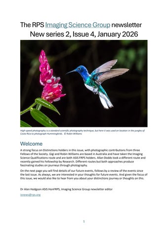
High-speed photography is a standard scientific photography technique, but here it was used on location in the jungles of Costa Rica to photograph Hummingbirds. © Robin Williams
Welcome A strong focus on Distinctions holders in this issue, with photographic contributions from three Fellows of the Society. Gigi and Robin Williams are based in Australia and have taken the Imaging Science Qualifications route and are both ASIS FRPS holders. Allan Dodds took a different route and recently gained his Fellowship by Research. Different routes but both approaches produce fascinating studies on journeys through photography. On the next page you will find details of our future events, follows by a review of the events since the last issue. As always, we are interested in your thoughts for future events. And given the focus of this issue, we would also like to hear from you about your distinctions journey or thoughts on this.
Dr Alan Hodgson ASIS HonFRPS, Imaging Science Group newsletter editor isnews@rps.org
1
Future events We have the Group AGM in February. Please come and join us and meet the committee. Mike has sorted out a stellar line-up for the next Good Picture on-line in April. Chris Dainty and Rita Hofmann-Sievert were the 2023 and 2024 Progress Medal and Honorary Fellowship recipients respectively. Should be an informative meeting.
2026 Annual General Meeting of the Imaging Science Group The Annual General Meeting of the Imaging Science Group will be held online using Zoom on Wednesday 25 February 2026 at 3pm. Members will be contacted nearer the time with an agenda and joining details. If you think you can help the committee in any way in running your Imaging Science Group please get in touch. Any help will be welcomed.
Good Picture – Online – 2026 Part 1 Following a successful inaugural meeting in October 2025, the IS Group intends to continue this series of regular meetings with the first of this year’s 2 annual events in April 2026. We have 2 speakers for this meeting, their names and the titles of their presentations are as follows: • •
Prof. Chris Dainty HonFRPS: “The Fundamental Limits of Mobile Phone Cameras” Dr. Rita Hofmann-Sievert HonFRPS “Material Science in Photography: Surfaces and Nanostructures”
The full details of this meeting, including how to apply, will appear on the Group’s website and will be sent, via an e-mail, to all members before the end of January. Dr Mike Christianson, Organiser.
HDR Displays: Show the Full Dynamic Range of RAW Files This event is being run by the RPS Digital Imaging Group and they have kindly offered free access to members of the Imaging Science Group. It takes place 10th January 2026, 16:00 GMT. Register here or through the short link https://tinyurl.com/RPSDIOnline073 . If the timing is inconvenient you should still register as you will subsequently get a link to watch the recording at your leisure. It is not made available to those who have not registered so make sure you sign up!
2
Recent events Good Picture – Online Zoom Meeting: 18th October 2025 Early in 2025 the Imaging Science Group committee made the decision to move its face-to-face Good Picture meetings online. For over 20 years this meeting had presented many talks on various technical aspects of Imaging. Unfortunately due to ever rising costs, particularly post-Covid, the Group was unable to continue its sponsorship without increasing the delegate fees to unacceptable levels. The meeting on 18th October was the inaugural online event with 2 speakers presenting, with “Q and A” sessions after each talk. The first talk by Adrian Davies MSC ARPS was entitled “Photographing Plant Behaviour”. He had given us a talk a few years ago describing the use of reflected and fluorescence UV photography to produce an insect’s eye view of flowers. This was briefly summarised showing examples where the image under UV illumination was significantly different from that under visible light. The main part of his talk was on “events that are either too fast or too slow for the human eye to see”. He was able to use his innovative methods of illumination to produce images which showed movement together with very sharp and colourful subjects. He showed many beautiful images of, for example, winged seeds falling to earth, hummingbirds approaching flowers, movement in touch sensitive plants, spore release from puffballs and pollen from tree flowers. Images were also shown of plants changing over days or weeks. Great care was necessary to ensure that the camera distance to subject and illumination were the same throughout so that the correct scale was apparent. This was a fascinating collection of images brought to life with an insightful description of the subject and methods used to capture them. The second speaker was Dr Alan Hodgson ASIS HonFRPS, a long-time supporter of our meetings, whose talk was entitled “Photography by Synthetic Aperture Radar”. It was based on his investigations into an unusual image in his archive; a 63x12 cm. black and white print from a satellite radar image. This image of Northern Iran (300x50 km) was taken during a NASA mission of the Colombia Space Shuttle in November 1981. His description of the relatively complicated process of “Side Looking Radar” was impressive and allowed the non-experts amongst us to get a good basic knowledge of the technique. One of the main problems with this form of imaging was the enormous amount of data per image, too much to be easily transmitted to ground stations with the technology available. Alan described NASA’s solution which was based on conventional silver halide photography. The Optical Film Recorder on board the Shuttle, which was identical to the instrument used on the moon landing mission Apollo 17 in 1972, was basically a camera receiving light from a CRT on which the radar echo signal was imaged. Each echo produced a stripe on a roll of 5 inch wide photographic film. The resulting image on the roll was printed to produce Alan’s final picture using an optical data processor. Surprisingly this whole imaging chain was analogue as late as 1981. His investigations into NASA’s use of silver halide scientific imaging continue! The organisers would like to take this opportunity to thank our speakers for giving such interesting, knowledgeable and enthusiastic presentations. Recordings of the 2 talks will appear on the IS Group website. The Group committee would like to continue this series of online talks with a Zoom meeting in April 2026. If you have a subject you would like to present, or know of someone else who might be willing, we would be very grateful if you could contact us at pandm.christianson@gmail.com.
Fraunhofer spectra – their place in the evolution of photography This joint event with the RPS Historical Group took place on 2nd December 2025. A recording of the presentation can be found here and we hope to have an event review in the next issue. 3
London Imaging Meeting 2025: Lighting and Imaging The Institute of Physics (IoP) organised this meeting during the summer; it a part of a series which was previously organised by the Society for Imaging Science and Technology (IS&T) which has its headquarters in Virginia, USA. Our Imaging Science Group is a Cooperating Society for these meetings and has provided both an organizer and sponsorship for previous events. Other Cooperating Societies include the CIE and Colour Group (GB). As part of this sponsorship, members of our Group received a significant reduction in delegate fees equivalent to full IoP members. This is a brief description issued by the organisers just prior to the event. “Following the tradition of previous years’ successful research events dedicated to the future of imaging science, the 6th London Imaging Meeting (LIM) takes place at the Institute of Physics (IoP) in London, UK, from September 8 to 10, 2025. The theme chosen for this year is ”Lighting and Imaging” with the intention of bringing together students, researchers, engineers and others from these two distinct fields of science and technology, which share so many fundamental concepts and methods, and whose research challenges are often deeply intertwined. Just think about trying to capture good quality images in really low light levels! The first day features a summer school with four insightful short courses on radiometry, photometry and colour science for lighting; materials, lights, algorithms, and software for light transport simulation; the colourful maths behind multichannel imaging systems; and methods for illuminant estimation and correction in colour imaging workflows. The main conference offers a rich two-day program, including three keynote talks covering topics like the convergence of lighting and imaging, innovation in the field of spectral imaging, and how perceiving the illuminant may or may not influence colour constancy. 5 invited focal speakers address hot topics around the effects of individual vision differences, metrological challenges using HDR imaging for luminance measurements, virtual relighting of images, colour definitions of non-uniform surfaces, and spectral and polarimetric imaging of human faces. Additionally, 11 peer-reviewed papers have been accepted for oral presentation, and 21 as poster presentations. It is our hope that this year’s LIM conference provides excellent opportunities for networking and discussions about the future of imaging science research., and that these proceedings will serve as an important reference for future research and innovation in the field(s) of lighting and imaging.” Here is a direct link to the pdf of the events programme with the title and authors of the papers presented.
4
Members’ projects An Artistic Life after Science? Gigi Williams ASIS FRPS and Robin Williams ASIS FRPS “To develop a complete mind: Study the science of art; study the art of science. Learn how to see. Realize that everything connects to everything else.” Leonardo da Vinci We have spent most of our lives interacting with people – photographing patients, working with medical colleagues, and all those management meetings! So we are not the classic landscapists, introverted and ’wandering lonely as a cloud’; but we do now spend most of our time out in the landscape, in tune with the natural world. When we both retired from full-time work a few years ago, leaving behind lifelong careers in medical and scientific photography, we decided to re-kindle our interest in landscape and nature photography, something we hadn’t done in decades. Photography is photography, right? Our skills should be easily transferable. Wrong! The transition from medical and scientific photography to landscape and nature photography showed us just how different both genres are. We had always respected the fact that specialist photographers were good at what they did because they understood their subject – food, fashion, cars – or in our case medicine: but we were unprepared for how steep a learning curve it was going to be moving to creative practice. The challenges would be significant. Scientific and medical photography are very clear cut. It's very easy to judge a good or a bad result. It's very straightforward to tell a trainee how to improve, for example, a diffracted photomicrograph, a flatly lit psoriasis, or multiple reflections in a deep wet surgical cavity. There are clear standards of performance. In respect of all creative photography it is virtually impossible to do that; there are no clear performance metrics – it's all opinion – completely subjective. This is why we find creative photography so very hard.
Moiré interferometry for the measurement of spinal deformity in scoliosis. © Gigi Williams
5
When we worked as medical photographers we had to work in a highly controlled and standardized way – the accuracy and repeatability of the images of a patient across time with different photographers was paramount. This required us to be in control of every aspect of the process – subject positioning, lighting and so on. This turns out to be the opposite in landscape photography where we have no control of the subject, or the light, or the weather! We had spent our careers in studios, clinics, laboratories, post-mortem rooms and operating theatres, where we totally controlled the lighting – its angle, quality, colour and so-on. In landscape photography you’re never in control of the light – despite it making the difference between an average and a great photograph. We can get ourselves into the right location, at the right time, with the right gear, but at the end of the day it’s the weather and the light that will make the difference, and this we can neither predict nor control – all we can do is mitigate with good technique and embrace a skill we never had in the fast-paced medical environment – patience. We often have to allocate several days to photograph a location, waiting for the light. On occasions it never materializes; we recently turned up seven mornings and seven evenings in a row to photograph a location in Tasmania with no satisfactory result.
As scientific/medical photographers we were always in control of the light: as landscape practitioners we have to plan, wait and pray for incredible light like this dawn over El Chalten in Argentina. © Robin Williams
Our scientific training has turned out to be one of our greatest strengths but also one of our greatest impediments. We routinely use a whole range of photographic techniques that are part of the basic training and arsenal of the scientific photographer and apply them to landscape and nature photography. From high-speed to Infrared; from macrophotography to HDR and tone mapping; from multi-image panorama construction to astronomical photography and perspective control, all these techniques give us a measure of control over the final image that many of our contemporary peers in landscape and nature photography do not have. But in our current genre – considered to be ‘artistic’ – our logical, rules driven, factual, approach is a significant obstacle to producing great work.
6
Serial documentation of skin graft healing following burns on the shoulder of an 18-month-old child exemplifying the standardized scientific approach to photography. © Gigi Williams
According to conventional wisdom people who are ‘left-brain dominant’ tend to be more logical and analytical with strong critical thinking and mathematical skills, whereas people who are ‘right-brain dominant’ tend to be more intuitive, empathic and creative with strong artistic skills. Contemporary neuroscience however teaches us that brain functions are not in fact neatly dichotomised between 7
the two hemispheres of the brain. Instead, both sides of the brain work together to perform tasks such as speech or mathematical calculations. While brain activity may occasionally be higher in certain regions depending on the task, brain scan studies show that both sides of the brain work an equal amount on average.
‘The Family of Man’ contour map. Images like this one were used by doctors all over the world for clinical measurement, formed the basis of Robin’s Fellowship in 1976, but have also featured in hundreds of magazine articles as ‘fine art’.
There is no doubt that many decades of scientific training and practice have had a significant impact on our ability to be truly creative. This may be partly our left-brain ‘wiring’ but it is also the way that our brains have learned to process information. We think this is not only why we find creative work hard but is also a significant reason why we take different images. Not only does Robin see the ‘big picture’, but he has over the years, become extremely efficient at processing large amounts of information and quickly honing in on the crux of the matter. He has also become convinced that particular ways of approaching a problem will aid in its satisfactory solution; he displays large amounts of cognitive inhibition. Gigi on the other hand is much more able to see a wide variety of alternatives. Robin has generally believed that once he has ‘got the shot’ – ie., his pre-conceived version of the scene – he is then free to explore other alternatives. In fact this doesn’t work. Capturing the ‘most likely to succeed’ image actually inhibits him from looking for other alternatives and he is often packed up and ready to go whilst Gigi is still happily exploring and photographing. There is no doubt that Gigi is able to use cognitive disinhibition.
8
The authors routinely used reflected infrared in medicine but here applied the technique to the trees in Joshua Tree National Park, USA. © Gigi Williams
9
There is an outdated idea that science and art are polar opposites. That science, associated with the left brain hemisphere, is logical and structured, whereas art, the domain of the right hemisphere is intuitive, creative, guided by practiced judgement and innate skill. In fact, scientists draw on highly trained, creative intuition and judgements, just as much as artists do. Scientists rely on the sort of skills we usually attribute to artists throughout their work, from conceptualising projects, to designing experiments, to interpreting and presenting data, to conceiving of new theories and models. Both science and art are human attempts to understand and describe the world around us. The subjects and methods have different traditions, and the intended audiences are different, but we think the motivations and goals are fundamentally the same. Creativity is evident in the work of every scientist who has ever had to ponder a novel problem and figure out a solution.
Reflected ultraviolet photograph of abnormal pigmentation in Chloasma. © Robin Williams
Fundamentally, art and science are both examples of human attempts to comprehend the world around us or our feelings and emotions and provide some explanation for it. A sense of curiosity drives both science and art. There is a strong desire for discovery, a critical view of the status quo, and an aspiration for further knowledge. There have been many examples of great scientists who were also great artists and vice-versa. Leonardo da Vinci is perhaps the best-known example. Today there are many examples: Zoe Keller producing great works of art that tell the story of threatened ecosystems, Adrien Segal who produces 3D sculptures of tidal data, Rob Kesseler working at Kew Gardens with multi-image, hand-coloured SEMs of plant cells to understand lifecycles or Hubert Duprat making jewellery by collaborating with Caddis Fly larvae. Several initiatives, such as the SciArt programme at the Wellcome Trust in London have started to institutionalize and fund art and science crossover projects.
10
Phase contrast microscopy of blood passing through a cardiac hyperthermia pump showing crenelation of the erythrocytes and consequential fibrin formation. Although scientifically extremely important the images produced have sold well to individuals and magazines as fine art. © Robin Williams
Interestingly photography sits at the very heart of the science-art debate. It’s hard to imagine how acrimonious the debate became about whether photography was an Art or a Science, or indeed what constituted photographic art. Photography’s ‘birth’ in the mid-nineteenth century was a collaboration between scientists, keen to discover a reliable process for fixing light on a flat surface as a means of examining the physical world, and artists searching for new ways to look at and capture nature’s beauty and create a visual narrative. The original aim of the Photographic Society (now The Royal Photographic Society) was ‘to promote the Art and Science of Photography’ and there was a reasonably happy co-existence of the two in the early activities of the Society. Inevitably there was more emphasis on technique, chemistry, equipment and so on, in the papers presented at meetings but the exhibitions and the Journal often reflected the commitment to the art of the discipline. Things came to a head in the 1880s with two groups of eminent photographers – ironically both committed to the view that photography was truly an art – led by Peter Emerson and Henry Peach Robinson – starting a very public debate about the correct technique to create ‘photographic art.’ Emerson and Robinson were both respected, leading photographers in their day – and their images are still iconic today over 140 years later. Both published texts defending their positions. Emerson a doctor advocating the technical and Robinson a pictorialist leading the creative faction. By 1890 Peach-Robinson had become increasingly exasperated by the influence of Emerson’s “Naturalistic” photographers on the council of the Photographic Society and the disproportionate amount of time devoted to photographic technique. By 1891 the two factions of the society had become deeply polarised. Peach Robinson (the Vice President of the Society) and 15 other senior members left the Society. We think as a photographers we are heavily influenced by these two disparate schools: on the one hand Pictorialism – Henry Peach Robinson, Turner and Constable – including the camera club images we saw in competitions as a youngsters and the 11
picture postcard views we sought to emulate as a teenagers; and on the other the work of Peter Emerson and the technical and scientific training we received.
The yellow stain from absorption of the antibiotic Tetracycline into the teeth of a child (top), is satisfactorily obscured by artificial facings (middle) but still revealed by ultraviolet induced fluorescence (bottom). © Robin Williams
12
As self-funded retirees, serious landscape photography is a very expensive activity and we are therefore attempting to sell our work to offset some of the costs. We have an extensive e-commerce enabled web-site, retail representation, lab and framing partners and all the paraphernalia of running a small business. In trying to make money from our landscape work we face other challenges: most particularly people’s perceptions of the value of photography. One of the most common and annoying perceptions that works against our ability to sell our work is the belief on the part of the majority of people that they could get pictures just as good as ours if only they had a better camera. It’s truly insulting. It’s like going to a dinner party where the host presents a wonderful meal and you compliment them by saying ‘You must have a really good set of pots and pans.’ Unfortunately, the world is full of great images. The days of the paid stock library have gone. There are literally millions of high-quality landscape images available to editors and publishers for nothing – often not even a credit! Photography has been seriously devalued; it is for this reason that we have concentrated on selling our work as individual bespoke fine art pieces. We are enjoying modest success: our work is selling and we are winning significant awards.
Distillation of the essence of the scene instead of trying to capture every detail is the hallmark of artistic work. This image is from a series called ‘A Year at the Beach’ and was short listed for Australia’s most important fine art prize. © Robin Williams
With all these challenges to overcome you might be asking why we bother –– why don’t we cruise the Rivers of Europe, or play golf, like other retirees? In response we have to say that there is nothing more rewarding than standing in some remote part of the world, with the one you love, as the scene you envisaged, and hoped for, unfolds in front of you. After months of planning and arduous travel, fully prepared and in command of your equipment and technique, you record the image of the moment. It is a moment of true creative flow – an extraordinary ‘high.’ There is something very fundamental in producing some tangible artefact outside of oneself from something intangible within oneself. The greatest reward for a creative life is not only in the things you create, but also in how you live: in how you train yourself to perceive and respond to the world, and how you assimilate the experiences, revelations and emotions, that come your way. As Gigi records in her
13
Artist’s Statement ‘I am passionate about recording the fragile nature of our world and its inherent beauty in a world full of hatred and conflict.’
Conceptual work sits at the heart of our artistic practice, but is underpinned by our knowledge of scientific techniques, in this case high-speed, Rheinberg, macrophotography. ‘Photo-Synthetic’ is a body of work that explores notions of creation, and therefore creativity; life in a liquid state. © Gigi & Robin Williams Photography.
Gigi and Robin have a combined experience of over one hundred years as photographers; as ‘retired’ scientific photographers now aspiring to create work with some aesthetic value we’re happy with the advice of Albert Einstein who once said: “The Best Scientists are Truly Great Artists.”
Fluorescein angiography of retinal vessel leakage. A fluorescent dye is injected into the patient’s circulatory system as rapid sequence photographs are taken of the retina with a specialized camera. © Robin Williams
14
About the authors: Gigi Williams BAppSc ASIS FRPS FAIMBI FBCA Gigi has been a professional photographer for over 30 years having graduated from the Royal Melbourne Institute of Technology. She is an Accredited Senior Imaging Scientist, a Fellow of the Royal Photographic Society, the BioCommunications Association (BCA) in the USA, and a Fellow of the Australian Institute of Medical & Biological Illustration. She has been the recipient of many awards for her photography in Australia and the United States. In 2006 she was awarded the top honour of the BCA, the Louis Schmidt Award. She has had many years experience as a practitioner and manager, most recently as the Director of a large creative department at the Royal Children’s Hospital in Melbourne providing photography, graphic design, video and web design to the Hospital and the wider community. In 2016 she left her busy job to concentrate on her personal work and combine her science background with art. Since then she has traveled extensively with her husband Robin and has been recognised with over 60 international awards for her landscape and nature photography. Dr Robin Williams BSc MPhil PhD ASIS FRPS FBCA FIMI FBIPP Robin is a distinguished photographer with fifty years professional experience as a practitioner, researcher and teacher. He holds undergraduate qualifications in both scientific and medical photography and Masters and PhD degrees in Medicine. He is an Accredited Senior Imaging Scientist and a Fellow of seven learned societies including the RPS. For 20 years he had a stellar career as a medical photographer starting at Cambridge Medical School and culminating as Director of Medical Illustration and Teaching Services at Charing Cross and Westminster Medical School in London. Then Robin moved into academia and was a Professor of Photography for over 25 years and Dean of the Faculty of Art, Design and Communication at RMIT University in Melbourne. Robin was also a Founding Governor and Chair of the Academic Board at Australia's top private photography college (PSC). He has published over 70 scientific papers and books and delivered over 400 presentations. His work has been widely exhibited, is in many public and private collections, and has been published in hundreds of magazines, such as, Time Life, National Geographic, The Observer and New Scientist. In recent years Robin has concentrated on his personal work and has quickly established himself as a recognised landscape and nature photographer. He has already won many international awards for his work in this genre; including recently International Garden Photographer of the Year.
15
Reflected infrared photography – de-oxygenated blood strongly absorbs infrared. © Robin Williams
16
Scanning macrophotography to obtain increased depth-of-field is one of the many techniques learned by the authors as part of their training but here it is applied to the feathers of endangered and extinct birds as part of a conservation awareness project concerning the disastrous loss of bird species. © Robin Williams
17
Head of Department with no qualifications! Dr Allan Dodds, FBPsS, FRPS. My first contact with photography as a full time, paid occupation had been when I had left school having failed to obtain any academic qualifications. This had been on account of having been given at the age of nine a Kodak Brownie box camera by a kind neighbour. By the age of fourteen I had become quite proficient at developing my own films and enlarging the negatives in a makeshift darkroom in my parents’ attic and, having preferred this rewarding activity to the drudgery of school homework, my academic career had adopted a steadily downward trajectory. At the age of seventeen and to my parents’ despair I left school prematurely to enrol as an apprentice photographer to the long-standing and respected firm in the City of Edinburgh known as J Campbell Harper Ltd, Commercial, Industrial and Portrait Photographers. The work soon proved to be repetitive and unrewarding, being limited to the developing and printing of other people’s amateur efforts at photography. However the manager encouraged all apprentices to become members of the Edinburgh Photographic Society. There we were able to observe the efforts of others whose seniority and enthusiasm had enabled them to produce images of which we teenagers were in awe. And in the fallow, dreary, Scottish winter months, when only one dedicated photographer’s film per day might require our attention if we were unlucky, there was plenty of time to experiment in the darkroom in order to produce, in eager anticipation of receiving praise, or conversely in fear of receiving harsh criticism from our betters, large mounted exhibition quality bromide prints fit for critical and only too public consideration. Each month a print competition would take place with members being invited to make regular submissions. At the end of the first year I was, to my astonishment and disbelief, awarded the President’s Trophy for the best print in the Beginners’ Section, together with a silver medal celebrating the Society’s Centenary Year in 1961; awards that I still cherish. In the following year I was promoted to the Intermediate level, but by this time I had resigned myself to the fact that my weekly wage of £2.00 as an apprentice was insufficient to enable me to keep up with the earnings of many of my contemporaries, and a fond wish to purchase a camera more sophisticated than my entry level Agfa Silette was far beyond my means of realisation, a new Leica camera costing the entire year’s wages of an apprentice before deductions. Consequently, that year was to be my last involvement with the Society, but it had provided me with a sound footing and a confidence in my abilities that would later stand me in good stead, although at the time I did not appreciate the fact or indeed have any career ambitions or clear idea as to what sort of photographer I might eventually become. Having served only two years of my five years’ apprenticeship and in recognition of the fact that I had nothing further to learn in such a limited environment, I applied for the post of photographer at the Scottish Central Library, recording on microfilm rare manuscripts and ancient publications. Occasionally a priceless artefact would be brought to my studio, secured in a steel box to which two porters from another library would, in the strict interests of security, be handcuffed. One of those was a rare tenth century Arabic book on parchment bearing meticulously drawn diagrams of optical phenomena such as reflection, refraction and the camera obscura: Alhazen’s ‘De Aspectibus’. Another was an original edition of Fox Talbot’s book of Calotypes entitled, ‘The Pencil of Nature’. Others were of a far more mundane nature such as, ‘The Oliphants of Gask’, a distinguished but virtually unheard of Scottish aristocratic family; or ‘The Paisley Pamphlets’, an odd collection of local gossip compiled by a nineteenth century Minister of the Church of Scotland that ran to several tedious volumes of endless trivia. Those cultural artefacts are still available in microfilm form for current researchers to view, long after the originals have now disintegrated. Uninspiring as those tasks proved to be, this was to be my first photographic contribution to cultural posterity, before the invention of the photocopier made such a post redundant.
18
Needless to say, after two years of such reprographic activity I had again become disenchanted with the repetitious nature of the work and, lacking the range of camera and darkroom equipment that had been at my disposal during my brief apprenticeship, I yearned for more varied and challenging photographic activities. I had already decided that it was time to broaden my skill set and venture into a specialist area where I felt I might make a more useful contribution to society: perhaps even making a personal statement through images rather than words, or even recording more significant events that were taking place around me instead of those that had taken place decades or even centuries before. So it had been that in 1965 my attention was drawn to an advertisement in The Scotsman newspaper for the post of Senior Medical Photographer at a nearby hospital. Accordingly, at the age of twenty-two and without the possession of any academic or formal photographic qualifications I submitted my application for a post at what I was later to learn to be the highest Whitley Council grade of appointment for an NHS Head of Department comprising one member of staff. As the advertisement had omitted to mention that no photographic department yet existed I had naively assumed that if appointed I would be accountable to a senior practitioner with whom I could gain clinical experience under close supervision, rather than to find myself the unwitting founder of a department and a medical photographic service that were yet only a twinkle in the eye of an eminent professor. To my surprise, I was short-listed for interview and was duly summoned to address a panel comprising eight orthopaedic surgeons and a hospital administrator. Also present on the panel was a Mr Robert Whitley, FRPS, Chief Medical Photographer at the Department of Medical Photography at the Royal National Orthopaedic Hospital in London. One by one the consultant surgeons quizzed me about my background and put a number of medical questions to me of which I was regrettably able to answer but a few. Changing tack to spare me any further embarrassment I was then asked why I thought that I could provide a photographic service to a busy teaching hospital. Not fully grasping the implications of this question and, instead of answering I took from a large 20x24 inch Kodak bromide paper envelope that I had brought with me a number of exhibition prints that I had submitted to the Edinburgh Photographic Society over the previous two years. The images were in the then pictorial tradition and, as each consultant surgeon inspected my portfolio of prints exclamations of, “Could I have a copy of this one please?”, were voiced. Mr Whitley, FRPS, beamed at me and tipped me a wink, giving me some small hope for success in my future career. After three other candidates, much more mature and experienced than myself had been interviewed, I was invited to return to the interview room. To my astonishment I was offered the post of Senior Medical Photographer. I was informed that on account of my lack of medical knowledge and relevant experience I would be required to undertake a placement at Mr Whitley’s department in London for a period of three months’ training. Barely comprehending my good fortune I readily agreed to this condition and in the following week I found myself aboard ‘The Flying Scotsman’ from Edinburgh Waverley to St Pancras station on the beginning of a journey that I shall never forget. This was in the ‘swinging sixties’ and London was to present itself to me as a far cry indeed from the genteel, dignified, conservative and God-fearing Edinburgh in which I had grown up. The ‘training’ turned out to be somewhat of a disappointment as I was simply permitted to observe other medical photographers at work and was not even allowed into the darkroom. After two weeks of frustration I appealed to Mr Whitley to allow me to participate under his tutelage but instead and, for reasons unknown and unexplained, I was shipped off to a subsidiary department at Stanmore where another medical photographer took me under her wing. There I was given experience in routine clinical photography and permitted to observe pioneering surgery such as hip replacement operations, although operating theatre photography was deemed to be out of the question for such a novice, fortunately for me as the sight of blood was to come as a shock to the system. I soon 19
became frustrated by this disempowerment and after two months I persuaded myself that I was sufficiently proficient to be set loose on the orthopaedic scene in Edinburgh. My premature return was motivated by anticipation of the prospect of working alongside other medical photographers and surgeons: a team of worthy professionals, I thought, and a far cry from the drudgery of developing and printing the efforts of amateur snapshooters or the recording for posterity of ancient, dusty documents. My enthusiasm proved to be short-lived as the department of medical photography did not yet exist. Indeed, the building in which it was to be housed was still under construction and it was to be a number of months before I was able to envisage a department that I was not only to head, but to found from scratch. Once this reality had sunk in I began to have doubts about being up to the challenge. So many things beyond the photography itself had to be considered, costed, pleaded for and acted on. Unbeknown to me most of the department’s budget had already been spent on equipment on the advice of Mr Whitley and one of the surgeons who himself was an accomplished amateur photographer who regularly undertook his own technically excellent photography in theatre. I again began to feel disempowered, hampered even, in what influence I might have by way of establishing my own department, photographic style and reputation, but there was precious little time available to indulge in the luxury of self-doubt. Pressure was already being placed upon me to get things up and running and this spurred me into action.
An anomaly of the ulnar nerve. © Allan Dodds
The equipment so far provided consisted of two thirty-five millimetre Exakta cameras with three lenses, a set of extension bellows, a Mecablitz electronic flash unit, a Paillard Bolex sixteen millimetre cine camera, a Ferrograph tape recorder and little else besides. The Exaktas had been recommended by Mr Whitley as they had represented for him the pinnacle of equipment for undertaking macro photography and, together with the bellows attachment they were indeed to 20
prove highly suited to the task of photographing operative procedures of small areas. However, by 1965 such equipment was already out of date, with single lens reflex cameras now available that featured through the lens metering. When I enquired as to what studio and darkroom equipment had been ordered the answer came, “None”. I was then faced with the unenviable task of persuading the hospital authorities to allocate further funding to allow me to purchase the bare necessities for the taking and processing of high quality black and white photographs, the norm for clinical records in the day. Mercifully, Professor of Orthopaedics was more than happy to support all of my requests and, after a number of calculated tantrums on his behalf the hospital authorities were persuaded to permit me free reign in furnishing the department with further but limited funding for the equipment that I deemed necessary to undertaking the various tasks in hand. These included an MPP 5x4 camera with associated film holders, sheet film developing tanks, film hangers, drying cabinet, a De Vere 54 enlarger, masking frame, developing dishes, rotary glazer, print trimmer, cine film splicer, etc. Regrettably, this did not permit the purchase of more state of the art cameras to replace the already ageing Exakta models. Nonetheless, I was by now feeling more optimistic that I could provide at least a basic and dependable medical photographic service. But there was soon to be another setback. As it was in the process of being completed, my studio simply consisted of a large, empty space with two patients’ changing cubicles attached. Recessed into the ceiling were three rows of 500 watt floodlights pointing at forty-five degrees to the floor. A backdrop consisting of three rolls of paper was mounted on the wall, permitting a choice of white, grey or black, and a pair of dark green curtains completed the studio setting. So far, so good, but the lighting was not at all appropriate, being suited more to colour cinematography than to black and white still photography. More worryingly, however, was the fact that no benches, storage cupboards, bookshelves or desk had been thought of, so that print finishing, negative filing, slide mounting, cine film editing and all the associated administrative work could not be carried out. A further request to the management produced a positive result. Two rows of benches were soon installed and once a desk had been commandeered my studio now looked almost complete, along with shelving to accommodate a set of ring-bound Kodak Data Sheets and copies of the British Journal of Photography, negative filing cabinets and a telephone bearing my own internal ‘phone number with a direct dialling outside line. Attention next turned to the darkroom, which had been clearly designed with input from Mr Whitley. Already installed were two large sinks, one of which could accommodate three 5x4 processing tanks and opposite a large bench on which an enlarger and developing dishes could be located. State of the art blinds and a lightproof door inspired confidence but as there were no developing tanks, print dishes, dish warmers, film drying cabinet or glazing machine to be found, all of those items had to be purchased from the residual budget or by means of special pleading. It soon became evident that although the photographic side of things had now been adequately taken care of other considerations began to present themselves. Mr Whitley had hinted that the surgeons often believed that the medical photographer was in some way psychic or clairvoyant; that is to say that they would often express a wish by means of a vague request and then subsequently complain that it had not been carried out to their satisfaction because the photographer had been unable to read their minds. Standardised photographic request cards were therefore developed along the lines of Whitley’s department, and these were distributed to all of the wards so that some degree of control over the process of identifying, specifying, communicating and undertaking the requisite clinical recording could be achieved. Clerical administration next reared its head and a system of filing negatives and cross-referencing them with other patient identifiers was established. Alongside this a duplicate set of patient images was retained in the department for the purposes of future reference, enabling replication of a non-standard viewpoint or lighting arrangement to be produced at a later date. This was to prove invaluable in the production of comparable images over long time periods as a patient might have to undergo a series of corrective procedures, each of 21
which could alter their outward appearance over several months and in some cases several years. Objectivity and reproducibility were to become the watchwords. Final preparations involved liaising with the hospital’s Linen Room where diligent seamstresses were employed to craft a number of bikinis (‘fripperies’ as Mr Whitley referred to them) in varying sizes to provide unclothed patients with a modicum of decorum and dignity during the psychological stress of the photographic recording of their orthopaedic conditions under the glare of studio lights and the clinical gaze of the photographer and soon the changing rooms were fitted out with an array of accoutrements, including a clothing locker, dressing gowns and slippers. Lastly, a policy of chaperoning was also insisted upon for the safeguarding of female patients as well as the male photographer. Once orders for the necessary film, print materials and chemicals had been placed with Kodak and Ilford, I felt ready to commence my new career as a professional medical photographer.
Scoliosis: one year apart. © Allan Dodds
Following this period of concentrated activity I was able to announce that the new medical photographic service was now fully up and running. Requests soon flooded in after a lengthy period 22
of anticipation among the surgeons. Such was the initial surge in demand that a single-handed photographer soon proved insufficient to meet the high volume of requests, and a ruling was made at senior management level that only Consultant grade surgeons could legitimately request photography, although in practice this stipulation was to be honoured more in the breach than the observance, such was the keenness of the photographer to display his new found talents to anyone who might show an interest in his craft, not to mention the eagerness of the surgeons to have their handiwork recorded for posterity through photographic means. This latter situation was to prove a problem as word got around, but a stern memorandum from the Professorial Chair soon put an end to any dreams of self-promotion through photographic recording of surgical handiwork by aspiring professorial candidates. But before the first patient had crossed the threshold my first commission as a more generic photographer had come from the hospital management with a request to photograph the New Clinical Research Unit for the purposes of illustrating a booklet that was being prepared for the opening ceremony in a few weeks’ time. This was a fortuitous event as it enabled me to establish that all of the equipment was now fully functional and that I was able to fulfil the great expectations awaiting me as well as my own ambitions. A number of glossy glazed bromide prints were duly despatched to the printers and on receipt of the publication a note of thanks from the Hospital Secretary gave me the confidence to consider that I had ‘arrived’ at what I hoped might be a valued member of our National Health Service. I was subsequently invited to give a talk to the medical staff on the importance of medical illustration, an invitation that I was happy to accept, and a lecture on the history of medical illustration over the centuries was duly delivered. After a few years the work began to take on a familiarly repetitive nature and, having rubbed shoulders with eminent surgeons I began to appreciate my relative ignorance. Also, I had observed that patients often responded to their conditions in very different ways. For example, an individual born without a limb would cheerfully accept their lot, whilst one who had recently lost one would present as anxious, depressed and with low self-esteem. I began to feel that I might become of better use to these patients other than by merely recording images of their bodies. So it had been that I commenced academic study at evening classes and following success, I relinquished my post and went up to Edinburgh University to read Psychology and Philosophy. Following a post-graduate doctorate I became head of the Blind Mobility Research Unit at Nottingham University where I remained for the next twenty years researching the effects of blindness on travel skills. Following that I again changed career to become a Clinical Neuropsychologist, assessing the brain function of individuals who had suffered a stroke or a brain injury. During those thirty years I barely touched a camera other than to take holiday and family photographs, but on retirement I came across a suitcase in the attic containing hundreds of images dating back to the 1960s, most of them medical. This reawakened my interest in medical photography and motivated me to donate those images to the University of Edinburgh’s Lothian Region Archive where they were gratefully received. At around the same time, my former assistant passed away before he had the opportunity of writing up his own history of the department that I had founded and which he had headed for over thirty years following my departure. In the process of doing this I realised that I was the only person who retained this knowledge which I felt compelled to make public. Accordingly, I set about researching other departments of medical photography on Scotland and became aware that very little had been written on the topic. With the help of other retired medical photographers I managed to garner the relevant information that enabled me to tell their stories, as well as my own, and with further research at the University of Edinburgh Library I was able to trace the origins of medical photography in Scotland back to the middle of the nineteenth century. After a year of research I submitted the thesis to the Royal Photographic Society and to my delight I was awarded a Fellowship in Research in 2024.
23
I consider that my first hand experience of medical photography, combined with years of academic publication, had furnished me with a unique combination of first hand practical and analytical skills, and I assume that the RPS had been sufficiently impressed by these to feel able to award me with the Fellowship distinction, although I have to confess that my print submissions of portfolios for a Licentiateship distinction had failed on three previous occasions! Dr Allan Dodds, FBPsS, FRPS.
24
Yet to come Details of our next events are included in the Future Events section. Ideas for future meetings are always welcome – send them to me and I will pass these on. Especially if you are willing to present or organise! In this edition I am grateful for contributions from Gigi and Robin Williams and Allan Dodds. It would be great to add your name to the list for future editions. Gigi and Robin took us through their journey across photographic genres and how they bridged the art/science “divide”. What is your genre and where does Imaging Science fit into this? It would be good to compare notes. We would also like to hear from readers about conferences or meetings they attend. We intend that this newsletter will provide a forum for members to show their work, completed or in progress. All for now – see you next time! Don’t forget to send me your content and thoughts for the next issue. At the moment we are aiming for an April 1 publication so the edition will be assembled midMarch.
Dr Alan Hodgson ASIS HonFRPS, Imaging Science Group newsletter editor isnews@rps.org
Past newsletters The Group is starting to create an archive of these newsletters. Issues 1-3 are now available on the Group website as pdf files.
25