ISSUE 83 SEPTEMBER 2026
IMC21: Picture Special Science in Your Mother Tongue: Why I Volunteer with Native Scientists The Hudson Transparencies: magical work of past RMS President revisited in modern exhibition Microbial masquerade: The rise of the synthetic specimen
Plus...News, Calendar, Reviews, Reports 1
ISSUE 83 SEPTEMBER 2026
JEM-120i
NEW!
Compact, Easy to Use, Expandable TEM
Specimen observation in 4 steps
We developed the JEM-120i with the concept of "Compact", "Easy To Use", and "Expandable". With the new external appearance, this instrument has evolved into a useful tool that anyone can use easily, from operation to maintenance. It takes only 4 steps from loading a specimen to completing observation. The JEM-120i is equipped with an enhanced TEM control system and fully automated apertures, eliminating the need for switching magnification modes and selecting an aperture. Observation operations can be performed more smoothly than with previous models.
Learn more 2
ISSUE 83 SEPTEMBER 2026
contents features 4 30 38
IMC21: Picture Special IMC21: “The Next Generation – Embracing the Responsible AI Revolution” – Your views Introduction by Kirti Prakash & Ian van der Linde Science in Your Mother Tongue: Why I Volunteer with Native Scientists Virginia Silio
50
The Hudson Transparencies: magical work of past RMS President revisited in modern exhibition Owen Morton
74
Microbial masquerade: The rise of the synthetic specimen Brian Ford, Hon FRMS
84
Imaging in Biomedical Research – Obtaining Patient and Public Perspectives Steve Thomas & Natalie Poulter
regulars 34 42 62 68 94 98
Calendar Journal of Microscopy New Member Welcome Office News Company News New Products
reports and other features 36
Microscopy: Advances, Innovation, Impact 2026 – watch online for FREE!
48
Early Career Researcher Prize-winner for Excellence in Imaging at the SBI2 European Meeting: Gantugs Atarsaikhan
90
Imaging Cell Dynamics Conference – report by Alan Wainman
92
In Memoriam: Kevin Christopher McNamee 1
MAGAZ I N E
infocus is the Magazine of the Royal Microscopical Society (RMS) – the only truly international microscopical society. The RMS is dedicated to advancing science, developing careers and supporting wider understanding of science and microscopy. infocus Magazine 37/38 St Clements Oxford, OX4 1AJ, UK Tel. +44 (0)1865 254760 Email: infocus@rms.org.uk Website: www.infocus.org.uk Scientific Editor Leandro Lemgruber, University of Glasgow, UK Editor Owen Morton Tel. +44 (0)1865 254763, Email: editor@infocus.org.uk Editorial Board Myfanwy Adams, John Innes Centre, Norwich, UK Maadhav Kothari, Zeiss Microscopy, UK Hilary Sandig, Cancer Research, UK Trevor Almeida, University of Glasgow, UK Tim Young, University of Cambridge Ferran Valderrama, City St George’s, University of London Thomas Slater, Cardiff University Advertising Email: advertising@infocus.org.uk ISSN: 1750-4740 © 2026 Royal Microscopical Society infocus is published four times per year by the RMS. Designed and produced by Creative Design. Reproduction in whole or in part without permission from the RMS is forbidden. Views expressed in the Magazine are those of the individual contributors and do not necessarily reflect those of the RMS.
RoyalMicroscopicalSociety @RoyalMicroSoc @RoyalMicroSoc
2
ISSUE 83 SEPTEMBER 2026
FROM THE SCIENTIFIC EDITOR Dear Readers, Welcome to our third issue of infocus Magazine for 2026! I write this introduction having just returned from the 21st International Microscopy Congress (IMC21) in Liverpool, UK. What an absolutely brilliant event this proved to be for the global microscopy community - from the groundbreaking research and latest technology on display, to the workshops, social events and networking opportunities. It really was a pleasure to be there, and meet so many researchers, manufacturers and enthusiasts from around the world. I hope you enjoy our report and pictures from the event in this issue! The subject of Artificial Intelligence (AI) and its impact on microscopy was an overarching theme of IMC21, and infocus was on hand to ask attendees for their thoughts on how AI is already transforming research, and what the future might hold.The result of those conversations is presented here in a special article, with an introduction by Kirti Prakash and Ian van der Linde. Special thanks to everyone who contributed to this piece, and to everyone who took the time to chat with us at IMC21. On the same subject in this issue, RMS Honorary Fellow Brian Ford casts a critical eye over the rise in AI-generated representations of microorganisms as online teaching resources. His impassioned defence of microscopy as “the disciplined observation of the real” is both timely and thought-provoking. Elsewhere, we hear from Natalie Poulter and Steve Thomas about a patient / public engagement event to inform approaches to imaging in biomedical research at the University of Birmingham. Their article is not only useful reading for anyone interested in carrying out similar activities, but also illustrates the huge importance of scientists building trust with the general public. In a similar vein, Virginia Silio shares her experience volunteering for the ‘Native Scientists’ initiative – a ground-breaking outreach project bringing the wonder of science to the children of first-generation immigrants in their first language. It’s a great example of how role models “who look and sound like you” can make a difference. And finally, for the history buffs among you, infocus examines how the work of a Victorian-era RMS President was recently brought to life at a London gallery. The ‘Hudson Transparencies’ really are visual treat. As always, I hope you enjoy reading these latest offerings, as well as all our other content in this issue. Slàinte!
Leandro Lemgruber
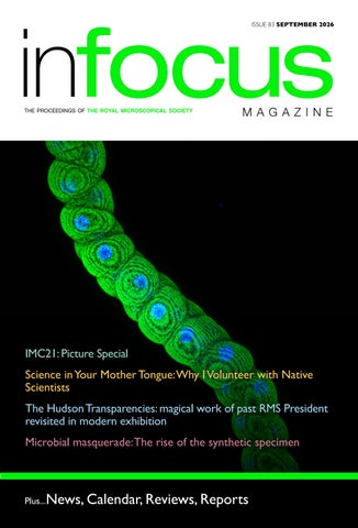
COVER IMAGE: Na+/K+ ATPase (488) and DAPI By Saurabh Chand Sagar, Banaras Hindu University, India.This image shows the projection image of the Drosophila Malpighian tubule (main segment) 3rd instar larvae (118-120 h old) stained with Na+/K+ ATPase (green) and nuclei stained with DAPI (blue). The image was acquired using a 40× objective at 0.5 zoom captured using Zeiss LSM 900 Confocal microscope.
3
Phew – what a week! IMC21: Picture Special
4
ISSUE 83 SEPTEMBER 2026
A
s the dust settles on the 21st International Microscopy Conference (IMC21), we take a look at some of the action from a memorable few days in Liverpool, UK (31 August – 4 September).
A total of 3,020 delegates, visitors and exhibition representatives from almost 50 different countries took part in this superb event for the global microscopy community. The Congress featured a world-class conference programme with 32 symposia stretching over four full days. Meanwhile, around 120 exhibitors – including many of the world’s biggest names in microscopy and imaging – brought their latest products and technology to the exhibition hall. With preconference workshops, a Young Scientist Assembly and a host of social events also featuring, the ‘Olympics of Microscopy’ certainly lived up to its billing. The RMS was honoured to deliver this year’s Congress alongside the International Federation of Societies for Microscopy (IFSM), Liverpool University and all our other partners. The Society would like to put on record its thanks to all the delegates, visitors, volunteers, exhibitors, sponsors and everyone else who helped make IMC21 such a success.
5
Young Scientist Assembly (YSA / ECR)
6
Before the main conference and exhibition got
the quality of their IMC21 abstract submissions - to
started at IMC21, we welcomed 50 successful
take part in the The Young Scientist Assembly (YSE
applicants - recognised for their achievements and
/ ECR).
ISSUE 83 SEPTEMBER 2026
After a fun day out in Liverpool on Saturday (30 August) and an evening meal at the Victoria Gallery and Museum, a full-day meeting took place on the Sunday at the University of Liverpool, with roundtable discussions, Q&A sessions and plenty of networking time. Micrographs entered into a specially organised image competition were also on display throughout the day. The winning image, titled: ‘Wormholes: Journey to the centre of the tooth’, was submitted by Maria Clara Muller de Andrade (Federal University of Pernambuco, Brazil) - pictured top right. 7
Pre-Congress workshops
8
Alongside the YSA on the Sunday, delegates took
There was plenty of time for some lively discussions
part in a wide range of pre-congress workshops at
on some of the hottest topics in microscopy – as
the University of Liverpool, covering the full range
well as some hands-on activities. The sessions also
of microscopical techniques and applications across
provided an early opportunity for delegates to
the sciences.
make new connections with their fellow attendees.
ISSUE 83 SEPTEMBER 2026
9
Welcome Reception To round off proceedings on the Sunday, we
It was a fabulous atmosphere and a wonderful way
welcomed delegates to the Liverpool Experience
to lift the curtain on what would prove to be a
Campus (LEX) for our IMC21 Welcome Reception
fantastic week in Liverpool. Attendees even posed
– featuring some great food, drinks and live music
for a giant group photo!
from a jazz band.
10
ISSUE 83 SEPTEMBER 2026
11
Conference The IMC21 conference provided delegates with a huge range of content delivered in 32 sessions across three main themes: Physical Sciences, Life Sciences and Instrumentation. The superb auditorium at the LEX was packed out each morning for our four Plenary speakers – Shirley Meng (University of Chicago, USA), Joanne Etheridge (Monash University, Australia), Peter Rosenthal (Francis Crick Institute, UK) and Sergei Kalinin (University of Tennessee, USA), and provided the perfect setting for the IFSM Symposium and Awards on the Friday morning (4 September). Elsewhere, the conference rooms were buzzing throughout the Congress, with hundreds of speakers presenting their work over the course of the week. We would like to thank all our presenters for bringing their research to IMC21.
12
ISSUE 83 SEPTEMBER 2026
13
14
ISSUE 83 SEPTEMBER 2026
“I appreciated all the positive feedback and really enjoyed meeting and connecting with everyone who showed up early before the conference for a run.” Running club at IMC21 For those keen to keep their fitness levels topped
The initiative provided a great opportunity to meet
up, IMC21 boasted its very own running club –
new people in an informal setting, get some fresh air,
courtesy of locally-based delegate and keen runner,
and shake off any pre-presentation nerves.
Richard Jinschek.
Richard, who is a PhD researcher at the University
Delegates of all abilities signed up online and met
of Liverpool, said: “I appreciated all the positive
for an early morning, 5km run throughout the week,
feedback and really enjoyed meeting and connecting
taking in the sights and sounds of Liverpool away
with everyone who showed up early before the
from the conference centre.
conference for a run.”
15
Exhibition Throughout IMC21, delegates and visitors were
breaks and lunch periods. It was also the location
treated to a fantastic array of the latest products
for the afternoon poster sessions, which were
and technology in microscopy, courtesy of around
extremely well attended throughout the Congress.
120 exhibitors who filled the enormous exhibition hall.
16
We would like to thank all the companies and partner organisations who took part in the IMC21
The exhibition hall was very much the central hub of
exhibition and helped make the Congress such a
IMC21 – especially during the regular refreshment
success.
ISSUE 83 SEPTEMBER 2026
17
18
ISSUE 83 SEPTEMBER 2026
19
Poster Sessions
20
IMC21 featured more than 800 scientific posters,
perfect opportunity for sharing ideas and making
presented across four afternoon poster sessions.
new connections in a relaxed atmosphere. Huge
With thousands of conversations taking place
thanks to all our poster presenters for bringing
throughout the week, these sessions provided the
your work to IMC21.
ISSUE 83 SEPTEMBER 2026
21
Gala Dinner Wednesday’s Gala Dinner was a spectacular affair,
of Liverpool’s Centre for Electron Microscopy
as more than 750 attendees returned to the
bearing his name, is renowned as the inventor of
conference centre for an evening of food, drinks
the scanning transmission electron microscope
and entertainment.
(STEM).
The atmosphere was buzzing across a sea of tables
After three courses of dinner and a glass of wine or
as the IMC21 Scientific organisers took to the
two, delegates took to the dance floor in customary
stage to welcome all the guests. As part of their
fashion, assisted by a troupe of local dancers.
introduction, they presented a signed, framed photograph of the late Albert Crewe to members of his family attending the dinner as special guests. Crewe, whose legacy is honoured by the University
22
ISSUE 83 SEPTEMBER 2026
It was certainly a memorable evening and a fitting way for the global microscopy community to celebrate together.
23
24
ISSUE 83 SEPTEMBER 2026
25
IFSM Symposium The final act of IMC21 was the International
The event also included a poignant talk from
Federation of Societies for Microscopy (IFSM)
Max Haider and Ute Kaiser in remembrance of
Symposium and Awards ceremony, which took
Professor Harald Rose, who passed away earlier
place on Friday morning (4 September).
this year. Harald was a world-renowned physicist
The symposium featured talks from each of the IFSM award-winners, who were also presented with their awards at the end of the meeting.
correction fundamentally transformed electron microscopy, opening up new avenues for research across different scientific fields. It was a fitting
Congratulations to:
tribute to a true giant in his field.
• Jennifer Zenker - Eduard Kellenberger Medal
As IMC21 was officially brought to a close, delegates
2023 (awarded for scientific excellence) • Lukáš Palatinus - John Cowley Medal
were offered a glimpse of the next instalment of this fantastic Congress series, with an introduction to
2025 (for pioneering contributions to electron
IMC22 due to take place in Berlin in 2031 – marking
diffraction and crystallographic analysis using
100 years of electron microscopy!
electron microscopy)
We can’t wait!
• Niklas Dellby - Vernon Cosslett Medal 2025 (for ground-breaking innovations in aberrationcorrected electron optics and advanced electron microscope instrumentation) • Jose Maria Carazo - Eduard Kellenberger Medal 2025 (for outstanding contributions to structural biology and cryo-electron microscopy image analysis)
26
and RMS Hon Fellow, whose work on aberration-
ISSUE 83 SEPTEMBER 2026
27
…Huge thanks to everyone who helped make IMC21 so special – including our generous sponsors:
28
ISSUE 83 SEPTEMBER 2026
From Millimetre to Nanometre Scale within one multimodal workflow
• Magnetic Imaging • 3D Structure • Surface Morphology & Properties • Chemical & Optical Nanoscopy • Elemental Composition • Crystallography & Diffraction
semiconductor devices
batteries
quantum materials
nano-structures
thin films
failure analysis
www.qd-uki.co.uk
(01372) 378822 | info@qd-uki.co.uk
magnetic materials
advanced materials r&d
IMC21: “The Next Generation – Embracing the Responsible AI Revolution” – Your views Introduction by Kirti Prakash and Ian van der Linde, School of Computing and Information Science, Anglia Ruskin University, Cambridge, UK AI at the microscope: seeing faster, but seeing true? AI has entered the microscopy room. It can steer instruments, sharpen acquisition, segment cells and writes code for data analysis before a researcher has finished their coffee (Krull et al 2020; Stringer et al 2021; Hohlbein 2026). The attraction is obvious: less time tracing boundaries, more time asking biological questions, at least until AI begins formulating the questions too, encroaching on the creative core of research. At IMC21, excitement about faster science was accompanied by a sharper concern: when does assistance become invention? “AI is here to stay, for good or bad,” says Leandro Lemgruber of the University of Glasgow. The question is no longer whether scientists will use it, but how and with what safeguards. Theo Andrews of University College Dublin offers a useful reality check: AI is “another tool in our toolbox rather than a magic fix-all.” For microscopists, its most immediate value lies in removing drudgery. Melanie Rug, President of the Australian Microscopy and Microscience Society, points to volume imaging, where AI can save “hundreds of hours” by targeting and separating regions of interest. Shrija Ghosh of IIT Kanpur sees the same potential in particle-size analysis: why spend days clicking on individual particles when a reliable model can do it in minutes? Mohammed Ilhami from Rouen is unequivocal: “We can definitely use AI to accelerate the way we do microscopy.” Speed, however, is only the first revolution. The deeper promise is to change what scientists spend their time doing. Sarah Farrow of Bitesize Bio describes the appeal of an autonomous laboratory
30
ISSUE 83 SEPTEMBER 2026
in which machines absorb routine operations and researchers return to ideas: “The idea of PhD students and experienced scientists being able to focus their energy on thinking full time is very exciting.” Yet there is a paradox: what looks like drudgery may be where discovery begins. Careful manual examination of images can expose confounds; reveal unexpected patterns and spark questions we had not thought to ask an automated agent. Such immersion also teaches PhD students patience, resilience and how to learn from failure. Not every repetitive task deserves preservation, but automating all of them risks weakening the link between observation and curiosity. The deeper risk is that tools designed to free scientists to think may increasingly do the thinking for them, while preserving the feeling that the ideas are their own. Sarah Farrow worries that outsourcing even routine writing could weaken critical reasoning and erode the foundations on which scientific judgement is built. Callum Perrett of the University of Birmingham captures the challenge in one line, hoping that: “We don’t automate the humanity out of research.” Nowhere is the boundary between assistance and authorship more consequential than in scientific imaging. In writing, AI may distort an argument; in microscopy, it may alter what appears to be the evidence itself. Hannah Dickinson of Utrecht University welcomes faster analysis “without manipulating data,” but warns that synthetic images can look disturbingly real. Her reminder is simple: “It’s important to remember that you are the one
with the microscope.” Anjuli Bali of the University of Oxford is similarly excited by AI-guided, realtime improvements in electron-microscopy acquisition, yet fears that hallucinations “might end up polluting my data.” For Shrija Ghosh, privacy and authenticity are equally fundamental. Artefacts and misclassification are longstanding risks in image processing; the question is whether AI makes these errors more frequent or harder to detect, further undermining trust in the scientific record. Alastair Gemmell of the University of Strathclyde sees promise in analytical AI but warns that generative outputs can lack the “refined detail expected of scientists” and raise questions about training data and consent. The wider challenge extends to both: research-quality standards exist, but their adoption remains uneven across researchers and journals. Consistent safeguards are needed, for dealing with a range of issues including preventing leakage between training and test data to choosing performance metrics that fairly account for imbalanced data.
beyond, this requires consistent adherence to shared quality standards: transparent methods, rigorous validation and fair reporting of both performance and limitations.
Kirti Prakash.
Ian van der Linde.
References: 1.
Krull, A., Hirsch, P., Rother, C. et al. Artificialintelligence-driven scanning probe microscopy. Commun Phys 3, 54 (2020). https://doi.org/10.1038/ s42005-020-0317-3
2.
Stringer, C., Wang, T., Michaelos, M. et al. Cellpose: a generalist algorithm for cellular segmentation. Nat Methods 18, 100–106 (2021). https://doi.
The mood at IMC21 was therefore neither anti-AI nor blindly enthusiastic.The challenge is to automate the repetitive, accelerate the difficult and reveal patterns the human eye may miss, while preserving provenance, judgement and doubt. In microscopy and
3.
Theo Andrews – Phd Student, University
Hannah Dickinson, Post-doctoral
College Dublin
researcher, Utrecht University
“My work is quite hardware-based so in terms of seeing AI as a potential threat, it’s probably less severe than in other areas, but even the work I’m doing includes automation so it somewhat depends on what is included in the definition of AI. I’m not really a fan of AI but I try not to be black and white about it, since I have seen some interesting presentations using it for protein structure prediction and things like that.The thing to remember, and I think most of the scientific community is not doing a bad job of this, is that AI is another tool in our
“I don’t know who said it but there is a quote around AI and it goes something like: “I don’t want it to do my art for me; I want it to do the boring tasks”. Especially in relation to science, if it can speed up analysis w i t h o u t manipulating data then I’m all for it. I recently took part in a group exercise where a colleague sent us some images and we had to guess which ones were AI. It underlined how generative AI can look very close to fact, and how it’s important that the metadata reflects that you were the one behind the
tool box rather than a magic fix-all.”
microscope.”
org/10.1038/s41592-020-01018-x Hohlbein J., webSMLM - a browser-based singlemolecule localization microscopy tool. (2026). https://doi.org/10.5281/zenodo.21445041
31
Mohammed Ilhami, PhD student, Groupe
Shrija Ghosh, PhD student, Indian
de Physique des Matériaux, Rouen
Institute of Technology, Kanpur
“AI can help us to
“When
do regular things to
publishing black and
save
for
white SEM images
instance, helping to
we can use AI to
automate the way
colourise
the
micrographs,
time
-
microscope
functions
or
in
we
are
the
integrating
the
processing the data.
image
I think, years ago,
part. We also need
processing
some of these tasks took one or two days, so we can
to do a lot of
definitely use AI to accelerate the way we do
particle size analysis, so if AI can help with any kind of
microscopy. But I think that AI will never interpret
image processing we are doing manually, selecting
the results like a human can, because we have a clear
each particle, that is a really good time saving thing. In
critique on what we see – rather than a binary ‘ones
terms of my concerns over AI, the first thing would
and zeros’ approach.”
be privacy - you never know if your information could be leaked to some other sources. The second
Melanie Rug, Australian Microscopy and Microscience Society President is
search for images, if I’m not the subject matter expert and I come across an AI-generated image, I
“AI will revolutionise (or
thing is authenticity, because a lot of times when we
won’t necessarily know if what I’m looking at is real.”
already)
volume imaging in all areas of microscopy
Sarah Farrow – Bitesize Bio
including light and
“I’m
electron
and
excited
correlative
EM,
freeing
up
man-
saving hundreds of
hours
that
were
hours of manually
historically spent on
targeting and separating regions of interest with
low-impact,
machine learning algorithms.
repetitive tasks that
AI is also changing the way we approach teaching, developing new ways of assessing student knowledge etc. Powerful – but also important to watch out for misuse.”
can
particularly about
now
be
automated.
I
recently toured an “autonomous lab” and the idea of PhD students and experienced scientists being able to focus their energy on thinking full time is very exciting.What does worry me is whether some of us are losing the ability to think – outsourcing writing basic emails to ChatGPT risks us losing the ability to write them for ourselves or apply critical reasoning. I also worry about what it means for training positions, as entry-level roles are inevitably the first to be cut.”
32
ISSUE 83 SEPTEMBER 2026
Alastair Gemmell, PhD student,
which is creativity. It will be important over the next
Strathclyde University
decade to ensure we don’t automate the humanity “AI (non-generative)
out of research.”
as a tool for analysis, in
a
pipeline, is
r e v o l u t i o n a r y. However, it is firmly a
tool.
It
Anjuli Bali, PhD Student, University of Oxford
will
ultimately
make
research
more
“I’m
both
apprehensive
and
excited about the use of AI in every
efficient, allowing for
stage of research.
more complex algorithms to take over monotonous
I’m glad that it might
tasks that take an extremely long time for humans to
save my time in the
do independently.
data
collection
However, generative AI is an exceptionally poor
phase with live, real-
recreator of human work. AI-generated work –
time imaging improvements in TEM acquisition.
be it art, diagrams, posters etc – are both easily
Depending on how this is achieved, though, I’m a bit
identifiable as AI and lack the refined detail expected
worried about the classic AI hallucination that might
of scientists. In addition, databases are trained from
end up polluting my data.
artwork or written work from authors and artists who did not consent to their work being used – which is bad.”
I’m also excited that I might not have to sit for hours in front of a FIB to prepare my sample. But the most worrisome part for me is the data post-processing. I enjoy this process and still believe human creativity
Callum Perrett, PhD student, University
is valuable at this stage. Fingers crossed that AI
of Birmingham
becomes a valuable friend and not a foe in research!” “AI
is
now
an
inevitability, and is something in science that you have to learn and integrate into your work, or you
will
be
left
behind.
Used
responsibly
and
appropriately, AI is possibly the most powerful tool we have right now, with essentially limitless applications. My research involves MRI and confocal microscopy, and AI makes my day-to-day much easier, with tasks such as counting neurites being made virtually entirely automated. Fundamentally, I think AI will
Leandro Lemgruber, infocus Scientific Editor; Head of Cellular Analysis Facility, University of Glasgow “AI is here to stay, for good or bad. We now have to teach students on how to use this tool and, importantly, how to critically
analyse
what you get when you use it. We also need better depositories that can be used as ‘ground truth’, since you can find anything online.”
never replace the most important skill in science,
33
Calendar We are very pleased to continue offering a range of ‘in-person’ and virtual events, in order to maximise accessibility and provide opportunities to those who might not otherwise be able to attend. The following information was correct at the time infocus went to print but could potentially be subject to change in the coming weeks. Please visit our event calendar at www.rms.org.uk for the latest updates. If you have any questions about a booking you have already made for an event, or need any help or advice, please contact us at info@rms.org.uk
2026
24 – 25 In Situ Workshop 2026, Harwell, Didcot, UK
September
24 – 27 32nd Annual New Zealand Microscopy Conference (RMS sponsored event), Wellington, New Zealand
15
Laboratory-based X-ray Phase Contras Imaging Workshop 2026, London, UK
30
Microscopy: Advances, Innovation, Impact 2026 - incorporating the RMS AGM & Section AGMs, London, UK (and online)
October 7
FIB&Prep Meeting 2026, Loughborough, UK (and online)
12
BioImagingUK 2026 Meeting (RMS attending), London, UK
November
December
3 – 4 Virtual European Flow Core Meeting 2026, Online
2027 January
21 – 22 EM-UKI 2027, Leeds, UK
March
3–4
Facility Management Training Course 2026,York, UK
21 – 25 Botanicals Meeting 2027, Cologne, Germany
12
flowcytometryUK 2026, Cambridge, UK
22 – 24 MSM2027, Cambridge, UK
16
Frontiers in BioImaging 2026, Manchester, UK
For further information on all these events, please visit our Event Calendar at www.rms.org.uk
34
ISSUE 83 SEPTEMBER 2026
Featured RMS events Microscopy: Advances, Innovation, Impact 2026 - incorporating the RMS AGM & Section AGMs 30 September, Royal Society of Chemistry, London, UK (and online) The global microscopy community is invited to join the Royal Microscopy Society (RMS) online for Microscopy: Advances, Innovation, Impact 2026 – a free-to-attend meeting incorporating the Society’s Annual General Meetings.
of microscopy, imaging and flow cytometry across the sciences. The meeting is being live-streamed from the Royal Society of Chemistry in London on 30 September – and while ‘in-person’ bookings are now closed, registration for online attendance will be remain open until the day of the meeting! Online attendance is completely FREE, and virtual attendees will still be able to register, up until (and including) the day of the event.
The event will feature a range of superb talks from recent RMS Award-winners, including some of the world’s leading researchers in their fields. It will also include contributions from each of the Society’s Science Sections covering all branches
Book now: https://www.rms.org.uk/rms-eventcalendar/2026-events/microscopy-advancesinnovation-impact-2026.html
flowcytometryUK 2026
a number of speakers covering immunology, marine biology, extracellular vesicles and other important research areas. Talks will be interspersed with commercial flash presentations. Sponsors will allow delegates to have access to information on recent developments in the field.
12 November, Cambridge, UK The flowcytometryUK 2026 Meeting will take place in person and will highlight the diverse areas in which cytometry is a vital resource. There will be scientific presentations from Frontiers in BioImaging 2026 16 November, Manchester, UK Frontiers in BioImaging 2026 will focus on correlative microscopy, leveraging the strengths of traditionally distinct imaging techniques to address questions that no single approach can answer alone. Sessions will span technical developments in light and electron microscopy, advances in image analysis and data interpretation, and
Read more on p36
biological applications across scales. We are creating a meeting where earlycareer researchers can learn, connect, and grow. By combining accessible scientific talks with dedicated networking opportunities and interactions with leading research groups, the event will provide a platform for early-career and established researchers to exchange ideas, build collaborations, and strengthen connections across disciplines.
35
R E P O RT
Microscopy: Advances, Innovation, Impact 2026 – watch online for FREE! Wednesday, 30 September, 2026, London The global microscopy community is invited to join the Royal Microscopy Society (RMS) online for Microscopy: Advances, Innovation, Impact 2026 – a free-to-attend
meeting
incorporating
the Society’s Annual General Meetings. The event will feature a range of superb talks from recent RMS Award-winners, including some of the world’s leading researchers in their fields.
It will also include contributions from each of the Society’s Science Sections covering all branches of microscopy, imaging and flow cytometry across the sciences. The meeting is being live-streamed from the Royal Society of Chemistry in London on 30 September – and while ‘in-person’ bookings are now closed, registration for online attendance will remain open until the day of the meeting!
A view of the impressive courtyard at the Royal Society of Chemistry’s Burlington House in London.
36
ISSUE 83 SEPTEMBER 2026
Former RMS Early Career Award-winner Akaash Kumar is pictured delivering his talk at Microscopy: Advances, Innovation, Impact 2024.
It means wherever you are in the world, you
Pantazis (Imperial College London) and Professor
are welcome to log-on and find out more about
Steven Lee (University of Cambridge).
the current and future activities at the RMS, and celebrate the contributions of some of the most ground-breaking scientists from across the microscopy community.
A further highlight will be the final of the RMS Early Career Award Competition, in which three shortlisted
presenters
will
deliver
quick-fire
presentations, with the winner to be announced
Among the speakers will be winners of the RMS
on the day. Our 2024 winner, Dr Akaash Kumar,
Section Awards, including:
is pictured here, delivering his talk at Microscopy:
• Professor Ilaria Testa, KTH Royal Institute of
A range of other RMS Awards will be presented
Technology Sweden (Light Microscopy Award) • Professor Emilie Ringe, University of Cambridge (Engineering, Physical & Material Sciences Award) • Dr Francisco de la Pena, University College London (Data Analysis in Imaging Award) • Professor
Kang-Nee
Ting
(University
Advances, Innovation, Impact 2024.
during the meeting, and we will also be hearing from RMS Patron, Baroness Julia Brown of Cambridge CBE, FRS. Online attendance is completely FREE, and virtual attendees will still be able to register, up until (and
of
Nottingham, Malaysia) Chris Hawes Award for Outreach and Education We will also be hearing from winners of the RMS
including) the day of the event. Book now: https://www.rms.org.uk/rms-eventcalendar/2026-events/microscopy-advancesinnovation-impact-2026.html
Scientific Achievement Award, Dr Periklis (Laki)
37
Science in Your Mother Tongue: Why I Volunteer with Native Scientists Virginia Silio
Virginia Silio.
Demonstrating fluorescence, using turmeric and alcohol under blue/ UV light.
38
ISSUE 83 SEPTEMBER 2026
The table-top set-up for one of my sessions.
Back in 2024 I saw a call on LinkedIn for scientists
that was the category of job that felt available to me.
whose mother tongue was Spanish to go into
I also wanted to be a dancer at one point, but that
a school and talk to some students. I signed up
didn’t sound realistic either. And now I’m also the
straight away, without thinking too much about it. It
mother of a first-generation immigrant child, who I
was only later, reading more about the project, that
hope will grow up bilingual.
something clicked: the idea is built around speaking to children of first-generation immigrants in their own language, and that really resonated with me.
Why outreach matters to me
There’s data showing that students from the same
I’ve always loved outreach, separately from any of
socioeconomic background go on to study STEM at
this. Scientists have drifted apart from the public over
very different rates depending on whether they’re
the years, and I think we have a real responsibility to
first or second-generation immigrants, compared
close that gap, to talk about what we do in language
with their peers (Schiefer et al., 2024). One of the
people will actually understand, rather than hiding
reasons is simple: they lack role models within their
behind jargon. If we don’t make that effort, someone
own community. There is nobody who sounds like
else fills the gap instead. At best that’s a journalist
them, or grew up the way they did, doing the kind of
getting the nuance wrong. At worst, increasingly, it’s
job that says “this is possible for you too.”
an AI chatbot with no particular stake in getting it
That hit home for me. I’m the first person in my
right.
family with a degree and a PhD. I’m the daughter
I did outreach during my postdoc, hosting donor
of a policeman and a waitress who later became a
visits from Cancer Research UK in the lab, talking
secretary when I was an adult. I grew up with no
through the science behind the work to people
scientist role models, none at home, none in the
who’d funded it but had no scientific background
family, none in the neighbourhood. I just didn’t know
themselves. In my current role at the Centre for
anyone who did this for a living. I remember saying I
Cell & Molecular Dynamics at UCL, we regularly
wanted to be a hairdresser ( if you know me, you’ll
host secondary school students in the imaging
understand how little sense that makes; I don’t even
facility, showing them the microscopes, walking
know how to do a plait!). I probably said it because
them through how we acquire images, etc. So when
39
brilliant. The children were aged seven to 12 - a wide enough range that I had to plan for two very different audiences in the same room. I started with a quiz about microscopes, just to get them talking.Then I moved into light decomposition, with a torch and a prism: a properly low-tech, highimpact experiment. After that came fluorescence, using turmeric and alcohol under blue/ UV light. This was, by a wide margin, their favourite part of the whole session. They could not stop playing with it. They made an enormous mess. I regret nothing. We finished with a set of microscopy images that they had to classify by animal, plant, or tumour. The session is structured like speed dating: roughly 20 minutes per group, five groups in total, each rotating through. In between rounds, the questions come thick and fast, and they go absolutely anywhere — telescopes, vaccines, cancer, how I ended up becoming a scientist, whether I have a pet, what my salary is, how to do an experiment ... Nothing is offLight decomposition, with a torch and a prism: a properly low-tech, high-impact experiment.
limits, and that’s part of the fun. I’d come prepared, though, as I have the best coach for this kind of
the Native Scientists call appeared, it wasn’t really a
thing: my daughter can ask you a hundred questions
new direction for me. It was outreach with an extra
a minute.
layer: outreach in Spanish, aimed specifically at kids who might not otherwise see anyone who looks or sounds like them doing this job.
generally don’t speak Spanish themselves. Mostly they just stand at the side, smiling, keeping a gentle
It’s worth saying that age range matters less for
eye on things, which leaves me free to focus entirely
the secondary school groups I see at UCL; they’ve
on the kids, something I’m genuinely grateful for,
already decided science is, at minimum, something
because trying to manage both the science and
that exists as a subject they study. The Native
classroom discipline at once would be a very
Scientists sessions are different. Seven-year-olds
different job. There is also a coordinator keeping an
haven’t decided anything yet. They’re still working
eye on the time, and helping if needed.
out what categories of grown-up job even exist,
What I find fascinating, session after session, is how
and whether ‘scientist’ is one of them. That’s a
differently the two ends of the age range behave.
much earlier, and arguably more important, point to
The seven-year-olds are curious, a bit mischievous,
intervene.
completely without filter in what they’ll ask you, and
My first session: chaos, mess, and a torch My first session, back in 2024, was chaotic but
40
The children are chaperoned by their teachers, who
ISSUE 83 SEPTEMBER 2026
playful. The twelve-year-olds are closer to (or fully into) puberty, so there’s a layer of wanting to appear like they already know everything. But underneath that, they’re just as curious as the younger ones.
Working with a group of children around the table.
At the end, every child gets a certificate. It’s a small
And honestly, it’s also just a good excuse to make
thing, but it’s a genuinely nice touch.
new friends. The other volunteers come from all
Getting better at it
kinds of scientific backgrounds, scattered across different institutions, and there’s a real camaraderie
I’ve done this every year since, in different schools,
in turning up to a school hall on a Friday morning
refining the session a little each time. It now includes
with a box of turmeric, a UV torch, and absolutely
an actual microscope and a set of slides: a toy one,
no idea how loud the room is about to get.
but surprisingly good quality for the price, bought on Vinted; the 40x lens is genuinely decent. At the most recent workshop, the kids were queuing up to look down the eyepiece. Genuinely queuing.
What it means, beyond the science Now, as the mother of a first-generation immigrant child, I see even more clearly why this matters.
For me outreach is about going back to the people we’re actually supposed to be working for: the ones we’re meant to help, or simply the people we share a world with and owe some kind of explanation to. Science doesn’t belong to scientists. The least we can do is show up, in a language people understand, and prove it.
There’s real value in hearing about science in your
Reference
own language, and in seeing role models who look
Schiefer, J., Caspari, J., Moscoso, J. A., Catarino,
and sound like you. That’s what I like so much
A. I., Miranda Afonso, P., Golle, J., & Rebuschat, P.
about the Native Scientists programme. It creates
(2024). Science and Heritage Language Integrated
inclusive, multilingual spaces that give children
Learning (SHLIL): Evidence of the effectiveness of
confidence, visibility, and a genuine sense that
an innovative science outreach program for migrant
science is something they can belong to, not just
students. Science Education, 108, 983–1014. https://
something they observe from a distance.
doi.org/10.1002/sce.21860
41
Journal of
Microscopy The Journal of Microscopy publishes top quality research articles, review articles and Hot Topic papers covering all aspects of microscopy and analysis. This includes cutting-edge technology and innovative applications in physics, chemistry, material and biological sciences.
You can read the latest Early View papers online at www.journalofmicroscopy.org They include: ORIGINAL ARTICLE
Enhanced biocompatibility of robotic microplasma sprayed Zr–2.5Nb coatings for orthopaedic implants Darya Alontseva, Yuliya Safarova (Yantsen), Sergii Voinarovych, Alexander Krasavin, Aleksei Obrosov, Bagdat Azamatov The surface bioactivity of orthopaedic implants plays a critical role in osseointegration and long-term clinical performance. In this study, zirconium–niobium (Zr– 2.5Nb) coatings were deposited onto gas-abrasively treated Ti6Al4V alloy substrates using robot-assisted microplasma spraying (MPS) to enhance implant bioactivity. The MPS parameters were selected to produce uniform coatings with controlled porosity (∼20%), rough surface, and satisfactory adhesion to the substrate. Surface topography and microstructure of coated specimens were characterised using confocal laser scanning microscopy and scanning electron microscopy, and compared with the uncoated Ti6Al4V alloy. Zr–2.5Nb coatings exhibited significantly increased surface roughness and a developed microrelief. In vitro angiogenesis assays using human umbilical vein endothelial cells demonstrated enhanced microvascular
42
ISSUE 83 SEPTEMBER 2026
network formation on Zr–2.5Nb–coated surfaces. Biocompatibility assessment with rat bone marrow– derived mesenchymal stromal cells revealed improved cell adhesion, spreading, and cytoskeletal organisation. These findings demonstrate that robot-assisted MPS of Zr–2.5Nb coatings on titanium implants with precise maintenance of selected spraying parameters can provide a biologically favourable surface that promotes angiogenic responses and represents a promising approach to enhancing the implant biocompatibility.
ORIGINAL ARTICLE - Open access
GloBIAS survey results – An insight into the global bioimage analysis community Laura R. de la Ballina, Christa G. Walther, Nicholas D. Condon, Alessandro A. Felder, Martin Schätz, Bettina Schmerl, Ko Sugawara, Clara Prats, Anna Klemm, Kota Miura, Paula Sampaio, Christian Tischer, Shuichi Onami, Rocco D’Antuono, Beth A Cimini, Robert Haase, Agustin A. Corbat, Florian Levet There is a global need for bioimage analysis (BIA) as advances in life sciences increasingly rely on cutting-edge imaging systems that have dramatically
expanded the complexity and dimensionality of biological images. Turning these data into scientific discoveries requires scientists with effective data management skills and knowledge of state-of-theart image processing and data analysis, in other words, bioimage analysts. The Global BioImage Analysts’ Society (GloBIAS) aims to enhance the profile of bioimage analysts as a key role in science and research.To better understand the needs and geographical representation of the BIA community, a worldwide survey was conducted, and 290 responses were collected across people from all career stages and continents. The survey underscores a strong interest of the BIA community in the activities proposed by GloBIAS to address shortcomings in work environment, funding, and scientific activities, and the enthusiasm of the community to actively contribute to the growth and sustainability of GloBIAS as a scientific society.
REVIEW ARTICLE
Convergence of cryo-electron microscopy and artificial intelligence in integrative structural biology: A critical review of advances, synergies, and implications for molecular biophysics and drug discovery Abinawanto, Alfi Sophian Structural biology has entered a period of rapid methodological change defined by the convergence of cryo-electron microscopy (cryo-EM) and artificial intelligence (AI)-driven structure prediction. Prior reviews have generally addressed cryo-EM instrumentation or AI structure prediction individually, or their combination primarily from a structural biology or drug discovery perspective; a microscopy-
centred synthesis tracing this convergence from detector physics and image-processing workflows through validation standards to in situ cryo-electron tomography (cryo-ET), while situating AI tools specifically as inputs to and complements of the cryo-EM workflow, has been comparatively underexplored.This review provides such a synthesis, organised around: (i) the instrumentation, imageprocessing, and validation advances underlying cryo-EM’s resolution gains, including detector physics, contrast transfer function (CTF) estimation, Bayesian and deep-learning-based particle picking and reconstruction, and map-to-model validation and deposition; (ii) cryo-electron tomography and subtomogram averaging (STA) for in situ structural biology, including deep generative approaches to heterogeneity analysis; and (iii) the AlphaFold2, AlphaFold3, and RoseTTAFold All-Atom systems, considered specifically in relation to how they interface with and depend upon cryo-EM data for model building, refinement, and validation. Building on this microscopy-centred account, we propose a three-tier classification framework for selecting between AI-primed, experiment-led, and in situ integrative pipelines, and we summarise performance, transparency, and accessibility differences between leading AI tools. We illustrate these methods with
43
examples from membrane proteins, ion channels, and viral glycoproteins, and briefly note implications for structure-based drug design and for structural biology capacity in resource-limited settings. Throughout, we distinguish between demonstrated capability, exceptional proof-of-principle results, and routine practice, and identify the experimental and computational limitations that constrain each approach.
ORIGINAL ARTICLE - Open access
Presynaptic actin nanostructures: A reproducibility case study Rensu P. Theart, Florian Levet, Paul Hernández-Herrera, Christophe Leterrier, Laura R. de la Ballina In an effort to assess the reproducibility of bioimage analyses in current publications, we took part in a Global BioImage Analysts’ Society (GloBIAS) initiative to try and reproduce results from published articles. We attempted to reproduce core findings from Bingham et al. (2023), who investigate the actin organisation in presynaptic structures by using diffraction-limited and super-resolution microscopy. While the original paper unveiled novel biological insight, it lacked sufficient detail in the bioimage analysis methodology, limiting the depth of reproducibility we could achieve. Through frequent contacts with the corresponding author, we were able to replicate qualitative aspects of the analysis of actin nanostructures in bead-induced presynapses. We performed image reconstruction from superresolution microscopy data, automatic image registration and visual inspection, followed by manual annotation of structures of interest in approximately 35 images. From this exercise, we provide concrete evidence that key practises such as sharing of example datasets, depositing manual annotations and documenting manual decision criteria and consensus procedures are essential for making bioimage analysis
44
ISSUE 83 SEPTEMBER 2026
workflows reproducible. Our experience further highlights that transparent data sharing, adherence to bioimage analysis standards, and close collaboration between experimentalist and bioimage analysis specialists are critical to ensure the reproducibility of today’s complex biological imaging studies.
ORIGINAL ARTICLE
Practical crystallography with a transmission electron microscope Benjamin L Weare, Kayleigh L Y Fung, Ian Cardillo-Zallo, William J Cull, Michael W Fay, Stephen P Argent, Paul D Brown Three-dimensional electron diffraction (3DED) is a powerful technique providing for crystal structure solutions of sub-micron sized crystals too small for structure determination via X-ray techniques. The entry requirement, however, of a transmission electron microscope (TEM) adapted with bespoke software for coordinated sample stage rotation and continuous electron diffraction data acquisition has generally inhibited the wider uptake of 3DED. To address this limitation, we present novel DigitalMicrograph script GiveMeED appropriate for controlled 3DED data acquisition. The collection of useable reflections beyond 0.8 Å makes 3DED crystallographic processing effectively routine, using standard software and workflows derived from singlecrystal X-ray diffraction (SCXRD) techniques. A full experimental workflow for 3DED on a conventional TEM is described in practical terms, in combination with direct imaging, and energy dispersive X-ray spectroscopy (EDS) and electron energy loss spectroscopy (EELS), for the return of comprehensive correlative descriptions of crystal morphologies and sample compositions, with due regard for the quantification of electron flux at each stage of the characterisation process. The accuracy and effectiveness of GiveMeED is demonstrated through structure solutions for case study paracetamol,
copper(II) phthalocyanine, and percholorocoronene samples, characterised in their near-native states under controlled low dose conditions at either room or cryogenic temperatures, with determined unit cell parameters and atomic connectivity matching accepted literature X-ray structures for these compounds. To promote the wider adoption of 3DED, we make GiveMeED freely available for use and modification, in support of greater uptake and utilisation of structure solution procedures via electron diffraction.
ORIGINAL ARTICLE - Open Access
UC2-ESP: A general-purpose framework for open-source microscopy control Benedict Diederich, Ingo Fuchs, Haoran Wang, Holger Bierhoff, Christian Kuttke, Rainer Heintzmann Building the optical setup for investigating biological questions comes with challenges. A major such challenge is setting up and synchronizing the control of multiple hardware components such as stages,
cameras and lasers. With UC2-ESP, we present a low-cost electronics system powered by the ESP32 microcontroller (∼$10), designed as an accessible, open-hardware alternative to commercial controllers for custom and opensource microscopy setups. Our system can interface with stepper motors, direct current motors, lasers (transistor-transistor logics, TTL or pulse width modulation, PWM), light-emitting diodes and analog voltage outputs (galvo mirrors and LED current control), allowing precise control over microscopy hardware. The platform is highly flexible, supporting custom pin configurations and multiple communication interfaces such as Bluetooth, universal serial bus (USB-serial) and HTTP via a builtin web server.A PlayStation controller can be used for haptic hardware manipulation, whereas commands are transmitted in a human-readable JSON format to ensure modularity and extensibility. The firmware is designed to receive parameters and execute actions dynamically, supporting complex control loops such as motor homing, stage scanning and temperature regulation via integrated controllers. Furthermore, the system integrates seamlessly with ImSwitch as well as Micro-Manager and offers a browser-based control tool using Web Serial. This open-source firmware enables microscopy research groups to develop custom setups and expand functionality efficiently, at low cost and high flexibility.
ORIGINAL ARTICLE
Supervised artificial intelligence classification of clinker processing conditions by optical microscopy 45
Daniela Gastaldi, Mirko Ippolito, Fulvio Canonico, Luca Contò, Enrico Boccaleri, Marco Dossena, Christopher Irwin, Stefano Nera, Luca Piovesan Optical microscopy is known to be a powerful technique for the characterization of industrial clinkers, allowing for obtaining valuable information on many aspects of the production process. The time required for the preparation of samples and collection, and interpretation of pictures strongly limits its application on a large scale. Notably, the analysis of samples requires skilled operators and is
often influenced by a significant degree of subjectivity. Consequently, the automation of this task is of great interest to the field. For this reason, in this study, we explore the possibility of adopting supervised artificial intelligence (AI) methodologies on clinker micrographs. This study aims to assess whether an AI model can be employed to support human operators in the interpretation of experimental results. Specifically, this paper focuses on the model’s ability to autonomously determine the parameters of the experiment.The results demonstrate a promising capability of AI models to perform this task.
Submit to the Journal of Microscopy 1.
No submissions fees
2.
No page or colour charges
3.
No page limit
4.
Simple online submission
5.
Helpful, friendly editorial team
6.
Average time from submission to first decision is less than 50 days
7.
High readership figures
8.
Online tracking system – authors can easily check the status of an article in production and receive emails at key stages
9.
Rapid publication with Early View papers published online in advance of print, significantly shortening time from acceptance to publication
10. Free electronic offprints
Journal of Microscopy App Available for iPhone and Android
Search for Journal of Microscopy on the App Store or Google play and access your personal or institutional subscription wherever you are, whenever you want.
Submit online at https://mc.manuscriptcentral.com/jmi View the Guidelines for Authors and full submission details online at:
www.journalofmicroscopy.org 46
ISSUE 83 SEPTEMBER 2026
Your microscopy automation partner Precision positioning | Imaging automation | OEM Solutions
High Precision Motorized XY Stages Up to 12-inch travel ranges, full range of sample holders, compatible with major microscopes
Piezo Stages and Objective Positioners High speed and nm resolution in Z, XY, XYZ, Z-tip-tilt
Laser Autofocus Sample detection, dynamic power adjustment, large focus recovery range, optical offset
High Speed Filter Wheel Large FOV, ultra-thin design, 38 ms switching time, C/F/TFL camera mount compatibility
Plus: OpenStand imaging system, motorized XYZ decks, motorized focus modules, nosepiece, filter cube turret and other OEM components.
Discover our full range: prior.com
R E P O RT
Early Career Researcher Prize-winner for Excellence in Imaging at the SBI2 European Meeting: Gantugs Atarsaikhan Award sponsored by RMS Life Sciences Section Project Description: Self-supervised Learning for Image-based Profiling of the Tumor Microenvironment
T
he spatial organisation of the tumor
prediction. In addition, the attention maps produced
microenvironment (TME) plays a critical
by vision transformers provide an interpretable
role in cancer progression and treatment
view of the learned spatial protein marker patterns,
response.
Multiplexed
immunofluorescence
microscopy provides detailed characterisation of the TME at single-cell resolution, but conventional single-cell analysis pipelines are often limited by segmentation accuracy, reliance on predefined features, and the inability to capture higherorder spatial relationships within tissues.
We
apply
the
framework
to
multiplexed
immunofluorescence datasets from lung, prostate, and renal cancer cohorts.The learned representations successfully identify patient subgroups with significantly different survival outcomes, consistent with previous expert-driven analyses. Our long-term goal is to
In this project, we developed a hierarchical self-
develop scalable and interpretable machine learning
supervised learning (SSL) framework for image-
methods for spatial proteomics that support biomarker
based profiling of multiplexed tissue microscopy
discovery and improve our understanding of the tumor
data. The framework learns representations directly
microenvironment.
from raw, unlabeled images by capturing both local cellular morphology and global tissue architecture, eliminating the need for cell segmentation or manual annotation. We benchmarked several state-of-the-art SSL methods, including DINO1, MAE2, SimCLR3, and VICRegL4, using vision transformers networks as the backbone architecture. The learned representations are evaluated through embedding quality metrics, downstream biological analyses, and clinical outcome
48
facilitating biological interpretation of the models.
ISSUE 83 SEPTEMBER 2026
1.
Caron et al.“Emerging properties in selfsupervised vision transformers.” ICCV. 2021.
2.
He et al.“Masked autoencoders are scalable vision learners.” CVPR. 2022.
3.
Chen et al.“A simple framework for contrastive learning of visual representations.” ICML. 2020.
4.
Adrien et al.“Vicregl: Self-supervised learning of local visual features.” Neurips. 2022.
Multiplexed immunofluorescence (mIF) tissue microarray (TMA) core images from lung, prostate, and renal cancer cohorts.The bottom row shows pseudo-colored cores highlighting the structural and biological hetetogeneity of the tumor microenvironment of lung, prostate and renal cancer tissues. The top row displays individual marker-channels from a small region in the prostate sample. DAPI labels cell nuclei, providing information about nuclear distribution and cell density, while PanEpi identifies epithelial cells within the tissue.The stromal compartment is characterized by SMA, FAP, and PDGFRβ, which together highlight myofibroblasts, cancer-associated fibroblast (CAF) subpopulations, and pericytes involved in tissue remodeling. Collagen I visualises the collagen-rich extracellular matrix, and pSTAT3 serves as a marker of activated STAT3 signaling associated with tumor progression and the tumor microenvironment. These raw, multi-channel images serve as direct input for our self-supervised learning framework, enabling the extraction of biologically meaningful spatial representations without requiring cell segmentation or manual annotation.
Biography Gantugs
Atarsaikhan
is
a
postdoctoral
researcher in the Bioimage Profiling Group at the Institute for Molecular Medicine Finland (FIMM), HiLIFE, University of Helsinki. His research focuses on the image-based profiling of multiplexed fluorescent microscopy images from cancer tissues. He develops novel methods leveraging deep learning architectures and high-performance computing to build scalable tools that enable the exploration of biomedical imaging datasets. Through this work, he aims to improve the characterisation of the tumor microenvironment and support the discovery of novel biomarkers. Gantugs Atarsaikhan
49
The Hudson Transparencies: magical work of past RMS President revisited in modern exhibition Captivating images created by a 19th-century microscopy pioneer and past president of the RMS were the subject of a fascinating exhibition at a prestigious London gallery earlier this year.
50
ISSUE 83 SEPTEMBER 2026
51
Born in London in 1828, Charles Thomas Hudson worked in
education as a teacher and headmaster. He was also a scholarly, amateur scientist at a time when advances in optical technology were opening up entirely new areas of research and discovery in Victorian Britain. Hudson was renowned for both his ground-breaking research and his ability to communicate the wonders of the microscopic world to wider audiences, notably through his ‘Transparencies’ - a set of 58 unique, handcrafted screens depicting microscopic plants and animals which he created to accompany his educational talks. Crafted during a period around the 1880s, the Transparencies consist of areas of painted paper, perforated with intricate lines and clusters of pinholes. When lit from behind and viewed in a dark environment, they depict magical visions of life previously only glimpsed when viewed under a microscope. The microorganisms are enlarged to gigantic proportions – the equivalent of drawing ants the size of elephants.
The exhibition Specially-produced
prints
of
Hudson’s
work
The original Transparencies measure 37.8 × 29.5
went on display during May and June at the Osh
inches (96 × 75 cm), and are contained within
gallery in Islington, London, offering visitors the
simple wooden frames, covered with thick brown
unique opportunity to experience how Hudson’s
paper. They are currently housed at the University
transparencies would have appeared
of Exeter’s Special Collections, where
to his captivated audiences. From
they are preserved as rare examples
swirling protozoa and delicate
of early scientific visual culture.
algae, to the mesmerising forms
The exhibition at the Osh was
of rotifers, the back-lit images
developed in collaboration with
helped recreate a moment
the University, whose research,
when cutting-edge science
previous displays and online
and visual culture met to
exhibition
stunning effect.The exhibition
the Hudson Transparencies
also
featured
including
ephemera
Hudson’s
years.
sketchbook and family tree.
Hudson’s Great-Great Grandsons,
Charles Thomas Hudson.
Luke Powell and Jody Hudson-Powell, who are Partners at Pentagram - the world’s largest independently-owned design studio. The exhibition was also accompanied by the latest Pentagram Paper – a publication designed by Luke, Jody and their team, bringing together all 58 Transparencies for the first time in print.
52
ISSUE 83 SEPTEMBER 2026
brought
renewed attention in recent
original
The project was conceived by
have
Exploring Hudson’s work A small number of the transparencies feature plants, but Hudson was primarily
a zoologist, and animals dominate the collection. While not all of these are microscopic, it was the tiniest animals that are almost or entirely invisible to the naked eye that particularly fascinated Hudson. One group of animals in particular, the Rotifera (whose name means ‘wheel-bearing’), became the
A collection of rotifer species.
53
A larva of a mayfly.
54
ISSUE 83 SEPTEMBER 2026
A planktonic zoea larva of a crab, and a megalopa larva of the Squat Lobster Galathea.
55
Carchesium Polypinum.
56
ISSUE 83 SEPTEMBER 2026
Collotheca Hoodi.
Dero digitata protruding from an empty tube of Alcyonella stagnorum.
Euchlanis Triquetra.
Planktonic larvae of the bryozoan Microporella.
57
A look inside the exhibition space at the Osh Gallery in Islington, London. Credit:Tian Kheesiong.
subject matter for some of his most decorative
Henry Gosse, the definitive nineteenth-century
slides. He became an international authority
two-volume monograph on the microscopic
on them, publishing twenty-six papers and co-
creatures.
authoring, with the great Victorian naturalist Philip
Hudson’s work was initially brought to the attention of Luke and Jody by their mother, Annie Hudson, some years previously. However, it wasn’t until the brothers published a social media post about their Great-Great Grandfather in 2020, that the idea for an exhibition began to take shape. Luke explains: “The response we received from friends and contemporaries encouraged us to explore his work more deeply, drawing the conclusion that the work was not only of interest both artistically (in its draftsmanship) and mechanically (in its construction), but that the transparencies’ unique position between creativity and rigour, fact and fiction, performance and presentation was in itself a space which feels as relevant now to contemporary art and design practice as ever, and warranted the gathering and presentation of his work to a wider audience.”
Charles Hudson’s obituary, as it appeared in the Journal of Microscopy in 1904.
58
ISSUE 83 SEPTEMBER 2026
Three views of a small sepiolid squid, Rossia, in different stages of colour change.
Rotifers of the genus Anurea.
Floscularia Ringens.
Various adult and larval water mites.
59
A row of Hudson’s images brought to life at the exhibition. Credit:Tian Kheesiong.
He added: “Our practice has always existed at an
favoured. His addresses were charming in style, and
intersection between function and aesthetics. Never
his lectures on his favourite topics were exceedingly
beauty for beauty’s sake – nor function to the point
interesting both to hear and see, for they were elegantly
of being disengaging, and always looking for a balance
illustrated by a method which he had made peculiarly
in which each supports and enhances the other. Our
his own.”
great-great-grandfather’s images of Rotifer sit on a similar intersection, between something magical, to be in awe of – and something more tangible and immediately understood.”
“A method made peculiarly his own” Hudson was elected a fellow of the RMS in 1872. He served as the Society’s President from 1888 to
In an age dominated by HD digital imaging and highpowered microscopes, The Hudson Transparencies invite us to reflect on how we visualise and understand the natural world. The beautiful, handmade images remind us of a much earlier moment of scientific discovery, led by dedicated amateur scientists and driven by both ingenuity and curiosity.
1890 and was awarded an Honorary Fellowship the following year. He was also elected a fellow of the Royal Society in 1889, in recognition of his work on Rotifera. Hudson died in October 1903, and the Journal of Microscopy published an obituary early the following year, alongside a list of his many papers (The Journal of Microscopy 1904, p49). The obituary states: “(Hudson’s) researches on the Rotifera are of world-wide knowledge and repute; in this branch of science he was the chief authority of his time.” It also notes: “He was the discoverer of several new genera and species of Rotifera, among which may be mentioned Pedalium Mirum. The results of his researches and observations were communicated to various scientific journals, our own being specially
60
ISSUE 83 SEPTEMBER 2026
Brothers Luke Powell and Jody Hudson-Powell pictured among some of the images in the exhibition.
Still relying on metal halide? The clock is ticking. From 24 February 2027, new restrictions will reduce the supply of mercury & metal halide lamps used in fluorescence microscopy. Fewer bulbs means higher prices, harder sourcing and a much greater risk of disruption.
Make the transition to LEDs now before you have to
Staying Ahead of the Mercury Bulb Phase-out
The True Cost of Using Mercury and Metal Halide
The University of Aberdeen replaced its mercury systems with direct-fit CoolLED LED illumination, with no major changes to existing microscope setups.
Independent testing at the Wolfson Bioimaging Facility found that moving away from mercury / metal halide to CoolLED LED Illumination could:
The transition was simple, quick and gave users a straightforward route away from mercury.
Use up to less energy
93%
when using CoolLED LED Illumination
www.coolled.com/no-more-mercury
Save over
£6k
in bulb & electricity costs over 25,000hrs
New Member Welcome The Royal Microscopical Society would like to welcome our new members who have joined us in the last three months. We hope they enjoy a long and rewarding membership with the RMS. Dr Lasse Klausen
Dr Lawrence Coghlan
Mr Mubarak Ismail
If you know of anyone who might be interested in becoming a member of the Royal Microscopical Society and if you would like us to contact them, please send their details to our Membership Administrator, Debbie Hunt – debbie@rms.org.uk Application forms are available to download at www.rms.org.uk/membership Don't forget you can now log into the RMS website and check your membership status, renew and download receipts. If you have never logged into the RMS website, please enter the email address that is linked to your membership and then click 'forgotten password'. If you have any queries or questions about your membership please contact Debbie Hunt debbie@rms.org.uk
Member Profiles Name Yin-Chen Mao Tell Us About You? I am a PhD student working on the structural mechanism of how SLC25 family mitochondrial transport work using Cryo-electron microscopy. Why did you become a member of the RMS?
How do you feel being an RMS
I would like to advance my knowledge in the field of electron microscopy.
Training; Travel support; Networking.
Name Lasse Hyldgaard Klausen
University of Copenhagen.
Tell Us About You?
Why did you become a member of the RMS?
I am a Staff Scientist at iNANO, Aarhus University. My work focuses on atomic force microscopy (AFM) and related scanning probe techniques, particularly scanning ion conductance microscopy (SICM). I am particularly interested in developing quantitative nanoscale measurement approaches and applying them to biological systems and functional materials. I hold a PhD in Nanoscience from Aarhus University and have been a postdoc at Stanford University and the
62
member benefits you?
ISSUE 83 SEPTEMBER 2026
I joined RMS to engage more actively with the microscopy community, particularly within AFM and scanning probe microscopy. How do you feel being an RMS member benefits you? RMS has great events, and I look forward to staying connected with developments in microscopy and the wider community.
experience in materials for nuclear energy. My research is focused on the degradation of materials in light water reactors and aims to inform industry to prevent corrosion and extend long-time safe operation of power plants. I love microscopy and spectroscopy techniques and utilise them in my everyday research activities. Why did you become a member of the RMS? RMS is a prestigious society for people whose life is heavily connected with exploration of materials structure, such as myself.
Name Mariia Zimina Tell Us About You? I am a Senior Researcher at the Interface Analysis Centre, University of Bristol with wide
Name Zaida L. Almeida Tell Us About You? I am a researcher with a PhD in Biological Chemistry and expertise in biophysics, protein aggregation, and amyloid-related diseases. My research interests focus on the structural and functional characterisation of proteins, particularly in the context of degenerative disorders. I currently work at the University of Coimbra, Portugal, where I study protein interactions, stability, and protein aggregation/ disaggregation mechanisms.
How do you feel being an RMS member benefits you? Participation in the events organised by RMS has a strong impact on my research enabling regular meet ups with colleagues in my field and pushing the boundaries of what is possible.
Being an RMS member provides access to valuable scientific webinars, events, and networking opportunities, which can facilitate interdisciplinary collaborations that support both my research and professional development. I particularly value the chance to stay at the forefront of microscopy advancements, engage with an international scientific community, and leverage RMS resources to develop my expertise in microscopy techniques.
Why did you become a member of the RMS? I became a member of the RMS because microscopy plays an important role in the study of biological systems and disease-related processes. In particular, I am interested in expanding my knowledge in cryo-EM applied to amyloid structures, including oligomers, aggregates, and amyloid fibrils. How do you feel being an RMS member benefits you?
63
Lawrence Coghlan
Name
when exposed to a high temperature helium atmosphere whilst also working as a member of the Interface Analysis Centre.
Tell Us About You?
Why did you become a member of the RMS?
I am a specialist in the use of electron microscopy techniques for the analysis of high temperature corrosion. I have over 10 years of experience using electron microscopy after completing my EngD at Loughborough University and working as a PDRA at the University of Manchester. My current research is focused on the characterisation of advanced nickel alloys
Name Robert Wells Tell Us About You? I am a PhD Student at the University of Warwick who is researching how 2D materials can be used to defeat the fundamental silicon scaling limits, which are halting Moore's Law, through the development of in-memory architectures and neuromorphic computers. In particular, I am interested in using operando atomic resolution microscopy to better understand the physical mechanisms behind the function of molybdenum disulphide memristors so that they may ultimately be deployed at the wafer scale.
64
After attending Microscopy of Oxidation 2026 and speaking to the conference organisers I realised all the great work the RMS undertakes. The field of microscopy is continuing to grow and the community offered by being a member of the RMS has a global reach. There are various opportunities offered by the RMS which will assist in my career and personal development into the future. How do you feel being an RMS member benefits you? It gives access to various global events, training opportunities and a community around the world.
I am excited to join the many other researchers discovering new technologies and tackling the technical challenges to deliver these new technologies at scale.
Why did you become a member of the RMS?
How do you feel being an RMS member benefits you?
I joined the RMS to be part of the diverse community of researchers applying advanced microscopy techniques to solve the greatest challenges at the forefront of a variety of fields from material science and chemistry to biochemistry and the life sciences. With my interest in using electron microscopy for in situ and operando imaging of 2D material devices,
Collaboration and discussion are key to driving innovation and new discoveries, and I believe the vast research community in the RMS is the perfect place to grow, collaborate, and discover. Through being a member of the RMS I hope to make new connections with like-minded scientists and world-leading researchers, as well as nurturing my existing collaborations.
ISSUE 83 SEPTEMBER 2026
Name Zaman Shafiq Tell Us About You? I am currently pursuing an MSc in Biomedical Sciences after completing my bachelor's degree in Biotechnology. I am passionate about biomedical research and its potential to improve human health and the quality of life. My interests lie in advancing scientific research that contributes to the betterment of mankind, particularly in understanding diseases, developing innovative therapies, and improving healthcare outcomes. Why did you become a member of the RMS? I became a member of the RMS because I wanted to connect with people who share the same passion and interests. Being part of the society gives me the opportunity to learn from others, stay informed about new developments, and contribute to a supportive community.
Name Faizan Rasool Tell Us About You? I am currently an undergraduate Student at The University of Faisalabad, within the Department of Medical lab sciences. My research focuses on APRI as a non-invasive predictor for liver cirrhosis, I look forward to connecting with other researchers and RMS members. Why did you become a member of the RMS? I joined the RMS to connect with fellow professionals and contribute to the wider microscopy community. The society is a fantastic hub for sharing technical knowledge, staying informed about the latest equipment advancements,
How do you feel being an RMS member benefits you? I am excited to be an RMS member and look forward to the opportunities it offers. I hope to expand my knowledge, connect with researchers, and learn from experienced professionals. I believe this membership will support my academic growth and help me contribute to advancing biomedical research and human health.
and supporting the next generation of microscopists through education and outreach. How do you feel being an RMS member benefits you? Being an RMS member benefits me by providing a direct link to a supportive, expert community. The discounted access to specialised training courses and conferences is incredibly valuable for developing my technical skills, while the networking opportunities help me build relationships that will support my future career in research.
65
NEWS
From the RMS President Dear readers, As this is my last contribution to infocus as RMS President (my three years in this fantastic role are up at the end of September!), I would like to take the opportunity to reflect on some of the progress we have made as a Society in that time. Serving as RMS President is an incredible honour and a privilege. It is also a big responsibility, and I’ve learned that sometimes it is the things you don’t say
that can be just as important as the things you do say – or even more so. As someone who often vocalises in real time, that has definitely been an important lesson! It has been so rewarding to represent the Society at meetings, conferences and in other settings, and to see the difference it is making at an individual level. Sometimes it’s the small things that strike a chord - like a friendly comment, or a small word of thanks to, or from our members, or the smiles on people’s faces at one of our events. At the same time as I became President three years ago, we welcomed our new Chief Executive, Sali Davis, who has done such a fantastic job in a short space of time – as well as filling some very big shoes left by her predecessor, Allison Winton. Across the Society, Sali has instilled a whole new ethos and dynamism, and it has been great to see how the staff, committee members and others have taken up the baton alongside her. Our RMS Scientific Committees are more proactive and empowered than ever, and it really feels as though the Society has begun to take its next evolutionary step. I’d also like to thank Victoria Masters, Adam Clay and the rest of the RMS staff, who have all adapted to new ways of working to meet the changing demands of our community.
Professor Peter O'Toole, RMS President.
66
ISSUE 83 SEPTEMBER 2026
In recent years the Journal of Microscopy has made
It has been so huge strides under the watchful eye of General Editor Michelle Peckham. It really has become the rewarding to go-to publication for a diverse and vital scientific represent the community, covering a huge breadth of research as well as serving as the pre-eminent Journal for Society at meetings, -Microscopy. It’s important to remember that the conferences and funds generated by the Journal are returned back to the Society and used to support all our activities. It’s in other settings, why publishing in the Journal is one of the very best ways to support the RMS. Meanwhile our very own and to see the infocus Magazine is reaching new audiences as an difference it is open access publication, and I hope you are enjoying this current issue! making at an I also want to stress the importance of continuity individual level. at the RMS. For all that has changed, many of our
It’s certainly been a busy three years across all our RMS activities, from our conferences, meetings and courses, to outreach initiatives, education and publications – all of which have made important progress in that time. We have also presided over three consecutive major events in elmi2024, mmc2025 and of course, IMC21 – upon which the dust has barely settled! (read more on p4). Delivering a series of events on this scale has been a huge challenge – and one which has been superbly met by the Society – underlining how important the RMS has become in leading and supporting the global microscopy community.
longest-established events and other activities
At times, global events over the past three years have also presented huge challenges for our community; in particular, we have seen significant cuts to university budgets and research programmes on an international scale. In September 2025, we stepped up to hold a virtual ‘Town Hall’ meeting for Core Facilities, providing a forum for scientists from more than 30 different countries to share their ideas for navigating a way through this difficult new environment. It was great to see the Microscopy Society of America (MSA) and Global BioImaging join us in what was such a positive meeting, with everyone discussing solutions – rather than dwelling on the many problems – and it will always be one of my proudest memories from my time as President.
in the Virtual European Flow Core Meeting (which
continue to make a big difference – not least our annual courses and workshops in Electron Microscopy, Light Microscopy, Flow Cytometry, Facility Management and more, all of which provide a great introduction to essential techniques and applications for so many attendees.To underline that point, next year will mark the 21st birthday of the Light Microscopy Facilities Meeting and the 10th anniversary of the Flow Cytometry Meeting – both returning to York where they both started. I’m also really looking forward to continuing my involvement attracted more than 400 attendees from across the world last year), running again in December as an open access online event. Signing off, I would just like to say a huge ‘thank you’ to everyone who has helped me navigate the role of RMS President these last three years, making my job much easier in the process. I hope I have done the Society proud and look forward to seeing it thrive in the future. It has been a blast! Professor Peter O'Toole
67
NEWS
RMS Wiley Book Series publishes new title ‘Live Biological Imaging Across
Single cells, covering single
Scales’, edited by Stephan
molecule tracking/FRET, actin,
Linder and Claire M.Wells
microtubules,
intermediate
filaments, septins, vesicles,
The RMS Wiley Book Series is
phagosomes, and confined
very pleased to announce the
environments
publication of its latest title, Live Biological Imaging Across Scales,
Multiple
edited by Stephan Linder and
collective
cells,
migration,
covering
Claire M. Wells.
endothelial
monolayers,
flow platelets, and flow,
The book is a comprehensive
wound healing, monocyte
reference exploring microscopy
transmigration,
techniques to support the study
cancer
spheroids/tumor models,
of living systems with a unique
and organoids
multiscale focus. It presents an overview of the technologies and
Organisms,
covering
experimental strategies available
Dictyostelium, C. elegans, Drosophila, and
for live imaging across scales. Each chapter
mouse-tumors
highlights a different technique, working upwards from small to large scale. Readers will find critical evaluation of these techniques and perspectives
Imaging of whole cells individually and collectively in both 2D and 3D platforms
on future developments in this evolving field,
Live Biological Imaging Across Scales is an essential
enabling them to effectively identify techniques
reference for microscopists and experimental
that may benefit their research activities.
biologists, particularly those interested and/or
Written by a team of leading experts and part of the RMS-Wiley series, Live Biological Imaging Across Scales includes information on:
working in the expanding field of live imaging in biology, as well as those working in the commercial microscopy industry supply and development.
Journal of Microscopy new special issue: Light Microscopy Core Facility Management Volume 2 The Journal of Microscopy’s first Special Issue
ELMI in Liverpool, this special issue has gone on to
on Light Microscopy Core Facility Management
become the most viewed issue of the Journal.
(Volume 294, Issue 3) sought to support core facility
68
managers by formalising and sharing experiences
The second volume of this special issue continues
across this wide range of adjacent yet essential
the Journal’s mission of supporting core facility
domains. Published in Spring of 2024, just ahead of
staff while expanding on ‘essential adjacent’ topics.
ISSUE 83 SEPTEMBER 2026
This second issue reflects the evolving role of core
Editorial
facilities as professional, strategic, and sustainable
Enhanced good practice for core facilities
research environments.
Michelle S. Itano, Sebastian Munck, Kurt I. Anderson
These new facilities are described as FAIRcillities:
Expertise is the new infrastructure
findable and accessible to a broad range of users,
Pablo Hernandez-Varas, Chiara Rossi, Erin M.Tranfield,
increasingly
interoperable
across
institutions,
disciplines, and technologies, and rigorous in their
Baubak Bajoghli, Clara Prats,Winnok H. De Vos, Sebastian Munck
commitment to quality and practicing the highest
A role for core facilities in improving research
standards of imaging science.
rigour
The issue was guest edited by Michelle S. Itano (UNC Chapel Hill, NC, USA), Sebastian Munck (VIB / KU Leuven, Belgium), and Kurt I. Anderson (The Francis Crick Institute,London, UK). The issue features the following papers:
Alison J. North, Kurt I. Anderson Built to be shared: Lessons from the Canada Foundation for Innovation (CFI) on funding core facilities Mark Lagacé, Heidi Bandulet, Natalee Rubec, Olivier Gagnon, Sandra Zohar
69
NEWS Making the invisible visible: A global
Stress and conflict management in core
examination of careers and recognition for
facilities—News from an imaging survey
Imaging Scientists in core facilities
Jan Peychl, Chiara Rossi, Nathalie Aulner, Audrey
Kerry Thompson, Johanna Bischof,Yara Reis, Constadina
Salles, Nadia Halidi, Maia Brunstein, Adeline Mallet,
Arvanitis, Olubunmi A. Balogun, Claire M. Brown,
Karin Aumayr, Jean-Marc Verbavatz,Thomas Heuser,
Mariana De Niz, Marcela Díaz, Lize Engelbrecht,
Rachel Santarella-Mellwig, Aurelien Dauphin, Eef
Eunice Fabian-Morales, Elnaz Fazeli, Georgina Fletcher,
Parthoens, Manuel Gunkel, Martin Fritsch, Sebastian T
Adan Guerrero, Michelle S. Itano, Eleanor Kable,
Bundschuh, Rym Chaabouni, Agnes Uhereczky, Erin M
Gregory T. Kitten, Shinya Komoto, Xiaoxiao Ma, Peter
Tranfield, Pablo Hernández Varas, Sebastian Munck, the
John O’Toole, Andreia Pinto, Cora Noemi Pollak, Clara
Core4Life Consortium
Prats, Joanna W. Pylvänäinen, James Douglas Riches,
3D printing in core facilities – Low pain, high
Jean Salamero,Virginia Silio, Stefan Terjung, Sophie
gain
Winter, Graham Daniel Wright, Lisa Yen, Antje Keppler
Mohammad Goudarzi, Maximilian Schuster, rthur
People-centred funding as a catalyst for
Milberger, Manuel Gunkel, Stefan Terjung, Gabriel Krens
sustainable imaging infrastructure
Global impact through train-the-trainer (TtT)
Yara Reis, Johanna Bischof, Claire M. Brown, Michelle
courses
S. Itano, Caron A. Jacobs, Leonel Malacrida, Caterina
Mar García-Ferrés, Amy L. Bottomley, Louise Cole,
Strambio-De-Castillia, Adriana A. S.Tavares, Antje
Marcela Díaz, Neftali Flores-Rodriguez, Gleb Grebnev,
Keppler
Adan Guerrero, Caron A. Jacobs, Laurence Lejeune,
Strengthening research infrastructure through
Xiaoxiao Ma, Leonel Malacrida, Paul J. McMillan, Alison
local networks of light microscopy facilities Ann Wheeler, Dale A Moulding The ancient trading hubs of modern science: Bridging the divide between microscopists and data scientists Yasmin M. Kassim, Uri Manor Building capacity in imaging data management Silvia Melina Velasquez, Georgina Fletcher, Jean-Marie Burel, Maddy Parsons, Jason Swedlow, Matthew Hartley, Seema Bagia, Alison Beckett, James H. Crichton, Stephen John Cross, Daniel M. Foulkes, James Grimshaw, Marie Held, Kenneth Ho, Martin Jones,
Repetto, Alejandra M Ross Beraldi, Andres Hugo Rossi, Lía I. Pietrasanta, Rajan Singh, Claire M. Brown Remotely accessible optical microscopy education through a wide (and global) lens Zachary Sanchez, Miguel de Jesus, Moses David, Ali Taheri, Sunday Yinka Olatunji, Mai Rahmoon, Kevin W. Eliceiri, Anita Mahadevan-Jansen, Bryan A. Millis International microscopy facility benchmarking survey Alfonso J. Schmidt, Ellie Cho, Nicholas D. Condon, Eleanor Kable, Paul J. McMillan
Martin Lee, Marco Marcello, Stefania Marcotti, Robert
Global survey of microscopy core facilities
Markus, Gail McConnell, Steven G.Thomas, Steven J.
Kirk J. Czymmek, Alexander Scott, Jessica B. Back,
West, Ann Wheeler, Aybüke Küpcü YoldaÅ
Derek Davies, Zhenxi Guo, Masilamani Elangovan,
Running an electron microscopy core facility Ilkka Miinalainen, Eija Jokitalo Practical strategies for marketing a core facility Austin Tagnani, Alexander Lovely, Adriana Nuñez, Heather Brown-Harding
70
J. North, Cameron J. Nowell, Naomi Okugbeni, María V.
ISSUE 83 SEPTEMBER 2026
Peter O’Toole Global survey of Flow Cytometry core facilities 2021 Jessica B Back, Alexander Scott, Zhenxi Guo, Masilamani Elangovan, Kirk J. Czymmek, Peter O’Toole, Derek Davies
Find out what’s coming up in the RMS Events Calendar – including Society’s Annual General Meetings The last few months of 2026 are set to feature
• BioImagingUK 2026 Meeting (12 October)
an action-packed calendar of RMS meetings,
• Thermo
conferences and courses.
Fisher
Scientific
-
Correlative
Analytical Workflow with STEM-CL and EDS
This includes the Society’s Annual General Meetings
for Photovoltaic and Optoelectronic Devices
(AGMs) taking place as part of Microscopy:
(13 October)
Advances, Innovation, Impact 2026 on 30 September in London. While booking for in-person attendance is now closed, all are welcome to register to attend this hybrid meeting online – up to and including the
• Facility Management Training Course 2026 (3 – 4 November) • flowcytometryUK 2026 (12 November)
day of the event itself. This is a fantastic, free-to-
• Frontiers in BioImaging 2026 (16 November)
attend event, featuring some great speakers from
• In Situ Workshop 2026 (24 – 25 November)
across the microscopy community – including recent RMS award-winners. Remaining RMS events from October to December 2026 are currently listed as follows: • FIB&Prep Meeting 2026 (7 October)
• Virtual European Flow Core Meeting 2026 (3 – 4 December) Find out more about all these great events – as well as what’s lined up in early 2027 by visiting the RMS online Events Calendar.
71
Flowers of ZnO By Daria Poloneeva King Abdullah University of Science and Technology The particles of zinc oxide were synthesized by co-precipitation and imaged in SEM with further false coloring. Shortlisted in the 2023 RMS Scientific Imaging Competition (EM Physical Sciences Category).
72
ISSUE 83 SEPTEMBER 2026
Advanced Sample Preparation SEMPREP SMART is a high-precision ion milling solution for SEM and EBSD sample preparation, enabling damage-free surfaces and precise cross-sectioning for demanding applications.
Widest Energy Range
Flexible Ion Source Options (0.1–16 kV)
Highest Precision
±1 µm Alignment Accuracy
Smart Process Setup AI-Assisted Operation
2 μm
Cross-section of a PE/PP multilayer battery separator membrane, prepared at room temperature
UK Inquiries For detailed information, please contact our official UK distributor at +44 (0) 161 524 0414 or via email at enquiries@scimed.co.uk
50 μm
Cross-section of paper, prepared at room temperature
35 μm
EBSD-ready WC-Co slope-cut at room temperature, prepared in a single step using the Ar-ion beam
technoorglinda technoorglinda.com
This wantonly misleading plate opens the survey of microbes on the mtishtech site, and must surely qualify as the worst illustration of the microscopic world in the history of science.Their illustrated site claims to offer ‘online training’ for teachers of biology.
74
ISSUE 83 SEPTEMBER 2026
Microbial masquerade: The rise of the synthetic specimen Prof. Brian J Ford, Hon FRMS, Hon FLS Microscopy
is
the
science
of
seeing
for
oneself.
From
Leeuwenhoek’s single lenses to the digital instruments of today, its authority rests on the honest recording of what is actually observed under the objective. That authority is now under subtle but serious pressure. A growing volume of synthetic images and videos of faked microorganisms – generated by Artificial Intelligence (AI) and circulated as educational or scientific material – is presenting non-existent or grossly distorted specimens as fact. Students, teachers and the wider public are being shown convincing pictures of cells and microbes that have never existed. The problem is not illustration itself. Stylised drawings, simplified diagrams and even cartoons have long been used to communicate the world of microscopy and the microbial universe. The difficulty now arises when synthetic images are offered as representations of real organisms.
75
76
Fig 1a:The bdelloid rotifer Philodina is some 400µm long, and this study by Frank Fox is extensively reproduced online. It appears on his website www.mikro-foto.de and conveys a representative impression of the organism under dark-ground microscopy.
Fig 1b: By the time a typical rotifer is illustrated online by microscopicworld28, it has already acquired ventral abdominal legs, which no rotifer possesses.This image (likely created with Google Veo 3.1 or Kling 3.0) conveys a deceptively realistic appearance.
Fig 1c:The yog-blogsoth.blogsp site states: ‘Drawing a rotifer is hard … their anatomy is a bit hard to parse’ and shows this example as a menacing microscopic monster.The casual individual browsing the web could easily accept this for scientific accuracy.
Fig 1d: A rotifer video created by experimen3D has whirling coronal cilia that make it look like a power-tool, the mastax being replaced by gear-wheels and cogs, as if powered by clockwork.The appearance is that of a genuine micrograph, though it is pure fakery.
ISSUE 83 SEPTEMBER 2026
Fig 2a: Paired nuclei give Giardia lamblia the characteristic appearance of a curious face.The cell measures only about 12µm long, so in stained preparations little internal detail is resolved. Living Giardia have flagella that flutter ceaselessly, as it twists and turns.
Fig 2b: In the video of Giardia created by experimen3D, the detail claimed to be resolved by light microscopy is unachievable. Many Cytological features (the ventral axostyle, anterior rhizoplasts, a parabasal body) are missing, though the simulation is convincing.
Fig 2c: A vivid version of Giardia posted by microscopicworld28 lacks many details (axostyle, rhizoplasts) and its movement is erroneously embodied. Their Giardia glides gently along with graceful, balletic flagella and slowly turns head to tail, all remote from reality.
Fig 2d: Most bizarre is the version envisaged by the AI Grok. Using published digital images as references, it renders the nuclei as eyes, the flagella as appendages, and creates a dorsal fin from the caudal flagella. Rather than a protozoan, it looks more like a startled fish.
77
Fig 3a:The abundant aquatic chlorophyte alga Chlamydomonas is an ovoid cell some 15µm long, with flagella of bacterial thinness (0.2µm). It is principally characterised by its red anterior eyespot and its morphology, somewhat reminiscent of a chicken’s egg.
Fig 3b: By the time the mtishtech site have prepared their pluralised version for teachers, it has acquired twice as many (mis-spelt) flagella, situated them at opposite poles of the cell, dispensed with its eyespot altogether, and acquired the shape of a cigar.
A Longer History of Invention
microscope in biology – its inherently restricted
Fictitious microscopical images are not new. In
depth of field, its translucency, its living motion
1694 he published an engraving of a homunculus
– is supervened by the trivial aesthetics of digital
curled inside a sperm head that he claimed to
illustration.
have observed as an illustration of the spermist hypothesis (1). Later writers amplified the idea, and
Compromising Knowledge
in the twentieth century, educational cartoons such
There are now growing numbers of online
as Harry Wilmer’s Huber the Tuber (2) and wartime
resources, aimed specifically at teachers and science
animations
anthropomorphised
students, that falsify reality. There is no malice in
bacteria. These were recognised as teaching
this; they are not bent on distortion as an end in
devices or entertainment. What is different now
itself, but they display the lack of insight into the
is the volume suddenly appearing online, their
microscopic world that our educational systems
photorealistic quality, and the frequent absence of
perpetuate (4). Each seeks to assure the reader that
any indication that the image is synthetic.
they have the key to understanding, and there is a
deliberately
The rise of AI heralded the creation of images of spurious authority. The first paper to raise the
78
sense of confident assurance on each site that is not matched by what they convey.
warning flag appeared late last year in Nature
Experimen3D makes bold claims. ‘Discover the
Nanotechnology (3), when Nadiia Davydiuk and her
secrets of biology, physics, and the human body. Real
team presented images of authentic-looking mineral
science powered by AI for incredible, educational
structures that were created by AI using nothing
visual learning,’ states the site.The videos are visually
but text prompts. There was little to suggest that
compelling, with clearly delineated intracellular
these fictitious creations were not genuine.
features created with vivid contrast in exquisite
When such material is presented as “real science
detail. To the tyro, there is a startling sense of
powered by AI for educational visual learning” then
reality. Regretfully, the structures are fiction, and the
the boundary between observation and invention
portrayal of living cells bears scant resemblance to
becomes compromised.The optical signature of the
any microbe. Their video of Amoeba shows it with
ISSUE 83 SEPTEMBER 2026
five pseudopodia, the anterior one with a distinct
like an egg-whisk, theirs glides majestically across
hint of a head, and the two on either side acting as
the slide, its flagella slowly undulating with balletic
permanent limbs. It moves like a swimming sloth.
precision, and not remotely like the living organism.
There is nothing remotely amoeboid about it. I sent
Yet the commentary omits any suggestion of AI:
a message asking about the images, but received no
‘Did you know,’ it intones in an authoritative voice,
reply.
‘this is a live Giardia lamblia trophozoite under the
Microscopicworld28 is described as ‘an engaging content creator and social media channel dedicated to showcasing the fascinating, invisible universe
microscope ...’ with no hint that this might be an idealised cartoon, a tarted-up twist on reality. I sent them an enquiry too. They blocked me.
of microorganisms’. Their page boasts of: ‘The
By far the worst of all is the mtishtech_international
things you were never meant to see. Discover the
site, and I make no apology for saying so. Mickey
hidden world of microorganisms through cinematic
Mouse is closer to Mus musculus than any of their
microscope-inspired
fascinating
organisms are to living microbes. They claim to
science in under one minute.’ Their videos are
offer ‘Online Training’ with ‘AI for Smart Teachers’
eye-catching and vivid, with crystal clear imagery
and they ask: ‘What if your lessons could come
showing pin-sharp organelles, every appendage in
alive in seconds?’ It is a valid question, though the
perfect focus and crisp cell walls sharply delineated
unmatched abominations they have created would
– thereby lies the problem. Under the microscope,
not provide the answer. They are approaching
that’s not how microorganisms appear.
70,000 followers after 11 years online, and boast
visuals.
Learn
Whereas real Giardia flutters like a leaf in the
one single response (a four star rating).
breeze, tumbling and turning, its flagella beating
Fig 4a: My earliest research publication on Spirogyra, which hangs like hair in ponds, concerned the connection between the pyrenoids in each spiral chloroplast and the centrally-disposed nucleus (6).This alga is instantly recognisable to all microscopists.
Fig 4b: Once the mtishtech video artists get to work, the fine filaments of Spirogyra are replaced with a bizarre concoction showing a ‘filamentallus’ organisation carrying out ‘chlorophyll-bear photosynthesis’ as it slowly twists and undulates unrealistically in water.
79
This site features an amoeba swimming with fins
and more aesthetically attractive than this synthetic
like a fish, a curious Chlamydomonas with twice
perfection.They should always be given prominence
as many flagella as it should, and Spirogyra looking
in educational resources. The Society has long set
like seaweed. They offer no link to send a direct
benchmarks for microscopical practice and for
message, which did not matter to me; there was
the communication of results. Extending those
nothing I could possibly have said.
standards to the use of synthetic imagery is a
Integrity at Stake
natural continuation of that role.
Public understanding of microorganisms has always
AI is a powerful tool for visualisation and for
been limited. Television natural-history programmes
explaining complex processes. Used openly and
rarely show real microbes (5). When they appear
accurately it can illuminate complex characteristics
they are usually crude computer graphics, and
and exemplify the extraordinary. But when misused
the new generation of AI images compounds the
without acknowledgement to invent spurious
problem. Students learn to recognise mythical
organisms, it erodes the very foundation of
forms rather than the organisms they will actually
microscopical science: the disciplined observation
encounter under the microscope.Teachers who lack
of the real. The living microbial world, recorded
extensive microscopical experience may themselves
honestly under the lens, is infinitely more
be misled. The cumulative effect is a progressive
astonishing than any synthetic substitute. Defending
contamination of the visual language of cell biology.
that record is now part of the responsibility of
The inner strength of microscopy has always been its insistence on recording what the microscopist can see. Photographs and videos have traditionally
References
been optimised for lighting, contrast and colour
1: Nicolaas Hartsoeker (1694) Essai de Dioptrique,
balance so that they can more accurately convey
page 230, Paris: Jean Anisson
the observer’s view. Retouching to remove artefacts
2: Harry A. Wilmer (1942) Huber the Tuber: A Story
or to clarify structure is legitimate when the aim is
of Tuberculosis, New York: National Tuberculosis
fidelity. Fabrication of imaginary structures is not.
Association
A Practical Response
3: Nadiia Davydiuk, E. Krieg, J. Gaitzsch, J. et al
The remedy is straightforward and within the
(2025) The rising danger of AI-generated images in
traditions of the Royal Microscopical Society.
nanomaterials science and what we can do about
Clear labelling should be routinely incorporated
it. Nature Nanotechnology vol 20, pages 1174–1177.
for any AI-generated or heavily stylised image
https://doi.org/10.1038/s41565-025-02009-9
used in teaching, outreach or publication. Online platforms and journals can require this; individual microscopists can emphasise it. Training at every level should include the characteristic appearance of living specimens under the microscope – the artifice of limited depth of field, the texture of living membranes, the coordinated
80
every microscopist.
4: Brian J Ford (1975). Microscopic blind spots, Nature vol 258 (5535) page 469. https://doi. org/10.1038/258469a0 5: Brian J Ford (2010) Censoring the Cell: How the Microscope is Abused by the Media, The Microscope, vol. 58, no. 3, 2010, pp. 121–129.
motion of cilia and flagella, the disposition in life of
6: Brian J Ford (1965) New Observations on the
organelles. Genuine photomicrographs – with all the
Cytology of Spirogyra, Biological Journal, vo 5 no 2:
artifice of imperfection – remain more informative
pages 5-8, October.
ISSUE 83 SEPTEMBER 2026
Brian J Ford is a research biologist, author, lecturer
where he continues to write, lecture, and research,
and broadcaster whose research has spanned
championing the primacy of direct observation in
decades at the frontier of microscopy and cell
an age of digital illusion.
biology. A Fellow of Cardiff University and former Visiting Professor at the University of Leicester and Fellow at the Open University, he has published over 30 books – including The Revealing Lens, the Optical Microscope Manual, and Genes, the Fight for Life, alongside hundreds of scientific papers ranging from algal ultrastructure, haemostatic mechanisms, and the history of microscopical observation. His pioneering research, ranging from Antony van Leeuwenhoek to cell intelligence, has been highly influential. Ford is a passionate advocate for microscopy, and presented the annual ‘Evening with Brian’ at the Inter/Micro conference in Chicago for some 40 years. He has recently turned his critical eye to the rise of generative artificial intelligence, warning that photorealistic synthetic imagery threatens to erode the empirical foundations of biological teaching. He lives in Cambridgeshire,
Brian J Ford
Contacting the Royal Microscopical Society The offices of the Royal Microscopical Society are at: 37/38 St Clements, Oxford, OX4 1AJ, UK Tel: +44 (0) 1865 254760 For general enquiries email info@rms.org.uk
For information about meetings and courses email events@rms.org.uk For membership enquiries email membership@rms.org.uk
www.rms.org.uk
81
Sub-units By Jessica Araujo Marques Universidade do Estado do Amazonas Image from thin films of manganese dioxide, developed for confection of flexible polimeric structures. The acquisition was performed by scanning electron microscopy (JEOL- JSM IT500 HR) in the Multi-user Center for Analysis of Biomedical Phenomena (CMABio-UEA). Short-listed in the 2023 RMS Scientific Imaging Competition (EM Physical Sciences category).
82
ISSUE 83 SEPTEMBER 2026
Faster, Smarter, Clearer Imaging FLUOVIEW™ FV5000 Confocal and Multiphoton Laser Scanning Microscope
Push the boundaries of microscopy further than ever before. Built on Evident’s renowned optical expertise, the FLUOVIEW FV5000 is redefining the boundaries of confocal and multiphoton imaging with extraordinary clarity, speed, and reliability.
EvidentScientific.com/FV5000
FLUOVIEW is a trademark of Evident Corporation or its subsidiaries.
Imaging in Biomedical Research – Obtaining Patient and Public Perspectives By Natalie Poulter and Steve Thomas, University of Birmingham As scientists we are fully aware of how important microscopy and other imaging methods are for carrying out biomedical research, and in its translation to clinical benefit for patients. However,
84
disease, diagnosis and treatments. Public and patient involvement and engagement (PPIE) is a cornerstone of modern health and care
to the lay person, these techniques are often
research, ensuring that the work being done reflects
obscured. They happen in dark corners of research
the needs, priorities and lived experiences of the
labs, are hidden behind complicated and confusing
people it ultimately aims to benefit. Rather than
jargon, or seem irrelevant to their understanding of
conducting research about or for the public, PPIE
ISSUE 83 SEPTEMBER 2026
brings patients, carers and members of the public
funding to boost imaging networks across the
into the process as active contributors — shaping
university. We were writing a grant for money from
what is studied and how.
a very patient-orientated charity with the proposed
However, for microscopy
microscopists
hardware,
protocols, and
developing
software
biomedical
or
researchers
new
imaging using
microscopy to study mechanistic aspects of disease,
project using microscopy and in vitro systems, and we were struggling to get meaningful PPIE into the application. The conversation quickly developed into a plan to run a public engagement event. We
it can be hard to achieve this. The work is often so
completed the form with some thoughts on how
far removed from clinical application and answering
we would do this and before long were in receipt
the question of “how does this help me or someone
of a “Congratulations, you have been awarded your
I know?” can seem a daunting one. To help break
flash funding” email!
down some of these barriers and to try to bring PPIE into our own imaging-based research, we
Fleshing out the details
organised a public engagement event focused on
Now the idea was a reality, we needed to think
imaging in biomedical research.
about how we might deliver it. Rather than having a
Getting the idea off the ground
“drop in” event in a busy public space, we opted for bringing together a small group of members of the public and providing a series of talks and activities
As ever, the idea started with a casual comment over
covering a range of imaging areas. One of the key
coffee and the promise of some internal university
aims of the day was to develop a network of people
money. The Institute of Advanced Studies (IAS) at
that we could begin to build a relationship with
the University of Birmingham had provided funding
and who could form an imaging PPIE group moving
to develop IMAGINE+, a campus-wide research
forward. Drawing on research strengths in the
network dedicated to advancing biomedical imaging
College of Medicine and Health, we thought about
sciences through transdisciplinary collaboration
how we might make the link between the research
and innovation. IMAGINE+ were offering flash
and how this ultimately can be used for patient
85
86
ISSUE 83 SEPTEMBER 2026
benefit. This led us to focus on four key themes: i)
together a programme with short talks setting the
Molecules, tissues and cells, ii) Models of disease, iii)
scene, followed by round table activities with small
Imaging people and iv) Image analysis and AI.
groups to explore each area in more detail. Overall,
To provide a hook and make each one relevant, we linked to common experiences in the NHS and how microscopy and imaging contribute. For example, most people have given a blood sample at some point in their life but may not have given thought to what is done with it. So, we brought a haematologist and researcher in to talk about how blood films and bone marrow samples are made and used for diagnosis, and how techniques like super-resolution microscopy are used to investigate protein localisation in blood cells to provide mechanisms for the causes of disease.This approach was also applied to cardiac imaging (both existing techniques and new techniques under development in Birmingham); the use of spatial tissue analytics and imaging to monitor drug effectiveness in clinical trials; how histology is unlocking the cellular mechanisms of rheumatoid arthritis; and how advances in image analysis and artificial intelligence tools are helping
we planned for four hours of activity, which also included a short talk from the PPIE lead from the Birmingham Biomedical Research Centre about how people can get involved in PPIE groups.
The where and the who
Whilst we initially planned to bring people to the Medical School, we ultimately moved to using The Exchange, a meeting and social space owned by the university in the city centre. This was driven by the practicalities of finding appropriate space during term time in the medical school and the excellent public transport links to The Exchange it's conveniently situated in the heart of Centenary Square in Birmingham. As we were asking people to come and spend around five hours at the event, our funding generously allowed us to provide plenty of food and drink throughout the day, and to provide each participant with a shopping voucher to say
to improve clinical diagnosis of disease (and even
thank you for their input.
getting people to help train a clinically relevant
Recruiting people to attend was our biggest worry
data set!). Working closely with each team we put
– how would we get people to attend? We wanted
87
a mix of people who were already part of patient
due to the enthusiasm of all the presenters, who
engagement, along with others who had no prior
worked hard to ensure that their presentations
involement. We advertised to existing PPIE groups
and activities were pitched at the right level and
in the Medical School to bring in people who
answered questions clearly. This was very pleasing
have experience of participating in these types of
to us as organisers as we had aimed to ensure that
activities already, along with using parent networks
we were able to break down barriers of jargon
at some local schools to attract some general
and complexity and project the science clearly and
members of public. We were concerned that
effectively.
holding the event during the working day might shift
Gathering feedback from the event was important
the demographics of the attendees, but ultimately
to understand if we had achieved our aims. All
were able to attract a diverse group of individuals.
participants were asked to complete a simple form
We successfully got to our target number of 20
(either on paper or online) and 12 out of the 19
sign-ups, with 19 attending on the day.
participants did so. This supported the positive
Reflecting on day
88
perception of the event with an average score of 4.8 out of 5 across categories including balance of
Despite some sleepless nights in the run up to the
talks / activities, quality of the talks and activities,
event, along with a few logistical challenges whilst
and engagement with presenters (which scored 5
setting up, the day itself ran smoothly. We were
out of 5). Free text comments provided further
really pleased with the energy and engagement in
context and supported these scores. It was also
the room and saw discussions continuing to run
really pleasing to see that the question “how likely
over coffee and lunch. This was in no small part
are you to take part in future public and patient
ISSUE 83 SEPTEMBER 2026
engagement events after attending this one?” scored an average of 4.7 out of 5. This circles back to our key objective of developing a relationship to build future PPIE activity around imaging.
Some lessons learned for future events…… Whilst the day was a resounding success, there are always things that we can improve or adapt for future events. As an outreach event, our fivehour day was suitable for what we wanted to cover. However, for a PPIE event related to a particular research project, we will need to shorten and focus on a specific area/question to ensure participants do not get too tired and relevant input is acquired. Another thing that came out of the participant feedback relevant to microscopy and imaging events, is that participants are interested to see the equipment that we are talking about, rather than just hear about it or see pictures. So, if possible, events should include this option and take place close to the facilities in question.
Summary Overall, this event was equally enjoyable for the participants and the researchers delivering the activities and both groups learned a lot. For many of the researchers, this was the first time that they had taken part in such an event, and it was a good opportunity to practice communicating their work to non-experts and highlighting the relevance of their work to the public. For the participants, they went away with a better understanding of what we do and an interest in contributing more to this research area. Events like these are important for scientists to build trust with the general public and it is certainly something that we will be doing again.
sometimes feel like a closed off subject to members of the general public.” “Speakers did a great job of understanding we had little knowledge of what they were going to talk about and adapted well.” “The presentations and the sessions on the tables were both equally valuable.” “Really enjoyed the format — short talks broken up with smaller group sessions. Most of the speakers were excellent.” “Talks were short and explanatory so easy to understand.” “Presenters and facilitators were enthusiastic and managed to pitch the research information to the audience at the appropriate level and made you feel welcome.” “I really enjoyed the interactive activity at the table about AI imaging.” “Probably the most engaging chats on these talks — thoroughly enjoyed meeting researchers and able to understand the work they do.” “It was a great workshop, really interesting, welcoming. I was pleased to be able to learn something as well as contribute.”
Participant comments: “A really interesting day. I would “I really appreciated the certainly enjoy doing another opportunity to gain some insight into healthcare research, which can similar one.”
89
R E P O RT
Imaging Cell Dynamics Conference Montanyà Hotel, near Barcelona, Spain 11–14 May 2026
I
n May 2026, I attended the Imaging Cell
Hamel, who used expansion microscopy to study
Dynamics conference at the Montanyà Hotel
centrioles and synapses. As someone working on
near Barcelona. Organised by The Company
centriole biology, the level of detail that can be
of Biologists and the Journal of Cell Science, the
achieved using this approach is impressive. Another
meeting brought together researchers working at
highlight was a presentation by Jana Kroll from the
the forefront of microscopy and cell biology and
Max Delbrück Center for Molecular Medicine in
provided an excellent opportunity to learn about
Berlin, who combined optogenetic stimulation with
new imaging approaches, present my work, and
cryo-ET to capture the nanoscale architecture of
establish new scientific connections.
synaptic vesicle fusion. By activating neurons with
The scientific programme was outstanding, and it is genuinely difficult to single out just a few highlights. The talks covered everything from live-
she was able to visualise an incredibly rapid process that would otherwise be almost impossible to capture. I also greatly enjoyed work from the
cell imaging and super-resolution microscopy to
laboratory of Pablo Sáez, which investigated how
mechanobiology and cryo-electron tomography
migrating cells decide whether to turn left or right
(cryo-ET). One thing that particularly struck
when navigating patterned environments. The study
me was how prominent cryo-ET has become
was both elegant and thought-provoking.
across many areas of cell biology. It seemed to feature everywhere and demonstrated just how powerful structural approaches have become for understanding dynamic cellular processes. One of my favourite talks was given by Virginie
90
flashes of light immediately before plunge freezing,
ISSUE 83 SEPTEMBER 2026
The poster sessions also provided an excellent opportunity for more detailed scientific discussion. One of my favourite posters was presented by Giulia Bertolin, who described work combining Förster Resonance Energy Transfer (FRET) with
great opportunity to share my work and discuss my results with researchers working in related areas. The questions and conversations following my talk were extremely valuable and gave me several new ideas to explore in the future. The
conference
had
a
wonderfully
friendly
atmosphere. The relatively small size of the meeting made it easy to strike up conversations with other delegates, and there were plenty of opportunities for networking and informal scientific discussion. The catering quickly became legendary among attendees. Michael Way, Editor-in-Chief of the Journal of Cell Science, jokingly described the meeting as featuring “some science talks at a food conference”, which perfectly captured the mood. The food certainly helped fuel many lively scientific discussions! Overall, I thoroughly enjoyed the conference and came away with new ideas, new contacts and a greater appreciation of the current imaging approaches. I eSRRF super-resolution imaging. The poster was a fantastic example of how innovative imaging
would like to thank the Royal Microscopical Society (RMS) for their financial support, which enabled me to attend the meeting and present my
technologies are continuing to push the boundaries
work. The experience was enormously beneficial
of what can be measured in living cells.
for my research and professional development, and
I presented a talk entitled “How Centrioles Disengage
I am very grateful for their support.
– Insights from High Resolution Imaging”. This was a
Alan Wainmain, University of Oxford
91
In Memoriam Kevin Christopher McNamee (1938–2026) A tribute from his family and colleagues in the microscopy industry. Kevin Christopher McNamee, who was one of the
apprentice at Charles Baker Scientific Instrument
most respected and well-liked figures in the UK
Makers - a fitting start for a man who would go on
microscopy community, sadly passed away recently
to spend more than thirty years in the field.
at the age of 87.
He
subsequently
joined
Carl
Zeiss
Born in Blackrock, Dublin, on 22 September
(Oberkochen) Ltd at Foley Street, London, as
1938, Kevin came to England as a boy and
a sales representative covering both Medical
settled in Warlingham, Surrey, where he would
Microscopy and Life Science Microscopy.
spend the rest of his life. He is survived by his
was the beginning of a remarkable career.
It
wife Lyn, his children Andrew, Dawn and Tony, and his grandchildren and great-grandchildren.
Following a restructuring of the business in the early 1980s, Kevin was appointed Manager of the Light
Kevin’s
professional
life
in
microscopy
Microscope Division (LiMi), before LiMi and the
began in London, where he served as an
Systems division were merged to create a combined Microscopy Division. As its leader, Kevin became responsible for the full range of clinical, industrial and research microscopes across the UK and Ireland. He oversaw the integration of Carl Zeiss Jena into the wider Zeiss organisation, the separation of electron optics into LEO, and the expansion into the sophisticated microscope systems we see today. Under his leadership, the UK team consistently delivered the best performance of any Microscopy Division in Europe — and became the benchmark against which other national teams were measured. Those who worked alongside Kevin will remember a man who combined encyclopaedic product knowledge — his recall of part numbers was, by common agreement, legendary — with a warmth and approachability that made him a trusted figure across the industry, within Zeiss and among its competitors alike. He
92
ISSUE 83 SEPTEMBER 2026
was
deeply
supportive
Kevin is pictured here with his wife, Lyn, and the Carl Zeiss team at their December 2001 gathering.
of his team whilst always encouraging them to
Beyond work, Kevin’s love of microscopy never
reach greater heights; his steely determination
left him. In retirement, he collected several fine
to be the best was a quality he actively
instruments, including a Carl Zeiss binocular
cultivated in those around him. His influence on
microscope of the very kind he had spent a
colleagues and the business endures to this day.
career championing - a quiet testament to a passion that was never merely commercial.
Kevin’s
beyond
Kevin McNamee gave more than thirty years to
his employer. He served on and chaired the
contribution
extended
well
the advancement of microscopy in this country.
Trade Advisory Committee (TAC) of the Royal
He is genuinely missed by all who knew him, and
Microscopical Society, and was nominated to
remembered with deep affection and admiration.
membership of The Worshipful Company of Scientific Instrument Makers, through which he was proudly awarded the Freedom of the City of London — a distinction he held in great esteem.
If you knew Kevin, and you would like to share your memories with his family, please contact Andrew McNamee (andrew.mcnamee47@ gmail.com)
93
C O M PA N Y N E W S
Seeing More with Correlative Microscopy: Unlocking the Full Story of Nanoparticles Correlative microscopy combines SEM and AFM to reveal nanoparticle structure, topography and properties in a single, efficient workflow. Nanoparticles underpin advances in fields ranging from energy storage and catalysis to pharmaceuticals and semiconductor manufacturing. Yet their small size presents a persistent characterisation challenge. Accurate analysis requires more than simply imaging individual particles; researchers must understand their morphology, topography, composition and, increasingly, their functional properties. Because no single microscopy technique can provide all of this information, combining complementary methods has become essential for obtaining a complete picture. Scanning Electron Microscopy (SEM) excels at locating and imaging nanoparticles across relatively large areas, delivering high-resolution surface detail and enabling rapid navigation to regions of interest. However, SEM alone cannot provide quantitative three-dimensional topography or nanoscale mechanical information. Atomic Force Microscopy (AFM), meanwhile, produces true 3D surface measurements with nanometre and even sub-nanometre vertical resolution while also measuring roughness, adhesion, stiffness and other local properties. The challenge has traditionally been correlating data from both techniques on exactly the same particle. Correlative microscopy addresses this limitation by integrating complementary imaging methods into a single workflow. Rather than transferring samples between separate instruments, researchers can identify individual nanoparticles using SEM before immediately performing AFM measurements on the identical location. This reduces the risk of contamination or losing the region of interest while significantly improving efficiency and confidence in the resulting data. The advantages become particularly clear when analysing heterogeneous nanoparticles or coated materials. SEM rapidly identifies particle size distributions and morphology, while AFM reveals subtle differences in surface texture, height and
94
ISSUE 83 SEPTEMBER 2026
roughness that are not evident from electron images alone. Mechanical mapping can further distinguish regions with different material properties, identifying coatings, contaminants or structural variations that would otherwise remain hidden. Adding elemental analysis into the same correlated workflow provides another dimension of information. Combining compositional data with quantitative topography enables researchers to differentiate particles that appear morphologically similar but differ chemically. Together, these complementary datasets provide a far richer understanding than any individual technique alone. As nanomaterials become increasingly important in advanced manufacturing, batteries, quantum technologies and biomedical research, efficient multimodal characterisation is becoming just as important as instrument performance. Correlative microscopy streamlines analysis while ensuring structural, topographical, mechanical and chemical information can be interpreted together. The result is a more complete understanding of how nanoscale features influence material behaviour, enabling researchers to answer increasingly complex scientific questions with greater confidence. Read the full application note here
Technoorg Academy: Learn. Share. Grow A free learning platform where scientists can develop
practical microscopy skills and connect with a growing international community.
doing science.
Although electron microscopy is at the heart of the platform, the community is much broader. We hope
Every scientist remembers how valuable it was to
to bring together materials scientists, metallurgists,
meet someone willing to share the practical side
geologists, engineers, microscopists, and anyone
of research. While papers explain the results, they
with a curiosity for understanding the world at
rarely show the small tricks and decisions that make
the microscopic scale. We also hope experienced
experiments successful. Technoorg Academy was
researchers will share the practical solutions
created to fill that gap.
that helped them move past the obstacles we all
Technoorg Academy is a free online learning
encounter in the lab.
platform and community for anyone interested
The beta version of Technoorg Academy will launch
in electron microscopy and sample preparation. Whether you are taking your first steps in the field or looking for fresh ideas after years of research, we want the Academy to be a place where practical knowledge is easy to find and enjoyable to explore. Registration is free and will remain free. Our goal is simple: publish useful content regularly in a format that fits into a busy day. From short videos that solve common laboratory challenges to longer webinars and searchable scientific articles, everything is designed to help researchers spend less time searching for answers and more time
during the M&M Conference in Milwaukee, with the official launch following at IMC Liverpool. Early users will help shape the platform through their feedback, ensuring that it grows into something the community genuinely finds useful. We would love for you to join us from the very beginning. Register, follow our progress, and become part of a community built around curiosity, practical science, and the belief that knowledge becomes more valuable when it is shared. https://technoorg.academy
95
C O M PA N Y N E W S
Radboudumc decreasing beam damage in electron microscopy with SenseAI software
Radboud University Medical Centre (Radboudumc)
images or get better resolution in the images that
in the Netherlands has its own electron microscopy
we’re recording.”
centre, providing services for both the medical centre and industry.
Dr Luco Rutten, a postdoc at the electron microscopy center adds: “An additional challenge
Professor Nico Sommerdijk heads up the centre and
when imaging biological processes is that you need
is focussed on bringing together advanced electron
a large volume to look at the process.With SenseAI
microscopy techniques to advance their research, in
we are now able to reduce the electron dose to
particular liquid phase electron microscopy.
one electron per square angstrom, still reaching a
Professor Sommerdijk says: “The problem with
resolution of five nanometer using STEM.
electron microscopy is that its use a concentrated
“Our next research focus is to be able to restart
electron beam that is not only giving us information,
frozen processes in the microscope and look at
but it’s also damaging the sample. So we have to be
them live, as they would be in a real tissue sample.
very careful in how many electrons that we can use.
The sparse imaging from SenseAI will be a really
What we want to do is to reduce the damage by
important tool in achieving that.”
reducing the number of electrons that are needed.
SenseAI
has
developed
compressed
sensing
“We are now working with SenseAI, who have
technology that reconstructs high-quality images
developed a methodology where we can use only
from substantially less data. By intelligently
20% or 10% of the pixels and electrons that we’re
undersampling the scan, SenseAI enables significantly
using to create an image. When we compare these
lower electron dose while simultaneously increasing
images with the ground truth, it’s very, very difficult
imaging speed.
to see the difference. We can reduce the electron
https://senseai.vision
dose by a factor of 5 maybe 10, to record more
If you would like your Company News to appear on these pages, please contact infocus Magazine at advertising@infocus.org.uk The announcements in this Section are compiled by the manufacturers. They in no way represent a recommendation by the Royal Microscopical Society for any particular instrument or equipment. The Royal Microscopical Society does not endorse, support, recommend or verify the information provided on these pages.
96
ISSUE 83 SEPTEMBER 2026
The EM technologies you’ve trusted have a new home. Following the transfer of the Labtech Electron Microscopy portfolio, Agar Scientific is now your trusted source for many of the world’s leading sample preparation technologies.
CCU-10 Compact Coating Unit
CDS-020 Compact Drying System
PIE Scientific Tergeo-EM Plasma Cleaner
Tousimis Autosamdri-931 Critical Point Dryer
CryoCapCell HPM Live High Pressure Freezer
CryoCapCell Freeze Substitution Unit
Diatome Diamond Knives
Kleindiek MM3A Micromanipulator
world - class technologies from :
Agar Scientific is an international supplier of scientific instruments and accessories to support all forms of microscopy. Find everything you need at www.agarscientific.com.
Please contact our Application Specialists to discuss your application and requirements: sales@agarscientific.com
NEW PRODUCTS EVIDENT Launches an Award-Winning 3D Optical Profilometer Evident announces the launch of its LEXT™ OLS5500
the field of view to support accurate measurements,
hybrid 3D optical profilometer, an award-winning
while high NA WLI optics expand the field of view
platform that sets a new standard in precision
for up to 40 times faster measurement throughput
surface measurements. Uniting laser scanning
compared to conventional LSM. The system’s smart
microscopy (LSM), white light interferometry (WLI),
automation, intuitive interface, and PRECiV™
and focus variation microscopy (FVM), the OLS5500
software integration help users of all levels move
helps labs move seamlessly from first discovery to
efficiently from observation to inspection and
final decision with precise surface detail, traceable
measurement.
accuracy, and smart automated workflows.
“Our goal was to build a 3D optical profilometer
Designed for R&D, quality assurance, and quality
that bridges the gap between exploratory research
control, the OLS5500 is world’s first 3D optical
and high‑throughput surface metrology,” said Cindy
profilometer
accuracy
Zhang,VP of Product Management, Materials Science
and repeatability1 for both LSM and WLI
at Evident. “By integrating laser scanning, white light
measurements, ensuring consistent, high-precision
interferometry, and focus variation technologies,
measurements
Guaranteed
the award-winning LEXT OLS5500 enables labs to
measurement noise levels2 in accordance with
move seamlessly from fine structural insights to
to
offer
across
guaranteed
surfaces.
ISO standards also ensure high-resolution capture
production‑ready measurement.”
of subtle topographic changes. For larger regions,
During its pre-release, the OLS5500 3D optical
the OLS5500 guarantees3 the accuracy of stitched
profilometer received the Good Design Award
image data for both LSM and WLI.
2025 from the Japan Institute of Design Promotion,
Built on more than a century of optical innovation, the OLS5500 offers exceptional precision imaging
98
recognising its precision in measuring a wide range of surface types.
with in-house engineered optics. LEXT objective
For more information, visit
lenses maintain clarity and shape fidelity throughout
EvidentScientific.com/OLS5500.
ISSUE 83 SEPTEMBER 2026
Introducing the Gatan F1-GIF System Gatan, Inc., an AMETEK business and a global leader in electron microscopy solutions, announced the F1-GIF™ System, a nextgeneration energy-filtered imaging and electron energy-loss spectroscopy (EELS) platform that delivers deeper chemical and structural insight for advanced transmission electron microscopy (TEM) research.
The F1-GIF overcomes the traditional trade-off between energy range and energy resolution, allowing researchers to capture both broad spectral coverage and fine detail in a single acquisition. This reduces the need for multiple exposures, lowering electron dose, shortening acquisition times, and helping protect beamsensitive samples. “Researchers consistently tell us they need more of the energy-loss spectrum without sacrificing resolution, efficiency, or sample integrity,” said Ray Twesten, Senior Product Manager at Gatan. “The F1-GIF combines unprecedented energy range, high resolution, and low-dose performance in one platform, enabling scientists to detect trace elements, reveal subtle chemical signatures, and gain deeper insights with confidence.” At the core of the F1-GIF is a next-generation 5,760 × 4,096-pixel direct detection sensor paired with advanced electron optics. Together, these innovations deliver more than 4,000 eV of energy range with sub-1 eV/channel sampling, enabling high-resolution analysis across a broader spectrum than previously possible. The system operates across the full TEM accelerating voltage range of 30 – 300 kV, supporting both lowand high-voltage applications on a single platform. This flexibility is especially valuable for emerging materials and advanced semiconductor devices that are increasingly thin, complex, and sensitive to beam damage. “By combining next-generation direct detection with
a fundamentally new spectrometer architecture, the F1-GIF enables researchers to correlate composition, chemical state, structure, orientation, strain, and electrostatic behavior from the same nanoscale region,” said Stephen Mick, Director of Global Sales and Product Management. “The result is richer data, more reliable characterisation, and faster answers from even the most challenging materials.” Beyond conventional EELS, the F1-GIF integrates high-speed dose fractionation, low-dose spectrum imaging, and synchronised 5D STEM workflows within a unified platform. By combining elastic and inelastic scattering information, researchers can correlate chemistry, bonding, structure, orientation, strain, and internal electromagnetic fields while minimising beam-induced damage. These advancements enable researchers to acquire higher-quality data faster, accelerating discovery across semiconductors, energy materials, quantum materials, catalysts, and other advanced research fields. www.gatan.com
99
NEW PRODUCTS
Vision Engineering launches EVO Cam AURA, the next generation of its industry-leading digital microscope range Vision Engineering Ltd announced EVO Cam AURA, its new digital microscope platform for industrial inspection, quality control and laboratory analysis, succeeding EVO Cam II — the system that became an industry standard across electronics, automotive, aerospace, medical device and research applications worldwide. EVO Cam II built its reputation as a fast, reliable Full HD workhorse. EVO Cam AURA moves the range to true 4K imaging and brings inspection, measurement and reporting into one continuous, on-screen workflow. What’s new for customers moving up from EVO Cam II 4K imaging resolves fine detail — hairline cracks, cell boundaries, contamination, pad geometry — at the level modern components demand. Bespoke optics and optical zoom take operators from overview to close detail without a lens change, while integrated autofocus and focus stacking bring uneven or complex parts into sharp focus. The biggest shift is software: AURA software puts zoom, focus, lighting, image control, capture, measurement and analysis in one interface, keeping operators in the workflow from live image to documented result. Intelligent auto-recognition keeps magnification and measurement scaling
100
ISSUE 83 SEPTEMBER 2026
correct, lighting recall restores settings when the ringlight changes, and metadata-based comparison can match live images to a golden sample. Supervisor Mode locks down critical parameters, and presets can be shared across systems and sites to keep results consistent. Built-in productivity tools include pulse, curtain and synchronised comparison modes, plus customisable go/no-go overlays built directly into presets. Annotation and calibrated measurement sit in the same interface, so images carry annotations, measurements, metadata and timestamps automatically. EVO Cam AURA comes in three models — 4K Touch, 4K and HD (keypad-controlled) — with a choice of illumination, objectives, stands and connectivity. It operates standalone with no PC required, connects via Wi-Fi or LAN, and supports live streaming for remote oversight. Secure variants of all three models suit restricted environments where wireless connectivity, Bluetooth or removable storage cannot be permitted — increasingly requested by EVO Cam II customers in defence and regulated sectors. An optional Oblique and Direct Viewer (ODV) accessory extends the range to angled inspection of connectors and assemblies. https://www.visioneng.com
QUALITY
Flagship 4K digital inspection for confident quality control. EVO Cam AURA is digital inspection upgraded. • See the detail that matters. 4K image quality shows the fine detail your inspection demands. • Handle challenging parts with ease. A range of integrated tools brings uneven, reflective or hard-to-see samples into clear view. • Same result, every time. Auto-recognition, Presets and Supervisor Mode keep settings consistent and traceable across your operation. • Document as you inspect. Annotate, measure and capture metadata directly on AURA. No separate PC required. Available in 4K and HD, with touch screen or buttons, and in standard and secure variants. Designed to suit your task and environment.
NEW EVO Cam AURA. Available now. Tel: +44 (0) 1483 248300 Email: enquiries@visioneng.co.uk
FOCUSED
INSPECT. MEASURE. REPORT.
NEW PRODUCTS
EVIDENT Enhances the SLIDEVIEW™ VS200 to Reveal More Biology in Every Scan Evident introduces its newly enhanced SLIDEVIEW™
slides, a manual rotatable analyser provides real-
VS200 universal slide scanner, a comprehensive
time control of the polarisation angle, and a 1 ×
platform designed to help life science researchers
2 in. slide tray expands the VS200 system’s broad
reveal more biology in every scan. VS200 version
specimen compatibility.
5.1 introduces 10-color fluorescence imaging and unmatched observation options in a modern interface, supporting efficient workflows for a wide variety of research applications from spatial biology to geology.
methods, used individually or in combination, within a single scanning session. With an unmatched range of imaging modalities, the VS200 system enables research labs to consolidate multiple instruments
With the new 10-color fluorescence imaging
into one platform and select the most appropriate
capability, enabled by SpectraSplit 10 filter sets from
imaging technique for each sample type.
Kromnigon and the pE-10 illumination system from CoolLED, researchers can image across the full UVto-NIR spectrum in a single scan, capturing up to 10 fluorescent markers in one preparation. Adding to this flexibility, the VS200 system features a modernised interface built for all experience levels, with streamlined batch scanning tools that enable users to customize settings per slide, save and recall projects, and run multiple scans within a single workflow. AI-powered rare event detection reduces acquisition time and data volume while helping ensure that critical events are not missed. Three new hardware additions expand the range of supported specimens and improve precision for
102
The VS200 system supports up to 10 observation
“By combining up to 10 observation modes, multiplexing capabilities with 10-colour fluorescence imaging, and time-saving tools for faster workflows, the new VS200 system allows research labs to capture meaningful data from every sample—without added complexity or the need for multiple instruments,” said Motoki Inoue,Vice President, Product Management, Life Science Research, Evident. “This added flexibility is important as core facilities are asked to support more users, work with more sample types, and use more modalities—often within the same lab space.”
specialised imaging applications. A slide insertion
evidentscientific.com/applications/
tool reduces the manual effort required to load
vs200-for-research
ISSUE 83 SEPTEMBER 2026
Introducing the Large Particle Sorting Upgrade for Sony Benchtop Cell Sorter The benchtop SH800 and MA900 Cell Sorters
performance sorting with minimal setup time.
from Sony Biotechnology bring powerful, reliable
Their design makes them suitable for multi-user
cell sorting to a broad range of research needs.
laboratories and core facilities sorting a wide range
Designed with user accessibility and flexibility in
of cell types for many applications.
mind, these instruments combine advanced optics, automation, and intuitive software to deliver high-
The Large Particle Sorting Upgrade supports highly efficient recovery of large cells and particles. Combined with the 130-μm microfluidic sorting chip, this purpose-built software enables reliable sorting of semi-large (25–35 μm), large (35–50 μm), and very large (>50 μm) particles. Key benefits of the software upgrade include: • Automated system calibration for large particle sorting that eliminates manual optimisation • Custom sort settings for maximum purity and recovery • Enhanced compatibility with microcarrier platforms, irregular cells, and spheroids, making it easy to sort a wide variety of applications and starting materials. https://www. sonybiotechnology.com
If you would like your new product information to appear on these pages, contact infocus Magazine at advertising@infocus.org.uk The announcements in this Section are compiled by the manufacturers. They in no way represent a recommendation by the Royal Microscopical Society for any particular instrument or equipment. The Royal Microscopical Society does not endorse, support, recommend or verify the information provided on these pages.
103
Submission Guidelines
infocus is the Royal Microscopical Society’s (RMS) vibrant and striking quarterly magazine for members. It provides a common forum for scientists & technologists who use any form of microscope, including all branches of microscopy. Published four times a year, infocus is free to members of the RMS.
• Spelling should conform with The Concise Oxford Dictionary and SI units must be used. • Abbreviations should be used sparingly and only if a lengthy name/expression is repeated throughout the article. When used, the abbreviated name or expression should be cited in full at first usage, followed by the abbreviation in parentheses.
infocus features articles on microscopy related topics, techniques and developments, an events calendar, news, event reports, book reviews, new product information, and much more.
• Authors should provide a photograph, brief biography as well as contact information that will be published.
infocus welcomes submissions of:
References in the text should be in the form Joy (2000) or Joy & Williams (2000). For three or more authors, use the form Echlin et al. (2000). The reference list should:
Articles - Full articles or reviews of general interest to microscopists, of approximately 30004000 words (excluding references), with images/ figures (as many as appropriate, 4-8 as a guide). Longer articles can also be considered. Short Articles - Short topical articles, review articles or articles providing hands-on help for microscopy methods. Primer Articles - Short general articles that are focussed on specific techniques. Debuts - Student articles publishing emerging results from a project. Results may still be incomplete, but areas of progress/problems should be highlighted, with the aim of provoking feedback. Book Reviews – if you are a member of the RMS and are interested in writing book reviews for infocus, please contact Owen Morton owen@rms.org.uk. Please see recent issues of infocus for examples of articles and reviews. If you are interested in submitting to infocus, contact: editor@infocus.org.uk
Article Text
References
• be listed in alphabetical order of first authors’ surnames. • (where a journal is cited) - include authors’ surnames and initials, date of publication, title of paper, name of journal, volume number, and first and last page numbers. • (where a book is cited) - include authors’ surnames and initials, title of book, year of publication, edition, followed by publisher and town, county/state (and country if necessary) of publication. • (where a URL is cited) – include authors’ surnames and initials, year of publication, title of page, URL and date accessed.
Images / Figures • Figures can be one column/half page width, 65.5 mm or two column/full page width, 135 mm. • Larger images may fill the page/spread. A full page of the magazine is 170 x 250 mm, a double page is 340 x 250 mm. • Text in figures (labels, axis labels, legends, etc) should be Helvetica or Arial, 8pt size.
• Text should be in a standard font (e.g. Times New Roman or Arial) at a size of 12 pt.
• Figure and table captions should be listed numerically at the end of the article text
• Articles should begin with a brief summary, which accurately summarises the content and is intelligible without reference to the text.
• Line weights and line strokes should have a maximum value of 1 and a minimum of 0.25.
• Footnotes and appendices should not be used unless absolutely necessary. • The hierarchy of headings within the text should be clear.
• For graphs and plots, whenever possible, please submit vectorized images. • As much as possible, please avoid white spaces. • All images must be high resolution – 300dpi or more.
Double page of magazine, 340 x 250mm (Trim size) 104
ISSUE 83 SEPTEMBER 2026
• Submission files should be in CMYK format and can be supplied as tiff, jpeg or eps files. • Images MUST include scale bars or field widths where relevant. • Total number of images/figures/tables should not exceed 15 including tables.
Proofs Prior to publication, authors will be sent a PDF of the article by email for approval.
Authors should ensure articles are thoroughly checked before submission – proof amendments should be limited to minor corrections only.
Copyright Authors are requested to assign copyright to the RMS. However, authors may make copies of their own articles without seeking permission from the RMS, provided that such copies are for free distribution only (they must not be sold) and provided that infocus is properly acknowledged (issue number, month and page number should be given). Permission to reproduce material from infocus in other publications will not be given to third parties except with the consent of the authors concerned. Authors are responsible for obtaining permission to reproduce copyright material from other sources. Approval for reproduction/modification of any material (including figures and tables) published elsewhere should be obtained by the authors before submission of the manuscript and the source of the material should be properly acknowledged. Authors are responsible for any copyright fee involved.
One column/half page width, 65.5mm
Authors are requested to complete and submit a signed copy of our copyright sign-off form. This is available on the RMS website (www.infocus.org.uk).
Figure 1. Width of figure or table confined to one column.
Two column/full page width, 135mm
Figure 2. Width of figure or table spanning full width of page.
105
Enabling Nanoscale Advances parksystems.com
Beyond the Image
Quantitative microscopy across scales
Explore surface structure, dynamic behaviour and thin-film optical properties with complementary microscopy and metrology solutions from Park Systems. Polymer Blend
Atomic Force Microscopy
Gyroscope @ 80 kHz
WS2 Flake on Si-SiO2
Digital Holographic Microscopy
Resolve nanoscale topography Capture quantitative full-field and characterise mechanical, 3D data and monitor fast electrical and other functional dynamic processes without properties. mechanical scanning.
Applications
Material Science
Life Science
Thin Films & Coatings
Imaging Spectroscopic Ellipsometry Visualise and measure film thickness and optical properties across microstructured samples.
Micro Devices
Semiconductor Research
Find the right measurement approach for your sample - Explore AFM, DHM and ISE Park Systems UK Ltd MediCity Nottingham, Thane Road Nottingham, NG90 6BH UK Tel: +44-(0)-115-784-0046 | Email: pse.sales@parksystems.com