Volume 30 | Issue 4 | Winter 2020
ipem.ac.uk
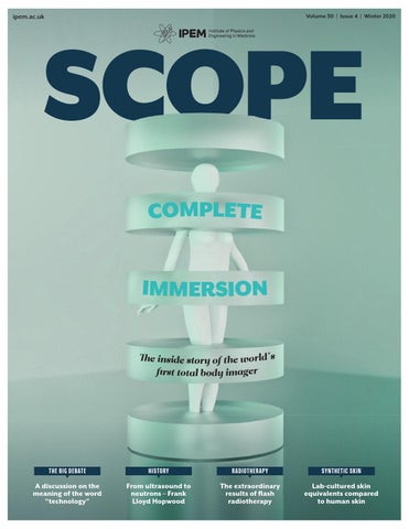
COMPLETE IMMERSION The inside story of the world’s first total body imager
T H E BI G D EBAT E
H I STO RY
RADI OTHERAPY
SYN TH ETIC SKIN
A discussion on the meaning of the word “technology”
From ultrasound to neutrons – Frank Lloyd Hopwood
The extraordinary results of flash radiotherapy
Lab-cultured skin equivalents compared to human skin
Cover_Winter 2020_SCOPE 1
16/11/2020 10:22
+ + +
CareMin650™ uses CareMin650 SKRWRELRPRGXODWLRQ D VFLHQWL¿FDOO\ DQG FOLQLFDOO\ YDOLGDWHG SKRWRWKHUDS\ IRU WKH SUHYHQWLRQ DQG WUHDWPHQW RI muscositis and dermatitis. 7KLV EUHDNWKURXJK GHYLFH JUHDWO\ VLPSOL¿HV WKH GHOLYHU\ RI SKRWRELRPRGXODWLRQ WKHUDS\ ZKLFK LV FRPIRUWDEO\ DSSOLHG WKURXJK SDWHQWHG luminous fabric. Designed to ease implementation DQG DFFHVVLELOLW\ RI WUHDWPHQW &DUH0LQ ™ SURYLGHV DQG DFFHVVLELOLW\ RI WUHDWPHQW &DUH0LQ SURYLGHV D FRVW HႇHFWLYH DQG SDWLHQW IULHQGO\ VROXWLRQ HDVLO\ integrated into therapeutic practice. ,QWHUHVWHG LQ D WULDO" &RQWDFW 2,6 WRGD\ Oncology Imaging Systems
IPEM.Winter2020.002.indd 6
CareMin650™ is a trademark of NeoMedLight. 2,6 LV D SDUWQHU RI 1HR0HG/LJKW DQG H[FOXVLYH GLVWULEXWRU RI &DUH0LQ Œ LQ WKH 8. ,UHODQG
06/11/2020 17:48
UF UPFRONT COMMENT
CH A I R OF I P E M S C O P E ED I TO R I A L A DV I S O RY B OA R D
Cutting-edge techniques and technology Usman Lula on the articles in the latest issue of Scope and the theme that is the driving force behind the content. elcome all to the Winter issue of Scope 2020 – a year when COVID-19 took hold and changed our take on precious life. With news of vaccines on the way, it may well be time to celebrate the New Year with some positive news. For this issue, approaching potential authors for an invited feature was something I was eager to undertake. With the existing limitations on travel, liaising with new authors was limited to email and phone. I set out looking for authors broadly representing as many areas under IPEM – who were able to provide features that met the theme of “cutting-edge techniques and technology”. We’ve got some really exciting features lined up for you in this
W
issue – Professor Simon Cherry kindly supplied his co-authored piece in record time – where he outlines the EXPLORER project, from the initial idea all the way to design, development and approval, whilst also trying to acquire the necessary funding. Professor Cherry was also able to join our debate question on the definition of technology and how his work impacted on improving diagnostic and therapeutic clinical services. The idea of the “Big Debate” topic actually came about when I had asked a former colleague of mine (Steve Lake from Liverpool) about supplying a potential piece.
On the phone, he started talking about how we currently view “technology” and how quickly we adopt it within our own medical physics and clinical engineering specialties. Specifically – how we should be driving continuous change (in diagnostics and therapy) and what we see as “technology” in doing so. So, along these lines, I thought this would set a theme for the March 2021 issue whilst we can debate the question in this very issue. I’ve tried to use a holistic approach to finding “debaters” based on the area of work – and with a limit of 400 words per piece and a total of 2,000 words, it meant I could only invite a maximum of five. Each piece provides a perspective different to another, trying to fulfil the much needed depth and breadth required to answer the key question. Have a great read and happy holidays!
With news of vaccines on the way, it may well be time to celebrate the New Year with some positive news
Usman Lula Chair of IPEM Scope EAB
F E E DBACK
Get involved in Scope Do keep your eyes peeled for the new Scope readership survey, as this is planned to be circulated after publication of this issue. If you have any feedback on the new Scope design and content,
or you have any ideas on future themes, features or series, or would like to contribute to Scope in any other way (e.g. how do we improve readership engagement?), then we will
review and potentially integrate it into our next three-year strategic Scope objectives. For the next issue of Scope (March 2021) – our theme has been set as “Driving technology to benefit patients and the healthcare system”. If you have
IPEM.AC.UK
3 Usman_Winter 2020_SCOPE 3
3
been working on a project that improves diagnostics or therapyy in any way (e.g. improvements in clinical pathways, systems, techniques, technologies and/ or infrastructure) and would likee your piece considered, please drop me an email.
WINTER 2020
16/11/2020 13:42
FEEDBACK Discuss, debate, share. mycommunity.ipem.ac.uk/login Scope is the quarterly magazine of the Institute of Physics and Engineering in Medicine IPEM Fairmount House, 230 Tadcaster Road, York, YO24 1ES T: 01904 610821 | F: 01904 612279 office@ipem.ac.uk | ipem.ac.uk | scopeonline.co.uk
Chair of IPEM Scope Editorial Advisory Board: Usman I. Lula Principal Clinical Scientist, 1st Floor, Radiotherapy, Building, Medical Physics – University, Hospitals Birmingham NHS Foundation Trust, Queen Elizabeth Hospital, Queen Elizabeth Medical Centre, Birmingham, UK B15 2TH 0121 371 5056 | usman.lula@uhb.nhs.uk
WEBSITE News, events, support. ipem.ac.uk
ARCHIVES Back issues of Scope online. bit.ly/2SRhh0E
CO
Vice Chair of IPEM Scope Editorial Advisory Board: Dr Matt Aldridge Clinical Scientist Radiotherapy Physics/Nuclear Medicine, UCLH matthewaldridge@nhs.net
14
Commissioning Editor, Meetings and Special Reports: Kirsten Hughes Radiotherapy Physicist, Royal Shrewsbury Hospital, Mytton Oak Road, Shropshire, SY3 8XQ kirsten.scope@gmail.com Commissioning Editor, Reviews: Usman I. Lula 0121 371 5056 | usman.lula@uhb.nhs.uk Commissioning Editor, Applied Academics: Vacant position
UPFRONT 1 4 / THE B I G D E BAT E : THE MEA N I N G OF TECHNOLOGY We asked five experts: “What is understood as the meaning of the word ‘technology’? Please give examples of how your work has impacted on the ability to learn and continually improve diagnostics and therapeutic clinical services.”
Commissioning Editor, International: Dr Mandy Price Principal Physicist, Radiation Safety, Barts Health NHS Trust 0203 594 1142 | mandy.price@bartshealth.nhs.uk Commissioning Editor, Medical Physics: Usman I. Lula 0121 371 5056 | usman.lula@uhb.nhs.uk Commissioning Editor, Medical Physics: Dr Paul Doolan Medical Physicist, German Oncology Center, 1 Nikis Avenue, 4108 Agios Athanasios, Limassol, Cyprus 00357 2520 8025 | paul.doolan@goc.com.cy Commissioning Editor, Clinical & Biomedical Engineering: Vacant position
COVER FE AT URE Scope is published on behalf of the Institute of Physics and Engineering in Medicine (IPEM) by
18 / COMPLETE IMMERSION
Redactive Publishing Ltd +44 (0)20 7880 6200 redactive.co.uk
Publisher: Daniel Butcher daniel.butcher@redactive.co.uk | +44 (0)20 7324 2728 Editor: Rob Dabrowski Lead designer: Carrie Bremner
Our brains are the scientific tools or technology that we use to continually improve diagnostics and clinical services..
Picture researcher: Claire Echavarry
Simon Cherry and Ramsey Badawi outline the EXPLORER project, from staring into the academic abyss, to standing on the precipice of a potentially transformational step in the history of nuclear medicine.
– Dr Anna Barnes page 14
Production: Aysha Miah-Edwards aysha.miah@redactive.co.ukk | +44 (0)20 7880 6241 Advertising sales: scope@redactive.co.uk | +44 (0)20 7880 7556
Scope is published quarterly by the Institute of Physics and Engineering in Medicine but the views expressed are not necessarily the official views of the Institute. Authors instructions and copyright agreement can be found on the IPEM website. Articles should be sent to the appropriate member of the editorial team. By submitting to Scope, you agree to transfer copyright to IPEM. We reserve the right to edit your article. The integrity of advertising material cannot be guaranteed. Recycle your Copyright: Reproduction in whole or part magazine’s plastic wrap. Check your by any means without written permission local facilities to find of IPEM is strictly forbidden. © IPEM 2020. out how. ISSN 0964-9565
U PFRO N T
03 / 07 / 10 / 12 /
CHAI R’S CO MMENT NEWS TECHNO LO GY NEWS STR ATEGY UP DATE
Cover image by
GET T Y
IPEM SCOPE
4-5 Contents_Winter 2020_SCOPE 4
4
WINTER 2020
16/11/2020 16:02
Volume 30 | Issue 4 | Winter 2020
NTENTS GENERAL FEATURES 2 2 / HOPWOOD O F BART’S In the third of his articles on the early pioneers of medical physics, Francis Duck describes the work of Professor Frank Lloyd Hopwood, whose studies in the 1930s ranged from ultrasound to neutrons.
2 6 / C LIN ICAL DECISIO N S UPPORT ICT-based clinical decision support solutions have the potential to transform the application of clinical guidelines through the implementation of dynamic clinical pathways. These standardised approaches can incorporate the capturing of extra data on which patient care can be studied and continually improved.
30 / IS T HE R AD I OTHER APY REVO LU T IO N HER E? The extraordinary results of flash radiotherapy challenge our accepted notions of radiobiology. An overview – from early research to implementation in departments.
34 / A M U LT I CENTR E DATA CO L L ECT ION TO O L
22
Antony Carver looks at a platform to ease the process of collecting outcome and treatment planning data together and streamline the process for analysis.
SPECIAL REPORT
38/ A M U LTI D I SCI P L I NARY T EAM EFFO RT The first of a two-part series in which Aquila Sharif and colleagues outline the process of installing and getting to grips with a new MR-Linac.
52 EDU CATI O N
Training in the pandemic
42 / SYRING E- D R I VEN INFU SIO N
How the Scottish Medical Physics and Engineering Trainee Network have been sharing training experiences virtually during lockdown.
Tarah Mc Ateer looks into whether syringe drivers are accurate infusion devices for neonatal drug delivery.
45 / DEVELO P MENT O F NEW TOM OSYNTHESI S T ECHNOLOGY
ENDNOTES
Matthew Rowlandson explores Adaptix’s new advances in tomosynthesis technology through work on his HSST project.
26
Laurence King, Principal Clinical Scientist at Royal United Hospitals Bath NHS Foundation Trust, gives on overview of this new publication by Timothy P Szczykutowicz.
48 / SKIN DEEP Akhil Kallepalli and colleagues compare lab-cultured human skin equivalents to human skin.
IPEM SCOPE
4-5 Contents_Winter 2020_SCOPE 5
54 / BO O K R EVI EW: THE CT HAND BO O K
5
WINTER 2020
16/11/2020 10:07
'RVLPHWU\ 6ROXWLRQV IURP 3KRHQL[ 'RVLPHWU\ /WG Phoenix Dosimetry Ltd are specialists in Dosimetry Peripherals and ‘Harshaw TLD Systems’. We also offer Service contracts, repairs and training on TLD Readers along with peripheral Dosimetry equipment.
• • • •
Express Repair Service—typically 1 week All Mini and Legacy NE/Thermo Equipment catered for Free inspection service with no obligation quote Some used parts available to save costs
•
RadEye B20 measures Alpha, Beta, Gamma surface contamination—Geiger based Light weight (300g) Apply H*10 filter and B-20 becomes a dose/doserate meter
• •
• • •
TruDose Electronic Personal Dosemeter replaces the well proven EPD-2 Dose and Dose rate display/alarms for Hp10 and Hp0.07 Use either stand alone or with RadSight software for full personal database and backwards compatible with EPD Mk2
• • • •
Custom made Dosimetry Panels and Cables All common QA connections catered for Optional mounting of panel in protective steel box Improves cable management
•
The original Farmer Chamber used throughout the world for the measurement of Photon and Electron beam dosimetry NE2571 delivered with Graphite thimble and 10m cable NE2581 is a robust Shonka Plastic version of the NE2571
• •
For more information please visit phoenix-dosimetry.co.uk or call 01252 871990
Phoenix Dosimetry HPH and6FP options.indd 1 IPEM.Winter2020.006.indd
07/08/2020 10:24 12:19 09/11/2020
NEWS/TECHNOLOGY/POLICY/DEBATES
FEEDBACK Discuss, debate, share my.community.ipem.ac.uk
WEBSITE News, events, support ipem.ac.uk
UPFRONT
RA DI OT H E RA PY
Lung cancer during COVID investigation new national project investigating changes in radiotherapy services during COVID-19 has been launched by a team of Manchester cancer clinicians. The project, Lung Radiotherapy during COVID-19 (COVID-RT Lung), aims to build a national database assessing radiotherapy treatments for lung cancer patients during the pandemic, and how this has affected patient outcomes. The project is also highlighting how NHS services across Greater Manchester are working as one to provide care and treatment for patients, as well as ensuring their safety during the pandemic. It is hoped the project will shed light on changes in the management of lung cancer patients and treatments during the COVID-19 pandemic, and its impact on patient outcomes. Professor Corinne Faivre-Finn, who recently published guidelines into radiotherapy treatments for lung cancer patients during COVID-19, alongside other UK oncologists, is one of the project leads. She said: “The pandemic presents many challenges to treating lung cancer patients. Lung cancer patients undergoing radiotherapy often have multiple medical problems. They are at much higher
A
IMAGE: ISTOCK
The pandemic means we will likely have to make some changes to services and treatment across the whole of the UK
risk of severe complications from COVID-19, which may require a hospital stay or ventilation to help them breath. “The pandemic means we will likely have to make some changes to services and treatment across the whole of the UK for quite some time, until we have a vaccine. Our recent recommendations aim to make treatment as safe as possible by reducing the number of hospital visits, which minimises the risk of exposing patients to coronavirus in hospital.” The news came at the end of October,
FAST FACTS
434
A TOTAL OF 434 nuclear medicine departments responded to the IAEA survey.
IPEM.AC.UK
7-9 Upfront_Winter 2020_SCOPE 7
when the Director General of the International Atomic Energy Agency (IAEA), Rafael Mariano Grossi, warned the global pandemic is disrupting key health services to diagnose and treat chronic conditions such as cancer and heart disease, putting many lives at risk. Speaking at the World Health Summit in Berlin, Director General Grossi said an IAEA survey on the impact of the pandemic on nuclear medicine services showed worrisome trends. “Diagnostic procedures fell on average by more than half in the countries surveyed,” he said. bit.ly/3kvJ02x
7
72
THE RESPONSES came from 72 countries around the world.
54%
RESPONDENTS REPORTED an average decline of 54% in diagnostic procedures.
WINTER 2020
16/11/2020 10:08
UF UPFRONT NEWS
ME DI CA L I MAG I N G
N EWS I N BRI EF
Ultrafast camera
New salivary glands discovered edical researchers have made a surprise anatomical discovery, finding what looks to be a mysterious set of salivary glands, which have never been identified before. This discovery was made by accident by doctors who were examining prostate cancer patients with a prostate-specific membrane antigen (PSMA) positron emission tomography/ computed tomography (PET/CT). When paired with injections of radioactive glucose, this diagnostic tool highlights tumours in the body. In this case, however, it showed up salivary gland, nestled in the rear of the nasopharynx. Wouter Vogel, from the Netherlands Cancer Institute, said: “As far as we
M
knew, the only salivary or mucous glands in the nasopharynx are microscopically small, and up to 1000 are evenly spread out throughout the mucosa. So, imagine our surprise when we found these.” They have been named “tubarial” glands, in reference to their anatomical location, above the torus tubarius. The PSMA PET/CT imaging technology was able to detect the salivary glands due to their superior visualisation capabilities, in comparison to technologies such as ultrasound, CT, and MRI scans. However, the team concedes that additional research on a larger, more diverse cohort will be needed to validate their findings. bit.ly/2J0AElV
ME DI CA L P O L I CY
RADIOTHERAPY FOLLOWING PROSTATE CANCER A systematic review and metaanalysis suggests that patients with localised and locally advanced prostate cancer may be potentially spared radiotherapy and its side effects following surgery. The authors recommend that if prostate cancer later recurs or relapses men should receive radiotherapy at that stage. The systematic review and metaanalysis is based on three new randomised controlled trials. The authors recommend that patients should be closely followed after surgery for treating localised and
locally advanced prostate cancer. If the cancer shows early signs of coming back, men should be offered radiotherapy. Dr Claire Vale, from University College London, who led the systematic review and meta-analysis, said: “Our findings suggest that following surgery, patients whose cancer is confined to the prostate, or has spread only to nearby tissues or organs, can safely be spared routine postoperative radiotherapy and its associated side effects.” bit.ly/3kk127Y
IPEM SCOPE
7-9 Upfront_Winter 2020_SCOPE 8
8
Medical engineering professors have created a camera that is capable of taking up to 100 billion frames per second. That is fast enough to take 10 billion pictures (more images than the global human population) in the time it takes to blink an eye. The scientists from the California Institute of Technology describe the device in a new paper in the journal Nature Communications. The camera not only records video at incredibly fast speeds, but it does so in three dimensions. The technology is called “single-shot stereo-polarimetric compressed ultrafast photography”. go.nature.com/37ymp1I
Radiotherapy tool RadCollision is a new an open-source collision detection tool designed to aid dosimetrists planning photon or proton beam radiotherapy. When embedded in a treatment planning system, the modular software platform takes seconds to automatically calculate whether a gantry head will collide with the patient or treatment couch. In addition to greatly reducing the need for replanning if a collision is detected during the dry run, the tool helps planners choose optimum and feasible irradiation angles to potentially increase the quality of treatment plans. bit.ly/37A16wT
Healthtech campus The Birmingham Health Innovation Campus is hoped to support health and life science businesses throughout their growth and provide a focal point for inward investment in the region’s healthcare cluster. The campus is being developed through a partnership between the University of Birmingham and Bruntwood SciTech. It is due to open in 2022. bit.ly/31Jus86
WINTER 2020
16/11/2020 10:08
MOL E CUL A R PAT HO LO GY
Algorithm to compare 3D genomes A study describes a new algorithm to compare the 3D genomes of cancer patients with that of a healthy reference. Using a computational technique called “structural similarity analysis”, the scientists developed an algorithm that compares the DNA organisation data from a healthy individual with that of a cancer patient, for example. The algorithm can then
identify which regions are packaged differently in the cancer patient, to help identify what might have gone wrong. The algorithm uses the healthy individual’s data as a reference, to spot differences in the cancer patient’s data. This tool helps researchers to scan the entire length of the DNA in an automated process, more quickly than by eye. It also allows researchers to give a region of the genome a
numerical value. Before, they would only have been able to say whether a given section of DNA was different or not. Now, the algorithm assigns regions of DNA a numerical value that denotes just how different that region is. The tool can also highlight differences in regions that the human eye can not see.
go.nature.com/3ow9nYA
UP CLOSE
3D PRINTING WHAT IS THE LATEST WITH 3D PRINTING? Researchers are now 3D printing cancer cells to help fast-track testing and better treatments for patients. The Australian innovation called Rastrum can create hundreds of 3D tumours in hours, instead of weeks.
WHAT ARE THESE TUMOURS USED FOR? The technology allows the printing of multiple mini tumours simultaneously, which can then be tested with different drugs. It is hoped to help patients receive the most effective drugs sooner. It could even lead to discoveries about the ways that tumours become resistant to therapy.
HOW ARE THE TUMOURS CREATED? A cartridge inside the printer contains different types of gel to create a matrix that mimics the tumour’s
environment. The malignant cells are then added to the mix for the printing process, which takes just 80 minutes.
WHAT DO THE SCIENTISTS SAY? Professor Justin Gooding, Chief Investigator at the Centre of Excellence in Convergent Bio-Nano Sciences, says: “It really opens up what we can do scientifically from not just understanding cancers but also personalising your treatment or developing new therapeutics for cancer.”
MRI AND CARDIAC DEVICES MRI examinations can be performed safely in patients with non-MR compatible cardiac devices, including those who are pacemakerdependent or have abandoned leads, says new research. Prior studies have demonstrated the safety of performing MRI exams in patients with non-MR conditional devices. But those studies did not account for pacemakerdependent ICD patients. To develop a more comprehensive picture of risk, researchers enrolled more than 500 participants who had undergone a total of 608 MRI exams, including 61 cardiac MRI exams. The results demonstrated that MRI exams can be performed safely in pacemaker-dependent ICD patients and in patients with non-MR conditional devices or abandoned leads. bit.ly/3dW77VU
WHO IS USING THIS TECHNOLOGY? A dozen research centres in Australia are using the machines and the Royal College of Surgeons in Dublin has also acquired the technology. Pharmaceutical companies have expressed interested, too.
IPEM.AC.UK
7-9 Upfront_Winter 2020_SCOPE 9
RADI OLOGY
9
WINTER 2020
16/11/2020 13:43
UF UPFRONT TECH NEWS
BI OT E CH NO LO GY
SENSOR FOR RAPID COVID DETECTION A new type of multiplexed test (a test that combines multiple kinds of data) with a low-cost sensor may enable at-home diagnosis of COVID-19. This would be possible in 10 minutes, without the help involvement of a medical professional, it is claimed. The device has been developed at the California Institute of Technology and uses rapid analysis of small volumes of saliva or blood. The research was conducted in the lab of Wei Gao, assistant professor essor in the department ent of medical engineering. gineering.
Previously, Gao and his team have developed wireless sensors that can monitor conditions such as gout, as well as stress levels, through the detection of extremely low levels of specific compounds in body fluids. Gao’s sensors are made of graphene. A plastic sheet etched with a laser generates a 3D graphene structure with tiny pores, which create a large amount of surface area on the sensor, which makes it sensitive enough to detect, with high accuracy, compounds that are only present in very small amounts. In this sensor, the graphene structures are coupled with antibodies, immune system molecules that are sensitive to specific proteins. bit.ly/3kpUpRI
M EDI CAL I M AGI N G
Consolidating imaging data regional informatics project that consolidates radiology and nuclear medicine imaging data has been completed. The Region of Southern Denmark now has a single system for storing, retrieving, and viewing clinical images across all the locations and specialties in its extensive healthcare system. The region encompasses four hospital groups with a total of 12 hospitals that serve a population of over 1.2 million people living on both on the mainland and the region’s many islands. The unified imaging ecosystem will sserve all of the region’s approximately 300 radiologists and nuclear medicine 3 specialists, performing 1.5 million exams sp yearly. The related images are now yea accessible to the over 5,000 clinicians in acc the region. Calum Cunningham, General Manager Ca Enterprise Diagnostic Informatics at of En Philips, which implemented the system, said: “Easy access to medical patient data across complex healthcare systems fosters
A
CELL SHEETS
ELECTROTHERMAL SOFT MANIPULATOR A new device inspired by octopus suckers rapidly transfers delicate tissue or electronic sheets to the patient, overcoming a key barrier to clinical application, according to researchers at the University of Illinois at UrbanaChampaign and collaborators. Current methods of transferring the sheets involve growing them on a
IPEM SCOPE
10-11 Tech news_Winter 2020_SCOPE 10
10
clinical collaboration and is essential to advance precision diagnosis. “We were able to implement our Enterprise Imaging solution in record time, helping the Region of Southern Denmark to move to the next step of digital maturity, improve collaboration, and ultimately enhance patient care.” The region decided to partner with Philips to advance its digital transformation towards precision diagnosis. The adoption of Philips Enterprise Imaging solutions has started with implementing the Philips VendorNeutral Archive (VNA) and Philips Universal Viewer as part of its strategy to replace the existing systems. These modules provide a single source of archiving and distribution of imaging information that enables viewing of clinical images virtually, anytime anywhere by authorized users via a web browser or mobile device. philips.to/2TnUk52
temperature-sensitive soft polymer that, once transferred, shrinks and releases the thin film. However, this process takes 30-60 minutes to transfer a single sheet, requires skilled technicians and runs the risk of tearing or wrinkling. The researchers wanted to quickly pick A new device inspired up and release the by an octopus’s sucker delicate sheets of cells rapidly transfers or electronics without delicate tissue or damaging them. Seeing the way an electronic sheets to octopus or squid can the patient
WINTER 2020
16/11/2020 10:08
O PTI C S
RA DI OT H E RA PY
Gating and MLC tracking esearchers in Australia have evaluated radiation doses delivered to 44 prostate cancer patients when using multileaf collimator (MLC) tracking and gating with a standard linac. The patients were enrolled in the TROG 15.01 SPARK trial, which examined the use of kilovoltage (kV) intrafraction monitoring (KIM) to measure tumour position during treatment. Paul Keall, Professor of Medical Physics, and Jarad Martin, a Radiation Oncologist, led the team from the ACRF Image X Institute of the University of Sydney and four other Australian cancer treatment centres. The researchers delivered 49 fractions using MLC tracking and 166 fractions using beam gating and couch shifts.
IMAGES: SCIENCEPHOTOLIBRARY / ISTOCK
R
pick up both wet and dry objects of all shapes with small pressure changes in their muscle-powered suction cups, rather than a sticky chemical adhesive, gave the researchers an idea. They designed a manipulator made of a temperatureresponsive layer of soft hydrogel attached to an electric
They performed motion tracking with KIM, which uses the linac’s on-board kV imager to acquire patient images during treatment. Intrafraction KIM-guided motion adaption was then performed using MLC tracking for 10 patients or gating for 34 patients. Of the 166 gated fractions, 65 included prostate motion that exceeded established thresholds and required treatment interruptions. The researchers estimated the radiation doses that would have been delivered without motion adaptation and compared these with doses delivered when employing MLC tracking or gating. To evaluate the efficiency of gated treatments, they also calculated the time required to gate and perform a couch shift for each fraction. bit.ly/3kznUR1
heater. To pick up a thin sheet, the researchers gently heat the hydrogel to shrink it, then press it to the sheet and turn off the heat. The hydrogel expands slightly, creating suction with the soft tissue or flexible electronic film so it can be lifted and transferred. Then they gently place the thin film on the target and turn the heater back on, shrinking the hydrogel and releasing the sheet. The entire process takes about 10 seconds.
10-11 Tech news_Winter 2020_SCOPE 11
US biomedical engineers are developing a novel method for the imaging and assessment of corneal elastic properties that could potentially be used for routine clinical diagnostics of different corneal diseases and treatment. The team, led by Kirill Larin for the University of Houston, has received a $1.6 million continuation grant from the National Eye Institute to improve current Optical Coherence Tomography (OCT) to provide ultrafast 3D clinical imaging. The technology will combine Brillouin microscopy with OCT and Optical Coherence Elastography (OCE). The new technology uses highly localised air pressure stimulation. Larin said: “We’re going to use an air puff that will produce very small waves on the surface of the eye. “The patient will not feel them, but we will be able to detect them. The speed of the waves will tell us about the elasticity of the cornea.” Using OCT, he will reconstruct volumetric biomechanical properties of the cornea. The team has developed a first prototype, demonstrated its capability to measure biomechanical properties of the cornea in vitro and in vivo, and has developed analytical models. The new grant will be used to accelerate the transition of this technology into clinics, influence the selection and application of corneal surgical treatments and to help understand the structural consequences of corneal disease and wound healing. bit.ly/3ouYI0
bit.ly/3dXZ5M8
IPEM.AC.UK
Ultrafast 3D eye imaging
11
WINTER 2020
16/11/2020 10:09
SU STRATEGY UPDATE
I P E M CH I E F E XEC U T I V E
Mission, Vision and Values Phil Morgan, IPEM Chief Executive, outlines strategic plans to develop the profession improve health and transform lives.
N
ot long after assuming the role of Chief Executive, at the start of 2020, I wrote in Scope that “IPEM’s job is to understand the shape of things to come and create a dialogue that addresses the challenges. This should profoundly shape IPEM’s future strategy.” It has been a year with a few unexpected twists, which IPEM members will know all too well, but we have made progress towards a new strategic outlook. Each autumn, the Board of Trustees is asked to set a budget for the following year. This year I asked them to sanction the overall shape of our strategy for 2021-24, alongside the financial profile, which was unanimously approved. The aim is not to reinvent the wheel or add shine without substance. We want to focus IPEM’s activity on a set of achievable objectives that deliver the greatest benefit to the public. As a precursor to a new set of operational plans, we embarked on a project to restate our Mission, Vision and Values. These will be rolled out imminently, but our restated mission - “improving health though physics and engineering in medicine” – is a
succinct and memorable statement of our purpose as a charity and member association. Our updated vision – “developing the professional, improving health, transforming lives together” – expresses how clearly we see the role of members in this. Our members, as professionals and as people, make the difference, in terms of the science and patient outcomes. IPEM’s job is to sustain communities of practice, support the creation of intellectual capital that develops professionals and be an impactful and influential voice for physics and engineering in medicine. To achieve this, we need to make changes to the way we work. We want to facilitate a stronger sense of community within the membership. Organisations like IPEM are the home of communities of practice – formal or informal networks which can support your career, provide access to information and advice and, outside of pandemics, a peer group social gathering. This is a volunteer-led enterprise and we will be putting more resources behind them. IPEM’s value is also found in the way we create and develop professional standards. This can be through the excellent work our
IPEM SCOPE
12-13 Sean colum_Winter 2020_SCOPE 12
12
members do in producing guidance and reports, and is also found in our training, education and events. My intention is for our educational activities to generate more income. Any surplus that IPEM generates directly supports the work of the charity. We plough it back into improving the organisation and the better we do, the more we can put behind the mission. This is the area in which we are keen to make an exciting expansion of our capacity and capability. Professional knowledge, which is the outcome of the work we do to create intellectual capital through IPEM, will be boosted by the creation of new full time roles dedicated to co-creating - with our
WE WANT TO FACILITATE A STRONGER SENSE OF COMMUNITY WITHIN THE MEMBERSHIP
WINTER 2020
16/11/2020 10:10
I believe we can shape the global discourse on science and healthcare
change in areas like technology. If we can do that more effectively, I believe we can shape the global discourse on science and healthcare. In the summer, we invited members to share their ideas in a horizon scanning exercise and I am pleased to say that many of you have done so. This will become a regular exercise, with members and others continually iterating ideas for the future. It will not have escaped your attention that in order to communicate and achieve these aims there are some things IPEM needs to improve. Our website is not keeping pace with digital change and nor has our visual presentation. We will use our communications channels to
members - the guidance and reports that support the wider professional groups. I am keen to add a new dimension to this – understanding “the shape of things to come”, as I said back at the start of the year. By understanding the drivers of change in the future operating environment for medical physics and clinical engineering, we can do more to help the profession prepare for
roll out our refreshed mission, vision and values to all, and a new IPEM logo and “look” will follow. These will feature in and drive the development of a new website – a larger project which is getting underway now. Together these ambitions will take shape in four operational plans that we are working on – plans to expand our capacity for the creation of intellectual capital, to sustain a professional community characterised by high standards and equality, diversity and inclusion, to step up the commercialisation of our education and events, and to increase our profile and impact. We are changing and, I believe, growing. It is an exciting time to belong to IPEM.
OSL is the exclusive distributor for Spectronic Medical within the UK & Ireland
MriPlanner MRI only radiotherapy planning A VLPSOH DQG DXWRPDWHG ZRUN̍RZ process easily converts T2w image into a synthetic CT for export into the planning system. MriPlanner also provides users with automated delineation of OAR contours from standard MR Images using its advanced AI technology.
Oncology Systems Ltd +44 (0)1743 462694 enquiry@osl.uk.com osl.uk.com
IMAGE: ISTOCK
Contact OSL to arrange a demo.
IPEM.AC.UK
12-13 Sean colum_Winter 2020_SCOPE 13
13
WINTER 2020
16/11/2020 10:10
UF UPFRONT DEBATE
T H E BIG DEBATE The meaning of technology We asked five experts a topical question about technology. Here’s what they said. What is understood as the meaning of the word “technology”? Please give examples of how your work has impacted on the ability to learn and continually improve diagnostics and therapeutic clinical services.
Q
healthcare. How many times has our knowledge about the practical use of science “saved the day”? This is very much highlighted within my day job in nuclear medicine, where the duty physicist is often the “go-to” person for all things going wrong during the day. So, I would argue that in this sense my entire day is geared towards helping colleagues, and sometimes patients and carers, learn more about, and consequently continually improve, diagnostics. One recent example, though not terribly glamorous, is using scientific knowledge to re-purpose a clinical examination room in our department so that we can administer molecular therapies. The technological solution to this required application of the theory of radioactive isotopes to calculate the thickness and location of lead sheeting required to reduce the amount of ionising radiation reaching into patient and public areas. Another example would be the optimisation of imaging protocols. Our brand new rather splendid PETCT scanner has a continuous patient table movement acquisition mode. We used our knowledge of the detection of ionising radiation and the mathematical algorithms used to reconstruct 3D images of the body to optimise the protocol in terms of maximum radioactivity administered and the total time for the whole-body scan. This has enabled us to improve diagnostics by reducing the burden on the patient, while maintaining image quality for radiological reporting. So, I argue that our brains are the scientific tools or technology that we use to continually improve diagnostics and clinical services.
DR A NNA BA RNES M ARK K N I GHT The Cambridge Dictionary states that the definition of technology is: “the study and knowledge of the practical (especially industrial) use of scientific discoveries”. So, by this definition my “knowledge” of scientific discoveries, for example, of the structure of the atom and how it is applied to medicine is “technology” in and of itself. I don’t think this is normally how we think of technology. Most people, I would imagine, would point to a phone, or a laptop, but this would be a very limiting definition of technology. I like the idea of my knowledge being technology or a tool to improve healthcare. I think we often underestimate ourselves (as scientists and engineers in healthcare professions) in respect of how important we are when it comes to delivering and supporting
IPEM SCOPE
14-17 Big debate_Winter 2020_SCOPE 14
Technology: is it our friend or foe? There are two things we can be sure of: 1) That technology is always with us –it’s there in the home, workplace, social arenas and all of the places in between. 2) Technology never ages – Individual items of technology age, but technology, as a set of products that develops and leaps forwards in capability each day, is forever young. For example, think about CT. From the sluggish, single-slice product of the 1980s,
14
MEET THE EXPERTS
WINTER 2020
16/11/2020 17:00
IMAGE: SCIENCEPHOTOLIBRARY
THERE HAS BEEN A CHANGE IN THE RELATIONSHIP WE HAVE WITH TECHNOLOGY RECENTLY
better. They’re not in the same room, but product developers in each camp keep a careful eye on what the other is planning next. These are examples of technology, our friend. So, what of our foe? The technology that captures our whims, changes the way we think, the way we see the world, maybe even the way we vote? The guest that never leaves, rifling through our private affairs and sells what has been collected? This is technology, our foe. There has been a change in the relationship we have with technology recently. This may have started at the time technology began to process information better than we can; seeing big data in a way that escapes us and now working in a world we can’t inhabit. How will this affect us? As bystanders, we hope for better diagnoses, better treatment management decisions, but will we also get a further divided society and more civil unrest? Our relationship with technology has changed forever: once we were controller and facilitator, now it’s something more like a partnership, and we can only guess at where it will go next. With appropriate checks and balances, including continual reflection on the powers of new technology as it is developed, we should hope that our on-going relationship is more “friend” than “foe”.
whose reconstruction times were measured in 10s of seconds, was born the multi-slice, helical scanner of today, which spits out images in fractions of a second. With siblings in radiotherapy and nuclear medicine, this status is not what my colleague foresaw when they said to me some 20 years ago “CT is on its way out. MRI is the new CT”. And MRI develops in parallel. It too has family in radiotherapy and nuclear medicine. CT and MRI complement one another, working throughout the fields of diagnosis and therapy to make our lives
DR ANNA BARNES
MARK KNIGHT
PROFESSOR DAN CLARK
SIMON R CHERRY
DR JOHN AMOORE
Principal Clinical Scientist, Nuclear Medicine UCLH, and Hon Associate Professor, Centre for medical Imaging, UCL
Head of Radiation Physics Maidstone and Tunbridge Wells NHS Trust
Head of Clinical Engineering Nottingham University Hospitals NHS Trust
Professor, Department of Biomedical Engineering and Department of Radiology University of California, Davis
Retired Clinical Engineer Edinburgh
IPEM.AC.UK
14-17 Big debate_Winter 2020_SCOPE 15
15
WINTER 2020
16/11/2020 10:11
UF UPFRONT DEBATE
SPARKLE5
I’ve devoted a significant proportion of the latter part of my career to the relatively new field of health technology assessment and been involved in the assessment of many hundreds of new technology products on behalf of my hospitals, for commercial companies and for NICE. I’ve seen many an expensive and exciting device that claims to revolutionise healthcare. But perhaps my personal favourite was a cheap, plastic flask produced with a small negative pressure and an integral catheter. This simple device is used for patients with ascites – the unpleasant build-up of abdominal fluid that is associated with certain, unfortunately usually terminal, cancers. It enables patients to stay at home and self-drain this liquid, rather than the necessity of repeated hospital visits, allowing them a degree of independence and family time in their final days. No glossy brochures, no “go-faster” stripes – just the application of science to bring about real patient benefit. That’s my definition of healthcare technology.
SI M O N R C HERRY
Think of technology and you might consider a range of smart new devices: the latest mobile phone, flat screen TV or games consoles. Think of healthcare technology and we might imagine the latest MRIs, ventilators or dialysis machines. But the definition of technology generally refers to the “art of creating” such products, not the products themselves. At its simplest, technology is the application of science. As healthcare scientists, that’s what we do: we’re all technologists. It’s easy to be seduced by the guiles of new technological products. The marketing people know this only too well and their adverts are full of images of new cars, computers, phones and coffee machines. New technology is sexy; it sells. It’s the same in our professional world – medtech companies have glossy brochures pushing their latest products, with shiny cases and “go-faster” stripes. But beware – all that glistens is not gold. The human part of us (am I suggesting that a part of us is not human?) might be drawn, magpie-like, to the shiny new toys, but the technologist in us wants to apply the science. As healthcare technologists, we need to review the scientific evidence, consider the benefits and risks and quantify the patient improvements and costs. The technologist is not fooled by glossy brochures or the “go-faster” stripes, but can analytically review the application of science. And the healthcare technologists can place this in the context of patient benefit.
IPEM SCOPE
14-17 Big debate_Winter 2020_SCOPE 16
IMAGES: SHUTTERSTOCK / ISTOCK
P ROF E S S OR DAN C L A R K
16
The word “technology” is quite widely and variably applied today and has a long history with origins partly borrowed from Greek and Latin. Our more modern understanding of the word traces its roots back to the late 18th century, where it is defined as the “branch of knowledge dealing with the mechanical arts and applied sciences, technical know-how, machinery or equipment”. In my own field of medical imaging, just like in many other fields of science, the impact of technology has been breathtaking. One has only to compare medical imaging scans from a few decades ago with those produced today to appreciate how technology has revolutionised our field. We are still using the same fundamental signal generation mechanisms and imaging physics in magnetic resonance imaging, ultrasound, x-ray imaging and nuclear medicine as when these techniques were first developed, but through technological innovation we can now image better, faster, at lower radiation dose and cover more of the body, than ever before. I take a fairly broad view of “technology”, beyond the classical view of better materials, detectors and sensors, and other hardware. While these are undoubtedly at the heart of many improvements, we must also hail the revolution in electronic and computational hardware that often underlie new detectors and sensors and that, in some cases, produce
WINTER 2020
16/11/2020 10:11
the technological innovation themselves. I also would not stop at “physical” technology. I would include algorithmic technology, for example the use of sophisticated statistical models in image reconstruction, and convolutional neural networks for image denoising, as “technological” innovations. My own laboratory has focused on technology innovation in the more traditional sense of the word, applied mostly to radionuclide imaging. We have surely made use of computational and electronic technology, but our niche has been to study how to take emerging radiation detection materials and sensors, and exploit their physical properties to yield better imaging signals, whether that be by improved signal localisation, more efficient signal detection or improved temporal localisation. After identifying the most promising technologies, we also have been patient in moving them from concept all the way through complete and functional imaging systems, very often in collaboration with industry. One example was the development of microPET, which helped develop the field of preclinical
imaging with a particular impact on drug development and the evaluation of new drugs in animal models of human disease. A second example is the EXPLORER total-body PET system, which provides a level of detection sensitivity that takes nuclear medicine into uncharted territory, and is already having clinical impact through the first installations in the US and in China. I see many more opportunities for technology, broadly defined, to further impact the field of medical imaging. One just has to look at cutting edge technological research today and imagine what it will do for medical imaging tomorrow!
D R J O HN AM O O RE The World Health Organization defines healthcare technology as “application of organised knowledge and skills in the form of devices, medicines, vaccines, procedures and systems developed to solve a health problem and improve quality of life”. Clinical engineers typically restrict the term to medical devices, instruments used to prevent, diagnose or treat disease, or that restore function, whilst increasingly recognising the procedures and systems through which the medical devices support health. “Buy it right, use it right, keep it right” are avenues in which our value-added management and application of medical devices impacts on healthcare, striving continually to improve clinical services. To “buy it right” we must understand the technology and how it supports care, and the context in which it will be used so that we can specify the most costeffective and appropriate devices. Teamwork and clinical engineers
IPEM.AC.UK
14-17 Big debate_Winter 2020_SCOPE 17
17
OUR BRAINS ARE THE SCIENTIFIC TOOLS OR TECHNOLOGY THAT WE USE TO CONTINUALLY IMPROVE DIAGNOSTICS AND CLINICAL SERVICES cooperating with clinical colleagues is required. The procurement of routine vital-signs diagnostic and monitoring equipment has a hospital-wide impact and needs to consider usage in medical and surgical wards and outpatient clinics. Different teamworking is required when procuring for a specialised department, for example acquiring a novel liver diagnostic device. Improved functionality of new technology might suggest new ways of clinical working, for example remote diagnosis utilising a technology’s on-line capability. “Using it right” includes deploying the technology to where it is needed, with support systems and consumable-supply logistics in place. Many medical devices can be configured to optimally support local practices: a trivial example is configuring the clinical thermometers to read in centigrade, not Fahrenheit. A not so trivial example is deciding whether the thermometer should record oral or core temperature; a clinical engineer had to resolve disputes between two adjacent clinical wards. Alarms and display formats are important areas to decide optimal configurations. Medical devices should be specified to be user-friendly and ergonomic, but clinical staff training is an important component of our work too, requiring us to understand the technology and how it supports care. This understanding is also required when investigating the causes of adverse events which caused or had the potential to cause harm. “Keep it right” summaries the processes of preventative and corrective maintenance, available on demand to problem solve, ensuring equipment is safe and functional.
WINTER 2020
16/11/2020 13:43
CF COVER FEATURE
COMPLETE IMMERSION The inside story of the world’s first total body imager Simon Cherry and Ramsey Badawi outline the EXPLORER project, from staring into the academic abyss, to standing on the precipice of a potentially transformational step in the history of nuclear medicine.
IPEM SCOPE
18-21 Explorer_Winter 2020_SCOPE 18
18
IMAGES: GETTY
T
his story starts in 2005, with a meeting between the two authors (both relatively recent arrivals at the University of California, Davis, and both of whom trace their PhD education back to the University of London). 1 We were discussing possible ways to work together. One of us had been simulating what would happen if the axial length of positron emission tomography (PET) scanners were increased. Making the scanner longer is particularly compelling for PET scanners, not just because you can image more of the body at once, but, since the radiation is emitted isotropically from within the body, you capture more of the available signal. Although conventional wisdom was that scattered and accidental coincidence events would overwhelm the system, rendering improvements in detection
WINTER 2020
16/11/2020 13:44
Figure 2 The concept of total-body PET (right) compared with a conventional PET scanner (left). sensitivity moot, the simulations suggested otherwise. The other one of us was coming from the perspective of preclinical imaging studies and had been developing PET systems that could image an entire mouse. The clear synergy that emerged at this meeting was to focus on building long axial field of view human scanners. And by long, why not a scanner that covers the entire human body? 2. This figure was originally published in the Journal of Nuclear Medicine (Cherry et al., 2018; 59: 3-12) © SNMMI. This would capture almost all the emitted radiation, giving the Berkeley National Laboratory, along with senior maximum signal for the administered radiation dose, and advisors, such as Tom Budinger and Terry Jones. allow us to image the entire human body simultaneously, One of our first actions was to come up with a name, something that no other medical imaging device could do. and EXPLORER (standing for EXtreme Performance Long Research ScannER) was proposed by Jinyi Qi at Funding trials and tribulations UC Davis. OK, It is a little contrived, but caught on and If the concept was easy, gaining acceptance and funding was has stuck to this day; invoking images of how this new not! Early grant applications to funding agencies in the US tool could be used to explore the workings of the in 2007-2010 were met with almost universal disdain. It was human body in much more detail than previously tempting to be upset, but a little introspection suggested possible. Working through the consortium, we went that we needed to build a much stronger case, both for the back into the funding shark tank full of confidence only likely performance of such an instrument and for its to find we had not greatly shifted opinions. Between applications. We hired a PhD student to work on detailed 2013 and 2015, attempts both in the US and the UK to simulations of the scanner, and formed the EXPLORER secure funding ended in rejection. At the same time, we Consortium, including two of the leading PET detector were engaging with manufacturers of PET scanners, groups at the University of Pennsylvania and the Lawrence and while the research engineers were largely enthusiastic and helpful, management could not see a business case for developing such an expensive device. These were the darkest hours. We had no money, funding for our labs had almost disappeared because we had spent so much time focusing on EXPLORER, and we were staring into an academic abyss.
Funding success The big breakthrough came in mid-2015. One of our proposals had been to the National Institutes of Health (NIH) Transformative Research Program, where we were competing against bold ideas across all of biomedical science. Given our huge budget request (approximately equal to the annual budget of the programme!), the relatively small exposure of nuclear medicine in the broader scientific community and the previous skepticism of reviewers, we held little hope. But perhaps precisely because it was reviewed by a broader panel of experts from across the medical sciences, the idea of an imaging device that could image
1 The authors with the EXPLORER total-body PET system.
IPEM.AC.UK
18-21 Explorer_Winter 2020_SCOPE 19
19
WINTER 2020
16/11/2020 13:44
CF COVER FEATURE
Figure 3 Photographs showing construction of EXPLORER system which is built from 8 individual rings of detector modules.
the entire body at once hit a resonant chord. After many machinations within NIH, we received the notice of award in September 2015, astonishingly receiving almost the entire requested budget, the largest award made under this programme by far. At this point, a new challenge suddenly raised its head. Because industry had been unwilling to make any commitment to engage in the project, our proposal had stated we were going to build this entire scanner (which rivals the complexity of the hadron calorimeter at CERN in terms of numbers of detectors and channels of electronics) by ourselves! This raises a problem in terms of manufacturing quality control – in an academic lab, we might be able to build a technology demonstrator that could be shown to function once or twice, but our goal was to build a tool that could reliably be used for imaging humans. We were not set up for this. So we hit the road with renewed vigour to knock on the doors of companies – but this time with a $15m cheque burning a hole in our back-pocket! Another matter had also become clear to us. If we wanted total-body PET to truly change the face of molecular imaging and to really impact human health, then we needed to make it available to other centres and hospitals. The only way to do that was to make total-body PET available as a product. So we looked for companies that would be prepared to obtain FDA marketing clearance for the scanner in the US, ACKNOWLEDGEMENTS and distribute the We would like to thank all at United Imaging Healthcare (UIH), scanner world-wide. for their amazing support. We also gratefully acknowledge the With FDA clearance, input from many scientists and clinicians. We especially thank we would be able to the other leaders in the EXPLORER consortium, Joel Karp and use the scanner for Bill Moses, and the entire EXPLORER team, with a particular clinical studies, and acknowledgement to Terry Jones for continuing to push us to so begin impacting make the best use of this new instrument. The authors have a healthcare right research agreement and their institution has a revenue sharing away. Unfortunately, agreement with UIH. this seemed far too
IPEM SCOPE
18-21 Explorer_Winter 2020_SCOPE 20
20
big of an ask for most of the companies at that time. But a chance meeting at a conference led us to a relatively new medical imaging company, with large ambitions. That meeting, and a visit to their factory a few weeks later, completely changed the course of the project. Finally, we had found management with the same vision and passion we had, and with a simple handshake, the decision was made. Of course, the formal agreements came later, but never underestimate the importance of people giving their word.
Construction and installation The project then moved at breathtaking speed, thanks in large part to the team of very talented engineers at the company. A mock-up of the scanner was completed in November 2016, a small-scale prototype, the “miniEXPLORER”, was finished in June 2017 and the full EXPLORER system was assembled between December 2017 and May 2018 3. The scanner uses over half a million lutetium oxyorthosilicate scintillator crystals read out by more than 50,000 silicon photomultipliers, covering an axial field of view of 194 cm, sufficient to simultaneously image the entire human body. We ran tests in the factory throughout the remainder of 2018 and the scanner received 510(k) clearance from the FDA in December 2018. Now we were ready to ship it to UC
4 Photographs of EXPLORER system operational at the EXPLORER Molecular Imaging Centre at the University of California, Davis. Two of the EXPLORER “Super Techs” are shown, Heather Hunt (left) and Kristin McBride (right).
WINTER 2020
16/11/2020 13:44
Davis for installation. Perhaps we forgot to mention that the factory where the scanner was assembled was in Shanghai, and that we now got caught up in the trade war between the US and China which saw a 25% tariff slapped on many goods, including all medical imaging equipment. A multi-million dollar tariff charge had certainly not been anticipated in our budget! So now we had to write another proposal, to the Office of the United States Trade Representative, requesting an exemption to the tariff. More than a year later, we learned that the exemption was approved. Meanwhile, the company had stepped forward and shipped the system and paid the tariff, in anticipation that we would get the exemption and a refund, allowing us to keep the project moving forward. After a further small hiccup due to a strike at the Oakland Port, EXPLORER was finally delivered and installed at UC Davis, in our newly renovated centre, in May 2019 4.
Early experience and results Our first subject was scanned at UC Davis on June 20, 2019, a hugely memorable occasion celebrated with late-night champagne after a gruelling day that included six imaging sessions spread over 12 hours to get maximum information about the kinetics of 18F-fluorodeoxyglucose (FDG) over six half-lives from the time of injection 5. It was exhilarating to see the sharp images coming off the scanner, qualitatively already supporting our expectations for dramatic improvements over conventional PET scanners. Fast-forward to the present day, and in just 16 months, despite the inevitable interruptions due to COVID-19, we’ve scanned over 500 patients and 100 research subjects. We have already seen indications of how the EXPLORER scanner could change clinical practice through its ability to capture dynamic information from the whole-body, or acquire images with higher signal-to-noise ratio, even while reducing the administered radiation dose. The breadth of research topics hints at new application areas, spanning systemic autoimmune diseases (e.g. arthritis), infectious disease and multiorgan inflammation following myocardial infarction, as examples. We are also undertaking efforts in methodological aspects, including image reconstruction, motion correction and kinetic modelling, in many cases incorporating deep-learning based approaches, which will further improve the
IPEM.AC.UK
18-21 Explorer_Winter 2020_SCOPE 21
images and data produced by EXPLORER. One stunning example has been the ability, for the first time, to acquire meaningful PET images in 100 milliseconds and see individual heart beats. Perhaps the greatest challenge, which was not unanticipated, has been handling the large amounts of data, especially in research studies where list mode datasets can easily exceed 1-2 TB in size, and reconstruction of this data into large numbers of high-resolution dynamic image sequences carries a significant computational burden. This is being addressed with continuing investment in the computational infrastructure that supports the scanner.
Figure 5 Representative images of the human body (maximum intensity projections) showing the distribution of FDG (350 MBq dose) in a normal subject 90 minutes after injection. PET images of the entire body can readily be obtained in as little as 30 seconds (left), and images with unprecedented resolution and signal-tonoise are acquired in 20 minutes (right).
Future outlook History will be the judge as to whether EXPLORER and total-body PET comes to be viewed as a transformational step in the history of nuclear medicine. It is far too early to tell. Yet the signs are encouraging that perceptions are changing and that it has brought a level of enthusiasm and excitement to the field. There are now seven EXPLORER scanners installed worldwide. Another 140 cm long axial field of view scanner has been developed by our collaborators at the University of Pennsylvania and other major companies are developing long (perhaps not total-body) systems as well. An international network of these advanced PET systems that can image all, or most of, the human body is essential for exploring the vast array of new application areas that imaging data with this body coverage and quality can support, as well as to study where this advanced, but admittedly expensive technology, can play an impactful clinical role. The field of total-body PET is in its infancy, but our eyes have been opened to a host of new opportunities. It is our sincere hope that our talented physician and research colleagues will find ways to use this powerful new instrument in contexts we have not even imagined, to further our understanding of human disease and to impact patient care.
21
WINTER 2020
16/11/2020 13:45
GF GENERAL FEATURES
HOPWOOD OF BA RT ’S In the third of his articles on the early pioneers of medical physics, Francis Duck describes the work of Professor Frank Lloyd Hopwood, whose studies in the 1930s ranged from ultrasound to neutrons.
F
rank Lloyd Hopwood (18841954) was the son of a mining engineer from North Wales. Their home, Oak House, lay on the outskirts of his native village of Buckley in Flintshire, surrounded by pitheads, potteries and brickworks. Hawarden Grammar School nurtured his talent for science. His ability secured scholarship support to attend the University of North Wales at Bangor, graduating in physics in 1905 with a creditable second-class honours degree. Ambition, or adventure, took him to London, where he continued his studies at the Royal College of Science (later, Imperial College). After a year he secured a part-time post as demonstrator in physics at St Bartholomew’s Medical School, commencing a lifetime applying physics to medicine.
Persistence and recognition In 1892 the General Medical Council had finally ruled that physics must be part of pre-clinical medical training. Most
universities responded by inviting senior physicists to teach medical students. The London medical schools, by and large, did not. St Bartholomew’s Medical School appointed Frank Womack MD, who had scraped a third-class degree in experimental physics in 1880. When Hopwood was appointed, Womack expected him to deliver lectures and manage the laboratories, but no more. The following year he supplemented his part-time income by securing a physics lectureship at Hackney Technical Institute. By 1914, now supporting his young wife and daughter in their terrace home in North London, Hopwood’s career was in the doldrums. Womack gave him no support and lecturing gave little time for research. Perhaps stimulated by the new Coolidge X-ray tubes, Hopwood initiated his own studies into the physical and electrical behaviour of heated filaments. His work attracted the interest of Alfred Porter at University College London (UCL), and led to the award of MSc and a couple of publications. One thing led to another.
IPEM SCOPE
22-25 History_Winter 2020_SCOPE 22
22
WINTER 2020
16/11/2020 13:45
William H Bragg had just received, jointly with his son, the Nobel Prize in physics “for their services in the analysis of crystal structure by means of X-rays”, and was now Quain Professor at UCL. He recognised Hopwood’s experimental skill, and gave him the opportunity to work on X-ray crystallography with an overseas visitor.
IMAGES: GETTY
Detecting submarines Wartime conditions created the governmental Board of Invention and Research. Ernest Rutherford was tasked to find new methods for submarine detection. Bragg recommended Hopwood to help and, in March 1916, he was drafted into Rutherford’s team at the Admiralty Experimental Station, first at Hawkcraig on the Firth of Forth and then, under Bragg’s leadership, at Parkeston Quay, Harwich. The team’s priority was to design a practical, ship-borne, directional hydrophone to detect submarine engine noise. The preferred design placed a carbon button microphone at the centre of a diaphragm, but this design detected sound on both sides of the hydrophone. Hopwood’s
IPEM.AC.UK
22-25 History_Winter 2020_SCOPE 23
23
PHYSICISTS WERE BECOMING INCREASINGLY WELCOME COLLABORATORS IN CLINICAL AND EXPERIMENTAL MEDICINE contribution was in perfecting the design of an acoustic baffle, shielding one side. After the war, he described this unidirectional hydrophone in a paper in Nature. The best ideas are the simplest. Hopwood pointed out that it was not the material of the baffle but the thin air layer that blocked the sound 1.
Return to Bart’s After the war, Womack’s departure from Bart’s gave Hopwood his opportunity to establish a creditable physics department. He replaced Womack as physics lecturer and, in 1924, became the first professor of physics. Physicists were becoming increasingly welcome collaborators in clinical and experimental medicine. He inherited just three radium sources when he returned to Bart’s. By 1929 he was the Radium Custodian in charge of over 1000 mg radium, half in solution
WINTER 2020
16/11/2020 10:13
GF GENERAL FEATURES
and the rest contained in several hundred platinum needles and capsules. In a rare exposé of the carelessness of some of his colleagues, he reported that half of the sources were damaged during careless clinical use 2.
Ultrasound At last Hopwood was free to explore innovative aspects of physics with medical potential. Radiobiology was a new growth area, and he worked with Thomas Strangeways in his Cambridge laboratory on the effects of X-rays on mitotic cell division. This may have kindled his interest in biological responses to other radiation. On 13 February 1931, at the Royal Institution (RI), he gave a lecture on ultrasound titled “Some Properties of Inaudible Sound”. He recalled that “some fifteen years ago, while carrying out experiments at sea, Langevin and his co-workers observed that fish which swam into a beam of ultrasonic waves were disabled or killed. Being informed at that time of this mysterious occurrence, the lecturer determined that when circumstances permitted he would investigate the matter”. Hopwood thus became the first British scientist to investigate the physical and biological effects of ultrasound. His apparatus included a 3 kW, 0.5 MHz amplifier driving a resonant quartz disc in oil. He used coke-dust to visualise the waves, using a model of the RI lecture theatre to show the whispering gallery effect 3. He demonstrated acoustic fountains, acoustic cavitation, emulsification and streaming. Amongst the biological effects he reported were haemolysis, changed behaviour of streptococci and viruses, and inhibition of nervous action. Hopwood lived just long enough to see ultrasound therapy become accepted, and for the earliest ultrasound imaging to emerge. Hopwood’s department was expanding. Growing from a staff of one technician, Miss F E Smallwood, he appointed Thomas Banks in 1930 and then Thomas Chalmers in 1932. Banks, with a PhD from Birmingham, took over the physics teaching. Chalmers was a Londoner who had been at the National Physical Laboratory since graduating from Battersea Polytechnic in 1927.
1 Hopwood’s directional hydrophone with baffle for submarine detection (1919).
2 Bent and broken radium needles. Hopwood & Smallwood BJR (1929).
3 Ultrasonic model of sound waves propagation in the Royal Institution Lecture Theatre. Hopwood: Proc RI (1931).
Szilard and neutrons It was a hugely interesting time in nuclear physics. In the summer of 1934, the Hungarian nuclear physicist Leo Szilard asked Hopwood if he could use some of his radium for some experiments. Szilard had arrived in London as a refugee from Germany the previous year. He wanted to explore the release of neutrons, discovered by Chadwick in 1932, and needed easy
IPEM SCOPE
22-25 History_Winter 2020_SCOPE 24
access to radiation from radium. Szilard, a quixotic genius, preferring to publish patents rather than papers, and had just filed a UK patent for a neutron chain reaction in beryllium. Hopwood released 150 mg radium from his stock, but stipulated that Szilard must involve his staff, and must publish in the open literature. In a groundbreaking experiment they “observed that a radiation emitted from beryllium under the influence of radium gamma rays excites induced radioactivity in iodine” concluding that “neutrons are liberated from beryllium by gamma rays”. They had generated 128 I, a beta-emitter with a half-life of 25 minutes. They separated the altered recoil atoms from the n,γ gamma reaction by precipitation as 128I silver iodide, leaving the stable 127I as ethyl iodide. This process of recovery became known as the SzilardChalmers reaction, later used for separating radioisotopes created using cyclotron neutrons. Then, with the help of Lise Meitner, they worked with Brasche and
24
WINTER 2020
16/11/2020 17:05
Lange in Berlin to show that neutrons could be also released using 1.5 MV X-rays, the beryllium surrounded by paraffin wax to increase yield. All this happened in three months. Chalmers, Hopwood and Banks continued to explore neutrons after Szilard’s departure 4. They demonstrated neutron release from heavy water using α particles and γ-rays, creating radioisotopes of bromine, rhodium, silver and indium. With JT Phillips, Hopwood investigated the action of neutrons on colloids and chemical reactions, studies that he described in his 1940 Silvanus Thomson lecture to the BIR. Chalmers explored the “canalisation” of slow neutrons, showing how thermal neutrons might be redirected for therapeutic purposes. Chalmers left Bart’s in 1938 to become physicist at the Liverpool Radium Institute. In the same year, Szilard emigrated to the US, where he was responsible for drafting Einstein’s persuasive letter to
5 Cockcroft-Walton generator (1937).
President Roosevelt that initiated the Manhattan project to create an atomic weapon. In 1945 he argued in favour of the use of the bomb on a Japanese military, rather than civilian, target.
Supervoltage radiotherapy In his BIR Presidential address in December 1933, Hopwood had noted that the National Physical Laboratory had a 1 MV X-ray
4 Hopwood making measurements from a multi-element radium source.
machine, but “if we tried to install such an apparatus at one of our hospitals, it would mean that the hospital became an adjunct to the X-ray department!” Nevertheless, in 1934, Bart’s contracted with Metropolitan Vickers to install the first 1 MV radiotherapy machine in Britain. This massive machine used a continuously evacuated X-ray tube, 10 m long, which weighed 10,000 kg. The voltage supply was a Cockcroft-Walton generator initially operating at 700 kV, reaching 1 MV by 1939 5. During the interwar decades, Hopwood took an active part in professional organisations. He was a founder member of the Institute of Physics in 1923. In the same year, he took part in the formation of the British Empire Cancer Campaign, becoming its honorary secretary in 1946. When pre-clinical medical training at Bart’s was transferred to Queen’s College Cambridge in 1940 Hopwood moved also, and became Vice Dean. While he was away, on 12 January 1941, a high-explosive bomb destroyed the Bart’s physics block. The 1983 History of the HPA described Hopwood’s personality: “There was unanimity among those who knew him that he was a big man in all ways, in mental and physical stature, voice, ‘presence’, sense of humour, vitality and generosity”. Hopwood had worked with X-ray crystallography, neutrons, ultrasonics and supervoltage therapy at a time when each was in its infancy. He had worked with three of the greatest physicists of his time: Rutherford, Bragg, Szilard. Hopwood retired from Bart’s in 1949. His successor, Joseph Rotblat, had returned from Los Alamos to Liverpool after the war, committed to applying his knowledge of atomic physics to medicine. His arrival at Bart’s completed Hopwood’s transformation of Womack’s mediocre unit into a modern, professional, medical physics department. Francis Duck is a medical physicist who has spent most of his career working in medical ultrasound.
IPEM.AC.UK
22-25 History_Winter 2020_SCOPE 25
25
WINTER 2020
16/11/2020 10:14
GF GENERAL FEATURES
ICT-based clinical decision support solutions have the potential to transform the application of clinical guidelines through the implementation of dynamic clinical pathways. These standardised approaches can incorporate the capturing of extra data on which patient care can be studied and continually improved.
CLINICAL DECISION SUPPORT A learning opportunity IPEM SCOPE
26-29 Clinical decisions_Winter 2020_SCOPE 26
26
C
linical guidelines and clinical pathways are accepted as instruments for the quality assurance and process optimisation in healthcare. Clinical guidelines are defined as: “Systematically developed statements to assist practitioners and patient decisions about appropriate health care for specific clinical circumstances.” (Field and Lohr, 1990). Clinical guidelines represent the results of the latest research. The positive impact of clinical guidelines on the quality of care has been scientifically proven but their influence on clinical routine is low due to their narrative and non-formulised form. Imprecise, non-formulated and abstract guidelines need to be implemented as processes. Clinical pathways are appropriate for this purpose. Clinical pathways are defined as:
WINTER 2020
16/11/2020 13:46
IMAGES: GETTY
to an electronic patient record makes the application of guidelines more personal and, therefore, acceptable at the moment of care. Lenz and Reicher (2007) distinguished between organisational processes (medical order entry and result reporting) and the medical treatment process (diagnostic and therapeutic procedures to be carried out for a particular patient). Different challenges arise for these two different levels. Organisational processes can be supported by pre-defined work flows. Patient treatment processes depend on medical knowledge and case-specific decisions, made by interpreting patient-specific data according to medical knowledge. It is generally accepted that medical decisionmaking cannot be automated. Treatment can be improved by providing information in the context of a treatment process. The difficulties with this are to: offer current information only offer relevant information according to the current context include the underlying evidence support all of this in a way which seamlessly integrates with the clinicians working practices. “A complex intervention for mutual decision making and organisation of care processes for a well-defined group of patients during a well-defined period.” (Vanhaecht, 2007)
in different ways and, consequently, there are many care pathways, which differ from medical guidelines.
“Routine or patient protocols are useful means to standardise care, to facilitate completeness of services, and to evaluate both the patients’ progress and the therapeutic efficiency of the program. They are also an educational tool. In essence, the development of protocols is the first step leading from anecdotal to scientific medicine. Protocols, routines, and other standards do not insure excellence, but sometimes they prevent disasters”. (Vanhaecht quoting Shoemaker, 2007)
Implementing formulated guidelines in a decision support system with an interface
The complexity of clinical pathways depends on the severity of a disease, presence of complications and methods of treatment. Because of patient variation, various hospital structures and the individual experience of a doctor, different patients are treated
ICT clinical decision support requirements
Figure 1 How do we assign a pathway for patients presenting with multiple chronic diseases? Screen for disease A
Screen for disease B
Confirm A
Confirm A and confrim B
Confirm B
Pathway for disease A
?
Pathway for disease B
IPEM.AC.UK
26-29 Clinical decisions_Winter 2020_SCOPE 27
The medical treatment process is often denoted as a diagnostic-therapeuticcycle, see figure 2, comprising of observations, reasoning and action. Each pass of the cycle is aimed at decreasing the uncertainty about a patient’s disease, or the actual state of the disease process.
27
WINTER 2020
16/11/2020 13:46
GF GENERAL FEATURES
The observation stage mostly starts with patient history taking and continues with diagnostic procedures, which may be influenced by guidelines and available information. Clinical decision support can assist healthcare personnel in making informed decisions by presenting relevant information at the time of data entry.
How do we determine what is relevant? Clinical guidelines are aimed at supporting clinicians in interpreting existing evidence by providing recommendations for decision making based on literature reviews and existing evidence and are not aimed at establishing “cookbook medicine”. Clinicians must know what they are doing as they have to estimate an individual’s risks and ensure that their decisions are consistent with a patient’s values. Implementing guidelines by translating them into prompts and reminders has proven to be effective if certain preconditions are considered and the patient treatment process is not recognised as a unit. It is important that ICT support for healthcare processes does not control
the course of the process and assists the healthcare professionals instead by reducing cognitive overload and improving the basis for their decisions. The following summarises the ICT requirements for delivering pathways in a flexible way: Incorporate clinical guidelines for screening, diagnosis and treatment Take into account the severity of the disease Accommodate other complications, e.g. chronic conditions Adjust for different hospital structures, e.g. the availability of a critical care outreach team Take into account the experience of staff, e.g. distribution of work between auxiliaries, nurses and doctors Support diversity in the process due to uncertain scientific evidence Adjust for individual patient characteristics Offer current information Offer relevant information to the current context Include the underlying evidence Seamlessly integrate into the clinicians working practices.
Figure 2 Diagnostic – therapeutic cycle (based on Lenz and Reichert, 2007) Patient-related information (varying certainty) Interpretation
Interpretation
Data
Pre-operative assessment and optimisation example An example of a clinical decision support solution developed by our department is used by pre-operative assessment nurses to prepare patients for surgery. A patient’s detailed medical history is taken and decision support rules run to provide advice on how to manage the surgical case and which investigations should be carried out. Investigations can be booked from within the application. A prioritised list of pre-op cases waiting on investigation results is used by each nurse to stay on top of their workload. An interface with the laboratory system is used to download investigation results and highlight those outside of reference ranges. The impact of the results on patient care is then recorded at this point. Once all the investigation results have been reviewed, a decision is made as to whether a case is fit to proceed, not fit to proceed, or needs further input from an anaesthetist, i.e. another diagnostic cycle, see figure 2, above. A virtual clinic may be held, where an anaesthetist reviews the records and makes a decision, or a patient may be booked in to an anaesthetic clinic to see an anaesthetist. In some cases, a third diagnostic cycle may be needed if a patient needs to see a specialist, e.g. cardiologist, before a decision on whether the case is fit to proceed, or not.
Data
Diagnostic treatment
Decision making
Diagnostic cycle
Therapeutic treatment
Therapeutic cycle
Medical knowledge
IPEM SCOPE
26-29 Clinical decisions_Winter 2020_SCOPE 28
28
CANCELLATION RATES ON THE DAY OF SURGERY DUE TO PREVENTABLE MEDICAL REASONS HAVE REDUCED
WINTER 2020
16/11/2020 10:15
FAST FACTS
CLINICAL DECISION SUPPORT SOLUTION IN NUMBERS
15 YRS the system has been operational for
150,000 pre-operative cases have been managed
5%-1.5% The initial remit was to develop the solution in an attempt to reinforce pre-operative assessment policy on investigations. The pre-operative assessment department is one of the main users of laboratory services. The NICE guidelines for pre-operative investigations are consensus based. The remit for the solution switched from reinforcing policy to becoming a learning opportunity. The design included the capturing of extra data to evaluate the benefit of carrying out specific investigations put forward by the decision support rules. Investigations were either suggested, where there was a weak indication to their need in a patient’s medical history, or were recommended, where there was a strong indication. If a recommended investigation was not carried out, then a nurse had to evidence this decision. The system has now been running for over 15 years and approximately 150,000 pre-operative cases have been managed through it. Cancellation rates on the day of surgery due to preventable medical reasons have reduced from around 5% to around 1.5% and the trust has made cost savings of around £500k per annum. The number of investigations has also been challenged
reduction in cancellation rates on the day of surgery due to preventable medical reasons
£500K is the annual saving thanks to the system.
and reduced, where possible. The latest work has been to align the development and operation of the solution with the Medical Device Regulations 2017, under the Health Institution Exemption, and to carry out detailed in depth scrutiny of the accuracy of the clinical decision support rules.
Technology The meaning of technology is often wrongly aligned to that of physical equipment, where we are more comfortable. The Cambridge English Dictionary online defines technology as: “The study and knowledge of the practical, especially industrial, use of scientific discoveries.” Technology is studying and improving
IPEM.AC.UK
26-29 Clinical decisions_Winter 2020_SCOPE 29
29
the way we go about something. Health technology management is managing the study and improvement of patient care routines, i.e. technique-ology, where many of these routines depend on equipment, like medical devices and ICT. As a sciencebased profession we are ideally placed to support clinicians in this endeavour, especially with our army of newly titled Clinical Technologists. Ste Lake is a Consultant Clinical Scientist, in the Clinical Engineering Department at Liverpool University Hospitals NHS Foundation Trust. References have been supplied and can be requested from the Editor on rob.dabrowski@redactive.co.uk
WINTER 2020
16/11/2020 10:15
GF GENERAL FEATURES
I S T HE RA DI O T HERA PY RE VOLUTI ON HE R E? The extraordinary results of flash radiotherapy challenge our accepted notions of radiobiology. Clinical Scientist Robert Ross gives an overview – from early research to implementation in departments.
IPEM SCOPE
18-21 Flash ther_Winter 2020_SCOPE 30
30
WINTER 2020
16/11/2020 16:50
History of flash research
IMAGES: GETTY / ISTOCK
D
evelopments in external beam radiotherapy tend to be iterative improvements on existing clinical practice. We have seen improved dose conformality with MLCs (multi leaf collimators), which lead to IMRT (intensity modulated radiotherapy) with multiple small segments defined by MLCs, then to VMAT (volumetric modulated arc therapy) where the MLCs and doserate change, while the linac gantry rotates around the patient. Advances in knowledge of the radiobiology of tumours have led to clinical trials that show hypofractionation (START took breast prescriptions from 50Gy in 25 fractions (#) to 40Gy in 15#, while the results of Fast Forward now shows we can treat at 26Gy in 5#; CHHIP has taken prostates from 74Gy/37# to 60Gy/20#, while PACE-B will establish if 5# will achieve the same tumour control and NTCP). This comes from the original fractionations being based on assumptions of 2Gy/# being needed for tumour control, while continued research has established that higher doses per fraction achieve equivalent tumour control and equivalent (or improved) normal tissue complication probability. These iterations to beam delivery and doses follow a standard radiobiological assumption of dose rate invariability – the cell survival is not related to the dose rate received by the cell. But research published by Favaudon et al in 2014 showed that ultra high dose rates (UHDR), delivering high doses of 20Gy in less than 0.5s (40Gy/s), preferentially spared normal tissues compared to conventional dose rates (CDR) of ~0.03Gy/s, while maintaining isoeffective tumour suppression. A startling paradigm shift had been revealed, challenging established radiobiological tenets. Favaudon et al named this dose rate regime flash radiotherapy. Benefits of dose escalation, enhanced NTCP and ultrafractionation were immediately apparent. A flurry of research activity was born.
IPEM.AC.UK
18-21 Flash ther_Winter 2020_SCOPE 31
31
Curiously, Favaudon et al’s 2014 paper was not the first to look at ultra high dose rate radiotherapy. Several papers were published in the 1960s and 1970s that investigated cell survival in vitro and in vivo effects of UHDR radiotherapy. But the first paper was Dewey and Boag (1959) who observed that bacterial cells irradiated with UHDR electrons had less cell kill compared to CDR irradiation. The cell survival curves appeared similar to those of hypoxic cells and the authors concluded this was caused by the radiation consuming all the oxygen quicker than oxygen could diffuse into the cell, because of the short radiation pulse duration of 2μs. Epp, Weiss & Santomasso (1968) concurred. Town (1967) and Nias et al (1969) followed up with UHDR experiments using HeLa (human cervical cancer) cells at different oxygenations. Todd et al (1967) found UHDR had greater cell kill than gamma CDR irradiation of human kidney cells, contradicting the other researchers. Wiess et al (1974) compared CDR and UHDR bacterial cell survival, finding a differential response. In vitro experiments indicated break points occurred in UHDR cell survival experiments compared to the same doses delivered at conventional dose rates. This is akin to oxygen depletion – a hypothesis supported by experiments that changed the oxygenation of the cells and found the breakpoint dose correlated with oxygen tension. Results were not always reproducible, perhaps because of differences in irradiation pulse duration, and some researchers didn’t find breakpoints. Authors pointed out that tumour cells are generally extremely hypoxic, but the tumour may be spared if cells are not completely anoxic, raising fears that UHDR may be less effective at tumour kill. Cygler et al (1994) looked at oxygen enhancement ratios in in vitro tumour cells for UHDR and concluded there was no advantage over CDR; Tillman et al (1999) and Shinohara et al (2004) both compared UHDR laser radiation sources to CDR and found no increased harm. Then in 2014, Favaudon et al published – and an explosion of publications followed. Favaudon et al was cited 226 times to date according to Google Scholar. Some key subsequent research findings are: Normal mouse brain injury sparing using both electrons and X rays; Improved minipig skin healing post-irradiation compared
IN 2014, FAVAUDON ET AL PUBLISHED – AND AN EXPLOSION OF PUBLICATIONS FOLLOWED
WINTER 2020
16/11/2020 10:16
GF GENERAL FEATURES
to CDR and a phase I clinical trial treating inoperable squamous cell cancer in six cat patients. Each cat received a different dose, with the highest dose of 41Gy giving toxicity equivalent to 25Gy using CDR, while the maximal tolerated dose was not reached. Normal tissue sparing in zebrafish. These findings demonstrate normal tissue sparing in a range of tissues, across species, organ sites and treatment modality. Flash and CDR comparison on in vitro hypoxic and normoxic prostate cancer cells, finding the flash effect only for hypoxic cancer cells – this does not necessarily mean less tumour control with flash. But arguably the most important finding is the report of the first human flash patient.
First patient An elderly patient with multiresistant T-cell cutaneous lymphoma had CDR radiotherapy on over 110 previous occasions, with poor tolerance, typically 20Gy in 10 fractions or 21Gy in 6 fractions to control his lesions, Bourhis et al (2019) report. High-grade, acute skin reactions would take 3-4 months to heal. A prototype linac, Oriatron eRT6, was used to deliver a single 15Gy fraction of 5.6MeV electrons in 10 pulses in 90ms to a field diameter of 3.5cm. Skin evaluation was carried out 3 weeks and again 5 months after treatment, using clinical evaluation and optical coherence tomography. Normal soft tissues had “a transient grade 1 oedema” and “grade 1 epithlitis” three weeks after treatment, but these were at the peak of reaction, while “the tumour response was rapid, complete and durable” at follow-up five months later. Flash spared the patient’s normal tissues and controlled the tumour.
Linear quadratic modelling Where no breakpoint is present, the curve can be fitted by the Linear Quadratic (LQ) model, which is familiar to researchers and consultants and gives us the α/β ratio commonly used in radiobiology. But LQ’s most commonly used formalism doesn’t include a term for flash-induced oxygen depletion. Such a term may be dependent on how quickly the oxygen is consumed, meaning the instantaneous dose rate may be a key parameter. Number of pulses of radiation, intrapulse duration and pulse duration may all play into this – research has yet to be published fully exploring these effects. This would be useful, but may not be the full story to predict how real life tissues respond to flash.
IPEM SCOPE
18-21 Flash ther_Winter 2020_SCOPE 32
Mechanism of action Oxygen makes cells more sensitive to radiation. Radiation breaks apart the molecular oxygen – the difference between CDR and UHDR is that UHDR delivers large amounts of radiation, breaking up the oxygen quicker than replacement oxygen can diffuse into the cells from surrounding media. UHDR sparing of normal tissues by inducing transient hypoxia is commonly thought to be important for the flash effect. Yet the mechanism for reduced tissue toxicity is not yet understood. Favaudon et al (2014) used doses too low to induce oxygen depletion in well oxygenated tissues, though mouse brain sparing is thought be at least in part due to this effect. Immune response may be different at the high (>10Gy) doses per fraction used, with an associated large release of tumour antigens. Lymphocytes have anti-tumour functions that are reduced with radiation damage - flash irradiation is complete within half a second, resulting in less lymphocyte damage. Sparing of hypoxic stem cell niches in normal tissues has been hypothesised, as has differences in radiation track structure and radical-radical interactions. While we don’t know the exact cause of normal tissue sparing, it may well involve more than one mechanism of action.
The future More research will be ongoing than has been published yet. The benefits of flash are obvious, and we have seen clinical translation has already taken place. The technology to deliver isocentric flash is beyond the capacity of extant linac designs, yet the required components to adapt these designs already
32
WINTER 2020
16/11/2020 17:10
WITH FEWER SIDE EFFECTS, THE PATIENT OUTCOME MAY BE SIGNIFICANTLY IMPROVED
exists – the high-speed detectors and signal processing required for clinical flash linacs are used in large particle accelerators and have been adapted for flash. New linac designs can be built using low-energy electrons for superficial treatments, such as was done for the Oriatron eRT6 prototype flash linac. Existing clinical linacs have successfully been adapted to be flash research machines, which will make further research more accessible and affordable. Increases in dose rate of >300 times are required for treating deep tumours; a new linac design, PHASER (Pluridirectional High-energy Agile Scanning Electronic Radiotherapy), has been proposed already. Conversion of a CDR Intra Operative Radiotherapy (IORT) mobile linac into a flash platform has been documented, opening the door to consider flash-IORT. Low linear energy transfer (LET) radiation (Electron and X-rays) were used for the majority of UHDR and initial flash research, but research now includes flash protons – enhanced proton flash normal tissue sparing has been reported, while Varian Medical Systems have created the Flash Forward Consortium to further investigate the use of protons in flash radiotherapy. Much work is required before routine clinical adoption of flash can start. Clinical trials will be needed, which will take
IPEM.AC.UK
18-21 Flash ther_Winter 2020_SCOPE 33
33
years to complete. Radiobiology work has started to look at the mechanisms of flash – can we adapt current tools like the Linear Quadratic model so that clinicians can relate familiar α/β ratios to flash α/β? Does flash sparing depend on total duration of radiation, number of pulses, or dose per pulse? Are the induced in vitro hypoxias related to the in vivo normal tissue sparing, or is there a biologically mediated effect that we are barely starting to understand and can’t replicate in a cell experiment? There are other barriers and opportunities to consider. Will radiation bunkers need expensive additional shielding to accept a flash linac? Will the reduced number of fractions require a change in payment tariff? (Currently the NHS is paid per fraction of CDR radiotherapy, going from 20 CDR fractions to 1 flash fraction would impact on finances.) Numbers of CDR treatment courses are predicted to rise by 16% from 2012 to 2025 with current equipment generally close to capacity. Declining numbers of therapeutic radiography students in NHS England raises concerns about being able to staff the required number of linacs, while physicist vacancies were 9.4% and 9.2% for clinical scientists and technologists, respectively, in 2019. Linac time and the associated staffing requirements will reduce with increased adoption of hypofractionation – the two most common treatments benefit from this. The CHHiP results save 17 fractions per eligible prostate patient, START saves 10 out 15 fractions for eligible breast patients. Flash potential offers ultrahypofractionation, but the saved fractions will be less compared to the newer hypofractionations. But with fewer side effects, the patient outcome may be significantly improved.
Summary Favaudon et al have challenged conventional radiobiological wisdom. Their results have been supported across different animals, different researchers and different tissues. It’s easy to get swept up in the hype – few people will want to publish negative flash research, even if they find it. We must temper our excitement with effective critical thinking, conduct high-quality, complex research, collaborate extensively, and look for reasons to embrace this paradigm shift. We have far more questions than answers at the moment, but one thing is clear: flash is a game changer for researchers, clinicians and, most importantly, patients. Robert Ross is a Clinical Scientist at Gloucestershire Hospitals NHS Foundation Trust. He is currently finishing his doctoral thesis, relating to flash radiotherapy.
WINTER 2020
16/11/2020 10:16
GF GENERAL FEATURES
A MU LT I C E NTR E DATA C OLLECT I O N TOOL
Principal Clinical Scientist Antony Carver looks at a platform that was created to ease the process of collecting outcome and treatment planning data together and streamline the process for analysis.
AÂ
s a modality, each radiotherapy treatment generates large volumes of precise data. Images of patient anatomy before treatment and contours of tumours and key critical structures are all generated as part of the treatment planning process. Three dimensional dose calculations yield a detailed estimate of the quantity and location of delivered dose not often available for therapeutic agents. These are accompanied by images gathered during treatment, together with detailed descriptions of the treatment delivery and its time course. Consequently there is enormous potential for monitoring of practice based on inter-centre comparisons and patient outcomes. Although radiotherapy has a distinct advantage in terms of the detail available about a planned treatment, the collection of
IPEM SCOPE
34-37 CTE data_Winter 2020_SCOPE 34
34
outcome data is still often reliant on spreadsheets or paper records. In 2015 NHS England began a commissioning through evaluation (CtE) study to evaluate the use of stereotactic ablative radiotherapy (SABR) for the treatment of oligo-metastatic tumours, pelvic re-treatments and hepatocellular carcinoma (HCC). As part of this programme, a platform was commissioned to collect outcome data and the associated data from the treatment planning process. PROPEL (Platform for Radiotherapy Outcomes, Plan Evaluation and Learning) was developed as the data collection platform. PROPEL was intended to ease the process of collecting outcome and treatment planning data together and to streamline the process for analysis, which can be a time consuming process. Outcome data may be in documents, spreadsheets or may even have to be transcribed from paper
WINTER 2020
16/11/2020 13:51
records. Treatment planning data has unified around DICOM for the storage and transmission of data. However, this is not an easy format to work with in an analysis context, although tools such as CERR and pydicom do make extraction simpler. With PROPEL the aim was to produce an analysis pipeline that would allow users to submit all their data via an online portal so that treatment planning and outcome data were all linked in a single database. To help solve the problems of analysing data the DICOM handling parts of PROPEL were required to parse and analyse the DICOM data in a pipeline and store the results in an analysis database.
ETHICAL HACKERS VALIDATED AND CERTIFIED THE INTERFACE
employed to validate and certify the security of the web interface. Each registered user was associated with a recruiting centre and was able to view and edit only their data. For each patient, the user could view the data completeness and whether or not DICOM data had been uploaded. Figure 1 demonstrates the summary page that users accessed. From this page a user could access each patient and add or modify data. Within the CtE study, DICOM data were uploaded to the website by the user, or automatically via sFTP. After virus scanning, the DICOM data was sent to an Orthanc instance for storage. Users were requested to anonymise DICOM data in advance of submission. However, to ensure that anonymisation was consistent, data were re-anonymised on import into Orthanc. Data sets were allocated a patient ID that could be used to relate the DICOM to the identifiable data in the outcomes database. Uploading data via the website was technically simple but time consuming. Submitting data via sFTP had a more significant overhead in terms of configuration but was more effective for uploading large amounts of data as the enduser’s browser was not tied up submitting large data files. The treatment planning data analysis component of PROPEL is implemented as a series of analysis pipelines. Each pipeline is written to perform a given analytical task, such as calculating a DVH. Currently, only DICOM data are
Figure 1 PROPEL data collection web interface
IMAGES: ISTOCK
Design and implementation A web application was designed for users to enter textual outcomes data. A key requirement of PROPEL was the ability to accept identifiable data so that PROPEL could be linked to external data sources, such as the Health Episode Statistics (HES) and Office for National Statistics (ONS) databases. This requirement made information security a crucial aspect of the web interface design. A team of ethical hackers was
IPEM.AC.UK
34-37 CTE data_Winter 2020_SCOPE 35
35
WINTER 2020
16/11/2020 10:17
GF GENERAL FEATURES
supported as inputs, as this format was specified for the CtE study and in any case is almost ubiquitous in the radiotherapy community. PROPEL filters DICOM instances by their frame of reference to ensure that, for example, a DVH is only calculated for a structure/dose pairing that makes geometric sense. It is then up to the DVH pipeline configuration to determine whether to apply any more sophisticated constraints, such as only calculating DVHs on dose grids marked as “plan” doses. Each pipeline passes its output back as a JSON object. For the purposes of CtE, a JavaEE application was built to store the results of the DICOM analysis and the relationship to the original patient and their associated demographic and outcome data. The analysis database could be accessed either directly to perform bulk extracts of data, or via RESTful web queries to extract data programmatically one case at a time. Figure 2 shows the structure of PROPEL for the CtE project. Following the completion of CtE, the DICOM components of PROPEL were re-factored to make PROPEL more flexible for plan quality monitoring. The result database and the analysis tools are now separate and need not be on the same machine, or indeed on the same hospital site. The analysis
platform is now plug-in based so that new functionality can be easily added. An instance of a plug-in is constructed by creating a JSON file that PROPEL loads at start-up. This file describes the location PROPEL should search for new data, a destination to which results are posted, and the plug-in to be loaded. This is then dispatched to the specified location via HTTP. Initial plugins were created from the quantities used in the SABR analysis; including the calculation of DVHs and distance target histograms (DTHs). DTHs give the distance of each voxel in a structure from a target volume. Another plug-in extracts either the dose or HU map inside of a given structure and uses Pyradiomics to calculate a suite of radiomic variables. More complex plugins being developed include the calculation of moments of images, metrics describing the dose gradient through a structure and the addition of analysis of plan complexity. Figure 3 shows re-factored PROPEL and plan quality and outcomes monitoring.
THE PLATFORM IS CURRENTLY BEING FURTHER DEVELOPED
Figure 2 PROPELTM as deployed for the SABR CtE project
Figure 3 Re-factored PROPELTM and plan quality and outcomes monitoring
IPEM SCOPE
34-37 CTE data_Winter 2020_SCOPE 36
36
WINTER 2020
16/11/2020 10:17
Figure 4 a) Rectum DVHs from two centres (solid=mean, dashed=+/- 1 sd) b) distribution of dose vs. distance for rectum volumes
Figure 5 Results displayed on a dashboard
Data analysis with PROPEL The CtE study has now completed data collection; the clinical results have been published. By the time the database closed to new submissions, data for over 1500 oligometastaic patients had been submitted, alongside uploaded DICOM data for many of the patients. Using the radiotherapy planning data, an analysis of plan quality and clinical outcomes is underway. Furthermore, the DICOM component of PROPEL is now intended to work autonomously from the web interface. This has resulted in a tool that can be used in a more automated manner to collect DICOM data outside of the CtE study. For example, to collect planning data without outcomes data to create plan quality dashboards or be used in inter-centre audits. Currently PROPEL is being used in a prostate plan quality audit between two centres in the West Midlands. Figure 4 shows example graphs created using the PROPEL analysis filters. Figure 4a shows rectum DVHs for the two different centres in the local prostate audit. Figure 4b shows the output from a more complex filter that, for a specific structure, calculates the dose to each voxel and its distance from the target. Alternatively the analysis database can be used as a data source for a dashboard. Figure 5 demonstrates PROPEL being used to power a very simple illustrative dashboard. Figure 5 shows a Statistical Process Control (SPC) example, created with Microsoft PowerBI, displaying results of the prostate planning audit. Both centres can see their data as a control chart with
IPEM.AC.UK
34-37 CTE data_Winter 2020_SCOPE 37
outliers automatically highlighted. This audit is ongoing but one preliminary finding is that plans from one centre generally have a smaller variation in rectum volume, but the data shows one or two outliers. The plans from the other have no outliers but a greater degree of variation. The reasons for this remain to be explored. Figure 5 shows results displayed on a dashboard.
Conclusions PROPEL has been successfully deployed to support the CtE program. The platform is currently being further developed to support plan monitoring and audit applications. The results of the plan quality audit have potential to stimulate changes in local practice. Antony Carver is a Clinical Scientist working in Medical Physics at University Hospital Birmingham (UHB). PROPEL was developed through close collaboration of members of the UHB IT, Informatics and Medical Physics teams. References have been supplied and can be requested from the editor on rob.dabrowski@redactive.co.uk.
37
WINTER 2020
16/11/2020 13:48
GF GENERAL FEATURES
R
utherford Health has installed its first Elekta Unity magnetic resonance (MR)-Linac in the Rutherford Cancer Centre North West. This is the UK’s third Unity and one of four MR-Linacs in the UK. The Rutherford has become part of the Elekta Unity MRLinac (MRL) consortium, which aims to share knowledge and experience from the use of this new technology. Presented is the first of a two-part series on the multidisciplinary team preparation and commissioning work completed to date. Unity provides visualisation of the tumour and surrounding healthy tissue with unparalleled soft tissue contrast and functional imaging. The ability to see clearly along with real-time adaptive radiotherapy and real-time tumour
monitoring capabilities will enable us to deliver true personalised radiation therapy. The higher precision deliveries can lead to dose escalation studies and hypo fractionation, reducing the clinic appointments and burden of cancer on an already stretched healthcare system while improving patient outcomes. A complete multi-disciplinary team approach is required to both commission and effectively run the Unity. It is an ongoing challenge to commission this new technology, create the protocols and processes required to enable this technology to be used effectively.
MR safety, knowledge and Unityspecific learning Magnetic resonance imaging is becoming increasingly prevalent in radiotherapy departments. However, many therapeutic
The first of a two-part series in which Aquila Sharif and colleagues outline the process of installing and getting to grips with a new MR-Linac.
radiographers have minimal experience with this form of imaging. Being able to use MR imaging, which provides higher soft tissue contrast presents its challenges, you can see more, but you have got to know what you’re looking at! Rutherford Cancer centres use MR sims for radiotherapy treatment planning and training is provided for all staff. The radiographer team has spent time using the MR scanners located at our other sites.
A M U LT I DI S C I P L I N A RY TEAM E F F O RT IPEM SCOPE
38-41 CT imaging_Winter 2020_SCOPE 38
38
WINTER 2020
16/11/2020 10:18
1 Multi-disciplinary team approach – clinical implementation team meeting with Elekta and Philips held at the Rutherford Cancer centre’s North West during installationpre-COVID-19
Phillips have supported us with excellent applications training, this has helped raise our awareness of MR safety guidelines and helped us familiarise ourselves with the benefits and challenges of MR imaging. As a multidisciplinary team, we attended Philips MR in RT user group meeting in January 2020, which gave us a brilliant introduction to the expanding use of MR in radiotherapy.
Throughout our training the importance of MR safety has been at both the forefront of the teaching and of our learning. To support and maintain learning new staff are trained in-house and encouraged to attend appropriate external MRI Safety courses such as Dr Kanal’s MRSO/MRMD course brought over to the UK/Europe by MRI Safety Matters. Staff are also encouraged
2 Graphs comparing the generated Monaco model to beam data collection performed using the PTW MR tank
IPEM.AC.UK
38-41 CT imaging_Winter 2020_SCOPE 39
39
and supported to sit one of the American Board of MR Safety (ABMRS) UK- modified MR Safety Officer (MRSO), MR Safety Expert (MRSE) or MR Medical Director’s (MRMD) exam, aimed respectively at radiographers, physicists and radiologists. There are plenty of courses available to support the increased use of MRI in radiotherapy, including study days run by vendors including Philips and Elekta. One of the most useful courses we were able to attend as a multidisciplinary team was the MRI in radiotherapy basics and peer-to-peer training course run by the clinical team in Utrecht sponsored by Phillips. It is an ongoing challenge to adapt our current processes so that they can be implemented for use on the MR-Linac. With the help of the consortium and applications training we are in the process of creating new work instructions for both adaptation methods on the MRL. The multidisciplinary team have attended specific Unity end-to-end training, allowing us to both create these work instructions but also test them prior to our go live date. As part of the Unity training there is also a five-day onsite course on MR safety and routine MR QA as recommended by Philips. As well as the challenges that Unity brings there are some great opportunities to learn at the annual MR-Linac Consortium meeting where founding members share their knowledge and experience with new users. There is also a related Yammer site, which is a perfect resource for new users were access is granted to recorded
WINTER 2020
16/11/2020 10:18
GF GENERAL FEATURES 3 Selection of photos of the install
presentations from meetings. It is important to record and learn from this new system. Audits are currently being developed which will allow us to record information from patient treatment, which will help us improve the service that we are developing. As shown across the consortium the full potential of the Unity has not been reached. We hope we can contribute to the consortium and to the continued development of the Unity.
Systems installation and device acceptance tests Unlike conventional Linacs, where the Linac is built and tested in the factory and then unassembled and transferred for on-site install, the Elekta Unity is built on site. Elekta engineers then perform device acceptance tests (DAT) which signify the end of manufacturing of the device, including the successful installation and configuration of Mosaiq (Oncology Information System) and Monaco (Treatment Planning Software). Although physics staff are not required to be present during the DAT it is an opportunity to learn how the Unity performs. The tests carried out include: Linac performance checks (Gantry angle, radius of radiation Isocentre, MV image quality, beam quality, MLC calibration, dose output with gantry, dose rate
THERE IS STILL A LOT OF WORK TO DO, BUT WE ARE LOOKING FORWARD TO TREATING OUR FIRST PATIENT
4 Ilias Billas (NPL)Kamran Fathi (RCC) and Michael Homer (NPL)
stability, to name a few) MR and Linac performance checks (effect of MV beam on MR image quality, effect of the gantry on MR image quality and MR geometric accuracy) There are no customer acceptance tests!
Physics commissioning and clinical readiness phase As part of the install, Elekta physics staff attended the site with calibrated equipment and performed beam data collection, cryostat characterisation and created the beam model in the Monaco Treatment planning system. Additional data was collected, as per the customer’s request, and post-model creation point dose verification is performed with the customers and the model verified using Monaco Commissioning Utility (MCU). There were an additional two weeks of physics training on site, combined with practical measurements which formed part of the commissioning. The two weeks were structured to guide the physicist through simple tests, such as treatment couch accuracy (there are no lasers on the Unity,
IPEM SCOPE
38-41 CT imaging_Winter 2020_SCOPE 40
40
apart from a sagittal laser which may not be accurately set to the MV isocenter), through to more complex end-to-end training.
MR to MV isocentre alignment The geometric centre of the magnet is mechanically aligned within 0.5mm of the gantry isocenter, which houses the linac. It is essential to verify the coincidence between MR and MV isocenter, as any error would result in systematic geometrical error. We are using MR to MV Registration phantom which consists of seven ZrO2 spheres placed at two depths in a known geometry surrounded by plastic and
WINTER 2020
16/11/2020 10:18
Copper Sulphate solution. The ceramic markers are visible on MV images (High density ball bearings) and as signal voids on T1-weighted MR images. The position of the sphere centres between the two systems indicate the MR-MV isocenter alignment, which is applied every time Unity MR images are imported to TPS. The training provided is comprehensive and covers a lot of material, it is Elekta specific and uses the equipment provided by Elekta as part of the Unity package. Customer-supplied equipment is used if calibrated and available. In practice, we encountered multiple issues, such as miscommunication between systems
due to configuration settings to MLC calibration errors. All issues were resolved with support from the wider Elekta global team and extended working hours. This is standard practice when commissioning new systems, so there were no real surprises.
RCC further commissioning We are currently in the process of completing the additional commissioning for all ancillary QA equipment, Monaco TPS and independent MU check program. We requested the NPL attend to advise on the optimum setup for the local Farmer –type chamber measurements and
5 MR –MV Isocenter Alignment
determine the calibration coefficients and associated correction factors for the BField strength based on the work they have already completed at the Royal Marsden and The Christie. As always it is encouraging to get an independent check of the output and tissue phantom ratio ( TPR20/10), specifically when it is the NPL team.
Summary The commissioning and training on the Unity MR- Linac has been a multidisciplinary team effort, which is also reinforced by the Elekta clinical training. There is still a lot of work to do but as we approach the end of this hard work we are looking forward to treating our first patient soon. We would like to take the opportunity to thank all the people who have supported us to get here. This includes Elekta, Philips, NPL and teams at The Christie and Royal Marsden Hospital, as well as many other members of the MR-Linac Consortium. Aquila Sharif is a Principal Physicist (MR Linac), Kamran Fathi is a Senior Physicist and Phil Ivens is a Senior Radiographer, all at the Rutherford Cancer Centre
IPEM.AC.UK
38-41 CT imaging_Winter 2020_SCOPE 41
41
WINTER 2020
16/11/2020 10:18
Gf GENERAL FEATURES
S
yringe drivers are considered to be the most accurate method of intravenous (IV) drug delivery. Due to the accuracy achieved when using a syringe driver, it is the recommended method for administration of high-risk medication. In a typical neonatal unit, IV drugs are delivered to virtually all patients. The flow rate for the infusion is calculated using the body weight of the neonate, the dose per weight and the volume to be infused. In neonatal drug infusion, low flow rates are typically used due to the low body weight of the neonate. Flow rate is of particular importance to patients who can only tolerate a limited amount of fluid intake. The syringe driver mechanism and use of syringes that have compliance, makes it challenging for clinicians to ensure the correct dose of drug has been delivered. Compliance is compressibility and can be calculated as the change in volume that occurs due to a change in pressure. Compliance effects are present within a syringe due to the elasticity of its components – plastic bore and rubber bung. Using Poiseuille’s law we can model the relationship between flow and pressure within a syringe (Figure 1). The syringe chamber length changes as the plunger is activated by the stepper motor to enable flow in the system. In order to achieve the set flow rate, a change in pressure is required. In an ideal system without compliance effects the set flow rate can be easily achieved and maintained by pressurising the system.
SY RI N G E DRI V E N I N FUSI O N An investigation Tarah Mc Ateer looks into whether syringe drivers are accurate infusion devices for neonatal drug delivery.
Flow measurement
In order to measure the flow rates within the syringe driver system I needed to use a flow measurement device. Previous studies have used a gravimetric flow method, which uses a weighing balance to determine the mass of the liquid. This method has been shown to have inaccuracies due to thermal drift and vibrations from the syringe driver. Coriolis flow measurement has accurate measurement capabilities for a wide range of flow Figure 1. Schematic of a syringe using Poiseuille’s Law rates. Coriolis flow measurement is based on the use of a vibrating tube, which rotates when fluid passes through the system. Due to
IPEM SCOPE
42-44 Neonatal_Winter 2020_SCOPE 42
42
WINTER 2020
16/11/2020 13:46
this twisting motion there is a phase shift, which is derived to give a value of mass flow. Bronkhorst Ltd provided the flow measurement device; M12 Coriolis module. The fluid passes through the inlet of the measurement module and through a series of valves, which can be opened and closed. This Coriolis device also has the added benefit of pressure measurement along with flow measurement. The mass flow accuracy for measuring liquids is ±0.2 % and the minimum flow that can be recorded is 0.1 g/hr.
IMAGE: SCIENCEPHOTO
Experimental Setup I conducted experiments using a Critical Care syringe driver and the Bronkhorst Coriolis device. The experimental setup also incorporated an extension set with in-line pressure monitoring and a 50 ml syringe. In order to better replicate the clinical conditions, I added a catheter to the outlet of the measurement module, for added resistance. The catheter I used was an umbilical arterial catheter (UAC). I conducted multiple tests on flow rates ranging from 0.5 ml/hr to 20 ml/hr.
Figure 2. Graph of flow rate over time graph showing graph segments
Results
(20 m/hr flow rate) A syringe-driven system has distinctive periods during an infusion, as shown in the flow graph 2. Each graph has a start-up curve, which is the time it takes for the set flow rate to be achieved. Then there is a period of equilibrium at which the flow rate and pressure remain steady and finally the relaxation curve occurs at the end of the infusion period. I analysed the it was observed that the measured flow rate pressure within the system during a typical was below the set flow rate during the infusion period. It was discovered that entire infusion period. The start-up times there is a linear relationship between the are particularly relevant when infusing set flow rate and the pressure required to drugs such as Dopamine, which reach that flow rate. The pressure required metabolises quickly. It is vital that drugs to reach the set flow rate decreases with a such as these reach the patient as soon as decrease in the flow rate 3. For flow rates such as 0.5 ml/hr, free possible and delays in delivering the drug flow occurs, as no pressure is required to could have harmful effects on the patient. deliver the low flow rate. After the start-up period there is a period Analysis of the start-up curve showed of equilibrium until the infusion has been that the set flow rate was not achieved stopped. The set flow rate should be upon starting the infusion on the syringe achieved and maintained during this driver. This start-up time varied between period, however, I discovered that this was the tests and was not consistent. It took on not always the case. There were average of 10 minutes or more to reach the fluctuations during the equilibrium period set flow rate for all flow rates tested. At with large fluctuations observed at low flow lower flow rates there was a large increase rates 4. These results show that the set flow in the time it takes to reach the set flow rate is not achieved throughout the rate. When testing a flow rate of 0.5 ml/hr, infusion period and thus the expected
Figure 3. Flow and pressure over time for an infusion period of sixty minutes A 10 ml/hr flow rate
IPEM.AC.UK
42-44 Neonatal_Winter 2020_SCOPE 43
B 5 ml/hr flow rate
43
C 0.5 ml/hr flow rate
WINTER 2020
16/11/2020 15:28
Gf GENERAL FEATURES
Figure 4. Graphs showing how the flow rate fluctuates around the set flow rate during the equilibrium period volume delivered cannot be achieved. At the end of the infusion period (typically one hour), the syringe driver will display the total volume delivered to the patient. For drug delivery the total delivered volume is the most important parameter. The investigations I undertook analysed the volume delivered shown on the syringe driver display, compared to the measured volume delivered using the Coriolis measurement device. The error value was calculated as the difference between the measured and the expected volume delivered. The error value that is considered to have clinical significance for dose delivered is ±5 %. Both underdosing and Conclusion volume delivered is also inaccurate. The set overdosing can have a detrimental effect on Coriolis flow measurement provided an flow rate is not achieved throughout the the patient. From the experiments I carried accurate method of analysing both the whole infusion period. The underdosing out, underdosing occurred at all flow rates pressure and flow rate during an infusion that was observed at flow rates of 5 ml/hr tested. The percentage error in the volume period. The experimental setup I used and less is clinically significant. delivered increased as the flow rate replicated the clinical conditions of Further investigation is needed to fully decreased (Figure 5). There is a significant neonatal IV drug infusion. Results showed understand the effects of compliance on the amount of the drug not being delivered that the displayed flow rate on the syringe flow rate being delivered by a syringe when infusing at low flow rates. driver is inaccurate and thus the estimated driver. Other factors to be investigated include the viscosity of the drug being delivered, the height of the pump and the size of the syringe. These factors could also Figure 5. Errors in the volume delivered for each flow rate tested affect the start-up time and ultimately the actual volume delivered. To conclude, the syringe driver and consumable setup I used in my experiment, which replicates the clinical setup has shown to have inaccuracies. At low flow rates these inaccuracies are considered clinically significant and warrant further investigation. The clinical staff operating this equipment daily need to be aware of these inaccuracies and solutions need to be devised in order to overcome the compliance effects in the system. Tarah Mc Ateer, Trainee Clinical Scientist, Department of Clinical Physics and Bioengineering, University Hospitals Coventry and Warwickshire NHS Trust
IPEM SCOPE
42-44 Neonatal_Winter 2020_SCOPE 44
44
WINTER 2020
16/11/2020 15:02
D E VE LOPMEN T O F NE W TO MOSYNTHES IS TECHNOLOGY Matthew Rowlandson, a Clinical Scientist at Royal Surrey NHS Foundation Trust, explores Adaptix’s new advances in tomosynthesis technology through work on his HSST project.
1 A mock- up of how the flat panel source may be used for chest imaging
D
igital tomosynthesis (DT) is beginning to reappear in mainstream imaging and the technology used to acquire these images is evolving. There are a number of new technologies that are beginning to be introduced to the market that enable users to capture high definition 3D images. One of the most promising candidates leading this technological evolution is Adaptix Limited, which has developed the Flat Panel Source. This device is capable of acquiring 3D images using technology that is portable, low-dose, low-energy and affordable. The vision of Adaptix is to revolutionise the X-ray imaging market by bringing DT in to the forefront of radiology and making it a standard “entry” imaging system.
IPEM.AC.UK
45-47 Imagaing d_Winter 2020_SCOPE 45
45
WINTER 2020
16/11/2020 14:29
GF GENERAL FEATURES
The development of DT One of the early developments of X-ray imaging was the development of conventional tomography in the 1930s. This allowed for the exposure of a single plane of interest by rotating an X-ray tube around a small-arc in an opposite trajectory to a film. DT evolved by using digital detectors that allow for the reconstruction of multiple planes from the projections created from the small imaging arc. Although originally introduced in the late 20th century, there hasn’t been a widespread uptake in DT. It is only in recent years with the adoption of Digital Breast Tomosynthesis (DBT) that the practice has become more widely adopted. With the growth of this adoption in mammography, tomosynthesis is beginning to produce results and, with evidence from the recent TOMMY trial, it is expected that this growth will continue. Nevertheless, competition with true tomographic imaging modalities, such as CT and MRI, has limited the need for DT and it’s only with the improvement of digital detectors that DT became viable for clinical scenarios. Although the digital detectors meeting the requirements for DT have been available for the last 15 years, there still
2 Adaptix’s prototype being used on a dog
hasn’t been a wide adoption. With the advantages over other tomographic modalities (cost, radiation dose, space and energy) DT could be utilised more widely so there is a question as to why this hasn’t happened already. There are a number of interlinking reasons: lack of clinical evidence, absence of need for DT with the availability of other modalities, cost of technology and availability. The combination of these has led to a scenario 3 How Adaptix square array functions compared to other tomosynthesis systems. that Dobbins describes as a “chicken-and-egg situation” as DT cannot be more widely adopted until the benefits have been established and the benefits cannot be established until it’s been more widely adopted. Through the introduction of new technologies, the hope is that this impasse can be broken by demonstrating advantages of DT and making it more widely available.
The technology Adaptix’s flat panel source uses an array of cold cathode emitters that allows for faster DT imaging than with other devices. Cold cathode
IPEM SCOPE
45-47 Imagaing d_Winter 2020_SCOPE 46
46
X-ray sources are being introduced by a number of manufacturers as a method to produce electrons without applying external heat, as conventional X-ray tubes do. Utilising cold cathode X-ray sources for medical imaging is a new innovation and will bring some challenges to medical physicists and will present some exciting possibilities in this field. Adaptix utilises this technology by addressing different emitters sequentially, therefore, allowing for the capturing of DT acquisitions without the physical movement of an X-ray source, as currently implemented in tomosynthesis systems. Figure 3 demonstrates Adaptix’s system. Images are captured using standard digital detectors, which are complemented with new reconstruction algorithms that can feed into existing image display software, allowing for the opportunity to retrofit these systems into rooms with very little adjustment to existing set-ups.
The advantages The use of cold cathode emitters and the reduced power requirement from bringing the panel closer to the detector means that the flat panel source is significantly lighter than existing X-ray tubes and, therefore, 3D imaging can become mobile. This could allow for the system to be taken to the beds of patients on wards, be installed in
WINTER 2020
16/11/2020 15:02
it means that high-detail 3D imaging will become more readily available and without a significant increase in risk.
Developing the flat panel source
IMAGE COURTESY STEVE WELLS, ADAPTIX
4 2D X-ray of a dogs jaw and Tomosynthesis images of a dogs jaw captured using Adaptix system demonstrates how dental abscess is immediately identifiable
temporary clinics and potentially even in the back of ambulances. This will all come with radiation protection challenges, but the possibilities of this could change several clinical pathways and impact the way many patients are treated. Considering the current pandemic, the advantages of having 3D imaging on the wards where COVID-19 patients are being isolated could have significantly benefits, saving infectious patients being sent through the hospital to the CT scanners. In addition, the radiation dose is notably lower than CT and Adaptix states the effective dose will be between one and two times that of planar X-ray. Along with this, there is no cumbersome mechanism needed to move the X-ray source, saving space and meaning that the cost of the flat panel source will be comparable to a planar X-ray system. If this is achievable
However, there are significant challenges facing Adaptix before the flat panel source is in clinical use. Setting aside the technical challenges in manufacturing, the flat panel source system and ensuring its stability, there are additional challenges in incorporating it into current systems of work. Some of these challenges could interest the community of IPEM: Finding methods to comply with the standards for medical X-ray equipment that don’t necessarily exist for tomosynthesis systems with arrays of cold cathode emitters Configuring systems to calculate and display the patient exposures Introducing systems for a quality assurance programme Finding a way to restrict the exposure for different size patients and assessing the effective dose to patients for different examinations. It is expected that this isn’t the full extent of what faces Adaptix as it introduces this system onto the market, but these are some of challenges will need to be overcome before this technology is introduced into clinical practice. This means there is still a wait before this technology can begin to demonstrate the
advantages of DT on a wider scale. Digital tomosynthesis offers a promising option for the future of imaging where clinicians want, and are becoming more reliant on, high-detail 3D images and there is a growing stress on existing tomographic imaging. The need for more CT and MRI scanners is increasing and DT could fill this gap and reduce the risk from radiation comparatively to CT for many patients. Through their prototypes (see figures 5-6) Adaptix has sought to demonstrate the advantages of this system. As the market for technology like this grows, medical physicists will have to familiarise themselves with a new piece of technology and figure out how to optimise its use. This will provide the community with the chance to be creative with DT and through collaboration with clinicians and manufacturers shape the medical imaging revolution.
5 Left, an Adaptix flat panel source with no cover. It has 45 individually controllable X-ray emitters and is a monoblock with an integrated high voltage generator. This example is about 20 cm wide and weighs around 4 kg. 6 Right, an orthopaedic imaging system integrating this source.
IPEM.AC.UK
45-47 Imagaing d_Winter 2020_SCOPE 47
THIS COULD ALLOW FOR THE SYSTEM TO BE TAKEN TO THE BEDS OF PATIENTS
47
WINTER 2020
16/11/2020 15:02
GF GENERAL FEATURES
S KI N DE E P Akhil Kallepalli and colleagues compare lab-cultured human skin equivalents to human skin.
W
ouldn’t it be fantastic if we could understand skin tissue and its finest nuances? What if we could do this assessment in the lab, and rapidly? What if there was never a lack of tissue models for assessment and treatments? Tissue engineering and biomedical optics are two distinct and rapidly evolving domains of research and innovation. Imagine an understanding of skin tissue so versatile that the progression of diseases and healing of wounds could be understood without physical contact, inexpensively and rapidly. Our recent study Kallepalli et al. 2020 (J. of Biophotonics), is the first step towards bringing these two domains closer together for a better understanding of the optical properties of cultured tissues. Biomedical optics involves understanding of light-tissue interactions to draw inferences regarding the tissue being observed. Tissue engineering, on the other hand, is constantly evolving to present accurate test models and substitutes. We combine these two domains in a novel attempt to examine the optical properties of synthetic skin, i.e. human skin equivalents (HSEs).
IPEM SCOPE
48-51 Novel device_Winter 2020_SCOPE 48
48
What did we find, you ask? Well, here’s the ending of the story and it is a happy ending – the HSEs are optically similar to human skin, but by a factor that depends on the thickness of the sample. This difference is also due to the lack of melanin and haemoglobin in the HSE. Skin, for its immunological and aesthetic purposes and as the final frontier between the environment and the body, occupies a special significance when it comes to considering alternatives. HSEs are lab-grown cultures replicating human skin. They are 3D, living models of human skin exhibiting the biological properties and interactions of skin tissue in vivo. HSEs were developed to provide models that find purpose in many applications. For instance, they have been used for a better understanding of biological interactions in the skin tissue and amongst the two layers themselves. HSEs can be used as an alternative for commercial purposes, such as cosmetic testing, and medical applications, such as drug trials, to monitor the growth and progression of infections, wound healing and for other surgical procedures. Herein lies the greatest value of these skin models. However, to be a dependable testbed in these applications, HSEs must accurately emulate skin tissue.
WINTER 2020
16/11/2020 10:20
1 Artificially-grown human skin being removed from a culture dish in order to make a skin graft. 2 A layer of artificial human epidermis tissue, stored in an incubator.
2
Optical investigation
1
This translates to experimental verification of the optical (appearance) and mechanical (physical) properties of HSEs. Through our novel study, using low-cost systems and simple methodology, we compare the optical properties of HSEs with human skin. The stochastic and experimental methods used in the study are quick, easy to reproduce and inexpensive in comparison to other experimental strategies in tissue optics.
IMAGES: SCIENCEPHOTOLIBRARY
Synthetic skin and its construction Engineered tissues and HSEs have been widely used for many applications ranging from skin drug progression, worm invasion and progression to wound healing. The synthesis of this tissue is key and prior research established the importance of several components in the synthesis, such as the basement membrane supporting the tissue and the microenvironment for growth. These conditions promote the proliferation of the cells. The synthesis of HSEs in our study follows the protocol present by Carlson et al. 2008 (Current Protocols in Cell Biology). The process begins with primary normal human dermal fibroblasts to build the basement membrane and the dermis layer. Two weeks subsequently, normal human epidermal keratinocytes are aliquoted onto the surface of the dermis layer and the cells proliferate to form the epidermis layer. The cells grow in a fibroblast medium that promotes cell division and growth. After a culture period of 28 days, the HSEs are fully formed. This tissue, at this time, is composed of live cells and forms the closest approximation of skin tissue. Note that the HSEs do not contain chromophores (melanin, haemoglobin) that give in vivo skin the characteristic colour.
IPEM.AC.UK
48-51 Novel device_Winter 2020_SCOPE 49
49
The investigation of tissue samples has previously been done with complex measurements, including usage of integrating spheres. While the accuracy of these systems is not questioned, the accessibility in different parts of the world is difficult and costs involved are relatively high. This fostered our motivation to look at low-cost, off-the-shelf solutions. This resulted in a Monte Carlo-based numerical assessment followed by simple, transmission-based experiments. The human skin’s optical properties can conveniently be differentiated based on its constituents – layers and constituent chromophores that absorb and/or scatter light. The epidermis is composed of stratified keratinocytes and suspended melanin. The distribution and concentration of melanin affect the degree of absorption of visible light. With increasing wavelength, greater depth penetration can be achieved. The dermis layer is composed of collagen fibres and perfused blood. Blood is responsible for absorption in this layer. In this study, HSEs are identical to human skin on the basis that the samples are cultured from fundamental cells. However, melanin and haemoglobin are missing in the HSEs and therefore, lower absorption is postulated – and confirmed. A detailed review of light interaction with biological tissues
Figure 3 Stratification and differentiation of the epidermis and dermis layers of the HSE, as seen in histology slices after optical investigations
WINTER 2020
16/11/2020 10:20
GF GENERAL FEATURES
is available and highly recommended. In our study, we used Monte Carlo simulations to understand how the skin would behave in natural conditions. Extensively researched optical properties were carefully selected for this study and the resulting simulation results formed the baseline for comparison. Experiments used a straightforward approach of measuring the transmitted light.
Figure 5 The experimental arrangement includes a measurement of transmitted energy
HUMAN SKIN EQUIVALENTS OFFER AN UNDENIABLY ACCURATE SUBSTITUTE FOR HUMAN SKIN
through the HSE samples. The inset shows a simplified scenario of attenuation of energy when light interacts with the tissue.
What we found When comparing the various results of the study, we drew key inferences, including reaffirming currently accepted norms and novel insights with regards to comparison with HSEs. The samples are predominantly forward scattering, like human skin in vivo The transmission through the skin samples depends on the thickness of the models. Thin samples (< 0.5 mm) do not behave like thicker samples due to lesser absorption within the tissue. The lack of chromophores results in the higher transmission of light but a trend comparable with skin simulations. This, by far, provides the most significant conclusions to fuel further research.
The bigger picture Human skin equivalents offer an undeniably accurate substitute for human skin that can be used for commercial and medical testing, and wound healing, to name a few.
We envisage an ability wherein tissue can be cultured in the laboratory, specific to individual requirements, to treat wounds that would today require skin transplant from different hosts or a different region of the body of the individual. Such procedures are incredibly difficult and painful, with a heightened risk of infection. This alternative scenario can be realised if the optical and mechanical properties of the tissue are almost identical to that of in vivo tissue. The possibilities that arise from being able to culture tissue in vitro effectively and inexpensively could indeed fill volumes. In the journey of realising such degrees of comparability, this method of assessing the optical properties is the first step.
Figure 4 Magnified view of the result of terminal differentiation, i.e. differentiation of keratinocytes to form the layers of the epidermis of the HSE.
Akhil Kallepalli is currently a research associate at the University of Glasgow, Dr David James and Prof Mark Richardson are affiliated to Cranfield University, Shrivenham, and Dr Blake McCall and Dr Sarah Junaid are at Aston University in Birmingham.
IPEM SCOPE
48-51 Novel device_Winter 2020_SCOPE 50
50
WINTER 2020
16/11/2020 10:20
5DGLDWLRQ 5) 6KLHOGLQJ 05 ; UD\ ,PDJLQJ $FFHVVRULHV
Ć”
^ĆšĆŒĆľÄ?ĆšĆľĆŒÄ‚ĹŻ yÍ˛ĆŒÄ‚Ç‡ Θ 'ĂžžĂ ^ĹšĹ?ĞůĚĹ?ĹśĹ?
Ć”
Z& Θ DÄ‚Ĺ?ŜĞƚĹ?Ä? ^ĹšĹ?ĞůĚĹ?ĹśĹ? Ä¨Ĺ˝ĆŒ DZ/
Ć”
DZ/ WÄ‚ĆšĹ?ĞŜƚ DŽŜĹ?ĆšĹ˝ĆŒĹ?ĹśĹ?
Ć”
ÄžĆ?ƉŽŏĞ ĹśĹ?Ĺ?ĹśÄžÄžĆŒĹ?ĹśĹ?
Ć”
Ç†Ć‰Ĺ˝ĆŒĆšĆ? Ĺ?ĞŜƚĆ? Ĺ?Ĺś Ĺ˝Ç€ÄžĆŒ Ď°ĎŹ Ä?Ĺ˝ĆľĹśĆšĆŒĹ?ÄžĆ?
d Íť & Íť Íť t Íť
нϰϰ͞ϏͿ ĎŽĎŹ ϴϯϾϴ ϾϾĎĎ Đ˝Ď°Ď°ÍžĎŹÍż ĎŽĎŹ ϴϯϾϴ Ď´ĎŹĎŻĎŽ Ć?Ä‚ĹŻÄžĆ?Î›Ç Ä‚ĆŒÄšĆŒÄ‚Ç‡Í˛Ć‰ĆŒÄžĹľĹ?Ć?Ğ͘Ä?Žž Ç Ç Ç Í˜Ç Ä‚ĆŒÄšĆŒÄ‚Ç‡Í˛Ć‰ĆŒÄžĹľĹ?Ć?Ğ͘Ä?Žž
YƾĂůĹ?ƚLJ Ç Ĺ?ƚŚŽƾƚ Ĺ˝ĹľĆ‰ĆŒĹ˝ĹľĹ?Ć?Äž Ä¨ĆŒĹ˝Ĺľ ƚŚĞ h<Í›Ć? ůĞĂĚĹ?ĹśĹ? ĆŒÄ‚ÄšĹ?Ä‚ĆšĹ?ŽŜ Ć?ĹšĹ?ĞůĚĹ?ĹśĹ? Ä?ŽžƉĂŜLJ
Wardray 117x180mm.indd 1
29/04/2020 16:28
OSL is the exclusive distributor for IBA Dosimetry within the UK & Ireland
SMARTSCAN™ Automated and Guided Beam Commissioning Premium water phantom solution from IBA Dosimetry. *HW 6PDUW ZLWK VLJQLĚŒFDQW 7,0( 6$9,1* $XWRPDWH \RXU ZRUNĚ?RZ DQG YDOLGDWH \RXU GDWD as you acquire it. Perfect for commissioning new techniques.
Oncology Systems Ltd +44 (0)1743 462694 enquiry@osl.uk.com osl.uk.com
Contact OSL to arrange a demo
IPEM.AC.UK
IPEM.Winter2020.051.indd 51
51
WINTER 2020
06/11/2020 17:54
SR SPECIAL REPORT
Pre-Registration Clinical Scientist Rebecca Stace on how the Scottish Medical Physics and Engineering Trainee Network have been sharing training experiences virtually during lockdown.
Becoming a clinical scientist: training in a pandemic
T
he COVID-19 pandemic introduced change and challenges across all of health and social care – the effects of which have been embraced by many in the Scottish Medical Physics and Engineering Training Network (SMPETN), as they prepared for a new normal way of training. With the cancellation of trainee meetings and the onset of remote working, the SMPETN launched a new platform on its website for trainees across all years to submit their own blogs, allowing us to stay connected and share ideas to keep the network motivated and inspired during lockdown.
Work-life balance: getting it right Striking a balance between work and home life has been the key challenge for many trainees who now find their bedroom the new office and their pet, the “new office mate” 2. Having said this, a face-to-face meeting with supervisors and colleagues has been made possible by the many platforms not so appreciated
before – Microsoft Teams, Zoom and Google Hangouts, to name but a few! It has been great to hear how these platforms have helped enhance the training experience and ensure training plans remain on track with the ability to hold scheduled meetings, share screens and collaborate on live documents. For many, aspects of practical work had been put on hold, but this has allowed trainees to prioritise other tasks that will equally benefit them and their portfolio, whether this be for equivalence or foundation year.
Increased responsibility and tasks underway: With some trainees still working on-site either full or part time, the blogs were encouraged as a way for them to share with the network any tasks they have been involved with and any specific work to help combat COVID-19 issues. Among our final year trainees are those who have chosen to specialise in radiotherapy, nuclear medicine, radiation protection and MRI – and all have felt the effects of the COVID-19 pandemic in different ways. In their roles as a training radiotherapy physicists, trainees have enjoyed
IPEM SCOPE
52-53 Special report_Winter 2020_SCOPE 52
undertaking signed-off tasks, such as those in routine treatment planning and in routine dosimetry QA. These trainees are among the few still working on site, relatively full time, and have experienced working within the clinical environment during the pandemic more than other specialism year trainees. It is great to hear how the trainees are able to “pitch in” and assist with as much of the routine work as possible, whilst maintaining their training workload. A trainee states that with the COVID-19
52
STRIKING A BALANCE BETWEEN WORK AND HOME LIFE HAS BEEN THE KEY CHALLENGE FOR MANY TRAINEES
WINTER 2020
16/11/2020 10:21
2 Pet Brando WFH
IMAGE: ISTOCK
1 Fresh air break in the Scottish Highlands from the office desk
pandemic affecting working practices within MRI, it has been important for them to understand and keep up to date with the MR safety aspects – “knowing about the ferromagnetic properties of facemasks was an unexpected deviation from the training plan”. Other exciting tasks where the trainee has been able to enhance the department’s safety is providing emergency evacuation procedure training whilst taking into consideration the challenges that COVID-19 introduces. A trainee specialising in radiation protection has been working behind the scenes performing analysis to ensure radiographic imaging units are in optimal working order and learning about how COVID-19 symptoms present on X-ray and CT images. The department has been
of lockdown during the second week of their nuclear medicine placement, leading to a clinical department with very few patients: “This meant I didn’t get to see some very common nuclear medicine tests, although had plenty of time with the cameras to carry out equipment work.” This trainee has since moved into their radiotherapy placement where MS Teams has been set up to allow them to learn from planners who are working from home – another example of how virtual training has helped the trainee to continue learning and stay on track with their training plan.
working hard to complete equipment surveys and maintain patient and staff safety in busy departments in this everchanging environment. Staying optimistic about change Whilst the pandemic has caused One aspect of the trainee blog was to disruption to many of the training plans, provide a fun read for the network and the start of something new was put share optimism in the uncertain times, into action for a few of the foundation offering tips/tricks for getting through year trainees. One trainee has taken the the day, a task or even training in general. opportunity to prematurely start their Trainees were encouraged to include photos, specialism year in MRI (and return to their especially if it could spark ideas for their foundation placements later in the year), fellow colleagues, such as their new home which they have found to be “fantastic office, a lunchtime walk, 1 or their new in many ways”. This has office buddy! When working allowed them to be involved from home, great tips to fellow THANKS Thank you to all who in COVID-19 research and trainees included: reaching contributed to the trainee help the MRI physics team out to each other (especially in blog to share their different in answering patient safety these uncertain times, if only training experiences and queries. Whilst there is to realise you are not alone), their tips/tricks for home working. The blogs have apprehension about revisiting catching up with supervisors provided an interesting their foundation year part frequently to stay motivated breaktime read for all the way through their specialism, and on track, and putting network. It is comforting the trainee agrees that the aside time for fresh air and and reassuring to know new skills and knowledge coffee breaks from the “office how well trainees are adapting to the changes acquired in this time have desk”. Many trainees agreed faced by their training plans on the invaluable resource been beneficial to their overall and training environments, training experience. Another of webinars and e-learning and to hear how fellow foundation year trainee modules and have shared these trainees have helped in offering support and advice. with the network. experienced the introduction
IPEM.AC.UK
52-53 Special report_Winter 2020_SCOPE 53
53
WINTER 2020
16/11/2020 10:21
EN
BOOK REVIEW
ENDNOTES
BOOK RE VI E W
The CT Handbook Laurence King, Principal Clinical Scientist at Royal United Hospitals Bath NHS Foundation Trust, gives on overview of the new publication by Timothy P Szczykutowicz.
T
here are many textbooks on computed tomography that provide a good technical guide on the physics and functionality of modern scanners, including those by Kalendar, Samei and Pelc, and Buzug. However, none of these are aimed as a day-to-day reference for the practicing medical physicist, with as much clinical insight as technical information. This is the aim of Tim Szczykutowicz’s handbook, which attempts to embody all the concepts that a medical physics expert should understand with respect to CT optimisation. The CT Handbook very impressively conveys an appreciation of specific CT capabilities and features, clinical workflows and scan techniques, radiology networking and informatics, and the language to effectively communicate with radiologists and radiographers. It contains a wealth of practical advice and examples to draw on. The book begins with an obligatory chapter on an introduction to CT, but instead of covering the familiar physics background it progresses fairly swiftly to example clinical workflows, commonly used anatomical landmarks, and the clinical significance of image quality metrics. In the next few chapters, protocol parameters, such as reconstruction options, scan mA and pitch, are dealt with in turn, describing the image quality implications
of scan protocol decisions with plenty of clinical images to back up the text. A chapter of almost 30 pages is dedicated to automatic tube current modulation, with plenty of insights into the main four vendors’ specific implementations. Further chapters deal with use of iodine contrast, kV selection, then 35 pages on robust protocol management and review, including overviews of dose management software (a chapter dedicated to radiology networking and informatics follows later), and common pitfalls of performing dose audits of multiseries CT protocols. A chapter on clinical cal CT includes patient positioning techniques, ues, the various cardiac CT gating strategies, commonly requested d image reformatting and considerations for or paediatric imaging. This goes a long way in giving a physicist thee language and ed understanding needed to have meaningful conversations with radiologists about specific imaging requirements. A lengthy chapter on ully artefacts is wonderfully illustrated with many y d, lastly, clinical examples and,
IPEM.AC.UK
54 Book review_Winter 2020_SCOPE 54
54
follows a buyer’s guide to CT, including common features and ancillary equipment to consider – potentially very useful for MPEs who by law in the UK are required to contribute to equipment specifications. This text is written from a US perspective, so it loses just a little relevance in the UK in a few places: use of patient shielding is discussed, although recent UK guidance advises it should not be used in the majority of situations, and DLP-to-effective dose conversion factors from various sources are presented, but without the updated conversion factors of Shrimpton et al that are commonly used in the UK and recommended by the HRA for research study review. These points aside, the unique selling point of this handbook is it comes from a practical and clinical perspective, aimed at the physicist who wants to dive into clinical optimisation. It I contains concepts and practical pieces of advice adv that I have taken years yea (sometimes painfully) pain on the job to appreciate. a This textbook text is not aimed at providing pro technical theory, the nor as an update to familiar f references. Instead, Ins it is a practical and pragmatic handbook han that will give medical me physicists much mu of what they need nee to know to immediately imm begin working wo more closely with wi clinical colleagues to effectively optimise CT C protocols. I can recommend this th handbook for both experienced and aspiring M MPEs alike.
WINTER 2020
16/11/2020 10:22
IPEM–IOP Series in Physics and Engineering in Medicine and Biology
Soft Biological Shells in Bioengineering Roustem N Miftahof Nariman R Akhmadeev
Institute of Physics and Engineering in Medicine
x3
H(α1,α2)
B R(α1)
R0
α2 o1
x1
o
Series in Physics and Engineering in Medicine and Biology
Series in Physics and Engineering in Medicine and Biology
Design and Shielding of Radiotherapy Treatment Facilities
Practical Radiobiology for Proton Therapy Planning
IPEM Report 75 Edited by Patrick Horton David Eaton
Bleddyn Jones
Series in Physics and Engineering in Medicine and Biology
Series in Physics and Engineering in Medicine and Biology
Magnetic Nanoparticles for Medical Diagnostics
Basics, applications and future prospects
Edited by Adarsh Sandhu Hiroshi Handa
Stephan Wieneke Christoph Gerhard
SECOND EDITION Institute of Physics and Engineering in Medicine
Institute of Physics and Engineering in Medicine
Lasers in Medical Diagnosis and Therapy
Institute of Physics and Engineering in Medicine
Institute of Physics and Engineering in Medicine
IPEM–IOP Series in Physics and Engineering in Medicine and Biology
IPEM–IOP Series in Physics and Engineering in Medicine and Biology
IPEM–IOP Series in Physics and Engineering in Medicine and Biology
IPEM–IOP Series in Physics and Engineering in Medicine and Biology
Anthropomorphic Phantoms in Image Quality and Patient Dose Optimization
Computational Anatomical Animal Models
Principles of Biophotonics
Biomechanical Modeling of the Cardiovascular System
A EUTEMPE Network book
Methodological developments and research applications
Kristina Bliznakova, Ivan Buliev and Zhivko Bliznakov
Edited by Habib Zaidi
Institute of Physics and Engineering in Medicine
IPEM.Winter2020.055.indd 6 EBOOKS 270x210 FP.indd 1
Institute of Physics and Engineering in Medicine
Linear systems and the Fourier transform in optics Gabriel Popescu
VOLUME ONE Institute of Physics and Engineering in Medicine
Ricardo L Armentano Edmundo I Cabrera Fischer Leandro J Cymberknop Institute of Physics and Engineering in Medicine
06/11/2020 10:13 17:48 06/05/2020
IF UNDELIVERED PLEASE RETURN TO:IPEM Fairmount House 230 Tadcaster Road York YO24 1ES UK
OSL is the exclusive distributor for IBA Dosimetry within the UK & Ireland
myQA® SRS Digital SRS/SBRT Patient QA Solution The new myQA SRS solution uniquely combines the best of both worlds:
• 8QULYDOHG DFFXUDF\ UHVROXWLRQ RI ̌OP 4$ • 3URYHQ HǏFLHQF\ RI GLJLWDO GHWHFWRU DUUD\ ZRUN̍RZ
Contact OSL to arrange a demo.
IPEM.Winter2020.056.indd 1
Oncology Systems Ltd +44 (0)1743 462694 enquiry@osl.uk.com osl.uk.com
06/11/2020 17:56