Correspondence
Hyperinflammatory shock in children during COVID-19 pandemic
to 2 million children in South East England. During a period of 10 days in mid-April, 2020, we noted an unprecedented cluster of eight children with hyperinflamma tory shock, showing features similar to atypical Kawasaki disease, Kawasaki disease shock syndrome,1 or toxic shock
South Thames Retrieval Service in London, UK, provides paediatric intensive care support and retrieval Age; weight; Clinical presentation BMI; comorbidities Initial
PICU referral
Organ support
Pharmacological treatment
syndrome (typical number is one or two children per week). This case cluster formed the basis of a national alert. All children were previously fit and well. Six of the children were of AfroCaribbean descent, and five of the children were boys. All children except one were well above the 75th centile
Published Online May 6, 2020 https://doi.org/10.1016/ S0140-6736(20)31094-1
Imaging results
Laboratory results
Microbiology results
PICU length of stay; outcome
6 days; demise (right MCA and ACA ischaemic infarction)
Patient 1 (male, Afro Caribbean)
14 years; 95 kg; BMI 33 kg/m²; no comorbidities
4 days >40°C; 3 days non-bloody diarrhoea; abdominal pain; headache
BP 80/40 mm Hg; HR 120 beats per min; RR 40 breaths per min; work of breathing; SatO2 99% NCO2
MV, RRT, Dopamine, VA-ECMO noradrenaline, argipressin, adrenaline milrinone, hydroxicortisone, IVIG, ceftriaxone, clindamycin
RV dysfunction/ elevated RVSP; ileitis, GB oedema and dilated biliary tree, ascites, bilateral basal lung consolidations and diffuse nodules
Ferritin 4220 μg/L; D-dimers 13∙4 mg/L; troponin 675 ng/L; proBNP >35 000; CRP 556 mg/L; procalcitonin>100 μg/L; albumin 20 g/L; platelets 123 × 10⁹
SARS-CoV-2 positive (post mortem)
Patient 2 (male, Afro Caribbean)
8 years; 30 kg; BMI 18 kg/m²; no comorbidities
5 days >39°C; non-bloody diarrhoea; abdominal pain; conjunctivitis; rash
BP 81/37 mm Hg; HR 165 beats per min; RR 40 breaths per min; SVIA
MV
Noradrenaline, adrenaline, IVIG, infliximab, methylprednisolone, ceftriaxone, clindamycin
Mild biventricular dysfunction, severely dilated coronaries; ascites, pleural effusions
Ferritin 277 μg/L; D-dimers 4∙8 mg/L; troponin 25 ng/L; CRP 295 mg/L; procalcitonin 8∙4 μg/L; albumin 18 g/L; platelets 61 × 10⁹
4 days; alive SARS-CoV-2 negative; likely COVID-19 exposure from mother
Patient 3 (male, Middle Eastern)
4 years; 18 kg; BMI 17 kg/m²; no comorbidities
4 days >39°C; diarrhoea and vomiting; abdominal pain; rash; conjunctivitis
BP 90/30 mm Hg; HR 170 beats per min; RR 35 breaths per min; SVIA
MV
Ascites, pleural Noradrenaline, effusions adrenaline, IVIG ceftriaxone, clindamycin
Ferritin 574 μg/L; D-dimers 11∙7 mg/L; tropinin 45 ng/L; CRP 322 mg/L; procalcitonin 10∙3 μg/L; albumin 22 g/L; platelets 103 × 10⁹
4 days; alive Adenovirus positive; HERV positive
Patient 4 (female, Afro Caribbean)
13 years; 64 kg; BMI 33 kg/m²; no comorbidities
5 days >39°C; non-bloody diarrhoea; abdominal pain; conjunctivitis
BP 77/41 mm Hg; HR 127 beats per min; RR 24 breaths per min; SVIA
HFNC
Moderate to severe Noradrenaline, LV dysfunction; milrinone, IVIG, ceftriaxone, clindamycin ascites
Ferritin 631 μg/L; D-dimers 3∙4 mg/L; troponin 250 ng/L; proBNP 13427 ng/L; CRP 307 mg/L; procalcitonin 12∙1 μg/L; albumin 21 g/L; platelets 146 × 10⁹
SARS-CoV-2 negative
Patient 5 (male, Asian)
6 years; 22 kg; 4 days >39°C; BMI 14 kg/m²; odynophagia; autism, ADHD rash; conjunctivitis
BP 85/43 mm Hg; HR 150 beats per min; RR 50 breaths per min; SVIA
NIV
Milrinone, IVIG, methylprednisolone, aspirin, ceftriaxone
Dilated LV, AVR, pericoronary hyperechogenicity
Ferritin 550 μg/L; D-dimers 11∙1 mg/L; troponin 47 ng/L; NT-proBNP 7004 ng/L; CRP 183 mg/L; albumin 24 g/L; platelets 165 × 10⁹
4 days; alive SARS-CoV-2 positive; likely COVID-19 exposure from father
Patient 6 (female, Afro Caribbean)
6 years; 26 kg; BMI 15 kg/m²; no comorbidities
5 days >39°C; myalgia; 3 days diarrhoea and vomiting; conjunctivitis
BP 77/46 mm Hg; HR 120 beats per min; RR 40 breaths per min; SVIA
NIV
Dopamine, noradrenaline, milrinone, IVIG, methylprednisolone, aspirin, ceftriaxone, clindamycin
Mild LV systolic impairment
Ferritin 1023 μg/L; D-dimers 9∙9 mg/L; troponin 45 ng/L; NT-proBNP 9376 ng/L; CRP mg/L 169; procalcitonin 11∙6 μg/L; albumin 25 g/L; platelets 158
3 days; alive SARS-CoV-2 negative; confirmed COVID-19 exposure from grandfather
Patient 7 (male, Afro Caribbean
12 years; 50 kg; BMI 20 kg/m²; alopecia areata, hayfever
4 days >39°C; 2 days diarrhoea and vomiting; abdominal pain; rash; odynophagia; headache
BP 80/48 mm Hg; HR 125 beats per min; RR 47 breaths per min; SatO2 98%; HFNC FiO2 0.35
MV
Noradrenaline, adrenaline, milrinone, IVIG, methylprednisolone, heparin, ceftriaxone, clindamycin, metronidazole
Severe biventricular impairment; ileitis, ascites, pleural effusions
Ferritin 958 μg/L; D-dimer 24∙5 mg/L; troponin 813 ng/L; NT-proBNP >35 000 ng/L; CRP 251 mg/L; procalcitonin 71∙5 μg/L; albumin 24 g/L; platelets 273 × 10⁹
SARS-CoV-2 negative
Patient 8 (female, Afro Caribbean)
8 years; 50 kg; BMI 25 kg/m²; no comorbidities
4 days >39°C; odynophagia; 2 days diarrhoea and vomiting; abdominal pain
BP 82/41 mm Hg; MV HR 130 beats per min; RR 35 breaths per min; SatO2 97% NCO2
Ferritin 460 μg/L; D-dimers 4∙3 mg/L; troponin 120 ng/L; CRP 347 mg/L; procalcitonin 7∙42 μg/L; albumin 22 g/L; platelets 296 × 10⁹
7 days; alive SARS-CoV-2 negative; likely COVID-19 exposure from parent
Moderate LV Dopamine, dysfunction noradrenaline, milrinone, IVIG, aspirin, ceftriaxone, clindamycin
5 days; alive
4 days; alive
ACA= anterior cerebral artery. ADHD=attention deficit hyperactivity disorder. AVR=atrioventricular valve regurgitation. BMI=body-mass index. BP=blood pressure. COVID-19=coronavirus disease 2019. CRP=C-reactive protein. FiO2=fraction of inspired oxygen. HERV=human endogenous retrovirus. HFNC=high-flow nasal canula. HR=heart rate. IVIG=human intravenous immunoglobulin. LV=left ventricle. MCA=middle cerebral artery. MV=mechanical ventilation via endotracheal tube. NIV=non-invasive ventilation. NT-proBNP=N-terminal pro-B-type natriuretic peptide. PICU=paediatric intensive care unit. RR=respiratory rate. RRT=renal replacement therapy. RV=right ventricle. RVSP=right ventricular systolic pressure. SARS-CoV-2=severe acute respiratory syndrome coronavirus 2. SatO2=oxygen saturation. SVIA=self-ventilating in air. VA-ECMO=veno-arterial extracorporeal membrane oxygenation.
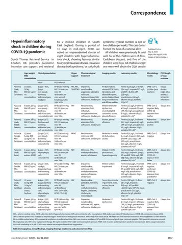
Table: Demographics, clinical findings, imaging findings, treatment, and outcome from PICU
www.thelancet.com Vol 395 May 23, 2020
1607
Correspondence
Published Online May 7, 2020 https://doi.org/10.1016/ S0140-6736(20)31100-4
See Online for appendix
Submissions should be made via our electronic submission system at http://ees.elsevier.com/ thelancet/
1608
for weight. Four children had known family exposure to coronavirus disease 2019 (COVID-19). Demographics, clinical findings, imaging findings, treatment, and outcome for this cluster of eight children are shown in the table. Clinical presentations were similar, with unrelenting fever (38–40°C), variable rash, conjunctivitis, peripheral oedema, and generalised extremity pain with significant gastrointestinal symptoms. All progressed to warm, vasoplegic shock, refractory to volume resuscitation and eventually requiring noradrenaline and milrinone for haemodynamic support. Most of the children had no significant respiratory involvement, although seven of the children required mechanical ventilation for cardiovascular stabilisation. Other notable features (besides persistent fever and rash) included development of small pleural, pericardial, and ascitic effusions, suggestive of a diffuse inflammatory process. All children tested negative for severe acute respiratory syndrome coronavirus 2 (SARS-CoV-2) on bronchoalveolar lavage or nasopharyngeal aspirates. Despite being critically unwell, with laboratory evidence of infection or inflammation2 including elevated concentrations of C-reactive protein, procalcitonin, ferritin, triglycerides, and D-dimers, no pathological organism was identified in seven of the children. Adenovirus and enterovirus were isolated in one child. Baseline electrocardiograms were non-specific; however, a common echocardiographic finding was echobright coronary vessels (appendix), which progressed to giant coronary aneurysm in one patient within a week of discharge from paediatric intensive care (appendix). One child developed arrhythmia with refractory shock, requiring extracorporeal life support, and died from a large cerebrovascular infarct. The myocardial involvement3 in this syndrome is evidenced by very elevated cardiac enzymes during the course of illness.
All children were given intravenous immunoglobulin (2 g/kg) in the first 24 h, and antibiotic cover including ceftriaxone and clindamycin. Subse quently, six children have been given 50 mg/kg aspirin. All of the children were discharged from the PICU after 4–6 days. Since discharge, two of the children have tested positive for SARSCoV-2 (including the child who died, in whom SARS-CoV-2 was detected post mortem). All children are receiving ongoing surveillance for coronary abnormalities. We suggest that this clinical picture represents a new phe n omenon affecting previously asymptomatic children with SARS-CoV-2 infection manifesting as a hyperinflamma tory syndrome with multiorgan involvement similar to Kawasaki disease shock syndrome. The multifaceted nature of the disease course underlines the need for multispecialty input (intensive care, cardiology, infectious diseases, immunology, and rheumatology). The intention of this Correspond ence is to bring this subset of children to the attention of the wider paediatric community and to optimise early recognition and management. As this Correspondence goes to press, 1 week after the initial submission, the Evelina London Children’s Hospital paediatric intensive care unit has managed more than 20 children with similar clinical presentation, the first ten of whom tested positive for antibody (including the original eight children in the cohort described above). We declare no competing interests.
*Shelley Riphagen, Xabier Gomez, Carmen Gonzalez-Martinez, Nick Wilkinson, Paraskevi Theocharis shelley.riphagen@gstt.nhs.uk South Thames Retrieval Service for Children, Evelina London Children’s Hospital Paediatric Intensive Care Unit, London SE1 7EH, UK (SR, XG); and Evelina London Children’s Hospital, London, UK (CG-M, NW, PT) 1
Zhang M-M, Shi L, Lin Y, Liu Y. Clinical analysis of Kawasaki disease shock syndrome. Chin Med J (Engl) 2017; 130: 2891–92.
2
3
Li Y, Zou L, Wu J, et al. Kawasaki disease shock syndrome: clinical characteristics and possible use of IL-6, IL-10 and IFN- as biomarkers for early recognition. Pediatr Rheumatol Online J 2019; 17: 1. Liu P, Blet A, Smyth D, Li H. The science underlying COVID-19: implications for the cardiovascular system. Circulation 2020; published online April 15. DOI:10.1161/ CIRCULATIONAHA.120.047549.
COVID-19: PCR screening of asymptomatic healthcare workers at London hospital The exponential growth in coronavirus disease 2019 (COVID-19) caused by severe acute respiratory syndrome coronavirus 2 (SARS-CoV-2) across the UK has been successfully reversed by social distancing and lockdown.1 RNA testing for prevalent infection is a key part of the exit strategy, but the role of testing for asymptomatic infec tion remains unclear.2 Understanding the determinants of asymptomatic or pauci-symp t omatic infection will provide new opportunities for personalised risk stratification and reveal much-needed correlates of protective immunity, whether induced by vaccination or natural exposure. To address this, we set up COVIDsortium (NCT04318314), a bioresource fo cusing on asymptomatic health-care workers (HCWs—doctors, nurses, allied health professionals, administrators, and others) at Barts Health NHS Trust, London, UK, to collect data through 16 weekly assessments (unless ill, selfisolating, on holiday, or redeployed) with a health questionnaire, nasal swab, and blood samples and two concluding assessments at 6 months and 12 months. HCWs were self-declared as healthy and fit to work for study visits. Participants were not given swab results, and those with symptoms or in selfisolation resumed study visits on return to work. Across London, case-doubling time in March, 2020, was approximately 3–4 days. The number of nasal swabs testing positive for SARS-CoV-2 www.thelancet.com Vol 395 May 23, 2020