ISSUE 6 | SPRING 2017
From the publishers of Plastic Surgery News
» Using a Compass for clinical decision-making When precision is crucial in reconstruction, this device can lead surgeons to the correct path
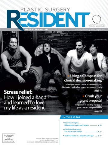
Stress relief: How I joined a band and learned to love my life as a resident
page 16
» Crush your grant proposal he pursuit of funding requires a good coach – and persistence
page 28 IN THIS ISSUE » InService Insights: Odontogenic cysts and tumors ................ p. 18 » Craniofacial surgery: The must-read articles
............................. p. 22
» TheTrend looks at a bony-trauma app ..... p. 27 AMERICAN SOCIETY OF PLASTIC SURGEONS 444 E. ALGONQUIN ROAD ARLINGTON HEIGHTS, IL 60005
Presorted Standard U.S. PoSTAge PAID RIPoN wI PeRMIT No. 100
READY TO USE
Coming soon. 16x20cm Perforated. ALLODERM SELECT™ Regenerative Tissue Matrix Perforated 16x20cm for reinforcement of weak soft tissue in pre-pectoral breast reconstruction.
For more information, contact a LifeCell representative, or visit lifecell.com.
Before use, physicians should review all risk information, which can be found in the Instructions for Use attached to the packaging of each ALLODERM SELECT™ Tissue Matrix. Rx only. CONTRAINDICATIONS: ALLODERM SELECT™ RTM Ready To Use is contraindicated for use in any patient who is sensitive to any of the antibiotics listed on the package or Polysorbate 20.
Copyright 2017 LifeCell Corporation. All rights reserved. All trademarks designated herein are proprietary to LifeCell Corporation, its affiliates and/or licensors. MLC5616/6851/2-2017
A note from the editor
T Alex Spiess, MD Chief Medical Editor Plastic Surgery Resident Pittsburgh
he Spring 2017 edition of Plastic Surgery Resident was an absolute pleasure to put together. From a brainstorming standpoint, it was truly a team efort that’s resulted in a solid compilation and interesting combination of unique content folded into several familiar sections. For this issue, we continue with favorites such as “InService Insight,” “Consult Corner,” “Journal Club” and, of course, the PSR Crossword Puzzle. We also provide a deep-dive into grant writing – and present the story of one young plastic surgeon’s successful battle against a well-known foe: stress. With an eye on the future, we’re working to cultivate an international lavor, as we bring to you interesting pieces from plastic surgeons around the world who will highlight their unique perspectives and experiences, as well as expand our “Pearls from the Masters” series to tap global contributors! We also continue to solicit content and submissions for our ledgling “Resident as Artist” feature (send your artwork to residentart@plasticsurgery.org) and the residents’ advice column “Dear Abbe” (send your conundrum to dearabbe@plasticsurgery.org). he next ofering of “Dear Abbe” will be moderated by Penn Medicine’s Ben Chang, MD, Philadelphia – the names, places and identifying characteristics of any submissions will be changed to preserve anonymity. Look for responses to those questions in an upcoming issue. I hope that you enjoy this edition – our irst of 2017. As always, I would like to thank our physician editorial board and our production staf at ASPS. Look for the Summer 2017 edition of PSR in June. |
Educate Congress and help shape policy that impacts your practice and patients at the inaugural ASPS Advocacy Summit.
» Meet with members of Congress from your home and business district » Participate in workshops on pressing state and federal legislative issues » Learn first-hand from political analysts how Congress is shaping health care » Network with leaders from ASPS and local, state and regional plastic surgery societies
» PlasticSurgery.org/AdvocacySummit
Advocacy Summit replaces Regional Fly-Ins in 2017.
Plastic Surgery Resident | Spring 2017
3
Table of
Contents How rock ‘n’ roll helped him achieve a stress-free life ............... 6 Phuong Nguyen, MD, felt the oppression of training and its accompanying lack of joy – until gigging threw him a life-preserver.
he mission of the American Society of Plastic Surgeons is to support its members in their eforts to provide the highest quality patient care, and to maintain professional and ethical standards through education, research and advocacy of socioeconomic and other professional activities.
Plastic surgeons as artists ...........10
A SPS PR E SI DEN T
Rafi Fredman, MD, used a study session on cleft-lip repair to master the “golden ratio” of facial anatomy – and put charcoal to paper.
EDITOR
Consult Corner: Finger amputation ........................12
Debra Johnson, MD | johnson@pscmail.com Alex Spiess, MD | amspiess@msn.com ASSOCI ATE EDITORS
Mohammed Alghoul, MD | mo.alghoul@gmail.com Sanjay Naran, MD | sanjaynaran@hotmail.com
This digit-trauma unit targets indications for revision amputation vs. replantation.
INTER NATIONAL EDITOR
Crossword ......................................15
RESIDENT EDITORS
Challenge your plastic surgery vocabulary.
Johan Sandberg, MD | ljsandbrg@gmail.com Kavitha Ranganathan, MD | krangana@med.umich.edu Deana Shenaq, MD | dshenaq@gmail.com
Finding the ‘true north’ of surgery with reconstruction Compass .....16
E X EC U T I V E V ICE PR E SI DEN T
Measuring complexity, risk, personal and system factors, and anatomical problems pre-op is more important than ever.
SENIOR DIRECTOR OF COMMU NICATIONS
InService Insights .........................18 Odontogenic cysts and tumors may initially present to plastic surgeons – so these are a regular part of the residents’ exam.
Michael Costelloe | mcostelloe@plasticsurgery.org Mike Stokes | mstokes@plasticsurgery.org M A N AG I N G E D I T O R
Paul Snyder | psnyder@plasticsurgery.org S TA F F E D I T O R
Jim Leonardo | jleonardo@plasticsurgery.org S TA F F W R I T E R
Kendra Y. Mims | kmims@plasticsurgery.org
Things I wish I knew when I was a plastic surgery resident ............21
SEN IOR GR A PHIC DE SIGN ER
Nicholas Tarola, MD, advises residents to choose their words wisely, and to absorb all they can from every experience.
GR A PHIC DESIGN ER
Journal Club .................................. 22 Ten articles on craniofacial surgery that every resident should read, particularly given the rapid progress of this field.
Book review .................................. 26 Plastic surgery’s “ultimate compendium” in the Michigan Manual of Plastic Surgery.
#TheTrend ..................................... 27 Jacob Grow, MD, and James Vargo, MD, launch a new tech column with a hands-on look at a new hand/face E.R. “call” app.
Jim Andrews | jandrews@plasticsurgery.org Anna Abbinante A DV ERTISING SALES
Connor Winther, Wolters Kluwer Health (215) 521-8797 Plastic Surgery Resident (ISSN 2469-9381) is published four times per year and distributed free to members of the ASPS Residents and Fellows Forum and plastic surgery training programs. Letters, questions or comments should be addressed to: Editor, Plastic Surgery Resident, 444 E. Algonquin Road, Arlington Heights, IL 60005. POSTMASTER: Send address changes to ASPS Membership Department Plastic Surgery Resident 444 E. Algonquin Road Arlington Heights, IL 60005 Postage paid at Arlington Heights, IL, and at additional mailing oices.
Kavitha Ranganathan, MD, shares how persistence, dedication and a good coach are keys to writing a successful grant.
he views expressed in articles, editorials, letters and other publications published by Plastic Surgery Resident (PSR) are those of the authors and do not necessarily relect the opinions of ASPS. Acceptance of advertisements for PSR is at the sole discretion of ASPS. ASPS does not guarantee, warrant or endorse any product, program or service advertised.
On the cover: Phuong Nguyen, MD (2nd from left)
ASPS Home Page: www.plasticsurgery.org
Grant writing as marathon .......... 28
4
Plastic Surgery Resident | Spring 2017 | Vol.1 No.6
Plastic Surgery Resident | Spring 2017
EPSTEIN ABDOMINOPLASTY RETRACTOR As the trend in todayÕs abdominoplasties is towards less upper lateral undermining, the upper tunnel has become narrower. During the dissection of this tunnel, it is difÞcult to maintain countertraction with available instrumentation. The Epstein Abdominoplasty Retractor was designed to assist in the performance of the dissection of the upper abdominal tunnel. The ergonomic handle is easily held by the surgical assistant. It is available in several blade lengths so as to best Þt the anatomy of the patient: whether the tunnel is long or short, there is a retractor to provide the best mechanical advantage in yielding exposure and reducing fatigue. The widened, curved working end spreads the tissues of the upper skin ßap apart as the teeth gently hold them in place without slippage. The leading edge of the dissection is easily seen and maintained, facilitating effortless cautery elevation of the skin ßap from the muscle fascia. The retractor is also extremely helpful in elevating the abdominal skin ßap over the narrow tunnel so that the underlying muscle fascia can be plicated.
Designed to assist in the performance of the dissection of the upper abdominal tunnel
The wide curved working end spreads and holds the tissue of the upper skin ßap apart
Facilitates cautery elevation of the skin ßap from the muscle fascia
Extremely helpful in elevating the skin ßap over the narrow tunnel for muscle fascia plication
ASSI.ABR31726 Epstein Abdominoplasty Retractor 80x27mm blade with 60mm wide working end with teeth and ergonomic handle ASSI.ABR31826 Epstein Abdominoplasty Retractor 110x27mm blade with 60mm wide working end with teeth and ergonomic handle ASSI.ABR32026 Epstein Abdominoplasty Retractor 140x27mm blade with 60mm wide working end with teeth and ergonomic handle ASSI.ABR31926 Epstein Abdominoplasty Retractor 160x27mm blade with 60mm wide working end with teeth and ergonomic handle
Æ ACCURATE SURGICAL & SCIENTIFIC INSTRUMENTS®
For diamond perfect performance ©2013 ASSIÆ
®
Designed By: Mark D. Epstein, M.D., F.A.C.S., Stony Brook, NY
accurate surgical & scientific instruments corporation 300 Shames Drive, Westbury, NY 11590
800.645.3569 516.333.2570 fax: 516.997.4948 west coast: 800.255.9378 E-Mail: orders@accuratesurgical.com Internet: www.accuratesurgical.com
Better living through gigging
How the pursuit of
LED TO A
(relatively) stress-free life By Phuong Nguyen, MD TH
E IN N TH H E THR ROES OF RESII D EN N CY OR knoow thatt itt s quuite the slog. mo t sc ooli t pu
Help the Doctor performs at L.A.’s Viper Room in 2014. Michelle Van Vliet
Furthermore, this doesn’t include the numerous personal sacriices of missed weddings, birthdays and funerals, or even something as simple as a night out with a friend or a loved one. I recall at one point during my second year of residency (or, as it was known in our program, the “re-tern” year) walking home post-call through the morning streets of Manhattan, dead to the world, completely envious of a dog being walked across the street, without a care in the world. How did I get here? I now have the true privilege of interviewing outstanding applicants for plastic surgery residency positions. he decision-making is extremely diicult, as everyone who makes it this far is truly outrageously qualiied. Indeed, if they don’t seem to walk on water, there’s probably something wrong with them. Included in this application usually lies a list of hobbies and talents. I’ve interviewed concert violinists who’ve performed at Carnegie Hall, painters who’ve had gallery showings all over the world and athletes who’ve been recruited to the NHL, among other achievements. So what happens to all these amazing talents when one is a slave to his or her beeper, constantly on the cusp of sleep deprivation, with minimal control over his or her schedule? Particularly if there’s another free lap starting at 6 p.m.? he answer is that these talents and interests are still present, though life and responsibility create waves of smoke and mirrors to disguise and redirect this once and future potential. In recent times, an ever-increasing focus exists with regard to physician burnout. A 2011 survey of ASPS members revealed that of the 1,691 respondents, 29.7 percent reported validated burnout.1 In addition, approximately one-fourth of plastic surgeons had signiicantly lower quality-of-life scores than the U.S. population norm. In his 2016 American Association of Plastic Surgeons presidential address and in recognition of these concerning trends, Michael Bentz,
MD, drew special attention to the importance of work-home balance for the plastic surgeon.2 Physicians have a well-documented propensity toward dissatisfaction with work-life balance and burnout when compared with a general population of working adults adjusted for age, relationship status, sex and hours worked per week.3 It’s never been more clear that we, as physicians, and in particular as plastic surgeons, don’t do a particularly good job of taking care of ourselves. Indeed, most physicians are unaware that they are experiencing burnout until it’s too late. his may be manifested in depression, suicide, alcoholism, substance abuse, marital problems, poor job performance, compromised patient care and disruptive behavior.4 However, there are ways to counteract this. (Editor’s note: ASPS launched a wellness efort at Plastic Surgery 2016 in Los Angeles, and it’s also organizing the Project Well Retreat this Fall to prevent or address physician burnout.) Shanafelt, et al., suggested general strategies for success toward improving surgeons’ quality of life. hese include inding meaning in your work; focusing on that which is important in your life; maintaining a positive outlook; embracing a philosophy that stresses work-life balance; and employing a broader repertoire of wellnesspromotion practices.4 he activities and relationships we engage in outside of the hospital oftentimes are what buttress and improve us to be our best self, for our patients and for our loved ones. hough a thorough discussion of burnout is beyond the scope of this article, I wanted to share some anecdotes on what has helped me personally throughout the training process. For background, after medical school, my professional journey included ive years of general surgery residency, two years of postdoctoral research fellowship, three years of plastic surgery residency and a one-year fellowship in pediatric craniomaxillofacial surgery. I’m currently in an academic practice,
board-certiied in general surgery and plastic surgery, and I’m a faculty member of a robust residency program with 18 very talented residents.
Phuong Nguyen, MD
I can certainly tell you that while I’m very proud to be an academic plastic surgeon, the road was fraught with many instances of doubt, selfquestioning and a lack of clarity as to what lied ahead. Ultimately, for me, an integral component of what kept me going throughout those years of school and training was music. I remember it quite clearly – the irst time I heard the opening notes of Slash’s intro to the Guns N’ Roses song “Sweet Child o’ Mine,” I knew. I had to learn how to play guitar. Of course, I was also 14 years old and knew that guitar equals girls. However, that irst lesson in a dingy practice room at Schmitt Music in Maplewood, Minn., learning the three chords to “Bad Moon Rising” by Creedence Clearwater Revival was the initiation of a lifelong love afair. Like many precocious students who liked and excelled in science, I had hoped and imagined someday I’d be able to go to medical school and become a doctor. However, that reality to a teenager was a distant future.
Plastic Surgery Resident | Spring 2017
7
As this was the era of grunge, like all the other disgruntled suburban teenagers illed with existential angst, I started a band. Amazingly, this year marks the 20th anniversary of the irst album I ever made, in my parents’ garage (Beow Mix – On this Afternoon). Of course, though quite proud of the record for existing, it remains quite unlistenable, I assure you. However, like any passion, music insistently kept with me. Ultimately, I did endure the
recorded and released throughout general surgery residency while at NYU (Killing Phantoms – Killing Phantoms 2007; Heroes Night 2009; and March Lions 2011). I relect upon them as little snapshots of a speciic time and place. It certainly required some crafty time management. I would take overnight trauma call as a chief resident at Bellevue Hospital in Manhattan and walk out bleary eyed to take the F train
Dr. Nguyen sings and shreds at the House of Blues in Los Angeles.
rigmarole of college, medical school, general surgery residency, post-doctoral research fellowship, plastic surgery residency and craniofacial fellowship. A short 15 years after I inished college, I was now a bona ide plastic surgeon. However, it was diicult not to compare a resident’s quality of life to those of non-medical colleagues. Was all this work worth it? hroughout each of those moments of school and training, I was still attempting to process life experiences – though to be sure, I was quite stunted in emotional intelligence. A surgical residency virtually ensures a delay in emotional maturation. With the daily rigor and stress, a way to process these experiences was desperately needed. For me, it was making music. So I wrote albums. One was recorded and released in medical school (Phuong You Norway – Sirens and Accidents, 2004), and three were 8
Plastic Surgery Resident | Spring 2017
want to be judged as uncommitted, sidetracked or, worse yet, lazy. I decided to keep it to myself. However, as I went through the ranks of the residency hierarchy – building a track record of dependability and work ethic – it became increasingly acceptable to have interests outside of medicine to which one could also be dedicated. In the insular world of medicine and training, one constantly fears judgment and labeling, as those irst impressions may carry a stigma throughout not just residency, but even into practice. However, with the aid of retrospection, I know now just how important it was for my own survival and well-being to have this external outlet. It allowed me to be my true and complete self. Not one part denying the other, the creative vs. the pragmatic. It took a long time to realize they were intertwined, and ultimately, helped me see more clearly what I truly wanted in life.
over to Brooklyn to the recording studio and lay down a few tracks. hen I would head back to take over the pager come evening. hough a little sleep deprived, doing something completely the opposite of my day job (or night job, as it were) was actually quite invigorating. hat band, Killing Phantoms, had multiple iterations and a few generations of bandmates. Most were not involved with medicine, which made for a perfect outlet to meet people outside of work. We played most of the clubs in the East Village and the Lower East Side, irst really to only a few people (it turns out that Sunday night at midnight is not the ideal time for people to see a rock show), but over time, and with building word-of-mouth not only from people within work, but also outside, we began to attract bigger and bigger crowds. When I irst started internship, I was quite hesitant to promote or bring attention to this side gig. I didn’t
After seven years of general surgery and research fellowship, the prospect of three more years of plastic surgery residency was a little discouraging. I was watching all of my former medical school compatriots move forward and immerse in their practices as I stared down the barrel of PGY-10. However, it seems, fortune always happens when least expected. Just months into beginning plastic surgery training at UCLA, I had already felt a vacancy at the dissolution of my band in New York. hough excited at the prospect of learning an entirely new discipline, I felt there was still something missing. What started as an innocuous invitation to check out the UCLAUSC football game by a co-resident became a deining moment not only in my music career, but also my medical career. After seeing a guitar in the corner of the living room while waiting for our ride, it turned out that we shared quite the similarity of musical taste. I had previously relegated in my mind that the music phase of my life had passed. We excitedly talked about playing music together. Fast-forward a month. While sitting across from the facial plastics Fellow,
after raising a radial forearm free lap and waiting for the oncologic resection of the head and neck cancer, discussion – of course – turned to music. he Fellow also played guitar, had been classically trained on piano since childhood and shared a love of early 1990s rock bands as well. Rounding out the bill was the fact that the current intern on the plastic surgery service, an OMFS resident, happened to have played drums his entire life. So, serendipitously, Help the Doctor formed – and we began writing songs and practicing in a spare room in a family member’s house. As much as I had resisted playing in a band of doctors for fear of lack of legitimacy, our lineup consisted of Jason Roostaeian, MD (bass; microsurgery/ aesthetic plastic surgeon at UCLA); Robby Kang, MD (guitar and synths; facial plastics, and head and neck reconstruction at City of Hope Cancer Center); Solomon Poyourow, MD (drums; oral and maxillofacial surgeon in private practice in Alamitos, Calif.); and myself on guitar and vocals. (We can be seen on the cover of this issue: Dr. Poyourow at the far-left; me; Dr. Roostaeian; and Dr. Kang.) Having played in multiple bands over the past nearly two decades, I had limited expectations of what lay ahead. hrough local contacts, we were fortunately able to secure a show at the historic Troubadour in West L.A. on a Saturday night, barely having been a band for a few months. his venue has hosted everyone from Bruce Springsteen to Elton John – Guns N’ Roses got their start there! Even with this history, it was a modest-sized club. We were early on the bill. After soundcheck in the afternoon, coming back to the club before our set, there was a line out the door and around the block. I assumed it was for one of the other bands on the bill. hen, a young man on the street asked me if I’d like to buy some scalped tickets for the sold out show for Help the Doctor. hough I’m certainly proud of the publications, grants and awards that accumulate on the road to academic medicine, it’s a special and utterly unique satisfaction of being sold
scalped tickets to your own show. (A practice I do not condone, by the way!)
to be mutually exclusive. It doesn’t have to be one or the other.
One thing led to another, and we began selling out shows at other major venues, including the Viper Room and the Roxy, and culminating in a headlining, sold-out show at the House of Blues on the Sunset Strip. Seizing the momentum, we wrote and recorded an EP (Help the Doctor, 2012) and a full-length album (Angeles, 2015) in a few Los Angeles studios that have hosted luminaries such as Pink
I’ve been reminded many times over the years of the late Robin Williams as the English teacher John Keating in the iconic ilm Dead Poets Society: “And medicine, law, business, engineering – these are noble pursuits and necessary to sustain life. But poetry, beauty, romance, love – these are what we stay alive for. To quote Whitman, “O me! O life!... of the questions of
With the daily rigor and stress, a way to process these experiences was desperately needed. For me, it was making music. Floyd and Mavis Staples. For admitted music geeks, this was cloud nine. We decided early on that any proits we obtained from our shows would be donated to philanthropic organizations that focused on reconstructive surgical care for children. his was a way for us to reconcile the careers that we had spent a fair amount of time and efort in pursuing with our lifelong love of music. Interestingly, these simple measures gave us some unique credibility within the music scene – and also became a hot topic, resulting in several network television news spots covering our shows. We were able to expose the iner parts of plastic and reconstructive surgery to a much wider audience than we ever thought possible. Ironically, our lives outside of medicine had now actually made us more notable within our own professions! Sometimes, I’m asked “Medicine or rock and roll?” Well, the truth of the matter is: We’re not exactly U2 selling out stadiums, with each member arriving on their own separate tour bus. he reality of life as a working musician for the vast majority is considerably less glamorous. Music for me was, and is, an essential outlet that helps complete the picture of who I am and provides me deep fulillment. It went from being a liability in the path of becoming a physician to an aid that helps provide balance and restoration in my day-to day practice. hough much time and efort go into becoming a doctor, your unique interests and passions do not have
these recurring; of the endless trains of the faithless, of cities ill’d with the foolish… what good amid these, O me, O life? Answer: hat you are here – that life exists, and identity, that the powerful play goes on, and you may contribute a verse.’ “hat the powerful play goes on, and you may contribute a verse. What will your verse be? Carpe, carpe diem, seize the day boys, make your lives extraordinary.” We are uniquely privileged to practice in plastic surgery, a discipline that encourages creativity and ingenuity. However, in the process of training and daily practice, there are signiicant constraints on our personal time and life balance. I urge you not to forget who you are, and don’t forget the dreams that you have. Indeed, make your life extraordinary. | Dr. Nguyen is an assistant professor of Surgery in the Division of Plastic and Reconstructive Surgery, Children’s Hospital of Philadelphia. He can be reached at pdnguyendoc@gmail.com. REFERENCES 1. Qureshi, H. A., Rawlani, R., Mioton, L. M., Dumanian, G. A., Kim, J. Y., Rawlani, V. Burnout phenomenon in U.S. plastic surgeons: risk factors and impact on quality of life. Plast Reconstr Surg 2015;135:619-626. 2. Bentz, M. L. he Plastic Surgeon at Work and Play: Surgeon Health, Practice Stress, and Work-Home Balance. Plast Reconstr Surg Glob Open 2016;4:e1081. 3. Shanafelt, T. D., Boone, S., Tan, L., et al. Burnout and satisfaction with work-life balance among US physicians relative to the general US population. Arch Intern Med 2012;172:1377-1385. 4. Shanafelt, T. D., Oreskovich, M. R., Dyrbye, L. N., et al. Avoiding burnout: the personal health habits and wellness practices of US surgeons. Ann Surg 2012;255:625-633.
Plastic Surgery Resident | Spring 2017
9
Drawing and lip anatomy: infusing life through the ‘white roll’ By Rafi Fredman, MD, PGY-3 plastic surgery resident, University of North Carolina, Chapel Hill
I
spent an entire weekend reading about cleft lip reconstruction in preparation for a cleft lip repair I was scheduled to perform the upcoming week. With cleft lip reconstruction, a detailed understanding of the diferent elements of lip anatomy is essential. For example, the position and length of the philtral columns, proportion of the lips, Noordhof’s point, etc.… hese are things I hadn’t previously thought much about when studying facial anatomy.
Feature your artwork in an upcoming issue of PSR – submit to ResidentArt@plasticsurgery.org.
10
trying to draw lips using the principles I had gathered from both cleft lip surgery and principles of facial aesthetics would be a rewarding challenge. It was! I used pencil and charcoal on drawing paper, with the accompanying images revealing how I used the golden ratio to determine lip proportions. Not only was it relaxing to spend time drawing, but the exercise also taught me a lot about the importance of certain anatomic details of the lip. For example, I had read about the importance of the white roll – but when I drew the lips without the white roll, and then used a ine eraser to create it, I found an efect that was incredible. It was like the lips came to life.
I read an article about the golden ratio and its applications in facial aesthetics. his fascinated me – and I thought that
It’s diicult to ind the time to sit and just draw – but I encourage anyone learning plastic surgery to give it a try. |
An understanding of the aesthetic relationships of the face are important when drawing.
I used the "golden ratio" to design the lip proportions.
Charcoal pencil was used to create shadows and contrast with light areas, i.e., the philtral columns and white roll.
For me, drawing is both relaxing and educational. I encourage all plastic surgery residents to try it!
Plastic Surgery Resident | Spring 2017
Some call a needle primitive We call it unnecessary Introducing the New Standard in Reconstruction
AEROFORM M® DOSAGE CONTROLLER
AEROFORM® TISSUE EXPANDER
Needle-Free Patient-Controlled Faster Expansion than Saline*
See how AeroForm® can change your practice at
www.airxpanders.com/contact * Ascherman, JA., et al. CO2-based vs. Saline Tissue Expansion for Breast Reconstruction: Results of the XPAND Prospective, Randomized Clinical Trial. Plastic and Reconstructive Surgery. December 2016 – Volume 138, Number 6. The AeroForm Tissue Expander system is used for soft tissue expansion in breast reconstruction following mastectomy, for the treatment of underdeveloped breasts, and for the treatment of soft tissue deformities in the breast. The AeroForm Expander is intended for temporary subcutaneous or submuscular implantation and is not intended for use beyond six months. AeroForm Tissue Expander System, the AeroForm logo and the AirXpanders logo are registered trademarks of AirXpanders, Inc. © 2017. All rights reserved. The products described in the document may be covered by U.S. Patents: 9,526,584; 8,394,118; 8,617,198 and 8,808,322. COL-0053 Rev. A
Management of finger amputation
“Consult Corner” addresses a consult commonly encountered by an on-call resident. he column begins with the reason for consult and assesses questions that might go through a resident’s mind as he or she heads to the emergency department to see the patient. Key aspects of the history and physical, as well as additional testing that should be obtained, are also presented. Finally, a review of the decision-making process will present possible management strategies, all of which are synthesized into the context of an actual case.
A multifaceted approach to an E.D. call for consult By David Stepien, MD
Y
our pager goes of at 2 a.m. on Saturday. It’s the E.D. with the following message: “21 y/o male transferred from outside hospital with distal index inger amputation from circular saw. Has amputated part with him.”
Having seen multiple hand traumas in the past, you realize the futility of asking why someone was using a circular saw in the middle of the night. Regardless, in this article we will discuss the approach to hand trauma with a speciic focus on the indications for revision amputation versus replantation. On your way to see the consult, ask the E.D. to obtain X-rays of the injured hand and the amputated part – preferably in the same image. X-rays are important for evaluating for associated fractures and foreign bodies. Make sure they’ve wrapped the amputated part in saline-moistened gauze within a plastic bag that’s illed with ice. his will ensure that the amputated part stays at a target temperature of 4 degrees Celsius (39.2 degreed Fahrenheit) to minimize the metabolic needs of the digit and improve the likelihood of success if replantation is performed. he patient should receive intravenous antibiotics in the setting of this open fracture and a tetanus shot if needed.
MESSAGE ON YOUR PAGER: E.D. CALL: TRAUMA PATIENT WITH FINGER AMPUTATED AND PRESENT As you return the page, your mind should begin to ruminate 12
Plastic Surgery Resident | Spring 2017
on the immediate considerations that must be addressed upon your arrival: • Mechanism of injury (sharp versus crush; clean vs. dirty): his is a critical determinant of whether a digit will be able to be successfully replanted. If a inger is crushed, the vessels are at much higher risk for thrombosis and replantation failure. Similarly, if a digit is amputated as a result of a farm injury, there’s a signiicantly higher risk of infection compared to an injury that occurred as a result of a kitchen knife. • Level of amputation: he more proximal an injury, the greater the likelihood of successful replantation given the increased size of the digital vessels (Fig. 1). Injuries within Zones 3-5 on the volar surface of the hand are more likely to tolerate replantation, given the larger size of the digital vessels that will be reapproximated compared to those in Zones 1 and 2. Particularly in Zone 2, replantation in the setting of a single-digit amputation at this level is unlikely to succeed, given the high risk for post-op adhesions and stifness. • Patient occupation/age/comorbidities/handedness: he recovery after replantation is signiicantly more strenuous than that after revision-amputation. If a patient’s main goal is to return to work as soon as possible, the best option may be a revision amputation. However, if the patient has appropriate resources and is willing to tolerate prolonged occupational therapy and additional procedures
Relative contraindications • Severe crush or avulsion injury • Segmental/multiple injuries within the same digit • Systemic illness • High contamination of part or wound • Injury distal to DIP he critical decision point in this case is deciding whether to perform a revision amputation or replant the amputated part. In this case, given the distal zone of injury, replantation is not indicated and revision amputation is the correct choice. Although amputations do not have the fame and glory associated with replantation procedures, performing a proper revision amputation is of utmost importance to prevent subsequent sequelae and complications including neuromas, quadriga efect and hook-nail deformities.
Figure 1. he zone of injury is critically important. Injuries in Zones 3-5 are easier to recover from after replantation compared to Zone 2 injuries which are at high risk for stifness.
to gain optimal function, replantation is the best option – if all other indications are met. If the amputation occurs in a child, replantation is almost always attempted. On physical exam, your patient has sustained a dorsal oblique amputation distal to the distal interphalangeal (DIP) joint of his left index inger within Zone 1. Bone is exposed at the base of the amputation, and the nail bed has been lacerated as well. he patient has complete lexion and extension of the digit at the metacarpophalangeal joint (MCP), the proximal interphalangeal joint (PIP) and the DIP joint. Normal sensation is present on both the radial and ulnar sides of the digit. Perfusion to the digit is normal. he X-ray demonstrates a mildly comminuted tuft fracture of the DIP joint, but there doesn’t appear to be any foreign bodies or additional fractures. he amputated part is macerated and dusky, and it consists of the amputated soft tissue pulp of the index inger (Fig 2A, 2B). REPLANTATION OR REVISION AMPUTATION? At a tertiary hospital with replantation capabilities, you must consider whether this amputated part is suitable for replantation. While appropriately selected replantations do have a high success rate (up to 80 percent), many patients don’t meet criteria for replantation. he indications for replantation • Indications for replantation • humb amputation • Loss of multiple digits • Palm, wrist or forearm-level amputation • Any amputation in a child
To provide adequate soft-tissue coverage over the exposed bone while maintaining as much length as possible of the digit, multiple options exist: A: Healing by secondary intention with dressing changes (good for areas without exposed bone) B: Use of local or distant lap-closure given paucity of mobile soft-tissue available C: Primary closure or advancement lap-closure with shortening of the bone and excision of the nailbed if needed For this particular patient, a revision amputation was performed by advancing the volar skin to allow for primary closure dorsally, with nail-bed repair ensured at the same time (Fig. 3). TO PERFORM A REVISION AMPUTATION IN THE E.D., GET THE FOLLOWING SUPPLIES: • Finger tourniquet (can also use the inger from a sterile glove wrapped around the digit with a hemostat) • 10 milliliter syringe with 18 and 27 gauge needles • 1% lidocaine without epinephrine • 0.5% bupivacaine • Sterile towels • Two 1-liter bottles of saline • Betadine/iodine prep • 4-0 nylon or prolene • 5-0 vicryl or monocryl • #15 blade scalpel • Minor handset PROCEDURE After completing your physical exam, perform a digital block to allow for proper anesthesia during the procedure. A mixture of 1 percent lidocaine without epinephrine and .5 percent bupivacaine will optimize pain control for the procedure, as well as six to eight hours later. After cleaning Plastic Surgery Resident | Spring 2017
13
Images courtesy of the Michigan Manual of Plastic Surgery
Figure 2. he patient presents with a volar oblique amputation of the left index inger. here is bone exposed at the base. he injury occurred as a result of a saw injury. he level of amputation resembles letter D (left). In this setting, revision amputation is required to allow for proper coverage over the distal phalanx (above).
the site with an alcohol pad, inject 2-3 milliliters of the local anesthetic mixture just distal to the distal palmar crease subcutaneously at the sites of the radial and ulnar digital nerves. Also inject 2-3 milliliters dorsally, as well, within the subcutaneous tissues. Allow ive to seven minutes for the medication to take efect and assess for anesthesia. Once adequate anesthesia is achieved, copiously irrigate the wound, making sure to remove all particulate matter. A scrub brush may be necessary, particularly if tenacious contaminants such as machinery grease are present. We frequently pour half of a small bottle of betadine into a liter bottle of saline to use as irrigation. Once cleaned, apply a inger tourniquet to the base of the digit to minimize bleeding. Prep the whole digit with betadine and drape sterilely. Under loupe magniication, identify the digital nerves and vessels. Use cautery or monocryl suture-ligation to address the vessels and trim back the nerves into the wound sharply to prevent neuroma formation. Remove the nail by passing a Freer elevator under the nail plate and gently sweeping the nailbed of the undersurface of the nail, all the way back to the nail fold. If the nail is intact, you can set this aside to splint the nail fold at the end of the case. Using a pair of small rongeurs, trim back the exposed distal phalanx to a level that maximizes bony length while allowing for primary closure using the surrounding tissues, avoiding the lexor and extensor insertions near the DIP joint. Make sure that you have adequate bone to support the nail matrix to prevent hook nail. If performing a ish mouth-type primary closure, trim the bone back to the point at which adequate soft tissue is present to cover the site. Ideally, it’s best to place the scar along the dorsal surface of the digit to prevent sensitivity with touch that may occur if the incision is placed dorsally. Approximate the incision using 4-0 nylon suture. Cover the 14
Plastic Surgery Resident | Spring 2017
digit with bacitracin and xeroform gauze, with a plan for suture removal in 10-14 days. Apply a volar resting splint to minimize trauma to the site. Place the ingers, hand and wrist in intrinsic plus position to minimize contractures. Tip: For injuries that present with tissue loss in a dorsal oblique orientation, volar V-Y advancement (Atasoy-Kleinert) laps are a good option. In this form of local tissue rearrangement, the volar soft tissues are advanced dorsally to improve soft-tissue coverage dorsally. If performing a V-Y volar advancement, position the base of the V centrally – approximately 1mm distal to the DIP joint lexion crease. Do not violate the crease, as you may induce a lexion contracture. Distally, the V should extend to the injury site and be as wide as the nailbed. Once marked, incise the drawn lap down through subcutaneous tissue, staying supericial to the lexor sheath proximally while freeing the ibrous septa from the distal phalanx. his lap can be advanced over the wound to evaluate the need for further dissection or modiication of the lap. Placing your stitches from proximal to distal allows incremental advancement that will ultimately reduce tension on the nailbed closure. | ADDITIONAL READING Louis, DS, Jebson PJL, & Graham TJ. “Amputations.” Green’s Operative Hand Surgery. Fourth Edition. 1998 Russell, RC & Casas, LC. “Management of Fingertip Injuries.” Clinics in Plastic Surgery. 16: 405-425, 1989 Shulebarger, JV & Scheker LR. “Nail Bed Injury.” Operative Plastic Surgery. 867-882, 2000 Zook, EG & Brown, RE. “he Perionychium.” Green’s Operative Hand Surgery. Fourth Edition, 1998 Lemmon JA, Janis JE, & Rohrich RJ. “Soft-tissue injuries of the ingertip: Methods of Evaluation and Treatment. An Algorithmic Approach.” Plast Reconstr Surg. 2008;122(3):105e–17e.
Put your plastic surgical vocabulary skills to the test! By Daniel Lyons, MD
2
1
3
4
5 6 7
8 9
10
11
12 13 14
15
17
16
19
18
20 21 22
23
24 25
ACROSS 3. Type of ulcer for which compression therapy is recommended 5. Gold standard for diagnosis of osteomyelitis (2 words) 7. _____-pressure wound therapy used to expedite healing in non-infected wounds 10. _____ sore often seen in non-ambulatory or insensate patients 12. Topical ointment derived from clostridium histolyticum, used to break down ibrinous debris 14. Type of dressing which maintains a moist environment and supports autolytic debridement (e.g., DuoDerm) 16. he most abundant protein in the human body 17. Dressing solution efective in controlling pseudomonas (2 words) 18. Microbial adherence to infected implants, often resistant to antibiotics 20. Bilayered skin substitute produced from bovine collagen and tissue-cultured human infant foreskin 22. _____ acetate: topical used in burn patients with excellent penetration of cartilage 24. Super-absorbent dressings derived from seaweed used in exudative wounds 25. Imaging modality with a 98% sen./88% spec. in detecting osteomyelitis 26. Silver _____ wet dressing solution efective for most staph and gram-negative aerobes, including pseudomonas
26
DOWN 1. First cell type present within wound sites 2. Lab study whereby a value >100 is often diagnostic for osteomyelitis (abbr.) 4. _____ sulfadiaznie: topical which may cause transient leukopenia 6. Bilaminar porous matrix that cross-links bovine collagen and glycosaminoglycans 8. Surgical procedure to remove necrotic tissue 9. Often fatal syndrome of cutaneous necrosis that tends to develop in patients with chronic renal failure 11. Type of dressing change regimen used for mechanical debridement of wounds (3 words) 13. Gauze dressing containing bismuth 15. Topical antimicrobial ointment used for minor cuts or abrasions 19. _____ gangrenosum: ulcerative cutaneous condition often exacerbated by surgical debridement 21. _____ Solution: antiseptic solution composed of dilute bleach 23. Golden-colored topical thought to have antibacterial properties Answer key on page 30
Navigating the ‘reconstructive ladder’ with the Plastic Surgery Compass he 360-degree evaluation and directional guidance for an unusual sacral wound By Johan Sandberg, MD
I
n today’s special and challenging practice environment – where demands from patients, insurance agencies and hospital administration are increasingly becoming greater – it is of utmost importance to make the right decisions in complex clinical situations. Results are no longer measured only in centimeters or milliliters – success now includes the measure of quality of life and satisfaction. hese new additions require an additional level of skill from the surgeon, as well as a sense of direction when evaluating a patient. When I returned home from the United States and entered practice in Norway, I had to “inherit” patients from other previously employed surgeons at my hospital. I often met my patients for the irst time only 30 minutes before surgery. his is stressful beyond what can be imagined, with the only reasonable comparison being the stress experienced in an oral board exam situation. Decisions have to be made on the ly. his enlightened me even further of the importance of the preoperative evaluation; patient-surgeon interaction with preoperative information; and the setting of expectations. I was very happy to have had clinical experience in a diferent system and I trusted my instincts – my “inner compass,” so to speak, in these situations. Eventually, I began to analyze the workings of this inner compass that had been instilled through years of American training.
To describe this thought process to my own residents in Norway, I came up with the concept of the Plastic Surgery Compass. he compass illustrates how we as plastic surgeons make complex clinical decisions1. In brief, the poles of the compass are designated North – Procedural Complexity; South – Risk; East – Personal and System Factors; and West – Anatomical Problem. he compass is built around the proven concept of the reconstructive ladder, but it focuses on the patient and his or her situation in the health system, rather than on the procedure. he northern pole represents a force in the direction of procedural complexity. he southern pole represents the force that risk (such as ASA classiication) constitutes and as such, it at times acts as a polar opposite of complexity. he eastern pole, which involve the personal factors, essentially represents the problem as it’s deined by the patients situation and the health-care system in which he or she is treated. he western pole is deined by the anatomy of the defect – the anatomical part of the problem. Each of the poles exerts a “magnetic force” that guides us as surgeons in a “grid” along the reconstructive ladder that essentially constitutes the backbone of the compass. Patients in Norway are evaluated in clinic where treatment decisions are made. hose deemed to be candidates for surgery are then placed on a waiting list. he waiting list can be long – ive years for postbariatric surgery, for example. One consequence of a long waiting list is that a lot can occur as time passes by, such as an AMI, a change in weight or a cancer diagnosis.
Aakerman/Sandberg
16
Plastic Surgery Resident | Spring 2017
Patients on the waiting list are sometimes not connected to a certain attending and can be randomly assigned,
depending on the hospital where one works. It’s a challenge to prepare patients for surgery when factoring-in a long lag time between consult and date of surgery. he “waiting list” was the place I encountered nearly all my elective patients for surgery during my irst six months of practice – until I had built-up a waiting list of my own.
for myoglobulinemia. He had been debrided three days previously and had a VAC placed over the wound. he patient was now due for further debridement; was it OK to add him to my operating list?
Certain parameters have to be fulilled to get treatment that’s covered by the state: for example, for an abdominoplasty, a BMI and a pannus measure from the inner surface of the skin fold in centimeters. hese guidelines do not speciically say to exclude other factors in the preoperative evaluation, but in my opinion “protocols” of this type can promote a lack of responsibility/ownership and of sound thinking.
Upon reviewing the chart, I found that this was a young male with a history of substance abuse controlled over many years with Methadone. he patient was found collapsed on the bathroom loor. A large bruised/necrotic area had been noticed on his sacral/buttock area and it had been deduced that he had somehow been electrocuted. Since there was a suspicion of electrocution, a CK level had been ordered, which had been elevated to a level that supported the suspicion of considerable muscle injury.
his can be further compounded in the event that the planning surgeon is not the same as the operating surgeon. he complexity of successful practice may in this way be reduced into the evaluation of two simple parameters with an additional substantial temporal delay.
He had been treated in the ICU. After his CK levels had been normalized, he had been debrided for his supposed burn injury. Due to his problem with painful wound changes, a VAC dressing had been placed on the wound to decrease the wound-change frequency.
It was during this period in my practice that I learned that cancelling patients who were not true candidates for their anticipated procedures was a painful and time-consuming process both for me and for the patients. hey rightfully felt entitled to the procedure; they had an approval for the procedure and they were good people with good values, good citizens and good taxpayers. However, the smoking patient with a recent MI, an out-of-control DM and a BMI of 35 will surely not beneit from a mini-abdominoplasty of their voluminous abdomen.
he possible diagnosis of a decubital/crush injury due to a loss of consciousness was also entertained. he matching scapular pressure-wounds with full dermal necrosis certainly pointed toward this. he patient denied any drug use in conjunction with the injury, except for a single benzodiazepine tablet used for insomnia. he patient was brought to the O.R. for debridement.
I was also quite sure from the start that it would be even more painful and outright unethical to perform a procedure that I was not in agreement with and that the patient did not tolerate, need or would not be satisied with. Interestingly enough, resetting expectations was often possible with careful and serious counseling. Patients in Norway are exceptionally receptive, kind, pleasant and considerate. So for surgeons, there’s a skill that can be developed to tactfully break this bad news and reverse a previous decision. After a couple of years in practice, I still on occasion have to operate on patients who’ve been seen by other surgeons. I try to limit this part of my practice to procedures where satisfaction rates are high – such as breast reduction – or where extensive guidelines are a part of practice – such as the treatment of malignant melanoma. I always see the patients preoperatively and cancel the patients or correct the plan as necessary. In my opinion, patient satisfaction relies on excellent preoperative information; the setting of reasonable expectations; and the eicient execution of the treatment plan.
According to the Compass, the anatomical defect was deined: A foul odor with extensive purulence was found when the VAC was removed. A defect measuring 20-by20cm and located less than 10cm from the anus was found. Considerable undermining was also seen laterally. Sacral periosteum was exposed. An epidural catheter had been placed superior to the defect. Debridement including necrotic great gluteus muscle was performed. Gluteal perforators were ligated. he wound was left open with daily wound care. Wound cultures showed E. coli in the wound. he patient was informed post-op about the extent of the defect. he need for a colostomy was explained, the patient agreed with considerable hesitation. Personal factors were further illucidated as the next step in the Compass model: A discussion about the patient’s drug use and social situation was held. he patient had a job, continued on page 30
In addition, it’s important to make patients feel like individuals and not just a number in the waiting list. All patients are equal, but not the same. he two terms are commonly confused in the socialist system. One case in particular led me to devise the Compass. I was called by one of my residents to see a patient with a severe burn injury to the buttock and electrocution damage. he patient had been treated for his burns and also treated
Figure 1 – he defect
Figure 2 – Flaps raised with ONQ pumps placed subfascially
Plastic Surgery Resident | Spring 2017
17
Odontogenic cysts and tumors By Kristoffer B. Sugg, MD dontogenic cysts and tumors are a perennial topic on the In-Service Examination for Residents (ISE). While these conditions are typically managed by our oral and maxillofacial surgery colleagues, patients may initially present to the plastic surgery clinic; therefore, it’s important to be able to diferentiate cysts and tumors of dental origin from other pathologies of the jaw.
O
epithelial cells of the irst pharyngeal arch migrate into the underlying ectomesenchyme and give rise to the tooth bud. In the cap stage, the tooth bud grows around an area of condensed ectomesenchyme and becomes the enamel organ. Ameloblasts located within the enamel organ eventually produce enamel, while ectomesenchymal cells form the dental papilla and dental follicle.
Odontogenic cysts and tumors are a perennial topic on the In-Service Examination for Residents (ISE). While these conditions are typically managed by our oral and maxillofacial surgery colleagues, patients may initially present to the plastic surgery clinic; therefore, it’s important to be able to diferentiate cysts and tumors of dental origin from other pathologies of the jaw.
he dental papilla consists of the pulp, which contains a rich supply of blood vessels and nerves to the tooth, and odontoblasts that secrete dentin. he dental follicle contributes to the supporting structures of the tooth by producing cementum from cementoblasts, alveolar bone from osteoblasts and the periodontal ligament from ibroblasts. In the bell stage, the crown of the tooth assumes its inal shape and tissue remodeling occurs.
Even though most of these lesions are benign, local growth can lead to mandibular enlargement and loss of teethsupporting alveolar bone. When this occurs, patients may complain of pain and facial swelling. Involvement of the mental nerve can also cause parasthesias of the lower lip. Panoramic radiographs are useful at establishing a diagnosis and ruling out pathologic fracture, but a CT scan may be indicated for preoperative planning or to delineate the extent of a lesion. Management usually consists of enucleation for simple or solitary lesions, and surgical resection with mandibular reconstruction for more severe cases. Like most topics on the ISE, a thorough understanding of the anatomy and developmental origin of a tissue can greatly improve your score. For primary dentition, odontogenesis occurs between the sixth and eighth weeks of gestation and consists of three overlapping stages. In the initial bud stage, 18
Plastic Surgery Resident | Spring 2017
Periapical or radicular cysts are the most common odontogenic cysts of the mandible. hese cysts arise from the epithelial cell rests of Malassez in the periodontal ligament and are thought to occur as a result of periapical inlammation from an infected tooth. hey tend to be asymptomatic and are often encountered as incidental indings during routine radiographic examination. he classic appearance of a periapical cyst on plain ilm is described as a small, well-demarcated radiolucency at the root or apex of a devitalized tooth. Histology demonstrates a stratiied squamous non-keratinizing epithelium. he preferred treatment for periapical cysts is endodontic therapy. Dentigerous or follicular cysts are the second most-common odontogenic cysts of the mandible. hese cysts arise from the crown of an unerupted tooth such as the mandibular third
molar or maxillary canine. Similar to periapical cysts, they also tend to be asymptomatic unless they become infected. On plain ilm, a dentigerous cyst appears as a small welldemarcated radiolucency at the neck or cervix of an unerupted tooth. Histology demonstrates a stratiied squamous nonkeratinizing epithelium. Given the increased risk for local growth, the recommended treatment for dentigerous cysts is enucleation and extraction of the involved tooth. Odontogenic keratocysts or keratocystic odontogenic tumors (KOTs) are the third most-common odontogenic cysts of the mandible. hese benign cystic neoplasms originate from the dental lamina and tend to be locally invasive. KOTs are highly variable in clinical presentation and can occur in any position along the jaw, but the posterior body of the mandible is the most common location. Histology demonstrates a stratiied squamous keratinizing epithelium with the occasional budding of daughter cysts from the basal layer. Often discovered incidentally on routine radiographs, KOTs typically grow longitudinally along the length of the mandible as an expansile unilocular radiolucent lesion with smooth, corticated borders. Treatment usually consists of enucleation with peripheral ostectomy, although recurrent rates as high as 60 percent have been reported. Of note, exam writers for the ISE frequently ask about the association of KOTs with the nevoid basal cell carcinoma syndrome, or Gorlin syndrome. his a hereditary disease of autosomal dominant inheritance that presents with multiple KOTs, multiple basal cell carcinomas, palmar pitting, biid ribs and calciication of the falx cerebri. Solitary or traumatic bone cysts are primarily seen in children and young adults. While trauma is the suspected etiology, little evidence exists in the literature to support this claim. Solitary bone cysts are not considered true cysts, since they are composed of a cystic cavity that lacks an epithelial lining. he majority of them are found in the body of the mandible. hey tend to be asymptomatic, but large cysts can lead to facial swelling.
associated with ibrous dysplasia. Similar to solitary bone cysts, ABCs are not considered true cysts. hey are composed of numerous blood-illed sinusoidal spaces that lack an endothelial lining and are surrounded by multinucleated giant cells. Radiographs demonstrate a characteristic soap-bubble appearance of an expansile multilocular radiolucent lesion with thin sclerotic margins. Curettage and bone grafting is an efective treatment strategy, although lesions recur in approximately 20 percent of cases. Odontomas are the most common odontogenic tumors of the mandible. hey are considered tooth hamartomas, since they comprised of multiple dental elements including epithelial and ectomesenchymal structures. Complex odontomas consist of irregular calciications with no discernible teeth, while compound odontomas contain malformed teeth with a characteristic “bag of marbles” appearance on radiographs. Surgical resection is the preferred treatment for odontomas. Ameloblastomas are the second most-common odontogenic tumors of the mandible. hey are true neoplasms that arise from ameloblasts in the odontogenic epithelium and tend to be locally invasive. Patients usually present with a hard, painless mass near the angle of the mandible in the third to ifth decades of life. As the tumor enlarges, the cortices of the mandible form a thin shell of bone that elicits a cracking sound or crepitus during palpation. here are four clinical subtypes of ameloblastoma: unicystic; solid/multicystic; desmoplastic; and peripheral/extraosseus. Radiographs display an expansile multilocular radiolucent lesion with characteristic soap-bubble appearance, usually in the region of the third mandibular molar. A classic inding on histopathologic examination of ameloblastomas is reverse nuclear polarity of the ameloblasts. he majority of lesions are successfully treated by surgical en-bloc resection followed by mandibular reconstruction.
A common radiologic feature of solitary bone cysts is the appearance of a unilocular radiolucent lesion with scalloping efect between the roots of adjacent teeth. Surgical exploration and curettage is the treatment of choice to help establish the diagnosis and encourage new bone formation within the cyst lumen.
Cementomas are rare, odontogenic tumors that arise from the cementum of the tooth root. hey are characterized by slow and painless growth, and commonly involve the apex of mandibular premolars and molars. Radiographs demonstrate a well-demarcated radiopaque density at the apex of the involved tooth surrounded by a radiolucent rim. No treatment is required, but surgical resection is generally curative for symptomatic lesions.
Aneurysmal bone cysts (ABCs) are benign, often rapidly expanding, osteolytic lesions of bone. hey are of unknown etiology and occur with increased frequency in children and young adults. Patients usually present with pain and facial asymmetry due to local destruction and enlargement of the involved bone. In addition to the mandible, ABCs are commonly found in the long bones of the skeleton and can be
he ability to distinguish these tumors from one another is contingent upon one’s ability to recognize nuanced descriptions in question stems and accurately discern radiographic indings on panorex images and CT scans. Fortunately, the predictability of these questions on the exam each year makes this a high-yield, low-cost investment that will enable you to study smarter, not harder. | Plastic Surgery Resident | Spring 2017
19
Facts, secrets and tips I wish I had known residency By Nicholas Tarola, MD Plenty has been said and written to help hopeful plastic surgeons transition from medical student to a resident of the specialty, but little has focused on the personal/ professional approach. Here are a few nuggets gleaned from experience living in this new world – such as when and how to speak up, and the mindset that can make your transition particularly enjoyable.
I
’ve read many “Top 10 hings I Wish I Knew Before Starting Residency” articles in other publications, and what struck me about those were their common-sense information and relative supericiality. So for Plastic Surgery Resident, I’ve taken a diferent approach: I will recount recall life lessons from residency, and the lasting efect these have exerted upon me.
Speak with conidence and never lie. he best advice I received early in my general surgery residency was to speak and answer questions with conidence, whether I felt conident or not. You should never lie, however – especially when it comes to patient care. For example, if your attending or senior resident asks you to explain the Parkland Formula, answer with conidence rather than with “I don’t know,” regardless of whether you feel you have the correct explanation. However, if you’re asked about the care of a speciic patient, it’s better to say “I don’t know” or to explain that you weren’t able to do something that you were supposed to do, instead of lying to make yourself look good. Not only
Got an issue?
could you compromise the care of your patient, but you could be ired over such an ofense. Stay true to who you are. As you progress in years as a resident, your conidence will increase and you will become better-known throughout the hospital. Don’t let accolades go to your head. Continue to be the good son/daughter and/or husband/wife you were when you started residency. Treat everyone with respect. Everyone. Whether you think they deserve it or not. his holds true for patients, fellow residents whether senior or junior, as well as attendings. Work hard and be humble. Everyone will recognize the amount and quality of work you do for the team and your patients without you bringing it to their attention. Don’t fall into the trap of making yourself appear stronger by denigrating others around you. Recognize the value of everyone in the hospital. I learned a tremendous amount from the nurses in the cardiac and trauma ICUs early in residency, as these nurses are the most experienced in the hospital in caring for the sickest patients. A number of my fellow residents did not form good relationships with the nurses and would treat them as subordinates. Don’t grumble – especially to “seniors.” Never say anything negative about any of your experiences as a resident to anyone more senior to you. Negative comments are usually interpreted as complaining and not constructive criticism. Regardless of how stellar your performance might be, such comments can dictate how you’re ultimately treated and evaluated. Absorb everything. Remember, there’s something to learn from every experience. I had a junior plastic surgery resident who only intended on doing cosmetic surgery after residency. He didn’t want to scrub on reconstructive cases – because he didn’t realize how reconstructive and cosmetic surgery are interrelated. When it comes down to it, the same things that make you a good person will allow you to be a successful resident – work hard, treat others as you would like to be treated, don’t complain, take in each experience and situation, be humble and remain true to yourself. You might be surprised how quickly success comes to you. |
We’re excited to bring back the “Dear Abbe” column in a forthcoming issue of Plastic Surgery Resident. Plastic surgery training is hard, and each of us has at one time or another struggled with a diicult decision or situation. Therefore, in the interests of revealing that which has vexed us in the past and to bring forth solutions, we invite and encourage our readers to share their dilemmas, quandaries and questions. The submissions will be “blinded” by the editorial staf (all identities and identiiers will be scrubbed) before being forwarded to a program director for his or her response and advice.
can help
We encourage questions from and for medical students, residents and Fellows. Entreaties to “Dear Abbe” can be submitted via email to dearabbe@plasticsurgery.org. Plastic Surgery Resident | Spring 2017
21
Journal Club; 2017 Spring; (6) In order to understand the great strides that have been made in craniofacial surgery since its inception by pioneering surgeon Paul Tessier, MD, the original procedures must be appreciated for their ingenuity and originality. Articles related to cleft lip and palate were excluded from this list, due to the diiculty in narrowing down the inal 10 articles, had this complex topic been included.
1. he deinitive plastic surgical treatment of the severe facial deformities of craniofacial dysostosis. Crouzon’s and Apert’s diseases. Tessier P / Plast Reconstr Surg. Nov 1971;48(5):419-442.
» In this seminal article, Dr. Tessier presents the basis for the
JOURNAL ARTICLES ON
CRANIOFACIAL SURGERY
EVERY PLASTIC SURGERY RESIDENT
SHOULD READ By Ian C. Hoppe, MD
T
he ield of craniofacial surgery is constantly evolving, with that evolution driven largely by the development of new techniques and the advancement of current practices. Within plastic surgery, craniofacial surgery is unique with regard to its focus on bony anatomy of the skull and face, as well as its close interactions with other specialties such as neurosurgery, otolaryngology, oral and maxillofacial surgery, and orthodontics. he principles of plastic surgery are incorporated into this milieu to allow for the remarkable techniques used for the correction of numerous congenital and post-traumatic abnormalities. It’s important to understand the basis of these techniques and the disease processes they treat in order to apply them efectively in the management of these complex patients. his collection of articles is arranged chronologically, in order to emphasize the step-wise progression of the breakthroughs made in craniofacial surgery. he decision was made to limit this collection to classic articles that describe a particular procedure or problem, with contemporary advances in the ield provided in the “Additional Reading” section located at the end of this article. 22
Plastic Surgery Resident | Spring 2017
subcranial LeFort III osteotomies used in the treatment of midface retrusion associated with the craniofacial dysostoses (Apert’s and Crouzon’s). he facial deformity present in these two disease processes is exquisitely described, including but not limited to exorbitism, maxillary hypoplasia, a constricted maxillary dental arch and soft-tissue changes as a result. Acknowledgement is given to Sir Harold Gillies, MD, for developing the initial description of a variation of the Le Fort III osteotomy, but improvements on his technique are described, which allow for a true craniofacial disjunction and the addition of bone grafting, thereby facilitating a result lesssusceptible to relapse. he surgical procedure is described in great detail with excellent illustrations. In brief, it comprises pterygomaxillary disjunctions, a sagittal split of the lateral orbital walls, orbital loor and medial orbit osteotomies, a keel-shaped frontonasal osteotomy below the cribiform plate (based on X-ray analysis), and ultimately an osteotomy of the vomer through the frontonasal osteotomy to complete the facial disjunction. Following advancement of the Le Fort III segment, this projection is maintained by the placement of bone grafts in the ive newly created gaps (one frontonasal; two frontomalar; and two pterygomaxillary). Variations, complications and outcomes are then described, with the article inishing with an eloquent description of the rationale for the procedure. Dr. Tessier provided the irst description of an operation to advance the recessed midface in a syndromic patient with stable results.
2. he kaleidoscopic world of rare craniofacial clefts: order out of chaos (Tessier classiication). Kawamoto HK Jr. / Clin Plast Surg. Oct 1976;3(4):529-572.
» In this epic volume, Henry Kawamoto Jr., MD, presents the Tessier classiication system of rare craniofacial clefts. A detailed account of the embryology of the craniofacial structures, various etiologies and pathogeneses, and previous classiication systems is provided as a foundation. Following this exhaustive description of previous groundwork in the ield of facial clefts, the Tessier classiication is described, with Dr.Tessier’s unique experience with a large number of these rare defects allowing him the opportunity to devise a comprehensive classiication system.he clefts are numbered 0-14 and are organized around the orbit in a kind of time zone or spokes on a wheel.he facial
clefts (numbers 0-7) originate around the mouth and fan out from extending through the midline to extending laterally from the oral commissure.he cranial clefts (numbers 8-14) originate superior to the orbit and fan out from the lateral upper-lid margin to extending through the midline of the forehead.he beauty of this system lies in the observation that frequently the clefts combine in such a way that the cranial and facial cleft numbers add up to 14 (i.e.: 1-13, 2-12, etc.).his is far from a rule, but it may assist the clinician in detecting subtle extensions of obvious clefts.he occurrence of bilateral 6, 7 and 8 clefts is represented by the malformation known as Treacher Collins syndrome.he article concludes with discussion of the treatment of hypertelorism, which can be manifest by the 12, 13 and 14 clefts. Dr. Kawamoto presents a cohesive and elegant explanation of the classiication system formulated by Dr.Tessier – and which is still in use today.
3. Early surgery for isolated craniofacial dysostosis: Improvement and possible prevention of increasing deformity. Whitaker LA, Schut L, Kerr LP / Plast Reconstr Surg. Oct 1977;60(4):575-581.
» Linton Whitaker, MD, et al., laid the groundwork for what would ultimately become doctrine in the treatment of isolated synostoses: early release/reconstruction in order to minimize later deformities. he authors posit that the dominant feature of facial growth is its response to extrinsic forces such as the rapid growth of the brain and orbits in infancy; a disruption of these extrinsic forces results in craniofacial deformity. A clinical description of unilateral plagiocephaly is provided and the authors experience with eight children under the age of 9 months is described. he operation performed consisted of a unilateral coronal incision with exposure of the afected orbit. A frontal craniotomy was performed on the afected side in order to facilitate near circumferential osteotomy of the internal and external orbit. his bone lap (containing the afected orbit) was then hinged forward based on a thinned area of bone medially until it was symmetric with the unafected side. A bone graft harvested from the frontal bone lap was placed between the lateral orbit and the temporal bone, and in the gap formed in the zygomatic arch. he beneits of performing this surgery in young infants include the exploitation of rapid neurocranial growth, as well as the ability to perform a more limited operation than what would need to be performed in an adult. No complications were noted with near-normal facial growth observed through at least one year following the intervention.
4. Advancement of the orbits and the midface in one piece, combined with frontal repositioning, for the correction of Crouzon’s deformities. Ortiz-Monasterio F, del Campo AF, Carrillo A. / Plast Reconstr Surg. Apr 1978;61(4):507-516.
» Fernando Ortiz-Monasterio, MD, et al., provide the basis for what will become the monobloc procedure to treat
the concomitant frontal and facial retrusion often seen in syndromic patients. he evolution of the subcranial Le Fort III procedure is discussed in the context of Dr. Tessier’s contributions. A brief account of the authors’ recent experience with orbitofacial repositioning is provided as a segue into the description of a series of seven patients with severe Crouzon’s disease who had undergone simultaneous orbitofacial and frontal bone advancement. he procedure is performed almost entirely through a coronal incision. Initially, a bifrontal craniotomy is performed with removal of a frontal bar just above the orbits extending into the temporal region bilaterally. Next, a lateral osteotomy from the frontal bone, through the lateral orbit, extending through the maxilla and ending just posterior to the last upper molar, is performed on both sides. he orbital roofs are then osteotomized behind the orbital rims, connecting in the midline with an anterior extension to avoid the crista galli. his cut is continued along the medial orbits into the orbital loors to complete the orbital portion of the procedure. Finally, the disjunction is completed by osteotomies at the pterygomaxillary sutures through a buccal vestibular approach. he face is advanced to the desired position and bicortical iliac-crest bone grafts are inserted between the pterygoid process and maxillary tuberosity, and the lateral orbit and the skull bilaterally. he authors report correction of exorbitism in all cases, with several suboptimal occlusal results attributed to bone graft extrusion. In addition, there was an avoidance of the frontal contour deformities seen with onlay bone grafting of the frontal region performed in a classic Le Fort III operation. his landmark paper proved the feasibility of moving the frontal bone and the midface at the same time to address deiciencies of the entire frontofacial unit.
5. Radical forehead remodeling for craniostenosis. Marchac D. / Plast Reconstr Surg. Jun 1978;61(6):823-835.
» Daniel Marchac, MD, presents the foundation for what would be become the fronto-orbital advancement for the treatment of deformities conined to the superior orbit and anterior cranium. he basis for the development of this technique is the author’s experience with a 9-yearold patient who had been treated for oxycephaly at age 6 months by strip craniectomies, yet presented with the deformities commonly seen with this condition. he treatment proposed incorporates tilting the superior orbits forward based on the frontonasal region and exchanging the misshapen forehead with a piece of cranium with a more suitable slope. he amount of forward angling of the supraorbital bar is determined preoperatively by X-rays and photographs. A standard approach to the supraorbital region is used via a coronal incision. A supraorbital bar is created 1 cm above the supraorbital rim following the bifrontal craniotomy to remove the misshapen forehead. After removal of the supraorbital bar, it is rocked forward at the predetermined angle and ixated with wire. he selected Plastic Surgery Resident | Spring 2017
23
portion of the cranial vault is then removed and usually rotated 180 degrees to recreate the forehead. he previous forehead is placed in the resulting gap at the vertex of the skull. Short- and long-term results were good, with no bone resorption noted and maintenance of the new nasofrontal angle. Dr. Marchac inishes by stating that despite the likelihood that the deformity in faciostenosis originates at the skull base, a forehead remodeling as described is often enough to treat the cosmetic and functional deformities. his procedure has evolved from this description to be a mainstay in the treatment of many cranial vault deformities.
6. Combined report of problems and complications in 793 craniofacial operations. Whitaker LA, Munro IR, Salyer KE, Jackson IT, Ortiz-Monasterio F, Marchac D. / Plast Reconstr Surg. Aug 1979;64(2):198-203.
» his paper presents one of the irst accounts of complication rates following craniofacial surgery in a large patient group spread across six centers. he inclusion criterion for a craniofacial operation was wide, circumferential subperiosteal stripping of the periorbita of at least one orbit to facilitate osteotomies or placement of grafts. he 793 cases included in this series were divided into three groups: intracranial operations with movement of one or both orbits; extracranial operations with movement of one or both orbits; and extracranial operations without movement of the orbits. Overall, there was a 16.5 percent complication rate, with the intracranial group experiencing the highest proportion (25.7 percent). here were a total of 13 (1.6 percent) deaths; only one was a result of uncontrolled intraoperative hemorrhage, while the rest were due to inadequate volume replacement, cerebral edema, respiratory obstruction and infection. Infections occurred in 35 (4.4 percent) patients, most commonly in the intracranial group. his was explained by the prolonged surgical time and frequent communication that occurred between the nasal and cranial cavities; the latter was remedied by ensuring a mucosal repair at the end of the case to separate the two spaces. here were two instances of post-op vision abnormalities, both within the extracranial group with movement of one or both orbits. Several improvements are noted to have contributed to a decrease in complications within the more recent cases at each center: hypotensive anesthesia, decreased operative time, rigid skeletal ixation, fewer incisions, improved antibiotic therapy, and repetition of the procedure to improve results. his article focused on one of the major criticisms of craniofacial surgery at the time: that it was too dangerous. Here, the authors honestly evaluated their results, and improvements were made to minimize complications.
7. Intracranial pressure in craniostenosis. Renier D, Sainte-Rose C, Marchac D, Hirsch JF. / J Neurosurg. Sep 1982;57(3):370-377.
» In this article by Dominique Renier, MD, et al., the debate regarding the necessity of performing surgery for 24
Plastic Surgery Resident | Spring 2017
craniosynostosis is addressed. Speciically, the authors attempt to delineate the efects craniosynostosis and the efects surgery to treat it may have on intracranial pressure (ICP). he study included 92 children with 75 undergoing ICP monitoring. he timeframe of ICP monitoring in relation to surgery was not standardized, but important observations can still be made. Preoperatively, the patients were placed into three groups: elevated ICP during slow wave (SW ) sleep (deined as >15 mmHg); borderline ICP during SW sleep (ICP 10-15 mmHg); and normal ICP during SW sleep (<10 mmHg). It was noted that in patients with single-suture craniosynostosis, the incidence of elevated ICP was 14 percent, while in multi-suture craniosynostosis this increased to 47 percent. In patients who had undergone preoperative and postoperative ICP measurements, there was a signiicant decrease of ICP seen at 15 days and 6 months following surgery – respectively, of 3 mmHg and 6 mmHg. Finally, an examination of intelligence quotient (IQ) and ICP were performed. Only preoperative IQ tests were performed and found a slight, but signiicant, decrease in IQ when ICP increased. he authors are careful to point out that this does not imply a causative relationship and that longitudinal studies are needed to delineate this.
8. Lengthening the human mandible by gradual distraction. McCarthy JG, Schreiber J, Karp N, horne CH, Grayson BH. / Plast Reconstr Surg. Jan 1992;89(1):1-8; discussion 9-10.
» his landmark article by Joseph McCarthy, MD, et al., proved the feasibility of performing distraction osteogenesis of the human mandible. Prior to this publication, the mandible had only been successfully lengthened in canines. What follows is the authors’ account of distracting the mandible in four patients, three with craniofacial microsomia and one with Nager’s syndrome. he procedure is discussed in detail and consists of a modiied Risdon incision, exposure of the mandible, placement of bicortical pins on each side of the planned corticotomy, the corticotomy, and skin closure. After a latency period of seven days, there’s activation of 1 mm per day for 20 days, and a consolidation period of nine weeks. he only complication noted in this series was when a patient fell and loosened one of the distraction pins; however, distraction was still able to be completed successfully. In the two patients old enough to undergo testing of inferior alveolar nerve function, no deicits were encountered. For the most part, the expansion was maintained through follow up (11-20 months), except for one patient who did not have adequate postoperative orthodontics. he technique presented in this article has had far-reaching applications in the treatment of craniofacial disorders and represents a valuable addition to our clinical armamentarium.
9. Rigid external distraction: Its application in cleft maxillary deformities. Polley JW, Figueroa AA. / Plast Reconstr Surg. Oct 1998;102(5):1360-72
» he introduction of rigid external distraction for the treatment of maxillary deformities by John Polley, MD, and Alvaro Alfredo Figueroa, MD, represents a revolutionary step forward in the management of this disease process. Previous attempts at distraction of the midface were performed utilizing a facemask device with elastics that provided advancements comparable to those achieved with conventional orthognathic surgery.he authors describe their experience with 14 patients utilizing rigid external-distraction with a halo device and four patients undergoing traditional facemask distraction.he procedure consists of fabrication of a custom, intraoral orthodontic splint that is cemented to the maxillary molars and further secured to the maxilla with circumdental wires to act as the link between the maxilla and the distraction device. A Le Fort I osteotomy was performed in all patients.he halo device was then placed for the group undergoing rigid external distraction, and an orthodontic facemask was itted for the traditional distraction group. In all patients, a latency period of four-ive days, an activation of 1 mm/day, and a consolidation period of two to three weeks was utilized (facemask patients underwent and additional four to six weeks of elastic retention at night). No complications were noted in either group.he mean maxillary advancement in the rigid external distraction and facemask groups was 11.6 mm and 5.2 mm, respectively (p = 0.001).he beneits of this technique are the ability to achieve a greater advancement; the stability of the result; and obviating the need for a bone graft.his paper solidiied the applicability of rigid external distraction of the midface in craniofacial surgery.
10. Posterior calvarial vault expansion using distraction osteogenesis. White N, Evans M, Dover MS, Noons P, Solanski G, Nishikawa H. / Childs Nerv Syst. Feb 2009;25(2):231-26
» In this article, Nicholas White, MD, et al., further expand the applications of distraction osteogenesis within the ield of craniofacial surgery. Problems encountered in cases of syndromic craniosynostosis involved raised intracranial pressure (ICP) and occasionally the presence of a Chiari malformation. he traditional treatment strategy for these has been a posterior cranial vault expansion. However, this is subject to high relapse rates due to the “back to sleep” campaign, and oftentimes wound-healing problems result
due to the tension of the scalp closure. In this paper, the use of posterior calvarial vault-distraction osteogenesis is examined in six patients, four with Apert’s and two with Crouzon’s syndrome. he procedure consists of a calvarial osteotomy, without elevation of the bone from the dura, and placement of three internal distractors. he latency period was three days and activation was 0.5 mm twice a day, with a variable consolidation period. here was a noted improvement in both anterior and posterior head shape of all patients. Complications consisted of two cerebrospinal luid leaks, two episodes of external trauma to distractors, three cases of footplate loosening and one incidence of coronal-incision dehiscence. he authors discuss various ways of minimizing these complications in the future and ultimately conclude that posterior calvarial vault distraction is a safe and efective method of treating patients with syndromic craniosynostosis. his method has been adopted throughout the ield of craniofacial surgery as a method of treating this unique subset of patients. I would like to thank Linton Whitaker, MD; Scott Bartlett, MD; and Jesse Taylor, MD, all of the Children’s Hospital of Philadelphia and the University of Pennsylvania, for their assistance in compiling this list. | Dr. Hoppe is a resident in the Division of Plastic and Reconstructive Surgery at Rutgers University, Newark, N.J. ADDITIONAL READING 1. Wilkie AO, Morriss-Kay GM. “Genetics of craniofacial development and malformation – Nature Reviews.” Genetics. Jun 2001;2(6):458-468. 2. Speltz ML, Kapp-Simon K, Collett B, Keich Y, Gaither R, Cradock MM, Buono L, Cunningham ML. “Neurodevelopment of infants with single-suture craniosynostosis: presurgery comparisons with casematched controls.” Plast Reconstr Surg. May 2007;119(6):1874-1881. 3. Czerwinski M, Hopper RA, Gruss J, Fearon JA. “Major morbidity and mortality rates in craniofacial surgery: an analysis of 8101 major procedures.” Plast Reconstr Surg. Jul 2010;126(1):181-186. 4. Warren SM, Proctor MR, Bartlett SP, Blout JP, Buchman SR, Burnett W, Fearon JA, Keating R, Muraszko KM, Rogers GF, Rubin MS, McCarthy JG. “Parameters of care for craniosynostosis: craniofacial and neurologic surgery perspectives.” Plast Reconstr Surg. Mar 2012;129(3):731-737. 5. Hashim PW, Patel A, Yang JF, Travieso R, Terner J, Losee JE, Pollack I, Jane J Sr, Jane J Jr, Kanev P, Mayes L, Duncan C, Bridgett DJ, Persing JA. “he efects of whole-vault cranioplasty versus strip craniectomy on long-term neuropsychological outcomes in sagittal craniosynostosis.” Plast Reconstr Surg. Sep 2014;134(3):491-501.
On behalf of ASPS and the Editorial Board of Plastic and Reconstructive Surgery, the PRS articles highlighted in this article can be read for free through May 1 by clicking “Residents” on PRSJournal.com (look for the Plastic Surgery Resident logo).
Plastic Surgery Resident | Spring 2017
25
TEXTBOOK R EV V IEWSS Michigan Manual of Plastic Surgery By Shoshana Woo Ambani, MD he tattered pages of my irst and second editions of the Michigan Manual of Plastic Surgery: he Quintessential Handbook of Plastic Surgery visually illustrate how critical this book has been to my education as a medical student and resident.
CosmetAssure− �e GOLD STANDARD in complications insurance REVOLUTIONIZES the industry! • REALISTIC Capsular Contracture coverage • ONLINE claims submission • AUTOMATIC patient registration
CONTACT US today to learn more about this INDUSTRY LEADING coverage!
CosmetAssure · 855.874.1230 info@CosmetAssure.com · www.CosmetAssure.com
26
Plastic Surgery Resident | Spring 2017
It’s the ultimate compendium of plastic surgery, an immediate go-to resource when time is of the essence and a surgical trainee needs to prepare for cases, rounds, consults or conferences at lightning speed. Its comprehensive yet concise text focuses on core plastic surgery principles while imparting many nuances and pearls of wisdom important for the wards and O.R. Written in outline format, the manual is easy to navigate and provides color illustrations to support key points. As a recent graduate of the University of Michigan (Ann Arbor) plastic surgery program, I hold the Michigan Manual near and dear to my heart. While working on the second edition, I witnessed the painstaking eforts made by my co-residents, Fellows and attendings to improve on the irst. By editing, re-editing and adding new chapters, we strove tirelessly to fully distill the depth and breadth of plastic surgery into just 641 pages of prime reading material. he Michigan Manual was crafted for students and residents alike. Even for the ABPS Written Examination, I kept the Michigan Manual by my side as a trusty study guide. In my humbly honest (and nostalgically biased) opinion, the second edition of the Michigan Manual is today’s quintessential handbook of plastic surgery – written and edited by plastic surgeons whom I know and trust. Go Blue! | Michigan Manual of Plastic Surgery. Lippincott Williams & Wilkins, Philadelphia, 2004
An app for residents about to tackle E.R. hand/face call By Jacob Grow, MD, and James Vargo, MD
#TheTrend #TheTrend is a column new to the pages of Plastic Surgery Resident. Plastic surgery residents Jacob Grow, MD, and James Vargo, MD, are “tekkies” who are immersed in trending resources and tech (apps, websites, etc.) available to the plastic surgery community. Through #TheTrend, they will endeavor to spotlight resources with the highest value and greatest potential to plastic surgery residents, that they can help protect a resident’s most valuable commodity: time. These reviews will be specific to plastic surgery – and unbiased, as the authors are free from financial disclosures ... “unfortunately,” they say. |
Hand or craniomaxillofacial call sucks. here, we said it. Combine incessant E.R. pages for inger lacerations that “look really deep” with oddities such as failed meat-grinder disimpaction, and your night can certainly feel like a three-ring circus. Although the nuances riddled throughout hand and face trauma make the topics feel like a “black box” to many, it’s time to lay your anxieties to rest – thanks to app developers at the AO Foundation. he AO Surgery Reference App truly sets the bar for clinical apps related to bony trauma, covering all major fractures in the body including craniomaxillofacial and hand trauma. he app serves as a mobile version of the AOSurgery.org website. Organized by an interactive full-body skeleton, users can narrow their searches by fracture location, and then by speciic fracture pattern; a level of detail that becomes extremely helpful when checking out a consult to ultra “type-A” hand staf. he app walks users through the decision-making process for operative versus conservative management, preparation for the O.R., surgical approaches for reduction/ ixation, non-operative splinting and aftercare. For those who enjoy literature citations, additional information can be found by selecting the “info” icon next to an option, which provides reference articles on every page. Equally as valuable is the extensive library of images, which provides examples of fracture patterns, patient positioning, relevant anatomy and surgical approaches. It’s also concise and extremely userfriendly, with high-yield images that can be accessed rapidly as opposed to thumbing through hundreds of outdated pictures in Green’s or a similarly heavy textbook. Most importantly, the app is free for both Apple and Android users, as is the website. Overall, the AO Surgery Reference is a great app to access while walking into the E.R. at 2 a.m., or prior to any hand or CMF trauma case. In our opinion, the content, organization and visual aids make this app a musthave for any resident taking hand/face call. It’s also detailed enough for any aspiring hand or CMF surgeon to use as a foundational reference. We recommend you check it out! | Dr. Grow and Dr. Vargo are both PGY-3 residents at the University of Kansas Medical Center, Kansas City. To contact Dr. Grow, submit email to jgrow@ topplasticsurgeryapps.com.
Grant writing basics for residents
Mental marathons and the race to the inish line
By Kavitha Ranganathan, MD
T
he skills required to write a successful grant are similar to those required to survive a Michigan winter – endurance is the key. Fortunately, endurance is a quality that can be learned. It is stamina that improves with training, a skill honed with patience, the weapon of choice in the sport of academia and, of course, the unrelenting source of grit in the marathon of a surgical residency. Endurance builds persistence, and persistence is, in fact, the required minimum to become a successful grant writer. Securing funding is important for many reasons. With funding comes recognition, status and promotion. Funding establishes both internal and external credibility, often leading to additional honors and accomplishments. Most importantly, however, funding ensures independence in thought and relatively unfettered freedom to develop your own ideas; with funding comes the authority to start your personal, distinct, “his is what I’ve worked hard my whole life for” legacy. It’s the opportunity to learn something that has never been known before, to teach others about something that has never been written about and to contribute to the advancement of your ield to enhance the care provided to your patients. Importantly, however, only 10 percent of all grant applications submitted to the NIH are accepted upon irst submission. his is why endurance – or perhaps more aptly, persistence – is critical. he list below is structured to help you start training for your marathon, to get you through the initial wide-eyed moment when you must squint to see the inish line, and to highlight the hurdles that must be cleared prior to feeling the glory of success. Each point is listed in order of importance for residents considering a career in academic surgery. Although it’s possible to become a successful grant writer no matter where you are in your career, your best chance of success as a new faculty member lies in building your foundation during residency itself. 28
Plastic Surgery Resident | Spring 2017
Find a coach. he irst step as a resident interested in an academic career is to ind two things: a mentor and a sponsor. A mentor is an experienced, trusted advisor – the person who always has your best interests at heart. Your mentor will likely be the person whose work interests you, and the faculty member whose work you hope to build upon in the future. A sponsor, on the other hand, is the faculty member who has already established himself or herself as an inluential and powerful igure within your ield of interest. A sponsor has already gained both internal and external credibility and has the authority to be your champion. Funding agencies like to see sponsor-level faculty on your collaborative team to mitigate the risk involved in supporting new scientists and to increase the likelihood of a proper return on investment. Studies show that students or staf members who have a sponsor progress to higher levels of achievement than those who do not. Your mentor and sponsor may or may not be the same person, but both roles must be illed. When selecting a mentor, try to choose a faculty member whose work interests you, but who’s also willing to share preliminary data and previous grants. As residents, our time in the laboratory is transient, arising suddenly amid demanding clinical rotations. Finding a mentor who has previous grant examples, preliminary data and time to advise you will make the process tremendously easier, as opposed to starting from scratch. Finally, try to select a mentor willing to share the glory of your work with you; this is critical in the process of building your academic reputation during residency itself. Write efectively. he most basic skill required to become a successful grant writer – other than achieving a baseline level of stubborn persistence – is to be able to directly, eiciently and thoughtfully convey the goals and impact of your proposal. he language you use to write a grant is drastically diferent from that used to write your personal statement in medical school. Scientists like clear and direct
styles of writing; avoid overly lowery or convoluted sentence structures. Although the science itself is very important, the nuances and novelty of complex methodologies are lost in the absence of efective communication. Practice writing and revising your grant as much as possible in preparation for your irst submission. Study the language used by your mentor in previous proposals. Use diagrams and igures as much as possible. Keep it tight. Make it perfect. Read the instructions carefully and know the goals of your potential funding source. Although funding for research has decreased in recent times, many organizations still exist to provide funding, especially in academic settings both within and outside of your institution. By applying to as many of these agencies as possible, you increase your chances of success. hat being said, you must target the goals of your proposal to clearly and indisputably match those of your potential sponsor. Do not simply copy and paste your proposal for each funding source. Print-out the instructions from each website, study the goals of the organization and outline exactly how you plan to make your aims it those of each funding source. he more your goals match those of your future funding source, the more likely your grant is to be funded. For example, if you’re working on a project that’s focused on evaluating the efects of radiation on breast reconstruction, your speciic aims should be diferent depending on whether you’re applying for funding from the National Cancer Institute (NCI) versus he PSF versus the National Institutes of Health (NIH). If you are focused on the NCI, you may want to deine your aims in terms of survivorship and potential therapies that may improve quality of life among patients who survive breast cancer and received radiation. If applying for funding through the NIH, you may want to deine your focus in terms of the mechanisms behind radiation injury and new treatments to be tested in animal models based on these mechanisms. If applying for funding through he PSF, your focus may be on identifying the best reconstructive technique for patients who have received radiation. Importantly, plan your portfolio in a manner that will allow you to use funding from smaller grants to generate preliminary data for larger grants; this is the best way to convert thousands in funding to millions.
Focus on perfecting your abstract and the “Speciic Aims” section. Most grants are structured into standardized sections: Abstract; Speciic Aims; Signiicance/Impact; Approach; Innovation; and Environment. However, the “Abstract” and “Speciic Aims” sections are the irst and sometimes the only parts of your grant that people will read. hese sections must be perfect – no spelling errors, no formatting mistakes, succinct and clear. If a grant reviewer sees errors in spelling, they may interpret this to mean you are not careful as a writer, or even worse as a scientist. If people do not immediately understand what you’re trying to do and the impact of your proposal, your grant will not be funded. hese two parts of the application are critical. he speciic aims should be two to three concrete objectives of your project. It’s critical that each aim is independent of the subsequent aim. For example, if Aim 1 is to create a questionnaire that can be used to evaluate quality of life among patients with breast cancer treated with radiation, Aim 2 should not be to use the questionnaire to evaluate quality of life in this patient population. If you are unsuccessful in creating a valid questionnaire (Aim 1), you will be unable to evaluate (Aim 2). If, however, Aim 1 is independent of Aim 2, your funding agency will know that you will at least be able to accomplish Aim 2 even if Aim 1 is unsuccessful. Don’t assume that you will be successful in Aim 1 and use this as justiication to ignore this advice. his is science, not history. Take a break. By the time the submission deadline arrives, you’ll likely have read your grant so many times that obvious spelling mistakes and illogical aims do not appear as absurd as they otherwise would. Giving yourself a break allows you to look at your proposal with fresh eyes. Consider asking others to read your proposal as well. Your collaborators and the other experts on your team can provide valuable feedback.
You’ll almost certainly find yourself against the ropes at some point in the fight, but when that happens, don’t throw in the towel. Remember, endurance is the key.
Start early. My attending, Kevin Chung, MD, tells us that we should be completely inished writing the grant at least one month prior to the deadline. I’ve never been able to meet this goal myself, but I do believe this is an important goal to achieve. he irst step in this process is becoming the expert within your ield of interest. Make sure your study goals and hypotheses are supported by what has already been published. It’s important to also make sure that your proposal advances the ield in some way beyond what has already been done.
As your career progresses, diferent portions of this list will require more attention and focus than others. As residents, however, inding the proper mentor/sponsor team is of utmost importance. Although many are capable of becoming coaches, only a few will be able to appropriately challenge you, constructively identify your strengths and weaknesses, and inspire you to go the extra mile.
If you want to win the marathon, you must train with a good coach. With or without the perfect mentor, however, if you hope to enact change – or at least question why things are the way the way they are – you must personally and resolutely square of early, keep your eye on the ball and move fullcourt-press toward your goal, no matter what. You’ll almost certainly ind yourself against the ropes at some point in the ight, but when that happens, don’t throw in the towel. Remember, endurance is the key. | Plastic Surgery Resident | Spring 2017
29
RECONSTRUCTION AND THE PLASTIC SURGERY COMPASS / continued from page 17 family and a home, and he had been in a methadone program for many years. he patient showed signs of impatience and had an unrealistic wish to quickly return to everyday activity. he pain situation was reviewed – opioid use, and epidural treatment and dosages, were charted. he patient’s main concern was pain-control and to avoid or at least expeditiously reverse the colostomy. He also wanted his lower back to have a normal contour and minimal scarring to avoid his children from seeing any scars on his torso. Risk was evaluated: he patient was deemed low risk for anesthesia and surgery. Evaluation according to the Compass: he patient was a low-risk patient with a substantial and contaminated defect of the buttock and sacrum with exposed periosteum and a lack of gluteal perforators. Also the contaminated wound and the proximity to the epidural catheter were of concern. His personal factors caused a concern about low compliance in general, and speciically concerning postoperative prone positioning. his patient would likely not tolerate any complications. His pain-management issues were of great concern. In this case, a polar tension is created between the anatomical defect and the personal factors. Procedural alternatives: Wound care and secondary healing was not working well in this patient, due to severe pain-management issues and poor compliance. Free lap reconstruction of sacral/back defects are notoriously complicated; good compliance would be absolutely necessary. Local laps were deemed to be the best alternative.
THE PROCEDURE It was decided that two, large sensate-fasciocutanous laps in a yin-yang fashion would be used. SGAP laps were not possible due to the defect, nor were gluteal laps. Retaining gluteal muscular strength was also a major priority in this ambulating gentleman. Appropriate antibiotics were given. he epidural catheter was removed. A large rotational lap was marked preoperatively on the back, with a smaller lap on the left buttock in a cut-as-you-go pattern.
Sweden to provide the product, as well as the education of the nursing and anesthesia stafs. Drains were placed. he lap was preliminary inset with staples. An ICG angiography was performed. he distal 1cm of the tip of the right lap showed a lack of perfusion and was trimmed to avoid wound necrosis. Wound closure was then performed. he patient healed well without wound necrosis. Pain was well managed. he patient had full sensibility of the laps. Due to compliance issues and to avoid necrosis or dehiscense, the patient was kept admitted for two weeks. On postoperative Day 8, the ONQ pump was discontinued. he patient was ambulating with crutches at the time of discharge. he patient never returned for follow-up, but he was noted to have had an uneventful recovery three months later when his colostomy was reversed at his local hospital. In my opinion, several decisions, based on the 360-degree evaluation of the patient, contributed to a successful solution in this case. • he correct diagnosis and appropriate anatomical evaluation of the defect • he diversion of bowel • he choice and execution of the lap • he use of ICG angiography to ensure total viability of the laps • he intraoperative placement of a pain catheter • he extended admission, attentive nursing and daily visits to encourage and support the patient hese were all essential, basic building blocks that together created a successful and satisfactory solution tailored to the patient. he Plastic Surgery Compass in no way constitutes a new way of practice for American surgeons, but rather it’s an instrument to illustrate the intricacies of plastic surgery decision-making for junior residents. he original Plastic Surgery Compass paper is available for download at PRS Global Open (www.prsgo.com). | References “he Plastic Surgery Compass – Navigating the Reconstructive Ladder in the Era of Personalized Health Care.” LJ Sandberg / Plast Reconstr Surg Glob Open, eCollection, 2016 September.
Initial debridement and lavage was followed by lifting the laps. Placement of bilateral ONQ pump catheters was performed. his was the irst-ever use of the ONQ pump in a hospital in Norway, with representatives lown in from
Answer Key 1
N B O N E U E G A T R 12 C O 13 P X H E I R 16 C O L L F I O F I R M 5
6 7
I N T E G R A
18
B
Figure 3 – PDE camera evaluation of the lap showing poor perfusion at the tip of the lap.
30
Figure 4 – he lap after inset.
Plastic Surgery Resident | Spring 2017
24
2
3
4
V E N O U S I L 8 V I D 9 10 P R E S S U R E C R L L A G E N A S E B
B
E I O P S Y R V E
A L 20
A
A L G
W E T 15 D T B O A T I C A C I D
R L H Y D R O C O L L O I I D 17 G E N A C E P H M I M Y T E L R N P L I G R A F A T 21 X C D 22 I M A F E N I D I N A T E S N K 25 M R I 26 N I T R N S
14
11
19
P Y O D E R M A T
R Y
23
H O N E Y
OUR BODY OF WORK Implantech has been developing innovative, quality implant solutions since 1989. A leader in the plastic surgery industry, Implantech offers a wide variety of facial and body implants in addition to our two scar-management product lines, Cimeosil® and GelZone®. Patient-specific implant options are available. All Implantech products are proudly made in the United States.
• Facial implants
• Silicone tubing
• Body-contouring implants • Silicone sheeting • Patient-specific implants
• Scar management
• Silicone carving blocks
• Surgical accessories
From top to bottom: Mandibular angle and chin implants: Barry L. Eppley, MD, DMD Submalar and chin implants: William Binder, MD, FACS Pectoral implants: Sam Gershenbaum, DO Calf implants: Howell Tiller, MD, PA Gluteal implants: M. Mark Mofid, MD
REWARDS Purchasing that pays off!
implantech.com | 800.733.0833