1 minute read
Photo feature: Views from a microscopic world

from Segment magazine 2
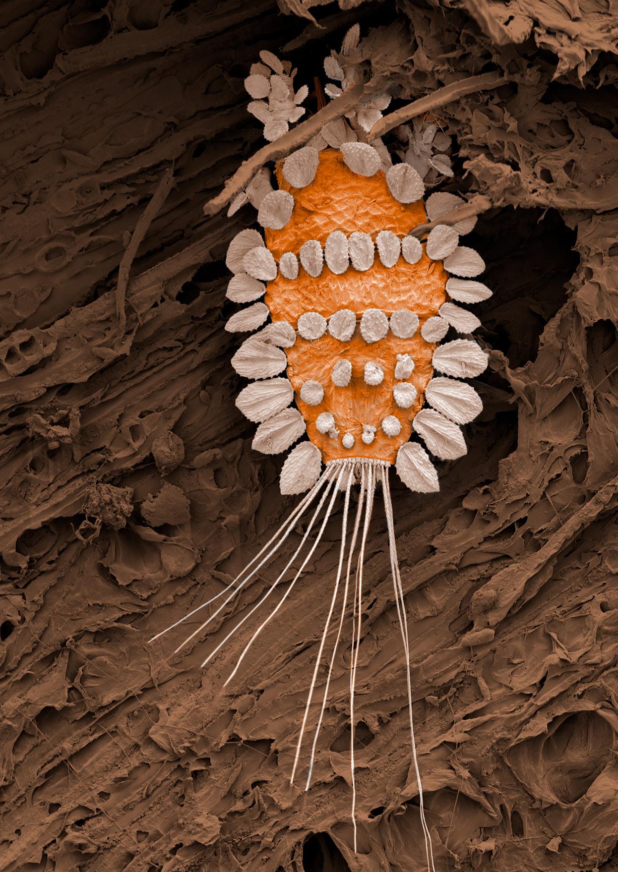
Above: A peacock mite on a branch. A significant pest in some regions, the name is evoked by their feather-like bristles or setae which can be used against predators. IMAGE BY RIA REBSTOCK Scientist, Postharvest Fresh Foods
IMAGES CAPTURED WITHIN THE PLANT & FOOD RESEARCH MICROSCOPY LABORATORY. CURATED AND HAND-COLOURED BY PLANT & FOOD RESEARCH PHOTOGRAPHER WARA BULLÔT.
Advertisement
The images in the following pages have been taken using a scanning electron microscope (SEM) as part of Plant & Food Research projects over the last 30 years. By scanning the surface of an object with a focused beam of electrons, SEMs can produce high-resolution images, at magnifications much greater than those captured with a light microscope. Specimens usually require special preparation to preserve or stabilise and increase electrical conductivity, prior to imaging.
SEM images have an intriguing 3D appearance. The original images are captured in grayscale but colour is often added to enhance their appearance and to make the images more visually appealing, as has been done in these photographs. In these images, hand colouring transforms seldom-seen views captured for science into images of curiosity and beauty.

A pollen grain from the common purple-fruited passionfruit variety.
IMAGE BY DR IAN HALLETT Team Leader, Microscopy & Cell Walls
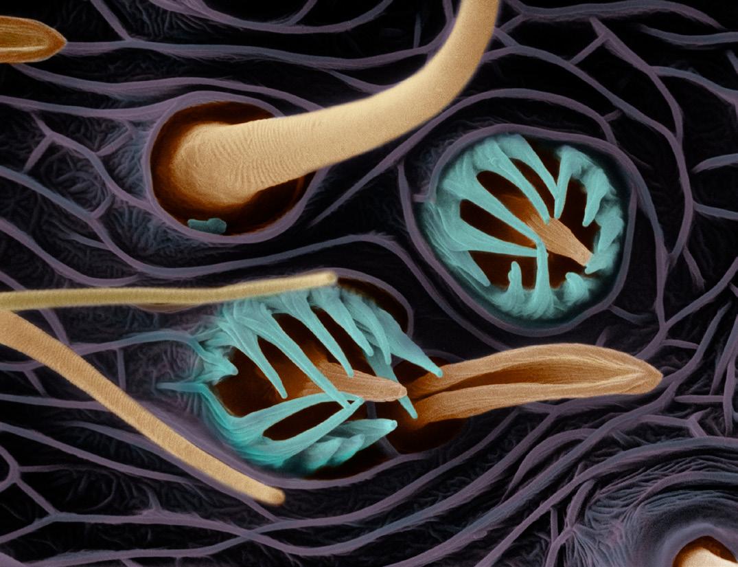
Detail of an antenna from a lightbrown apple moth. The wavy design (background) is the cuticle. Shown in orange are sensory hairs, called sensilla. Volatile odorants enter these from the air and these make up the insect’s nose. The aqua-coloured parts are protective hairs found around sensilla pits.
IMAGE BY DR MELISSA JORDAN Team Leader, Molecular Sensing

The reproductive spores of a rust fungus. Image taken as part of plant pathology research. The mass of spores on an infected plant appear as rust-like patches. When conditions are favourable, the spores can germinate to cause additional infections and spread the disease.
IMAGE BY DR IAN HALLETT Team Leader, Microscopy & Cell Walls

Detail of a developing kiwifruit flower, showing the pollenbearing anthers (cream) and the inner stigmatic whorl (centre).
IMAGE BY MIDORI JONES Previously Research Associate at Plant & Food Research

The detail on the head of a weevil (Pleosporius bullatus). The image shows the compound eyes, coloured olive green. The hexagonal facets are each a lens that focuses light on a receptor. The antenna and mouthparts are visible in forest green. Insect setae (hairs) can be sensitive to movements or chemicals (smell and taste).
IMAGE BY DR IAN HALLETT Team Leader, Microscopy & Cell Walls

A gorse thrip image taken as part of a study into whether thrips can distribute spores or pollen. Gorse thrips were introduced into New Zealand from Europe as a means of biological control of the gorse weed.
IMAGE BY DR IAN HALLETT Team Leader, Microscopy & Cell Walls
