






Editor-in-Chief
Douglas B. Berkey, DMD, MPH, MS
Senior Publisher
Annie Zhao
Journal Manager
Sangamithrai S
Abstract Writer
Elaine Steinborn
© March 2026, Elsevier Inc. All rights reserved.
No part of this publication may be reproduced, stored in a retrieval system, or transmitted, in any form or by any means—electronic, mechanical, photocopying, recording, or otherwise—without prior written permission from the publisher.
Publication Information Dental Abstracts (ISSN 0011-8486) is published bimonthly by Elsevier Inc.,1600 John F. Kennedy Boulevard, Suite1818 Market Street, Philadelphia, PA 19103, United States. Months of publication are January, March, May, July, September, and November.
Customer Service Office: 11830 Westline Industrial Drive, St. Louis, MO 63146.
Periodicals postage paid at New York, NY, and additional mailing offices. Annual subscription rates for 2018 (domestic): $144.00 for individuals and $71.00 for students; and (international) $194.00 for individuals and $105.00 for students.
USA POSTMASTER: Send address changes to Dental Abstracts, Elsevier Health Sciences Division, Subscription Customer Service, 3251 Riverport Lane, Maryland Heights, MO 63043.
Copyright 2026 by Elsevier Inc. All rights reserved. Dental Abstracts is a trademark of Elsevier Inc. Dental Abstracts is a literature survey service providing abstracts of articles published in the professional literature. Every effort is made to ensure the accuracy of the information presented in these pages. Statements and opinions expressed in the articles and communications herein are those of the author(s) and not necessarily those of the Editors or the publisher. The Editors and the publisher disclaim any responsibility or liability for such material. Mention of specific products within this publication does not constitute endorsement.
All inquiries regarding journal subscriptions, including claims and payments, should be addressed to: Elsevier Health Sciences Division, Subscription Customer Service, 3251 Riverport Lane, Maryland Heights, MO 63043. Tel: 1-800-6542452 (U.S. and Canada); 314-447-8871 (outside U.S. and Canada). Fax: 314447-8029. E-mail: support@elsevier.com (for print support); support@elsevier.com (for online support).
Notice: Journals published by Elsevier comply with applicable product safety requirements. For any product safety concerns or queries, please contact our authorized representative, Elsevier B.V., at productsafety@elsevier.com or Radarweg 29, 1043 NX Amsterdam, Netherlands.
Practitioners and researchers must always rely on their own experience and knowledge in evaluating and using any information, methods, compounds or experiments contained herein. Health care practitioners must exercise their professional judgement and make all treatment-related decisions based solely on the specific conditions of each patient. Because of rapid advances in the medical sciences, independent verification of diagnoses and drug dosages should always be made. The content is provided “as-is” and Elsevier makes no representations or warranties, whether express or implied, as to the accuracy, completeness, or adequacy of any content. To the fullest extent permitted by law, Elsevier assumes no responsibility for any damages, adverse events, or liability arising from use of information contained herein including for any injury and/or damage to persons or property, whether as a matter of product liability, negligence or otherwise.
Inclusion of any advertising material in this publication does not constitute a guarantee or endorsement of the quality or value of such product or service or of any of the representations or claims made by the advertiser.
Printed by Sheridan at 450 Fame Avenue, Hanover, PA 17331

DentalAdvance .org is the gateway offering high-quality research, news, jobs and more for the global community of dental professionals.
Journal profiles with quick links to Tables of Contents, author submission information, and subscription details
Important information and valuable resources on how to submit a journal article
Dentistry Articles in Press from participating journals
Quick links to the leading dentistry societies worldwide Dentistry News from Elsevier Global Medical News (formerly IMNG)
Dentistry Jobs powered by ElsevierHealthCareers.com
Editor-in-Chief
DouglasB.Berkey,DMD,MPH,MS ProfessorEmeritus, SchoolofDentalMedicine, UniversityofColorado, Aurora,Colorado
AssociateEditor
DanielM.Castagna,DDS AssociateProfessor,DepartmentofPreventiveandRestorativeDentistry, UniversityofthePacific,ArthurA.DugoniSchoolofDentistry, SanFrancisco,California
P.MarkBartold,DDSc,PhD,FRACDS(Perio) ProfessorEmeritus SchoolofDentistry, UniversityofAdelaide Adelaide,Australia
RobBerg,DDS,MPH,MS,MA ProfessorandChair, DepartmentofAppliedDentistry, UniversityofColoradoSchoolof DentalMedicine, Aurora,Colorado
TylerH.Berkey,DMD GeneralDentist Aurora,Colorado
FionaM.Collins,BDS,MBA,MA ConsultantandEditor, GeneralDentist Longmont,Colorado
AnthonyJ.DiAngelis,DMD,MPH ChiefofDentistry, HennepinCountyMedicalCenter, Professor,UniversityofMinnesota, SchoolofDentistry, Minneapolis,Minnesota
RaulI.Garcia,DMD,MMedSc ProfessorandChairman, DepartmentofHealthPolicyandHealthServicesResearch, BostonUniversitySchoolofDentalMedicine, Boston,Massachusetts
MichaelSchafhauser,DDS GeneralDentist, St.Paul,Minnesota
JoeVerco,DClinDent PaediatricDentist NorthAdelaide,Australia

MARCH/APRIL2026
VOL.71 No.2
DryNeedling108
Treatingtemporomandibularjointpainwithdry needling
E-CigarettesandPeriodontalDisease109
Evaluatingtherisksandbene fitsofe-cigarettes
EndodonticMaterials112
Evidenceforendodonticintracanalmedicamentsand agents
Gagging114
Managingasensitivegagre flex
GlassionomerConditioners117
Cavityconditionersforglassionomerrestorations
LocalAnesthesia119
Intraligamentaryinjectionversusincisivenerveblock
ManagingBleedingDisorders120
Bleedingdisorderofunknowncause
Mouthwashes122
Adviceforcliniciansregardingmouthwashes Non-dentalToothaches123 CausesanddifferentialdiagnosesofNOTAs PediatricDentalTrauma127 Epidemiologyandmanagementofdentaltraumain children
RecordKeeping129
Updatingdentalrecordkeeping
ThirdMolars132 Evidenceforandagainstextraction Inquiry
AntibioticResistance/OdontogenicInfections135
Needforantibioticresistancemeasuresrelatedto odontogenicinfections
BurningMouthSyndrome136
Approachestomanagingburningmouthsyndrome CariesandGutMicrobiota138
Causalrelationshipsbetweengutmicrobiotaandcaries
DeepCaries/CavityLiners139 Efficacyofcavityliners
DentalImplants140
Patientsatisfactionratingsofsingleimplants
Consecutivedentalimplantreplacements
OcclusalReduction/Pain144
Effectofocclusalreductiononpainafterendodontic therapy
OdontogenicSinusPathologies144
Connectionbetweenmaxillarysinuspathologyand dental findings
OralIrrigators146
Managingperi-implantdiseases
Periodontal/SystemicDisease148
SRPplusadjuvanttherapiesforperiodontal/diabetic care
TinnitusandTMJ149
TMDandtinnitusconnections
ToothBleaching150
Externaldentalbleachinggels
Findingtherightamountandqualityofsleep
Sleeptrumpsdiet,exercise,andsocialtiesforliving longer
Notes
TofacilitatetheuseofDentalAbstractsas areferencetool,allillustrationsandtablesincluded inthispublicationarenowidentifiedastheyappear intheoriginalarticle.Thischangeismeanttohelp thereaderrecognizethatanyillustrationortable appearinginDentalAbstractsmaybeonlyoneof manyintheoriginalarticle.Forthisreason, figure andtablenumberswilloftenappeartobeoutof sequencewithinDentalAbstracts.
StandardAbbreviations
Thefollowingtermsareabbreviated:acquiredimmunodeficiencysyndrome(AIDS),humanimmunodeficiency virus(HIV),andtemporomandibularjoint(TMJ).
Between 70% and 90% of the population have experienced at least 1 traumatic event in their lives and respond to the event as a trauma. Even relatively common life effects can be traumatic, including illness, bullying, loss of a loved one, dysfunction in the family, and medical or dental care. Oral health care providers should be prepared to deal with individuals suffering trauma knowingly or unknowingly and should understand that they can also be affected by the traumatic history. A need for oral health care and the experience of orofacial injuries can occur with domestic violence, child abuse, or other physical events. Some patients experiencing trauma tend to avoid preventive or curative treatments to avoid having flashbacks to the event. Triggers for trauma can include a white coat, sharp and metallic instruments, bright head lights, or physical closeness with the oral health practitioner. Among the reactions to these triggers are dental fear and anxiety, gagging, comorbid mental illnesses, and disruption in body systems such as chronic inflammation. Even the patient’s common trauma-coping strategies, such as smoking, substance use, and poor diet, can lead to long-term negative oral and overall health experiences. Trust and self-regulation can suffer, which can also compromise the quality of care and treatment success. Practitioners may also suffer from secondary traumatic distress, leading to chronic fatigue, disturbing thoughts, poor concentration, emotional detachment, avoidance, and physical illness. Lasting adverse psychological damage can result. A trauma-informed care model was developed to incorporate an understanding of trauma into all care provision practices. However, oral health care practitioners have not widely implemented traumainformed care compared to emergency medicine, pediatrics, and nursing professionals. Trauma for oral health care was defined and a patient care approach for interactions, decisions, and treatments provided by oral health care providers was outlined.
Misunderstandings regarding the meaning of trauma-informed care in oral health care arise from the view that the treatment of orofacial injuries or patients who have experienced an adverse dental encounter is what is meant. Dissecting the terminology
can help to more accurately define what the name means in a particular framework. Trauma is defined as experiencing actual or threatened death, injury, or sexual violence by psychiatric physicians, whereas medicine focuses on physical injuries or the individual’s psychologic response to events or injuries, and behavioral science is limited to an emotional response to a serious adverse event. The Substance Abuse and Mental Health Service Administration (SAMHSA) states that trauma can result from events or circumstances an individual experiences as physically or emotionally harmful or life-threatening and that the experience creates adverse effects on an individual’s ability to function and maintain mental, physical, social, emotional, or spiritual wellbeing. Even though this broadens the elements, it focuses on what trauma results from and not what it is.
In trauma-informed care for oral health, the 3 Es of trauma adopted by SAMHSA and the recognition that trauma is an integrated response of the entire person are essential (Table 1). The 3 Es are traumatic Events, the individual’s Experience of the event as traumatic, and the lasting adverse Effects of the trauma. Trauma is a personalized experience of the event that actually produces or threatens physical or psychological harm. The person’s interpretation of the event is influenced by many factors, such as cultural beliefs, life history, resilience, guilt, sense of responsibility, availability of social support, and developmental stage of growth. The adverse effects can affect the individual’s mental, physical, social, emotional, and/or spiritual stability. Effects can be immediate or delayed.
A succinct definition of trauma in trauma-informed care situations is a patient’s personal response to distressing life effects that carries lasting adverse effects on all aspects of his or her life. This includes effects on oral health and the ability or desire to access oral health care.
An overarching patient care approach allows oral health care providers to access information on interactions, decisions, and
Table 1. Definition of Trauma in Trauma-informed Care for Oral Health. Trauma is a Patient’s Personal Response to Experiencing Distressing Events Which Has Lasting Adverse Effects on All Aspects of Their Life, Including Oral Health and Access to Oral Healthcare
Actual or threat of physical or psychological harm
• Personal and subjective response which determines whether the event is traumatic or not
• Contributed by how the individual labels and assigns meaning to and is disrupted physically and psychologically by the event
• Influenced by a range of factors such as cultural beliefs, life history, resilience, guilt, responsibility the individual imposes on the event, availability of social support and the developmental stage
• Lasting adverse effects on the individual’s functioning and mental, physical, social, emotional, or spiritual wellbeing
• Can be an immediate or delayed onset, e.g., fear, anxiety, helplessness, horror, inability to cope with normal stresses and trust others
Oral health impacts of trauma:
• Avoidance of preventive treatments and frequent reschedules, missed and cancelled appointments
• Various and common oral health settings can trigger traumatic memories and re-traumatise the patients
• Dental fear and anxiety
• Increased gagging
• Complex medical history, mental illnesses and chronic inflammation
• Trauma coping strategies such as smoking, substance use and poor diet can increase the risk of periodontal diseases, dental caries and oral infections
• Difficult to trust and self-regulate, compromising the therapeutic relationship, quality of care and treatment outcome
• Oral health practitioners treating patients with trauma history can suffer from secondary traumatic distress
(Courtesy of Choi KS, Sohn W, Leadbeatter D: Safe haven – a trauma-informed care model for oral health practitioners: A commentary. Br Dent J 239:717-721, 2025.)
treatments. SAMHSA’s 4 key competencies are used in a care model (Figure 1) with the goal of protecting everyone involved in care directed at adverse impacts and potential retraumatization, as follows:
• Realize the widespread impact of trauma
• Recognize trauma signs and symptoms
• Respond using trauma-informed care principles
• Resist re-traumatization
The oral health care practitioner must realize the widespread occurrence of trauma and its perverse impact. Oral health care practitioners are highly likely to encounter patients who have been traumatized, so they should be aware that everyone delivering care can be affected by the trauma.
The dental staff should undergo continuing education, learning to recognize the signs and symptoms of trauma and updating as indicated. Challenging behaviors from patients with trauma can be diverse and complex, so the practitioner should be sensitive to the possibility that the individuals can manifest uniquely.
The response to a patient includes incorporating traumainformed care principles. These principles are safety, trustworthiness, choice collaboration, and empowerment, all of which should be incorporated into every aspect of care from the first interaction onward. Patients should be asked what would
make them more comfortable, and they should be informed about what will happen next. The practitioner should employ nonjudgmental empathetic communication skills, ask permission to touch or examine, and practice show-and-tell techniques. It is important to provide the patient with choices, collaborate with him or her, extend the appointment time so rapport can be established, and involve the patient in the treatment planning process. Practitioners should share findings, options, and recommendations rather than dictate and take ownership and control. The patient can be asked if he or she would share the trauma experience, with this information received without discrimination or bias.
To resist re-traumatization, the practitioner must continue to learn about situations that can be triggering of post-traumatic memories and may cause the patient distress. The principles of trauma-informed care should be incorporated throughout the patient encounter.
Just as infection control is a standardized practice in oral health care for all patients, trauma-informed care is recommended as a standard or universal precaution when treating patients. This creates a sense of safety and is unlikely to trigger traumatization memories. Some patients will require more attention because they are at risk for exposure to traumatic events. Groups that
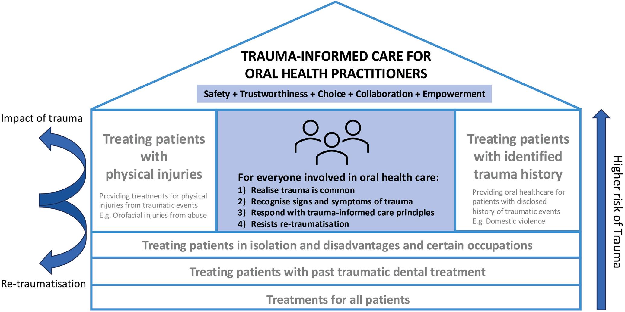
Figure 1. Safe haven – trauma-informed care for oral health. (Courtesy of Choi KS, Sohn W, Leadbeatter D: Safe haven–a trauma-informed care model for oral health practitioners: A commentary. Br Dent J 239:717-721, 2025)
should be considered for these enhanced efforts include minority ethnic groups, women, children and young adults, refugees, First Nations people, and those in certain occupations, such as emergency services. Patients shouldn’t feel the need to share their trauma history with the health care practitioner or be required to make connections between trauma and current challenges. Regardless of risk, history, or disclosure of trauma, trauma-informed care practices should be implemented routinely.
When practitioners demonstrate an understanding of trauma and its effects on their patients, they are better able to interact in ways that create a safe and healing environment for the delivery of oral health care. The goal should be to avoid unnecessary stress and prevent the re-traumatization of patients.
Oral health care practitioners should become familiar with the definition of trauma and how its effects can alter all aspects of life for patients. Being equipped to identify traumatic responses, addressing them with shared decision-making and treatment planning, and avoiding behaviors or situations that could trigger traumatic memories will make the interactions with patients more conducive to the successful delivery of oral health care.
Choi KS, Sohn W, Leadbeatter D: Safe haven – a traumainformed care model for oral health practitioners: A commentary. Br Dent J 239:687-692, 2025
Reprints available from KS Choi, School of Dentistry, Faculty of Medicine and Health, Univ of Sydney, Australia; e-mail: kristy.choi@sydney.edu.au
Dental hygienists are sensing changes in their occupation, noting faster schedules, greater demands, and an increased emphasis on business metrics rather than the human side of dentistry. These changes are leading to a loss of joy and meaning as dentistry becomes stretched between providing care and making good business choices. It's possible to manage these changes in the work environment without losing the vital belief in oneself by focusing on the essentials of recognition, respect, and recovery, with a subsequent sense of reconnection.
Practicing dental hygiene demands precision, compassion, and consistency with each patient and every day. Balancing these technical and human elements can drain the emotional and physical reserves of hygienists, with many developing a feeling of detachment from the work, persistent pain and fatigue, mental fog and irritability, diminished compassion, a sense of being unseen and undervalued, losing one's professional identity, and isolation. These stresses accumulate over time. If they aren't addressed, they can erode the sense of joy and pride in one's work.
The other major component hygienists must face is the evolution of dentistry into a highly commercialized industry. Decisions are based on profitability rather than relationships or outcomes. When financial goals consistently take precedence over clinical integrity, the loss is felt by the entire dental staff. Because they deal with prevention and education, dental hygienists feel the disconnect most acutely, since these elements don't show immediate returns on the balance sheet. To preserve professional integrity and emotional wellbeing in such an environment demands that the hygienist recognize his or her worth, stand in authority, and undertake self-care along with patient care.
Even if the system appears not to show it, the dental hygienist must know his or her worth. Recognition means being seen
for the true value brought, which begins with a self-awareness of that value.
Results often matter to the owner in a patient-centered practice because that individual can see oral health and business success as connected entities. Corporate settings may be less likely to consider individual value. Success in a corporate practice is generally based on profitability and not prevention, relationships, or long-term health outcomes. Hygienists can become discouraged because they see the difference they make in each patient's health, the patients who now accept dental care, measures that guide better health practices, and other results that can't be put on a balance sheet.
The hygienist can build evidence supporting a sense of purpose and pride through the following.
• Track improvements in patients' oral health.
• Keep brief records or care notes on transformations witnessed.
• Document success stories with photographs or narrative notes (with patient consent).
• Work with peers to share observations, compare outcomes, or develop case studies.
• Engage with research through professional associations or journals.
Corporations are unlikely to reward these efforts but they document the hygienist's connection to the meaning of the profession. Validation is about self-acknowledgment from within.
Respect isn't arrogance but rather ownership of one's professional expertise. As a licensed health care provider, the hygienist makes clinical decisions, accepts ethical responsibility for patient care, and has earned respect from the dental team, the leaders, and oneself. However, in some practices, hygienists are micromanaged, rushed, or considered just revenue producers rather than clinical professionals.
If the hygienist isn't being treated with respect, if his or her voice is silenced, or if ethical boundaries are stretched, it's time to reevaluate and consider how to build the mindset and confidence to set things back on course. Some activities to engage in are as follows.
• Reclaim authority through knowledge of the hygienist scope of practice, ethical obligations, and rights as a clinician.
• Communicate clearly, using assertive, professional language.
• Build credibility through continued learning, seeking advanced certifications, specialty training, or leadership courses to build skills and self-assurance.
• Find a support network, connecting with mentors, local associations, and online groups who can share their journey and reveal options.
• Set boundaries and maintain them to establish an ethical, sustainable practice.
If respect isn't earned, the hygienist should leave the practice and look for a situation where professionalism is valued. It's important that the workplace and the values of the hygienist align and honor his or her contribution.
The practice of dental hygiene is physically intense, emotionally demanding, and cognitively exhausting. Practitioners need to understand how essential self-care is. Ignoring recovery makes one vulnerable to injury, additional fatigue, irritability, and disengagement. Recovery is the way to maintain a meaningful career. It involves physical recovery, emotional and mental recovery, and professional recovery.
Over 85% of hygienists report musculoskeletal pain and almost half consider it moderate to severe. The repetitive strain, static posture, and fine motor control required to make hygienists excellent at their work also makes them vulnerable to injury. Hygienists need to see physical discomfort as a red flag rather than just normal. Prioritizing physical recovery can involve the following.
• Take microbreaks.
• Engage in movement and not stillness.
• Use ergonomic loupes, saddle stools, and adjustable lighting to reduce strain.
• Engage in therapeutic maintenance activities such as massage, physical therapy, or yoga.
Each day is filled with opportunities to manage patient anxiety, guilt, fear, and anger while remaining calm, empathetic, and professional. An emotional toll is the result. Over time, unprocessed emotions can build up and cause irritability, detachment, or compassion fatigue. The way to manage the situation is to engage
in debriefing, which allows the hygienist to recover emotionally and mentally.
In debriefing, the hygienist intentionally acknowledges the experiences, feelings, and lessons learned before moving on. This structured pause closes the loop on the day's events so the stress doesn't linger in the mind or body. Debriefing can be a personal or a team event.
A personal debrief is a reflective practice for self-repair and is done in private. It may be done in a brief 5 minutes and requires no formal format. The hygienist needs to give his or her emotions a place to go rather than remain and build up. Some examples are as follows.
• End-of-day journaling
• Voice notes during a commute
• Quick reset between patients
• Weekly reflection questions.
The goal is to validate one's experience and not analyze everything. Hygienists are trained to remain calm, but debriefing offers the opportunity to be human.
Team debriefs normalize reflection in the workplace. They cultivate a practice culture where it's safe to talk about difficult cases, emotional experiences, or challenges and there is no fear of judgment. Some practices incorporate them into morning huddles or at day's end, and others may make them a weekly gathering. Debriefs may be structured around questions such as “What went really well with patients today?” “Were there any difficult moments we can learn from?” or “What support was given or needed today?”
Taking time to talk honestly replaces silent emotional turmoil with shared understanding. This can prevent the development of resentment, improve communication, and build trust. During dental training, students should be taught how to debrief in preparation for real-world emotional challenges. This skill can be considered a professional growth tool.
Debriefing allows the brain to switch from reaction into integration, letting the nervous system settle and reducing cortisol and adrenaline accumulation during stress. Rather than carry challenges, it encourages learning from them.
Recovery of one's sense of purpose requires stepping back to realign values and goals. Operating in a fast-paced, profit-driven environment for extended periods of time can cause the loss of a clear understanding of why one does this job. Professional recovery relies on the following.
• Reflection
• Refreshing skills
• Reassessment of the environment
• Reconnection with peers
Mindset Shift
No one can sustain high-quality care when they are in a depleted state. Recovery can begin when the hygienist stops viewing rest, reflection, or boundaries as signs of weakness and begins to see them as hallmarks of professional maturity. This involves a shift in one's mindset that realigns how one views endurance and sustainability, pausing, reframing, what professional identity is outside of production, and how strength should be redefined.
Dental hygiene students should be taught as part of their curriculum how to recover from physical, emotional and mental, and professional strain related to providing care to patients. Clinicians need to be able to value both precision and preservation and prioritize sustainability over endurance. Longevity in the dental hygiene field depends on having inner equilibrium. Taking control of one's recovery and emotional balance should be considered part of being an excellent dental hygiene practitioner as well as a wise person.
Bromage L: Recognition. Respect. Recovery. Reconnection. BDJ Team 12:452-456, 2025
Reprints not available
When it comes time to sell a dental practice, a consulting agreement may help with the transition. Each situation will be different, but it’s important to understand what a consulting agreement is, how it will benefit sellers and buyers, and its influence on taxes.
In a consulting agreement, the seller makes himself or herself available to the buyer during the transition period. Some possible ways the seller can help are coaching the buyer on patient treatment plans, handling human resources issues with the dental staff, providing guidance regarding business matters and processes, or simply answering questions as they arise in the transition period. When the purchase price is agreed upon, it must be allocated among the various assets that are sold or purchased. A consulting agreement can also be used with a stock or equity interest structure.
In most asset acquisition structures for the sale of a dental practice, the seller and buyer may not only agree on the price of the various assets but also on the allocation of the money. If the price
is $500,000, a typical example is $25,000 for dental supplies, small tools, and instruments on hand; $175,000 for tangible assets such as furniture, equipment, and computers; and $300,000 for goodwill. The buyer may also suggest a different allocation that may lower goodwill and increase allocations to the other entities, or insert a consulting agreement to absorb some of the price allocation. Depending on the state where the sale takes place, the bulk sales tax may focus on tangible assets and may be due from the buyer at closing. If the tangible assets allocation is $175,000 and the state bulk sales tax is 6%, the buyer must spend an immediate cash outlay of $10,500 at closing from his or her working capital funds. If the allocation is reduced to $100,000, the buyer would only owe $6000, which immediately saves the buyer an outlay of $4500, something a new dentist could certainly appreciate.
Many sellers and their advisors object to the consulting agreement allocation of $75,000 because it would be taxed at ordinary income rates rather than at long-term capital gains rates, which are generally lower. However, a consulting income of $75,000 might generate a greater tax savings than that obtained with long-term capital gains rates. The allocation is considered consulting income, so the seller continues to have business income for their business tax return, usually as an S corporation. He or she may also have ongoing business expenses to apply
against the $75,000 income, including business travel, business use of a vehicle, continuing education fees, dues and subscriptions, licensing, business meals, office supplies, furniture, computer equipment, etc. These legitimate business deductions would reduce the seller’s income. If these expenses total $15,000 and the seller also has $20,000 of health insurance premiums for the year that can be deducted, the $35,000 expenses shelter some of the $75,000 and leave a taxable income of $40,000. An ordinary tax rate of 40% on this income would be $16,000, or less than the $18,750 tax on $75,000 at a longterm capital gains rate of 25%.
Retirement Plans
If sellers retain a 401(k) retirement plan or implements a similar type of retirement plan to replace one they had, they may pay themselves wages of $30,000 and defer $25,000 so only $5000 remains taxable income. An additional profit-sharing contribution of $5000 may also be claimed. Their $40,000 minus $30,000 of wages minus $5000 of profit-sharing contribution yields an S corporation income of $5000. They only pay ordinary taxes on $10,000, with retirement plan contributions deferred. Rather than paying $18,750 of tax on $75,000 at the 25% long-term capital gains tax rate, they only pay $4000. Even combined with the payroll tax cost on wages and the taxes on deferred income, the eventual cost is just $16,000 rather than $18,750. This is a conservative scenario. With a savvy taxpayer and advisor
with higher consulting income, the income can be spread over at least 2 years and result in greater savings.
A hybrid sale structure may include part stock, part personal goodwill, and part consulting payment. This structure can similarly shelter consulting payment into a retirement plan. Using an existing or new cash balance or defined benefit plan may permit the deferral of even more income into the future at a lower tax rate.
Sellers and buyers should ask their advisors about the concept of a consulting agreement as part of the practice transition. If the seller takes back a seller note, consulting agreements may also be a part of the exchange. Sellers, buyers, and their advisors should explore the pros and cons and the costs and savings that can be realized with these approaches.
Reprints not available
Currently, the dental mergers and acquisitions (M&A) market is active. There seem to be more entrepreneurial dentists, more practices ready for transition, consolidations related to dental support organizations (DSOs) and multi-practice owners, more focus on maximizing efficiency through managing fixed overhead expenses, and the availability of bank financing and other financial aids for purchasers. Several components contribute to a successful M&A transaction and can prepare buyers and sellers to avoid pitfalls that can cause these transactions to fail.
Letter of Intent
A letter of intent (LOI) is a nonbinding offer to purchase a practice and usually includes high-level deal terms along with specific key terms. A nonbinding LOI sets the expectations of the parties
yet,
regarding purchase price, closing date, payment structures, employment, and holdbacks and escrows, among items. The letter should be negotiated and signed before the definitive deal documents are drawn up to save time and money.
An LOI may contain some binding clauses. For example, “noshop” and “exclusivity” clauses can prevent parties from seeking other deals while negotiating the current deal. “Confidentiality” provisions help to protect the parties from unauthorized disclosures of confidential information. These provisions are meant to protect the parties during negotiations.
If a seller leases the practice property, the buyer must assume the lease, which usually means the parties must obtain the landlord's consent to assign the lease to the buyer. Any leases should be reviewed early in the process to ensure the landlord's rights are clear, the requirements for personal or corporate
guarantees are considered, and whether the landlord or lease terms impose unreasonable demands for lease assignment. Landlords can request a review of the buyer's financials before consenting to assignment, which can add time to the process. The lease should be addressed early to avoid having problems with lease assignment.
Because reputation matters, both buyer and seller should be aware of the publicly available information that exists about themselves. This includes researching Google, ChatGPT, and social media sites and may stretch to background checks. Sellers often conduct their own research on buyers so they understand what they're getting into. It's important to be truthful and not lie about potentially damaging publicity.
Having more than 1 practice or multiple locations can complicate the transaction, especially if not all are for sale. Thorough financial due diligence is needed to understand the financial health of the practice being sold and ensure that the revenue report the buyer gets is only derived from the practice being purchased. Dental practices may also have cross-collateralized debts across multiple practices, so the assets of a practice being sold could be encumbered by loans to practices that aren't included in the sale.
If the dentist has 2 practices in close proximity but is only selling 1, the post-closing covenants that are agreed to can be compromised. The buyer should have proper non-solicitation provisions to prevent patients, referrals, and employees from moving to a practice not in the sale. Non-compete provisions must meet the needs of both parties.
Generally the seller will continue to operate the practice through closing, which means that outstanding accounts receivable (A/R) and patient credit balances will exist. These must be allocated to either the buyer or the seller. Sometimes they are considered part of the transaction and other times they are excluded. If they are considered an excluded asset, the parties must decide who will collect the A/R after closing and for how long. Buyers may charge a collection fee to collect and remand the outstanding A/R to the seller. In addition, if the collected A/ R is applied on a last in, first out (LIFO) basis, it will be applied to the newest invoice and likely paid to the buyer. If it is applied on a first in, first out (FIFO) basis, it will likely be applied to the oldest invoice and will go to the seller.
Any patient credits are a liability for the selling practice. The buyer won't want to assume this liability and often requires that the purchase price be reduced by the patient credits owed at closing.
The purchase price is allocated among different types of assets for tax purposes. Seven different asset classes have been developed by the Internal Revenue Service. The purchase price allocation can greatly impact the amount of tax each party owes. Sellers want to allocate as much of the purchase price toward goodwill as possible because that is taxed at long-term capital gains rates. Buyers usually want to allocate as much as possible to tangible assets because they can be depreciated quickly. If the seller has already fully depreciated the assets, the amount allocated to physical assets will be taxed at ordinary income tax rates. Working with an accountant is recommended to determine the most appropriate and tax-efficient allocations.
A restrictive covenant limits the seller's conduct after closing. The 4 restrictive covenants that are most commonly used in dental situations are confidentiality, non-disparagement, noncompetition, and non-solicitation. Non-competition covenants differ by state, but most restrictive covenants are enforceable in the context of a business sale. The guiding principle with respect to enforceability is that the restrictive covenant must be reasonable in terms of time and geographic scope. Five-year restrictive covenants are usually enforceable in dental sales. Employment agreement restrictions are usually for just 1 to 2 years, but also differ by state. Location is a significant component when determining if the geographic scope is reasonably restrictive, with limits of blocks in population-dense cities but 10 to 50 miles in rural areas.
Generally the buyer of a dental practice will purchase the assets of the practice rather than the seller because the buyer doesn't want to assume any risk of liability associated with the seller's entity. In this purchase, the buyer can choose which practice liabilities he or she will assume after closing, but buyers can also have successor liability simply because they purchased the operation of the practice.
Buyers can protect themselves against successor liability by conducting due diligence on the practice using their own accountant. The accountant will review all financial records, inspect equipment, interview staff members about how the operations work and whether they are in compliance with applicable laws, audit the charts and billing records, review insurance policies, and require the seller to obtain tail insurance coverage, which extends the reporting coverage period.
Another protective measure against post-closing liability is including a robust indemnification provision, requiring the seller to reimburse the buyer for any out-of-pocket expenses incurred as a result of the seller's pre-closing actions or misrepresentations. Having a good M&A attorney can protect by obtaining a
tax clearance letter, including a setoff against amounts owed to the seller in the purchase agreement. It may also be possible to structure the purchase price payments over a period of time to guard against future liability.
Consulting with an experienced accountant or tax lawyer is an important step to take before engaging in any transaction. This can ensure tax efficiency. Several decisions in the M&A process can affect tax liability, including choice of entity formation, purchase price allocation, employment status, and rollover equity.
In buying into a practice or selling a practice that results in the individual owning equity in the buyer entity after closing, it's vital to understand the rights and limitations associated with that equity. The partnership agreement should be clear in spelling out the situation. Among the provisions the dentist and attorney should review when making a partnership agreement are voting rights, management rights, veto powers for minority partners, fees paid to managers, management responsibilities, clinical
responsibilities, work hours and compensation, buy/sell terms, drag-along rights, minimum standards to retain equity, restrictive covenants, and the corporate opportunity doctrine.
Buying, selling, and merging practices can present many opportunities for the dentist to learn about the financial details of these activities. Hiring an experienced dental attorney and financial manager who is knowledgeable about these activities is an important first step once you feel the need or desire to engage in these transactions.
Gocel C, Rhone J: Dental transactions through the looking glass. Dentaltown 26:36-38, 2025
Reprints not available
Many practices in the United States suffer from a poor working relationship between general dentists and hygienists. The problems that exist from the dentist’s and the hygienist’s perspective were outlined, including the management of periodontal disease in general practice, wages and preferred provider organization (PPO) reimbursements, hygienist shortages and retention, and the office culture. Possible approaches to address the situation were suggested.
Dentists and hygienists differ in their view of how periodontal disease should be managed in a general practice. Dentists complain about hygienists who don’t recommend scaling and root planing (SRP) to avoid offending patients or whose bloody prophylaxis is labeled maintenance. Some dentists simply refer patients to periodontists to avoid any drama. Periodontists and hygienists tend to be more alike in their practices than dentists and hygienists. Hygienists believe a healthy periodontium is foundational for restorative work and support the use of complete charting and documenting criteria before beginning any major restorative work.
Agreement among groups of dentists and hygienists was found for instituting written, co-developed protocols that set forth clear expectations for probing, staging, SRP indications, and referral thresholds. If few offices take the time to institute these protocols, the dentist and hygienist will remain misaligned, patients will be confused, and tensions can increase.
After the COVID-19 pandemic, hygienists have been able to command higher wages, with $65 to $100 per hour not uncommon in urban markets. However, insurance reimbursements have not kept up, and dentists experience pressure based on staffing costs versus stagnant production.
Most dentists depend on hygiene to drive examinations and restorative work, but lose money on the hygiene work. Hygienists push back, arguing that they produce well above their hourly rate. It’s been pointed out that in well-run offices, raises and bonuses come without ultimatums, building loyalty and performance. Some hygienists would prefer an hourly rate of pay to avoid the stress of no-shows that might erase a day’s paycheck.
Others prefer commissions that are structured fairly and agreed upon upfront. In contrast, dentists have suggested eliminating the role of hygienists and using assisted hygiene models or performing cleanings themselves. However, most would agree that this isn’t sustainable at scale.
The shortage of hygienists has led to expectations of higher salaries, less accountability, and flexible schedules. Some hygienists have left clinical practice to take on temp work or educational roles. Retention has also been an issue, with some arguing that it isn’t that more hygienists are needed but that more hygienists who are willing to work 5 days a week and who care about patient outcomes are needed. Many hygienists are moving out of clinical roles because of the working conditions. Having more hygienists who choose this route won’t solve the problems of a hygienist shortage.
Hygiene instructors have been accused of fostering adversarial attitudes toward dentists and teach students to see themselves as patient advocates who need to police dentists and assistants. This view has undermined trust between hygienists and dentists.
Office culture—how teams communicate, resolve conflict, and respect each other’s roles—has been identified as a more important stressor than pay or protocols. A veteran hygienist reported having worked in great practices and toxic ones, with the difference not being money but rather whether the dentist respected the hygienist and whether the team had each other’s backs. Practices with strong leadership, open dialogue, and common expectations can lead to good relationships among the team. Those lacking this kind of foundation can have constant power struggles over timing, diagnosis, and communication style.
Some dentists believe the problem is the business model, with practices becoming too dependent on hygienists for the diagnosis, production, and patient retention in the practice. When hygienists leave or demand more, the model collapses.
Suggestions to produce solutions include the following:
• Diversify marketing and scheduling to attract more patients who need treatment, such as emergencies, new patients with decay, and surgical consultations. The diagnostic engine shouldn’t have to rely on hygiene to drive production.
• Split the general practice model to either integrate hygiene under strict systems and leadership or restructure the model to a leaner, fee-for-service, treatment-focused format.
• Hygienists and dentists could compromise to find solutions, such as allowing sufficient time for the hygienist to address the patient’s needs, co-authoring periodontal protocols, and establishing a clear pay model that aligns incentives.
The goals should be to reduce friction by establishing clear periodontal protocols and regular team calibration. Values should include fair pay, accountability, and communication. To achieve these will require strong leadership and a well-constructed and collaborative culture.
Dentists and hygienists agree that neither group can succeed by itself. They need to create structures together that honor the value of each role so that patient care can be successful. In addition, the PPO system continues to be a problem and may require radical restructuring to avoid more exits, burnout, and tension.
Why dentists and hygienists clash. Dentaltown 26:50-51, 2025
Reprints not available
There has been an estimated 8% decline in the number of actively working dental hygienists in the United States, with 40% of dental practices having a hard time filling vacant positions. As a result, patients who need care and treatment aren’t being addressed. Positive communication between hygienists
and dentists is an important aspect in the hygienist shortage and can lead to an inability to achieve positive, effective patient outcomes. Factors that influence positive and effective communication and methods to improve interprofessional communication were suggested, with the goal of achieving positive patient treatment outcomes.
The perception of the job roles of dentists and dental hygienists has an important influence on communication. Dentists and dental hygienists share job responsibilities in ways that can shape the relationship they have with one another. Often hygienists feel undervalued by the dentist, leading then to a negative perception of the dentist. If either party sees the other’s profession and skills negatively, communication can be impacted. They need to see the value of each profession and communicate positively and respectfully.
Mutual respect, communication, and understanding of the separate roles and responsibilities of the dentist and hygienist are critical to effective collaborative care. When there is conflict between health care professionals, the quality of treatment provided to patients can suffer. The conflict can lead to a lack of communication and failure to provide the patient with a collaborative treatment plan. The patient’s satisfaction level and comfort in the dental office can be affected.
A positive dentist-hygienist communication model significantly improves the patient’s and professionals’ health and reduces fear in patients. With a collaborative work approach and no dominating behavior between the dental professionals, patients feel safer. Interprofessional collaboration also allows the team to adapt to changes in oral health care, dental treatment delivery, and patient preferences. As the dentist and hygienist understand one another’s roles and services, they learn to value one another, which influences the patient’s perception. Dentists who fail to value the hygienist can fall short in delivering care. Hygienists also need to be aware of how important quality restorative services are to ensure their preventive services are successful. Without preventive services, restorative services such as implants can be compromised, with bone and gingiva unable to successfully support the restorations. A symbiotic working relationship provides successful services to the patient.
Several methods can be used to improve interprofessional communication. These include the following:
• Conduct communication and leadership seminars, courses, classes, and team-building exercises to strengthen communication and leadership skills.
• Offer courses on stress management and communication classes as part of the dental hygiene and dental school curriculum.
• Practice positive interprofessional collaboration during patient treatment and dental hygiene work.
In addition to these educational and practice changes, access to dental care can be improved by expanding the scope of practice for dental hygienists by integrating dental hygiene therapists (DHTs) into the dental practice. DHTs provide their normal preventive services but can also offer basic restorative services under the direct supervision of a dentist. Fourteen states have currently approved this position. Effective collaboration and communication between dental health care professionals could increase the acceptance of the DHT role.
Oral health care benefits when there is positive, effective communication between the dentist and hygienist. Patients are more likely to receive quality, comprehensive, and collaborative treatment, and hygienists will be more likely to be satisfied with their work and have improved mental health. Approving the DHT training and skills can add to the hygiene scope of practice.
The quality of life, job satisfaction, and mental health of the dentist and dental hygienist are intimately connected to the type of communication adopted in the dental practice. Poor communication contributes to the hygienist shortage. The hygienist experiences significant stressors every day, including uncooperative patients, fully booked schedules, practice management conflicts, and physical demands related to care delivery. These can eventually lead to burnout, reduced work satisfaction, poor quality of life, and issues with mental health that lead to hygienists leaving the profession. Job satisfaction is closely related to communication experiences with patients, colleagues, dentists, and other health care providers. Negative communication can lower motivation and job satisfaction, lead to reduced compassion, and result in poor quality of care.
Aube S: Enhancing patient treatment outcomes through effective dentist-hygienist communication. RDH 45:17-19, 2025
Reprints not available
Dental hygienists can feel that they carry too much stress and are suffering both emotionally and physically. Adding constant mental strain to physical demands related to precise dental hygiene often leads to the need for a nap or a massage. Stress hormones such as cortisol are released, your heart rate elevates, muscles are tense, digestion is compromised, and your head hurts. The question is, are we truly burnt out on dental hygiene or experiencing social fatigue from overstimulation in a world that is always on? Determining if its overstimulation and addressing digital troublemakers can allow you to engage in ways to achieve inner peace and tranquility
All day it seems that you hear pings and notifications, then you have to check your health status and add your food intake, then evaluate your rings to see how many you’ve closed. After all of this and the mental, physical, and emotional strain of work has taken everything out of you, you’re likely depleted to the point of being resentful, intolerant, and irritated. That’s when you need to disconnect, remove distractions, and reinstall a healthier version of yourself. Chocolate, soaking in the bathtub, and binge watching may seem good, but they don’t deal with technology-induced overstimulation and overload.
An intentional disconnection from social media may be what’s needed. Research shows social media overuse to be linked to mental health decline, poor concentration, and reduced productivity at work. Your disconnection should begin with an evaluation of your digital status. If some accounts or online communications cause negative feelings, reduce your exposure and see how you feel. Schedule some screen-free time or some days away from the phone as a digital detox. Extended breaks from social media can improve happiness levels. You can also designate time for social media and remain present and disconnected outside of that period. App notifications can be silenced to keep you from responding.
Spending time outdoors helps with being present. It also increases sleep quality, enhances immunity, increases metabolism, and elevates your mood through the effects of vitamin D and serotonin. It allows your senses to take a break.
Solo activities can offer similar benefits by forcing us to focus on the present, whether that’s the next breath, the next rep, or the next step. We become disconnected from our inboxes.
To achieve an immediate response, you should consider doing slow, deep breathing exercises. They stimulate the parasympathetic nervous system, which lowers blood pressure and reduces anxiety. Yoga also deals with breathwork techniques that can reduce stress and calm your mind.
Meditation can be done in a myriad of ways—whatever fits you. The Mayo Clinic found that meditation clears the mental clutter and allows you to become calm, at peace, and balanced. You can pray or read/recite scripture, use a guided meditation, or simply focus on the rhythm of your breath.
Disconnection is a way to build self-care into your world. You should feel free to choose what works for you and institute a dedicated break away from the noise of the world. Intentional disconnection restores mental clarity and emotional balance, resulting in inner peace.
Klein K: The power of disconnection: Finding peace in a hyperconnected world. RDH 45:21-22, 2025
Reprints not available
Health is more than the absence of disease. Components include not just the ability to smile and respond enthusiastically to others but also aspects such as sufficient sleep, a wellbalanced diet, and good cardiovascular status. When considering the health of patients, dental care providers should evaluate the entire person to determine his or her health status. In addition, it’s important to see how well we as dental health care providers meet health criteria by asking ourselves the same questions we use for patients and considering how well we score on our ability to meet the 6 health dimensions used for self-care efforts.
Dental health care providers should ask their patients and themselves certain questions regarding meals, stress, physical activity, and choosing convenience over proper nourishment. Some of the concerns that should be raised are as follows:
• What barriers are being faced?
• Have educational efforts been presented in ways that make sense?
• Are efforts to address trauma influencing actions?
• Does the dental care provider embody the health he or she is advising patients to pursue?
The 6 health dimensions to be evaluated are physical health, mental and emotional health, nutritional health, environmental health, relational and social health, and spiritual health.
Physical Health Essentials
Sleep, hydration, and movement are essential components of physical health to ensure long-term vitality. Chronic disease can result from chronic fatigue and low-grade inflammation.
Mental and Emotional Health Essentials
When emotional exhaustion develops without regulation, the patient can develop depression and disengagement. Survival tools at this point include therapy and support systems—these approaches are no longer optional but essential.
Nutritional Health Essentials
About 60% of the US diet consists of ultraprocessed foods that promote inflammation, insulin resistance, and oral dysbiosis. Help with planning a healthier diet can establish a better health status, better rest, and improved function.
Environmental Health Essentials
Long-term health can be adversely affected by toxic load, ergonomics, and noise. Often dental professionals are exposed to mercury vapor, radiation, and volatile sulfur compounds (VSC), impacting respiratory and hormonal health.
Relational and Social Health Essentials
Situations in the dental office that can have an adverse effect on health include poor team dynamics and ongoing conflict among coworkers. These conditions increase cortisol and lead to emotional fatigue. Having an environment marked by supportive relationships can reduce stress and increase job satisfaction.
Spiritual Health Essentials
Having a clear purpose can reduce burnout, boost resilience, and possibly extend life expectations. Components include the individual’s faith, service, and sense of calling.
Dental care professionals need to answer the questions regarding elements of their health and assess themselves in the 6 health dimensions. Once they have a clear understanding of their current status, they need to determine how to transform their well-being in positive ways, such as scheduling time to recover, fueling their bodies intentionally, and reconnecting with their sense of purpose. Connecting in this way can reduce burnout. Setting healthy priorities is a key to being able to help patients do the same.
Dental care providers need to take care of themselves so that they can take care of others. Scheduling time to check in with themselves and evaluate the status of their own health is an important part of their preparation to offer effective care to others. Most patients prefer to deal with healthy and well-balanced dental care providers, as well as other health care professionals. By maintaining their own health, dental care providers can better serve their communities and maintain their healthy status longer.
Lepicek K, Hudson M: What does healthy mean, anyway? RDH 39-40, 2025
Reprints not available
New dentists can face daunting challenges for which they haven’t been prepared. These adverse events may fit into various categories, with more than one at the same time. Advice for managing specific challenges in the dentist’s financial and clinical buckets was offered by a dentist with 10 years under his belt.
Having an emergency account can make those unexpected expenses easier to manage. The amount to keep in your account is determined by calculating your monthly living expenses and multiplying them by 6 months. Components to consider include rent, utilities, food, gas, renters’ insurance, and auto insurance. Spend time saving this amount and don’t touch it unless you are facing a true emergency.
The money in the emergency account should be kept in a highyield savings account. Currently, this can grow about 5.1% per year, so a $30,000 savings could yield $1530 per year rather than just 0.01% a year ($300/year) in a checking account.
If you need a car, you should realize that your new car will depreciate by about 10% when you drive it off the dealer’s lot. Instead, you should look for a reliable pre-owned car. Oil changes and tire rotations should be done by a trusted local mechanic rather than at the dealership, where there tend to be over-diagnosis and over-treatment of the situation.
What about other purchases? The goal is to avoid being swayed by consumerism and materialism and instead embrace a minimalist lifestyle. It’s likely that the things you believe you can’t live without now won’t be important to you later. Buy some wants but limit them.
Saving for retirement is something that should begin as early as possible. Because of compounding interest, money invested at age 30 years will always exceed that not invested until age 40 years.
Once you’ve begun your career, many people—especially family and friends—would see you as a rich dentist and believe that you would be able to lend them money. They are discounting the impact of your student debt and life expenses. If you lend them money and they can’t pay you back, the relationship can be eroded. The debt may remain unpaid because the borrower never catches up financially or because he or she doesn’t plan to
pay you back. You should never lend more money than you are willing to give someone.
If your job offers a 401(k) with a matching percent, you should accept it and not leave this free money on the table. In addition, you should open a non-retirement investment account that may grow up to 10% a year if invested in the S&P 500. This avoids having to pay the major financial penalties and taxes when you take money out of the retirement plan early, before age 591/2 years.
Other expenses to consider include the following:
• Disability insurance—be sure to sign up for long-term disability insurance, which can protect you until age 70 years.
• Malpractice insurance—a federally qualified health center offers excellent malpractice insurance through the government at no added cost, but if you’re in a private practice or are the practice owner, the cost is yours. It’s a crucial expense.
• Medical, prescription, and dental insurance—be certain to carry these insurance plans because you never know what medical situations may develop in your life or the lives of family members.
• Life insurance—this is a vital coverage if you have children.
• Student debt—The situation differs for each person, but you need to know that you can’t file bankruptcy on student debt. Do your research to see what fits your situation, whether that is public service loan forgiveness or refinancing the loans through a private company that offers a lower interest rate. Make the payments a part of your monthly budget. Be certain that you know what happens if you must break a contract early. This could be an apartment lease or an employment contract.
Much of the clinical aspect of dentistry can be impacted by your mental and physical maturity in interpersonal relationships and your openness to growth experiences as you practice dentistry.
Dentists can experience disappointing and humbling situations and feel they aren’t living up to the degree of perfection they want. It’s important to learn to practice radical acceptance, reminding yourself that you made the best decision possible with the knowledge you have. Then you should try to learn from your errors so you can avoid those mistakes in the future.
Some patients will attempt to dictate the course of treatment or how it’s delivered. You must be free to diagnose and treat disease, so you should try to understand what’s going on with patients who are seeking to prevent your care delivery. You should try to understand their reasoning and educate them, but if these attempts fail, recommend that these patients seek care elsewhere. You need to be free to do those things that are not optional, such as obtaining a full health history, taking radiographs to diagnose, performing endodontic testing, doing periodontal charting, and performing deep cleanings, fillings, and the extraction of non-restorable teeth.
Some people will develop opinions of you that aren’t factual or true. Sometimes it’s a result of their backgrounds or mental or physical disorders. Take a deep breath and recognize that the problem isn’t yours.
New dentists can have difficulty projecting confidence because they are often overwhelmed, anxious, and inexperienced. The benefits of projecting confidence include reducing patients’ anxiety and increasing their trust, while you are also reducing your own stress. Power poses such as standing tall, taking up space, avoiding dodgy eyes, and not apologizing for existing can convey the needed confidence.
It’s important to remember that patients aren’t nearly as interested in what you know as they are in how you care about them. You may not know everything but you should know where to go to get the answer. Patients can live with you telling them you aren’t sure but you will check things out and get back to them. Then you need to do the research and get back to them. Patients tend to value this honesty, feel valued when you do the research, and recognize you as reliable when you get back to them.
Don’t panic when you don’t remember something. Remember the principles of evidence-based dentistry and what the
primary sources are, then use them to find things out. You can check the International Dental Traumatology Guidelines and product manufacturers’ guidelines. The best practice is to create a binder that contains all the package inserts for dental materials and equipment, as well as notes based on research you’ve done.
You will learn valuable things from many people and eventually you will piece them together. Having an actual mentor is ideal, but it may be hard to find someone who will spend time mentoring you. An alternative is to become self-sufficient. This includes putting in the work; investing in your education and additional training; consulting with specialists, physicians, and pharmacists; and seeking out answers in the literature. In addition, you need to learn from patient encounters. You may develop a mantra that you see every day that helps you overcome anxiety and develop confidence. You should also review the day’s work, noting what went well, what could have been done differently, and where you should go to find the answers to patient questions.
By keeping your focus on lifetime learning, you can become a better dentist and more in tune with patients and co-workers. Advice regarding taking measures to ensure the financial stability and comfort as you begin your dental career and working to grow in your clinical role is both practical and supportive of your role as a dentist.
Jacobson W: 19 financial and clinical lessons I wish I knew as a new dental grad. Dentaltown 26:56-59, 2025
Reprints not available
Oral diseases affect almost 3.7 billion people worldwide, with the most common manifestation being untreated dental caries. Periodontal disease has been classified as the sixth most prevalent noncommunicable disease (NCD) and is grouped with cardiovascular disease, cancer, diabetes, and chronic respiratory disorders. Common modifiable factors in these disease states include smoking, poor diet, alcohol
intake, stress, and inadequate sleep. Periodontal guidelines indicate a comprehensive risk management approach is the foundation for care. This approach includes behavioral and lifestyle change. Lifestyle medicine (LM) is an evidencebased specialty focused on modifying behaviors across several key pillars. The role of LM in periodontics, the clinical model for integrated periodontal treatment, clinical outcomes, and challenges going forward were explained.
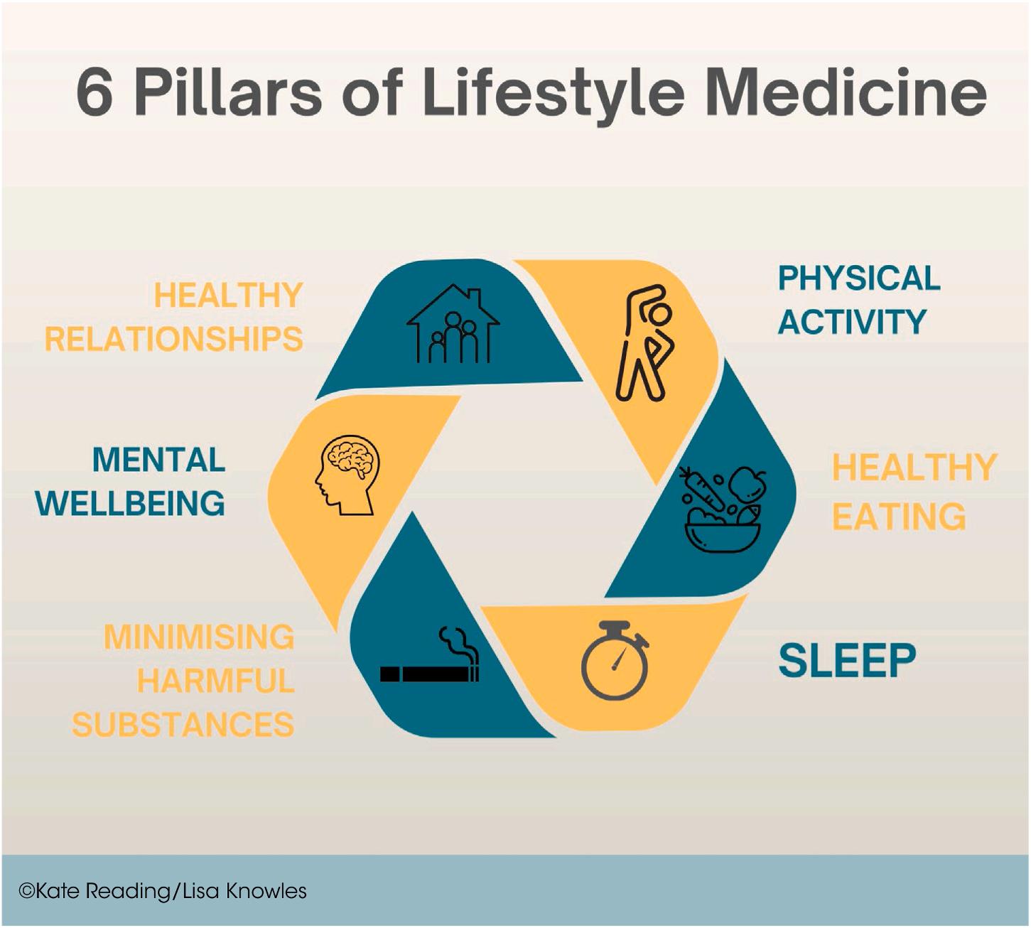
Periodontal disease is largely preventable but is a chronic inflammatory condition that has been linked to systemic health issues such as diabetes, cardiovascular disease, and obesity, which are also characterized by chronic inflammation. Periodontal therapy focuses on motivating the patient, supporting behavioral change, and addressing modifiable risks. LM is designed to prevent chronic diseases by focusing on inflammation. The LM interventions can be primary, secondary, or tertiary. The 6 pillars of LM ( Figure ) address the root causes of inflammation and systemic disease that overlaps with periodontal disease. Coupled with good oral hygiene and dental care, this approach achieves a holistic, sustainable method for preventing and managing periodontal disease.
In practicing lifestyle-based periodontal care, the patient pathway is reconfigured from a reactive, treatment-focused model to one that prioritizes prevention, holistic support,
and long-term health advances. The patient undergoes screening and assessment and is educated and engaged with how daily behaviors connect to gum and systemic health. This is done under the guidance of an integrated team of oral health care providers.
The foundation is a structured lifestyle screening tool that documents diet, smoking, activity, stress, sleep, and substance use so that care providers and the patient have a personalized view of the patient’s health context. Sharing these results visually with the patient improves health literacy and increases patient motivation.
The staff provides accessible resources, leaflets, digital content, and self-tracking tools that connect daily behaviors to periodontal and systemic health. Seeing gum health as part of the patient’s wider well-being increases motivation and raises the value of periodontal care in
the patient’s eyes. Accountability is achieved through the use of food diaries, activity trackers, and validated apps, promoting a gradual change and reinforcing new habits outside of the clinic.
Motivational interviewing and coaching strategies transform conversations from directive advice to a collaborative dialogue. Patients are encouraged to share their values, set achievable goals, and progress at their own pace. The discussions are integrated into the periodontal treatment sessions as part of the patient’s routine care.
Teamwork is an essential and involves the participation of hygienists, therapists, periodontists, and support staff. The team pursues consistent protocols, uses shared language, and engages in collaborative treatment planning. Holding regular case discussions encourages reflective practice, problem solving, and coordinated patient support. Patient outcomes are improved and the staff feels empowered and able to expand their professional competencies.
The evidence indicates that poor lifestyle habits compromise healing, shift the microbiome toward dysbiosis, and support systemic inflammation. When these habits are addressed in conjunction with mechanical therapy, the patient can see marked benefits, reductions in bleeding on probing, better tissue repair, and more stable long-term outcomes. Patients experience better oral health as well as enhanced energy, mood, and systemic metrics. These results reinforce their choice to continue their behavior changes in the future. The integrated clinical model positions periodontal care as the gateway to general well-being and reduced chronic disease.
Patients tend to value this broader approach to better oral health. The screening tool used shows how the pillars of LM interact. Recognizing the interconnections can trigger a chain reaction across all the pillars, making the importance of these relationships more apparent. Patients then become more likely to actively engage in their health management.
Integrating LM into periodontal practice still faces some barriers. These include time constraints, costs, role clarity, patient receptiveness, evidence-to-practice gap, and documentation. In particular, especially in private practice, it’s unclear how to structure, price, and value the services in patient care plans. In addition, some dental professionals worry about crossing boundaries
when discussing systemic health, which needs to be addressed by identifying referral pathways and structuring teamwork with medical colleagues. Not all patients are receptive to the lifestyle discussions, especially if they perceive them as judgmental or unrelated to their more immediate needs. Research may support LM’s impact, but it can be challenging to distill this into simple actionable steps for patients. Overall, there is no robust framework to track intervention outcomes, which is essential to clarify long-term effectiveness and justify the use of the model.
Going forward, the integration of LM into periodontal care will likely grow in the areas noted here:
• In-clinic health metrics: Incorporating HbA1c checks and blood pressure screening into the routine dental office visit can achieve early detection of systemic risks that influence oral health.
• Expanded patient education: The use of handouts, digital tools, and small group workshops can reach the community beyond the dental facility. As a result, the community’s awareness, motivation, and support for good oral health and the dental practice itself can grow.
• Generating evidence: Data from research and case studies can demonstrate measurable outcomes, leading to broader professional recognition and integration.
• Multidisciplinary collaboration: Adding dietitians, physicians, and other specialists to the team allows for more holistic and patient-centered care, reflecting the connections between oral and systemic health.
Addressing the underlying causes of disease through LM is a proactive, patient-focused solution to managing the root causes of systemic and oral inflammation. Empowering patients through lifestyle changes can improve clinical outcomes and help to overcome socioeconomic inequalities in treatment. The LM interventions are cost-effective, scalable, and beneficial at both the individual and system levels. The current dental hygienists and dental therapists should be able to help patients set achievable goals and develop effective plans, combined with their preventive expertise and regular patient contact.
Reading K, Knowles L: Lifestyle medicine in periodontal practice: The whys, wherefores, and pitfalls. BDJ Team 12:462-465, 2025
Reprints not available
When looking to institute a marketing campaign, several key performance indicators (KPIs) are needed to determine the impact and return on investment (ROI). KPIs provide the most actionable insights. Vanity metrics, such as drawing 1000 visitors to the website, can appear impressive but if only 50 are local, the large number is a basically irrelevant. Actionable metrics provided by KPIs can reveal meaningful insights into the effectiveness of the campaign. When planning for a marketing campaign, it’s important to specifically define and quantify success; determine the cost of acquiring a new patient; understand the ROI, projected revenue, and campaign costs; and consider other ways to measure campaign success.
Before beginning a campaign, the dentist should recognize that the 2 key metrics for a dental practice to measure results of a marketing campaign are the number of new patients and the production value those patients represent. To set the goals for the campaign, the dentist should consider how many new patients are desired and the revenue per patient expected. It’s important that everyone involved in the campaign, including a marketing partner, agrees on the goal for the campaign and how success will be determined, whether that is through call tracking and recordings, reports from the practice, or other factors.
Before beginning the campaign, the dentist should decide the maximum investment he or she is willing to spend to acquire a new patient, or the cost per acquisition (CPA). For cosmetic or implant treatment plans, a higher CPA may be acceptable, but a lower CPA is appropriate for new patient or emergency special offers.
To calculate an acceptable CPA, the items to consider are the number of new patients brought in over the past 6 months or year divided by the total revenue for the group. This yields the average production generated per new patient. A more accurate average can be obtained if the total of the practice’s net production rather than the gross revenue is used. Another more accurate CPA may result if you use the lifetime value of a new patient (LVT). This number is obtained by going back to the new patients who first entered the practice 7 years ago (or another data range). The dentist can calculate how much lifetime revenue has been generated from each of them and how long the patients stayed in the practice. Based on these calculations, it’s possible to
determine the CPA that would be appropriate for the practice in acquiring a new patient.
Once the dentist has determined how much production will need to be generated from the campaign to hit the goals, it’s time to determine the cost of the campaign and if it supports the expected results (ROI). This indicates the profitability of an investment or marketing campaign. If a campaign cost $10,000 and brought in $15,000 gross revenue, the practice would see a net profit of $5000. The ROI is 50%.
Precursor Actions and Google Analytics 4
If ROI can’t be determined because data on the revenue generated by the campaign, the associated costs, and the number of new patients added are unavailable, it’s possible to take some precursor actions, note key events, and use Google Analytics 4 (GA4). The precursor actions include calling from the website, submitting a contact or appointment form, or calling from the Google Business Profile. Measuring these actions provides insight into campaign effectiveness. It’s also possible to monitor changes in calls, contact requests, and appointment inquiries over time.
GA4 should already be installed on the practice website and the dentist should request access to the GA4 data from the marketing company if he or she doesn’t already have it. To tract the numbers over time, the dentist must set up each key event in GA4. The key events to be considered are contact form submissions, appointment form submissions, and phone number clicks. Tracking phone number clicks is limited to the raw number of clicks on the phone number and won’t tell whether the call went through or who called.
Insurance Por tability and Accountability Act (HIPAA)
More accurate data are available using HIPAA-compliant call tracking, which integrates with GA4, so the dental team member can link who is calling with where they came from on the Internet. When the name is known, it’s possible to determine if he or she became a new patient and how much production was generated.
Organic traffic is the measurement of how many visitors came to the website from a Google search (or any search engine). With
more people coming to the website, the chance that they will become new patients increases. Although this is a useful metric, it doesn’t give complete information. The real indicator is how many visitors become contacts or patients. A filtered, statelevel 90-day organic traffic measurement can provide some meaningful information, but it’s still less meaningful than tracking direct conversions.
The value of a marketing campaign rests on how it represents a balance between cost and revenue. This rests on knowing how much each new patient is worth and how much must be invested to gain a new patient. With this knowledge, it’s possible to make informed decisions and choose truly profitable options. Long-term success rests on careful tracking and thoughtful analysis of the process.
Wank D: 4 steps to evaluate metrics. Dentaltown 26:40-43, 2025
Reprints not available
Panic attacks can occur in anyone and may have no obvious cause or warning. Possible causes are dental anxiety, fear of dental procedures or needles, a repeat of past negative experiences, and underlying mental health conditions. To avoid these attacks, patients may choose to not attend dental consultations, leading to dental issues and deterioration of oral health. The patient may develop bruxism and TMJ disorders because of chronic stress and anxiety. Some patients at particular times of life develop panic attacks, such as teenagers who face high stress times, perimenopausal women, and persons with Alzheimer’s disease. However, panic attacks can also occur in persons with no underlying condition. Dentists should be aware of preventive measures to reduce dental anxiety, be able to recognize the signs of stress and anxiety in patients, provide care for patients during an attack, and help patients after the attack resolves.
The physical structure and d � ecor of the dental practice should be designed to be soothing and calming. This is achieved with relaxing colors, comfortable seating, and distractions such as music or television. The waiting area should be free of stress and welcoming.
The dental team should encourage open communication, allowing patients to share their fears or concerns. It should be okay to feel nervous. The procedures should be explained in simple, nonthreatening language, with techniques such as tell-show-do to ensure the patient is comfortable with the process.
Among the possible distractions used in dental settings are music, videos, and guided imagery. These can be used during procedures so patients can focus on something other than their anxiety.
Patients can also be positively reinforced for their cooperation and bravery during and after appointments. Verbal encouragement or follow-up calls can make a significant difference to the patient.
Among the physical signs that dentists should be aware of in patients are sweating or trembling, rapid heartbeat or breathing, nausea or dizziness, clenching the fists or jaw, and shaking. It’s important to differentiate an asthma attack from a panic attack. Generally patients with asthma have medication they can use to manage their disease, whereas panic attacks are usually short lived and resolve without medication.
The emotional signs include expressions of fear or worry, avoidance behaviors such as canceling appointments or refusing to sit in the dental chair, irritability, and difficulty concentrating or making decisions.
When patients suffer a panic attack, the dentist can help to calm and relax them. Often they can’t explain what caused them to panic and the dentist should not question them if they’re upset.
It’s important that the dental staff speak in soft, soothing voices and help the patient realize he or she is safe. Panic attacks can be frightening, but aren’t dangerous. They don’t generally cause physical harm or require a trip to the hospital.
Among the measures that can be taken are encouraging breathing exercises, with slow, deep breaths. This can be demonstrated by the dentist, who has the patient mimic his or her breathing. The patient may want to sit up or take a break from the procedure. Having the patient drink some water or take a moment to relax can be calming. The dentist should not rush the patient or force him or her to continue the procedure. It’s wise to remind the patient that panic attacks pass. Generally, panic attacks last from 5 to 20 minutes.
Grounding techniques can keep patients connected to the present and avoid panic responses. Some of the techniques are as follows:1.
The 5-4-3-2-1 technique: Have the patient sit comfortably with the eyes closed, then instruct him or her to take a deep breath. Have the patient open the eyes and think of 5 things they see, 4 things they can touch, 3 things they hear, 2 things they smell, and 1 thing they taste, then instruct the patient to take a deep breath.2.
Distraction techniques: Have the patient focus on a color and think of all the things in the room that are that color. Another technique is to have the patient count backward from 100 in sevens.3.
Tell the patient to choose a small item, then hold it and close the eyes. The patient should feel its texture, temperature and weight. With the eyes open, have the patient observe its color, pattern, and details. The same item can be reused to help ground the patient during stressful moments.
The dentist shouldn’t suggest breathing in and out of a paper bag. This can cause the individual to become dependent on the bag breathing and panic if there isn’t one at hand. In addition, if the patient is actually having an asthma attack, breathing into a paper bag is dangerous and can make the situation worse.
The dentist should follow up with the patient, asking how he or she is feeling and if anything is needed. The appointment should be rescheduled or continued, depending on whether the patient is agreeable. Reassurance that the patient did well and that panic attacks are manageable is helpful. If the patient has frequent panic attacks or severe dental anxiety, he or she may be advised to seek help from a mental health professional or the patient’s primary care provider. Patient preferences for their appointments should be noted so that future episodes can be minimized.
Panic attacks can feel frightening to patients, so dental staff members should be aware of the need to remain calm and supportive. Using preventive measures, handling the signs and symptoms with calmness and helpful interventions or distractions, and following up to ensure the patient understands the attack wasn’t serious can help patients to learn to undergo dental appointments without suffering future attacks.
Hammett E: Stress, anxiety and panic attacks for dental professionals. BDJ Team 12:418-420, 2025
Reprints not available
Oral pathology refers to any deviation from a healthy oral status. This may include diseases, injuries, or disorders that involve the teeth, gums, jaws, and other oral structures. Examples are TMJ disorders, oral cancer, facial trauma, cleft lip and palate, and severe periodontal disease. Often these disorders are accompanied by chronic pain, which can cause sleep disturbances, anxiety, and depression, making it difficult for the patient to
function and compromising his or her mental health. The mouth plays a critical role in communication, self-expression, and social interactions, so oral pathologies and their treatment can have a significant impact on the individual’s ability to function and make social connections. Dental hygienists not only maintain their patients’ oral health but also are trained to recognize and address the psychological and social effects of oral pathologies. The psychological effects of oral pathologies, care approaches and their
effects on quality of life, and the role of dental hygienists were discussed.
A strong link has been discovered between oral health and overall well-being. Persons experiencing oral pathology often have reduced life satisfaction levels, higher degrees of psychological distress, and a diminished sense of self-worth. Oral care must then address the interrelationship between physical symptoms and psychological well-being. Oral health care professionals should express empathy, engage in holistic treatment plans to address pain, and collaborate with other care professionals to address functional limitations, ultimately improving the quality of life for patients.
The psychological challenges are unique to each oral pathology. Dental hygienists must therefore personalize their care to meet each patient’s emotional and psychological needs.
The chronic pain and functional limitations of TMJ disorders can lead to a cycle of psychological distress. This can include frustration, anxiety, and depression, all of which are complicated by unpredictable flare-ups. Women may be especially susceptible and struggle with increased pain sensitivity and social pressures to suffer pain silently. Chronic stress related to TMJ disorders can cause physiological changes, including vasoconstriction and increased muscle tension, increased pain, and dysfunction. Sleep disruptions and bruxism can also develop, underscoring the complex interactions between physical and psychological factors in TMJ disorders.
With a diagnosis of oral cancer, patients can go through emotional distress related to a profound fear of death, disfigurement, and potential loss of important oral functions. This psychological dysfunction adds to the challenging physical aspects of cancer and its treatment.
Head and neck cancer significantly alters patients’ quality of life. After treatment, they must cope with altered functions, including swallowing, chewing, eating, and speaking, and are faced with disfigurement and its social adjustment challenges. Many patients can feel stigmatized and negative social experiences. Surgical interventions cause more social distress than radiotherapy or chemotherapy. It’s possible that facial disfigurement can change social engagement dynamics for patients with head and neck cancer, leading to more distress regardless of the relationship status involved.
Craniofacial anomalies such as cleft lip and palate (CLP) cause many challenges in terms of speech, hearing, and social acceptance difficulties. These can cause the patient to feel
isolated and anxious and to have low self-esteem. Such reactions are especially common in patients during the formative years in childhood and adolescence. Emotional challenges can result from the physical manifestations of craniofacial anomalies.
Adults with CLP have been found to have high levels of social anxiety and exhibit avoidance behaviors. Such conditions are indicative of the profound impact of craniofacial anomalies on social wellbeing.
Severe periodontal disease can also have profound effects on a patient’s psychological health. The consequences of tooth loss, receding gums, and halitosis can lower self-esteem and social confidence, making the individual feel isolated and vulnerable.
Further research is needed to detail the effects of periodontal disease on mental wellbeing. Recent studies found a strong link between mental health challenges and compromised oral health, even when socioeconomic and demographic factors are considered. Internalizing disorders, such as depression and anxiety, seem to have the most consistent association with adverse oral health outcomes. Mental and oral health are in a dynamic relationship, with the condition of one influencing the status of the other over time. A collaborative, compassionate approach to care is needed to address any gap between mental health and dental care professionals.
Dental hygienists can empower patients to confront and manage their challenges using a holistic approach that integrates physical and psychological care. Hygienists can foster clear, empathetic communication and provide comprehensive patient education. They should use simple language, visual aids, and gentle techniques; create a safe, trusting environment; and encourage patients to feel comfortable when asking questions and to actively participate in any decisions made. To ensure patients feel heard and understood and facilitate the construction of a strong therapeutic alliance requires active listening and validation of the patients’ emotions. Hygienists can also refer patients to appropriate mental health resources if signs of emotional distress develop. Effective communication should include the exchange of ideas in clear, accurate, concise, comprehensive, and well-organized ways. Patients need information so they can make informed decisions about their care and achieve a healthier state of wellbeing.
The current evidence indicates that psychological interventions can improve the quality of life for those with oral pathologic conditions. Among the effective measures are mindfulness-based interventions (MBIs), cognitive-behavioral therapy (CBT), and peer support groups. These approaches achieve reduced
anxiety, depression, and chronic pain and enhance coping skills and self-esteem. The changes in wellbeing include improved cognitive biases, emotional regulation, and interpersonal skills.
Dental hygienists not only fulfill their role as clinical care givers, but can play a vital role in acknowledging and addressing their psychological and social impact on patients with oral pathologies. Patients are more prepared to navigate the emotional challenges of their conditions when the hygienist creates a supportive environment and communicates clearly, shows empathy, and engages with other care providers. This holistic approach can promote the patients’ healing, resilience, and wellbeing.
Dental hygienists are uniquely positioned to not only provide needed oral care but also to offer support for patients who are dealing with the psychological and physical effects of poor oral health. They can ameliorate the pain patients are experiencing, provide a safe and calm environment, provide information to help patients make appropriate decisions, and support patients in their journey. Patients’ lives are enriched and dental hygienists’ professional expertise is enhanced as they make a profound impact with their approach to care.
Sucre A: The mind-mouth connection: Psychological implications of oral pathologies. RDH 44:40-42, 2025
Reprints not available
Dental care focuses on working with patients to ensure they develop critical self-care habits that will lead to lifelong oral health. A 4-step approach was developed to create and maintain behavior change (Figure 1). The support for this approach involves an ongoing, trusting relationship between the oral health care team and the patient that recognizes the difficulty in changing behaviors, the lack of perfection in pursuing these behaviors, and the different lengths of time that individuals take to reach good oral care behaviors. The 4 stages are teaching the skill, creating intention, developing a plan, and engaging in selfmonitoring with implicit cues. These stages are also known as capability, motivation, opportunity, and habit.
The oral health care team and their patients need to have a relationship of trust and mutual respect to create the joint decisions needed to change behavior. The dental team must build rapport, show interest in their patients, and be willing to help. Patients should observe the dental professionals listening to their concerns and the challenges they are facing. The dental team should offer appropriate empathic responses to show that they understand the patients’ frustration or stress and acknowledge their emotional state.
Patients should also be involved in decision-making. The dental team should seek patients’ opinions of the various options for care and accept their choices. Change should be clearly depicted as a continuous process that will have the dental team and patients working together. The timing of change can differ because of external events.
The key manual skills, including tooth brushing and interdental cleaning, must be practiced to reach a high level of expertise. These skills are generally taught through observation of another person (modeling) along with feedback on how well the patients did. Identifying where changes are needed will help them focus on those areas. Interdental cleaning is also done by modeling the behavior, sometimes on a model, with feedback offered to help patients improve the technique. Patients should be asked whether they prefer floss or interdental brushes.
The most effective methods for enhancing the motivation to change behavior are discussing the patient’s susceptibility to disease, emphasizing the benefits of changing behavior, and enhancing the patient’s perception of their ability to change (self-efficacy). To convey the patient’s susceptibility to oral disease tends to work best graphically rather than in the context of statistical susceptibility. Patients should also be asked about what is important to them regarding their oral health. Focusing on the benefits they see is usually more motivating than focusing on avoiding harm.
Patients tend to have a low sense of their ability to control their oral health, especially when compared to their overall health. The dental team should ensure the patients recognize their responsibility for daily care, with the team only involved at dental visits. The 3 primary ways to achieve this are to provide supportive statements to encourage patients, to make small changes that over time create a larger effect, and to share patients’ success (with their permission) with other patients to show how people can make changes even when it’s difficult.
People often have difficulty transforming their intention to change behavior into action. The most common barrier is the busy lives of individuals that keep them from changing their routine. To make a behavior change, patients may be asked to plan the change, which creates time and opportunity for the effort. Plans involve working with the patient to identify when the new behavior will begin, where it will take place, and what resources will be needed. The plan should be realistic and focused on the specific needs of the patient.
Moving from a plan to a habit takes an average of 66 days of daily repetition. Once patients begin to change a behavior, the dental team should support and encourage them to continue until a habit has been established.
Techniques to encourage habit establishment include building cues to behavior and encouraging patients to focus on the rewards they will reap from the change. Cues indicate when to
Capability –Teach Skill
Motivation –Create intention
Oppor tunity –Plan
Habit –Self monitoring
Implicit cues
2025.)
engage in a behavior and can be a certain time or be associated with another behavior, like going to bed. Linking a new behavior cue to an established behavior cue can more firmly establish it in the individual’s routine.
When the dental care provider emphasizes the benefits of the behavior change, such as a fresher mouth or less bleeding when brushing, the behavior becomes intrinsically rewarding. Behaviors linked to intrinsic rewards are generally more firmly established than those relying on extrinsic rewards.
Working with patients to make changes in their oral health behaviors can be complex and challenging. Taking it a step at a time helps to avoid overwhelming the patient and may end up with more firmly established behaviors than rushing through the process.
Jones R, Newton T: Behaviour change in dentistry: What really works? BDJ Team 12:370-373, 2025
Reprints not available
Dental caries and periodontitis are noncommunicable diseases (NCDs) of the oral cavity, yet they are not listed as such in the United Nations and World Health Organization (WHO) lists of NCDs, limiting that list to cardiovascular disease, cancer, chronic respiratory disease, diabetes, and mental health disorders. Evidence links severe periodontitis with premature death and shows a causal relationship with diabetes complications, major cardiovascular events, and poor kidney function in chronic kidney disease patients. It’s also a risk factor for rheumatoid arthritis. Policymakers and the public consider oral diseases to be the domain of dentists and therefore sees them as less important than diseases of internal organs. In fact, the oral cavity is the primary entry point for threats from the environment and offers a direct access point for microbial organisms to reach the systemic circulation. Oral health is also an entry portal for smoking,
alcohol intake, and eating unhealthy diets. With all of these connections, it’s important that all health care practitioners take oral health seriously. A discussion of the divide between oral and other health priorities, the importance of gingivitis care, the inequality of care opportunities, remuneration, and the responsibility of oral professionals was offered.
In 2019, severe periodontitis and dental caries had an estimated prevalence of 3.5 billion people, which was more than 1 billion greater than the cumulative burden of diabetes, cardiovascular disease (CVD), cancer, chronic respiratory disease, and mental health disorders. The worst cases are found among the poorer communities. The financial impact is also enormous, with a direct treatment cost of $357 billion.
Oral diseases are also associated with a high human cost, with more years lived with disability related to oral disease than any other Level 3 human disease in 2021. To be an accurate reflection of the harms related to NCDs, the focus should be on the quality of life of those affected rather than simply what mortality results.
Health should be seen as a basic human right. Oral diseases are largely preventable, with preventive approaches saving millions of dollars. Both economic cost savings and improved oral health equality can be achieved by managing oral diseases preventatively. Oral health care practitioners (OHPs) and medical health care teams should be integrated more effectively in these preventative approaches, with shared medical/dental records and investigations; shared primordial, primary, secondary, and tertiary prevention strategies for common risk factors; and shared outcome reporting. The conclusions of a workshop in 2022 between the European Federation of Periodontology (EFP) and the World Organisation of National Colleges, Academies and Academic Associations of General Practitioners/Family Physicians (WONCO) were the need for a closer collaboration between OHPs and family doctors in the early detection and management of NCDs, the promotion of healthy lifestyles, and the establishment of pathways for early case detection of periodontitis in family medical practices and of NCDs in dental practices. Remunerated systems and approaches must be developed to enable and incentivize the call for collaboration by the OHP and medical professions.
The focus has been on periodontitis, with little attention paid to gingivitis, yet the EFP concluded that treating gingivitis was the primary preventive strategy for managing periodontitis. Gingival inflammation has also been identified as a risk factor for major adverse cardiovascular events. Engaging gingivitis patients in improved oral hygiene practices lowers their C-reactive protein (CRP) levels in blood and reduces cardiovascular risk.
Changing the current practice of managing Stage 2 periodontitis to one that eliminates incident gingivitis gave positive returns on investment between 15.2 in Italy and 57.5 in Germany. In addition, the entire oral health care team can improve oral hygiene to reduce the burden of gingivitis. This expands the scope of practice of all OHPs, including dental nurses, receptionists, dental hygienists, and dental therapists.
The most deprived populations have the highest oral disease burden. In addition, the lack of oral health literacy throughout life presents a major challenge. A national curriculum on oral care must be developed that extends from antenatal classes, through primary, secondary, and tertiary education, into the
workplace, and finally into assisted living and elder care homes. In addition, urgent care services are needed for those who haven’t seen a dentist for years. It will require incentivizing offers to encourage these patients to attend urgent care plus receive prevention advice and risk factors counseling. Triage services may be required that use the entire oral health care team to improve cost effectiveness. Additional health care professionals such as pharmacists can be trained to offer advice and guidance regarding oral health care products. Health professionals may visit schools so that children in their early years will be engaged with oral health practices.
The population of those who work hard to look after themselves and their families but who earn minimal wages and can’t afford oral health care must also be addressed. Ways to support this population and develop an awareness of their specific needs must be part of the changes to the care delivery system.
Most health care remuneration systems focus on intervention and assign fees per item of work done or per a group of items. This encourages the performance of invasive interventions and doesn’t reward prevention. Some success has been made in lobbying for remuneration systems that reward prevention. A system in England allows phased courses of treatment to apply for each stage of periodontal care, providing OHPs who focus on getting patients to control risk factors and take responsibility for their own health.
Globally, the remuneration processes need to shift toward oral care systems based on a more holistic care pathway and supporting patient self-care and prevention. The decisions to make these changes would move oral health care into the 21st century.
For oral professionals running a practice or hospital, maintaining a sustainable and financially viable business is essential. A disconnect remains, however, among the principles that create the foundation of clinical practice, how undergraduates have been educated over the past 30 to 40 years, and the financial imperatives needed to sustain the organization. Most practitioners struggle because of remuneration models. Governments must trust OHPs as ethical and caring professionals to the same degree that they trust family medical practitioners.
The 2023-2030 WHO Global Strategy and Action Plan on Oral Health demonstrates the energy and commitment of the WHO even when heavy financial stresses and the policies of the current US administration created a significant strain. In addition, the health care community is joining in efforts to raise awareness of the massive global burden of oral diseases and conditions and to advocate for change in the policies of health institutions and governments.
If no action is taken, the prevalence of severe periodontitis will reach 13.1% by 2050, which means it will affect more than 1.5 billion people. In addition, edentulism will rise to 4.18% by 2050, with more than 660 million people affected. It’s time to join forces and lobby for oral health to be coupled with overall health and wellbeing. The mouth should be considered an important component of the body, with significant effects on the body’s ability to maintain a healthy status.
Chapple I: The global burden of oral disease—what is the fuss?
BDJ Team 12:364-327, 2025
Reprints not available
No universally reliable diagnostic method able to diagnose all types of carious lesions currently exists. The traditional approaches include radiography and visual-tactile examinations, but they are flawed by intra- and inter-clinical variation, which limits their accuracy in diagnosis. Visual methods suffer from a limited reproducibility and an inability to monitor lesion progression or discern active from inactive lesions in a single visit. Visual caries scoring systems are plagued by inconsistencies in the criteria and implementation selected, throwing off standardization and reliability. The ionizing radiation used for radiographs is a health concern. Radiographs also fail to properly estimate lesion depth, especially in early-stage caries. Low sensitivity in detecting incipient caries limits the effectiveness for early interventions. Alternative imaging techniques include fiber optic transillumination (FOTI), quantitative light-induced fluorescence (QLF), digital image fiber optic transillumination (DFOT), laser fluorescence (LF), and optical coherence tomography (OCT), all of which suffer from involving complex procedures, a need for training, and a lack of clinical trials. Intraoral scanners (IOSs) and intraoral cameras (IOCs) offer high-resolution, real-time imaging with digital analysis capacities, but their true positive rate (SE) and true negative rate (SP) values have varied widely. In addition to differences in image projection geometry and lesion characteristics, examiner-related variables such as training and experience can cause discrepancies. Both SE and SP directly influence treatment decisions, possibly resulting in diagnostic misclassification, overtreatment, or undertreatment. The current evidence of the diagnostic performance of IOSs and IOCs was synthesized, with special attention to lesion type, lesion location, and examiner-dependent variations.
A search of the Scopus, Web of Science, PubMed, Cochrane Library, and Dentistry & Oral Sciences Source databases yielded 28 studies that met the inclusion criteria for this systematic review. All were published between 1998 and 2024, with 6 experimental studies and 22 comparative studies. The latter included 1 retrospective audit, 12 in vivo investigations in human patients, 13 in vitro studies using only extracted teeth, and 2 using both in vivo and in vitro methods.
When a moderate diagnostic accuracy was selected for IOSs and IOCs in detecting dental caries, the values suggested that intraoral diagnostic tools perform similarly in identify SE and SP. The diagnostic performance is influenced by contextual and technical factors.
A comparison of the diagnostic methods showed IOSs exhibited the highest SE and significantly outperformed radiography, being more reliable in identifying true positive cases. IOCs had higher SE than radiography, underscoring how limited radiographic techniques are in detecting caries.
Visual methods showed moderate SE that wasn’t significantly different from the results of IOCs or radiography. IOCs may hold a marginal advantage in SE, possibly reflecting better surface-level visualization than IOCs. Variability across studies and the influence of examiner-dependent factors may be involved as well.
Radiography was the most reliable method to avoid false positives, achieving the highest SP. It significantly outperformed IOCs and IOSs in accurately detecting noncarious lesions. Visual methods had moderately high SP and were better than IOSs but not significantly different from IOCs.
There were highly significant differences in SE and SP across the 4 methods, showing that these tools vary significantly depending on lesion location and diagnostic metrics. For occlusal lesions, IOCs and IOSs tended to have higher SE than radiography. The differences didn’t reach statistical significance but the differences may reflect radiography’s limitations in detecting early or surface-level occlusal lesions. Examiner calibration heterogeneity, device imaging geometry, and lesion interpretation may also influence these trends and require further study.
Radiography had significantly higher SP for occlusal lesions compared to IOCs and IOSs, proving it is the most reliable tool to rule out false positives. No significant differences in SE were noted between IOSs and visual methods or between IOSs and IOCs.
No significant differences in SE or SP were noted in proximal lesions. This may reflect the limited visibility of these lesions regardless of diagnostic modality. The intrinsic diagnostic challenges of interproximal surfaces can overcome all of the modalities’ abilities. IOCs and radiography had similar SE, and radiographs and visual methods had nearly identical SE and SP for proximal lesions.
The SE and SP vary depending on lesion type. Radiography should be selected to achieve better SP for occlusal lesions, but IOCs and IOSs tend to have higher SE.
Moderate diagnostic performance was found for cementoenamel junction (CEJ), enamel, and dentin lesions, demonstrated by a pooled SE consistently at 50%. Significantly higher SE was noted for enamel and dentin combined lesions, which indicates better accuracy in detecting multilayered caries. The lowest SE was for supragingival lesions. The SP for supragingival lesions was exceptionally high, which indicates strong true-negative detection (non-caries) even with poor SE. SP was 50% with moderate variation for other lesions. The uniformly low SE highlights how difficult it is to detect early stage lesions in these areas.
The tools had varying results, as follows:
1. IOCs: Comparatively higher SE and SP for dentin lesions but no significant differences in SE and SP for CEJ lesions compared to the results of radiography and visual methods.
2. IOSs: Pooled SE of 50% and a slightly higher SP of about 87% for dentin lesions. SE had low heterogeneity, but SP varied considerably, probably as a result of differences in scanner software, examiner interpretation, reference standards, or misclassification of noncarious conditions. IOSs showed no significant differences from other tools in diagnostic performance for CEJ lesions, but low SE for supragingival lesions along with high SP.
3. Visual methods: Visual methods had the highest SE for enamel and dentin combined lesions along with lower SP and substantial heterogeneity. Variations may be caused by inconsistencies in examiner training protocol, subjective interpretation criteria, or inconsistent application of visual scoring systems, all of which can impact reproducibility, especially for early-stage lesions. Visual methods used for CEJ lesions achieved similar outcomes to IOCs and radiography, with SE and SP near 50%.
4. Radiography: Radiographic methods outperformed the other methods in diagnosing enamel and dentin combined lesions and had strong consistency of outcome. They were less
effective for supragingival lesions and had low SE but the highest SP, indicating the ability to minimize false positives. There were no significant differences in SE and SP for CEJ lesions.
Examiner -dependent Variabilitie s
The examiner-specific factors noted to affect diagnostic outcomes were most common for operator-dependent tools like IOCs, IOSs, and visual methods. The factors identified as problematic were training, experience, technique, and protocol adherence.
The comparison of diagnostic methods indicated that radiography consistently had the highest SP. IOSs and IOCs had moderate diagnostic accuracy overall, with higher SE for occlusal and dentin lesions but variable SP. Variations related to the examiner significantly influenced outcomes, especially for image-based and visual methods. Those examiners who had better training were more accurate but their results were more heterogeneous.
Intraoral scanners and cameras had moderate accuracy in diagnosing caries. Occlusal and dentin lesions were evaluated with greater sensitivity. Radiography consistently had higher specificity, especially in advanced caries lesions. The tools demonstrated important variations in diagnostic accuracy depending on lesion type, with supragingival and early enamel lesions associated with lower sensitivity for all methods. The experience, training, and calibration of the examiner significantly influenced the accuracy and resulted in some variations. Consistent reference standards and standardized diagnostic protocols are needed to accurately compare results across numerous studies.
Rashid F, Farook TH, Dudley J: Comparison of the accuracy of intraoral scanners, intraoral cameras, radiographs, and histological methods for the diagnosis of dental caries: A systematic review and meta-analysis. BDJ Open 11:82, 2025
Reprints available from F Rashid, Adelaide Dental School, The Univ of Adelaide, Adelaide, SA 5000, Australia; e-mail: qazifarah.rashid@adelaide.edu.au
Temporomandibular joint disorders (TMDs) are sufficiently painful that 5% to 12% of the population seek treatment. Most complain of pain, limited jaw movement, joint noises, and popping, with significant effects on oral function and quality of life. The cause is multifactorial, and treatment usually involves pharmacological interventions, physical therapy, behavioral therapies, and occlusal devices. A minimally invasive technique for TMDs is dry needling (DN), in which fine needles are inserted into myofascial trigger points (TPs) to reduce pain, maintain function, and improve organ movement. DN is also widely used to manage back pain, myofascial pain syndrome, and whiplash, but its exact mechanism of action requires further investigation. The current knowledge regarding the impact of DN on TMDs was explored.
A search of the PubMed, Scopus, and Google Scholar databases identified 10 randomized controlled trials (RCTs) where DN was applied to various muscle groups. The outcome measures included the visual analog scale, verbal rating scale, electromyography, extent of mouth opening, pain symptoms, myofascial TP pain, sonographic measurements, bilateral muscle palpation with a pressure algometer, Tinnitus Handicap Inventory (THI), and mandibular mobility.
Technique
The TPs were found through manual examination. Sterilized disposable stainless steel acupuncture needles with a diameter of 0.18 to 0.3 mm and a length of 13 to 60 mm were used. The skin overlying the TPs was fixed between the index finger and middle finger or index finger and thumb, then the needle was inserted perpendicular to the muscles to a depth between 10 and 35 mm. Once the needle was placed, the practitioner performed rotary movements or bidirectional manipulation to create local responses, such as aching, tingling, deep pressure feeling, warmth, or heaviness. The time the needle remained in each TP was 10 sec to 30 min. The technique was carried out weekly for 3 to 6 weeks in most studies.
DN was able to target specific TPs and facilitate structural changes. These included reduced muscle and disk thickness and relief of pain. DN also improved functional alignment and addressed associated conditions.
In addition to TMDs, DN was able to reduce tinnitus intensity and discomfort. It can be combined with counseling, manual therapy, and other therapeutic approaches.
No major adverse effects were reported. Among the side effects were muscle soreness, ecchymosis, headache, drowsiness, and nausea, which lasted from several hours to a week. Patients were instructed to apply ice or moist heat on any painful region after treatment.
DN is able to manage TMDs and other painful conditions in some patients and causes few major adverse effects. It can also be integrated with other modalities for even better outcomes. Patients tend to be satisfied with the results in most cases.
DN along with other adjunctive therapies may be able to improve clinical outcomes in patients with TMDs. Pain reduction and functional improvement are the most common outcomes.
Khayamzadeh M, Razmara F, Tavassoli A: Dry needling in treatment of temporomandibular joint disorders: A systematic review. Clin Exp Dent Res 11:e70214, 2025
Reprints available from A Tavassoli; e-mail: Afagh.tavassoli93@ gmail.com
E-cigarette use or vaping has reached usage rates of about 9% in the United Kingdom. However, the scientific literature on ecigarette use is limited, although clinicians may be able to guide patients toward making an informed decision about the use of these aids. The current information regarding the impact of ecigarette use in terms of user characteristics, systemic links, and periodontal effects; comparisons among e-cigarette use, conventional smoking, and non-use/non-smoking; and guidance for practitioners and the public were documented.
To better understand the impact of e-cigarette use, it’s necessary to examine the characteristics of users, the systemic links suggested by research, and the effects on periodontal health.
Most users of e-cigarettes are current or past smokers, with never-smokers quite rare. Among men age 16 years or older, about 9.5% are users daily or occasionally; among women, just 7.9% fall into this category. Fewer of those over age 60 years choose to use e-cigarettes. Younger users tend to use them for enjoyment, whereas individuals over age 45 years tend to use them for smoking cessation (Box 1).
Not all e-cigarettes are the same. They vary in nicotine levels as well as flavors. The components of each type vary.
Little evidence is available on the systemic effects of e-cigarette use, and long-term studies are lacking. However, the current evidence indicates neurological, pulmonary, cardiovascular, and oral and dental effects (Figure 1). Higher rates of alcohol and cannabis use as well as other high-risk behaviors are linked to daily or heavy e-cigarette use. This association has deleterious effects, with such behaviors considered co-morbidities for several disorders. Better information will only be obtained through long-term human studies on e-cigarette use.
E-cigarette use can contribute to periodontal disease through elevated inflammatory biomarkers in gingival crevicular fluid and saliva, which indicates an inflammatory response similar to that created by traditional smoking. Persistent inflammation can cause accelerated bone resorption and tissue degradation—both features of periodontal disease. Increased levels of
periodontal pathogens are also linked to e-cigarette use. The microbial imbalance can raise the risk of periodontal infections and tissue destruction, leading to the progression of periodontal disease.
E-cigarette use, conventional smoking, and non-use or nonsmoking status have been compared to determine the relative concern for periodontitis and the user/non-user response to periodontal treatment as well as the relative effects on implants and peri-implantitis treatment.
Periodontitis
E-cigarette use appears to be less harmful to periodontal health than smoking conventional cigarettes. Lower plaque index scores, probing pocket depths (PPDs), attachment loss, marginal bone loss, and pro-inflammatory mediators are seen with e-cigarette use compared to traditional smoking. However, most of the studies are observational and don’t establish a causative relationship between vaping and periodontal health. Long-term studies are needed.
E-cigarette use is linked to an increased prevalence of periodontitis compared to non-smoking. Concurrent use of other tobacco products may contribute to the findings, but this has not been documented.
Studies of the severity of periodontitis involve several potential confounding factors in comparisons of clinical attachment loss (CAL), a critical parameter of periodontal severity. The most recent studies suggest a nonsignificant increase in CAL in e-cigarette users compared to non-smokers and a higher risk for ecigarette users to have periodontal disease than non-smokers but a lower risk than cigarette smokers. The evidence regarding CAL levels remains inconclusive.
A comparison of 4 groups (former smokers, former smokers now using e-cigarettes, current smokers, and non-smokers) after nonsurgical periodontal treatment (NSPT) showed those who switched to e-cigarettes had less favorable responses to NSPT than both former smokers and nonsmokers. This included a deeper PPD and a greater need for surgery. The clinical outcomes for those now using e-cigarettes were similar to those for current smokers, suggesting that e-cigarette use may compromise periodontal healing in ways similar to those seen with conventional cigarette use. Because of the lack of statistical power and the confounding
• 40.6% of older people prefer tobacco-flavoured e-cigarettes versus only 4.8% of younger people; 51.1% of younger people prefer fruit flavours
• Being young and perceiving e-cigarette use as less harmful than conventional smoking increases the chance of recent (within the last 30 days) e-cigarette use
• Women from white and multiracial backgrounds were more likely to have tried e-cigarettes
• Lowest use was among Black African and Indian men, and women from all Asian and Black African backgrounds
• E-cigarette users are more likely to have parents with a lower socioeconomic status at birth
(Courtesy of Robson G, Lim XC, Chaudhari I, et al: Trying to clear the air: E-cigarette use and periodontal disease. Br Dent J 239:687-692, 2026.)
effects of past tobacco use, the findings should be considered suggestive instead of definitive.
E-cigarette users had worse clinical and inflammatory periimplant profiles than non-smokers over the course of at least 3 years when implants were in function. E-cigarette users had
increased PPD, plaque accumulation, marginal bone loss, and levels of pro-inflammatory cytokines compared to nonsmokers, but lower levels than cigarette smokers.
Research in the area of responses to peri-implant treatment noted that the care was less effective in e-cigarette users than in conventional smokers or non-smokers. After treatment, the clinical parameters, specifically, gingival color and consistency, plaque index, bleeding on probing, PPD, and keratinized tissue width, were assessed after 1 month, 6 months, and 1 year. Higher clinical and salivary inflammatory marker levels were noted in e-cigarette smokers than in never-smokers and cigarette smokers. The sample size of this study was only 60 participants and the details on power calculations or effect size assumptions were lacking. Lack of control of confounding variables and self-reporting of habits further undermine the value of these findings.
Limited evidence is available regarding the periodontal health of users of cigarettes who switch to e-cigarettes. Preliminary evidence suggests switching may improve periodontal health. The findings of a fairly reliable study indicate improved bleeding on probing, papillary bleeding index, and plaque levels in those who switched from conventional cigarettes to e-cigarettes.
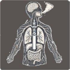
Figure 1. Confirmed or potential oral and systemic associations of e-cigarette use suggested in various papers. Please note varying degrees of support for these conditions—not conclusive evidence. (Courtesy of Robson G, Lim XC, Chaudhari I, et al: Trying to clear the air: E-cigarette use and periodontal disease. Br Dent J 239:687-692, 2026.)
Large-scale, high-quality studies that measure clinical and biological markers of periodontal inflammation and disease are needed.
Practitioner Guidance
The current guidance available to those who provide smoking cessation support and advice suggests that e-cigarettes are a stop-smoking aid but are not classified as licensed medicines. E-cigarette users are at a higher risk for periodontal disease compared to non-smokers but lower than tobacco users. This makes e-cigarettes a transition tool in smoking cessation. A quick toolkit helps to increase the success of smoking cessation efforts (Figure 2), suggesting ways dental practitioners can Ask, Advise, and Act at opportunities or recall appointments for adults and adolescents. Practitioners should also inform patients that validated knowledge regarding full effects of e-cigarette use is lacking.
It’s suggested by the National Institute for Health and Care Excellence in the UK that practitioners do the following:
1. Tell the individual there is insufficient evidence about the long-term harm of e-cigarettes.
2. Advise that e-cigarettes are likely to be substantially less harmful than smoking.
3. Recommend that the use of e-cigarettes should be coupled with complete cessation of tobacco smoking.
4. Discuss how long the patient plans to use nicotine-containing e-cigarettes.
5. Suggest using e-cigarettes long enough to prevent a return to smoking.
6. Offer advice on how to stop e-cigarette use when the patient is ready.
A 2023 survey indicated that 43% of adults believe e-cigarettes are as harmful or more harmful than conventional cigarettes. More of the population see e-cigarette use negatively, perhaps as the result of media coverage. This negative view can amplify fears regarding e-cigarette use and contribute to misperceptions.
Patients should be advised that both the risks associated with ecigarette use and how the effects of use compare to the effects of conventional smoking should be carefully considered. Individuals should be helped to make informed decisions about their health, especially their oral health.
Clinicians need to know the detrimental effects of e-cigarette use on the periodontium but also acknowledge that the current evidence indicates that e-cigarette use may be less harmful than smoking cigarettes. Patients shouldn’t be dissuaded from switching to a potentially safer option than conventional smoking. Ideally, smoking cessation will be chosen, but in the meantime, e-cigarettes offer a less harmful behavior than cigarette smoking.
Robson G, Lim XC, Chaudhari I, et al: Trying to clear the air: E-cigarette use and periodontal disease. Br Dent J 239:687-692, 2026
Reprints available from J Hurley; e-mail: Josh.hurley2@gmail.com
The disinfection of the root canal system (RCS) is required for the success of endodontic treatment. An intracanal medicament (ICM) is a chemical agent placed in the RCS and retained to alleviate pain and inhibit microbial activity. The effectiveness of root canal disinfection is enhanced by these medicaments, with irrigants, ICMs, and sealers all used to ensure optimal disinfection. The traditional gold standards are sodium hypochlorite (NaOCl), ethylenediaminetetraacetic acid (EDTA), and calcium hydroxide (Ca(OH)2). Zinc oxide eugenol (ZOE) and resin-based sealers are used for obturation. Advances in material science and a better understanding of microbial resistance and biofilm behavior have prompted the development of new agents with enhanced abilities, including antimicrobial properties, biocompatibility, and sealing ability. Newer nanoparticle (NP) or herbal agents, bioactive medicaments, and bioceramic-based sealers (BCS) offer alternatives (Figure 1). The evidence-based applications of older and newer irrigants, ICMs, and sealers were discussed.
NaOCl, chlorhexidine (CHX), 17% EDTA, citric acid, and 37% phosphoric acid solution, as well as a few other agents, have been suggested for use in canal preparation. Ideally an irrigant should completely remove the smear layer, lubricate the canal, efficiently kill microorganisms, induce no inflammation, and avoid damaging the dentin, but no one irrigant has as yet met all these requirements. None of the irrigants can dissolve both organic tissue and inorganic matter. NaOCl is the gold standard, but is lacking in antibacterial efficacy and is associated with toxicity.
Irrigant activation systems have been developed to overcome NaOCl drawbacks. They include photon-induced photoacoustic streaming (PIPS), photodynamic therapy (PDT), and shock waveenhanced emission photoacoustic streaming (SWEEPS). PIPS and PDT increase the penetration of NaOCl into dentinal tubules, and SWEEPS allows the use of NaOCl in lower concentrations without minimizing the antimicrobial effect. These techniques have negligible apical extrusion so they are safer for root canal disinfection. Postoperative pain has been minimal with laser activation compared to manual dynamic activation.
Among the newer and alternative irrigants are those addressing the cytotoxicity of NaOCl and avoiding severe tissue damage when the irrigant extrudes beyond the apex. These include Tetraclean NA, nanoparticles, herbal agents, and oxonated water.
Use of 17% EDTA achieves enhanced potential to remove the smear layer, release growth factors in regenerative endodontic procedures (REPs), and sustain compatibility with NaOCl. Modifications have been required to manage the cytotoxicity and inflammatory effects of EDTA. A mixture of tetracycline, citric acid, and detergent (MTAD) removes the smear layer with significantly less erosion of the dentin surface than occurs with EDTA. Citric acid aids in chelating, allowing for the penetration of doxycycline into the dentinal tubules and the achievement of a sustained antibacterial effect. Other aids include EDTA with cetyltrimethylammonium bromide (EDTAC), which facilitates optimum smear layer dissolution, and nisin incorporated into EDTA, which not only helps remove the smear layer but also exhibits enhanced antibacterial and antibiofilm properties.
QMix helps achieve better debridement of the smear layer from the apical third of the RCS compared to NaOCl, EDTA, and CHX. It’s biocompatible, has higher bond strength, and achieves greater sterilization of canals. QMix can also reduce dentin microhardness.
SmearClear offers chelating and disinfecting properties. It can remove calcium ions from the root canal more efficiently when coupled with MTAD.
Although 1.5% NaOCl and 17% EDTA are considered the standards for irrigants, in cases with a higher risk of NaOCl extrusion (such as open apex, apical bony fenestration, and iatrogenic factors), alternative irrigants can be considered.
With the shift toward biocompatible and targeted antimicrobial agents, Ca(OH)2 was widely adopted as a standard ICM. It offered high alkalinity, bactericidal properties, and the ability to induce hard tissue formation. Antibiotic-based pastes such as triple antibiotic paste (TAP) were used to address persistent infections, but concern over antibiotic resistance led to the development of herbal and nanoparticle-based medicaments, which were antibacterial with fewer side effects.
Although Ca(OH) 2 is considered the cornerstone of endodontics, newer products were developed to address some of the challenges of Ca(OH) 2 in certain applications. These include TAP, steroid and antimicrobial paste, and newer bioceramics.
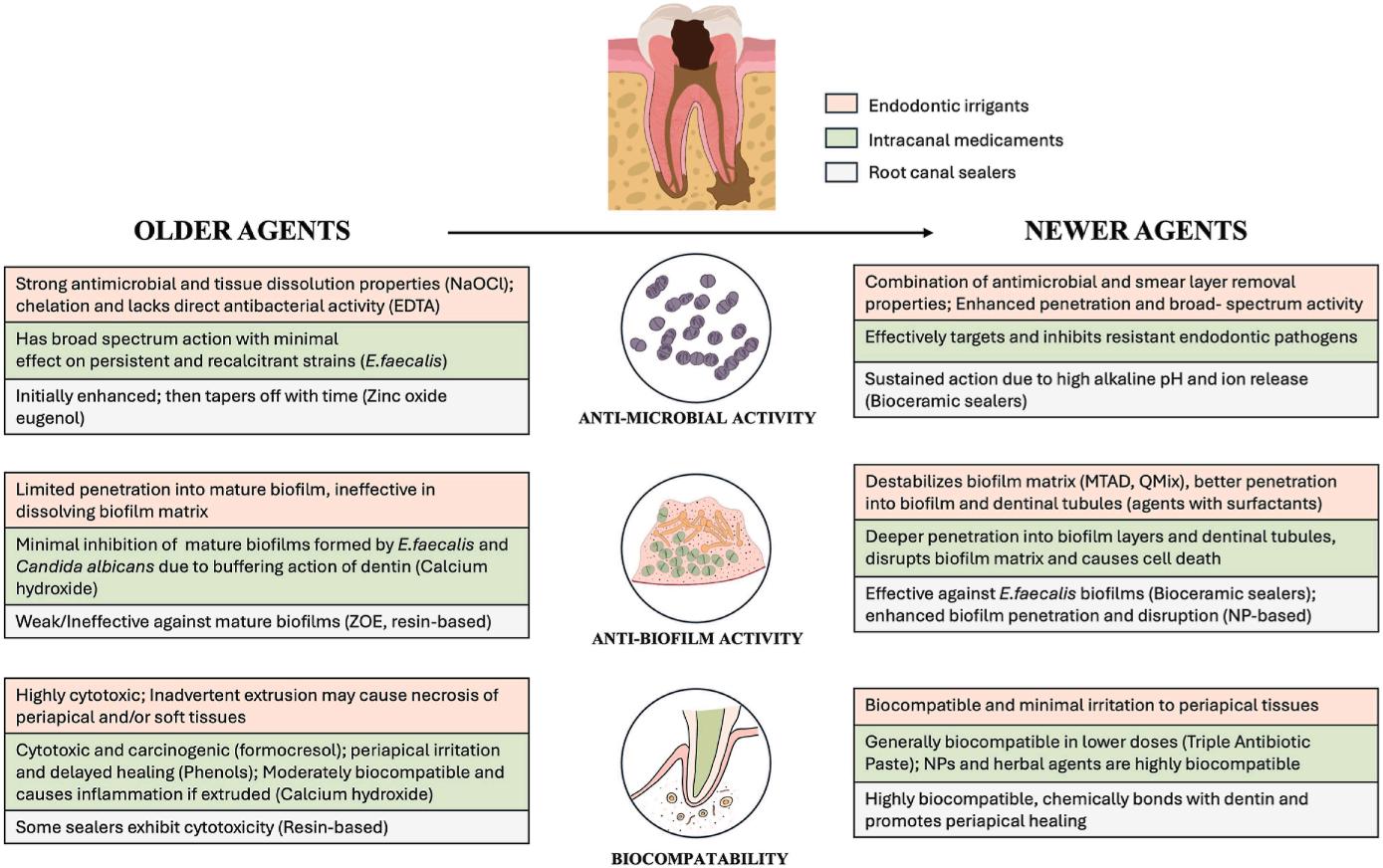
1. Schematic comparison of older versus newer intracanal agents based on antimicrobial activity, antibiofilm properties, and biocompatibility. (Courtesy of Pandian APG, Bellani D, Jibu RM, et al: Endodontic intracanal medicaments and agents; The old and the new, What is the evidence? Dent Clin N Am 70:45-62, 2026.)
TAP eliminates Enterococcus faecalis biofilms and has better antibacterial activity than Ca(OH)2 but is cytotoxic and has concentration-dependent effects on step cells of the apical papilla (SCAP). Low concentration of TAP has no significant adverse effects on SCAP survival and remains antibacterial against E faecalis. TAP also has some disadvantages, including tooth discoloration and structural alternation of the radicular dentin. Double antibiotic paste (DAP) without minocycline or with a substitute such as amoxicillin, clindamycin, or cefaclor can overcome the discoloration problem. Careful attention to the concentration used of Ca(OH)2 and TAP must be paid to optimize stem cell viability, antimicrobial effectiveness, and dentin integrity. TAP is also effective in managing teeth with periapical lesions, necrotic teeth with open apices, and traumatic dental injuries. It may prove beneficial for external inflammatory root resorption, controlling the resorptive process.
Triamcinolone and demeclocycline with anti-inflammatory and antibacterial properties has minimal local and systemic toxicity and facilitates the reduction in postoperative pain. Periodontal tissues tend to heal better and inflammatory root resorption is avoided.
Among the newer bioceramics are mineral trioxide aggregate (MTA), which has emerged as the most promising alternative to Ca(OH)2. The clinical outcomes and tissue healing are similar for Ca(OH)2 and MTA, but MTA is more desirable because of its shorter duration of treatment and single visit application. Among the applications for MTA are vital pulp therapy, apexification, root-end filling, management of perforation, and root resorption.
Metallic ions have been incorporated into bioceramic scaffolds to optimize antimicrobial activity. However, their reduced antimicrobial effectiveness and higher risk for tooth discoloration compared to Ca(OH)2 make them less than optimal for regular clinical applications.
The need to control drug-resistant bacteria has prompted the development of new, alternative antimicrobial treatments. In endodontics, these include antimicrobial peptides (AMPs), nanoparticles, and herbal-based medicaments.
AMPs offer potent antimicrobial activity, excellent biocompatibility, and minimal risk of bacterial resistance. AMPs have greater antibacterial and antibiofilm properties against E faecalis than conventional Ca(OH)2. In addition, AMPs don’t target specific proteins in bacteria, which makes it much harder for the bacteria to develop resistance to AMPs compared to traditional antibiotics.
A natural alternative to ICMs that are commercially available uses bioactive compounds from medicinal plants for root canal disinfection. They are readily accessible, affordable, have extended shelf-life, offer minimal toxicity, and have reduced microbial resistance. Some natural agents include curcumin, neem, aloe vera, propolis, and triphala. The drawback is the limited documentation of these agents in the literature. They are considered an adjunct rather than a replacement for conventional agents.
Nanoparticles encapsulating antibacterial agents are a recognized approach to enhance root canal disinfection. Ca(OH)2loaded poly lactic-co-glycolic acid (PLGA) nanoparticles have more antimicrobial activity because of the sustained release of
CA2+ and deeper penetration in the dentinal tubules compared to conventional Ca(OH)2. They are more able to inhibit osteoclastic differentiation, helping with root resorption.
Silver nanoparticles (AgNPs) offer potent antimicrobial, antibiofilm, and antifungal properties. Contact time is an important consideration, with the greater duration of exposure providing maximum action against the microbial agents. They may prove more useful as an ICM than as an irrigant. AgNPs also can enhance the properties of Ca(OH)2.
ICMs should be selected based on an accurate endodontic diagnosis. To eradicate the microbial species that causes inflammation, corticosteroids and antibiotics need to be accompanied by anti-inflammatories such as Ca(OH)2
Endodontic sealers with antimicrobial activity inhibit remnant microorganisms and prevent reinfection. The clinician should be aware of the value of traditional sealers as well as that of newer endodontic sealers.
ZOE has inherent antibacterial properties and serves as a traditional sealer. Ca(OH)2 can be used as a sealer and facilitates the calcification and repair of dentinal tissues. It has limited antibacterial activity against E faecalis, so newer antimicrobial alternatives are required.
Bioceramic sealers (BCS) have high alkalinity and ion release during setting, providing sustained antimicrobial efficacy. BCS has extended antibiofilm and antibacterial properties after setting, but the degree of antimicrobial activity differs with the different commercially available BCS. It can also be cytotoxic. Microbial
When the gag (pharyngeal) reflex is triggered, sensory nerves signal the brainstem, which activates motor responses in the throat muscles. This causes gagging or retching to expel any object, substance, or stimulus from the pharynx (Figure 1). The neurological pathways of the reflex include the trigeminal, vagus, and glossopharyngeal nerves, which also control nausea and vomiting, another protective mechanism to remove unwanted substances from the throat or stomach. Higher brain
growth can be hindered by incorporating nanoparticles into root canal sealers. Integrating AgNPs boosts the antimicrobial properties of Ca(OH)2 and other root canal filling materials. Incorporating quaternary ammonium polyethyleneimine (QPEI) nanoparticles and chitosan nanoparticles can produce robust antibacterial and antibiofilm properties.
BCS is the most biocompatible and bioactive sealer and forms chemical bonds to dentin. It creates an environment unsuited for bacterial survival.
Irrigants, medicaments, and sealers have changed modern endodontic practice, offering not just the traditional methods for root canal treatment but adding newer agents with potential advantages, including enhanced bioactivity, reduced cytotoxicity, and greater effectiveness against resistant microorganisms. In addition, conventional methods remain effective for successful endodontic outcomes. Clinical and long-term evidence is still lacking, so the use of these agents should follow careful evaluation of their properties and thorough investigation into the scientific evidence available.
Pandian APG, Bellani D, Jibu RM, et al: Endodontic intracanal medicaments and agents: The old and the new, What is the evidence? Dent Clin N Am 70:45-62, 2026
Reprints available from APG Pandian, General Dentistry Dental Clinic, 6130 Nevada Ave, Apt E107, Woodland Hills, Los Angeles, CA 91367, USA; e-mail: dr.anupriyagowtham@gmail.com
centers can also trigger the reflex, especially when the individual is experiencing disgust in response to textures, smells, or sights, for example. Somatic triggers are those stimulated directly through touch, and psychogenic triggers are stimulated via higher brain centers without contact. In a dental patient situation, the reflex can be triggered unnecessarily and complicate treatment. The Gag Severity Index and Gag Prevention Index are scales to categorize the reflex (Table 1). Possible methods for managing the gag reflex and suggested
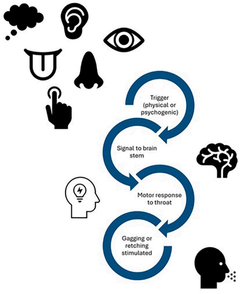
Figure 1. The gag reflex. (Courtesy of Tarnowski A: Gagging matters: A general dental practitioner’s perspective
Table 1. Gag Severity Index and Gag Prevention Index
Gagging Severity Index Grades (GSI)
Grade I Very mild Occasional and controlled by the patient
Grade II Mild Control is required by the patient with reassurance from the dental team
Grade III Moderate Consistent and limits treatment options
Grade IV Severe Some treatment is impossible
Grade V Very severe Affecting patient behaviour and dental attendance and making any treatment impossible
Gag Prevention Index (GPI)
Grade I Obtunded gag reflex Proposed treatment successful
Grade II Partially controlled gag reflex Proposed treatment possible
Grade III Partially controlled gag reflex but frequent gagging; Only simple treatment possible
Grade IV Inadequately controlled gag reflex Simple treatment unable to be completed
Grade V Gag reflex severe No treatment possible
(Adapted with permission from Fiske J, Dickinson C: The role of acupuncture in controlling the gagging reflex using a review of ten cases. Br Dent J 190:611-613, 2001. Courtesy of Tarnowski A: Gagging matters: A general dental practitioner’s perspective on managing the reflex. Br Dent J 239:624-629, 2025.)
guidance for specific circumstances in the dental setting were outlined.
Gagging is an involuntary response and no single method of control will fit all circumstances. Often the gagging is linked to patient anxiety, and failure to manage the reflex can contribute to the Berggren Cycle of Anxiety. The failure to manage the reflex in a dental intervention can cause patients to feel guilt and shame, leading them to avoid dental care and creating barriers to overcoming the response once a cycle has been established. A structured behavioral approach incorporating principles from cognitive behavioral therapy (CBT) has been recommended for developing behavioral strategies, practical steps, and coping mechanisms that can be tailored to the individual patient (Box 1).
CBT approaches involve a structured but flexible style that leads patients to gain confidence in managing their gag reflex, which can produce more successful and comfortable dental experiences. The steps recommended are as follows:
• Acknowledge the issue
• Normalize the reflex
• Clarify the need for management
• Agree on a strategy based on the patient’s needs
• Evaluate progress
• Build on success
• Record and apply, noting the effectiveness of techniques for future visits
The Singer’s Marble Technique is a 7-step desensitization program that begins with patients placing and holding 5 marbles in the mouth before the impression appointment, but this may no longer conform to health and safety requirements. Reassurance and encouragement, additional topical anesthesia, dullfinished plates, and a “training bead” can guide tongue position in preparation for a denture fit appointment. Other approaches include the use of gentle massage of the soft palate to sensitize the trigger area over time, the main amelioration of gagging indoctrination by communication (MAGIC), and psychogenic airway protection.
• Strategies described literature
• Psychological and behavioural approaches
• relaxation techniques
• Controlled breathing, such as MAGIC technique
• Distraction techniques, game playing
• Physical movement such as toe wiggling or leg lifting
• Audio-visual
• Progressive desensitisation, Singer method, Appleby & Bays method
• Behavioural modification (systematic desensitisation and reinforcement)
• Cognitive behavioural therapy
• Hypnosis
• Clinical and pharmacological interventions
• Friedman method which uses salt application on denture or tongue
• Topical anaesthesia, local anaesthesia or nerve blocks
• Inhalation sedation or intravenous sedation
• Herbal remedies, tanin patches
• Anti-nausea medications
• Acupuncture or acupressure
• Low-level laser therapy
• General anaesthesia
• Rubber dam use
• Adaptive and prosthodontic adjustments
• Desensitisation training
• Conditioning or training prosthesis
• Modifications to prosthetic design with reduced palatal coverage
• Scanning
(Courtesy of Tarnowski A: Gagging matters: A general dental practitioner’s perspective on managing the reflex. Br Dent J 239:624-629, 2025.)
Examination
Patients may mention having a sensitive gag reflex, and the clinician should conduct a thorough examination while avoiding sensitive areas. This requires staying buccally and avoiding touch of the soft tissues as much as possible. If gagging occurs, the patient should be encouraged to swallow, drink water, rinse, or take a deep breath to re-center and regain control. The initial encounter is critical in setting the tone for how care will be conducted and whether the patient has confidence in the clinician’s ability to help.
The tell-show-do approach is highly effective in managing patients with a sensitive gag reflex. The patient can be shown the radiograph film, noting especially the corners and edges, with an explanation of how it will be positioned in the mouth. Topical local anesthetic can be helpful in reducing sensitivity. Patients are encouraged to focus on their breathing and consciously squeeze the film holder between their teeth. Patients should experience the duration required for exposure to help them gauge the timing and control needed. It can be
helpful to have the patient count over the length of time taken by the clinician moving to the doorway for exposure. Children can have difficulty even with the smallest size 0 film, so extraoral radiography may offer an alternative to build patient confidence for future intraoral imaging.
Local anesthetic (LA) and inferior dental blocks can help suppress triggers involving the tongue or palate. Patients may benefit from practice tolerating an aspirator tube, tongue flange, or curly straw at home to reduce their sensitivity and build resilience in extending the duration of the process. If such home practice isn’t possible, a structured time limit during treatment helps patients know how long they need to maintain an open mouth while concentrating on breathing. Adjustments can be made based on patient response. The dentist should establish clear hand signals to indicate when to pause and when to continue. A rubber dam can isolate the working area and reduce stimulation to the soft palate and posterior tongue. Caution should be taken because rubber dams can induce claustrophobia in some patients. A tongue flange or curly straw may also provide a more secure alternative to a dental mirror in protecting soft tissues.
Clinicians can help patients during impression taking by encouraging controlled breathing and remaining calm, while they position the patient upright and leaning slightly forward. Impression trays should not be overfilled, especially in the palatal area. Smoothing the material and using an appropriate quantity can minimize gagging. The use of quick-setting materials and allowing the patient to practice desensitizing exercises and to use distraction techniques as indicated can help maintain the needed compliance. Some patients may have good results by pairing distraction with rhythmic breathing or guided focus. Repeating these techniques over time can train the brain to release its focus on discomfort. Visual and auditory distractions such as watching TV, listening to music, or using virtual reality (VR) headsets can divert attention away from the mouth, reduce a hyperaware gag reflex, and minimize discomfort. Among the alternative approaches that may be tried are the following:
• Digital intraoral scanning
• Inhalation sedation
• Acupuncture and acupressure
Patients with a hyperactive gag reflex may struggle to adapt to wearing dentures. Their ability to tolerate dentures can be dictated by the design and fit of the prosthesis as well as their personal adaptive process. The clinician can facilitate a better adaptive process through several measures. Engaging with the clinician can help patients adjust their individual preferences, expectations, and concerns, enhancing satisfaction levels. Learning
to wear a denture is like learning to walk with a prosthetic limb and requires time. Managing the patient’s expectations, especially if he or she has a strong gag reflex, is an essential part of the process.
Patients can also be given smaller dentures with minimal palatal coverage to help reduce gagging. Extension and peripheral seal are critical, especially for mucosal-borne dentures. However, smaller dentures may also reduce retention and increase mobility and gagging.
Patients can gradually increase their wear time. Setting up a structured approach, such as tolerating the denture for the duration of a song, a short video, or eventually a full-length film can help in the adaptation process. Taking rest breaks as they adapt allows patients to make incremental progress and not jump into full-time wear. Some patients can benefit from a gradual adaptation using a training plate with no teeth before moving into full dentures. Patients who have never successfully worn a denture before will require a period of learning and adapting while the denture is being constructed.
The sensitivity of a gag reflex in some patients can make dental procedures challenging. Management strategies can be fit to the patient’s needs and improve comfort and the chance of success.
Clinicians should consider how the denture will fit each unique patient and tailor their approach to what works best for the patient. Treatments can be made more tolerable through a variety of approaches, and patients and clinicians should work together and exercise patience in adapting to the needs required for their oral health care procedures.
Tarnowski A: Gagging matters: A general dental practitioner’s perspective on managing the reflex. Br Dent J 239:624-629, 2025
Reprints available from A Tarnowski; e-mail: a.tarnowski@nhs.net
Primary teeth are of vital importance for the development of young children, so oral health care providers make every effort to maintain their functionality in the mouth as long as possible. Usually this involves addressing dental caries, but dentoalveolar traumatic injuries, developmental dental anomalies, and erosive tooth wear, among other conditions, may also need to be managed. The choice of restorative materials must consider the patient’s anatomical and functional characteristics and how these primary teeth differ from immature or mature dentition. Among the materials often used are resin-based restorative materials, prefabricated or custom-made crowns, and glass ionomer-based materials. The newest glass hybrids (GHRs) have an increased level of reactivity and significantly improved mechanical properties. In addition, since they are applied with a light-cured surface coating material containing nano-filled resin, their performance is comparable to that of resincontaining systems. Adhesion of glass ionomer to dental tissues is an indicator of success, with close contact between the material and tooth surfaces required. To attain this degree of
adhesion, removal of the smear layer is strongly recommended. Cavity conditioners provide an appropriate adhesion by removing the smear layer on the cavity before placing the glass ionomer. These products generally consist of various dilutions of polyacrylic acid, citric acid, acrylic acid, ethylenediaminetetraacetic acid (EDTA), or phosphoric acid. Tests to measure the success of restorative materials via in vitro studies include internal adaptation (IA), which focuses on the adhesion of restorations. An in vitro study was conducted to compare the effects of various cavity conditioners on the IA of glass ionomer−containing restorative materials applied to primary teeth.
The 4 groups of cavity conditioners consisted of 80 extracted primary second molar teeth that were subjected to 10% or 20% polyacrylic acid, 17% EDTA, or 35% phosphoric acid. Class V cavities were prepared on the buccal surfaces before the cavity conditions were applied (Figure 1). Samples in each group were randomly assigned to GHRs or conventional glass ionomer restoratives. Restorative materials were applied, then the samples
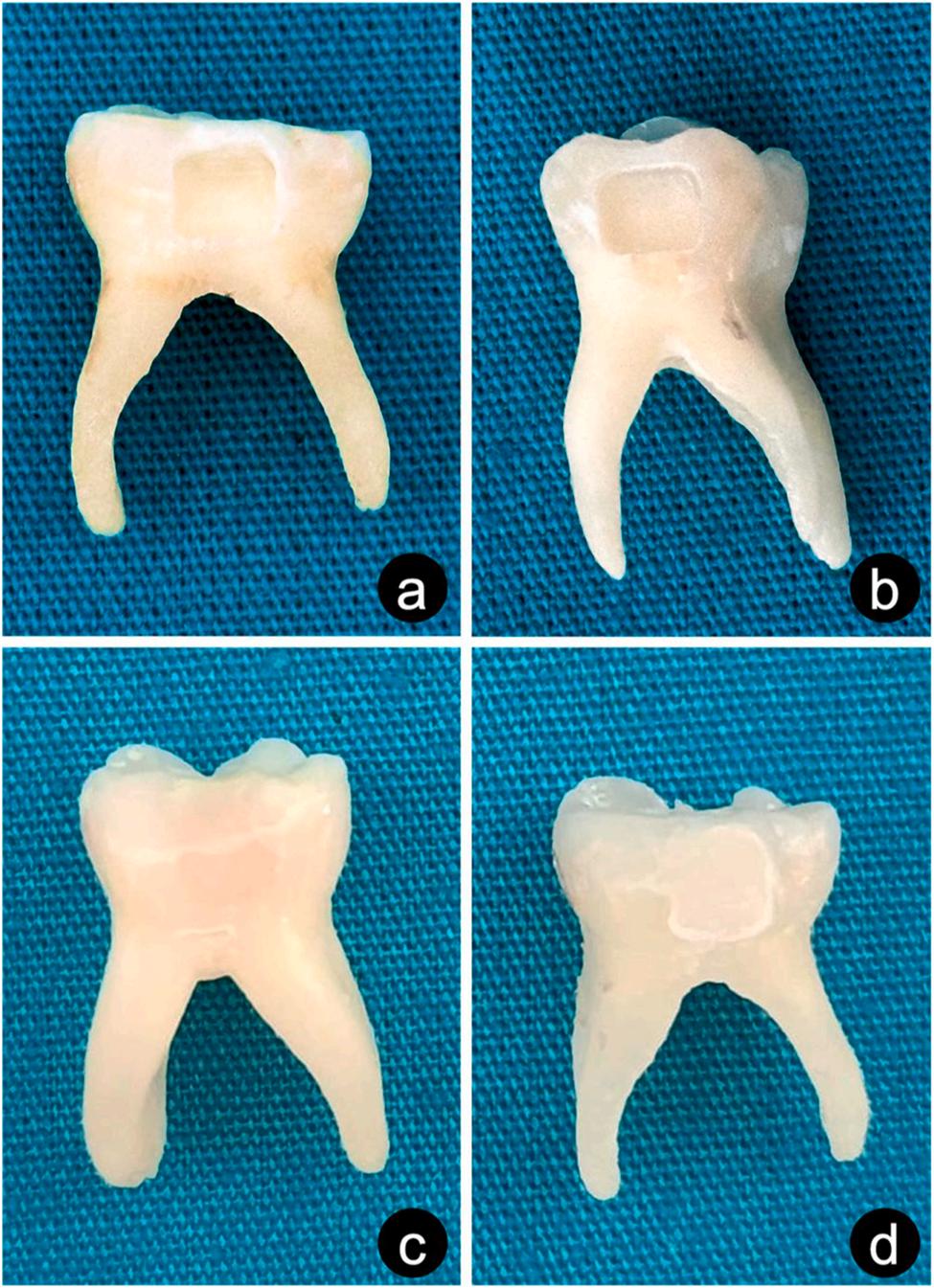
Figure 1. Representative images of the cavities prepared in the teeth (A and B). Samples with completed restoration procedures (C, glass hybrid restorative; D, conventional glass ionomer restorative). (Courtesy of Gurler S, Demirel A, Buyuksungur A: The evaluation of internal adaptation of glass ionomer restorations applied after the use of different cavity conditioners in primary teeth: An in-vitro study. BMC Oral Health 25:36, 2025.)
were thermocycled before having the IA calculated volumetrically.
The first comparison focused on the gaps seen at the cavityrestoration interface in GHR restorations. The highest IA quality was seen with 35% phosphoric acid, and the worst was found for 17% EDTA. The differences between the gaps in the cavityrestoration interface were statistically significant.
The post-hoc tests found no statistically significant differences between 10% and 20% polyacrylic acid, between 10% polyacrylic acid and 17% EDTA, between 10% polyacrylic acid and 35% phosphoric acid, or between 20% polyacrylic acid and 35% phosphoric acid. Significant differences were noted between 35% phosphoric acid and 17% EDTA.
Statistical comparisons between the gaps seen at the cavityrestoration interface in conventional glass ionomer restorations
found a significant difference between the conditioners for gaps in the conventional glass ionomer system at the cavityrestoration interface.
Post-hoc tests showed no significant differences between 20% polyacrylic acid and 17% EDTA or between 20% polyacrylic acid and 35% phosphoric acid. Significant differences were found between 10% and 20% polyacrylic acid; between 10% polyacrylic acid and 17% EDTA; between 10% polyacrylic acid and 35% phosphoric acid; and between 17% EDTA and 35% orthophosphoric acid.
When comparisons were made between the restorative materials in terms of IA quality, after 10% polyacrylic acid was applied, the gaps between conventional glass ionomer restorative and cavity surfaces were significantly greater than those between GHR and cavity surfaces. The gaps between the conventional glass ionomer restorative material and cavity surfaces were significantly greater than those seen between the GHR restorative material and cavity surfaces after 20% polyacrylic acid application. No significant difference in IA quality was noted after 17% EDTA application between conventional glass ionomer restorative and GHR restorations. After 35% phosphoric acid was applied, the gaps between conventional glass ionomer restorative and cavity surfaces were significantly greater than the gaps between GHR restorative material and cavity surfaces.
The most successful cavity conditioners based on IA were 35% phosphoric acid and 20% polyacrylic acid. The use of GHR restoratives had superior IA quality compared to conventional glass ionomer.
Although the results of this study remain to be validated through further, comprehensive in vitro tests and randomized clinical trials, it appears that 35% phosphoric acid and 20% polyacrylic acid achieved better results as cavity conditioners. The GHR system provided better results as a restorative material compared to the other selections.
Gurler S, Demirel A, Buyuksungur A: The evaluation of internal adaptation of glass ionomer restorations applied after the use of different cavity conditioners in primary teeth: An in-vitro study.
BMC Oral Health 25:36, 2025
Reprints available from A Demirel; e-mail: akifdemirel@ankara. edu.tr
Pain management during dental procedures helps provide comfort for the patient, reduces anxiety, improves the experience, and may help to prevent complications and achieve a successful result. Potent anesthetic agents can be used, along with anesthetic techniques that minimize any discomfort. Mepivacaine is the third most widely used local anesthetic in dentistry and has the same potency as lidocaine but is milder in vasodilation, ensuring a longer duration of anesthesia. The advised 3% mepivacaine has a rapid onset, greater absorption in the maxillary anterior area, ideal anesthetic effect, and few adverse effects on the cardiovascular system. Techniques that reduce discomfort during injections include the intraligamentary injection (ILI) technique and the incisive nerve block (INB). A comparison of the anesthetic success rates of the ILI and INB techniques using 3% mepivacaine hydrochloride for extractions of the mandibular anterior and premolar teeth was conducted, noting the patients’ ability to tolerate anesthesia and the extraction of mandibular anterior or premolar teeth.
One hundred participants (42 women and 58 men, mean age about 51 years) were undergoing the extraction of mandibular anterior teeth and premolars. They were randomly assigned to 1 of the 2 techniques (INB for group A, which served as the control group, and ILI for group B), but topical 3% mepivacaine was applied to both groups. The 2 techniques were compared for effectiveness using the Modified Dental Anxiety Scale for Dental Extraction Procedure (MDAS-DEP) and a visual analog scale (VAS) during the injection and extraction stages of the procedures.
No failures occurred with INB, so no further injections were required. The mean VAS score for the INB group was 6.14 after injection and 3.86 after extraction. In comparison, the ILI group
had a mean VAS score of 5.46 after injection and 2.90 after extraction. The pain level values were statistically significantly different between the 2 groups.
The MDAS-DEP scales revealed a statistically significant difference within each group after injection and extraction. Mean MDAS-DEP score for the INB group was 15.86 during injection and 11.26 during extraction. The values for the INB group were slightly higher than those in the ILI group, which were 15.68 after injection and 10.94 after extraction. The difference was not significant when MDAS-DEP scores from injection to extraction between the 2 groups were compared.
The ILI technique was less painful than the INB technique during injection and achieved profound pain relief during extraction. ILI may be appropriate for use as the only anesthetic technique when lower anterior and premolar teeth are being extracted.
The adverse effects associated with INB were not seen with ILI, which also eliminated lip biting. For patients who have infections or inflammation, care should be taken when using the ILI technique.
Salem S, Saad I, Elmoazen R, et al: Anaesthetic efficacy of intraligamentary injection compared to incisive nerve block using 3% mepivacaine hydrochloride: A randomized clinical trial. BMC Oral Health 25:90, 2025
Reprints available from S Salem; e-mail: Suzan.salem.dent@ alexu.edu.eg
Patients with mild bleeding disorders (MBDs) aren’t unusual in the dental office, but up to 60% of these individuals may have a bleeding disorder of unknown cause (BDUC), with no identifiable etiology even after a hematological evaluation. BDUC is a diagnosis of exclusion and is defined as abnormal or excessive bleeding in patients whose hemostatic evaluation is normal and who have no known cause for the bleeding. These cases can be complicated if the patient experiences excessive bleeding before BDUC has been diagnosed. The incidence of bleeding in patients with BDUC after dental surgery ranges from 36% to 84%. Dentists need to be aware of BDUC and follow the proposed guidelines and precautions for patients requiring dental surgery.
Clinical Presentatio n
BDUC has a bleeding phenotype similar to that of other MBDs and may cause epistaxis, hematomas, hemarthrosis, menorrhagia, postpartum menorrhagia, hematuria, or post-surgical bleeding. Women are affected more often than men. Patients with BDUC can have reduced health-related quality of life, including role limitations, reduced mental health, impaired social function, and body pain. They may be unable to access dental care because of physical impairment, fear of bleeding, and dentists who are unwilling to treat them because they are unaware of the condition.
A complete medical history, physical examination, family history, surgical history, and hemostatic evaluation are needed to make the diagnosis. If the results are normal, BDUC may be diagnosed, or advanced tests may be done to rule out rare causes of bleeding. The pathophysiology remains unknown but is most likely a result of a heterogeneous group of underlying defects that cause bleeding symptoms or a collection of disorders not yet identified. A family history of bleeding is seen in up to 40% of patients.
The differential diagnosis of BDUC includes mild hemophilia, von Willebrand disease, platelet function disorders, connective tissue or vascular disorders, and ascorbic acid deficiency. If the patient bleeds excessively after dental extractions, he or she can be referred to hematology for assessment.
Among the oral signs of BDUC are significant bleeding during dental extractions and a history of bleeding after dental extractions. Oral mucosal bleeding is seen in 19% to 53% of patients, along with bruising or petechiae of the oral mucosa, and gingival bleeding without signs of periodontal disease. BDUC can be associated with anemia or iron deficiency, resulting in angular cheilitis, glossodynia, mucosal pallor, ulceration, and glossitis intraorally. If the patient has ascorbic acid deficiency, the dentist may note gingival manifestations such as bleeding, swelling, or hypertrophy along with loosening or loss of teeth. Hypermobile Ehlers-Danlos syndrome (EDS) may cause oral ulcers, TMJ dislocation, microdontia, enamel and dentin defects, pulp calcification, and root abnormalities.
Before invasive dental surgery, the patient should provide a medical, surgical, and pharmacological history and be evaluated for the need for specialist hematological treatments. The dentist should consider the complexity and invasiveness of the procedure and his or her ability to safely perform the treatment. Historical information to be noted include history of a previous episode of prolonged bleeding after trauma, surgery or dental procedures and any family history of coagulation disorders. Some medications can increase the risk of bleeding, as can underlying medical conditions such as liver disease, advanced heart failure, and alcoholism.
The dentist should also take into account any operator and procedural risks, including the experience of the practitioners and familiarity with the treatment or with specialist care. HEMSTOP (Table 1) can be used to assess bleeding risk and the need for referral for additional testing or management.
The 2024 guidelines from the International Society on Thrombosis and Haemostasis offer general information about managing BDUC patients who require surgery. Treatment options are observation only, tranexamic acid (TXA) only, and TXA and desmopressin (DDAVP). For more invasive surgery, fresh frozen plasma, platelet transfusions, or recombinant factor VIIa may be considered (Figure 1).
Table 1. HEMSTOP (Haematoma, haEmorrhage, Menorrhagia, Surgery, Tooth Extraction, Obstetrics, and Parents) Questionnaire to Assess Bleeding Risk.
Question
Have you ever consulted a doctor or received treatment for prolonged or unusual bleeding (such as nosebleeds, minor wounds)?
Do you experience bruises or haematomas larger than 2 cm without trauma or severe bruising after minor trauma?
After a tooth extraction, have you ever experienced prolonged bleeding requiring medical or dental consultation?
Have you experienced excessive bleeding during or after surgery?
Is there anyone in your family who suffers from a coagulation disease (e.g., BDUC, haemophilia, von Willebrand disease)?
Have you ever consulted a doctor or received treatment for heavy or prolonged menstrual periods (e.g., contraceptive pill, iron supplements)?
Did you experience prolonged or excessive bleeding after delivery?
to
Male/female 1
Male/female 1
Male/female 1
Male/female 1
Male/female 1
Female 1
Female 1
Interpretation: <2 points = low risk of bleeding, no further investigations needed; ≥2 points = increased risk of bleeding, further investigations recommended.
Maximum score: 7 for female: 5 for male only.
Note: This table uses ‘female’ to reflect the language used in the original study, while recognizing that not all people who experience menstrual periods or can get pregnant identify as female, and not all those who identify as female experience menstrual periods or can get pregnant.
(Adapted with permission from Bonhomme F, Boehlen F, Clergue F, et al: Preoperative hemostatic assessment: A new and simple bleeding questionnaire. Can J Anesth 63:1007-1015, 2016. Courtesy of Khan I: Dental surgery for patients with bleeding disorder of unknown cause. Br Dent J 239:402-405, 2025.)
Figure 1. A summary of BDUC for dentists. (Adapted with permission from Bonhomme F, Boehlen F, Clergue F, et al: Preoperative hemostatic assessment: A new and simple bleeding questionnaire. Can J Anesth 63:1007-1015, 2016. Courtesy of Khan I: Dental surgery for patients with bleeding disorder of unknown cause. Br Dent J 239:402-405, 2025.)
The guidelines indicate that patients having simple extractions may be covered by TXA only, but those undergoing surgical or multiple extractions may require TXA and DDAVP or DDAVP only. Patients should be reassured that spontaneous life-threatening bleeding from BDUC is very rare and the precautions taken can see them through dental surgery. A hematologist should evaluate the patient before surgery and other invasive dental procedures.
Khan I: Dental surgery for patients with bleeding disorder of unknown cause. Br Dent J 239:402-405, 2025
Reprints available from I Khan; e-mail: dental.research@mpft.nhs.uk
The number of mouthwashes available over the counter (OTC) continues to grow. Some are clinically effective for managing dental caries that involve hard tissues, and others are more effect for periodontal diseases that affect soft tissues. Among the chemicals in these mouthwashes are fluoride, chlorhexidine (CHX), essential oils (EOs), and cetylpyridinium chloride (CPC), all of which can effectively reduce bacterial plaque biofilms on teeth and reduce gingival inflammation. Dental practitioners need to remain current on the evidence found through well-conducted studies. They should consider the patient’s specific oral disease and the active ingredients in mouthwashes that can address the problem and then be able to share that information with patients. Updates for dental clinicians regarding the use and effectiveness of antimicrobial mouthwashes for oral disease, information about the benefits and risks of the formulas, and advisable techniques for using mouthwashes in their oral health care regimen were detailed.
Caries Guidelines
Systematic reviews show that fluoride toothpastes prevent plaque formation and reduce dental caries in children. Current UK guidelines suggest performing twice-daily brushing with 1350 ppm fluoride toothpaste in children age 8 years or older to reduce dental caries. Mouthwashes with approximately 225 to 230 ppm of fluoride can be used daily to also address caries in children. The fluoride in mouthwashes has not been sufficiently studied in adults other than for root
caries, with other agents included to address plaque and gingivitis.
CHX and/or hydrogen peroxide have been seen as appropriate for the treatment of gingivitis, but CHX alone is relatively ineffective against more advanced periodontal disease. Mouthwashes containing adjunctive EOs and/or CPC can significantly reduce plaque and bleeding scores along with clinical signs of gingivitis. They can form a personalized preventive approach for early periodontal disease.
For more advanced periodontitis, adjunctive CHX mouth use should be continued for 2 to 4 weeks to prevent extrinsic staining. The patient should also perform oral hygiene and see the dental practitioner for professional mechanical plaque removal (PMPR). Differing percentages of CHX are advised in different situations. The PMPR−0.2% CHX regimen is advised for reducing pocket depths. EOs and CPC can be used adjunctively for periodontitis.
In the most severe periodontal disease (Stage IV), tooth prognosis is hopeless, so no amount of mouthwash will reach the depths of periodontal pockets or restore lost bone. Dental practitioners need to explain to patients why mouthwashes aren’t effective at this stage.
Based on current guidelines coupled with the best available evidence, clinicians should use fluoride mouthwash if the patient is
at high caries risk along with an antimicrobial mouthwash adjunctively for periodontal disease as indicated. Dental healthcare providers should also advise patients to use the mouthwash after brushing but should not use water. This contradicts the previous stance that patients should “use mouthwashes at some time other than after brushing.” Additional research is needed for further clarification.
Because antimicrobial mouthwashes tend to contain a mixture of active ingredients, it can be difficult to select specific mouthwashes to manage specific diseases or avoid side effects. Natural antimicrobial mouthwashes can vary in their effectiveness because of the different extracts and formulations.
Antimicrobial mouthwashes are either bactericidal or bacteriostatic, killing or reducing the bacterial load in the mouth. However, a healthy mouth contains microbial diversity, including commensal species. Mouthwashes that destroy healthy species can support the overgrowth of healthy bacteria, causing dysbiosis and compromising oral health. Ideally, mouthwash constituents should address oral disease without causing oral microbiome dysbiosis or reducing the abundance of ‘healthy’ bacteria. One mouthwash tends not to do it all. This requires a balance of the clinical benefits versus the oral and systemic risks of mouthwashes. Dental professionals can advise fluoride use for caries prevention because the beneficial effects on caries prevention currently outweigh the unsubstantiated risks or fluorosis as a result of overdose.
Dental caries is caused by bacteria, and if they enter the bloodstream through an infected tooth, a systemic situation can result. Links have been found between periodontal disease and diabetes, hypertension/cardiovascular disease, rheumatoid arthritis, Alzheimer’s disease, and various cancers. Poor oral health puts a patient at higher risk for systemic disease, so using mouthwash effectively as an adjunct to daily oral health practices should outweigh its risk.
The byproduct of the alcohol is a carcinogen, and smokers are at increased risk of oral cancer when they consume alcohol. Alcohol is cytotoxic to cells in the laboratory, but no definitive proof exists that alcohol-containing mouthwashes cause oral cancer. Clinicians can advise their patients who worry about alcohol that there are many alcohol-free mouthwashes available, so they should choose one they like.
Some evidence indicates that patients can have anaphylactic reactions to CHX mouthwash, although it’s rare. The choice of a mouthwash should be based on a risk-versus-benefit decision by the patient. Clinicians should ask patients about their oral home care regimen and be prepared to sensitively correct any misinformed routines.
Use of mouthwashes along with mechanical cleaning is an appropriate measure for preventing dental caries and periodontal diseases. Plaque biofilms and inflammation can be managed, and early periodontal disease can be addressed.
Clinicians are responsible for providing valuable information about the use of mouthwashes and other oral home care products to their patients. It’s important to ensure that these products are selected based on a careful risk-to-benefit analysis and take into account a complete knowledge of the patient’s oral health status.
Brookes Z, Bescos R, Witton R, et al: An update on mouthwashes: Advice for dental practitioners. Br Dent J 239:312-315, 2025
Reprints available from Z Brookes; e-mail: zoe.brookes@ply mouth.ac.uk
Differentiating between the site of pain and the source of pain is essential to reproduce, locate, and manage the discomfort patients experience. Mounting evidence shows that toothaches can be of nonodontogenic origin. A number of pain referral sources exist for nonodontogenic “toothaches”
(NOTAs), which account for about 2.5% to 4% of toothaches in dental offices. About 10% have concomitant odontogenic causes. NOTAs are more common in patients between the second and sixth decades, affect maxillary dentition more than mandibular dentition, and are more common in molars, followed by premolars and incisors. The various
Causes of NOTAs Key Clinical Features Management
Myofascial
Psychogenic
Infections
GCA andother vascular entities
Cardiac
Neurovascular
Sinus/nasal mucosal origin
Systemic
TN and other neuropathic pain
Central lesions and malignancies
Bisphosphonates and chemotherapeutic drugs
Miscellaneous
Toothache referred from muscle; presence of trigger points; and increased pain on function
Toothache; hallucinations; delusions; and absolute nonreproducibility of familiar pain from any somatic structures
Toothache; history of systemic infections
Toothache; jaw claudication; gingival pain; and temporal pain
Toothache; jaw pain; neck pain; shoulder pain; left arm pain; and chest pain
Toothache; unilateral facial pain; and ipsilateral autonomic features
Toothache; nasal discharge; facial fullness; and nasal congestion
Toothache; burning pain; and other symptoms
Toothache; trigger zones; extraoral and intraoral triggers; and refractory period
Numbness, tingling, toothache, hyperalgesia, and paresthesia
Toothache; jaw pain; numbness; and tooth mobility
Tooth ache and other symptoms
Thorough medical history; clinical and radiographic examination; reproduction of pain; diagnostic block;trigger point injection; physical therapy; laboratory investigations; dietary modifications; pharmacologic management; and interdisciplinary approach
Thorough medical history; clinical and radiographic examination; cognitive behavioral therapy; appropriate referral; and interdisciplinary approach
Thorough medical history; clinical and radiographic examination; laboratory investigations; and interdisciplinary approach
Thorough medical history; clinical examination; laboratory investigations; imaging; and interdisciplinary approach
Thorough medical history; clinical and radiographic examination; pharmacologic testing; prompt referral; and interdisciplinary approach
Thorough medical history; clinical and radiographic examination; pharmacologic testing; laboratory investigations; and interdisciplinary approach
Thorough medical history; clinical and radiographic examination; diagnostic block; and interdisciplinary approach
Thorough medical history; clinical examination; imaging; laboratory investigations; and interdisciplinary approach
Thorough medical history; clinical examination; imaging; diagnostic block; and interdisciplinary approach
Thorough medical history; clinical examination; imaging; and interdisciplinary approach
Thorough medical history; clinical examination; imaging; and interdisciplinary approach
Thorough medical history; travel history; clinical examination; imaging; and interdisciplinary approach
(Courtesy of Thomas DC, Somaiya T, Ajayakumar A, et al: Toothaches of non-odontogenic origin. Dent Clin N Am 70:209-224, 2026.)
pathophysiologies and differential diagnoses seen in NOTAs were discussed.
Convergence occurs when multiple presynaptic neurons signal input onto a single postsynaptic neuron. Diverse pain referral patterns are observed as a result of convergence in the orofacial region. Because of the presentation, both patients and clinicians can be confused, causing underdiagnosis, misdiagnosis, and suboptimal pain management of NOTAs. The lack of knowledge about the mechanisms can eventually cause chronicity of pain, reduced quality of life, and disability.
NOTAs can be of myofascial, neurovascular, trigeminal neuropathic and neuropathic, cardiac, infectious, giant cell arteritis and other vascular causes, systemic, related to biosphosphonate and chemotherapeutic drugs, of sinus/nasal mucosal nature, central lesion related, resulting from miscellaneous conditions, and
of psychogenic origin (Table 2). Myofascial pain, trigeminal neuropathic pain, and neurovascular pain syndromes are seen most often (Figure 1).
Myofascial pain originates from muscles and fascia and is a major cause of NOTA. The disorders are characterized by pain referred from a trigger point in the muscle to a tooth or teeth. The myofascial trigger point is a hyperirritable spot in the skeletal muscle and is part of the patient’s chief complaint, with the ability to reproduce the known pain held by an active trigger point, whereas those incidentally found are considered latent trigger points.
The NOTA of myofascial origin can mimic the pain of pulpitis or tooth sensitivity, which makes misdiagnosis and improper treatment not uncommon. Diagnostic local anesthesia may be used to anesthetize the tooth, which is the site but not the source of the pain. When the pain isn’t relieved and the toothache is reproduced by activating a trigger point, the clinician is more likely
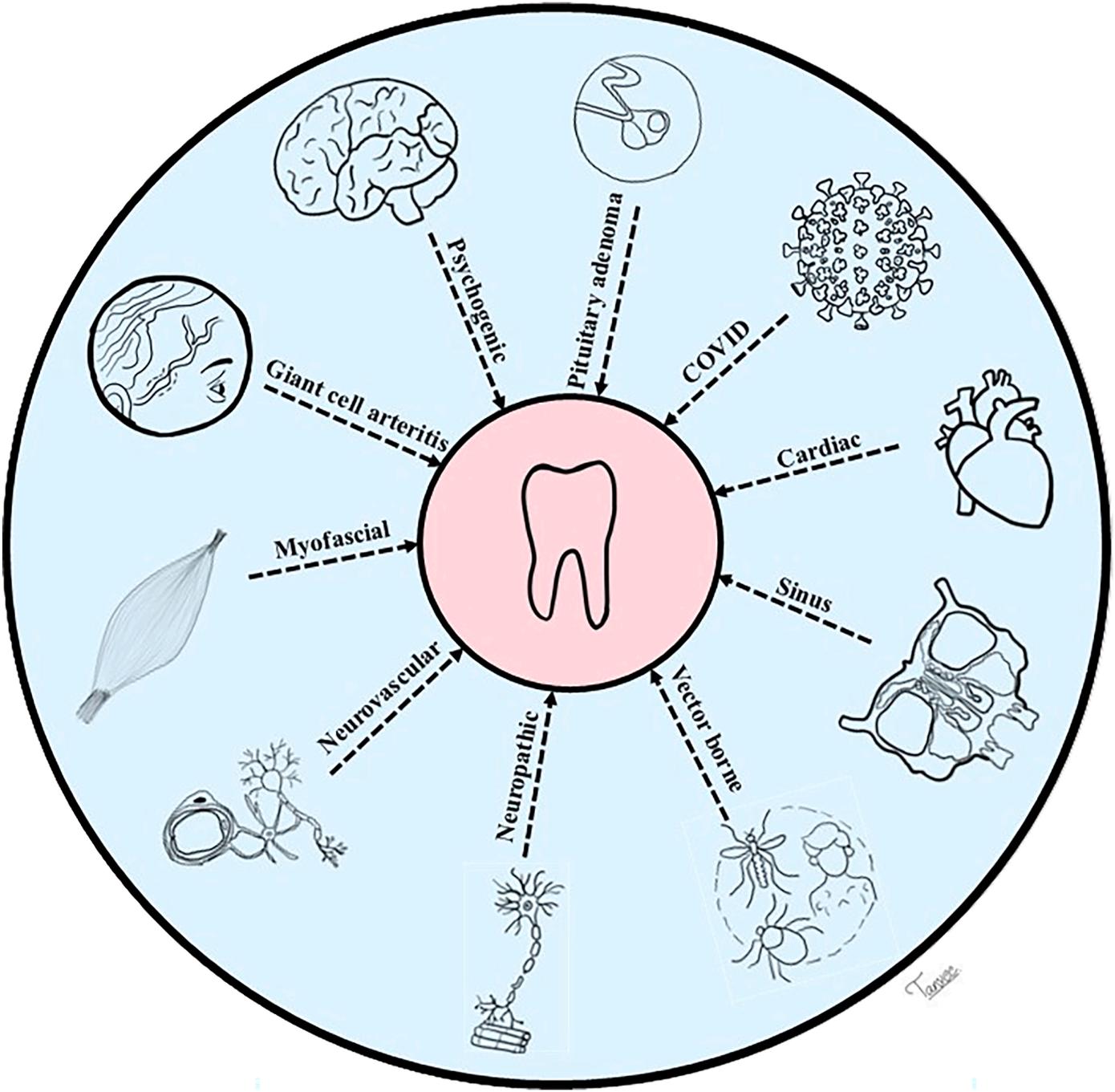
to make a proper diagnosis. Other indicators of a myofascially induced odontalgia include muscle function increasing toothache intensity, no abnormal clinical and radiographic dental findings, negative pulp testing, and reduced or relieved toothache when the proper trigger point is injected.
Included in neurovascular headache disorders are neural and vascular pathophysiologies such as migraines and trigeminal autonomic cephalalgias (TACs). The manifestations include pulsatile cephalalgia, allodynia, photophobia, phonophobia, nausea, and periorbital pain. Autonomic features can accompany pain attacks. Facial migraine or midface migraine may affect the face and teeth. TACs have unilateral facial pain and sometimes dental pain. Unnecessary invasive dental procedures may be undertaken if misdiagnosed. Comprehensive clinical assessment and investigative protocols are vital.
Trigeminal neuralgia (TN) is marked by paroxysms of pain triggered by innocuous stimuli and causing toothaches. The probability of maxillary and mandibular teeth being affected by NOTA secondary
to TN could be high. The NOTA manifests as paroxysmal unremitting pain lasting more than 3 months in the affected tooth. Triggers for NOTA secondary to TN can be brushing the teeth, eating cold foods, chewing, pressing or touching the gums, cold breezes, and drinking beverages. Patients with chronic NOTA may have complete or partial edentulous arches, whereas others may have dentinal sensitivity without any obvious cause. Interdisciplinary management approaches are needed to determine the need for surgical or medical interventions.
If there has been trauma to the trigeminal nerve fibers, persistent posttraumatic trigeminal neuropathic pain may develop and is seen in 2% to 15% of patients. Traumatic neuroma involves abnormal proliferation of a damaged nerve. Oral traumatic neuroma may present as NOTA and can be diagnostically challenging. Patients may experience altered sensation.
Convergence may lead angina of cardiac origin to present as jaw pain or toothaches. Even without chest pain, the most commonly reported orofacial pain sites of cardiac origin are the throat, teeth, ear, and temporomandibular areas. After ruling out the
orofacial source or dental source of pain, cardiac origins may be suspected. In these cases, the patient should be promptly referred to emergency care for urgent evaluation and management.
Lyme disease can affect multiple systems and cause symptoms such as toothache. A complete travel history, clinical examination, and detailed blood investigations are required for a proper diagnosis of Lyme disease, which can then help to diagnose NOTA. Dental pain can also accompany coronavirus disease 2019 infections, human immunodeficiency virus infections, herpes zoster infections, and chikungunya (oral mucosal pain and TMDs).
Giant cell arteritis (GCA) affects the medium and large arteries, especially the temporal artery. GCA has been associated with pain on chewing, toothaches, and gingival pain. NOTA should be considered one of the differential diagnoses if other clinical presentations are seen. Monckeberg medial sclerosis may cause facial pain in a similar manner. An interdisciplinary approach may be required to identify similar vascular entities that present as NOTA.
Sickle cell disease, benign and malignant neoplasms, and demyelinating disorders such as multiple sclerosis can manifest as NOTAs. The vaso-occlusive crises of sickle cell disease may cause pulpal necrosis in an otherwise healthy tooth. Multiple sclerosis patients may develop NOTA as a result of secondary TN. Burkitt’s lymphoma and Wegener’s granulomatosis as well as genetic factors can contribute to NOTA.
Bisphosphonates cause osteonecrosis of the jaws, producing jaw pain, numbness, and tooth mobility, along with other symptoms. Patients may experience and complain of toothache. Neurotoxicity caused by chemotherapeutic drugs can cause NOTAs. Clinical and radiographic evaluations coupled with a thorough medical history including medications can help in making an accurate diagnosis and referral for interdisciplinary management.
Both anatomy and close relationships between the maxillary sinus and maxillary teeth coupled with common innervations and neural convergence can allow NOTA to develop. Pain can be referred from the maxillary sinus to the teeth. Nasal mucosal infections can also refer pain to the maxillary teeth. Perineural inflammation, which causes pain in the target organ innervated by the nerve, can lead to tenderness to percussion on the maxillary posterior teeth, cold hypersensitivity and secondary hyperalgesia, and pain in the maxillary posterior area. NOTA caused by sinusitis can be equivocal with local anesthetic blocks. Careful history gathering, clinical examination, and radiographs are needed to differentiate between odontogenic
toothache and NOTA caused by maxillary sinusitis. Diagnosis and management are challenging because of the complex interactions between sinonasal symptoms and NOTAs. An interdisciplinary approach with an otorhinolaryngologist and radiologist is needed to evaluate the symptoms. Imaging, culture and sensitivity tests, and careful management approaches are advised.
Benign and malignant tumors and vascular malformations affecting cranial nerves V to XII can cause various orofacial pain symptoms. These can include tinnitus, hearing loss, and numbness, but sometimes NOTA, pain on chewing, reduced muscle tone on the affected side, ipsilateral burning mouth, hyperalgesia, and paresthesia in the orofacial region can develop. NOTA can also occur if the patient has malignancies with metastatic lesions in the jaws or brain. Diagnosis can be challenging and requires a thorough case workup and various investigations to ensure accuracy and minimize the patient’s suffering.
Some rare conditions are also associated with NOTAs. Included are angioleiomyoma of the nasolabial groove, vertebral artery pseudoaneurysm, foreign body in the nasal cavity, and inflammatory myofibroblastic tumor of the nasal cavity and intranasal sinuses. Cervical/neck conditions such as peritonsillar or parapharyngeal abscess, cervicofacial necrotizing fasciitis, calcified/calcifying stylohyoid ligament, and alcohol-induced vasospastic angina can also cause NOTAs. Barodontalgia is toothache caused by pressure/altitude changes and is seen most often in the maxillary teeth of air travelers, divers, patients having hyperbaric oxygen therapy, and those traveling back home after dental tourism appointments. The diagnosis demands a proper travel history and clinical examination. The management of these patients should include regular dental checkups, the performance of any required dental treatment, and careful adherence to post-procedural instructions.
Rarely do patients have psychogenic toothaches, but psychological factors can impact chronic pain. Pain perception may be affected by emotional or psychological factors, with patients often complaining of sensitivity or reporting sharp, stabbing, or intense pain in multiple teeth with no obvious dental pathology. Delusions, hallucinations, and other psychiatric conditions can accompany the NOTA. Accurate diagnosis is based on a thorough clinical examination and detailed history. Management involves referral to an orofacial pain specialist and a psychologist or psychiatrist. Knowledge and understanding of these conditions can avoid underdiagnosis, misdiagnosis, and irreversible overtreatment of these patients. Because psychogenic NOTAs are rare and complex phenomena, the diagnosis can be challenging. Somatic etiologies should be ruled out.
Clinical Significance
The accurate diagnosis of NOTAs is based on the ability to reproduce the patient’s chief complaint. Obtaining a thorough patient history, performing a complete clinical examination, using laboratory investigations to rule conditions in or out, performing diagnostic and pharmacological tests, and obtaining images as indicated contribute to a clear diagnosis and proper management options. Interdisciplinary approaches are advocated to avoid taking irreversible steps in management.
Thomas DC, Somaiya T, Ajayakumar A, et al: Toothaches of nonodontogenic origin. Dent Clin N Am 70:209-224, 2026
Reprints available from T Somaiya; e-mail: tanveepalan@ gmail.com
Epidemiology and management of dental trauma in children
A high prevalence of dental trauma is seen in children, with injuries increasing in response to more active lifestyles and increased sports participation and outdoor activities. Injuries can affect the hard tissues with or without pulp involvement or injury to the supporting tissues. Both children and their care providers tend to be distressed by these injuries, especially with the need for immediate intervention to increase the chances of successful treatment. Without urgent measures, children can suffer long-term complications and may develop compromised permanent dentition. With immediate and appropriate treatment, the risks for pulpal necrosis, infection, and damage to developing permanent teeth are mitigated. However, both children and their parents require reassurance so that parental anxiety doesn’t lead to further distress in the child. A conservative approach is preferred so that the vitality of the injured tooth is preserved and unneeded extractions are avoided. A systematic review of the current literature was undertaken to consolidate the relevant knowledge about the prevalence, types, management, complications, and prevention of dental trauma in deciduous teeth (Figure 1).
A review of the Scopus, Web of Science, and PubMed databases identified 12 publications eligible for qualitative analysis. The epidemiology of dental trauma, diagnostic and management information, treatment innovations, clinical decision support
tools, and impact on quality of life were identified as critical elements in childhood dental injuries.
Falls and sports injuries were identified as the most common causes of dental trauma. Prevention of long-term complications required immediate care and proper management. Preventive care and educational programs were key in managing and preventing dental injuries in children. The most common injuries were subluxation and avulsion in primary teeth, with crown fractures more common in permanent teeth. Maxillary incisors were affected most often in both dentitions. Among children under age 16 years, the permanent dentition was more commonly affected, along with soft tissue injuries, especially in younger children. Prevention of falls was identified as a way to avoid complications, along with education for parents and teachers regarding emergency response to dental trauma.
Early and accurate diagnosis is essential to mitigating long-term dental anomalies. The development of permanent teeth can be compromised by dental trauma, which often produced crown and root dilacerations in permanent incisors, intrusive luxations, and avulsions. Intrusive luxations are a significant risk factor for enamel hypoplasia and eruption disturbances. The diagnosis must be accurate and treatment promptly instituted.
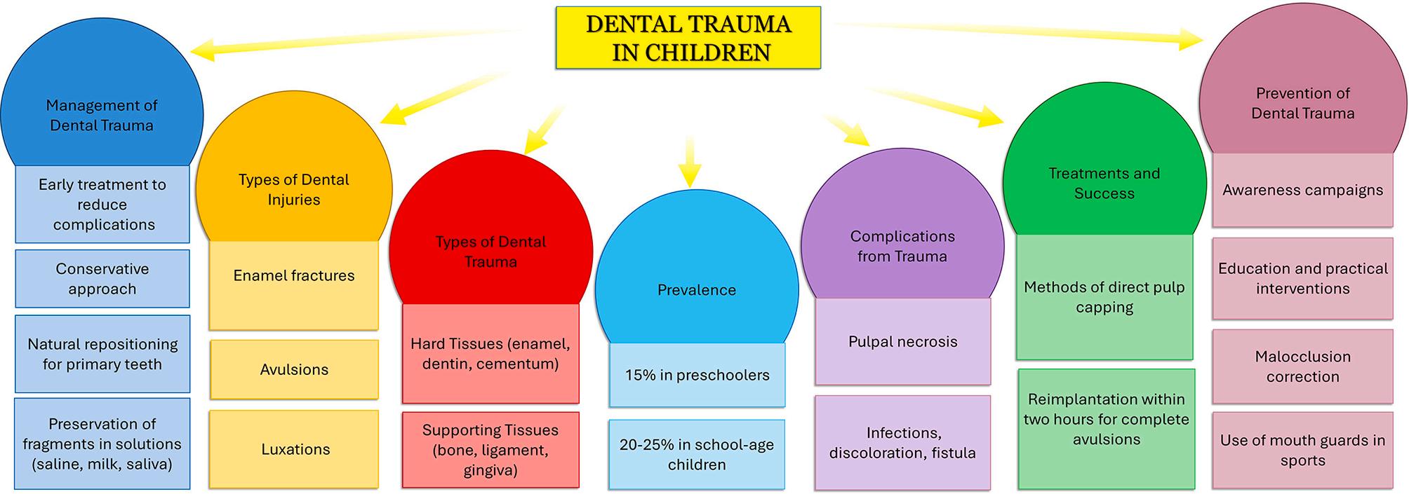
1. Concept map of dental trauma in children. (Courtesy of Laforgia A, Inchingolo AM, Inchingolo F, et al: Paediatric dental trauma: Insights from epidemiological studies and management recommendations. BMC Oral Health 25:6, 2025.)
Treatment Innovation s
The efficacy of different pulpotomy materials in managing traumatized immature permanent teeth has been the focus of some studies. A higher pulp survival rate was noted with iRoot BP Plus, which may be a promising alternative to calcium hydroxide (CH). Findings support the use of newer materials that offer better sealing properties and greater ease of use.
Significant improvements have also been made in vascular and neural formation, along with increased root length and width. These suggest that the use of deciduous autologous tooth stem cells (hDPSCs) may be a viable option to salvage young teeth and enhance root development. This option is part of a broad trend in dental research that focuses on tissue regeneration and conductive microenviroments created for healing.
CDSTs show promise in managing dental trauma. Both paperbased and mobile app CDSTs significantly improved diagnostic and management skills, especially among medical students. Integrating CDSTs into educational programs may improve the quality of dental trauma management and outcomes of treatment.
Higher rates of trauma have been found among special needs children, especially those with cerebral palsy and obesity. Dental trauma and caries can negatively affect children’s oral health −related quality of life (OHRQoL). This underscores the importance of early prevention and parental education. Cavitated lesions and traumatic dental injuries negatively affected OHRQoL but malocclusions did not. The impact of cavitated posterior teeth can be greater than teeth in other areas. Other factors to consider are sociodemographic factors, mother’s educational level, and parental perception of poor oral health.
Dental trauma in deciduous teeth is not only common but also significant among pediatric dental patients. Prompt and suitable management are needed to avoid long-term consequences.
Falls, sports injuries, and accidents are common causes for dental trauma in children. Early diagnosis and intervention are essential to support the vitality of injured teeth and avoid the development of complications. The management of dental trauma should be undertaken as quickly as possible to improve the outcomes. For example, fractured tooth fragments should be reattached, root canal therapy performed, and avulsed teeth reimplanted within 2 hours of injury. Conservative approaches and close monitoring with follow-up care are needed to avoid complications. Innovative techniques such as stem cell therapy along with novel pulpotomy materials may achieve better outcomes. Policymakers should support preventive measures and educational programs to reduce dental trauma among children.
Laforgia A, Inchingolo AM, Inchingolo F, et al: Paediatric dental trauma: Insights from epidemiological studies and management recommendations. BMC Oral Health 25:6, 2025
Reprints available from F Inchingolo, Dept of Interdisciplinary Medicine, Univ of Bari “Aldo Mora,” Bari 70124, Italy; e-mail: francesco.inchingolo@uniba.it
Objective and subjective patient information is documented in dental records (Box 1). The purposes of these records are multiple, as are the functions they fulfill. In the UK, the General Dental Council (GDC) provided fitness to practice (FtP) reports and reports a 22% reduction in overall concerns raised from 2018 to 2023. About half of the concerns originated from or on behalf of patients and concern clinical or conduct-related issues. In contrast, over the same period, dental services complaints have increased and usually address access to National Health Service (NHS) dentistry, treatment, and fees. Dentists tend to adopt an overly defensive approach to their clinical care and invest substantial time, effort, and resources to maintain detailed patient records. These records can contain excessive and nonessential information that may obscure important aspects of patient care. A review of the purpose of dental record keeping, guidance in record keeping, cautions related to the delegation of record keeping and ensuring accuracy, and possible future considerations.
Using the UK as an example, dental professionals are governed by legal, professional, and ethical obligations in the creation and maintenance of dental records. According to the GDC, dental professionals must make and keep contemporaneous, complete, accurate patient records as an essential part of understanding oral health, tracking patient progress, guiding clinical decisions, supporting audits and research, and recording the consent process. The key elements of dental recordkeeping should be included (Table 1).
Usually clinical record entries are read only by the treating dentist and possibly colleagues, but a wide range of authorities can access clinical records, as can the patient. Often dental professionals err on the side of quantity rather than clarity and keep records as a way to reduce the risk of criticism and accusations of inadequate charting. However, comprehensive records aren’t necessarily voluminous and lacking in clarity and accuracy. Information overload can hinder efficiency and impair the reader’s understanding, possibly leading to having important information overlooked. It’s recommended that records clearly outline what needs to be done next and why in readily understood language.
Although accurate records promote patient safety and avert errors in care, minor errors may not necessarily harm anyone. In addition, if excessive, superfluous, or template notes are used indiscriminately, they may not reflect the actual care provided. If
clinicians attempt to cover up errors, inflate claims, or manipulate patient data, these efforts are indefensible and carry severe legal, dental, and ethical considerations. The appropriate measure is to add an addendum that notes the inaccuracy.
Dento-legal situations often depend on accurate recording of the treatment delivered along with any pertinent information. A detailed account of the events should include the differential diagnosis, the process of obtaining informed consent, and financial transaction details. If the entries are inadequate or incomplete, any defense argument could be compromised. It’s important to note the “why” of clinical decisions to give a complete picture of the process.
The GDC doesn’t set clinical standards, and the guidance for clinical examinations and record keeping have been set by various laws, regulations, and organizations. Guidance refers to nonbinding recommendations of advice and isn’t enforceable by law, although it is highly influential and can be used by courts and regulatory bodies to judge whether the practitioner has met accepted standards. Not following guidance, however, may also result in disciplinary actions from regulatory agencies. There is also a risk that guidelines will be misapplied by third parties for purposes beyond their original intent.
The GDC’s primary purpose is to protect patients and maintain public confidence in the dental profession. Its FtP process has 4 stages (Table 2), with a clinical advisor required to provide a written report determining if the standard of care provided has met the level of professional practice reasonably expected. The clinical advisor generally considers the pretreatment aspects, obtaining consent, the treatment phase, and the aftercare provided. Because the clinical advisor doesn’t directly contact the informant, clinical records and the information they contain are the critical elements of the process. The overall conclusion of the process will fall into 5 possible outcomes, including the following:
1. At the reasonably expected level of professional practice
2. Below the expected level for some aspects but not unsatisfactory to the extent that overall care falls below the standard
3. Below the reasonably expected level of professional practice
4. Significantly below the reasonably expected level of professional practice
5. Information provided is insufficient to draw a conclusion
Box 1. To Show Items Which Make Up a Set of Patient Dental Records
• Handwritten and computerised clinical notes, to include details such as data to identify the patient and further charting, such as hard tissue charting and periodontal charting
• Medical history forms
• Radiographs, scans and other imaging records
• Digital scans
• Photographs and videos
• Models, to include study models, working models and diagnostic wax-ups/tooth set-ups
• Correspondence with patients/carers and other healthcare professionals/providers, to include referrals correspondence
• Recording of any telephone conversations
• Written treatment plans
• Consent forms
• Statements relating to custom made devices under the Medical Devices regulations
(Courtesy of Mehta SB, Rattan R, D’Cruz L: Note bloat. Br Dent J 239:343-349, 2025.)
Table 1. A Summary of the Key Elements of Dental Record keeping
Accuracy and clarity
Comprehensive documentation
All entries must be precise and unambiguous, ensuring that the information is correct and easily understood by other healthcare professionals who may be involved in the patient’s care. Avoid unnecessary jargon and ensure clarity in all documentation
Records should cover all essential aspects of patient care, including medical and dental history, diagnosis, treatment plans, procedures and outcomes. They must also include discussions of risks, benefits, alternatives, patient preferences, and any follow-up instructions, advice or care provided. It is also prudent to record the patient’s compliance to advice – from a dentolegal perspective, a non-compliant patient helps to advance the contributory negligence argument – for example in cases relating to periodontal disease progression
Table 1. Continued
Timeliness
Documentation should be completed as soon as possible after the patient interaction to ensure accuracy and prevent memory gaps. Any delay in writing of the record increases the risk of omitting important information. This
Legibility
Confidentiality
Continuity of care
Audit
Professional responsibility
helps maintain a contemporaneous record (we take ‘contemporaneous’ to mean writing the notes before the next patient is seen in the dentist’s appointment schedule) which is vital in defending the care provided in any future legal or regulatory review especially when addressing concerns that arise long after care has been provided. While civil claims must be filed within three years of the alleged negligence or when the patient becomes aware of it, GDC investigations have no time limit
Whether handwritten or digital, records must be easily readable. Poor handwriting or unclear digital formatting can lead to misinterpretation and compromise patient safety or legal defence
Patient records must be stored securely and accessed only by authorised personnel, in compliance with data protection laws like General Data Protection Regulation. This ensures patient privacy and builds trust in the healthcare system
Ensure that records support continuity by documenting all relevant communications, referrals, handovers and follow-up appointments. This ensures that other healthcare providers can seamlessly continue the care of the patient based on the documented history
In digital systems, every entry, edit and amendment should be tracked with an audit trail showing who made the change, when and why. This transparency ensures the integrity of the records and helps identify any unauthorised alterations
The treating clinician is ultimately responsible for ensuring that the records accurately reflect the care provided. Even if assistants or other staff are involved in record-keeping, the clinician must review, verify and amend the records as necessary
Consent and communication
Documenting informed consent is crucial. This not only involves recording that consent was given but also ensuring the patient’s understanding of the risks, benefits and alternatives discussed. Discussions must be noted to protect against potential future complaints or legal challenges
(Courtesy of Mehta SB, Rattan R, D’Cruz L: Note bloat. Br Dent J 239:343-349, 2025.) (Continued on next column)
Table 2. The Four Stages of the General Dental Council’s Fitness to Practice Process and the Tests Applied. Progression Will Take Place Only When the Test Has Been Satisfied
Stage number Name Test applied/outcomes
1 Initial assessment Does the information give rise to a concern that:
• Harm has been caused, or may have been caused, to a member of the public, and/or
• Public confidence in the profession has been, or may have been undermined?
2 Assessment Does the conduct alleged:
• Give rise to a concern that harm has been caused to a member of public, or
• Give rise to a concern that public confidence in the profession has been or may be undermined;
• And the circumstances are such that the allegation is serious enough that, if proved, it raises the issue that the FtP of the practitioner may be impaired.
3 Case examiner Is there a real prospect of a practice committee finding the facts, statutory ground of impairment and current impairment all proved?
4 Practice committee -
(Courtesy of Mehta SB, Rattan R, D’Cruz L: Note bloat. Br Dent J 239:343-349, 2025.)
When the records are made by persons other than the treating clinician, the clinician is still responsible for carefully reviewing the record and documenting if a delay in recording occurred and why. Auto-notes and prefilled templates have been used to save time, but they must be carefully checked to ensure their accuracy. Clinical advisors and dento-legal consultants find errors in template records when the copypaste feature creates anomalies. These can raise questions about the validity of records and the honesty and integrity of the dental professional.
If serious events with the potential for harm occur, they must be clearly documented. In addition, timely handling of complaints and local resolution efforts should be made to influence investigation outcomes. Engaging with indemnity providers can avoid future investigations. If the GDC requests records, the dentist should seek advice from the practice’s
indemnity provider and ensure all records (paper and digital) are disclosed.
In the 1970s and 1980s, minimalistic dental record keeping was practiced, and today more detailed and extensive records are common. An equilibrium must be reached between the 2 approaches, producing concise and clinically relevant notes that contain under- or over-documentation. All stakeholders must buy into this approach to promote context-specific documentation that supports patient care while falling into the patterns required by legal, compliance, and quality considerations.
Artificial intelligence (AI) and speech-to-text software can contribute to the future recording tasks. Appropriate AI applications may enhance accuracy, consistency, and efficiency, thereby freeing clinicians to spend more time on patient care. The data protection and storage implications of AI applications must be considered.
To record procedural details more succinctly, standard operating procedures (SOPs) should be created and adopted for use with the clinical record. SOPs can reduce variations, ensure greater consistency, improve safety, and add efficiency. Extensive details could be covered by the SOP rather than the recording clinician without giving readers note bloat.
The dental profession should set standards for record keeping in dental practices. This could help avoid some of the stress and burnout that dentists suffer and would create succinct, high-quality clinical records that clearly show the sequence, safety, and effectiveness of the dental care provided. Stakeholders should support these standards if they improve efficiency, safety, and accuracy. As a result, lawyers will be required to apply the revised guidance in their practices. Overall, this is an area of dental practice that needs work.
Mehta SB, Rattan R, D’Cruz L: Note bloat. Br Dent J 239:343-349, 2025
Reprints available from SB Mehta; e-mail: smehta@gdc-ok.org
The third molars are the most varied teeth in presentation and the most likely to become impacted. The question is whether it’s medically necessary to remove them, especially if they’re not symptomatic. The situation requires a thorough diagnosis and detailed workup, focusing on the chief complaint, medical history, and clinical examination. Included in this should be measurements of probing depths and attachment levels around the third and second molars, along with panoramic radiographs. The most recent literature was reviewed to outline the reasons for addressing third molars, conservative and trending methods to manage third molars, and patient concerns and preferences for removal or retention.
Third molars have a highly variable development pattern and complex interactions with adjacent teeth and other oral structures. Their unpredictable eruption patterns and potential for impaction can be challenging to assess and manage. Asymptomatic third molars may not be disease free, so they require regular evaluations with radiographs to determine the best way to manage them.
Orthodontists may favor retaining asymptomatic third molars. However, pain is present in 6% to 55% of patients, with complications such as cellulitis and osteomyelitis seen in less than 5%. Other concerns include functional disorders such as interference with occlusion, cheek biting, masticatory dysfunction, limited mouth opening, and TMJ dysfunction. No etiologic link has been shown despite the distress that patients report.
Periodontal disease is reportedly significantly higher in the mandibular third molars than in the maxillary teeth and has been suggested to be both persistent and progressive, but may improve with extraction of the teeth. The teeth adjacent to partially impacted third molars have increased plaque scores and gingival indices, with external root resorption of the adjacent molars seen in up to 50% of these teeth. To assess the problems related to retained third molars requires 3-dimensional radiographs using cone beam computed tomography (CBCT), which may be difficult to arrange in some clinical practices and research studies. The decisive factors in the incidence of external root resorption are patient’s age, impaction depth, and inclination.
The removal of asymptomatic third molars raises some controversies, with a number of indications and contraindications (Table 1). If the decision is made for extraction, patients should be prepared for sequelae such as discomfort, pain, inflammation, ecchymosis, limited mouth opening, infection, and hematoma. In addition, neuronal structures may be impaired.
In the event that nonsurgical management of asymptomatic impacted third molars is selected, the clinician must schedule regular monitoring to catch caries, periodontal pathologies, root resorption of the second molar, cysts, and tumors early on. Retained mandibular third molars are significantly more likely to suffer caries than maxillary third molars.
The 2 most common approaches to managing impacted third molars are (1) retaining impacted third molars until symptoms or evidence of pathology develops (nonintervention or third molar retention technique) and (2) removing the impacted third molar before symptoms or pathologic indicators develop (preventive or interceptive removal). A coronectomy may be required if there is an impacted tooth adjacent to vital structures so that infectious alveolar nerve injury, lingual nerve injury, and dry socket don’t develop. The clinician must be aware of the failure rate of this approach and the increased likelihood that additional surgery will be needed.
Among the complications related to third molar extraction are alveolar osteitis, infection, inferior alveolar nerve or lingual nerve involvement, bleeding, hematoma, TMJ dysfunction, and jaw fracture. Oral and maxillofacial surgeons may integrate minimally invasive techniques and artificial intelligence (AI)−driven tools to enhance the multidisciplinary care required. These approaches have shown promise in accurately evaluating the relationship between the third molar and neuronal structures.
The management of other potential complications also involves use of a range of methods to minimize complications, such as the judicious use of analgesics, steroid therapy, antibiotics, flap designs, sutures, drains, ozone therapy, cryotherapy, platelet-rich plasma (PRP), platelet-rich fibrin, piezoelectric surgery, and lasers. Various factors have been identified as making surgical procedures more or less difficult (Figures 2 and 3).
Table 1. Indications and Contraindications for Third Molar Extractions
Indications
Relative Contraindications
Pain Associated with possible damage to the adjacent structures on removal
Prevent periodontal disease progression
Ectopic position
Assist in prosthodontic and orthodontic treatment planning
Fractured tooth and irreparable caries
External resorption of the adjacent teeth
Tooth in association with tumor resection
Prophylactic removal in patients with medical or surgical indications
Informed refusal of nonsurgical treatment options
Abnormal tooth anatomy
Association with pulpal and periapical pathology
Elective therapeutic removal
Management of jaw fracture complicating fracture reduction
Trauma, orthognathic, or reconstructive surgery
Insufficient space to facilitate eruption
Arch size discrepancies
Proximity between a mandibular third molar and the inferior alveolar nerve
Risk of mandibular fracture
Risk of adjacent teeth fracture
Oroantral communication and tuberosity fracture
Systemic associations, such as bleeding disorders
Patients on intravenous antiresorptive agents
Patient undergoing chemotherapy and/or radiotherapy
Pregnancy
Inadequate space for eruption of the third molar
Serves as an abutment tooth
Third molars with no underlying etiology
Patient’s right to refuse treatment
Immunocompromised patients
(Courtesy of Mathai BC, Talaat W, Keni PK, et al: Third molars in dentistry. Dent Clin N Am 70:1-14, 2026.)
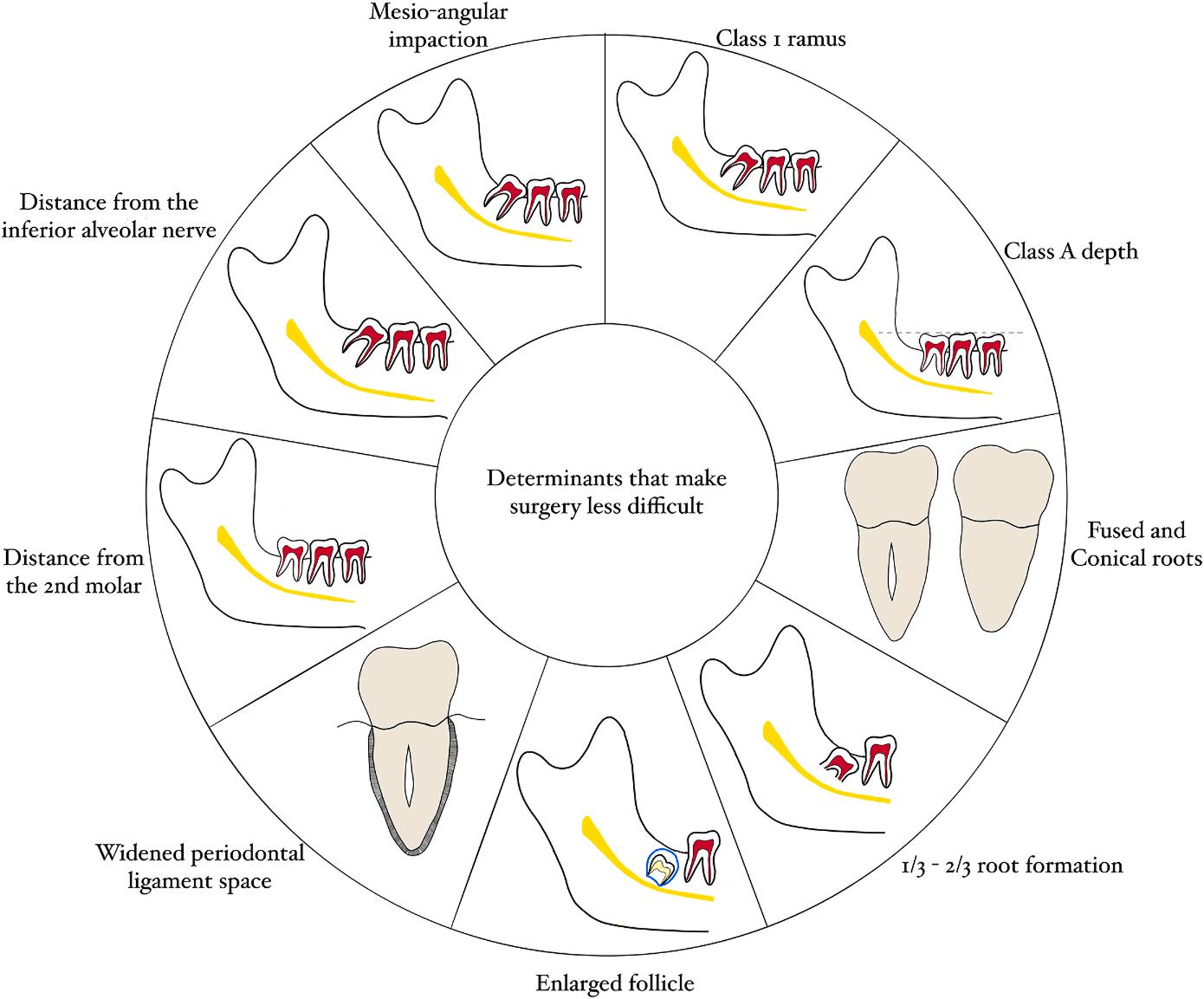
Figure 2. Factors contributing toward making third molar extractions less difficult. (Class 1 ramus—The space between the ramus and the second M is larger than the mesiodistal diameter of the second M. Class depth—The occlusal plane of the third molar is as high as that of the second molar.)
(Courtesy of Mathai BC, Talaat W, Keni PK, et al: Third molars in dentistry. Dent Clin N Am 70:1-14, 2026.)
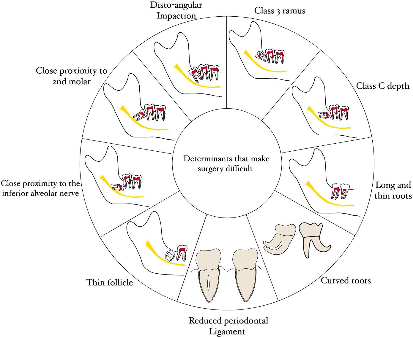
Figure 3. Factors contributing toward making third molar extractions difficult. (Class 3 ramus—There is no space between the ramus and the second M; Class C depth—The occlusal plane of the third molar is below the cervical line of the second M.) (Courtesy of Mathai BC, Talaat W, Keni PK, et al: Third molars in dentistry. Dent Clin N Am 70:1-14, 2026.)
Most patients today have a proactive attitude toward third molar management and want to discuss concerns with professionals. A few still feel anxious or even fearful about their third molar situation. The extraction of impacted third molars has been ranked as the most stressful surgical procedure by patients. Preoperative anxiety is associated with a higher degree of postoperative pain and prolonged surgery. Patients may report increased heart rate and can have a higher risk for complications. Clinicians should assess the patient’s anxiety levels and autonomic responses before surgery. A positive association has been shown between preoperative anxiety and postoperative pain and inflammation. Body mass index is also related to higher levels of C-reactive protein and pain before surgery. Body mass index and the eruption pattern of the impacted third molar have been considered predictors of postoperative complications.
The decision to manage third molars depends on the clinician’s assessment of the risk-to-benefit ratio, the position and inclination of the third molars, patient concerns and preferences, and evidence based on the most current research. Patients should be informed of potential risks, with clear explanations of the problems that may develop and how they will be managed, along with any alternative options and their costs and risks. This is part of the informed consent process.
Mathai BC, Talaat W, Keni PK, et al: Third molars in dentistry. Dent Clin N Am 70:1-14, 2026
Reprints available from BC Mathai; e-mail: blessy2009@gmail.com
Odontogenic infections prompt consultations with dental, oral, and maxillofacial medicine practitioners, with most infections of endodontic in origin, usually from apical pulpitis with bacterial invasion of the periapical tissues. A range of aerobic and anaerobic bacteria are involved. Treatment generally involves surgical incision and drainage to eliminate pus and ventilate the area. The odontogenic source is also addressed. Uncomplicated odontogenic infections can be managed with this approach, but severe cases may require adjunctive systemic antibiotic therapy. Selecting the appropriate empirical antibiotic therapy for these more severe infections is challenging. The recent inappropriate antibiotic uses such as unneeded prescriptions, deviations from clinical guidelines, and selfmedication have contributed to the development of antimicrobial resistance. Penicillin was once the first-line agent for these infections but now it often has insufficient coverage and broader spectrum or alternative agents are needed. Recommendations for empirical antibiotic therapy vary widely, and few studies have evaluated the potential impact of antimicrobial resistance on outcomes. Patients with complicated odontogenic infections were studied, noting associations between antibiotic resistance and adverse clinical outcomes and providing evidence to guide empiric treatment strategies and support antimicrobial stewardship by dentistry.
Seven hundred forty inpatients age 2 to 95 years with 225 localized odontogenic infections and 515 maxillofacial space infections were studied over the course of 12 years. All had microbiological tests to identify the causative organisms for their surgically treated odontogenic infections. Data included the resistance rates, time trends, and associations between resistance rates and clinical variables.
Patients had surgical management such as intraoral incision and drainage (342 cases) and extraoral incision and drainage (398
cases). Systemic complications occurred in 34 cases. Fiftyseven patients required extended hospitalizations.
The infections were predominantly polymicrobial, with a mean of 2.5 microbial species per infection. One hundred sixty cases had monomicrobial infections. A total of 1476 bacterial and 40 fungal isolates were found, including 116 different bacterial species in 39 genera and 6 fungal species from 3 genera. A total of 796 isolates were aerobic or facultative anaerobic and 680 involved strictly anaerobic organisms.
The highest resistance rate was seen for clindamycin (about 39%), then penicillin (about 24%), and amoxicillin (about 20%). Lower resistance rates were found for amoxicillin/clavulanate (about 7%) and moxifloxacin (about 5%), Annual resistance rates did not differ significantly for any of the antibiotics. A reported impact of penicillin hypersensitivity on antibiotic resistance was noted. Significantly increased risk of resistance to clindamycin was seen in patients who reported penicillin hypersensitivity, with resistance rates of about 56%. In addition, a significant association was noted between resistance to moxifloxacin and an increased risk for systemic complications. The risk of systemic complications was increased by a factor of 10.875 in infections resistant to moxifloxacin.
Dentistry has been identified as a major source of antibiotic prescriptions. The current resistance patterns of odontogenic infections and their potential impact on clinical relevant outcomes were evaluated to determine empiric treatment strategies. There is a need for structured antibiotic stewardship initiatives in dentistry and better adherence to guideline-concordant prescribing practices.
Persistent high resistance to clindamycin and its implications, especially for penicillin-hypersensitive patients, underscores the need for pursuing antibiotic stewardship measures and reevaluating current treatment guidelines. Patient outcomes should be improved with stronger diagnostic practices and hypersensitivity verification processes.
Fischer M, Rodel J, Schultze-Mosgau S, et al: Clinical impact of antibiotic resistance in odontogenic infections: A 12-year analysis of 740 cases. Clin Oral Invest 29:597, 2025
Reprints available from M Fischer, Dept of Oral and CranioMaxillofacial-Surgery/Plastic Surgery, Jena Univ Hosp, Friedrich Schiller Univ of Jena, Am Klinikum 1, 07747 Jena, Germany; e-mail: Martin.Stephan@med.uni-jena.de
Burning mouth syndrome (BMS) is characterized by recurrent burning or dysesthetic sensations in the mouth. These last more than 2 hours a day for more than 3 months and are accompanied by no identifiable clinical lesions. This chronic, idiopathic orofacial condition may be accompanied by somatosensory changes, including numbness, tingling, xerostomia, or altered taste and is seen in about 0.7% to 5% of the population, most often women over age 50 years undergoing menopause and postmenopause. The bilateral symptoms can vary in intensity and can be triggered by stress or certain foods. Primary BMS is idiopathic and has no identifiable cause, whereas secondary BMS is associated with local or systemic factors. The diagnosis is challenging, relying on the accuracy of patient-reported symptoms and the exclusion of other conditions. Generally a multidisciplinary approach to treatment is required. The efficacy of various therapeutic options being used currently to treat BMS and to relieve patients’ pain was evaluated to chart the path of clinical care going forward. The publications reviewed used topical, systemic, and combined pharmacological and nonpharmacological treatments, physical therapies, and alternative medicine approaches.
A systematic review of the PubMed, Google Scholar, and Scielo databases was conducted. Twenty-one randomized clinical trials (RCTs) were identified that covered the pharmacological, topical, systemic, physical, and combined interventions for BMS (Figure 1). The outcomes were assessed for symptom relief, adverse effects, and comparisons with placebo and other treatments.
The clinical implications and practical considerations for each treatment were evaluated (Table 8). Only a qualitative synthesis was performed.
The efficacy of topical clonazepam reflects the gamma-aminobutyric acid (GABA)−modulation of peripheral nociception and central sensitization. GABA-ergic modulation approaches are undermined by systemic absorption causing adverse effects, specifically, drowsiness and dizziness, in up to a quarter of patients. The absolute clinical benefit is limited in most patients.
Topical capsaicin achieves a modest efficacy in patients. The symptoms tend to increase initially, then the pain is relieved once capsaicin depletes substance P from the nociceptive terminals. Intolerable burning tends to prompt about 18% of patients to discontinue treatment.
Low-level laser therapy (LLLT) achieves variable efficacy, with infrared protocols demonstrating better outcomes than red laser approaches. Because of the protocol heterogeneity, no definitive conclusion could be made about optimal treatment parameters.
Alpha-lipoic acid (ALA) had a 64% response rate, which likely reflects its antioxidant properties and neuroprotective effects on damaged trigeminal nerve fibers. This may relieve the oxidative stress component of BMS.
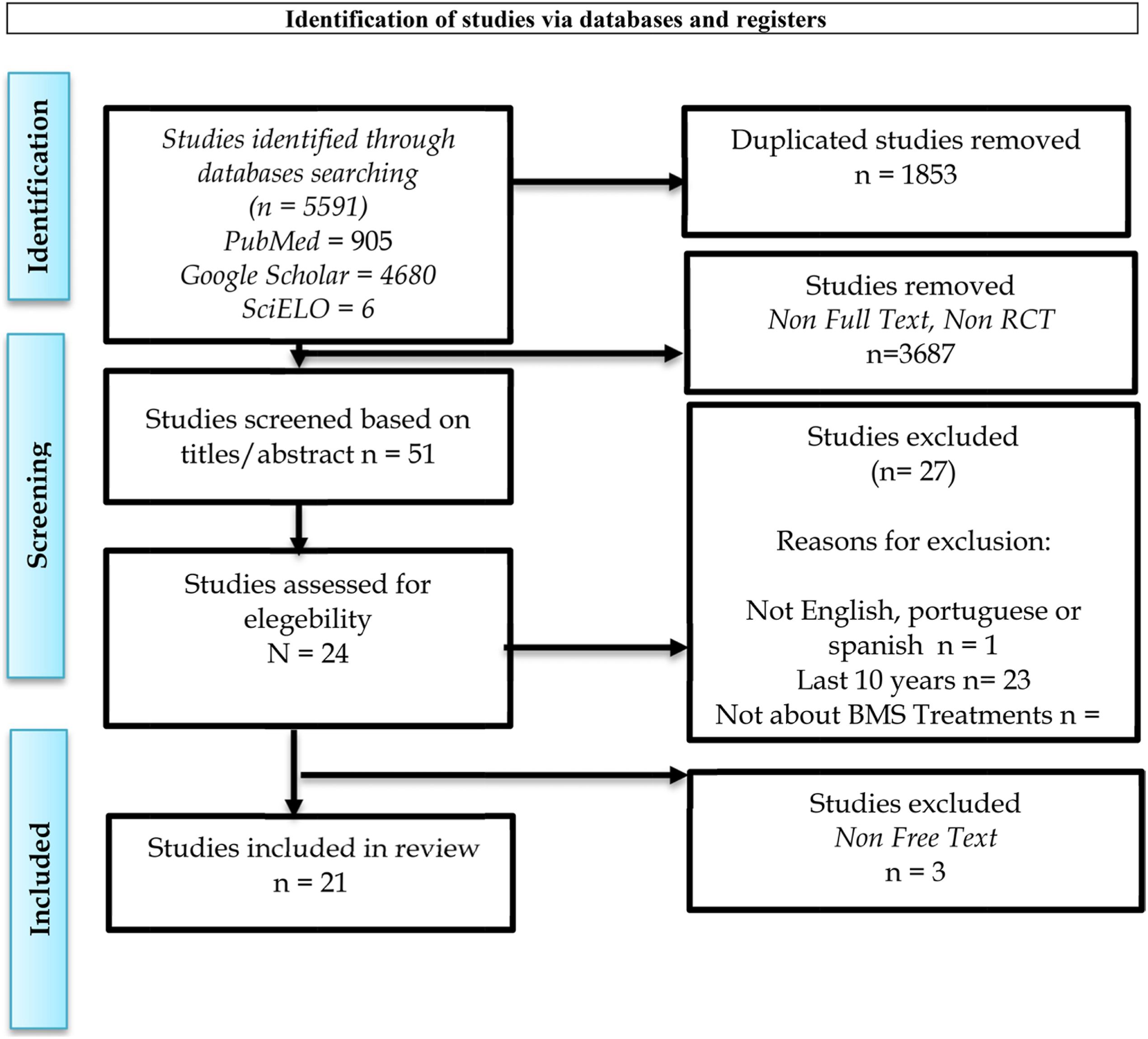
Figure 1. PRISMA flow diagram of the systematic review. (Courtesy of Rossetti A, Teixeira A, Milhazes N: Efficacy of different therapeutic options for pain relief and treatment of burning mouth syndrome: A systematic review. Clin Oral Invest 29:551, 2025.)
APPROACH
TOPICAL
TREATMENTS
Clonazepam, Laser (LLLT/ PBM), Capsaicin, Chamomile, Lidocaine, Topical Gabapen tin, Bupivacaine Lozenges, Oral Protectors
SYSTEMIC ALA, Duloxetine, Amitrip-tyline, Paroxetine, Sertraline, Vortioxetine, Amisulpride, Pregabalin, Gabapentin, Mela-tonin, PEA, Herbal medicine (DZX + Methylcobalamin)
COMBINED Medications + Laser + Herbal Medicine + Nutritional Supplementation
PSYCHOLOGICAL
Cognitive Behavioral Therapy (CBT)
PHYSICAL/ NEUROMODULATION Repetitive Transcranial Mag-netic Stimulation (rTMS)
MULTIDISCIPLINARY Combination of Drugs, Laser, Psychotherapy
CLINICAL IMPLICATIONS
– Good local efficacy, few adverse effects.
– Clonazepam and LLLT are f irst-line options.
– Capsaicin effective in some cases but may increase burning sensation.
– Topical Gabapentin and Bupivacaine Lozenges are safe alternatives.
– ALA, Pregabalin, and PEA showed positive results, espe cially in neuropathic pain.
– Antidepressants yielded vari able outcomes, more effective in patients with psychiatric comorbidities.
– Amisulpride and Vortioxetine effective in refractory cases.
– Few relevant adverse effects.
– Potentiates analgesic effects.
– Useful for patients with par tial response to monotherapy.
– Effective as an adjunct, espe cially in patients with anxiety or depression.
– Should be considered in mul tidisciplinary approaches.
– Promising in refractory cases.
– Significant pain reduction reported in some studies.
– Requires greater technical availability and cost.
– Individualized treatment is essential.
– Combinations tend to improve treatment adherence and clinical response.
– Collaboration among physi cians, dentists, and psycholo gists promotes better outcomes.
(Courtesy of Rossetti A, Teixeira A, Milhazes N: Efficacy of different therapeutic options for pain relief and treatment of burning mouth syndrome: A systematic review. Clin Oral Invest 29:551, 2025.)
Systemic therapies such as ALA, pregabalin, melatonin, and other antidepressants have variable efficacy. Their greatest effects were noted in refractory cases or patients with psychiatric comorbid conditions. Vortioxetine is an antidepressant that showed superior efficacy and tolerability compared to traditional antidepressants in BMS patients. Its highest rates of clinical response and remission were achieved after 6 to 12 months of follow-up. Vortioxetine has a unique multimodal mechanism of action, with serotonin reuptake subtypes that may address both neuropathic pain and associated mood symptoms related to BMS. Further studies are needed, with just 1 study used to derive these findings.
Some patients benefited from a combination approach consisting of pharmacological, physical, and psychological agents. The analgesic effects were improved as was treatment adherence. Placebo effects have also been reported in several studies.
Because of the variability in outcomes, treatment choice should be individualized, ideally with a multidisciplinary team and careful clinical monitoring. Further high-quality and better populated RCTs are needed before first-line recommendations can be made.
The current evidence and therapeutic protocols indicate that clinical management of BMS should be tailored to the patient and consider symptom severity, psychological comorbidities, tolerability of the treatment, and patient preferences. A multidisciplinary approach combining pharmacological, physical, and psychological interventions may be the best option for improving symptom control and quality of life for BMS patients.
Rossetti A, Teixeira A, Milhazes N: Efficacy of different therapeutic options for pain relief and treatment of burning mouth syndrome: A systematic review. Clin Oral Invest 29:551, 2025
Reprints available from A Teixeira, Univ Inst of Health Sciences—CESPU, Gandra 4585-116, Portugal; e-mail: ana.teixeira@iucs.cespu.pt
Dental caries can have a significant impact on quality of life. Caries is caused by interactions between microorganisms and various compounds. Gut microbiota are bacteria, fungi, viruses, and archaea that colonize the host’s intestinal tract. The gut microbiome is regarded as the body’s second brain because of its remarkable diversity and the role in plays in maintaining the host’s health and preventing disease. Diet and routine can influence the abundance of gut microbiota and influences host physiological functions. Research is showing that gut microbiota dysbiosis can promote systemic inflammation. Dysbiosis leads to lipopolysaccharide (LPS) being introduced into the circulatory system, ultimately producing systemic inflammation. LPS can also contribute to the development of dental caries. Because these links have relied on observational studies that can skew the results, a Mendelian randomization (MR) approach was used to investigate further. The potential causal link between gut microbiota and dental caries was explored.
The gut microbiota and dental caries data from genome-wide association studies were subjected to MR analysis. Inverse variance weighted (IVW) was used as the primary criterion.
MR analysis identified 6 caries-related bacterial genera: Escherichia. Shigella, Oscillibacter, Eubacteriumbrachygroup, Terrisporobacter, RuminococcaceaeUCG014, and Oscillospira The IVW method showed significant differences, with Eubacteriumbrachygroup and Terrisporobacter positively associated with dental caries and Escherichia. Shigella, Oscillibacter, RuminococcaceaeUCG014, and Oscillospira negatively associated with dental caries.
Causal relationships between gut microbiota and dental caries were shown. Some microorganisms have a negative relationship and others have a positive relationship.
With the significant relationships between gut microbiota and the development of dental caries, dental care has another approach to managing caries in patients. The composition of the gut microbiota may be a novel way to approach caries prevention.
Wang Y, Li Q, Hua J, et al: Causal relationship between gut microbiota and dental caries: A two-sample mendelian randomization study. BDJ Open 11:35, 2025
Reprints available from N Feng, Dept of Urology, Jiangnan Univ Medical Ctr (Wuxi No. 2 People’s Hosp), Wuxi 214000, China; e-mail: n.feng@njmu.edu.cn
When teeth with dental caries reach an advanced stage, it’s important to protect the pulp tissue and thereby preserve tooth vitality and avoid further complications. Cavity liners have been used in such cases to eliminate the danger posed by thermal, chemical, and mechanical stimuli and to enhance patient comfort. However, the clinical efficacy of liners versus no liners is debated. Various biomaterials have been used in liners, including calcium hydroxide formulations, mineral trioxide aggregate (MTA), and hydraulic calcium silicate cements. Pulp preservation, reduced postoperative hypersensitivity, and prevention of secondary caries were evaluated in patients with deep caries restorations along with patient-reported symptoms.
A systematic literature search was done in the Cochrane Review, PubMed (Medline), and Ovid databases. Twelve studies with participants ranging in age from 6 to 76 years were identified for review. Most patients had deep carious lesions in permanent teeth with vital pulps. The recall periods were primarily 12 to 24 months for the assessment of restoration failure, pulp vitality, and secondary caries, although some outcomes were reported at 6, 12, and 36 months. Postoperative pain and hypersensitivity were usually evaluated within 7 days to 3 months.
The most common cavity liners were made of calcium hydroxide−based materials, resin-modified calcium silicates, glass-ionomer cements, hydraulic calcium silicate cements,
and flowable composite liners. Less conventional materials included resin-based ion-releasing liners, desensitizing polycarboxylate cement containing potassium nitrate, and silver diamine fluoride (SDF) applied before liner application. The restorative materials included composite resin, amalgam, and glass-ionomer materials.
Failure was variously defined, with the most common definitions being loss of pulp vitality requiring endodontic intervention, and restorative retreatment caused by loss or fracture or tooth extraction. Pulp vitality was determined using cold and/or electric pulp testing. Radiographs were obtained to identify periapical changes or resorptive processes, but not all studies used them.
The various studies differed in their restoration success rates, but most reported long-term success whether liners were used or not. Successful outcomes tended to be in the range of 84% to 99%. Low failure rates were seen with both lined and unlined restorations.
The measures patients reported most were those that involved postoperative pain or hypersensitivity, with a few reports of functional satisfaction or long-term comfort. Generally, cavity liners didn’t significantly enhance patient comfort. No significant differences in postoperative hypersensitivity tended to be reliably reported between restorations with and without liners, but a study with a low risk of bias did report potassium nitrate liners significantly reduced postoperative hypersensitivity.
Selective rather than routine use of liners in restorations was suggested as the most evidence-based perspective drawn from this investigation.
Although the evidence doesn’t support the regular use of cavity liners in deep caries management in permanent teeth to reduce failure, secondary caries, or postoperative hypersensitivity, liners aren’t without clinical benefit. Their use should be tailored to the clinical context. Deep caries approaching the pulp may benefit from the use of a liner to protect the pulp from mechanical or chemical damage. Further research and longer periods of follow-up should be undertaken to further refine the clinical recommendations for liners.
Kosan E, Kosan D, Sturm R, et al: Efficacy of different cavity liners compared to no cavity lining in managing deep caries in vital permanent teeth – A systematic review. Clin Oral Invest 29:529, 2025
Reprints available from E Kosan, Dept of Periodontology, Oral Medicine and Oral Surgery, Charit� e – Universitaetsmedizin Berlin, Berlin, Germany; e-mail: zahnaerztinkosan@gmail.com
Single implants are the most common type of implant therapy. Most studies focus on the clinical outcomes of these implants, but future research must include patients’ perceptions and patient-reported outcome measurements (PROMs). When the implants are placed in the esthetic zone, patients’ expectations are especially high and the esthetics and functional status should be evaluated both objectively and subjectively. Function and esthetic outcomes were evaluated by patients who received single dental implants 10 to 15 years earlier.
PROMs were evaluated by 45 patients (mean age 40 years, range 30 to 80 years) using questionnaires that related to their satisfaction with the treatment they received. A visual analogue scale (VAS) was completed that ranged from 0 to 10. The first questionnaire rated function and cleaning ability of the single dental implant. The second questionnaire
rated the patient’s perception of the esthetics of the implant. In addition, each implant was photographed and the photos were evaluated by a dentist according to the Pink Esthetic (PES) and White Esthetic Score (WES) index, which evaluated mesial papilla fill, distal papilla fill, curvature of the facial mucosa, level of the facial mucosa, root convexity, and color and texture of the soft tissues, along with the crown form, volume/outline, color, surface texture, and translucency ( Figure 1 ). Probing pocket depth (PPD) and bleeding on probing (BOP) were measured at the implant site. Peri-implant mucositis and periimplantitis were diagnosed, and presence of suppuration was assessed.
When rating the function and cleaning ability of the implant, the highest satisfaction levels were for chewing function, followed by speech function and cleaning ability. The patients’
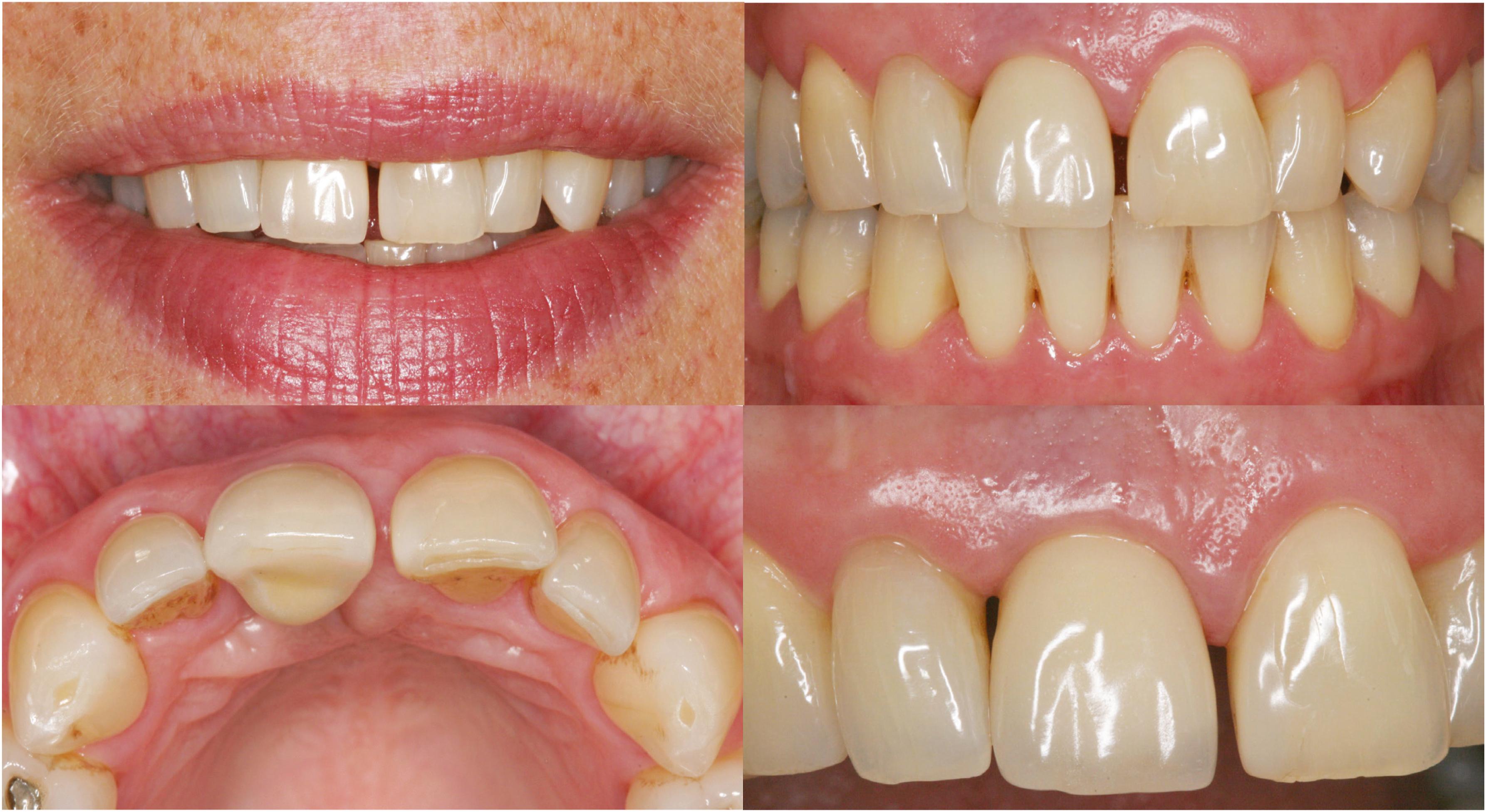
Figure 1. A patient with a single dental implant in the region of tooth 11, 13 years after placement. Pink esthetic score (PES) = 5, White esthetic score (WES) = 8, according to Belser et al. 2009. The patient’s perception of the esthetics on a visual analogue scale (VAS) = 40. (Courtesy of Bengtsson VW, Lindahl C, Scholander S: Patient-reported outcomes of esthetics, function and oral hygiene with single dental implants 10-15 years after placement: A cross-sectional study. Acta Odontol Scand 84:47-53, 2025.)
rating of the esthetics of the implant showed the highest satisfaction levels for the color and shape of the crown. Less satisfaction was noted for mucosal margins surrounding the implant.
The overall PES mean score was 6.2 and the median was 6.0. The WES mean score was 8.4 and the median was 9.0. The combined PES/WES mean score was 14.6, with a median of 15.0.
A diagnosis of peri-implant mucositis was made in 40 patients and a diagnosis of peri-implantitis was made in 2. The mean PPD was 3.8 mm, with 22.2% having a PPD over 6 mm.
Patients with peri-implant mucositis reported an overall esthetic satisfaction level of 8.7 and a median value of 9.0. Patients with peri-implantitis reported an overall esthetic satisfaction level of 7.5 and a median value of 7.5.
In patients with peri-implant mucositis, the PES score was 6.1 and the median was 6.0, whereas the WES score was 8.5 and the median was 9.0. In patients with peri-implantitis, the PES score was 7.0 and the median was 7.0, whereas the WES was 7.5 and the median was 7.5. The combined PES/WES score was 14.5 and the median was 14.5.
Patients reported high levels of satisfaction with cleaning, chewing ability, and overall esthetics for their single implants. They were less satisfied with the peri-implant mucosa.
Compared to the crown of their implant, patients were less satisfied with the peri-implant mucosa. In a few cases, patients never reached clinical acceptability in their ratings of the implant.
Bengtsson VW, Lindahl C, Scholander S: Patient-reported outcomes of esthetics, function and oral hygiene with single dental implants 10-15 years after placement: A cross-sectional study. Acta Odontol Scand 84:47-53, 2025
Reprints available from VW Bengtsson, Dept of Oral Health, Faculty of Oral Health Science, Kristianstad Univ, Kristianstad SE291 88, Sweden; e-mail: viveca.wallin_bengtsson@hkr.se
Dental implants often are a means of restoring function and aesthetics while improving patients’ quality of life. The survival rates are typically 95% to 98% after 10 years. However, failure is a significant clinical concern and can affect treatment planning and patient satisfaction levels. Early failures may develop during osseointegration or just after prosthetic loading and are generally caused by surgical trauma, poor stability, bacterial contamination, impaired healing, or systemic conditions. Late failures don’t occur until after successful integration and are often associated with biomechanical overload, peri-implantitis, trauma, parafunction, or compromised bone remodeling. Clinicians must determine the next course of action after a failure, which may be an alternative prosthetic strategy, site preservation, or reimplantation. With reimplantation, the implant is generally placed in the same location, but is subject to complications related to such events as residual infection, altered bone structure, or prior grafting. Survival rates after reimplantation vary widely, and success rates tend to decline after each repeated procedure. These failures are influenced by multiple patient- and site-specific factors. Not only are there anatomic conditions but patients can be subject to psychological stress that leads to decreased confidence, motivation, and cooperation; reduced compliance; and a decline in interest related to continuing treatment. The outcomes of consecutive implant replacements at previously failed sites, the survival rates across reimplantation attempts, and patient- and site-specific risk factors were documented.
A review of 5201 dental implants placed from 2017 to 2021 identified 203 failures (3.9%), with 189 of these undergoing reimplantation. Mean follow-up was more than 59 months. The clinical variables collected were patient age and gender, smoking status,
hypertension (HTN), diabetes mellitus (DM), antithrombotic medication use (especially acetylsalicylic acid [ASA]), bone grafting, implant location, timing of the failure, and patient willingness to continue treatment. Failure risk was determined for each reimplantation done.
The initial implants were followed up for about 63 months, the first reimplanted group was followed for about 62 months, and the group having a second reimplantation was followed for about 60 months. One hundred implants survived the first reimplantation (about 79%) and 40 failed (about 21%). Thirty-three underwent a second reimplantation, with 17 survivals (about 52%) and 16 failures (about 49%).
The potential factors influencing success or failure included age, gender, HTN, DM, ASA use, smoking, surgical site, bone graft, and timing of the failure.
Mean patient age was significantly greater in those who had failures. Mean age for the initial placement was about 47 years in the survival group and about 53 years in the failure group. Ages in the first reimplantation cohort were about 52 years in the survival group and about 58 years in the failure group, whereas of those in the second reimplantation most were about 44 years in the survival group and about 50 years in the failure group.
Gender data showed 3.9% of the failures were men and 3.7% were women in the initial placement group. These percentages grew to 22.08% for men and 19.4% for women in the first reimplantation and 50% versus 46.7% for men and women, respectively, in the second reimplantation. In none of these settings were the differences between genders statistically significant.
Among those patients who were managing HTN, 7.1% failed the initial placement, whereas only 2.6% of non-HTN patients failed. No statistically significant association was noted between HTN and failure in those having a first or second reimplantation. Similarly, the difference in failure data between those with and without DM was statistically significant in the initial placement but not in either the first reimplantation or the second.
A total of 5.8% of the failures in the initial placement were in ASA users, whereas 2.7% of the failures were in nonusers, which was a statistically significant increase in failure among ASA users. The first reimplantation cohort had a significantly reduced risk of failure compared to nonusers (15.0% versus 29.3%, respectively). The second reimplantation cohort had failures in 64.3% of ASA users and 36.8% of nonusers, a nonstatistically significant difference.
Smoking was found in 4.1% of the original implantation failures, with 3.8% among nonsmokers. The difference wasn’t statistically significant in any of the implantation or reimplantation groups. Surgical site also wasn’t connected to any statistically significant difference in outcomes. However, bone grafted sites were associated with failures in 7.2% of cases, with nongrafted sites associated with just 3.4%, a statistically significant difference. The differences were not statistically significant for the first or second reimplantation.
Of the 203 failed implants in the initial implantation, 93.1% failed in early healing phases and 6.9% in late healing phases. Of the failed first reimplantations, 85% were early and 15% were late failures.
Logistic regression noted that both age and ASA use were significant predictors of outcomes. No consistent predictors were noted for the second reimplantation cohort. Among the factors significantly associated with the initial implantation failures were older patient age, HTN, DM, antithrombotic medication use, and
grafted surgical sites. For the first reimplantation, ASA use was the only factor significantly associated with a lower risk of failure. None of the variables had a significant effect for the second reimplantation. It was also noted that there was a declining trend in patient willingness to undergo repeated implant procedures after consecutive failures.
Progressively declining survival rates were noted in patients having consecutive implant reinsertions at failed sites. Several patient-related factors were significantly related to the initial implantation, including older age, HTN, and DM but these were not significantly related to failure in reimplantations. ASA use was associated with a significantly lower risk of failure during the first reimplantation. Knowing what placed a patient at higher risk of repeated implant failure should help in determining a personalized treatment plan and improving long-term success.
Implant survival decreased with each attempt at reimplantation, with the second reimplantation associated with a marked reduction in good outcomes. Several factors had effects on outcomes, but further studies are needed to confirm the current findings.
Guzel C, Munevvero� glu S, Bes ¸ kardes ¸ B, et al: Risk factors and outcomes of consecutive dental implant replacements at failed sites: A retrospective cohort study. Clin Oral Invest 29:581, 2025
Reprints available from C Guzel, Dept of Oral and Maxillofacial Surgery, Faculty of Dentistry, Istanbul Medipol Univ, _ Istanbul, Turkey; e-mail: ceylan.guzel@medipol.edu.tr
A significant number of patients develop postoperative pain after root canal therapy. Factors contributing to the development of pain include preoperative discomfort, traumatic occlusion, root canal obturation technique, tooth type, age, and gender. Occlusal reduction may alleviate the pain by minimizing mechanical allodynia and reducing the activation of hypersensitive nociceptors. However, research hasn’t definitively clarified the role of occlusal reduction. A study was done to evaluate the impact of occlusal reduction on postoperative pain in symptomatic and asymptomatic molar teeth after endodontic treatment.
One hundred forty patients with or without symptoms who needed root canal therapy participated in a prospective, singlecenter, single-blind, randomized clinical trial. The treatment was done in a single visit, then the teeth were restored with composite resin. The patients were randomly assigned to 4 treatment groups, with 2 including occlusal reduction and 2 leaving the occlusional contacts intact. Pain was evaluated using a 0 to 3 verbal rating scale 1, 3, and 7 days after the treatment. Pain incidence and intensity were compared.
One hundred twenty-one patients completed the study. Sixty patients reported preoperative pain, with half of the individuals belonging to the intervention group.
Mean pain scores indicated that asymptomatic patients had significantly less pain than symptomatic patients on postoperative days 1 and 3. The symptomatic patients had significantly
more intense pain than the asymptomatic patients. By day 7, no significant differences in postoperative pain incidence or intensity was seen between the patient groups. Overall, occlusal reduction in molar teeth had no significant impact on postoperative pain after the single-visit root canal therapy.
Occlusal reduction had no significant effect on postoperative pain after root canal therapy done in a single visit for teeth that were symptomatic or asymptomatic. Although both groups had pain incidence and intensity during the first 3 days postoperatively, the pain significantly decreased by 7 days after the treatment.
Even symptomatic teeth had substantial periapical healing within the first 7 days after root canal therapy. Performing occlusal reduction did not have a significant effect on postoperative pain for these patients.
Esen AK, Furuncuo� glu F, Basturk FB, et al: Effect of occlusal reduction on post-operative pain of symptomatic and asymptomatic molar teeth. Acta Odontol Scand 84:371-376, 2025
Reprints available from AK Esen, Dept of Endodontics, Mithatpas ¸a, Adnan Menderes Cd. No: 122/B, 54100 Sakarya Univ, Sakarya, Turkiye; e-mail: a.kamaci@windowslive.com
Connection between maxillar y sinus pathology and dental findings
Traditionally, maxillary sinus inflammation has been linked to respiratory infections and allergies, but the literature indicates that as much as 40% of chronic maxillary sinusitis and between
45% and 75% of unilateral maxillary sinus opacification identified on computed tomography (CT) can originate in dental structures. It’s reasonable to accept this connection in light of the close anatomic positions of premolar and molar teeth root
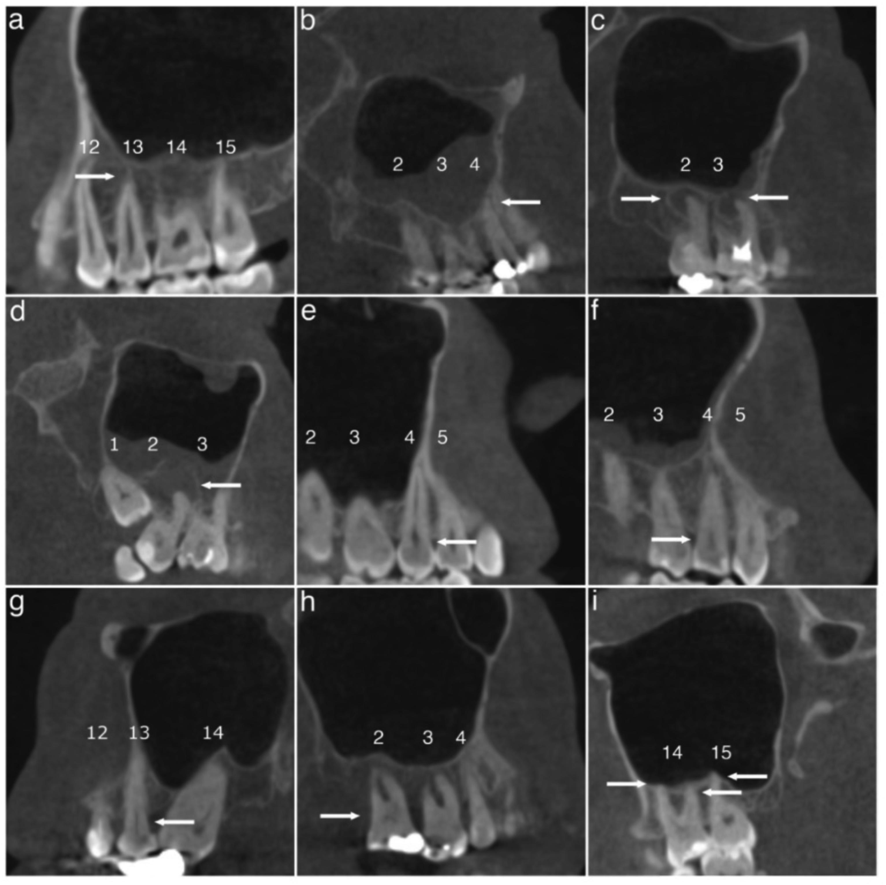
Figure 1. Representative CBCT scans of dental findings. Categories of periapical lesions: A, normal; B, widening of the radiolucent periodontal ligament space and minor changes of radiopaque lamina dura; C, periodontitis with well-defined and corticated radiolucent area; D, severe periodontitis with exacerbating structures. Categories of periodontal bone loss: E, normal (2 mm or less); F, mild (2.1-3 mm); G, moderate (3.1-5 mm); H, severe (more than 5 mm); I, at least 1 of the tooth roots in contact with the maxillary sinus floor. Teeth are numbered according to World Dental Federation notation (FDI) and each category/finding is indicated by arrows. (Courtesy of Riekki V-P, Nevalainen MT, Haapea M, et al: Associations between odontogenic and sinus pathologies—a low-dose CBCT study. Acta Odontol Scand 84:310-317, 2025.)
apices to the maxillary sinuses. Cone beam computed tomography (CBCT) now serves as the gold standard for identifying sinus problems originating in dental structures. Using low-dose CBCT to image the paranasal sinuses reduces the radiation dose patients receive and provides diagnostic accuracy comparable to that of standard CBCT. A study of the maxillary sinus pathology and dental findings noted on low-dose CBCT was done to assess associations between them.
Two hundred twelve consecutive CBCT scans were evaluated, covering 1565 teeth including 8 implants. Various odontogenic parameters were noted, including periapical lesions (PAIs), marginal periodontal bone loss (PBL), root contact with the maxillary sinus, and the presence of crowns, implants, defective restorations, and extensive caries (Figure 1). The maxillary sinus
findings included mucosal thickening (MT, generalized or localized), mucosal retention cysts, and opacification/fluid level.
The findings were divided into sinus pathology, relationship of sinus pathology and odontogenic findings, PAIs, and PBL.
Sinus Pathology
A total of 84.4% of the maxillary sinuses showed some abnormalities. Bilateral pathology was noted in 75.9%, and unilateral pathology was present in 16.0%. The most common finding was generalized MT, which was found in 65.6% of patients, with 45.3% bilateral cases. Just 2.3% of the men and 12.0% of the women had no sinus findings. Men had more retention cysts, but no other sinus pathology differed significantly between genders. Age showed no relationship to sinus alterations.
Sinus
Maxillary sinus pathology was found in 73.8% of the teeth, with the most common being generalized MT. Root contact with the maxillary sinus floor was the only dental finding significantly related to sinus pathology. The contact increased the risk of generalized MT, mucosal retention cysts, and fluid/opacification.
The severe grade 3 PAIs and moderate grade 2 lesions were combined. Severity of the lesion didn’t increase the likelihood that maxillary sinus pathology would be present. The combined analysis found root canal therapy and defective restorations in 52.1% of the grade 2 lesions and 46.5% of the grade 3 lesions. Sixty-eight percent of 25 teeth with inadequate root canal therapy and 34.5% of 58 teeth with adequate root canal therapy had grade 2-3 PAIs. Few teeth had root canal therapy and other restorations.
PAI alone or combined with root canal therapy or defective restorations wasn’t statistically associated with any sinus pathology.
Patients with dental implants can develop peri-implant diseases such as mucositis and implantitis, with the prevalence of these disorders increasing with the increased use of implants to replace compromised teeth. Peri-implant diseases are inflammatory, caused chiefly by the formation of peri-implant biofilms on the implant surface. The soft tissues become inflamed, leading to mucositis that presents as swelling, color change, and
In addition, adequate root canal therapy was not linked to sinus findings.
Grades 0 to 3 of PBL were documented, with 4.1% having grade 0, 29.7% having grade 1, 54.9% having grade 2, and 11.3% having grade 3. The proportion of PBL grades 2-3 was greatest in the generalized mucosal thickening. However, no statistically significant association was found between PBL and sinus pathology compared to healthy sinuses.
Root contact was the only dental finding that was significantly related to sinus pathology. This indicates that there may be fewer links between radiological dental findings and sinus pathology than expected.
Radiological dental findings accompanied by sinus pathology may not be as common as was previously thought. Having odontogenic sinus findings may not be as widely seen in all populations. Dentists should ensure that clinical examinations are thorough, and detailed analysis of the imaging is performed, since radiographic results can be difficult to interpret and understand.
Riekki V-P, Nevalainen MT, Haapea M, et al: Associations between odontogenic and sinus pathologies—a low-dose CBCT study. Acta Odontol Scand 84:310-317, 2025
Reprints available from V-P Riekki, Dept of Diagnostic Radiology, Oulu Univ Hosp, PO Box 50, 90029, Oulu, Finland; e-mail: vp. riekki@fimnet.fi
bleeding from the peri-implant mucosa, although marginal bone health is retained. Left untreated, peri-implant mucositis progresses to implantitis as the inflammation spreads to underlying bone, causing bone loss, swelling, pocket formation, implant mobility, and pus from the peri-implant sulcus. Generally, the progression of peri-implantitis leads to loss of the implant and the implant-supported prosthesis. Early diagnosis and management are essential, since the peri-implant mucositis
is reversible. Patients need to be educated regarding adequate home maintenance and plaque control coupled with effective periodontal debridement by dental professionals. Toothbrushes paired with interdental aids such as interdental, unitufted brushes or oral irrigators can help in plaque control. An oral irrigator uses a pulsating or continuous stream of water at high pressure to flush or remove biofilm and debris attached to teeth or implants. They can be effective adjuncts to tooth brushing and help control gingival inflammation and biofilm formation, avoid problems with dental prostheses, and maintain healthy dental implants. The current evidence is limited regarding the efficacy of oral irrigators for managing periimplant disease. The efficacy of oral irrigators at home was compared to that of other mechanical plaque control methods for patients managing peri-implant diseases.
The Medline (via PubMed), Scopus, Web of Science, EMBASE, and Cochrane databases were searched, along with the clinical trial registry. Seven articles—2 cross-over studies and 5 parallel-arm randomized studies—covering 360 participants were included in this review. Patients ranged from age 22 to 89 years. The primary outcomes were probing pocket depth (PPD), clinical attachment loss (CAL), gingival inflammation as measured by any gingival index (GI), bleeding on probing (BOP) as measured by any index, and plaque scores as indicated by a plaque index (PI). The secondary outcomes were bone loss around the implant, degree of osseointegration, recession around peri-implant mucosa, soft tissue healing measured by any index, ease of use and patient satisfaction, costeffectiveness, and quality of life after using the oral irrigator.
The results were considered in the context of primary outcomes, effectiveness against microbial agents, and patient factors, such as reported outcomes and ease of use.
The oral irrigator coupled with mechanical brushing was more effective in reducing PI, GI, and BOP than using just the oral irrigator or just mechanical brushing. The effectiveness in reducing bleeding around implants was increased 2.45-fold when the oral irrigator was paired with a manual toothbrush compared to using floss. Oral irrigators were able to increase the width of keratinized gingiva around implants and achieved a width comparable to that created by floss. PI and GI reduction was better with a combination of oral irrigator, manual toothbrush, and interdental brush compared to using just the interdental brush and oral irrigator. PI and BOP were significantly lower in oral irrigator groups than in the toothbrush-alone group.
After 12 weeks, no significant differences were seen for BOP scores when using an oral irrigator, mechanical brushing, and
interdental brush. Proinflammatory cytokine levels were reduced more effectively with oral irrigators than with tooth brushing only.
When oral irrigators were used with antimicrobial agents such as chlorhexidine, their efficacy was improved compared to using plain/tap water. Peri-implant mucositis prevalence was 5% with an oral irrigator and 0.06% chlorhexidine but 35% when plain water was coupled with the irrigator and 50% for tooth brushing alone. Patients who used chlorhexidine and oral irrigation had significantly lower BOP-positive sites after 12 weeks compared to those using tooth brushing only.
Oral irrigators were effective in reducing the microbial load compared to tooth brushing. The levels of Campylobacter rectus, Veillonella parvula, and Porphyromonas endodontalis were reduced more with oral irrigator use than when just tooth brushing was done.
No statistically significant differences were expressed regarding the ease of use or preference for using an oral irrigator or floss in patients with dental implants. Quality of life and patient satisfaction were similar whether the patient used mechanical tooth brushing alone or an oral irrigator. The comfort, cleaning sensation around the implants, and overdentures were all rated about the same. Patients with endosseous implants or overdentures had similar opinions regarding the ease of use.
Oral irrigators provide a good adjunct to mechanical tooth brushing, effectively removing microbial biofilm and controlling inflammation around implants. They proved comparable to other interdental aids in managing peri-implant diseases.
Patients need to be aware of the importance of performing excellent oral hygiene and plaque control measures when they have had an implant placed. Oral irrigators can be a valuable aid in reducing inflammation, plaque scores, and BOP, regardless of the solution used with the irrigator.
Gandhi G, Masanam BSL, Nair AS, et al: Efficacy of oral irrigators compared to other interdental aids for managing peri-implant diseases: A systematic review. BDJ Open 11:7, 2025
Reprints available from A Chopra, Dept of Periodontology, Manipal College of Dental Sciences, Manipal, Manipal Academy of Higher Education, Manipal, Karnataka, India; e-mail: aditi.chopra@manipal.edu
Periodontitis is a common oral disorder that has been found to have a bidirectional relationship with type 2 diabetes mellitus (T2DM), with both conditions affecting disease progression and treatment outcomes. Glycemic control is critical in managing T2DM, with reduced glycated hemoglobin (HbA1c) linked to reductions in myocardial infarction risk, diabetes-related mortality, and microvascular complication risk. Scaling and root planing (SRP) or SRP plus adjuvant therapies can improve both periodontal status and glycemic control in patients with periodontitis and T2DM. The optimal approach, however, has not been defined. No evidence clearly indicates whether adjuvant therapies combined with SRP are effective for patients with periodontitis and T2DM, nor have the specific therapies plus SRP that are most effective been singled out. A Bayesian network analysis was performed to evaluate whether SRP plus various adjuvant treatments can achieve better periodontal and glycemic control compared to no intervention or SRP alone in patients with periodontitis and T2DM.
A search of the Cochrane, PubMed, Embase, and Web of Science databases identified 37 studies eligible for analysis. Thirty-four were found in the published literature and 3 from international clinical trial registries. A total of 1989 patients with periodontitis and T2DM were included. The adjunctive treatments studied were satranidazole (SZ), 0.05% zoledronate gel (ZLN), amoxicillin (AMX), metronidazole (MTZ), azithromycin (AZM), doxycycline (Doxy), minocycline, diode laser (DL), Er-YAG laser (ERL), lowlevel laser therapy (LLLT), antimicrobial photodynamic therapy (aPDT), oral hygiene instructions (OHI), grape seed formulation (GSE), melatonin, symbiotic, omega-3, vitamin D3, and ginger.
The various treatments were evaluated for their ability to reduce probing depth (PD), clinical attachment level (CAL), bleeding on probing (BOP), HbA1c%, and fasting blood sugar (FBS) levels when used with SRP. Their effects were compared to those of SRP alone.
Compared to SRP alone, SRP + SZ, SRP + ERL, and SRP + melatonin all had superior improvements in PD. The most
significant results were achieved with SRP + SZ. Local adjunctive treatment with SZ was likely to be the most effective measure, followed by systemic treatment with melatonin and local adjunctive treatment with ERL. The least effective results were found with OHI.
Compared to SRP alone, SRP +SZ, SRP + melatonin, and SRP + vitamin D3 had superior improvements in CAL. SRP + SZ demonstrated the most effective results. Local adjunctive treatment with SZ had the highest probability of being the most effective approach to reducing CAL, followed by systemic treatment with melatonin and systemic adjunctive treatment with vitamin D3. However, OHI had the least positive effect on CAL.
Compared to SRP alone, SRP + AZM, SRP + aPDT, and SR + Doxy had superior improvements in BOP. The systemic adjunctive treatment with AZM was likely to be the most effective strategy for improving BOP. Local adjunctive treatment with aDPT and systemic adjunctive treatment with Doxy were the next most effective approaches.
Sixteen studies adopted HbA1c% as an indicator of glycemic control. Compared to SRP alone, SRP + Doxy + aPDT and SRP + DL were more effective in lowering HbA1c%. SRP + Doxy + aPDT was the most effective approach, followed by SRP + DL. The other adjunctive therapies and SRP alone demonstrated no significant differences in effectiveness.
Nine studies used FBS as an indicator of glycemic control. Compared to SRP alone, SRP + DL had superior effectiveness and was better than any of the other adjunctive treatments. The other treatments had results comparable to those achieved with SRP alone.
Periodontal treatment with SRP + SZ was the most efficacious in lowering PD and CAL in patients with periodontitis and T2DM. Treatment with SRP + AZM was the most efficacious choice for
lowering BOP. HbA1c% was improved most effectively by SRP + Doxy + aPDT. FBS was improved most efficaciously by SRP + DL.
Clinical Significance
Further studies are needed to confirm the findings of this analysis because the quality of the evidence tended to be low or very low. However, the treatments tested could prove useful for patients who suffer from both periodontitis and T2DM.
TMDs affect the masticatory muscles, TMJ, and related structures, causing orofacial pain. Painful TMDs are the result of the patient’s psychological profile, state of pain amplification, general health, and global symptoms, making these disorders biopsychosocial in nature. In addition to the TMJ and masticatory muscle pain, patients often have impaired jaw movements, TMJ sounds, and ontological symptoms such as hearing loss, vertigo, dizziness, ear pain, and tinnitus. The mechanisms by which tinnitus causes noises without an external source are as yet undetermined but the destruction of outer auditory structures may be involved. The prevalence of tinnitus in young adults is 13.7%, whereas older adults have a prevalence of 23.6%, but in both cases quality of life is compromised. Evidence does suggest that the existence and co-existence of TMDs and tinnitus may result from clinical connections between the conditions. The presence of these associations and whether the associations differ between painful and non-painful TMDs were investigated. Possible relationships connected to psychosocial status were also assessed.
A search was made in the MEDLINE, EMBASE, CINAHL, Cochrane Central Registry of Controlled Trials (CENTRAL), and
Xie X, Xu J, Li Y, et al: Efficacy of nonsurgical periodontal treatment on patients with periodontitis and type 2 diabetes mellitus: A systematic review and Bayesian network meta-analysis. Acta Odontol Scand 84:201-211, 2025
Reprints available from G Awuti, The First Affiliated Hosp of Xinjiang Medical Univ, 137 Liyue Rd, Xinshi District, Urumqi, Xinjiang Uygur Autonomous Region, China; e-mail: guawuti@ sina.com
Web of Science databases. Thirty-two full texts were included in the systematic review.
The vast majority of patients diagnosed with tinnitus have a diagnosis of TMD. However, among patients suffering from TMD, only about half also have been diagnosed with tinnitus. A strong association was noted between having tinnitus and having been diagnosed with TMD.
Six of 7 studies clearly differentiated between painful and nonpainful TMDs. It was noted that 2 to 8 times more patients with painful TMD also had tinnitus compared to those who had non-painful TMDs.
Six studies evaluated the psychological status of persons with tinnitus and TMD, focusing specifically on depression. In 4 studies, persons who had both tinnitus and TMD had higher levels of depression compared to those who only had tinnitus. Having tinnitus was also associated with significantly elevated levels of depression when compared to a control group. A
positive relationship between tinnitus severity, chronic pain, and depression appears to indicate a significant correlation between chronic pain intensity and tinnitus.
A strong significant association seems to exist between tinnitus and TMD. The prevalence of TMD in patients with tinnitus is significantly greater than the prevalence of tinnitus among patients with TMD. Further research is needed to explore this relationship.
Based on these findings, clinicians who have patients with either tinnitus or TMD should also evaluate their patients for the opposite condition. In addition, tinnitus may be a red flag indicating other lifethreatening diseases since other comorbidities of TMD include multiple sclerosis, vestibular schwannoma, meningitis, syphilis, Lyme disease, and complications from medications.
De La Torre Canales G, Christidis N, Grigoriadis A, et al: Associations between temporomandibular disorders and tinnitus – a systematic review. CRANIO 43:969-985, 2025
Reprints available from G De La Torre Canales, Dept of Dental Medicine, Karolinska Inst, Alfred Nobels All� e 8, Huddinge SE141-52, Sweden; e-mail: giancarlo.de.la.torre.canales@ki.se
Because tooth discoloration is a common phenomenon, patients often seek whitening to restore or improve their smile. External dental whitening involves oxidizing the chromogenic substances that discolor the teeth. Hydrogen peroxide and carbamide peroxide are commonly used, although a variety of bleaching gels can achieve whitening. However, the gels have inconsistent effectiveness, which should be addressed to achieve a better and more predictable outcome. The initial stage of teeth discoloration, the concentration of the bleaching agent or gel, time factors during the application, and individual patient factors influence the efficiency of bleaching gels. Side effects such as tooth sensitivity and gingival irritation must also be evaluated. A review was done to evaluate the bleaching efficacy of various products, how the different products affect changes in tooth color and how long they last, and possible side effects.
A search was conducted in the PubMed, Scopus, Web of Science and Cochrane Library databases seeking studies whose key end measures were changes in tooth color before and after external bleaching. Twenty-three articles were selected. Measurements used shade guide units (SGU) or a spectrophotometer/colorimeter (ΔE). Effectiveness was compared after combining research in subgroup analyses. The gels were based on hydrogen peroxide
(HP), carbamide peroxide (CP), and other substances often used for teeth whitening procedures. Eight articles combined HP with CP and 11 used HP in gel. Patient satisfaction was determined using questionnaires.
Bleaching Protocol and Patient Satisfacti on
The percentage of CP gels was between 10% and 37%, with most studies using a 10% CP gel. HP was used in concentrations from 5.4% to 40%, with 35% being the most commonly used amount. Most of the questionnaires didn’t reveal any changes between patient groups, generally reporting that patients were satisfied with the treatment.
In 19 studies, a significant reduction in ΔE was reported, but in 4, no significant change was found. Thus all studies either found a significant reduction or no change in ΔE.
CP gels tended to produce greater tooth-whitening effects than higher-concentration HP gels, but HP had a relatively lower impact on post-bleaching sensitivity. It was deemed more suitable for patients who were at risk for discomfort. Desensitizing agents such as potassium nitrate reduced sensitivity without compromising bleaching efficacy. Sodium perborate had limited effectiveness as
a bleaching agent. Sonic activation and in-office bleaching protocols had no significant effect on whitening outcomes or sensitivity.
Stained teeth can be significantly whitened by using bleaching gels. A greater concentration of CP bleaching gel was associated with less tooth sensitivity than the higher concentration of HP bleaching gel.
Being aware of the findings of various studies will provide practitioners with the evidence needed to make well-informed decisions regarding tooth whitening. Subtle differences in the bleaching agent, concentration, application, and duration were noted between the various studies, so dentists and dental specialists must choose the best course of action for each individual patient. Several approaches are possible, and dentists should be aware of the needs of the patient and the effect of the products and techniques.
Das G, Qasim M, Rana MH, et al: Comparative efficacy of external dental bleaching gels: A systematic review of whitening performance and longevity. Clin Oral Invest 29:546, 2025
Reprints available from G Das, Dept of Prosthodontics, College of Dentistry, King Khalid Univ, 61421, Abha, Kingdom of Saudi Arabia; e-mail: drgotam2000@gmail.com
Sleep experts tend to recommend getting 7 to 9 hours of sleep each night to have good health and emotional well-being. Sleeping fewer than 7 hours a day may increase the risk of obesity, high blood pressure, heart disease, and other problems related to sleep deprivation. The actual recommendation, however, should take into account the quality of sleep, which can have more significant effects than the quantity of sleep.
Dr. Tony Cunningham, clinical psychologist and director of the Center for Sleep and Cognition in Boston and assistant professor of psychology at Harvard Medical School, said, “There’s 2 different things going on in our bodies that dictate both the type of sleep that we’re getting and the quality of sleep that we’re getting, and that is our sleep pressure and circadian rhythms.”
Sleep pressure or sleep drive builds up throughout the time we’re awake and decreases when we’re asleep. It’s the reason we start feeling tired after being awake for an extended period of time. To get a good night’s worth of sleep, we should get into bed when we’ve built up a lot of sleep pressure.
Circadian rhythm is the body’s internal clock. Although it can be influenced by light and other external factors, the pattern our bodies follow is guided by our brains. Cunningham said, “The circadian rhythm can fluctuate and send either sleep-promoting signals or wake-promoting signals throughout the course of the day. So, if you’ve ever pulled an all-nighter and you’ve gotten a second wind in the middle of the night, and you felt less tired, then that is your circadian rhythm kicking in.”
For better sleep quality, sleep pressure and circadian rhythm should work together, which means any abrupt changes or an irregular sleep schedule can influence sleep and sleep quality. Cunningham suggests that a way we can improve sleep quality “is to start waking up at the same time every day, as it can be a little bit more impactful than going to bed at the same time every day— because it’s not always a good idea to go to bed if you’re not sleepy yet.” Once you have a general sleep schedule, your body naturally starts to seek out its optimal sleep time.
The amount of time each person sleeps will vary. Some actually fall into the average range of 7 to 9 hours, but it’s not unusual for people to need just 5 or 6 hours. Other people will need 10 or 11 hours each night. To determine what’s best for you, you can do a couple of things, as follows:
• Maintain a consistent bedtime when you’re pretty confident you will be able to fall asleep within 20 to 30 minutes. Make sure that you feel sleepy and not just tired. You need to have sufficient sleep pressure, and if you can’t fall asleep within 20 to 30 minutes, you should do some low-arousing activities such as taking a bath or meditating with the lights dimmed until you feel sleepy.
• Find a period of time when you can sleep until you wake up naturally with no alarm. Cunningham suggests that you hide your clocks, block out the windows, possibly use a noise machine, or wear an eye mask. You should have no sense of the time, then go to sleep and sleep until you naturally wake up.
It can be hard to fit this into your schedule, but it could be worthwhile to try the experiment. For example, when you’re home for the holidays, you probably will sleep longer than normal the first few days. You can go to bed at midnight and sleep until 10 or 11 in the morning to catch up on your sleep pressure. You’ll know you’ve found your sleep time when you wake up for 3 or 4 days in a row at about the same time with no external cues, lights, or alarms.
[Park G: You Might Not Need 8 Hours of Rest. Here’s How to Find Your Perfect Sleep Time. CNN Health, Dec 8, 2025]
Insufficient sleep is proving to have a greater impact on decreased life expectancy than other lifestyle factors such as diet, physical activity, and social isolation. Six of every 10 adult Americans don’t get sufficient sleep. Past studies have found inadequate sleep is related to conditions that can adversely affect quality of life and life expectancy, such as cardiovascular disease, type 2 diabetes, obesity, depression, anxiety, gastrointestinal issues, and dementia.
Andrew McHill, associate professor and director of the Sleep, Chronobiology, and Health Laboratory at Oregon Health & Science University and senior author of a new study in the journal Sleep Advances, noted that “Sleep plays a vital role in nearly every biological process within our body, yet it is still one behavior that I think we commonly take for granted. It’s something that is easy to put off until the weekend or deprioritize based on work or social events.” The new study found that insufficient sleep has a more significant impact on decreased life expectancy than other lifestyle factors.
The study collected data from the 2019-2025 Centers for Disease Control and Prevention Behavioral Risk Factor Surveillance System surveys to check for links between insufficient sleep and life expectancy. “Time and time again, when we analyzed the relationship between rates of insufficient sleep and life expectancy at the county level, regardless of state, there were strong relationships across the country,” McHill said. At the end of the study, when compared to diet, physical activity, and social connection, which are common behaviors associated with life expectancy, inadequate sleep was a more powerful driver of lower life expectancy than any other factor. In addition, researchers found that insufficient sleep showed a significant link with life expectancy in most US states from 2019-2025.
McHill noted, “These findings really deliver the message that regardless of where you live, whether it be rural, urban, north, south, east, or west, sleep plays a vital role in our health and wellbeing. Moreover, because we had multiple years of data, these findings also highlight that even when faced with extreme circumstances (i.e., COVID pandemic), sleep still plays a key role in our health.” Dr. Pakkay Ngai, medical director for the Sleep-Wake Center at Palisades Medical Center in New Jersey, said, “As a doctor who regularly treats patients struggling with sleep, my first reaction to this study is one of profound validation, yet also surprise at the sheer magnitude of the findings.” Ngai also noted that “It reinforces the message that sleep is not a luxury or something to be sacrificed; it is a biological necessity on par with, and in some ways more impactful than, other cornerstone health behaviors.”
[Pelc C: Sleep Is More Important for Longevity Than Diet, Exercise, Social Ties, Study Says. MedicalNewsToday, Dec 12, 2025]