






Editor-in-Chief
Douglas B. Berkey, DMD, MPH, MS
Senior Publisher
Annie Zhao
Journal Manager
Sangamithrai S
Abstract Writer
Elaine Steinborn
© November 2025, Elsevier Inc. All rights reserved.
No part of this publication may be reproduced, stored in a retrieval system, or transmitted, in any form or by any means—electronic, mechanical, photocopying, recording, or otherwise—without prior written permission from the publisher.
Publication Information Dental Abstracts (ISSN 0011-8486) is published bimonthly by Elsevier Inc., 1600 John F. Kennedy Boulevard, Suite 1600, Philadelphia, PA 1910, United States. Months of publication are January, March, May, July, September, and November.
Customer Service Office: 11830 Westline Industrial Drive, St. Louis, MO 63146. Periodicals postage paid at New York, NY, and additional mailing offices. Annual subscription rates for 2018 (domestic): $144.00 for individuals and $71.00 for students; and (international) $194.00 for individuals and $105.00 for students.
USA POSTMASTER: Send address changes to Dental Abstracts, Elsevier Health Sciences Division, Subscription Customer Service, 3251 Riverport Lane, Maryland Heights, MO 63043.
Copyright 2025 by Elsevier Inc. All rights reserved. Dental Abstracts is a trademark of Elsevier Inc. Dental Abstracts is a literature survey service providing abstracts of articles published in the professional literature. Every effort is made to ensure the accuracy of the information presented in these pages. Statements and opinions expressed in the articles and communications herein are those of the author(s) and not necessarily those of the Editors or the publisher. The Editors and the publisher disclaim any responsibility or liability for such material. Mention of specific products within this publication does not constitute endorsement.
All inquiries regarding journal subscriptions, including claims and payments, should be addressed to: Elsevier Health Sciences Division, Subscription Customer Service, 3251 Riverport Lane, Maryland Heights, MO 63043. Tel: 1-800-6542452 (U.S. and Canada); 314-447-8871 (outside U.S. and Canada). Fax: 314447-8029. E-mail: support@elsevier.com (for print support); support@elsevier.com (for online support).
Notice: Journals published by Elsevier comply with applicable product safety requirements. For any product safety concerns or queries, please contact our authorized representative, Elsevier B.V., at productsafety@elsevier.com
Practitioners and researchers must always rely on their own experience and knowledge in evaluating and using any information, methods, compounds or experiments contained herein. Health care practitioners must exercise their professional judgement and make all treatment-related decisions based solely on the specific conditions of each patient. Because of rapid advances in the medical sciences, independent verification of diagnoses and drug dosages should always be made.
The content is provided “as-is” and Elsevier makes no representations or warranties, whether express or implied, as to the accuracy, completeness, or adequacy of any content. To the fullest extent permitted by law, Elsevier assumes no responsibility for any damages, adverse events, or liability arising from use of information contained herein including for any injury and/or damage to persons or property, whether as a matter of product liability, negligence or otherwise.
Inclusion of any advertising material in this publication does not constitute a guarantee or endorsement of the quality or value of such product or service or of any of the representations or claims made by the advertiser. Printed by Sheridan at 450 Fame Avenue, Hanover, PA 17331
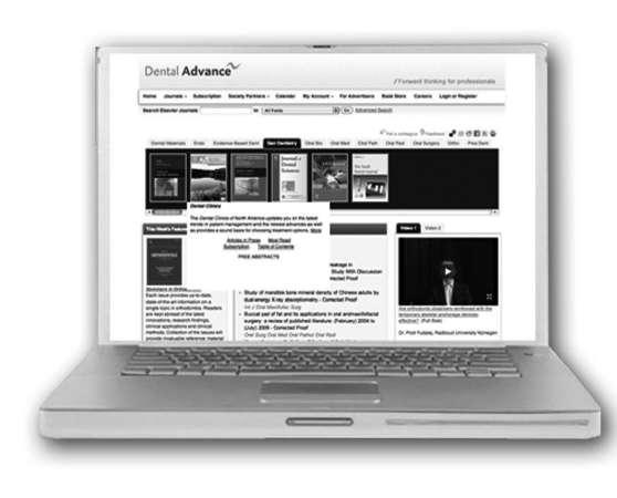
DentalAdvance .org is the gateway offering high-quality research, news, jobs and more for the global community of dental professionals.
Journal profiles with quick links to Tables of Contents, author submission information, and subscription details
Important information and valuable resources on how to submit a journal article
Dentistry Articles in Press from participating journals
Quick links to the leading dentistry societies worldwide Dentistry News from Elsevier Global Medical News (formerly IMNG)
Dentistry Jobs powered by ElsevierHealthCareers.com
Editor-in-Chief
DouglasB.Berkey,DMD,MPH,MS ProfessorEmeritus, SchoolofDentalMedicine, UniversityofColorado, Aurora,Colorado
AssociateEditor
DanielM.Castagna,DDS AssociateProfessor,DepartmentofPreventiveandRestorativeDentistry, UniversityofthePacific,ArthurA.DugoniSchoolofDentistry, SanFrancisco,California
P.MarkBartold,DDSc,PhD,FRACDS(Perio) ProfessorEmeritus SchoolofDentistry, UniversityofAdelaide Adelaide,Australia
RobBerg,DDS,MPH,MS,MA ProfessorandChair, DepartmentofAppliedDentistry, UniversityofColoradoSchoolof DentalMedicine, Aurora,Colorado
TylerH.Berkey,DMD GeneralDentist Aurora,Colorado
FionaM.Collins,BDS,MBA,MA ConsultantandEditor, GeneralDentist Longmont,Colorado
AnthonyJ.DiAngelis,DMD,MPH ChiefofDentistry, HennepinCountyMedicalCenter, Professor,UniversityofMinnesota, SchoolofDentistry, Minneapolis,Minnesota
RaulI.Garcia,DMD,MMedSc ProfessorandChairman, DepartmentofHealthPolicyandHealthServicesResearch, BostonUniversitySchoolofDentalMedicine, Boston,Massachusetts
MichaelSchafhauser,DDS GeneralDentist, St.Paul,Minnesota
JoeVerco,DClinDent PaediatricDentist NorthAdelaide,Australia

CreatingaSafeHaventoDeliverTrauma-informed DentalCare
ManagingSurvivorsofTrauma
VerticalMarginPreparationsForPosteriorZirconia Crowns
Inquiry
Implant/RootCanalCostComparison478
CalculatingCost-effectivenessforVariousTreatment Approaches
TemporomandibularDisorderTreatments479
OcclusalSplintandBotoxInjectionsforTMDs
CompositeRestorations480
Self-adhesiveandConventional/Bulk- fillResin CompositeRestorations
ChoosingtheBestUniversal,Etch-and-rinse,and Self-etchAdhesives
ExtractionComplications/Hygiene
PredictiveValueofFullMouthPlaqueScoreBefore LowerThirdMolarSurgery
ClearAligners/DeepBites
ClearAlignerstoAddressDeepDentalOverbites
ImplantLength/FailureRates
ChoiceofShortorLongImplants
Pain/AnxietyControlMeasures
CryotherapyforReducingInjectionPain
DistractionMethodsforChildrenwith Attention-de ficitHyperactivityDisorder
PediatricPulpotomyOutcomes
PartialPulpotomyforChildrenwithIrreversiblePulpitis
BloodSugarVariationsWithOurCircadianRhythm DentalProblemsCausedbyGLP-1Drugs
FromtheExecutiveDirector e1
FromtheInternationalPresident-electe2
FromtheInternationalVicePresidente3 SectionNews e5
Notes
TofacilitatetheuseofDentalAbstractsas areferencetool,allillustrationsandtablesincluded inthispublicationarenowidentifiedastheyappear intheoriginalarticle.Thischangeismeanttohelp thereaderrecognizethatanyillustrationortable appearinginDentalAbstractsmaybeonlyoneof manyintheoriginalarticle.Forthisreason, figure andtablenumberswilloftenappeartobeoutof sequencewithinDentalAbstracts.
StandardAbbreviations
Thefollowingtermsareabbreviated:acquiredimmunodeficiencysyndrome(AIDS),humanimmunodeficiency virus(HIV),andtemporomandibularjoint(TMJ).
The Hippocratic Oath is perhaps the oldest expression of medical ethics. It instills the obligations of a physician into students participating in medical training and the duties of students to their teachers. This was generally done for the first time at the students’ commencement ceremonies. White coat ceremonies were begun in 1993 and first held at Columbia University College of Physicians and Surgeons to underscore the strong commitment among medical students to humanistic practice. The entering class of participants received physician coats and recited a modern version of the Hippocratic Oath. The first white coat ceremony at a dental school began at the University of the Pacific in the mid-1990s and marked the transition of the dental students from preclinical to clinical care and included an oath that emphasized integrity, ethical behavior, respect for patients, and responsibility to the community. Dentists are primarily responsible to individual patients, whereas scientists have a responsibility to the wider public. Scientific discoveries have offered benefits but sometimes also had consequences for society. New researchers may be helped along in their understanding of their responsibilities to society and moral obligations if they also speak an oath similar to that used by health care providers. Various scientists and engineers have used oaths, usually on graduation day, but the oath ceremony may be more beneficial at the entry point to scientific studies so students recognize ethical and professional concepts early in their careers. Attitudes toward ethical codes or oaths, the justification of the timing of oath ceremonies, and the development and institution of the Dentist-Scientist Oath were outlined.
Ethical codes can be dismissed or at least met with skepticism because of the difficulty enforcing them. However, taking an oath with peer groups offers a symbolic bonding process that can reinforce one’s identity. Group identity reinforcement can improve the individual’s level of identification, with low identifiers who share in taking the oath perhaps being given a point of connection with the group, leading to
improved adherence to the group’s norms and values. The public declaration in front of family and friends as well as their peers may lead to a greater acknowledgment of the students’ responsibilities at a formative stage in their development.
The American Association for the Advancement of Science has identified 16 oaths for scientists and engineers that were used in the 20 th century. Most were taken on graduation day, but taking the oath at the students’ entry into their scientific studies may improve the students’ adherence to these values. This harkens back to the dental white coat ceremonies where oaths were taken at the junction between preclinical and clinical care.
A Graduate Student Oath was introduced in 2007 at the University of Toronto at the first meeting of each year’s new graduate student body. This oath was designed to increase student awareness of ethical obligations, publicize concern over academic misconduct, raise awareness of the intense competition in training programs, and assert the consequences of plagiarism, falsifying data, and practicing scientific fraud. Community, professionalism, and ethical conduct in research are the important factors this oath stresses.
The Dentist-Scientist Oath and its ceremony were begun at the University of the Pacific, with the oath developed through the principles espoused by Davis and colleagues regarding pride, integrity, and knowledge pursuits (Figure). It also focuses on inclusivity in clinical research to reduce biases, promote social justice and health equity, and produce more innovative science. The ability to generalize biomedical research across the research continuum depends on considering inclusivity in preclinical research as well. An additional inclusion in the oath is the importance of avoiding bias in research to produce knowledge applicable to all people.
"I, [NAME], have entered the serious pursuit of new knowledge as a member of the community of dentist-scientists at the University of
"I solemnly declare the following:
Pride: I declare my pride in belonging to the international community of research scholars.
Inclusion: I will incorporate diversity into preclinical and clinical studies, and foster a culture of prioritizing and sustaining diversity at all levels of research and be inclusive of racial and ethnic minority groups, as well as other populations experiencing health disparities. Integrity research and scholarship.
Pursuit: I will pursue knowledge and create knowledge for the greater good, but never to the detriment of colleagues, supervisors, research subjects or the international community of scholars of which I am now a member
By pronouncing this Dentist-Scientist Oath
Figure. Arthur A. Dugoni School of Dentistry, University of the Pacific’s Dentist-Scientist Oath. (Inspired by and modified with permission from Davis KD, Seeman MV, Chapman J, et al. A graduate student oath. Science 320:1587-1588, 2008.)
This oath was part of the celebration of students’ completion of the didactic portion of their clinical research training and in recognition of their transition to applying their clinical research skills to patients for the first time. At the first Dentist-Scientist Oath ceremony on August 20, 2024 at the University of the Pacific, 171 students in the doctor of dental surgery class of 2026 and the international dental studies class of 2025 recited the oath in the presence of their friends and families as well as staff and faculty members. Each participant received a ceremony booklet containing the oath, the list of those taking the oath, and a list of resources for responsible and ethical research activities.
An oath such as the Dentist-Scientist Oath should be included in clinician-scientist training programs, whether at the graduate research program, formal undergraduate training programs of dentist-scientists, or undergraduate dental training programs where research is valued. Ideally, the oath would be part of a ceremony held in the early years of a formative training period. As with the Hippocratic Oath, the Dentist-Scientist Oath should be reviewed regularly and updated to accommodate shifts in health care and social institutions.
Lam DK, O’Hara R, Sunwoo JB, et al: A dentist-scientist oath. J Am Dent Assoc 156:513-515, 2025
Reprints available from DK Lam, Dept of Oral and Maxillofacial Surgery, Univ of the Pacific, 155 Fifth St, San Francisco, CA 94103, USA; e-mail: dlam@pacific.edu
Surges in technology and screen time use have raised concerns about the effects of these influences on children. Children can freely access screens in a way that supports an “always-on” culture. As a result, children may become unable to properly process their own thoughts or disconnect from the external pressures of the digital technology. An overview
of the negative and positive effects of screens in the lives of children was offered.
The internet offers a magnet for attention that can disrupt and distract from our ability to concentrate and contemplate. While daily production may be increased with multi-tasking
and quick decision-making practices, we may become shallower and too-often disconnected from reality. Screen time affects subconscious thought and relationships. Constantly swiping through images and videos can lead to distorted views of our lives. When the screen is full of idealized physical images online, we can suffer from a sense of unattainable body goals. Scarcity and loneliness can also result. If all that’s shown is lavish vacations, lifestyles, and possessions, our surroundings don’t feel sufficient and we can suffer from the fear of missing out (FOMO).
Professional lives can also be undermined. Seeing how others constantly over-perform can cause a sense of self-doubt or insecurity about our own progress. The popularity we see online can cause us to feel isolated or unable to experience close friendships.
The psychological effects of digital dependency include depression, suicidal tendencies, hyperarousal, increased stress hormone levels, desynchronization of our body clock or circadian cycle, changes in brain chemistry, reduced mental energy, and altered development. Our physical health can suffer as well, from developing higher levels of risk factors for obesity or cardiovascular disorders, to lower levels of high-density lipoprotein (HDL) cholesterol, and to increased insulin resistance. A number of studies have linked increased digital device use to higher levels of stress, anxiety, and depression, as well as poor sleep quality.
Having access to digital devices has allowed for unprecedented availability of information, educational offerings, and global connectivity. Online groups help to build and also strengthen communities. Social media also help in building networks. Posts and videos often empower us and allow those who have been marginalized to speak up. Teledentistry and remote working help in reaching those who don’t have access to care. Even bureaucracies have been improved when digital services are put into place.
Some balance should be sought in our digital and natural experiences. Adults and children should institute control over their screen time and identify the usefulness of nondigital living. Important skills that we need to ensure are taught and nurtured in our children include practicing mindful habits, setting healthy boundaries, developing resilient behaviors, and learning coping mechanisms to manage stress and anxiety. The strong and resilient will be the ones who survive without giving in to digital devices.
Sahota RK: Mission Smartphone. J Calif Dent Assoc 53:2500455, 2025
Reprints available from RK Sahota; e-mail: ruchi.nijjar@gmail.com
Artificial intelligence (AI) is a newer advancement in dentistry that may offer benefits for dental hygienists. Knowing what AI is available; how it can affect the hygienist’s workflow, efficiency, and quality; and how best to incorporate AI into practice will guide hygienists in taking advantage of its abilities to enhance their patient care.
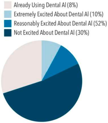
AI is software that can execute cognitive tasks similar to those performed by humans. This includes perception, reasoning, interactions with surroundings, and problem solving. A recent survey indicated how AI can aid dental hygienists in performing their duties (Figure 1).
The use of AI can allow offices to switch from paper charts to electronic health records (EHRs), which can facilitate the sharing of patient information with various providers. AI can collect data, assist in clinical decisions, predict prognoses, and provide healthrelated results, all of which enhance patient management. An interdisciplinary approach to patient care and comprehensive oral-systemic care can benefit from the use of AI systems.
Limitations in AI technology include possible inherent bias and problems adhering to the Health Insurance Portability and Accountability Act (HIPAA). In addition, the dental equipment may be outdated, so that AI isn’t compatible. A lack of training can hinder the adoption or use of AI systems. Automation bias, where clinicians view AI as perfect and a replacement of clinical skills and critical thinking, is a risk. AI should be viewed as an auxiliary method and not a replacement for professional judgment. It’s vital that open discussions of what AI can and cannot do be held among patients, dental hygienists, and product developers.
Meeting the Standard of Care
AI may be helpful in the thoroughness and quality of dental care provided. The periodontal standard of care was expanded by the 2017 Classification of Periodontal and Peri-Implant Diseases and Conditions. The new standard of care requires dental hygienists to adapt to a system that assesses the extent, severity, and
progression of periodontal diseases and conditions. As a result, hygienists are inundated by an immense body of data.
Complete periodontal charting is the foundation for accurate assessment. This includes identifying radiographic bone loss, assisting with periodontal charting, recording intuitive clinical notes, classifying disease, using patient education tools, and recommending patient self-care approaches.
Among the demands dental hygienists must meet in patient care is monitoring A1C levels to ensure they don’t exacerbate periodontitis and gingivitis, raise the risk of infection, cause xerostomia, or delay wound healing. Head and neck examinations must be done to assess for cancer. All these evaluations must be properly and completely documented. Dental hygienists must also help inform patients regarding their overall health and steps they need to take to improve their health status. All of this must be accomplished within the confines of an appointment lasting an hour or less. Adding older, inadequate equipment to the mix can compound the difficulties hygienists must manage.
Health evaluations that must be assessed to complement periodontal charting include radiographic bone level measurements, oral trauma and past history of periodontal diseases, and pH and microbiological determinants of systemic health. Evidence-based caries prevention involves the thorough assessment of individual risk factors, the identification of caries, the integration of the patient’s medical and dental histories, and the monitoring of glucose and A1C testing to evaluate for diabetes. AI can be used to address limitations on the hygienist’s ability to perform all these evaluations.
AI is most applicable in diagnostic imaging techniques. Radiographs help in identifying calculus, determining bone levels, and helping to chart existing conditions. AI can improve the hygienist’s ability to properly interpret radiographs, assist with thorough caries assessments, and reduce human inaccuracies while supporting the clinician’s expertise. Five different areas can benefit from radiographic AI: restorative therapy, endodontics, orthodontics, oral surgery, and periodontics. Adding AI to the hygienist’s clinical expertise will improve diagnosis and allow for better efficiency, accuracy, and precision.
1.
perceptions of
Radiographic AI can detect incipient caries and automatically chart existing restorations. Accuracy can vary, however, depending on the AI platform, image quality, and lesion type. Images also make it easier to educate patients on areas of concern, which can build the patient’s trust in his or her treatment plan.
Trained AI algorithms can recognize indicators of periodontal diseases in intraoral images. Mesial and distal radiographic bone levels can be confirmed using periapical radiographic AI, helping to classify periodontal diseases. Patients can also be shown educational visual aids overlaid with their teeth, be informed about the precise measurements of any bone loss, and even be given information on their condition based on the 2017 Classification of Periodontal and Peri-Implant Diseases and Conditions.
The diagnosis of periodontal bone levels based on panoramic and cone-beam computed tomography images can be more accurate using AI technology. This can enhance the classification, diagnosis, and treatment of gingivitis and periodontal diseases by
reallocating the time required for radiographic examinations toward greater in-depth patient care. Combining AI with radiographs, intraoral photographs, clinical findings, and periodontal examinations can increase the effectiveness of dental hygiene practices. Intraoral images may include AI to identify inflammation in gingival tissues. This allows for prompt treatment by making an early diagnosis, which can avoid the loss of connective tissue, the destruction of supporting tooth structures, and bone loss.
AI-based smartphone applications may allow for self-monitoring of gingival health. Patients can take photographs of their own mouths (Figure 2). The effectiveness of this self-monitoring has promoted better patient compliance with follow-up treatment, better oral health, less debris, and improved periodontal health. With compatible EHRs, the AI use from the smartphone can integrate seamlessly into the dental hygiene workflow with tools and technologies that are both accessible and user-friendly.
Clinical chart notes are available that allow AI to pull dental information from the conversations between patients and clinicians and add it into clinical chart notes. This offers the ability to optimize workflow and raise the quality of patient care. Intuitive clinical notes gathered by AI can free the hygienist to focus on patient care. Patients must give consent to have their data shared with AI and understand who can access the data, how the information will be stored or used, and how their privacy will be safeguarded.
AI voice-activated periodontal charting may free up more time and allow flexibility in accurately documenting the periodontal chart, leading to proper classification and treatment planning (Figure 3). The AI tool can adapt to anyone’s voice and doesn’t require an assistant. Charting out loud can add to the patient’s experience, encouraging him or her to listen and participate in the discussion. Having a deeper understanding of the patient’s treatment options could promote better infection control and support ergonomic efforts by removing the need to access a keyboard. Dental hygienists can more accurately classify, diagnose, and treat periodontal diseases by adding AI support.

Figure 2. Artificial intelligence (AI) based smartphone applications may provide a way to self-monitor gingival health. Here, AI evaluates a patient’s gingiva via “selfies.” Used with permission from Alalharith DM, Alharthi HM, Alghamdi WM, et al: A deep learning based approach for the detection of early signs of gingivitis in orthodontic patients using faster region-based convolutional neural networks. Int J Environ Res Public Health 17:8447, 2020. (Courtesy of Miller R, Haddlesey C: How artificial intelligence is reshaping patient care. Dimensions Dent Hyg 23:32-35, 2025.)
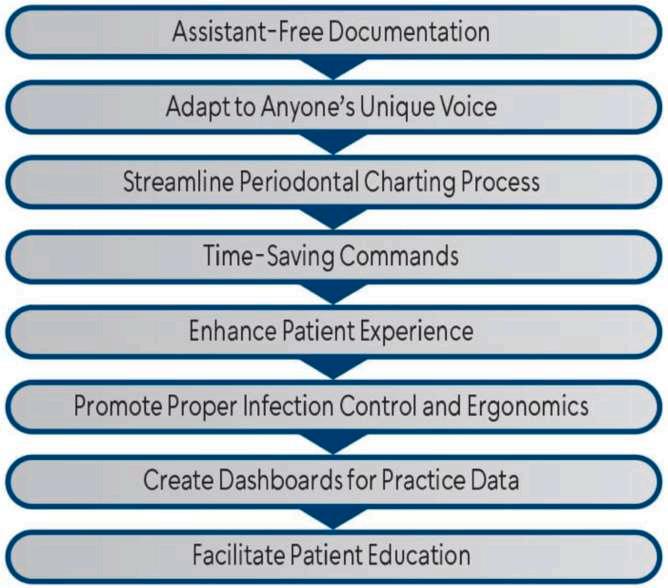
AI can also generate reports and educational information for patients. Qualified AI can pull information from medical histories that can be used to create a comprehensive oral-systemic approach to patient education. With power toothbrushes guided by AI and a smartphone application, patients can track their duration of brushing. They can be given information about brushing habits or bleeding and can receive rewards and personalized coaching.
AI isn’t yet fully integrated into dental practice, but has great potential to help dental hygienists collect and evaluate information that can improve patient compliance and build trust. The areas of most application for AI are charting and radiographic analysis.
AI technology needs to be studied further to pinpoint how it can benefit not only the dental hygiene profession but also patients and other dental or medical personnel. Dental hygienists should proactively explore the available AI technologies to determine how they can improve patient care both in the office and at home.
Miller R, Haddlesey C: How artificial intelligence is reshaping patient care. Dimensions Dent Hyg 23:32-35, 2025
Reprints not available
Pivoting from being in an insurance network to having a fee-forservice practice requires careful planning and a concerted effort to build up the patients’ perception of value. Each interaction should contribute to a positive patient experience, and every member of the team needs to be prepared to deliver that to every patient. Communication is the key to reach each patient, with the team also focusing on delivering an experience that exceeds anyone’s expectations. The process of transitioning to a fee-for-service format requires an in-depth assessment of the pros and cons of each option, a focus on customer service, and a plan for introducing the insurance change and managing cost concerns.
It’s vital to have an accurate picture of the current state of business of the dental practice. Current collections, accounts receivable, and treatment acceptance numbers will reveal if the business is overall healthy and if some aspects are struggling and need attention. Some specific concerns to address are as follows:
• Track treatment acceptance over 4 months, aiming for 85% by the fourth month.
• Run aging reports, with the goal of having an accounts receivable total that is half of the monthly production and not older than 60 days.
• Monitor write-offs, with a focus on identifying how much of the production is being written off because of nonreimbursement or bad debt.
• Keep track of the maximization of patient financing in the practice. A financing utilization report can show how many applications come from the practice and the status of these applications.
With these numbers in hand, it’s possible to make better decisions. The goal is to have strong financial options and collections processes in the practice.
Communication is essential and should be done using multiple touchpoints, including in-person conversations during appointments, written communications (letters and e-mails), and phone calls. Opportunities to communicate include when scheduling appointments and following up after care. Social media platforms and the practice website can be used to update and reinforce the key messages.
Before shifting to a fee-for-service model, the dentist must ensure the condition of the practice with respect to customer service is optimal. Patients often choose a dental practice that handles their insurance billing, and the fee-for-service model will require more involvement in customer service. The internal processes and skillsets for delivering excellence in all areas must be in pristine condition. Each team member can contribute to or detract from the patients’ relationship with the practice and their perception of value for treatment. All team members should be clear on why the change is happening, know the expected benefits, and be prepared to discuss these in conversations with patients.
The team should also understand and be prepared to confidently explain the payment options available to patients. Financing options should be explained early in the patient’s experience. The goal is to make it convenient for patients to pay for care even though insurance is no longer accepted.
Explaining the process will require having the team wellpracticed in communication strategies. Each member should be comfortable discussing objections, finances, and how insurance will be handled. Role-playing scenarios can improve their skills.
The Initial Interactions
When discussing the change of relationship with the patients’ insurance programs, the dental team should address the positive aspects of this move. The reason for making the change will be the motivator of behavior, and its positive aspects should be stressed. The face-to-face conversation with an insured patient during his or her next appointment is a key component in making the patient comfortable with the new payment options. The team members should be prepared, strive for a positive attitude, and be ready to stress the benefits offered by the practice, the dental care performed, the dentist, and the dental team.
Patient financing is an important resource that allows patients to move forward with recommended dental care within a financially manageable framework. To address cost as a barrier to care in a practice with patient financing, the options available to patients should be stressed. It’s good to explain that the practice is working with a trusted third-party
financing partner that can set up monthly payment arrangements over time. The positive aspects of the long-term options, such as convenience, should be the focus of the discussion. It’s important to use active listening skills to address any remaining questions and to empathize with the patient. Patients should be given the information needed regarding financing and reassured that it should be sufficient to cover the costs of care and still fit in their budget.
Transitioning into fee-for service requires proactive planning, communication, and providing what the patients are looking for. The entire process can take 6 to 12 months when done well. The 3 key components for having a strong fee-for-service practice are (1) give the patients with insurance a reason to stay by delivering outstanding treatment and service, (2) give patients various ways to pay for the treatments, and (3) allow patients the opportunity to say yes to the very best. Using a confident, prepared, and practiced team approach when communicating with patients and delivering excellence in every aspect of customer care will help to pass along the message effectively.
Webber C, Halverson D: Beyond the network. Dentaltown 26:20-22, 24, 2025
Reprints not available
Dental practices aim to establish a smooth workflow and deliver excellent patient care. These goals require strong teamwork, clear communication, and proper coordination. Colleagues often work in close proximity, and tasks can be interdependent, both of which can be compromised if harmony in the workplace isn’t established and maintained. Dentists need to understand the importance of conflict resolution and implement proactive strategies to manage conflict in the dental practice.
Dentistry is currently experiencing increased levels of stress and high employee turnover, making the culture of the dental practice an important component of being successful and maintaining excellent staff. Being proactive is more likely to avoid having job issues, tension, and dissatisfaction in the dental office. More than half of the workers in 1 survey expressed dissatisfaction with their job and described their workplace as toxic, with many planning to seek a new position in the next year.
Preventing and managing conflict will require the dentist to understand its root causes and the impacts that result. The causes of conflict uncovered in a Myers-Briggs’ “Conflict at Work” report included poor communication and lack of role clarity as the primary causes of conflict. Excessive workloads and personality clashes were the next most likely causes. Other reasons for conflict included dysfunctional teams, poor leadership, and inadequate resources and training. The impact of these causes were poor relationships at work, loss of trust, lack of cooperation, lack of motivation, and anger or frustration.
Workers expressed the belief that supervisors and team members were both responsible for ensuring workplace conflicts were managed effectively. Greater job satisfaction was reported in cases where the managers were considered strong conflict resolvers. However, more than half of the workers surveyed indicated their leaders’ conflict resolution skills needed improvement, especially in not avoiding conflict, listening more effectively, communicating both regularly and clearly, and resolving conflicts early, quickly, and directly.
The strategies for conflict resolution should include several important components. These include having a clear definition of roles and responsibilities, holding regular one-on-one meetings with staff, outlining the conflict resolution plan, and providing training during professional development opportunities.
When job duties aren’t clearly communicated, tasks can be left undone, causing conflict and confusion. Team members should be given detailed job descriptions and clearly understand their roles and responsibilities. When tasks change or new ones are needed, the leader should make it clear who should do them and how they should be done. Vague, last-minute instructions fail to give workers the information they need. With clear expectations, dentists can promote accountability for themselves as well as their workers.
Dental team members should feel safe to voice any concerns without fearing retribution, and the perfect opportunity to do so is in a one-on-one meeting. These regular check-ins can allow open sharing of any issues. For large practices, employees may be given the opportunity to speak to a higherlevel supervisor in a skip-level meeting. Leaders need to proactively connect with employees on a regular basis to avoid or manage conflicts early.
Workplace conflicts will occur. Having a written plan so that everyone is aware of the steps to take in resolving differences can guide the resolution process. The plan should be simple but structured. Among the components of the plan are definitions of the various possible conflicts, reporting guidelines, the conflict resolution process, and assigned roles and responsibilities for resolution. Plans should also emphasize how vital it is to maintain clear
In 2024, 181 health care providers faced ransomware attacks, with most targeted for their data and limited security measures. Demands have been reported to average $5.7 million, with actual payments averaging $900,000. Even smaller practices can face significant penalties. Ransomware attacks actually extend to every part of the dental practice, including operations, reputation, and the ability to retain and attract
communication, confidentiality, and mutual respect throughout the process.
Open communication, conflict resolution, active listening, mediation, and teamwork can all be addressed through ongoing professional development training. With informed team members, workplace tensions can be reduced and team members can actively participate in both preventing and resolving situations that may arise. This investment in the dental team creates a culture of continuous learning, collaboration among the employees, and mutual respect, which can strengthen the teamwork approaches and patient care delivered.
Having a cohesive, productive dental workforce is essential today with the challenges in employee turnover and stress that currently plague health care. Being able to manage conflict and support a wellfunctioning dental team can contribute significantly to ensuring the employees will want to remain in their dental practice setting. Dentists need to convey their willingness and ability to value and practice open communication, set clear expectations, address conflict early, and provide ongoing educational enhancements. The results can be satisfying to both patients and employees and create a harmonious atmosphere in the dental practice.
Morgan : 4 Dynamic conflict management strategies. Dentaltown 26:28-30, 2025
Reprints not available
patients. Rebuilding trust and restoring normal operations after this crisis can be challenging. During the system downtime, a breach can cost lost revenue, but fixing the problem can require forensic investigations and repairs, regulatory fines and legal expenses from the Health Insurance Portability and Accountability Act (HIPAA) violations, reduced long-term patient retention and attraction, and increased insurance premiums and possibly insurance
coverage denials. The most effective security measures cost little to implement but require attention and consistency.
Passwords are the first line of defense, with the most important defensive measure being not using the same passwords across different systems. Strong passwords are essential and should be secure and memorable when protecting sensitive information. Lengthy passphrases are advised rather than complex combinations. Personal information (birthdays, addresses, phone numbers, practice names, or family members’ names) and any passwords used in previous breaches should be avoided.
Systems to include in password defense are enabling multi-factor authentication (MFA) and the use of password managers. MFA can significantly reduce the risk of unauthorized access and protect sensitive information by adding a second verification step. Authenticator apps should be used instead of short message service (SMS) when possible. MFA is appropriate for all critical systems, especially those containing patient information. Staff members need training in how to setup and use MFA.
Password managers can securely store and generate unique passwords, which will reduce breach risks and simplify access across all devices. They can allow secure credential sharing with team members and support audit logging to track password usage.
The regular maintenance and monitoring of the practice’s technology can significantly reduce attack risk. Some of the components in this maintenance are removal of unused technology, monitoring of secure remote access, and checks of network security.
A regular house cleaning of the system can reduce vulnerability. This includes removing unused software and applications, creating and maintaining a software inventory, updating systems and software, disabling unneeded remote access tools, documenting the inventory of technology, scanning for unauthorized software, and monitoring system performance.
MFA for remote connections should involve enterprise-grade virtual private network (VPN) solutions, monitoring and logging remote access attempts, and reviewing access permissions. Time-based access restrictions and geographic access controls should be implemented when possible, with remote access security also subject to regular testing.
Networks should be segmented to isolate critical systems. Firewalls should be implemented and maintained, with any
unauthorized devices identified. Strong encryption is useful for securing wireless networks, with guest networks isolated from the practice systems. Reviews of the network security logs are advised on a regular basis.
Providing cybersecurity courses offered through reputable organizations, local consultants, or industry associations equips the dental team to protect the practice’s digital assets. Annual security training is needed, along with regular phishing simulations and updating information and usage tips monthly. Role-specific training for the various staff positions should also be available.
The team should be part of a security-aware culture. Staff members are encouraged to reporting suspicious activities and the entire team should celebrate security wins and learning moments. During team meetings, real examples of security activity can be shared. All team members should be aware of the incident reporting procedures for the practice.
Some security procedures that can be developed are as follows:
• Written security policies and procedures that clearly spell out roles and responsibilities
• Incident response plans
• Business continuity procedures
• Regular policy reviews and updates, plus documentation of security measures
• Training materials and resources
Because of these invaluable assets, robust data protection practices must be in place. These include strong encryption, backup strategies, and data management. All devices containing patient information require encryption, as do all data during transfer and storage and key management procedures. The industry-standard encryption tools should be used, with regular verification of the effectiveness of the encryption. Encryption audits and testing are considered regular exercises.
The 3-2-1 backup rule is 3 copies, 2 different media, and 1 offsite. The 2 local copies help achieve faster recovery after hardware or data failures. The 1 offsite copy aids in recovering from disasters. A full and incremental backup is needed, with periodic snapshots of the data. The backups must be secure and encrypted, with verification and testing of the backups done regularly. Full restoration drills should be conducted and restoration procedures fully documented.
Among the best data management procedures are regular data inventory and classification, data retention policies, secure data disposal procedures, access control reviews, data flow documentation, privacy impact evaluations, and regular data security audits.
When dealing with vendors, the dental practice should manage the relationship proactively, monitor the vendor’s security measures, and set clear expectations. Management includes requiring Business Associate Agreements (BAAs) for protected health information, using access control procedures, monitoring data security and storage, requiring incident reporting, using service-level agreements, scheduling regular security updates, and using a vendor security program. Vendor controls include identifying potential security risks, limiting vendor access to providing required services, maintaining an updated vendor inventory, performing regular security assessments, and reviewing vendor incident response procedures.
Maintaining strong cybersecurity measures is essential in ensuring the privacy of patient information, protecting digital practice data, and facilitating the recovery of the practice if a breach or a disaster occurs.
The most effective security systems aren’t necessarily the most expensive. Using strong passwords and MFA, keeping software updated, training the team, maintaining reliable backup systems, and managing vendor security are the best ways to protect important data and allow for quick recovery and return to productivity. It all doesn’t have to be done at the same time, but the practice should ensure that cybersecurity measures receive timely attention, consistent maintenance, and regular updating.
Niles R: Protect your practice. Dentaltown 26:34-37. 2025
Reprints not available
Dentistry is ranked as one of the most dangerous professions from a health perspective, even outranking firefighters, mine shuttle car operators, and nuclear equipment technicians. Dental professionals face physical risk factors from repetitive force, mechanical stress, or poor posture, as well as psychological risk factors, including job-related stress, depression, emotional exhaustion, and depersonalization. Over the course of a career, dental professionals can develop chronic and progressive conditions that can cause disabilities. Because it can be a considerable amount of time until the dental professional achieves the ability to self-insure and remain financially stable if he or she can’t practice any longer, it’s wise to purchase disability coverage early on and increase coverage whenever possible. Collecting disability benefits, dealing with the disability insurers’ demands for ongoing intrusions, and recognizing the value of having a disability attorney to guide dentists through the disability claims process present questions that need to be answered.
Fellow dentists may share that collecting disability benefits is both time-consuming and difficult—and this can be true. Some claims are straightforward and others involve complexities. Factors often must be established on an ongoing basis. Some of these involve securing ongoing sufficient medical support to establish
the disabling condition and the extent of any medical limitations. This may mean having contractually required testing to provide objective evidence of the condition’s severity. Input from various specialists may also be required, or medical experts may be brought on board to explain conditions and engage with the insurance company’s in-house doctors to verify the disability. Sometimes establishing the date of disability rests solely on the disabling condition and isn’t related to other factors. It may be necessary to prove that no worksite modifications could allow the dentist to continue working or to work part-time. The insurer may require a satisfactory explanation for why the dentist was able to work 1 day but not the next, which may rest on objective evidence of medical changes. Insurers may also require the dentist to establish that he or she is under the ongoing care of a doctor and that care is directed toward a return to work and/or maximum medical improvement. Typically the dentist must establish that further care wouldn’t be of any benefit. Finally, the policy may require the dentist to establish that he or she is entitled to lifetime benefits. This may rest on whether the condition is a sickness or an injury, leaves the practitioner totally or only partially disabled, and/or will be a total disability that continues without interruption until retirement age.
Dentists tend to avoid filing for disability and instead choose to continue working as long as possible. This can complicate the claim timelines and make it more difficult to collect benefits. In
addition, most disability policies define the practitioner’s occupation as what was done the day before he or she became disabled even if the policy was applied for when the holder was a full-time clinical dentist. Occupation definitions now include all sources of earned income as part of the job and specifically focus on the ability to perform the most recent work duties. This can be problematic because most dentists manage disabling conditions by modifying their schedules or seeking other sources of income. By the time they determine the need for a disability claim, they have transitioned to nonclinical roles, begun other nonclinical jobs, started working part-time, or attempted to return to work unsuccessfully after surgery or a medical procedure—all without filing a timely claim. Self-treating with over-the-counter pain medications or periodic vacations or rest can be seen as not medically limited because there is no medical documentation. Many dentist claims involve these difficult, complex timelines, especially since dentists tend to have slowly progressive conditions that make it hard to know when a claim should be filed and what benefits should be claimed.
Unlike other types of insurance, which result in a single, 1-time claim determination, disability policies issue benefits monthly and the disability insurance company can require proof of loss monthly for the duration of the insured’s eligibility. As a result, disability insurers can and do reevaluate the dentist’s disability status at any time. They may initially approve a claim, then terminate it later, even after years. The disability insurer must demonstrate a change of circumstances, but many relevant factors can change over time.
Some disability claims use the date of a medical intervention as the initial date of disability. This isn’t usually challenged at the onset, but actuaries at the disability insurer determine how long each medical condition should last and how long patients take to recover. Once that time frame is reached, the claim is reevaluated and doctors working for the insurer challenge the extent and duration of ongoing limitations. The process involves the following:
• The insurance company schedules a peer-to-peer call between their in-house doctor and the patient’s care provider. The call is then summarized, which can be inaccurate and skewed in favor of the insurance company.
• Treating providers can be too busy to read the summary carefully and object within the required timeline.
• If the insured’s doctor doesn’t return the call, the insurance company may determine there is no medical support for ongoing limitations.
• Alternatively, some doctors sign off on the letter without carefully considering it, and any approach for clarification made by the patient may prompt a hesitancy to admit the doctor made a mistake.
Disability claims tend to end when the insured goes back to work. If that return is unsuccessful, insurers routinely refuse to reopen claims after they are closed. Some policies contain a “recurrent disability” provision that is supposed to address this situation, but a closed file remains closed in many cases. Returning to work effectively resets the claim and, without any medical information, the insurer may determine the return to work was successful, even if the dentist feels it didn’t go well and believes he or she isn’t safe to return to seeing patients.
Insurers want to know if the dentist is returning to work, but they can also evaluate whether any new job duties are inconsistent with any reported limitations, even in a new occupation. Earning any income can trigger offsets in some policies. Beginning a new business and having other family members being paid may prompt the insurer to determine if their income should be attributed to the dentist because they are in the same household. Variations in income from month to month will be addressed in benefit calculations, with some companies taking averages over certain periods and others addressing month-by-month income. Bonuses or deductible expenses can also complicate the income determination. Some policies won’t pay total disability benefits if the dentist is working at all in any capacity.
Surveillance is used by disability insurers to analyze claims, but the results can be taken out of context or misconstrued, leading to unfair disability denials. Usually surveillance begins in online sites, such as social media activity, but investigators are paid to follow claimants in person to detect and record any physical activity on camera and use it to disprove claims of disability. Often this is the first step the insurance company takes when the desire is to limit liability or terminate benefits.
Reviews can occur throughout the claim process for several reasons. If the insured has fallen behind on regular visits to medical providers, the company may claim he or she isn’t meeting the policy’s ongoing medical care requirement. The company may also assume the dentist has recovered and is no longer seeing doctors. Social media posts or other online platforms may be searched to see if the dentist is engaging in behaviors that conflict with the disability claim. If the insured switches doctors or has a doctor who chooses not to be involved in a disability claim, it may be difficult to gather sufficient medical documentation to remain on the claim. If prior calls to the disability insurance company weren’t documented, the dentist may say something inconsistent with the claim file and his or her credibility may become suspect.
For a straightforward, severe medical condition with a simple claim timeline and no contract or legal issues, no attorney should be needed to collect total disability benefits. Most dental practitioners have slowly progressive radiculopathy caused by degenerative disc disease or other more nuanced conditions, such as tremors that may not hamper the ability to perform other activities but significantly impact their ability to perform dentistry. Mental health conditions are also difficult to verify with a single, definitive, and objective test. The fact is that disability insurance is a business with the goal of making money, and a dentist’s claim of disability may represent a million-dollar-plus liability, depending on the dentist’s age at disability. Even well-intentioned insurance companies will devote substantial attention and resources to dentist claims. Typically a team is tasked with handling dentist claims, including a senior analyst who communicates with the policyholder or attorney, in-house doctors and medical experts, vocational experts, and attorneys. The team is focused on finding ways to limit or deny benefits, and unsuspecting dentists may make mistakes in completing forms or during interviews. Therefore it’s prudent to approach the claim process carefully.
The areas where problems arise with dental claims include the timing of the claim, claims made by dentists who own their practice and must be decide how to proceed (sell, add associates, or keep working in a limited capacity), claims that involve both individual disability policies and business overhead expense policies or disability buy-out policies, claims involving unclear or
ambiguous definitions of key terms, claims involving an irregular work timeline immediately before the disability date, claims where the underlying condition is a diagnosis by exclusion, claims involving recommendations for or against procedures or surgeries, and claims involving mental health conditions. Other claims may also cause problems, but having 1 or more of these problematic situations should prompt the dentist to contact a disability attorney to discuss any potential complications that can arise during a claim.
Dentists should consult with an experienced disability insurance attorney if they are diagnosed with a condition that could prevent continuing to work as a dentist or could limit their ability to practice dentistry. Waiting until there is no possibility of working at all can mean that the dentist has missed deadlines for filing for certain benefits or that the dentist’s action or inaction has prevented any recovery from the policy.
Reprints not available
The Occupational Safety and Health Administration (OSHA) was created to safeguard workers from hazards encountered on the job. The OSHA Hazard Communication Standard (HCS), also termed the Right to Understand standard, is especially valuable in protecting workers from chemical hazards. HCS guidelines provide a classification of hazardous chemicals and communicate their risks through the use of labels and safety data sheets (SDS). The HCS was updated in 2012 to align with the third edition of the Globally Harmonized System (GHS), then in 2024, OSHA updated the seventh revision of the GHS to enhance chemical safety, especially for industries (including dentistry) where hazardous substances are used in everyday operations. Among the chemicals used in dentistry are disinfectants, acids, sterilants, adhesives, and impression materials, many of which can pose health risks if
not used properly. The latest revisions are especially applicable to dentistry where staff members are exposed to chemicals in small, patient-centered environments. The need for changes, key update elements and compliance, and specific strategies for dental practices were detailed.
Manufacturers and importers of chemicals are responsible for providing information about the hazards of the chemicals they produce or ship. In addition, employers who use chemicals in the workplace must comply with the OSHA HCS to ensure worker safety. The OSHA updates improve worker protection, decrease illnesses and injuries, address issues that have arisen since 2012, and more clearly communicate about chemical hazards to employees.
1.
Substances
• Alcohol
• Bleach
Mixtures
• Dental materials
• Cements and bases
• Acidetch
• Varnishes
• Resin composites
• Disinfectants
• Bonding agents
(Courtesy of Garland KV: Update on the OSHA Hazard Communication Standard. Dimensions of Dent Hyg 23:18-21, 2025.)
Clarification is the most important benefit of these updates and includes the definitions of substances and mixtures. Substances are single chemicals, whereas mixtures are combinations of substances ( Table 1 ). A second benefit is related to emerging technologies. New classifications and categories have been developed, such as explosives and aerosols that are routinely used in dentistry. Third, the label warnings or precautionary statements have been streamlined to enhance understanding.
Updates have addressed several categories of topics. The key changes to be noted are as follows:
• Labeling for small containers: Containers for 100 mL or less can carry simplified labeling to maintain legibility, whereas those 3 mL or less may only need product identifiers in some cases. These small containers are widely used in dental offices for etchants and bonding agents, among other substances.
• SDS enhancements: Additional details are needed for SDS, including disclosure of concentration ranges for hazardous components and more information for chemical reaction products. The goal is to improve transparency but protect trade secrets.
• Revised physical and health hazard classification: Flammable gases and aerosols categories have been expanded, and new classifications for hazards not otherwise specified carry clearer guidance on labeling and handling chemicals in dentistry.
• Precautionary statements: Labels and SDS have expanded guidance on safe chemical handling, storage, and disposal.
• Pictogram requirements: The flame pictogram is used for desensitized explosives, while the exclamation mark pictogram is used for hazards not otherwise classified. These visual aids make the message more understandable even for non-English speakers or those with limited literacy.
Table 2. Implementation Timeline
Compliance Date
Transition Period July 19, 2024
January 19, 2026
Requirement(s) Responsible Party
Can comply with curren: standard 29 CFR 1910.1200 or the previous (2012) starcard
Update labels and safety data sheets (SDS) for substances
Manufacturers, importers, distributors, and cmployers
Manufacturers, importers, and distributors
July 20, 2026 Update workplace labels, hazard communication program and training Employers
July 19, 2027 Update labels and SDS for mixtures
Manufacturers, importers, and distributors
January 19, 2028 Update workplace labels, hazard communication program and training Employers
(Data from Occupational Safety and Health Administration: Questions and Answers for OSHA’s Update to the HCS Final Rule. Available at osha.gov/ sites/default/files/HCS_Q-and-As.pdf. Accessed July 21, 2025.)
• Harmonization with other agencies: Labeling requirements are now integrated with those of the Department of Transportation, which is especially beneficial for dental suppliers who distribute large quantities of chemicals.
Compliance with the new rule for substances is required for chemical manufacturers and importers by January 2026, whereas compliance for mixtures is required by July 2027 (Table 2). An additional 6 months is extended to employers such as dentists to implement the updates in Hazard Communication Programs (HCPs) and employee training. The components of compliance include developing and maintaining a written HCP; ensuring all chemicals are accessible and listed in an updated inventory with the corresponding SDS; applying proper labeling to secondary chemical containers; and conducting training mandated by OSHA for all staff members regarding chemical hazards and proper handling. The components of the HCP must be documented in written or electronic form and made available to all team members (Table 3).
For dental practices, several specific compliance regulations apply. Training and education are a key component, so the OSHA HCS mandates revised training to ensure that staff members are familiar with the new labeling and SDS formats. Guidance is provided on how to handle chemical spills, dispose of waste, and follow response protocols in cases of exposure.
Table 3. Guidelines for Compliance with Occupational Safety and Health Administration Hazard Communication Standard 29 CFR 1910.1200
Written Hazard Communication Program
• Create and maintain a written program that outlines how your office complies with the Hazard Communication Standard.
• Include procedures for labeling, maintaining safety data sheets (SDS), and train- ing employees.
• Specify responsibilities for implementing the program.
Chemical Inventory
• Maintain an up-to-date list of all hazardous chemicals present in the workplace.
• Ensure the list is easily accessible to all employees.
Safety Data Sheets
• Obtain an SDS for every hazardous chemical used in the office.
• Keep SDS organized and easily accessible to all employees during their work shifts, either electronically or in hard copy.
• Ensure SDS are current (aligned with the Globally Harmonized System format)
Labels and Warnings
• Ensure all containers of hazardous chemicals are labeled with:
• Product identifier
• Signal word (eg, "Danger" or "Warning")
• Hazard statements
• Pronsutionary statements
• Pictograms
• Name, address, and telephone number of the manufacturer or importer
• Use secondary labeling systems (eg, National Fire Protection Association or Hazardous Materials Information System) for workplace containers if they are consistent with HCS requirements.
Employee Training
• Provide training to all employees on:
• The hazards of chemicals in the workplace.
• How to read and interpret labels and SDS.
• Safe handling, storage, and disposal practices.
• Procedures for spills, exposure, and emergencies.
• Conduct training at the time of initial hire and whenever new hazards are introduced.
Exposure Prevention Measures
• Use proper personal protective equipment such as gloves, masks, or eyewear, based on the hazard level of the chemicals used.
• Ensure proper ventilation when handling chemicals like dental amalgam or steril- ization sclutions.
Review and Updates
• Periodically review and update the hazard communication program and inven- tory to reflect any new chemicals, equipment, or processes.
• Ensure compliance with updates to OSHA regulations.
Recordkeeping
• Document all employee training sessions, including attendance, dates, and top- ics covered.
• Maintain records of SDSs, chemical inventories, and training for OSHA inspections.
(Courtesy of Garland KV: Update on the OSHA Hazard Communication Standard. Dimensions of Dent Hyg 23:18-21, 2025.)
Small containers or secondary containers must be properly labeled. The simplified labeling requirements make it easier for dental offices to comply and ensure staff members know which materials are hazardous.
Dental practices may need to reassess their inventory to ensure the new or expanded hazard categories are used properly and flammable or reactive substances are handled according to the guidelines. The HCS must be integrated into the practice’s existing infection control policies and procedures. Updated precautions and hazard classifications should be followed to ensure safe practices when disinfecting operatories or handling sharps.
Some challenges can arise with the OSHA regulations for dental practices, especially smaller ones. Strategies that can help navigate the requirements include the following:
• Appoint an OSHA or infection control coordinator to oversee compliance efforts. This individual should be charged with maintaining updated SDS, conducting training, and ensuring that labeling and record-keeping requirements are observed.
• Provide the dental staff with comprehensive training on the updated HCS, especially the new labeling standards and hazard classifications.
• Conduct periodic audits or reviews to identify any potential hazards, ensure proper labeling, and verify the accessibility of SDS.
• Update the documentation regularly. This includes training, record keeping, and monitoring of the practice’s compliance.
• Leverage resources and tools to support and remain informed about any changes in regulations or guidelines.
The 2024 updates to OSHA’s HCS are significant advances in protecting workers in all industries. The clarity and accessibility of hazard information is improved, which will empower dental professionals to create safer working conditions. Maintaining the standards that have been set and conducting proactive training, comprehensive documentation, and compliance with all the requirements will be required to protect the health and safety of both dental teams and patients.
Garland KV: Update on the OSHA Hazard Communication Standard. Dimensions of Dent Hyg 23:18-21, 2025
Reprints not available
The 19th annual Dental Economics/Levin Group Annual Practice Survey focused on general dentists in private practice, with just 6% of respondents employed by dental service organizations (DSOs) or extremely large group practices. The respondents’ average age is 54 years, which is fairly representative of US dentists at the midpoint in their careers. The survey focused on production, overhead, challenges faced by dentists, dental insurance, and staffing, with recommendations noted for each of these areas.
The results of the survey reflect a more positive dental practice environment than in previous years and appear to have overcome the ill effects of the pandemic. A new normal is emerging, with an average production per doctor (including hygiene production) of about $1 million, which is considerably higher than the production in 2023. If the average practice generates 75% of production through doctors and 25% through hygiene, average private practice dentists would be producing about $750,000 per year.
The doctors’ production was higher in 2024 in 73% of the practices, with the remainder about the same as in 2023. Sixteen percent of respondents reported an increase in production exceeding 25%, which is 4 times the level reported last year. The number of practices that experienced large increases in production increased significantly over past years.
Total production showed an average change of about +11%, one of the highest increases reported in the 19 years this survey has been taken. The average change in profit was about +7%. It appears that dental practices are learning to control costs while focusing more on increasing production.
Having the right practice management systems and approaches in place can increase production. Significant opportunities exist that will increase production more than what has been reported. The higher costs of overhead can be offset by increasing production through scheduling 97% of all active patients, reducing no-shows, and adding new services or new technology, to name a few measures. Each practice needs to determine what strategies are appropriate for that team and implement them to increase the production potential of the practice.
Sixty-five percent of the practices had higher overhead in 2024, 14% said it declined, and 21% reported it remained the same. A third had an increase exceeding 10%, but the overall increase appears to be about 5%, or less than half a point higher than in 2023. Overhead increases of about 5% per year may be the new normal.
Overhead that exceeds industry standards can kill a practice. For each dollar of overhead that’s reduced, profit will increase by a like amount. An $800,000 per year practices that reduces overhead by 4% will increase annual profit and income by $32,000, which can allow doctors to pay off their debt and reach financial independence more readily.
Although most respondents optimistically expected to have the same or better revenue than in 2024, challenges can arise that makes this hard to attain. Respondents were asked to identify and prioritize the greatest challenges they face (Table 1).
Sixty-two percent said their top challenge remains rising overhead, as they reported for the previous 2 years. Fifty-three percent identified the ability to hire clinical staff as the greatest challenge. Half of all the private practice dentists found declining insurance reimbursements to be the top challenge. The fourth was not enough new patients, which was chosen by 36% of the respondents. This moved up 3 places from where it ranked in the previous year. Concern over patients being worried about their personal finances and economy was selected by 36% of the dentists, a challenge that barely made the list last year. Thirty-two percent selected hiring front office staff as a challenge, although it wasn’t as serious a problem as hiring clinical staff.
Dentists should review the list of challenges with the dental team and prioritize them according to what concerns their specific practice. Each challenge should be followed by a list of actions and a plan to manage it.
Thirty-two percent of respondents noted 1 or more of their insurance plans had lowered reimbursements, which was better than the previous year when 50% of respondents faced this challenge. Only 12% of the practices had no insurance participation.
1 Rising overhead
2 Ability to hire clinical staff
3 Declining insurance reimbursements
4 Not enough new patients
5 Patient concern about their finances and the economy
6 Ability to hire office team members
7 Not enough active patients
8 Competition from other dental practices
(Courtesy of Levin RP: Higher production helps offset chronic overhead increases. Dent Econ 115:25-28, 2025.)
Most of the dentists were frustrated by their need to rely on insurance, with 38% stating that they plan to drop at least 1 plan during 2025. Most practices have a payer mix of 54% insurance and 46% fee-for-service. This makes it very difficult to convert to a totally fee-for-service model. About 64% of practices reported over half their revenue came from insurance reimbursements.
All participation decisions should be approached with caution. The first step should be to analyze the practice, including its collective position with insurance plans, revenue from plans, number of patients in each plan, and other factors. The dentist should understand the potential downsides of specific changes.
More than 91% of the dentists believe there’s a shortage of dental staff—but in fact, it’s a crisis (Table 2). For the 57% who are currently seeking to hire a staff member, this hasn’t improved since the last survey. In addition, 53% said they had at least 1 open position to fill, meaning someone left. Only 29% of dentists had no staffing turnover, but about 167% had lost 3 or more team members, which has had a significant impact on the practice.
Many perks can be offered to lure new employees into a dental practice. These include bonuses for signing and longevity, addressing the culture to be more positive and energized, gifts for the team, providing lunches or a refrigerator full of snacks, and out-of-office gatherings. Applying a new leadership model where the doctor and office manager
Table 2. Staffing Strategies
Adding technology for productivity enhancement or automation
5 Using a staffing agency
6 Reducing office hours due to decreased labor 14%
(Courtesy of Levin RP: Higher production helps offset chronic overhead increases. Dent Econ 115:25-28, 2025.)
spend time complimenting, inspiring, and recognizing people can create a highly desirable work environment. People want to feel important and appreciated, and they want to look forward to coming to work.
The survey also found that 14% of practices saw an increase in how much third-party financing was being used to pay for treatment. In addition, 41% had patients enrolled in an in-house dental membership plan designed to retain patients, increase case acceptance, and create a sense of loyalty among the patients.
Dentists need to increase practice production each year and by an amount that offsets overhead. Overhead will likely remain and may increase by about 5% per year. Practice leaders need to focus on increasing production by using strategies such as production-oriented education and developing ideas on how to grow the practice. The numbers reported in this survey provide more positive news than the past 3 years, but concerns remain regarding the economy and the changing geopolitical environment.
Levin RP: Higher production helps offset chronic overhead increases. Dent Econ 115:25-28, 2025
Reprints
The Quality of Health Care in America Project was published to improve quality, encourage accountability, and facilitate the identification of characteristics and factors that support the continuous growth of quality care. Safety in health care was the goal of this report, which offered recommendations for quality assurance (QA), documentation, and effective provider-patient communication.
The American Dental Association (ADA) released “Quality Assurance in Dentistry” and its executive summary through the Journal of the American Dental Association in 1979. The definitions and components of the QA system and QA assessment and how the QA process differs in dental educational settings were covered in this report, which recommended 6 major aims for improving the quality of dental care. The focus was on safety, effectiveness, patient-centered focus, timeliness, efficiency, and equitability ( Figure 1 ).
The ADA established the Dental Quality Alliance (DQA) with 37 members that included professional groups, dental education bodies, other stakeholders (providers, payers, and the public), and federal agencies. The DQA focuses on measures to identify and monitor strategies that support a reduction in the incidence of oral disease and more effective care ( Table 1 ). Both quantitative and qualitative data from patient records and surveys can be used in quality evaluations. The quantitative focus includes measures related to profit/loss and revenue reports, number of patients seen and procedures performed, and treatment and referral completion rates. Qualitative measures include reviews of how well the standard of care is met, periodic review or audits of patient charts, and patient surveys, but these are not universally implemented. Clinical record−based measures of patient health status offer the best predictive measure of quality, but QA is complicated by no standardization in dental information systems or clinical records documentation.
The Commission on Dental Accreditation (CODA) recommends educational programs have a formal written QA plan that can be broadened to practices outside of education. CODA recommends an ongoing review process of patient care, with comprehensive standards to
determine the appropriateness, necessity, and quality of care provided. Treatment deficiencies will be identified, along with their source, and lead to better policies, procedures, and patient outcomes.
Patient care is documented in the patient records, which are used to support both legal and ethical responsibilities related to the delivery of care. A dental record should document all interactions between dental professionals and patients, proving the provider’s ethical responsibility in the provision of care. Dental professionals must ensure that proper recordkeeping is done, noting informed consent from the patient as well as many other documentations. Omissions in the record may lead to misdiagnosed conditions, among other problems. The thorough, precise, and accurate documentation in the record is the strongest defense against malpractice claims. Any information in the dental record can be used in legal proceedings. Both subjective and objective data can be included (Table 2).
Dental documentation can be inadequate, incorrect, or falsified, all of which may lead to a significant issue in legal challenges. The most common errors are missing or incomplete details of the dental findings, diagnoses, procedures, treatment plans, medical histories, and informed consent. The results of these errors can include having an incorrect or inaccurate diagnosis, which can cause the patient harm and open the dental professional to disciplinary action and/or litigation. Tampering with documents, changing them deliberately, and falsifying the information are all illegal acts.
All dental records should be reviewed for accuracy. Treatment notes should be recorded as soon as possible. Clinical notes should follow a set format such as subjective, objective, assessment, and plan (SOAP) to ensure all details are recorded. Each chart entry should be unique to the patient. A designated provider must sign each entry. All patient or legal guardian signatures should be on file. If a correction is needed, there should be an addendum that is signed by the authorized user. Compliance with insurance regulations is another step to take in documentation.
Dental records are also regulated by the Health Insurance Portability and Accountability Act (HIPAA), which guarantees that patients may request access to their records at any time. In
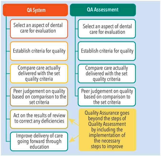
addition, dental records may be shared with and reviewed by other health care professionals and insurance companies. Omissions in dental records can have a negative impact on interprofessional communication and may serve as the basis for insurance fraud litigation. In addition, patient-related communications, including any instances
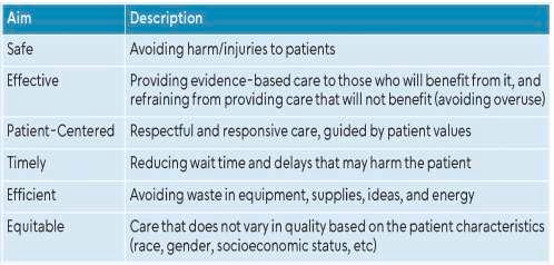
(Data from Institute of Medicine (US) Committee on Quality of Health Care in America. Crossing the Quality Chasm: A New Health System for 21st Century. National Academies Press: Washington, DC, 2001.)
of patient aggression toward clinicians or other dental personnel, should also be recorded as part of quality control and risk management protocols.
The World Health Organization (WHO) has found that 8% to 38% of heath care workers throughout the world will experience either physical violence during their careers or will encounter threatening verbal aggression, usually from a patient. Those most vulnerable to these aggressive or dangerous situations are usually involved in patient care.
New York University College of Dentistry did a study that found about 22% of dentists experienced physical assault, 55% were verbally assaulted, and about 44% were subjected to reputational assault. Over the practitioners’ career, rates increased to about 46%, 74%, and about 69%, respectively. Even dental students report encounters with aggressive patients. As a result, the American Psychiatric Association Task Force on Psychiatric Emergency Services recommends that dental staff members have annual training in managing behavioral emergencies.
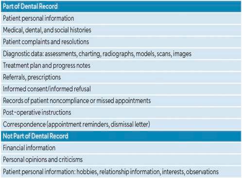
(Courtesy of Matthews A, Vyprynyuk K, Marsh I: Strengthening dentistry’s quality assurance. Dimensions Dent Hyg 23:32-35, 2025.)
When patients are agitated, the situation necessitates prompt intervention. Swift assessment and decision-making skills are needed to defuse a hostile situation. Four main objectives should be considered: safeguarding the patient, staff, and others; helping the patient manage emotions and concerns to bring behaviors under control; avoiding the use of restraints if possible; and avoiding coercive interventions, which can escalate the situation.
The American Association for Emergency Psychiatry has a standardized scale that helps in accurately assessing patient agitation. Each patient is approached in a practical, noncoercive way that respects the patient’s personal space and avoids becoming provocative or authoritative in demeanor. Agitation assessment includes looking for motor hyperactivity, pacing, tapping on surfaces, repeating statements, and raising of the voice. The surroundings should be evaluated for safety, accessible exit locations, and the removal of objects that could cause injury. The dentist should remain a minimum of 2 feet from the patient. Verbal de-escalation and nonverbal communication can help to engage the patient and help him or her become an active partner in the calming down of the situation. Techniques that work in these cases include openended questions and reflective feedback to identify the patient’s wants and needs, maintaining clear and concise
communication, agreeing or agreeing to disagree and maintaining respectful dialogue will maintain clear limits and expectations that allow better control of the interaction. Offering the patient choices, maintaining a positive, optimistic demeanor, and summarizing the exchange can all build mutual understanding and respect.
Dental professionals should adopt the best practices with regard to documentation and effective provider-patient communications, both of which contribute to the quality assurance process. With continuous advances in the quality of care delivered, better communication skills, and defusing aggressive or dangerous situations, dental care providers can maintain effective management of patient behaviors. Periodic training for all dental care providers and office staff will improve their awareness and readiness to prevent negative outcomes.
Matthews A, Vyprynyuk K, Marsh I: Strengthening dentistry’s quality assurance. Dimensions Dent Hyg 23:32-35, 2025
Reprints not available
In dentistry, unwanted outcomes can result from slips, mistakes, or malpractice and occur along a spectrum of severity or resulting consequences. Although it’s impossible to completely eliminate these unwanted results, it’s possible to correct them in many cases. Steps can be taken to make them less likely to occur and more readily resolved with less damage by building service recovery into an opportunity to make it a professional investment. Several wise moves are suggested, the service recovery continuum is explained, and the continuous quality service improvement model is proposed as a proactive approach to managing undesirable outcomes.
View Ser vice Recover y as a Professional Investment
In general, the cost of ignoring or avoiding recovery tends to be greater than recovery costs. Costs can be reduced by avoiding errors through a reduction in any unwanted variation in technical work, by managing the dental practice professionally, and by working with patients to develop realistic and shared expectations.
Service recovery is well managed when the situation is addressed promptly without blame, when an offer to apologize is made, when patients are a part of the solution, and when the damage is made right to the extent that this is possible. Not taking these steps tends to waste personal time, lower morale, open the practice to large settlements, lose patients, and increase the negativity of one’s public image. Following an incident requiring service recovery, the dental practice can learn from the experience and avoid future problems and may even increase patient loyalty, depending on the solution. The unwanted outcome could be repaired and the feedback from the experience may build stronger practice routines, making the experience an investment in better oral health care.
Among the wise moves in this process are preparing and preventing, knowing which role to play, listening, reaching a mutual decision, and taking the benefit (Table 1).
Several parts of the office routine can reduce the likelihood service recovery will be needed. It’s important to study the outcome patterns of commonly performed procedures to identify which procedures may be more accident prone. New procedures, new staff, and patients with complications or contributing factors should be closely monitored. It’s also
wise to check the patient’s expectations, because giving consent doesn’t mean that the patient understands all the things that may happen. The entire dental team should be involved, whether that is to identify what caused the problem or to be on the alert for problems.
Preparation and prevention also involve periodic but regular staff meetings devoted to reviewing the practice policy for service recovery. The dentist and team should determine what patient issues need to be shared, who needs to know, and what staff members can do on their own, what should they delegate, and what shouldn’t be done. Because many service complaints involve money, regular and accurate reports should be done in this area. All the lessons learned should be shared with the team.
In multiple ownership situations, a written defined service recovery policy should be developed. All the principles in a group practice should be harmonious. The service recovery policy can be shared in community clinics, schools, and corporate ownership situations.
The role of each individual should be determined so they can be well-prepared and contribute consistently. The various roles in addressing a problem include ignoring the problem and assuming it will just go away; determining you have nothing to correct, you will fight it, or you will delegate it to someone else; seeking the best available mutual solution; or accepting any resolution the patient suggests. In general, the first 2 and the last choices are unwise because the control of the conflict is handed over to the patient.
The wisest course of action is to seek the best available mutual solution. Proposing at the outset that the dentist and patient should see if they can work together to make it right establishes a relationship, offers willingness to work together, and encourages patient cooperation. This creates the ground rules that optimize the mutual acceptance of what comes next.
Listening is the only way to learn about all possible options and have a degree of understanding about what the patient’s expectations are. The goal is to find out how the patient defines the problem and what he or she wants. It’s also possible to learn how the patient might react to solutions the dentist would consider.
Because both facts and values are involved in conflicts, the focus of discussions should be on fixing the problem and not fixing the blame. Everyone involved with a service problem has multiple
Make it an ongoing policy to reduce the occurrence of unwanted outcomes and create policy for how problems should be managed and by whom.
State your goal of optimizing mutual acceptance of a corrective response. Listen to identify all the viable paths forward for you and the patient. Agree on what actions you and the patient will take so neither has any reason to renege on what they have promised. Thank the patient, execute as promised, check to see whether the practice can be improved by what has been learned, look forward to more loyal patients in the future.
(From Chambers DW: Maintaining professional relations with patients when unexpected things happen: The case for service recovery. J Calif Dent Assoc 53:2460733, 2025. © 2025 The Author(s), taken from Journal of the California Dental Association reprinted by permission of Informa UK Limited, trading as Taylor & Francis Ltd, https://www. tandfonline.com This is an Open Access article distributed under the terms of the Creative Commons Attribution-NonCommercialNoDerivatives License (http.//CreativeCommons.org/licenses/byNEC-nd/4.0/), which permits non-commercial re-use, distribution, and reproduction in any medium, provided the original work is properly cited, and is not altered, transformed, or built upon in any way.)
might apply to cases involving insurance, corporate interest, peer review, litigation, or disciplinary action, all of which involve paying costs to third parties.
A successful service recovery should prompt the following responses:
• Acknowledge the role of the patient in meeting the dentist partway in the best available course of action.
• Look forward to having converted a practice liability into an asset.
• Circle back to step 1 and share with the staff what has been learned about preventing problems and service recovery.
The experience can inform improvements in the treatment protocol and office policy. The best outcome is to avoid making the same mistake again.
options, including agreeing to the other’s offer, making a counteroffer, negotiating in hopes of finding new alternatives, walking away, or involving others. The goal is to maximize the options in play before being forced to pick one.
It’s possible that both parties have a common path forward that addresses the problem. If both sides have been honest, the best solution is likely on the table. The goal is to identify a pair of joint future actions that is rational and stable for both sides. Choosing the best service resolution approach should observe the following rules:
• Don’t pass up a “win-win” as a way to resolve the conflict.
• If you do, don’t pass up a “next best” agreement.
• If you do, negotiate, explore, and consider compromises. Clear up any misunderstandings or unexplored assumptions.
Some general considerations can be helpful for difficult situations, especially when third parties are involved. Computer simulations have been used to analyze the 78 possible combinations of encounters between pairs of individuals. Problems tend to arise when 1or both parties refuse reasonable solutions because they know they are right. In some cases no rational standard favors 1 or the other individual. It’s possible to simply flip a coin or graciously concede and earn points for being professional. The “Hawk & Dove” or “Chicken” approach is complicated by ego and status or self-image. The best strategy in these cases is to bluff and then concede. The worst case is the Prisoners’ Dilemma, wherein both sides are motivated to avoid personal harm by damaging the other. In this case, there is usually a third party rigging the situation for their own benefit. This
Multiple approaches can be used in service recovery, although the objective is to identify the best possible mutual commitment to corrective action. There is a continuum for conflict resolution (Table 2). As one moves to the right, from informal to formal, patterns can be seen showing how personal control diminishes, the number of persons involved increases, the time to resolution shows dramatic increases, cost grows with the number of others involved and the time and resources needed, and the predictability of the outcome becomes more questionable. Various factors push service recovery from the left to the right along this continuum.
The exception to the trend for greater cost and greater risk on the right involves disciplined licenses. All licensed persons have a large presumption of economic freedom. In dentistry, about a third of licenses are disciplined for technical reasons, and the rest are divided between faulty business practices and personal reasons, such as conviction for driving under the influence, sexual misconduct, or general incompetency.
Four levels exist in the continuous quality service improvement model (Table 3). In progressing from the lowest to the highest level, customer satisfaction, value, and provider fulfillment in work and value all improve. The first level is ignorance, which is when no attention is paid to the effects of variations in patient outcomes because random variation happens even if it is an unfortunate outcome that could have been prevented. The second is shifting the blame, which amounts to refusing to take any responsibility for problems. One of the costs at this level is the dentist’s ego. The third level is service recovery, where the focus is on fixing the problem.
Form Continuous quality improvement In office, manage and corrections
Prompt Constant Upon detection
Those Involved Whole office Dentist or staff and patient
Both agree to third-party exploration
Help sought to frame options
Dentist, patient, trained mediator
Both agree to third-party investigation and decision
Help sought to decide resolution
Dentist, patient, investigators,
Dissatisfied party seeks formal penalty
No agreement on resolution
Dentist, patient, lawyers, expert decision makers, judge
Official sanction for violating standard of care
Pattern of publicly unacceptable conduct
Dentist, patient investigators witnesses Time Continuous On-the-spot, within days
Cost An investment Minimal
Trade Cost of building better practice
Small costs, small gains
Trained insured, delay
Perhaps avoid moderate loss at moderate cost
Trained, insured processing costs, delays
Perhaps avoid large loss at some cost
Trained, insured large processing costs, delays, potential loss
Defend against very large loss at large cost
Trained, insured very large processing costs, delays, significant potential loss
Slight chance of catastrophic loss at very large cost
(From Chambers DW: Maintaining professional relations with patients when unexpected things happen: The case for service recovery. J Calif Dent Assoc 53:2460733, 2025. © 2025 The Author(s), taken from Journal of the California Dental Association reprinted by permission of Informa UK Limited, trading as Taylor & Francis Ltd, https://www.tandfonline.com This is an Open Access article distributed under the terms of the Creative Commons AttributionNonCommercial-NoDerivatives License (http.//CreativeCommons.org/licenses/by-NEC-nd/4.0/), which permits non-commercial re-use, distribution, and reproduction in any medium, provided the original work is properly cited, and is not altered, transformed, or built upon in any way.)
The goal of continuous quality service improvement is in the fourth level, which involves building predictably better practices. Problems can only be avoided if they are recognized and understood, followed by changes in the process to prevent or reduce issues in the future. The greatest opportunity for practice improvement is to practice conflict resolution approaches.
The 5 steps in good service recovery are as follows:
1. Policy and prevention
2. Communicating openness to satisfactory resolution
3. Listening to find out what options are possible
4. Agreeing on the best available mutual resolution
5. Learning from the experience and integrating what is learned in continuous quality improvement
Table 3. Four Levels of Quality
Ignorance
Denial
“What is all this ruckus? Ignore it; things always even out.”
“Somebody is going to have to pay for this. I may settle, but I am not to blame.”
Outcome correction Inspect out the duds. Redo. Compensate. Process correction Redesign the way things are done to reduce the chances of future mishaps.
(From Chambers DW: Maintaining professional relations with patients when unexpected things happen: The case for service recovery. J Calif Dent Assoc 53:2460733, 2025. © 2025 The Author(s), taken from Journal of the California Dental Association reprinted by permission of Informa UK Limited, trading as Taylor & Francis Ltd, https://www. tandfonline.com This is an Open Access article distributed under the terms of the Creative Commons Attribution-NonCommercialNoDerivatives License (http.//CreativeCommons.org/licenses/byNEC-nd/4.0/), which permits non-commercial re-use, distribution, and reproduction in any medium, provided the original work is properly cited, and is not altered, transformed, or built upon in any way.)
Successful service recovery is the systematic approach to managing problems that occur to the maximum mutual satisfaction of dentists and patients. Done well, it has the potential to produce stronger dentist-patient relationships than existed before the service disruption. As the California Dental Association’s Code of Ethics states, “While serving the public, a dentist has the obligation to act in a manner that maintains or elevates the esteem of the profession.”
Chambers DW: Maintaining professional relations with patients when unexpected things happen: The case for service recovery. J Calif Dent Assoc 53:2460733, 2025
Reprints available from DW Chambers, Arthur A Dugoni School of Dentistry, Univ of the Pacific, San Francisco, CA, USA; e-mail: dchamber@pacific.edu
Even though tens of millions of potential dental patients visit dental websites and provider directories each month looking for dental care, many struggle with the new patient acquisition methods currently available. Patients often find it nearly impossible to connect with dental practices through the access points that exist today. Forward-thinking dental practices can end this disconnect with a revision in their thinking that eliminates the digital disconnect by addressing the siloed dental ecosystem and shifting it to a business-to-consumer-to-business (B-C-B) model.
The Problem
It’s been estimated that about 75% of patients who begin searching for a dental provider don’t ultimately schedule an appointment. When potential patients visit provider directories, manufacturer sites, or professional organizations resources, they often find themselves provided with a static list of providers and their phone numbers but no actual access to making an appointment. This creates frustration because modern consumers expect convenience and ease from health care similar to what they see in booking appointments or making reservations. If the dental office is closed, if staff members are too busy with in-office patients, or if the potential patient is sent to voicemail systems that don’t result in returned calls, it’s not uncommon that the search will be abandoned and no appointment will be made.
The dental industry is composed of distinct segments that don’t communicate with one another effectively. Patients may use various channels to reach care, but the siloed nature of the dental ecosystem can frustrate their efforts (Figure 1). Insurance carrier provider directories offer limited functionality. Dental manufacturers and brands use marketing campaigns to reach consumers and convey information about their products, but patients’ efforts to make an appointment are stymied.
Dental practices tend to invest in their own practice management systems and patient communication methods while building out scheduling protocols. They rely on casting their marketing efforts into the community and hoping to attract new patients with local advertising, referrals, and visibility. As a result, patients looking for providers can be unsuccessful in scheduling appointments with the best provider for their care. This disconnect creates a patient journey that is full of dropout points. Other industries use technological solutions to connect the disparate systems, but dentistry has yet to undergo this transformation.
In the B-C-B marketing model in dentistry, large dental manufacturers and insurance companies market directly to consumers, creating interest in specific procedures or services. Interested consumers then follow a seamless pathway to dental practices. With this model, practices benefit from the greater marketing investments made by larger industry players.
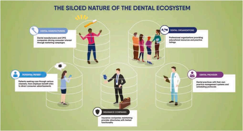
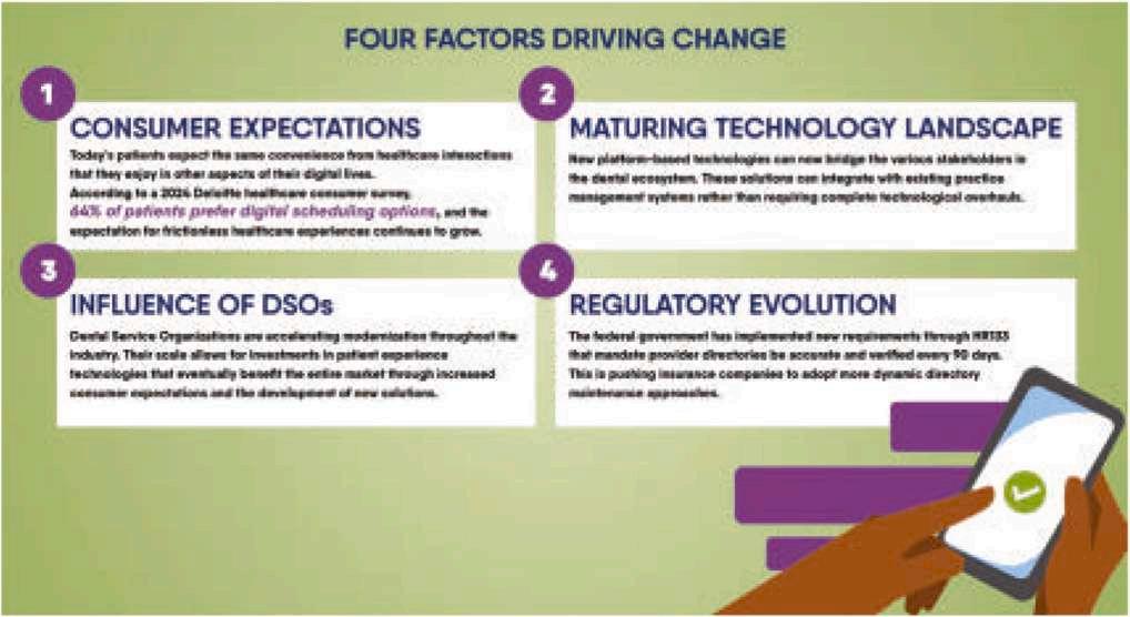
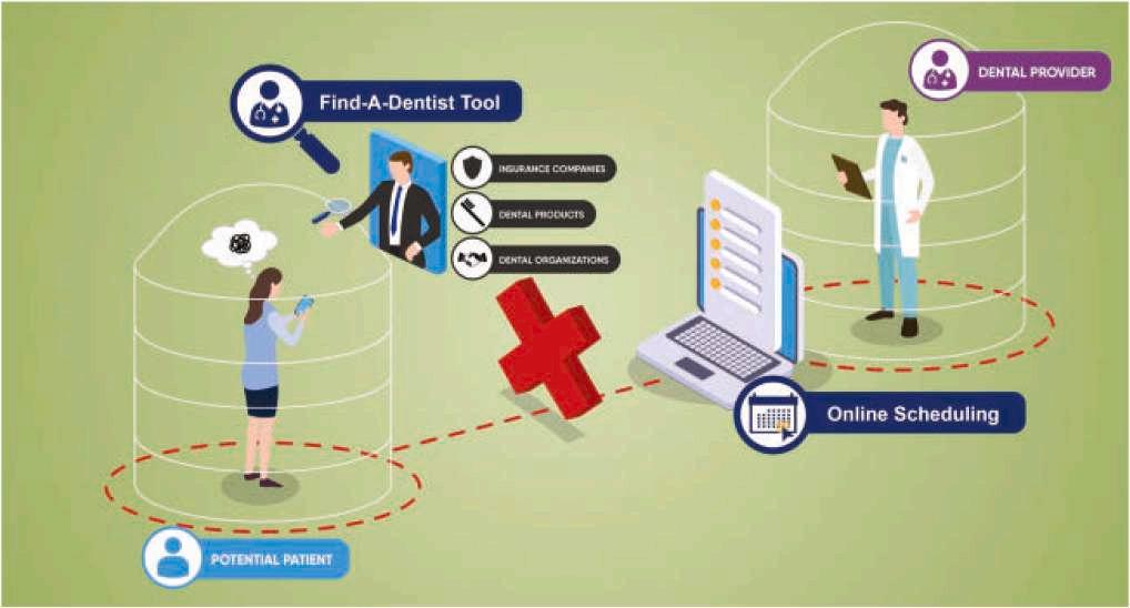
Dent Econ 115:28, 30-
The motivators for these changes are consumer expectations, better technology, the influence of dental service organizations (DSOs), and a regulatory evolution (Figure 2). Patients now expect convenience from health care interactions similar to what they experience in other digital interactions. Most patients want digital scheduling options, with the outcome being frictionless health care experiences. New platform-based technologies offer the stakeholders in the dental ecosystem a seamless integration with existing practice management systems. Complete technological overhauls aren’t required. In addition, DSOs are accelerating modernization throughout the dental industry. Their scale means investments in patient experience technologies will eventually increase consumer expectations and the development of new solutions throughout the entire market. Regulatory evolutions are taking place at federal levels, where legislation that mandates provider directories be accurate and verified every 90 days is being developed. This pushes insurance companies to adopt more dynamic directory maintenance measures.
The 3 critical success factors for these technology platforms for health care are their integration within established existing ecosystems, the creation of value through scale with multiple stakeholder participants, and proactive measures that address regulatory changes (Figure 3). Practices that embrace connected patient acquisition strategies may see significant advantages moving forward. With the evolution of consumer expectations,
these efforts will continue to grow in value and support the expansion and sustainability of dental practices. Dental practitioners are beginning to see the power of integrated scheduling technologies that connect provider directories directly to their practice management systems.
As practices move toward taking advantage of new technology platforms that increase the functionality of their existing systems, potential patients will see fewer barriers to accessing care. Rather than redirecting patients to call for appointments, patients will be able to view real-time availability and book from any digital touchpoint where they’ve researched dental care. New patient leads will be obtained through connected directories and scheduling programs. Practices that can integrate these advances into their existing systems will be able to grow even in an increasingly competitive market.
Rindler E: Breaking down silos: The changing landscape of dental patient acquisition. Dent Econ 115:28, 30-31, 2025
Reprints not available
The dental materials most often used for restorative dentistry are amalgam and composite resin. Each of these materials offers advantages and disadvantages that must be considered when selecting a restorative approach. Providers have tended to shift toward using composite resin for aesthetic reasons and because of concerns over the presence of mercury in amalgam. Amalgam is strong and durable and especially useful in posterior restorations because of its ability to withstand considerable occlusal forces. However, it requires preparation depths sufficient to ensure mechanical retention, which can compromise the preservation of healthy tooth structure. Its mercury content has been linked to environmental and health concerns. In contrast, composite resin allows more conservative approaches to restorations and these preserve healthy tooth structure. Its drawbacks include its high technique sensitivity, which compromises the restoration of posterior teeth with proximal involvement; polymerization shrinkage; and some evidence that it has a higher failure rate than amalgam. The 2 materials were evaluated using the most current data to compare the failure risk of amalgam and composite resin restorations in posterior teeth.
A search of PubMed/MEDLINE, Cochrane Central Register of Controlled Trials, and Google Scholar from 1990 onward identified 13 studies that were appropriate for inclusion. Five were randomized controlled trials, 4 were prospective cohort studies, and 4 were retrospective cohort studies. The participants in the studies ranged from 25 to 4856, and duration ranged from 24 to 144 months.
Amalgam’s failure rate was 0% to 50%, whereas that of composite restorations was 0% to 62.7%. The risk ratios (RRs) for the 2 materials were 0.96, which indicated no statistically significant difference between the failure risk of amalgam and composite resins. Studies published after 2013 noted higher success rates in resin composite restorations compared to amalgam restorations,
which may indicate advancements in the quality of the physical and mechanical properties of the composite resins.
Eleven studies consistently identified secondary caries as the definitive criterion for restoration failure, and 7 of these found that composite resin restorations had consistently higher numbers of failures compared to amalgam restorations, whereas 4 studies found amalgam restorations to have more failures. Eight studies defined tooth or restoration fracture as indicative of failure, with 4 reporting higher failure rates in resin composite restorations. When failure was defined as the need for endodontic treatment or tooth extraction, a higher failure rate (9.3%) was found for amalgam restorations than for composite resin restorations (4.5%). Studies also indicated failure was associated with pain/sensitivity, poor marginal adaptation or staining of overhanging margins, and restoration displacement or loss.
The studies failed to report factors that can influence restoration failure, which included operative techniques, material brand or type, use of base/liners, isolation techniques used, and data on adhesive systems used.
Previous meta-analyses suggested higher failure rates and reduced longevity were associated with composite resin restorations in posterior permanent teeth compared to amalgam restorations used posteriorly. The current meta-analyses indicated that the failure risks of composite resin and amalgam are comparable.
No statistically significant difference in failure risk was noted between amalgam and composite resin restorations placed in posterior locations. However, further studies should report on other factors that could contribute to failure and should standardize the criteria used in defining restoration failure.
Al-Sulimmani W, Al-Rasheed A, Al-Daraan H, et al: Failure risk of composite resin and amalgam restorations: A systematic review and meta-analysis. Int Dent J 75:100871, 2025
Reprints available from W Al-Sulimmani, Block No. 3, Street No. 38, Bldg No. 12, Kaifan, Al Jahra 78103, Kuwait; e-mail: Woroudnm@hotmail.com
Dental plaque control has relied on brushing, flossing, scraping, and applying antiseptics or even antibiotics to it. Research indicates that control and adjustment of dental plaque to keep it in health-promoting condition may be achieved through research into the structure and organization of the biofilm. The bacterial community, calcifying and health-promoting components, and spatial organization and structure are all aspects to be considered when dealing with plaque control.
Dental plaque consists of dozens to hundreds of bacteria, viruses, and fungi that exist together, forming a complex, interacting community. This microbiome contains microbes that are usually harmless or even beneficial to human health as well as those that are in the wrong place or wrong circumstances and cause problems. All plaque microbes require the right environment to grow and often need other microbes to be their neighbors. They depend on one another for nutrients
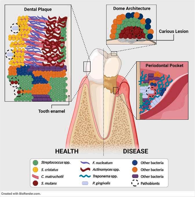
Courtesy of Mark Welch JL, Borisy GG: Dental plaque: Who is in the neighborhood and why it matters. JADA Found Sci 4:100051, 2025.
Figure 1. A, A photograph of dental plaque taken using a hyperspectral imaging microscope and using multiplexed fluorescence in situ hybridization, a technique to label different kinds of bacteria with different colors. The photograph shows the radial organization of the hedgehog structure and the central position of Corynebacterium bacteria, shown as magenta-colored strands. Streptococcus bacteria are shown in green and grow primarily on the outside of the structure. Fusobacterium nucleatum is shown in yellow. (Reproduced with permission of the publisher from Mark Welch JL, Rossetti BJ, Rieken CW, et al: Biogeography of a human oral microbiome at the micron scale. Proc Natl Acad Sci USA 113:E791-E800, 2016.) B, C matruchotii, an important bacterium for the structure of dental plaque, can elongate rapidly (i), and reproduce by separating into many different cells at once (ii) that then shatter and sprout new filaments (iii). This unusual growth strategy helps this bacterium form large bushy clumps on teeth. (Reproduced with permission from Chimileski S, Borisy GG, Dewhirst FE, et al: Tip extension and simultaneous multiple fission in a filamentous bacterium. Proc Natl Acad Sci USA 121:e2408654121, 2024.)
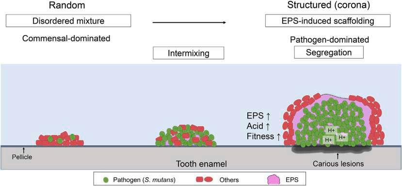
Figure 2. The caries-causing bacterium Streptococcus mutans can exist harmlessly in dental plaque when it is mixed with other bacteria, but it causes caries when it forms large clumps and domelike structures. (Reproduced with permission from Kim D, Barraza JP, Arthur RA, et al: Spatial mapping of polymicrobial communities reveals a precise biogeography associated with human dental caries. Proc Natl Acad Sci USA 117:12375-12386, 2020.)
and protection from toxins. Investigations into what microbes exist next to each other in natural dental plaque, their structures, and their behavior in various contexts may make it possible to determine what conditions cause them to thrive and how we might control them.
The earliest colonizers attach to clean tooth surfaces, then mature with time to become colonies of bacteria that can bring disease. Originally Fusobacterium nucleatum was considered the controlling factor in plaque maturation, but new imaging techniques (specifically, fluorescence in situ hybridization and hyperspectral imaging microscopes) indicate that Corynebacterium matruchotii is the structural core of the hedgehog dental plaque community (Figure 1). This bacterium doesn’t just divide but first grows rapidly longer and then splits into multiple cells at once, called multiple fission. It then forms a dense bush with long filaments that stretch from near the tooth surface to the tip of the plaque biofilm. C matruchotii creates the habitat where other bacteria live, with some on the filaments with direct access to saliva and others deep in the mass of bacteria where they are sheltered in a low-oxygen environment.
C matruchotii triggers dissolved calcium in saliva to combine with phosphate to form hydroxyapatite, which is a mineral found in tooth enamel. When it forms around bacteria, it turns soft dental
plaque into hard dental calculus. When dental plaque is rich in filaments, it forms calculus more quickly than plaque dominated by other types of bacteria.
These corynebacteria are also associated with oral health. C matruchotii recruits other bacteria that ward off potential pathogens. Having hedgehog structures made up of C matruchotii plus Streptococcus cristatus changes the biology of the periodontal pathogen Porphyromonas gingivalis and causes it to act less virulently, preventing problems in the oral cavity. Thus oral health relies not just on the quantity of dental plaque, but also on what kind of organisms make up the plaque. Our bodies may have the ability to encourage the growth of health-promoting and pathogen-inhibiting bacteria in our mouths.
The spatial organization of the dental plaque biofilm is important in the strategy of Streptococcus mutans. Although this bacterium can dwell harmlessly dispersed in the dental plaque, under the right conditions, it can proliferate and form a dense dome next to the tooth enamel. Other plaque bacteria form more layers above it (Figure 2). It begins to produce acid and degrade the enamel. Thus the spatial organization of the microbes can change their behavior and interactions with the human host. It may be possible that rather than eliminating S mutans to control caries, we can disrupt its ability to form a dome structure and achieve control that way.
Research is needed to shift the composition of plaque in precise, targeted ways to restore and maintain health. It may be possible to adjust the spatial organization and behavior of organisms in plaque to reduce any harmful traits and enhance beneficial ones. Precision tools are needed to edit the microbiome, removing the harmful bacteria and adding the beneficial bacteria without removing the entire community. Narrowly targeted antimicrobial peptides or bacteriophages may be employed to selectively remove pathogenic bacteria from the mouth. We also need to know more about how bacteria stick to one another or to surfaces in the mouth and what conditions are needed for beneficial bacteria to thrive in the mouth. All of this must be studied to determine how we can shift the ecological system of the mouth toward a healthy state.
Mark Welch JL, Borisy GG: Dental plaque: Who is in the neighborhood and why it matters. JADA Found Sci 4:100051, 2025
Reprints available from JL Mark Welch; jmarkwelch@forsyth.org
Oral health is recognized as essential for overall well-being and plays an important role in communication, facial expressions, sensory perception, touch, chewing and swallowing, and the ability to express emotions effectively. The World Health Organization (WHO) has expressed an urgent need to consider oral health within the overarching umbrella of noncommunicable diseases (NCDs). In addition, the FDI World Dental Federation advocates for the use of fluoride as a cost-effective caries prevention strategy that should be part of public health policy. The age-standardized prevalence of oral disease is 34.1%. An estimated 2.3 billion people have untreated caries in permanent teeth and 532 million children experience caries in their primary teeth. Effective caries prevention measures must be both community-based and individualized, with the goals of managing biofilm, modulating oral microorganisms, and promoting a lowsugar diet to lower the caries risk and prevent lesion progression. Topical or systemic fluoride delivery plays a critical role, whether it is through water fluoridation, milk fluoridation, salt fluoridation, or fluoridated toothpastes, rinses, and gels. Although systemic fluoride provides lifelong benefits because the fluoride is incorporated into the developing enamel before tooth eruption and ensures long-term resistance to acid attacks,
some harmful systemic effects are associated with fluoride use. A loud anti-fluoride movement has arisen, leading to a court decision in the United States that community water fluoridation presents a risk of reduced IQ in children. A contemporary look at the benefits, controversies, and potential risks associated with fluoride and the scientific evidence and clinical practices surrounding fluoride use was offered to educate the dental profession and the general public on the appropriate and safe use of fluoride in dental circles.
Fluoride offers several unique properties in the oral cavity. Fluoride interacts with the oral environment to influence bacterial metabolism and enhance tooth resistance to acidic attacks. It offers antimicrobial action and reduces extracellular polysaccharide production by bacteria and profoundly affects the dynamics of tooth mineralization and demineralization.
Fluoride has the ability to acidify the bacterial cytoplasm and exert stress on bacterial cell membranes. Higher fluoride concentrations can inhibit bacterial growth, lower the pH, and
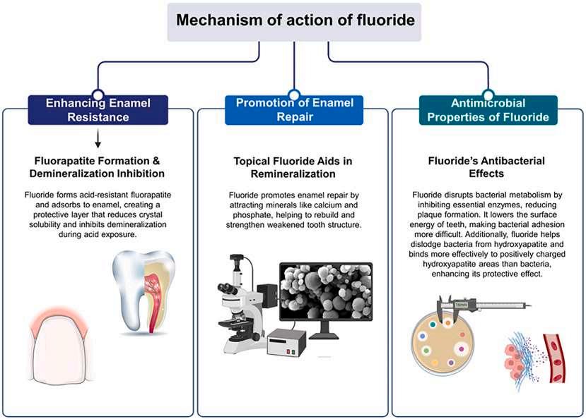
increase the population of extracellular hydrogen ions so that bacteria are inhibited at lower extracellular fluoride levels.
Fluoride also reduces the production of extracellular polysaccharides by bacteria, which disrupts biofilm integrity and interferes with bacterial adhesion to tooth surfaces. Vital bacterial enzymes are also inhibited. Being able to concentrate in dental
plaque allows the protective effects of fluoride to be localized to those areas most at risk for demineralization.
The remineralization process is enhanced by fluoride’s ability to support the deposition of calcium and phosphate ions into demineralized enamel. Fluorine-containing apatites, which are more
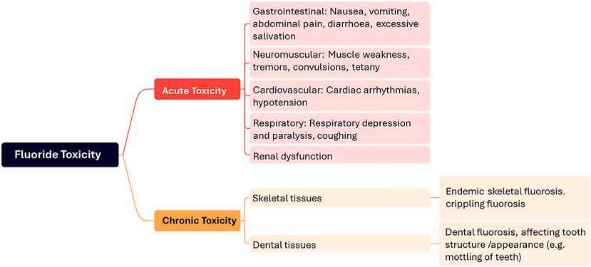
resistant to acid attacks and demineralization than hydroxyapatite, are formed. The apatites have unique structures that make enamel less soluble in acidic environments.
The predominant effects of fluoride are topical and posteruptive, so it’s important beyond childhood, with optimal effectiveness in small, sustained concentrations in dental plaque and saliva. It can continuously interact with the oral environment to maintain a balance favoring remineralization over demineralization. Adults, especially those with demineralized enamel, benefit from the increased uptake of fluoride by compromised areas.
Public health is enhanced by having fluoride serve as a part of the community caries prevention programs. Water fluoridation ensures equitable access to the protective effects of fluoride irrespective of affordability. However, its effectiveness relies on regular exposures in optimal concentrations (Figure 2).
Fluoride is naturally present in freshwater at varying concentrations, depending on the geographic location and water source. Once researchers observed that populations who consumed water with naturally higher levels of fluoride had a lower incidence of dental caries, controlled adjustments to the fluoride levels in public water supplies was introduced as a preventive public health measure. Large populations can receive the fluoride without any individual effort and have their caries risk lowered. The Centers for Disease Control and Prevention (CDC) has reaffirmed that community water fluoridation is a foundational strategy for caries prevention in the United States, being practical, costeffective, and equitable in providing better oral health to residents. The CDC and US Public Health Service (USPHS) do not mandate community water fluoridation and the standards they recommend aren’t enforceable. However, caries reductions of 40% to 50% were seen in primary teeth and 50% to 60% in permanent teeth in communities with fluoridated water. Recently, the deleterious effect of stopping community water fluoridation has been evident, with a 2-fold increase in dental treatments for children within 5 years after fluoridation was abandoned.
The other most common sources of fluoride supplementation are milk, salt, and fluoridated toothpaste. About 15 countries implement milk fluoridation programs and have documented a significant reduction in dental caries in children who regularly consume fluoridated milk. The most effective results are in early childhood (before age 4 years) and when the first permanent molars are erupting. Just over 1.5 million children worldwide participate in fluoridated milk programs.
Salt fluoridation is used when water supplies are compromised by political, geographical, financial, and technical challenges.
The approval of sodium and potassium fluorides as food additives by the European Union has yielded favorable results. The fluoridated salt is distributed as salt for household use, in meals in schools and large kitchens, and in baked goods such as breads. Fluoride is therefore able to work systemically and topically.
Fluoride toothpaste use can significantly reduce the risk of cavities. For children whose teeth are still developing, fluoride is critical to the development of strong, healthy enamel. The combination of community water fluoridation and fluoride toothpaste has led to a dramatic decline in dental caries.
A dose-response relationship exists, so that consistent exposure to multiple low-concentration fluoride sources has an enhanced effectiveness in caries prevention. Individuals at higher risk for developing caries experience especially significant effects. The risk of fluorosis is generally linked to excessive fluoride intake during tooth development stages, especially from the late secretion phase to the early enamel maturation. For adults, the simultaneous use of topical fluoridated products and fluoridated water has no fluorosis risk because their teeth are already fully formed.
Excessive fluoride exposure is associated with skeletal and dental fluorosis and systemic issues such as gastrointestinal, neurological, and urinary complications. Over 80% of fluoride toxicity cases occur in children under age 6 years and are usually related to an accidental ingestion of fluoride-containing products. Both acute and chronic adverse effects can occur with fluoride, with immediate, severe effects often seen in the acute and usually accidental cases. Chronic toxicity generally results from long-term exposure to elevated fluoride levels, which are more common in areas where high concentrations of fluoride occur naturally (Figure 3).
Debates over the risk of fluoride toxicity and the safety of community water fluoridation continue. Some countries have chosen to limit fluoride exposures and intake. Various claims have been made, with many disproved by evidence-based scientific findings (Table 1). However, several major governmental and nongovernmental bodies, including the CDC, WHO, American Dental Association, and FDI, fully endorse community fluoridation as both safe and effective.
The FDI has addressed topical and systemic fluoridation, including water fluoridation, on several occasions. It supports fluoridation as an effective measure for preventing dental caries,
Misconception or Concern Claim
Uncontrollable Fluoride Intake
Risk of Water Contamination
Lack of Monitoring in Some Countries
Dental Fluorosis
Adverse Health Effects
Alternative Sources of Fluoride available
Fluoride intake varies depending on water consumption, making it difficult to control.
Fluoridated water may contain harmful contaminants like arsenic.
Some nations do not enforce certification of fluoride additives.
Excess fluoride causes cosmetic or structural damage to enamel.
Fluoride may cause neurotoxic, kidney, or thyroid effects.
Fluoride is already present in food, beverages, and dental products.
Topical Benefit Over Systemic Use
Fluoride is more effective topically than systemically.
Tooth Decay in Low-Income Countries
Lack of RCT Evidence
"Unapproved Drug” Claim
Professional Opposition
Fluoridation hasn't eliminated caries in disadvantaged regions.
Fluoridation lacks support from RCTs.
The U.S. FDA classifies fluoride as an unapproved drug.
Thousands of professionals oppose water fluoridation.
Evidence-Based Clarification
Public health recommendations (e.g., 0.7 ppm) include a wide safety margin. Even with variation in water intake, toxicity thresholds are not reached.
Fluoride additives for water treatment are regulated and monitored for purity and safety. Contamination risks are rare in countries with strong public health infrastructure.
This is an issue of regulatory enforcement, not fluoride itself. Proper policy and oversight can mitigate this risk without dismissing the value of fluoridation.
Mild fluorosis is typically cosmetic and often undetectable. Severe forms are rare and usually linked to excessive fluoride from multiple sources during early childhood.
Such effects have only been observed with chronic exposure to high levels (well above 1.5-2 ppm). No harm is associated with fluoride levels used in community water (0.7 ppm).
Multiple sources improve dental protection. Total intake from all sources in most populations remains within the recommended safe range. Fluoridation provides a simple, cost-effective way to reduce cavities across entire populations, especially in communities with limited dental care access
Topical fluoride is important, but systemic exposure during tooth development and the equitable delivery through water remain vital for population-level prevention. Being incorporated into the enamel structure prior to eruption, systemic fluoride enhances long-term resistance to acid attacks.
Various studies have shown that fluoridation reduces overall caries risk in low income populations. Fluoridation is a preventive tool, not a cure-all.
Ethical and logistical barriers limit RCTs in public health. However, decades of high-quality observational and cohort studies provide strong evidence for effectiveness.
This label applies to specific supplements, not to water fluoridation. Major bodies like the CDC, WHO, American Dental Association (ADA), and FDI endorse community fluoridation as safe and effective.
A small minority oppose it, while over 100 national and global health organizations support fluoridation based on solid scientific evidence.
(Courtesy of Samaranayake L, Porntaveetus T, Tsoi J, et al: Facts and fallacies of the fluoride controversy: A contemporary perspective. Int Dent J 75:100833, 2025.)
offering clear policy statements regarding systemic fluoridation, topical fluoridation, and the safety and efficacy of fluoridation. As far as public health policy, the FDI encourages governments and
health officials to implement and maintain fluoridation programs that are based on scientific evidence and the public health needs of their citizens. Table 1. Misconceptions on Fluoridation and Evidence-based Counterpoints and Clarifications
The controversy surrounding community water fluoridation emphasizes the need for public health policies to be based on scientific evidence and to prioritize the safety, efficacy, and equitable access to fluoride. Strategies that emphasize the use of fluoride toothpaste and professionally applied treatments offer somewhat acceptable alternatives or adjunctive measures to water fluoridation, but for children whose permanent dentition is developing, systemic fluoridation is the only measure that promotes lifelong benefits by incorporating fluoride into the enamel structure. Additional high-quality research is needed to evaluate long-term outcomes and further refine fluoride recommendations and policies.
Samaranayake L, Porntaveetus T, Tsoi J, et al: Facts and fallacies of the fluoride controversy: A contemporary perspective. Int Dent J 75:100833, 2025
Reprints available from L Samaranayake, Faculty of Dentistry, 34 Hospital Rd, Hong Kong SAR, China; e-mail: lakshman@hku.hk
Porphyromonas gingivalis, Fusobacterium nucleatum, Actinomyces israleii, and Streptococcus mutans are well recognized for their relationship to oral diseases. In the past 10 years, oral microbiology has moved from focusing on the individual pathogens in isolation
to considering the oral polymicrobial community in an ecological model. With advances in imaging and sequencing techniques, researchers have now identified more than 700 species of bacteria that live in complex communities in distinct micrometer-scale structures in the oral cavity. The entire microbial community
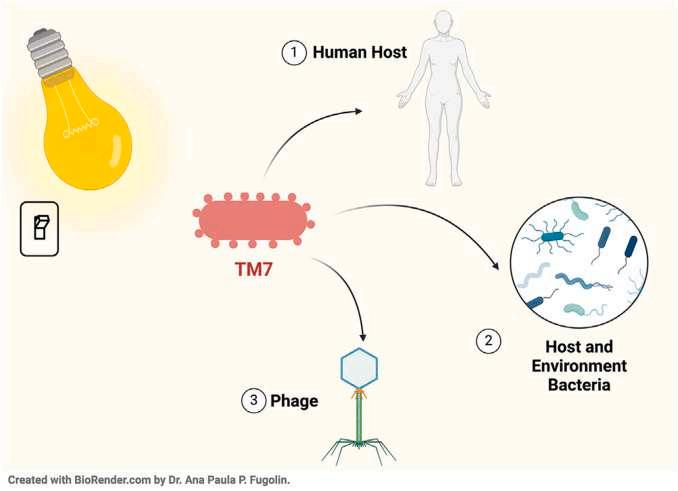
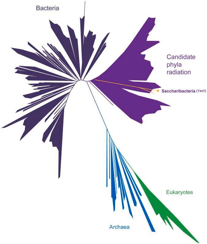
Figure 2. TM7 and the new tree of life. The image highlights the new category of bacteria, candidate phyla radiation, which represents at least 25% of all bacterial diversity. The orange line represents just 1 of the 73 families identified within candidate phyla radiation, TM7. It is the only strain in the group that scientists have been able to cultivate. All the bacteria in this group are episymbiotic to other bacteria. Adapted with permission of the publisher from Hug et al. (Adapted with permission from Hug LA, Baker BJ, Anantharaman K, et al: A new view of the tree of life. Nat Microbiol 1:16048, 2016.).
constantly interacts with one another and with the human host to influence oral and systemic health (Figure 1). A new group of bacteria has led to a new field of study with implications for dentistry and systemic health.
Next-generation sequencing has revealed a group of bacteria called candidate phyla radiation (CPR) that makes up about 25% of all bacterial diversity on the earth. CPR is found in different environments and animals, including the oral cavity of humans. Aside from their existence and their lack of the large genetic repertoire found in other bacteria, little is known about these bacteria. Just 1 lineage (Saccharibacteria, which was first known as TM7) has been successfully cultured, mostly from the human oral cavity. These microbes have dramatically
expanded and complicated our understanding of the human oral microbiome. Both TM7 and Actinomyces offer the potential to transform our current knowledge of oral health and our practice of dentistry.
Dark matter in microbiology refers to all the microorganisms that scientists recognize as existing but that can’t be studied because there’s no way to culture them in a laboratory. TM7 was considered dark matter until scientists cultured them in 2014. It’s not unusual for many bacteria to live in polymicrobial communities with other bacteria but they don’t usually depend completely on other bacteria as their host. The cultured TM7 species were growing on other bacteria as a parasite or episymbiont, the first time episymbiotic behavior has been seen between 2 different bacterial species.
TM7’s host bacterium is Actinomyces, which is associated with periodontal health and disease and is considered a pathobiont. Although Actinomyces doesn’t cause periodontitis, once the disease has begun, the bacteria follow it and contribute to inflammation. The more severe the periodontitis, the more intense is the inflammatory response. As a result, scientists first considered TM7 as a pathogenic species that contributed to inflammation in irritable bowel syndrome and vaginosis along with periodontitis.
Further study found that TM7’s interactions with Actinomyces in periodontal disease may have beneficial qualities. In a mouse model, researchers introduced Actinomyces and saw inflammatory activation leading to alveolar bone loss. When TM7 was added, the inflammation and bone loss decreased significantly. Although the mechanism is still unknown, TM7 may help to regulate the inflammatory response created by the Actinomyces, meaning it could help maintain a healthy balance of microbes and keep disease-exacerbating bacteria in check.
The TM7x strain studied is just 1 of hundreds of TM7 lineages found in the human oral cavity. Going beyond the oral cavity, 73 families of TM7-like bacteria in the CPR group are found, and each of these also has hundreds of lineages. Three families are part of the human microbiome. With all these data, scientists are realizing how much they don’t know (Figure 2). As a result, a completely new field of study within microbiology has developed.
Scientists now believe that the episymbiotic microbes are potentially involved in and contribute to many factors related to human health or disease. TM7 has been living with humans for a long time based on its presence in the dental calculus of ancient humans. It appears that TM7 evolved to live with humans, specifically in the oral cavity.
TM7 and their host bacteria are also some of the most successful bacteria to be readily transmitted from person to person. This transmissibility may be related to TM7’s successful evolution with humans.
TM7 may influence the future of dentistry along several potential paths. Based on TM7’s ability to reduce inflammation in periodontal disease, it may be used to target specific oral pathogens and decrease their inflammatory effects in human disease. TM7 may also act through colonization resistance, which means the oral cavity or gut acquires protection from opportunistic pathogens and diseases through the colonization of commensal or beneficial bacteria. The transmissibility of TM7 and Actinomyces and their stability in colonization may provide colonization resistance against opportunistic pathogens. Finally, TM7 has been found in human inflammatory diseases other than oral diseases, including inflammatory bowel disease and Crohn’s disease. It may offer potential therapeutic strategies for disease management once its activity is better understood.
Dentists need to be aware of the presence of microbial episymbiosis and TM7 bacteria in the human oral cavity. It is hoped that the fact that bacteria can be episymbiotic with other bacteria will lead to the development of future target treatments for oral diseases.
Sirko J, Bor B, He X: Microbial dark matter and the future of dentistry. JADA Found Sci 4:100043, 2025
Reprints not available
Trauma to the orofacial complex, which includes the teeth, soft tissues, facial bone, and TMJs, occurs during sports activities in up to 15% of participants, with higher rates among athletes who don't use protective mouthguards. Players who participate in sports that require physical contact have a 10% chance of facial injuries each season, with rates rising to about 50% by the end of their careers. Tooth-related injuries can affect the functional,
aesthetic, and psychological health of athletes, and such injuries can inflict irreversible damage, requiring long-term, costly care. Mouthguards and general preventive oral care measures for athletes have been developed to protect against this danger, with associations such as the International Association of Dental Traumatology (IADT) supporting the use of mouthguards and the education of athletes and coaches regarding such preventive measures. However, no internationally agreed-upon standards
exist for the use of sports mouthguards because of the variation in awareness among athletes and coaches, the regulations in place, and cultural attitudes toward sports safety in different countries. Custom-made sports mouthguards (CSM) are removable appliances that are resistant to deformation, reduce the risk of dental trauma, and protect surrounding structures. Their protection, fit, and comfort are superior to what is provided by prefabricated and boil-and-bite mouthguards, being designed to effectively absorb and distribute forces. The most current and valid information regarding CSMs and guidelines for their selection, construction, clinical use, and maintenance was provided in a comprehensive position statement and recommendations offered by the European Association for Sports Dentistry (EA4SD).
CSMs are recommended and strongly considered for sports that involve full contact or collisions, an increased risk of falls, equipment interactions, and special needs or situations. The materials used should be approved by the US Food and Drug Administration and the EU public health authorities, with recommended materials being ethylene vinyl acetate (EVA) copolymer, cyclic olefin copolymer (COC), soft acrylic resin or polyvinyl acetate-polyethylene (pEVA), elastomers, or 3D materials. A multi-layered design is recommended, with a wide range of these designs available.
A dental laboratory or office should use a vacuum- or pressureforming process on a working cast made from intraoral impressions. The mouthguard should accommodate any areas with tooth loss, implants, or orthodontic conditions, offering comfort and protection.
The recommended specific measurements are as follows.
• Labial surface of central incisors: Dictated by the material used and the fabrication method, but a minimum thickness of 3 to 4 mm and the maximum used for high-risk contact sports.
• Thickness: Maintained at 1.5 to 2 mm over the incisal edges of the anterior teeth.
• Extent: At least up to the distal surface of the first maxillary molars. Labially, conically tapered as far as possible to the vestibular sulcus, depending on anatomical status. Palatally, as small as possible to allow for the best retention.
With the form based on a dentist-made bite registration, the mouthguard should be articulated and adjusted to the opposite dentition in a balanced, preferably myocentric position. The clinical evaluation during insertion should be conducted by a
well-trained dentist. The labial flanges should have rounded edges and palatal flanges (if any) with tapered edges.
The CSM should provide optimal intraoral adaptation and maximum retention, but offer resistance to dislocation during impact as well as dislodgement during speech, allowing clear communication between athletes. No soft tissue irritation or ulceration should develop, nor should there be any tooth migration with long-term use.
The CSM should maintain space between the maxillary and mandibular teeth and between the condyle and temporal bone in the TMJ. In general, the CSM should accomplish the following.
• Provide comfort without impeding communication, breathing, lip closure, or deglutition.
• Be appropriate to the athlete's age, needs, frequency of use, and type of sport.
• Undergo clinical evaluation by a well-trained dentist at scheduled recall appointments.
• Avoid negative effects on performance-relevant functions such as nasal breathing, tongue positioning on the palate, and speech.
The athlete should be advised regarding the importance of maintenance and care, including the following.
• Rinse the CSM with water before and after use and store in a container.
• Clean the CSM after each use, either using a neutral hand soap and a soft toothbrush or by using chemical dissolving tablets.
• Disinfect the CSM following manufacturer's instructions using ultrasonic cleaners.
• Store in a perforated plastic container in dry conditions at room temperature and out of the sunlight.
• Avoid chewing the CSM.
• Keep the CSM correctly positioned in the mouth throughout the competitive or training period. Avoid constant removal and reinsertion to prevent infection and ensure protection.
• Wear the CSM correctly, with replacement when flanges become deformed.
• Ensure the ability to drink liquids with the CSM in place.
Selecting a CSM should be specific to the individual who will wear it. Specific fabrication, function, maintenance, and care criteria will enter into the decision of which CSM to choose. CSMs offer the best safety without impairing the athlete's functions and performance.
The EA4SD recommends CSMs rather than prefabricated or boil-and-bite mouthguards. Amateurs, professionals, and semi-professional athletes should be required to wear these protective devices. They should be fabricated by dentists who are aware of the best practices and understand the protection that is needed in each sport. It has been postulated that polymer-based mouth protectors may also prevent or reduce the risk of concussion, which is the most common type of traumatic brain injury in youth sports. Collision sports are associated with a higher risk for concussion, which can leave athletes unable to participate in physical activities and may lead to post-concussion syndrome. In addition to protecting the orofacial complex, the possible reduction in the risk and severity of concussion should be considered as an important reason for wearing a CSM during sports participation.
Avgerinos S, Stamos A, Nanussi A, et al: Position statement and recommendations for custom-made sport mouthguards. Dent Traumatol 41:246-251, 2025
Reprints available from C Rahiotis; e-mail: craxioti@dent.uoa.gr
Patient safety in dentistry has focused on the extraction of the wrong tooth, but many other aspects of patient safety should be addressed. Surgical safety checklists are effective, but only when used along with positive measures to ensure patient safety and team engagement and training with the checklists. Patient safety in primary care settings must include a framework of activities that create a positive culture and environment creating consistently lower risks for errors and less impact for any errors that occur. The frameworks that have been identified and the factors that contribute to the safety culture were explained. Elements of a positive patient safety culture were described.
The Swiss cheese model describes how adverse events or systems failures occur (Figure 1). Ideally, there are layers of safeguards and barriers in place that work together to prevent errors from occurring. In the real world, these barriers have potential holes in each layer, and when the holes line up, a
trajectory of accident opportunity is created, increasing the likelihood that an error will occur. This model assumes that the adverse outcomes can be fixed by compliance, standardization of the processes, and training, which is not always true. In addition, interactions among the interdependent layers (clinicians, patients, equipment, the environment) can occur.
The Safety I model was concerned with studying events retrospectively to learn how they happened. A Safety II model now focuses on why things go well and how systems can be resilient when they work effectively, with the recognition that most outcomes are successful.
Human
Clinicians make mistakes, which can result in harm and suffering. The reaction to errors must be considered. One response involves challenging or trying to rectify processes or clinicians who are responsible for the error. This is unlikely to create
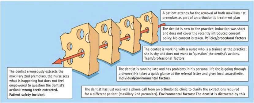
(Reproduced with permission from Bailey E, Tickle M, Campbell S: Patient safety in primary care dentistry: Where are we now? Br Dent J, 217:339-344, 2014.)
meaningful, lasting change and lacks any guarantee errors will no longer occur. It also doesn’t address the usually complex nature of how the error occurred.
A second response addresses the perfection myth, which assumes that practicing a task many times will improve quality and outcomes. This also doesn’t guarantee the problem won’t recur and lacks clear evidence of who should receive added training and if the training can address all the interactions and processes that contributed to the error. Many factors, such as fatigue, poor wellbeing, pride, overconfidence, and ego, contribute to errors.
Performance can be compromised by burnout and fatigue and a paucity of meeting basic needs, psychological needs, and needs for self-fulfillment. Burnout involves emotional, physical, and mental exhaustion caused by excessive and prolonged stress. It became especially prominent during the COVID-19 pandemic, and it has the potential to compromise the performance of team members.
The basic needs that must be met include nutrition, hydration, adequate and appropriate clothing, personal protective
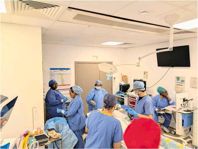
2. Who is who? A busy theatre team, some of whom have never worked with one another before. Similar outfits can lead to confusion and reluctance to speak up, particularly if team members are unaware of names and roles. (Courtesy of Bailey E, Dungarwalla M: Patient safety in dentistry – a decade in the making. Br Dent J 238:814-821, 2025.)
Table 2. Qualities and Behaviours That May Be Considered ‘Functional’ or ‘Dysfunctional’ in a Primary Dental Care Setting. These May Also Be Considered as ‘Pathological’ or “Generative’
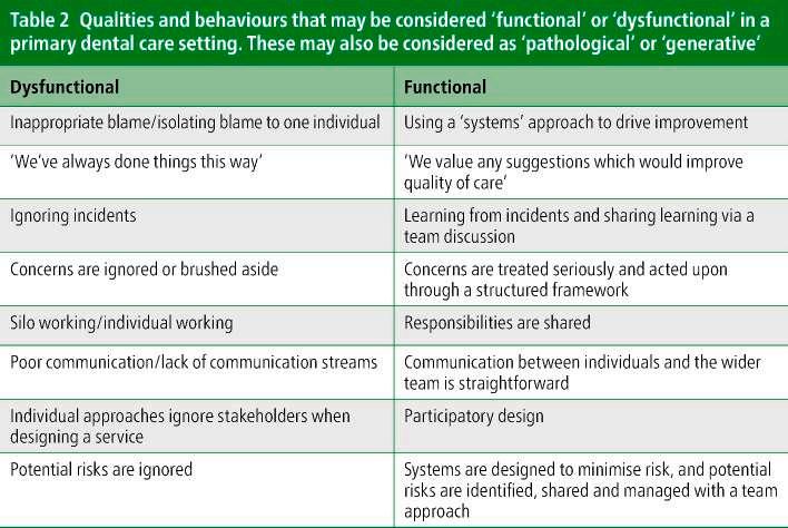
(Courtesy of Bailey E,
M: Patient safety in dentistry – a decade in the making. Br Dent J 238:814-821, 2025.)
equipment, toilet and sanitary facilities, and provisions for breaks. These are essential ingredients in having a prepared and fully functioning staff.
Psychological needs include the ability to speak and ensure senior leadership and management personnel hear what the individual is saying. Each team member should be recognized as a valued individual who is rewarded for his or her work and has a meaningful personal development plan.
Among the self-fulfillment needs are the ability to support and inspire others, deliver excellent patient care, and develop within one’s role. Additional needs may exist depending on the individual, the team, and the environment. The development of the team relies on having the appropriate resources, time, and faculty to receive excellent clinical teaching. Processes should be in place to allow clinicians who are at risk of becoming unsafe to be supported and helped to mitigate negative feelings.
In aviation, crew resource management (CRM) empowers junior pilots and crew members to participate in and intervene in decisions made by senior pilots or captains when they are perceived to have a negative effect on operations. This parallels the situation in a clinical setting where multiple staff who are unfamiliar with one another and under stress and time pressures related to complex surgery don’t share concerns for patient safety. All team members have the right to share their concerns when
they believe patient safety has been or will be compromised (Figure 2). However, in practice, the implementation of this theory can vary. Team members may be intimidated by the hierarchy of the team and fail to speak out to address patient safety issues.
To address this problem, dedicated induction talks can be conducted to address the concept of speaking up and not fearing a reprimand, engaging in theater briefings and morning huddles, and having all team members take part in the exercise, sharing their names and roles throughout the day. Junior clinicians may be tasked with planning complex cases and discussing the plans with more senior clinicians before surgery so that all team members have a stake in the process. In a primary care setting, pre-clinic huddles serve a similar purpose, allowing team members to discuss their roles, address matters that affect how the clinical session is run, and complete “huddle sheets” that guide the discussions, covering the people and teams, tasks, tools and equipment, working environment organization, and external factors.
For patient safety initiatives to be effective, an appropriate culture should be present, where safety is prioritized, resourced, and practiced by the team and leaders (Table 2). Safe care is delivered through ongoing learning and improvement in safety
risk levels, a supportive and psychologically safe team, and having all members of the team encouraged to speak up as needed.
Components of the safety culture include aspects providing psychological safety, such as civility, a positive leadership framework, and the appreciation of all team members who experience not just permission but also freedom to innovate. Qualitative evidence suggests that dental teams have high levels of knowledge and experience in support of patient safety.
When incidents occur, the impact on the patient and family is considered primary, making these the first victims. Some patients and families avoid using the term ‘victim’ in relation to their own experiences. Those on the clinical team can suffer psychological harm and may be considered second victims. Second victims progress through a cycle that includes hypervigilancestress-anxiety-shame, feelings of inadequacy, risk avoidance, self-doubt about knowledge and skills, insomnia, and difficulty concentrating. Clinicians can also suffer underperformance and depression, which have a detrimental effect on patient safety. Early intervention and support are required to minimize the long-term effects. Interventions to support the dental team include the Patient Safety Incident Response Framework (PSIRF), the ‘learn from patient safety events’ (LFPSE) process, and the Primary Care Patient Safety Strategy.
Strategies have been proposed or initiated to increase the uptake and scope of dentistry. In England, it has been proposed that dental hygienists and therapists be allowed to prescribe and administer a broader range of medications independently, that newly qualified dentists work for the National Health Service (NHS) for a minimum period, that provisional registration be given to overseas applicants while they wait for complete registration, and that overseas registration examination spaces be increased by 30%.
Many dental practices have ‘in-house’ specialists to manage the patient demands for better treatment and the pressure of litigation. With the growing interest in facial aesthetics, dental tourism, and cone-beam computed tomography to plan treatments, patient safety may require changes in the legislation and curricula.
Patient safety must be incorporated into the undergraduate curricula and early postgraduate training should be provided. Evidence suggests that patient safety in dentistry is perceived currently as adhering to rules made by senior faculty. Safer care will rely on adhering to established processes and taking care to complete incident reports, surgical safety checklists, consult with other practitioners, identify students and trainees with specific learning needs, and access mental health first aid caregivers more readily. Work pressure and pace, leadership support for patient safety, and openness regarding errors all need to see improvements.
Dentistry now appreciates the complex nature of the health care systems and processes as well as the psychological aspects of patient safety. Surgical safety checklists are widely used in hospital dental services and staff and students are engaged in the process. Primary care settings require additional measures to be taken to achieve better patient safety practices. There is much room for improvement.
Bailey E, Dungarwalla M: Patient safety in dentistry – a decade in the making. Br Dent J 238:814-821, 2025
Reprints available from E Bailey; e-mail: e.bailey@qmul.ac.uk
Periodontal disease (PD) and Alzheimer’s disease (AD) share a rising global prevalence and are both chronic conditions. Studies find that both are linked to inflammation and proinflammatory enzymes, with interactions between bacteria and host immunity determining the severity of brain damage present. The existing
evidence was surveyed to determine if there is a relationship between PD and AD.
Sixteen systematic reviews were identified through a search of the PubMed, Cochrane Library, SciELO, Web of Science, Scopus,
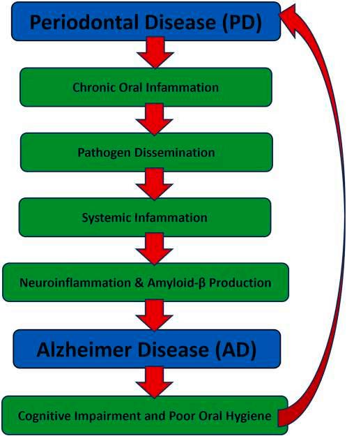
Figure 1. Hypothesized pathways linking periodontal disease and Alzheimer’s disease. The diagram illustrates the proposed bidirectional relationship between periodontal disease (PD) and Alzheimer’s disease (AD). Chronic inflammation originating in PD promotes pathogen dissemination and systemic immune activation, which can lead to neuroinflammation, amyloid-β accumulation, and Alzheimer’s pathology. As AD progresses, cognitive decline and reduced self-care capacity impair oral hygiene, exacerbating periodontal inflammation. This creates a selfperpetuating cycle involving bacterial virulence factors (e.g., gingipains), proinflammatory cytokines (e.g., IL-1β, IL-6, TNF-α), and disruption of the blood-brain barrier. (Courtesy of Arbildo-Vega HI, Cruzado-Oliva FH, Coronel-Zubiate FT, et al: Association between periodontal disease and Alzheimer’s disease: Umbrella review. Front Dent Med 6:1635200, 2025.)
Google Scholar, ProQuest, Dissertations and Theses, and OpenGrey databases.
The research suggests a bidirectional relationship between PD and AD, with PD increasing the risk of AD and AD adding to the risk of PD (Figure 1). Nine of the systematic reviews
reported an association between PD and AD. One found an association for patients in Asia but not in Europe and North America. Another found an association between the 2 diseases for sex. Four systematic reviews identified an association between PD and AD for severe PD but not for mild or moderate PD. Two reported an association between tooth loss and AD, but a third didn’t find this link.
Three systematic reviews found an association between PD and cognitive impairment, dementia, and AD. One found an association between PD and dementia and AD for patients in Asia but not for those in Europe or North America. One found an association between PD and dementia and AD for age. One reported an association between PD and dementia and AD for moderate or severe PD but not for mild PD.
The primary findings of this investigation were that PD and AD are associated. The link becomes more pronounced with age and is common in both men and women. Severe PD was more likely to be linked to AD than less severe cases.
Oral health care providers should inform patients about the link between PD and AD and empower them to take measures that might lower the risk of AD. This includes consistent brushing, flossing, and use of mouthwashes to control plaque formation. Periodontal evaluations should be included in the routine screening and patient education sessions. Multidisciplinary interventions should ensure a comprehensive and coordinated approach to the dental and medical management of AD patients.
Arbildo-Vega HI, Cruzado-Oliva FH, Coronel-Zubiate FT, et al: Association between periodontal disease and Alzheimer’s disease: Umbrella review. Front Dent Med 6:1635200, 2025
Reprints available from FT Coronel-Zubiate; e-mail: franz.coronel @untrm.edu.pe
The primary goal of artificial intelligence (AI) is to create systems that can perform tasks normally requiring human intelligence. Large language models (LLMs) are advanced AI systems that are designed to understand and generate human language. Normally they are trained on large amounts of text data so they can take on a wide range of language tasks, including generation of text, translation, summarization, and answering questions. LLMs go through a pretraining and finetuning process. In pretraining, the model is trained on a large body of text data to learn how to understand language without focusing on a specific task. In fine-turning, LLM is trained on a dataset specific to a task or domain, such as health care or customer service. An unhealthy level of stress related to health information technology is estimated to affect about 70% of dental clinicians. About 13% of dentists exhibit signs of professional burnout, with substantial staffing shortages in dental facilities contributing to their concern. The most common areas of stress are hiring, recruiting, training, and retaining staff
members, with more than 60% of dentists reporting these areas as causing concern. LLM chatbots have shown facility in tasks related to health care, health care education, and research. They potentially could participate in patient communication, administrative tasks such as electronic patient messaging, and training for the dental workforce. In a public social media forum, chatbot responses were rated as being of better quality and more empathy than those of physicians. The effectiveness of chatbots in addressing dental patient queries with respect to quality and empathy was compared with these qualities in the responses of human dentists.
Twenty patient questions and responses were randomly sampled from Reddit’s dental advice community. The quality and empathy of ChatGPT-generated responses (Version GPT-3.5, Open AI) were rated by 9 blinded dentists (Figure). These evaluators ranked the information quality
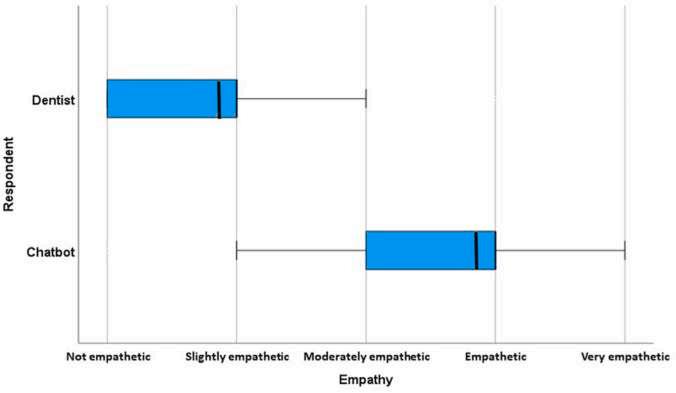
Figure. Created with BioRender.com by Dr. Ana Paula P. Fugolin. (Courtesy of Maruska EE, Elgreatly A, Madaio W, et al: Comparing dentist and chatbot answers to dental questions for quality and empathy. JADA Found Sci 4:100044, 2025.)
and empathy of chatbot and dentists on a Likert scale of 1 to 5. They also selected the best response, whether of the dentists or of AI. One hundred eighty potential responses were generated.
Mean word count was 83.95 (range 12 to 253 words) for the dentists and 277.3 words (range 67 to 363 words) for the chatbot. Evaluators scored ChatGPT’s responses higher in terms of quality and empathy than dentists’ responses. About 93% rated the ChatGPT responses as better and about 7% chose the dentists’ responses. No AI hallucinations (false fabrications generated by an LLM and presented as factual) were observed in the chatbot responses.
This study identified the chatbot’s responses as more efficacious than the dentists’ responses in answering dental patient queries.
The responses provided by ChatGPT Version GPT-3.5 to random online patient inquiries evidenced higher quality and greater empathy than the responses of human dentists. In addition, LLMs can provide written responses in just seconds, which could increase the efficiency of patient-dentist communications, allowing dentists the time to edit them, freeing staff members to perform more complex tasks, preventing burnout, and potentially delivering more compassionate and comprehensive answers to patient questions. Further research is needed to determine if these findings can be generalized and the extent to which chatbots can be used in dental communication tasks.
Maruska EE, Elgreatly A, Madaio W, et al: Comparing dentist and chatbot answers to dental questions for quality and empathy.
JADA Found Sci 4:100044, 2025
Reprints not available
Artificial intelligence (AI)−supported tools used in health care services offer advanced skills in mimicking human language processing abilities. In dentistry, ChatGPT can provide services in digital data recording, image analysis diagnosis and treatment planning, dental telemedicine, and dental and patient education. Health care professionals are using ChatGPT as a convenient source for medical and dental information, and patients can get instant feedback and information about their health questions. It may also have potential uses in patient information regarding oral and maxillofacial surgery. The American Academy of Pediatric Dentistry (AAPD) has set standards for pediatric dental care. This includes having a first visit in the first year of life, providing counseling for parents, evaluating any symptoms related to tooth eruption, assessing the child’s oral health risk, and training children in oral hygiene. This visit is important in detecting any early childhood caries (ECC) and beginning the use of fluoride.
Parents can be confused because of varying views on the usefulness of fluoride and should also be informed about traumatic dental injuries, which are common in young children. The reliability and effectiveness of AI-supported information was investigated, noting the quality, readability, and originality of pediatric patient/parent information and academic content generated by ChatGPT in pediatric dentistry.
Three topics were selected to investigate the quality, readability, and originality of the ChatGPT content: dental trauma, fluoride, and tooth eruption/oral health. Both pediatric patient/parent questions and academic questions were used, with a total of 60 questions. The quality of the answers was assessed using the modified Global Quality Scale (GQS). The readability was evaluated using the Flesch Reading Ease and Flesch-Kincaid Grade Level tests. A similarity index was used
to compare the answers given by the software with the guidelines and academic references in various databases. Ten pedodontists were contacted to assess the quality of the answers provided by ChatGPT.
The average modified GQS score for quality was 4.3 for the answers given to patient/parent questions, but the average score for the answers to academic questions was 3.7, which differed significantly. The average Flesch Reading Ease score was 41.5 for the answers to pediatric patient/parent questions and 34 for the answers to academic questions, which also differed significantly. Averages between 30 and 50 indicate the text is understandable at the college student level. The average Flesh-Kincaid Grade Level score for the answers to pediatric patient/parent questions was 12.7, whereas the average score for the answers to academic questions was 13.7. The difference did not reach statistical significance. Averages between 12 and 15 indicate the text is understandable at the college student level. The average similarity rate of the answers to pediatric patient/parent questions was 8.5, and the average rate for the answers to academic questions was 5.7; the difference between these values was not statistically significant.
No statistically significant difference was noted in the average quality scores of the answers to pediatric patient/parent questions regarding dental trauma, fluoride, and tooth eruption/ oral health. The average quality scores of the answers to academic questions were highest for the subjects fluoride and tooth eruption/oral health. They were statistically significantly higher than the answers to academic questions regarding dental trauma.
The Flesch Reading Ease and Flesch-Kincaid Grade Level scores of readability of answers to pediatric patient/parent questions were highest for tooth eruption/oral health and lowest for fluoride, which differed significantly. Readability of the answers to academic questions did not differ significantly among dental trauma, fluoride, and tooth eruption/oral health. Flesch-
Kincaid Grade Level scores were not statistically different between the topics in the readability of answers to pediatric patient/parent questions and those to academic questions. The averages of the answers to academic and pediatric patient/parent questions were between 12 and 15, indicating they were comprehensible at the college student level.
Similarity rate of the answers was highest for those given to the pediatric patient/parent questions regarding fluoride. Among the answers to academic questions, the similarity rate was highest for tooth eruption/oral health questions. No statistically significant difference in similarity was noted between the topics for answers to pediatric patient/parent questions and academic questions.
The quality of the information given by ChatGPT to parents of pediatric patients was high, but the readability tended to be at a higher level than was comfortable for that audience. The similarity rate was acceptable.
ChatCPT offers great potential for use as a patient information tool in pediatric dentistry. However, it needs to improve its answers to questions so that it can interact with parents more efficiently and with greater fluency.
Nahir CB: Can ChatGPT be guide in pediatric dentistry? BMC Oral Health 25:9, 2025
Reprints available from CB Nahir, Dept of Pediatric Dentistry, Faculty of Dentistry, Tokat GaziosmanpaS ¸ a Univ, Tokat, Turkiye; e-mail: canannbayraktarr@gmail.com
The European Association of Periodontology published clinical guidelines for the prevention and treatment of peri-implantitis, but many clinically relevant questions haven’t been studied sufficiently to provide reliable recommendations. This lack of clinical evidence allows the focus to be on therapeutic steps that
have been studied and are well-recognized and may lead to their recommended use in clinical practice. Once the patient has been evaluated for peri-implantitis, the main tasks to address the situation are oral hygiene instructions, professional dental cleaning, and adhering to proper recall intervals specific to the patient.
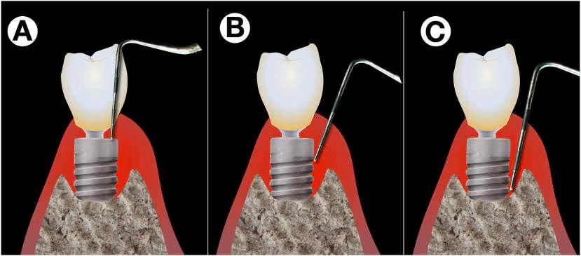
1. Difficulties with peri-implant probing. Exact peri-implant probing is difficult on different levels. While the shape of the implant’s shoulder and platform switching renders probing difficult in the upper part (A), correct probing is difficult due to pain sensations of the patient while the operator tries to verify whether the tip of the probe is still stuck in the implants’ threads (B) or has already reached the most coronal point osseo-integrated part (C).
(Courtesy of Sahrmann P, Giannopoulou C: Clinically relevant aspects of professional follow-up care for implant patients. Frontiers Dent Med 6:1565151, 2025.)
Diagnostic Process
Diagnosing peri-implantitis is more complex than diagnosing periodontitis because of the morphology of the collar area of implants, which is in the emergence zone from surrounding soft tissue (Figure 1). To allow for an aesthetic appearance with the narrower implants, the crown and implant complex are given a strongly funnel-shaped widening in the supracrestal soft tissue, and this complicates access for peri-implant probing.
To simplify the diagnostic process, a mild infiltration anesthesia allows precise, reliable probing with causing patients pain. The first probing is done when the restoration is inserted to detect any deepening pockets. Vertical implant position with respect to the bone margin helps in obtaining an accurate measurement. In addition, gently stroking the peri-implant soft tissue apically to coronally under light pressure with the practitioner’s fingertip allows for an accurate analysis of any post-implant exudate. The sulcus should first be air dried, then observed to see if any fluid is exuding during stroking. No exudate is a sign of health, but clear mucosal crevicular fluid indicates a serous inflammation. Often a drop of cloudy exudate or pus emerges from a place not considered remarkable during a cursory probing.
When analyzing potential marginal bone loss, single-tooth radiographs using a simple individual stent are recommended. After inserting the prosthetic restoration, the occlusion is recoded using a quick-setting A-silicone material in the bite area of the conventional radiographic holder during the baseline radiograph. The bite registration is left on the radiographic stent, which can be reused during each comparison follow-up evaluation.
The radiographs used for full-arch restorations should be evaluated to determine which radiograph is the best compromise of each single implant and to record the number of radiographs required. Storage and continuous use of the stent may be challenging for practices with many implant cases. In this case, stents may be fabricated only for patients and implants at higher risk for failure or for more complex restorations.
Problem Areas
Optimized oral hygiene is a prerequisite for peri-implant health and preventing inflammation. However, no data specifically identify the best brushing technique. Plaque distribution can be measured precisely in a few seconds using commercially available plaque relevators. The problematic sites include the area just adjacent to the mucosal margin of the implant crown restoration (Figure 3), the gum line−prosthetic reconstruction line that has an accentuated step at the margin and isn’t readily reached with conventional toothbrushes, and the interdental spaces and interdental/inter-implant areas, where interdental brushes can reach some areas but the spaces between the line angles, termed interdental funnels, are often left nearly uncleaned.
Instru ctions
Although no scientific studies support the superiority of any brushing technique around implants, practitioners can rely on the evidence that exists. In daily practice, the problem areas can be cleaned effectively using small, round, tapered brush heads. When activated by sound, these brushes replicate the manual, challenging shaking motion of the Bass technique, which allows them to provide efficient oral hygiene (Figure 4).
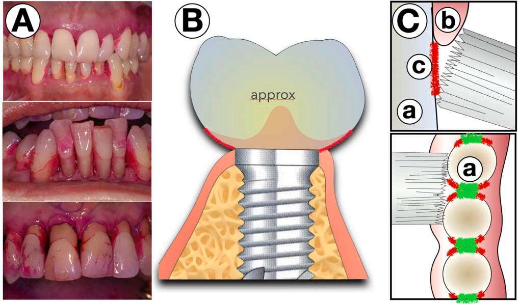
Figure 3. Areas with residual plaque after conventional brushing. A, Areas which typically remain uncleaned by conventional brushing revealed by plaque revelator. B, Sketch that displays these areas from approximally. C, On the prostetical parts (a) closed to the margo mucosae (b) the even bristle field of a conventional brush cannot enter the residually uncleaned surfaces (c). The green areas may be brushed well by a brush. (Courtesy of Sahrmann P, Giannopoulou C: Clinically relevant aspects of professional follow-up care for implant patients. Frontiers Dent Med 6:1565151, 2025.)
Dental floss is effective for areas just below the mucosal margin, but fluffy “superfloss” products may leave small fibers that remain in the sulcus and become colonized by biofilm, leading to inflammation within days. Removing these filaments can quickly resolve the inflammation. Dentists should recommend specific dental floss products and demonstrate proper usage that meets the patient’s needs. With perfect peri-implant conditions and perfectly fitting restorations, dental floss can be used effectively and without potential risks.
For implant-supported bridges, the pontic area can be cleaned with floss products that have stiff ends to facilitate insertion. Pontics make a tight contact to most keratinized soft tissue and fully epithelized surface, but not to a sulcular area that is highly responsive to biofilm.
The guidelines generally recommend mechanical tools and advise against using antibiotics, antiseptics, or other pharmacological
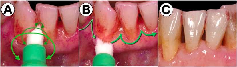
Figure 4. Specialized sonic-activated brush head for the residually uncleaned areas. With a small, round, and pointed brush head, which is ideally activated sonically, the problematic areas are easy to clean by rotating movements (A) and movements long the margo mucosa with a bristle angulation of 45◦ against the sulcus (B) to efficiently remove biofilms from the respective areas (C). (Courtesy of Sahrmann P, Giannopoulou C: Clinically relevant aspects of professional follow-up care for implant patients. Frontiers Dent Med 6:1565151, 2025.)
agents. Laboratory studies show the mechanical treatment of implant surfaces can be problematic. First, these instruments are usually made of hard materials and can alter the surface morphology of implants, which can reduce the biocompatibility of the surface areas and affect osseointegration. Their use is advantageous in that the smoother surface is less susceptible to biofilm accumulation. Second, the machined or polished surface areas are roughened by the instruments, which is a disadvantage in the sensitive area of the emergence profile because biofilm can adhere. Even softer materials, such as plastic and carbon, can abrade the material and leave small particles on the surfaces being cleaned. Biofilm can accumulate on these particles.
A compromise between surface changes and abrasion on the implant surfaces is the plastic PEEK, which changes the surface morphology less significantly than steel or titanium but still leaves particles on the titanium substrate. Low-abrasive powders such as glycine and erythritol have shown superior cleaning efficacy compared to other mechanical methods without visibly altering the surface morphology. However, few clinical studies document their advantages. The same is true of photodynamic therapy, laser treatment, and probiotics.
A significant benefit can be seen with strictly managed recall programs including good patient compliance. One study found patients observing these conditions have an incidence of inflammation in the remaining dentition of 33% over 20 years, with no inflammation. Comparable patients with poor compliance had peri-implant inflammation over the same period.
The guidelines for recall frequency aren’t clear, but several components should be considered when selecting the time between recall appointments. These include risk factors such as general health, increasing probing depths, maintenance factors for the implant restoration, and bleeding on probing (BoP), the last of
which is the most sensitive. A recommended rule of thumb is as follows: As long as the remaining dentition and implants show no increased risk for inflammation, the recall interval is adequate. If more sites with BoP develop, the interval should be shortened to address the inflammation at the peri-implant mucositis stage. This protocol appears to be safer and easier when the initial recall intervals are short (up to 3 months), then extended to an optimal frequency for the patient. If potential risk factors are well controlled, the intervals could be as long as 12 months.
Peri-implant tissue degradation progresses rapidly, making proper diagnosis an essential first step. Measures such as stroking the vestibular gums to detect sulcular fluid and probing under anesthesia can aid in the diagnostic process. Oral hygiene must be accomplished with significant discipline and consistency on the patient’s part. Tools for cleaning clinically relevant areas are required but must also minimize any potential damage to the implant or surrounding tissues. Recall intervals must be tailored to the needs of the patient and the early detection of any periimplant inflammation.
Sahrmann P Giannopoulou C: Clinically relevant aspects of professional follow-up care for implant patients. Frontiers Dent Med 6:1565151, 2025
Reprints available from P Sahrmann, Dept of Periodontology, Endodontology and Cariology, Univ Ctr for Dental Medicine, Basel UZB, Univ of Basel, Basel, Switzerland; e-mail: philipp. sahrmann@unibas.ch
Conventional complete dentures are the first therapeutic intervention for edentulous patients. Some patients with complete dentures are dissatisfied with their prostheses because of anatomical conditions or difficulty adapting, with most complaints related to inadequate retention of the mandibular dentures. The use of an overdenture retained on 2 implants is the treatment of choice in these cases, and it can improve comfort
and chewing performance, which has a positive effect on the patient’s satisfaction level and quality of life. Alternatives that may be offered in these cases include the reduction of abutments to 1 standard-sized implant in the symphyseal region, known as single standard implant-retained overdentures (SSOs). These restorations tend to have long-term stability if carefully planned and followed up. Another alternative is the use of mini-implant −retained overdentures (MOs), which involves the insertion of
2 or 4 reduced-diameter implants in the canine or premolar region. This is especially useful for patients with reduced bone availability. Multiple 1-piece implants may not be able to compensate for axial deviations, and the activation of worn attachment elements can be challenging with 1-piece implants. These alternative approaches are approved treatment options, with the primary goal of enhancing patients’ oral health−related quality of life (OHRQoL). A comparison of the different approaches and their long-term impact on patients’ quality of life was carried out.
Thirty-nine patients (78 implants) with advanced mandibular bone atrophy were randomly assigned to different treatment groups. These groups received single standard implantretained overdentures (SSOs) or mini-implant−retained overdentures (TMs), a group that was subsequently divided into those receiving 2 mini-implant−retained overdentures (TMOs) and those receiving 4 mini-implant−retained overdentures (FMOs). Over a period of 10 years, these patients’ technical and biologic parameters and OHRQoL were evaluated. Among the measurements noted were probing depth (PD), approximal plaque index (API), and sulcus bleeding index (SBI). Patients reported their OHRQoL by completing the Oral Health Impact Profile (OHIP)-G14 questionnaire.
At the 1-year follow-up evaluation, 1 participant in the SSO group had died and 1 implant in the SSO group and 1 in the MO group were removed because of a lack of osseointegration. At the final evaluation, 98.4% of the implants were functioning. Additional implants were used for 1 patient in the SSO group and 1 in the TMO group. After 10 years, 23 patients were reexamined.
Technical complications first developed and required interventions after 3.8 years (TMO group), 4.2 years (SSO group), and 4.6 years (FMO group). The most common intervention was the replacement of worn attachment parts.
The mean API at the final evaluation differed from that at the initial assessment. At 1 year, the API was 13, whereas it was 36 after 10 years. The PBI was 8 at the first year and 28 after 10 years. The highest PD was 2.8 mm after 1 year and 3.9 mm
after 10 years. Average bone loss was 1.6 mm after 1 year and 0.6 after 10 years.
Seventy-four percent of the patients had healthy peri-implant and mucosal conditions after 1 year. Twenty percent had mucositis and 1 patient had mild peri-implantitis. At the final evaluation, 40% of the patients had healthy periodontal tissues, 17% had mucositis, and 23% had various degrees of peri-implantitis. Peri-implant tissue conditions did not differ between the groups.
The average OHIP score before implantation was 22.6, which decreased to 7.6 after 1 year. After 10 years, OHIP score was 5.4.
The OHRQoL of edentulous patients significantly improved after they received a mandibular total denture on a standard implant or a set of mini-implants. OHRQoL remained at a high level at the 10-year follow-up.
Prosthetic restoration achieved an improvement in OHRQoL in edentulous patients that exceeded the average OHIP score of patients who wore a complete denture. Both the use of complete dentures on single implants and the use of mini-implants achieved a positive effect on patients’ quality of life. The significant improvement was sustained over 10 years, and the approach was associated with a lower surgical effort in placing the implants. Older patients with an atrophic edentulous mandible may benefit from this approach to managing their oral health.
Bacher H, Kasaliyska M, Arnold C, et al: Minimally invasive approaches for implant-supported overdentures in the atrophied mandible. Gerodontology 42:283-292, 2025
Reprints available from H Bacher, Dept of Prosthetic Dentistry, Univ School of Dental Medicine, Martin-Luther-Univ, Halle-Wittenberg, Magdeburger Str. 16, Halle 06112, Germany; e-mail: helene.bacher@uk-halle.de
The influence of the internet, social media, and celebrity culture coupled with the increased availability of implant treatments has focused patients’ attention on achieving immediate satisfaction and the fulfillment of their aspirations. As a result, patient expectations tend to focus on cosmetic perfectionism and are harder to satisfy, with desires that have grown exponentially from the original satisfaction in having a missing tooth replaced. Dentists must deal with these expectations and ensure that patients fully understand their treatment options, risks, costs, and time commitment before beginning any procedure. The changing dynamics in patient care was discussed, noting consentexpectation mismatches, misconceptions, unrealistic expectations, and the dentist’s role in addressing their care of patients who fall into these categories.
An important distinction must be made between lack of consent in dental or medical treatments and a failure to meet patients’ expectations. Generally the 2 can be seen as a dentist’s legal obligation and the patient’s perception. Lack of consent is a legal and ethical issue. Dentists are required to obtain the patient’s informed consent by fully explaining the risks, benefits, and alternatives of treatment before beginning any procedures. Any material risks that could influence the patient’s decision must be explained, with the patient holding the right of autonomy.
Failure to meet expectations occurs when the outcome of treatment doesn’t match what the patient expected would happen. It doesn’t reflect the fact that the treatment meets professional standards. Often the problem is poor communication between patient and dentist or unrealistic expectations on the part of the patient.
Failure to meet expectations can involve dissatisfaction with the outcome, whereas lack of consent is based on the absence of proper communication and agreement and has significant legal implications. Often the complaints that are made are based on unmet expectations or simple misunderstandings. If addressed before treatment, a simple explanation may be sufficient, but addressing the situation after treatment tends to be perceived by the patient as elaborate excuses and failure to obtain consent. If the patient’s expectations aren’t carefully managed, even
successful procedures may lead to a complaint when the outcome doesn’t match what the patient anticipated.
Patients should take responsibility for their problems, which can only happen if they are given the means to understand how they arrived at their situation. Having patients blame others for their issues isn’t a good foundation for the patient-dentist relationship. The dentist can share the cause of any original tooth or implant failure or loss honestly, which may help ground the patient and establish a rational baseline. Patients who suffer from periodontal problems or persistently fracture teeth or restorations must be told that treatment may give them the opportunity to continue to experience these issues, eventually causing worn, loose, fractured implant prostheses or peri-implant disease (Figure 1).
Patients should also recognize the complexity associated with treatment. Implants are seen as readily available, but for some patients, having simpler alternative treatments may fit with their expectations better than complex implant solutions. Patients can experience “treatment fatigue” from the repeated visits needed for treatment. If the case will involve challenging treatments with increased risk and extended time lines, the dentist may want to recommend the patient seek a second opinion or a referral.
Patients can also fail to understand the cumulative cost of treatment. Frustration can develop in patients who are financially challenged or who don’t see the results they anticipated. Similar frustration can occur when the patient experiences discomfort during surgery that exceeds his or her pain tolerance, when the patient with a self-image of being strong and resilient with a high pain threshold experiences more pain than anticipated, or for patients who have conscious sedation and remember what they went through rather than being unconscious during the entire treatment.
If the implant fails soon after surgery, both patient and dentist are disappointed. Both must realize that there is always a risk of failure and the possibility should be thoroughly discussed before treatment begins. The discussion should include how the failure will affect the overall treatment plan and how it will be addressed, both practically and financially.
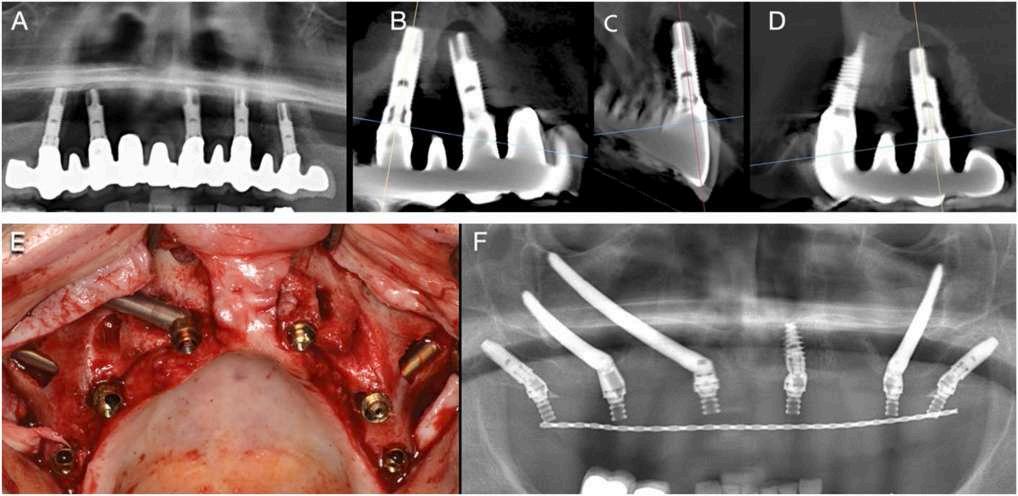
Figure 1. Panoramic radiograph (A), and cross sections (B, C, D) through each implant site show extensive peri-implant disease and multiple implant failures. Subsequent treatment is inevitably more complex, in this case necessitating reconstruction with a combination of dental and zygomatic implants as seen at the time of surgery (E) and in a subsequent panoramic radiograph (F). Patients need to be made aware of the requirement for ongoing maintenance and repair, with an appreciation of the longevity of a reconstruction, and the possible need for eventual replacement. (Courtesy of Dawood A: Patient expectations in implant and aesthetic dentistry. Br Dent J 238:770-776, 2025.)
Most patients are eager to get treatment over quickly, but some expedited procedures may be associated with a higher risk of failure or complications. Examples include immediate implant placement, immediate loading of implants, combinations of procedures, or the placement of implants where periodontally involved teeth are present. It’s wise to document the discussion of the heightened risk these situations present before beginning treatment so that the dentist can defend against a claim of having rushed the procedure.
Health care providers are bound by the duty of candor, wherein open, honest conversations are held when something goes wrong. All complications, failures, unexpected outcomes, and mistakes must be disclosed. The patient is less likely to feel misled or frustrated if he or she has been properly informed.
Other aspects associated with implants that can cause patients to have misconceptions about the outcome involve lack of understanding about all the changes that occur with the loss of a tooth or teeth, the possibility that screw channels or the use of soft, potentially unreliable provisional adhesive may occur, the concept of bridgework (Figure 2), jaw remodeling and its results, and possible difficulties with speech, air escape, lisping, spitting when speaking, food impaction, and cheek biting. Most patients will eventually adapt, although there may be a period of frustration and lack of trust. If the patient is intrinsically difficult to please and demands extra attention, the dentist should
ensure that all discussions where information about these possibilities was shared with the patient are fully documented.
Some patients don’t comprehend or accept realistic boundaries and dismiss professional advice about what is possible. These individuals are more likely to become disappointed with the outcomes of procedures and instead expect results that exceed what is technically or biologically feasible. When patients become emotionally attached to the treatment and expect it to solve broader emotional or psychological issues, significant disappointment can result.
Unrealistic expectations can also be a problem when patients have a history of multiple procedures. They may be chasing unattainable goals. Patients may also be unable to articulate their goals, offering vague or unrealistic expectations that can’t be met. Comparisons with celebrities or digitally altered images, seeking a dramatic transformation based on unrealistic standards, and a focus on others’ reactions rather than personal satisfaction are also associated with unrealistic expectations and disappointment with the outcome of treatment. Not listening to the dentist and dismissing or minimizing risks related to themselves, including smoking or certain medical conditions, may lead to frustration and expectations that aren’t in line with what’s possible.
Patients may become increasingly difficult to satisfy as they near a definitive result, with minor improvements devalued and the pursuit of perfection becoming more elusive. Hedonic treadmill or hedonic adaptation is a term describing the actions of people who quickly return to a baseline level of happiness or dissatisfaction after major life changes. The initial loss of teeth may feel catastrophic and leave a deep emotional impact. Replacing a denture with a fixed bridge may bring a surge of emotion when function is restored, but as the patient adapts, the sense of joy diminishes. Nearing normality causes patients to compare themselves to others, often on social media, and raise their expectations beyond their personal journey. Patients continue to set new baselines that make each treatment feel
less impactful. As a result, the patient becomes dissatisfied, stressed, and anxious.
If the hedonic treadmill becomes extreme, it eventually looks like body dysmorphic disorder (BDD), where minor or imagined flaws dominate the patient’s thoughts and lead to compulsive desires for more improvement, which can eventually create a poor quality of life. BDD is a mental condition where the patient has an obsessive focus on perceived flaws in appearance. The individual is significantly distressed and engages in repetitive behaviors, checking the mirror, grooming, or comparisons with others. As a result, the patient develops social avoidance behaviors, relationship difficulties, and challenges in working or studying.
Genetic, neurobiological, psychological, and environmental influences contribute to BDD. Because the face is often an area of focus, patients may seek out cosmetic dental or implant procedures to fix their flaws, but their unrealistic expectations aren’t met and they experience ongoing frustration and demands for more revisions or other procedures to obtain perfection.
When the dentist encounters an individual with BDD, he or she should seek to collaborate with the patient’s general practitioner and enter into a multidisciplinary approach that provides psychological care for the underlying condition. It’s wise to avoid unneeded dental interventions.
Patients can develop treatment fatigue and become exhausted and disengaged if the treatment is prolonged or repeated with minimal benefit. Often those who have repeated treatment failures develop profound psychological effects that can influence any future treatment outcomes. Learned helplessness is a condition where patients begin to believe they have no control over their situation, producing passivity and reluctance to engage, even when viable options for improvement exist. A history of failure can cause hopelessness, which makes it more difficult to treat them. They may be vulnerable to the nocebo effect, where their negative expectations for treatment cause poor outcomes and further complicate treatment. Demoralization syndrome can also develop in patients dealing with chronic issues or repeated failures. This is manifest as low self-esteem, hopelessness, and loss of motivation, especially if social support is lacking. This impacts their entire wellbeing.
The psychotherapeutic aspect to dental care can be seen when the bond between clinician and patient is strained by repeated unsuccessful treatments. This is similar to the therapeutic alliance rupture in psychotherapy, and it involves a breakdown of trust and collaboration on any future treatments.
Dental professionals are increasingly called on to manage not just the physical aspects of treatment but also patients’ psychological and emotional responses (Figure 3). The influence of social media on patient expectations must be tempered by biological realities to yield appropriate implant treatment. Clinicians must provide comprehensive patient education and informed consent, but are also responsible to ensure patients fully understand realistic goals and limitations on treatment. Maintaining trust between clinician and patient requires transparent, empathetic communications, with open discussions about potential risks and realistic expectations. In addition, dental practitioners must deal with patients who demand treatments with diminishing returns and those who have increasingly higher expectations.
All of these scenarios can leave the entire dental team feeling mental fatigue. They must justify every decision, explain every step, and manage the patient’s emotional responses in addition to providing demanding clinical care. Often these patients bombard them with calls, questions, or concerns between appointments that further tax the dental practice’s resources and lead to burnout.
Dentists and dental teams must maintain open communication and support one another, especially when dealing with emotionally tiring patients. Regular team meetings can be used to debrief on difficult cases, receive support from mental health professionals, and learn to support one another with the goal of avoiding burnout. Seeking second opinions or referrals for complex cases may alleviate some of the pressure on the team and gives patients a broader perspective on their treatment.
The dental team is called upon to display both clinical expertise and emotional resilience. Dentists need technical skills but also psychological tools to manage difficult cases successfully. Although providing care for these patients can be rewarding, the significant challenges along the way can affect the team’s wellbeing.
Dentists might be reluctant to treat patients who complain, threaten litigation, or demand unrealistic outcomes and can shy away from taking those who appear to have psychological complexities. As a result, these patients may have difficulty receiving appropriate care, causing them to feel rejected and to distrust the dental profession. Patients with legitimate needs may not receive proper treatment, so their condition worsens and they experience increasing frustration. This can cause an emotional and psychological burden on both patient and dental professionals. To avoid this cycle, dental professionals must balance the call to validate the patient’s concerns and the duty to manage their unrealistic expectations. To be successful, clinicians must practice within a framework of professional and personal resilience that allows them to provide needed and reasonable treatment while managing the patient’s emotional and psychological complexities.
Dawood A: Patient expectations in implant and aesthetic dentistry. Br Dent J 238:770-776, 2025
Reprints available from A Dawood, The Dawood and Tanner Specialist Dental Practice, London, Univ College London Hosp, England, UK: e-mail: andrew.dawood@dawoodandtanner.co.uk
Bitewing (BW) radiographs are useful for detecting caries in children at high risk for caries after a visual/tactile examination to plan treatment. The as-low-as-diagnostically-achievable-beingindication-obtained-and-patient-specific (ALADAIP) principle is followed, and the information obtained can contribute significantly to decision making. Clinicians must choose the most appropriate radiograph to obtain the needed information. The film for BW radiographs can be placed vertically or horizontally, with the horizontal placement used most. The diagnostic value depends on the quality of the radiograph obtained. BWs allow clinicians to estimate the extent of caries in dentin, the proximity to the pulp, and the size and extent of the pulp chamber in response to caries lesions, such as pulp retraction caused by tertiary dentin deposition. Over half of primary molars contain accessory canals in the furcation area, so the interradicular and periapical areas must be visualized. They aren’t always visible on horizontal BWs, requiring an additional radiograph. Vertical BWs (VBWs) can visualize the furcation area of primary molars, pathological root resorption, and any permanent successors to the primary teeth. The film is narrower in the vertical direction and won’t encroach as far distally on soft tissues. Children also tend to tolerate V BWs better than horizontal ones. Ideal VBWs allow clinicians to visualize occlusal and interproximal caries lesions from the distal surface of primary canines and the first and second primary molars to the mesial surface of the first permanent molars. They don’t capture the entire first permanent molar crown. The use of VBWs in children was
evaluated for overall quality and common errors that occur in film and tube positioning. The visibility of primary molar crowns, furcation area, and permanent successors on VBWs was also assessed.
The participants were 75 children (mean age 7.6 years) who had at least 1 active carious lesion. A total of 550 primary molars were visualized. VBWs were taken by undergraduate dental students as part of their clinical training. The type of BW positioner (Figure 1) and film sizes (Figure 2) selected was based on operator choice and child’s characteristics. Parameters noted were the overall quality rating of the radiographs as well as specific errors in processing, distortion, and positioning that could have contributed to the quality score. Positioning errors were subdivided into vertical errors of film positioning, horizontal errors of film positioning, and coning off due to collimation (Figure 3).
Three films were deemed unacceptable and were repeated, with just the second one considered in the analysis. Overall quality of the radiograph was excellent for 10.67%, diagnostically acceptable for 72%, and diagnostically compromised for 17.33%. The compromised radiographs had processing errors in 0.67% and distortion errors in 2.27%. Positioning errors included the child not biting fully in 7.33%, vertical errors in
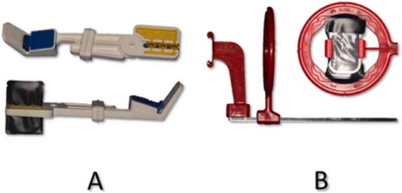
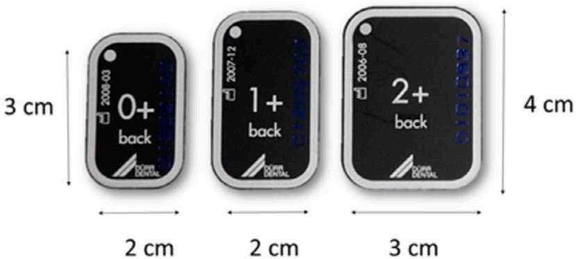
2. Different film sizes (0, 1, and 2) used with measurement in cm. (Courtesy of Oleg � ario IC, Leith R, O’Connell AC: Quality of vertical bitewings radiographs and common errors of positioning in children. Int J Paediatr Dent 35:869-875, 2025.)
13.33%, horizontal errors in 48.67%, and presence of central ray entry errors in 38.67%. Use of the Red Rinn XCP vertical holder was associated with better VBW quality than the use of the Snap-A-Ray holder.
The visibility of the primary tooth crown, furcation, and permanent successor was evaluated. Permanent successor teeth were not visible in 13.63% of the cases, but in 10 radiographs, the successor tooth was congenitally missing. Most (90.54%) cases showed the furcation area, but the entire crown of the primary molar wasn’t seen in 0.55% of the cases. Primary molars were
often (18.90%) only partially visible because the film was poorly positioned. Overlaps were seen in enamel (13.27%) and dentin levels (2.18%), which were the result of inappropriate positioning of the x-ray beam.
Younger children with primary or mixed dentition should be evaluated using VBWs to detect caries. Their diagnostic information is better than with horizontal BWs and children find the experience more comfortable.
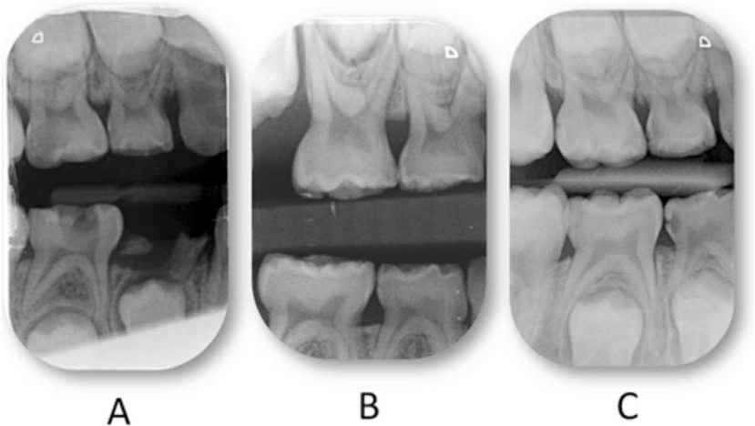
Figure 3. Common errors of film/tube positioning for vertical bitewings. A, Conning off (cone cute). B, Vertical error of film positioning. C, Horizontal error of film positioning (too far posterior). (Courtesy of Oleg � ario IC, Leith R, O’Connell AC: Quality of vertical bitewings radiographs and common errors of positioning in children. Int J Paediatr Dent 35:869-875, 2025.)
With VBWs, the film is narrower in the mesial-distal dimension, which can minimize problems with the gag reflex and discomfort when the film is placed posteriorly. Both of these make it easier to use with children. As a result, children are more cooperative, the information gained on a single image is maximized, fewer exposures are needed, and the final quality of the radiographs is enhanced. The use of VBWs is associated with the same quality as horizontal BWs and they allow visualization of corona caries in primary molars, furcation areas, and permanent successors. Correct film size selection and positioning are critical, with optimal film size and holders essential to accommodate pediatric patients’ smaller oral cavities and dental structures.
Oleg� ario IC, Leith R, O’Connell AC: Quality of vertical bitewings radiographs and common errors of positioning in children. Int J Paediatr Dent 35:869-875, 2025
Reprints available from AC O’Connell; e-mail: anne.oconnell@dental.tcd.ie
Direct composite resin restorations can be used to restore the aesthetic appearance of a discolored tooth. Matching the polychromatic dental tissues and harmonizing the color of a single element with the surrounding teeth can be challenging. Singleshade resin-based composites (RBCs) have been developed for such cases because the material can match the surrounding tooth color. They have spherical and uniformly sized filling particles that alter how light is transmitted through chromatic mirroring after polymerization. Blocker resin can be added to obscure the background color before the single-shade RBC is applied, allowing it to mask or mitigate the darkened substrate color. Usually these materials are used for Class IV and large Class III restorations where the dark background of the oral cavity interferes with the aesthetic qualities. The material can also be used to mask slight staining or reconstruct a highly opaque tooth. An anterior tooth that had become discolored was replaced by a direct composite veneer restoration using a singleshade RBC and blocker resin.
Man, 53, reported a discolored maxillary left central incisor. On clinical examination the dentist identified a severely discolored tooth that had undergone trauma 10 years previously. Radiographs showed endodontic treatment without any abnormalities.
A restorative test using a blocker and a single-shade RBC was performed. A condensation silicone matrix was made to guide the amount of tooth to be removed. Veneer preparation was performed with a spherical burr and a diamond bur, yielding a preparation thickness of 1.5 mm (Figure 2).
After rubber dam isolation of the anterior labial teeth, the enamel underwent selective acid etching and application of the adhesive before polymerization (Figure 3). The restoration had a 1.0 mm layer of blocker resin applied and photopolymerized for 40 seconds, with a silicone guide and millimeter probe used to verify the thickness of the layer. A 0.5-mm layer of single-shade RBC was then applied and photopolymerized for 40 seconds.
Initial finishing used a number 12 scalpel blade and a sequence of sandpaper disks. Rubber cups with coarse-grained, spirals with medium- and fine-grained, and a silicone carbide brush were used for the initial polishing. Felt disks and polishing paste completed the polishing.
The match between the restoration and the surrounding tooth color was satisfactory (Figure 4). The patient completed a questionnaire and reported an excellent result with respect to color, gloss, and form after 9 months. No adjustments have been made, with the patient still followed up to evaluate the maintenance of the optical and functional characteristics of the restoration.
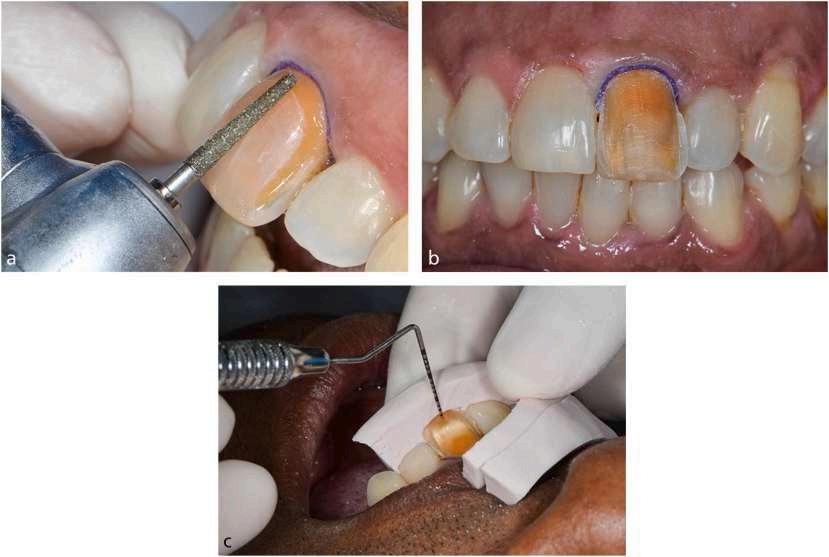
2. A, Veneer cavity preparation with diamond bur. B and C, The final preparation and the check of 1.5 mm wear thickness using a silicone guide and millimeter probe. (Courtesy of Ramos AB, Barquete CG, Pinhao MJB: Can a single-shade resin composite mask darkened teeth in a veneer restoration? A clinical case report. Br Dent J 239:31-33, 2025.)
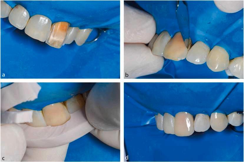
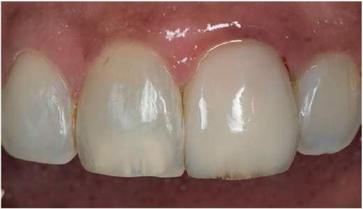
Figure 4. Final veneer restoration with resin blocker and single-shade RBC. (Courtesy of Ramos AB, Barquete CG, Pinhao MJB: Can a singleshade resin composite mask darkened teeth in a veneer restoration? A clinical case report. Br Dent J 239:31-33, 2025.)
Combining a resin blocker and single-shade RBC to address the problem of a discolored anterior tooth achieved satisfactory results. This simplified technique eliminates the color selection step.
Satisfactory aesthetics were achieved using the single-shade RBC and resin blocker to manage the discolored tooth. The patient was satisfied, as was the clinician. This approach avoids the need to choose a color-matched material and relies on altered aesthetics that match the resin’s color to that of the surrounding teeth.
Ramos AB, Barquete CG, Pinhao MJB: Can a single-shade resin composite mask darkened teeth in a veneer restoration? A clinical case report. Br Dent J 239:31-33, 2025
Reprints available from AB Ramos, Integrated Clinical Procedures, Faculty of Dentistry, Rio de Janeiro State Univ (UERJ), Rio de Janeiro, Brazil; e-mail: amandabarretoramoss@gmail.com
Dental caries is the most common chronic childhood condition and occurs at a rate over 5 times that of asthma. The prevalence of early childhood caries (ECC) varies, but those children from marginalized backgrounds are at higher susceptibility with no effect due to race or culture. ECC can cause pain and distress; hinder the child’s ability to eat, speak, and learn; and compromise the child’s development during critical stages. When lesions progress, these children may need care in hospital settings. Silver diamine fluoride (SDF) at a 38% concentration can be used to arrest caries without removing healthy tooth tissue. The fluoride component promotes remineralization, while the silver component offers antimicrobial properties. The “zombie effect” refers to the fact that destroyed bacteria reactivate silver ions that kill visible pathogens, which further enhances the efficacy of SDF. The frequency of applying SDF varies, ranging from weekly, to biannually, to annually, and to multiple times per year. Tribal children in India suffer from significant inequalities, including oral health disparities that are reflected in their high caries prevalence. Research was targeted at this population of preschool
children to assess the efficacy of applying 38% SDF 4 times per year, twice a year, or annually.
The 89 participants were age 2 to 6 years and had a total of 440 carious teeth falling into stage 5 or 6 based on the Modified International Caries Detection and Assessment System (ICDAS) criteria. The outcome assessments were conducted at 6- and 12-month intervals. SDF-treated cavities in teeth with arrested caries were defined as surfaces that had active cavitated caries at baseline that were arrested in subsequent assessments.
Caries lesions were more common in anterior teeth than posterior teeth. The mean number of carious teeth per patient was 5.3. A third of the children had a moderate risk for dental caries.
The mean difference in caries arrests from baseline to the 12month evaluation was 16.8% for the annual group, 56.4% for
the biannual group, and 61.0% for the 4 times per year group. Children age 3 years or older had more arrested caries than those younger than 3 years in all of the groups. Boys had more arrested caries compared to girls in the annual (72.4%) and biannual (81.8%) groups but had fewer arrested caries in the 4 times a year group.
The cessation of caries was highest among anterior teeth compared to posterior teeth. Having more applications per year significantly increased the likelihood that caries arrest would occur.
More frequent applications of 38% SDF was associated with a higher rate of arrested caries. Often teeth that weren’t arrested at 6 months had arrested by the 12-month evaluation.
The 4 times per year frequency of administering SDF showed significant efficacy against caries. However, dental clinicians should tailor the frequency based on individual needs, social determinants, and caries risk factors. Those children at higher caries risk who had poor oral hygiene or low parental education had lower arrest rates. Increasing the frequency of SDF applications can improve these outcomes.
Devan I, Janakiram C, Ramanarayanan V, et al: Effectiveness of silver diamine fluoride application at various frequencies for dental caries cessation among preschool children in India: Randomized controlled trial. Contemp Clin Dent 16:28-35, 2025
Reprints available from C Janakiram, Dept of Public Health Dentistry, Amrita School of Dentistry, Amrita Vishwa Vidyapeetham, Kochi –682 041, Kerala, India; e-mail: sekarcandra@gmail.com
Remote dental monitoring has the potential to improve access to care, reduce the need for additional in-person visits, and manage the unique needs of people with special needs. Intraoral photography and artificial intelligence (AI)−driven tools facilitate remote diagnosis, continuous monitoring, and personalized treatment plans. TestMyTeeth is an AI-powered smartphone application (app) designed to provide at-home dental plaque screening. The smartphone’s camera and advanced image analysis algorithms are used to help capture intraoral photographs and give real-time feedback regarding dental plaque. With this tool, individuals can take proactive steps to maintain their oral health, reduce the burden on dental professionals, and enhance preventive measures. The successful adoption of mobile health apps relies on usability, which is how effectively users can interact with an app to achieve their goals. Tools such as the system usability scale (SUS), heuristic evaluations, and user interviews measure usability and user experiences. High usability tends to increase user satisfaction, engagement, and adherence, all of which are needed for an app to effectively promote health. The usability and user experience of the TestMyTeeth app were investigated.
The 132 adult participants were 75% women and 90% under age 44 years. About 58% had completed university-level education, about 38% held an undergraduate degree, about 21% had a postgraduate degree, about 18% had completed college, and about 14% had attended secondary school. Over half of the participants had experience with dental photography using mobile phones. These individuals used the app and completed an online questionnaire, which included SUS and user experience elements. Usability differences were compared across age, educational level, and sex.
Median SUS score was 56.2, which is in the marginal acceptability category. The app was considered easy to use by about 42% of the sample, and about 38% remained neutral. Nearly half of the participants expressed confidence in using the app, but half indicated they might need technical support to use it effectively. The app was considered unnecessarily complex by more than 51% of the users, and about 49% believed they
needed to learn several things before being able to use it effectively. Almost 55% of the sample agreed or strongly agreed that they were likely to use the app frequently. Most respondents were positive or neutral when asked if they would recommend the app to friends and family.
Some users found the app easy to use, but 21% rated the difficulty level between 8 and 10, with 10 representing the highest level of difficulty. About 24% had no problems with navigation, but about 46% had minor to moderate difficulties. Following instructions was moderately to majorly difficult according to about 44% of participants. Significant challenges were reported for capturing intraoral photographs. About 50% had moderate to major difficulties obtaining photographs of their front teeth, and about 42% had similar challenges obtaining photographs of their back teeth.
Both positive and negative perceptions were reported for the selfmonitoring of oral health. Many saw self-monitoring as valuable for early detection of dental issues and empowering individuals to take responsibility for their own health. In addition, the educational aspect was appreciated, noting that people can check their oral health regularly and reduce unneeded visits to dental professionals.
Concerns were expressed about the accuracy and reliability of self-monitoring without professional input. Other participants thought self-monitoring might make one anxious or lead to a misinterpretation of the symptoms that delayed seeking professional help. User empowerment and accurate guidance and support within the app needed to be balanced.
Several technical challenges were encountered by participants. The most common were difficulty taking clear photographs, especially in the back, and problems with the camera focus, leading to blurry images. Correct positioning and angles for capturing images were unclear and required more time to become adept at these tasks. Some found the app to be user-friendly and straightforward, whereas others found it was time-consuming and required streamlining.
The focus of most suggestions was enhancing the photo-capture experience and overall user interface. Among the ideas offered were to add a selfie mode or enhanced reality guide to help users frame their teeth correctly; make it possible to import or adjust photographs after capture; integrate reminders or notifications; sync with health apps; and provide more instructional videos to improve user engagement and motivation.
The TestMyTeeth app was found to have potential benefits for addressing oral health. The users felt empowered and appreciated the benefits of detecting pathology early. Usability challenges were found with capturing clear intraoral photographs and navigating the app. Because these challenges weren’t confined to a particular age, education level, or sex, it appears that they are widespread and need to be addressed.
Users of AI-empowered devices may face a learning curve before they feel comfortable with their functions. They may also have concerns about the accuracy of self-assessment without professional input. Investigations are needed to identify the positive and negative features of these devices and offer input regarding usability and improvements that would yield a better user experience. The AI algorithms need to be tested for reliability before confidence in these tools can be expected. In addition, professionals should give clear, actionable feedback as part of the process.
Al-Zubaidy D, Innes N, Galloway J, et al: Evaluating user perceptions and usability of an AI-powered smartphone application for at-home dental plaque screening. Br Dent J 239:46-52, 2025
Reprints available from W Al-Yaseen, Cardiff Univ School of Dentistry, School of Dentistry, Univ Hosp Wales, Heath Park, Cardiff, CF14 4XY, UK; e-mail: al-yaseenw1@cardiff.ac.uk
Creating a Safe Haven to Deliver Trauma-informed Dental Care
Psychological trauma is an emotional response to a highly stressful and potentially threatening situation so that an individual feels
unsafe. It can involve a single incident, witnessing a natural disaster, or experiencing repeated events such as domestic violence or war. A survey by the World Health Organization
Table 1. Identified Barriers and Enablers to Trauma-informed Care in Dental Settings
• Uncertainty from staff as to how to practically apply trauma-informed approaches in clinical practice
• The potential limited mental health focus within dental settings, including education on mental health within training programmes
• Dental services having limited education and training in trauma-informed care
• Having limited resources and time for training and the evaluation of implemented trauma-informed initiatives
• Limited flexibility in service delivery to enable a change in clinical practice
• Staff having some ambivalence toward addressing and speaking about psychological trauma with patients
• Service pressures
• Lack of policy endorsement
• Having staff training that provides a shared understanding of psychological trauma across the dental service
• Staff asking about psychological trauma and having organisational structures to support this
• The use of the ‘therapeutic community’ approach to implementation
• Managerial promotion of wellbeing and reflective spaces for staff
• Having political backing and funding for trauma-informed care
• Staff having opportunities for reflection and gaining service-user feedback
• Collaboration between dental services and mental health professionals
(Courtesy of Smith M, O’Donald F, Sevier-Guy L-J, et al: The barriers and enablers to implementing trauma-informed care in dental settings. Br Dent J 239:21-24, 2025.)
of 70,000 people in 24 countries found that about 70% of the participants had experienced at least 1 traumatic life event. Based on these data and other studies, a significant portion of the global population have experienced psychological trauma. Rather than focus on the situation as an individual problem, the trauma response is now handled by looking at the root causes that produced the trauma. Psychological trauma is relevant to dental care because patients may feel unsafe when they are placed in an unfamiliar environment with unfamiliar people who are touching their face and mouth and lean into a close supine position to deliver care. Dental services need to adapt to acknowledge and address the widespread prevalence of psychological trauma in their patient population. Among the elements that must be managed are barriers and enablers of implementing traumainformed approaches, which includes staff training, structural factors, and leadership (Table 1). Practical measures to be implemented by dental practice clinicians or managers offer ways to incorporate this knowledge into daily practice.
A central concern is providing sufficient training to the dental workforce to effectively support patients who have a history of psychological trauma. The education provided should address the effects of psychological trauma and how to minimize the risks for repeat traumatization. The primary challenge is helping the staff understand the practical measures to take in adopting a trauma-informed approach and all that entails. Training for staff members should also include how to manage trauma among health professionals and support those who are struggling. Staff members need to learn how to screen for a history of psychological trauma, promote awareness of the health effects of trauma, and identify risks related to vicarious traumatization.
Compassionate and effective care is needed while safeguarding the mental health and wellbeing of those whose physical and emotional distress is impacting their work. The training should
address 3 broad domains: understanding trauma and its impact, considering how to respond to people dealing with trauma and reduce any barriers hindering care, and being aware of any needed self-care while delivering trauma-informed care.
Frameworks for Implementatio n
Various trauma-informed care models have been developed to manage the specific needs and support changes in health care environments. The effectiveness of these approaches is seen in their ability to guide staff toward more patient-centered responses. One framework, the Attachment, Regulation and Competency (ARC) Framework, focuses on supporting children and adolescents who experience psychological trauma. Within the ARC approach, staff can form strong working relationships with patients and proceed proactively to find ways to reduce distress. Its specific focus includes recognizing and responding to signs of trauma, creating safe environments for care delivery, and building trust through collaborative conversations with patients.
The combination of an effective framework and a well-trained staff facilitates the development of a trauma-informed dental workforce and helps to ensure high-quality, person-centered care as well as improve treatment adherence and prevent burnout among the staff. Cross-professional collaboration to adapt frameworks for dental settings and to determine how to best implement them is required.
It’s vital to have sufficient resources to facilitate change as well as the time needed for implementation. Time constraints can be managed by making minor changes such as offering choices and asking patients if modifications would make them more comfortable and facilitate trauma-sensitive care. Prioritizing innovation in existing health care procedures will help to sustain changes. A ‘therapeutic community’ approach, reflected in the Sanctuary
Model, shares strategies found in frameworks such as ARC. They promote patient safety and trust in the patient-provider relationship. Modifications to the physical environment to promote safety and comfort for the patient; the creation of a positive, hopeful, holistic culture; and better communication in various settings are core values in these models. A drawback is the requirement for additional training for all staff and the model’s resource-intensive approach.
Health care professionals may be unsure about how to initiate a conversation with patients concerning psychological trauma and how to manage their responses. They may also feel concern over possibly unintentionally causing harm to the patient through such conversations. To develop the best approach to patients, health care professionals will require training in supervision and reflective practice developed through regular meetings with an experienced staff member. This can help in fostering any new skills. Collaborations with mental health professionals and the support of the dental practice manager can be developed through training of groups of staff every few months after a comprehensive training session when the trauma-informed care approach is adopted.
Organization leaders who are committed to the change process, set achievable targets, and communicate clearly that traumainformed care initiatives are a priority are most likely to be successful in implementing a trauma-informed approach. Meaningful implementation also requires political backing, funding, and an ongoing ability to evaluate the result. The ability to reflect on practice, develop better approaches based on service-user feedback, and access supervisors to enable trauma-informed care are important components in the process.
Suggestions for the dental professional or dental practice manager who wants to develop a trauma-informed care approach include the following:
A trauma-informed approach to dental care is needed for patients who have a history of sexual abuse or adverse childhood experiences (ACEs), which include potentially traumatic events in childhood that evoke toxic stress responses and breach the child’s sense of safety, security, trust, or bodily integrity (Box 1). Trauma-informed care is based on understanding how the trauma exposure can impair the individual’s neurological, biological, psychological, and social development. Patients with a history of these experiences can react with heightened anxiety,
• Start small and learn about psychological trauma and the impact it can have on dental patients.
• Use models to adapt trauma-informed care to the existing clinical practice.
• Consider training needs, respond to patients with a trauma history, plan how to create a safe clinical environment, and plan for staff self-care enhancement.
• Ensure that the team leader clearly communicates the importance of trauma-informed care and the value of the model being used.
• Respond to patient feedback and make adjustments.
• Use supervision and reflective practice initiatives to support the process.
Psychological trauma is a widespread problem and will be seen in dental practices. The response of dental practitioners to individuals with a history of psychological trauma should be well-planned and compassionate. Overcoming barriers and enablers, increasing knowledge for the dental workforce, and ensuring that the leader is vocally supportive and committed to the effort form a good foundation for achieving a high-quality person-centered approach to dental treatment.
Smith M, O’Donald F, Sevier-Guy L-J, et al: The barriers and enablers to implementing trauma-informed care in dental settings. Br Dent J 239:21-24, 2025
Reprints available from M Smith, Dept of Special Care Dentistry, Dundee Dental Hosp, NHS Tayside, Dundee, Scotland, UK; e-mail: Molly.Smith@nhs.scot
fear, and mistrust in dental settings, which are especially reminiscent of abusive situations. Because these reactions compromise routine dental care, dental professionals need to understand how trauma affects patients and be prepared with strategies to create a supportive and nonthreatening environment for the patient. Dental practitioners should be aware of the responses that might be exhibited by people who have a history of abuse, take steps to create a trauma-informed approach to care, be sensitive to patients who qualify for a special care dentistry service, and be open to receive a patient’s disclosure of abuse.
1.
• Physical abuse
• Sexual abuse
• Emotional abuse
• Living with someone who abuses drugs
• Living with some who abuses alcohol
• Exposure to domestic violence
• Living with someone who has gone to prison
• Living with someone with serious mental illness
• Losing a parent through death, divorce or abandonment
(Courtesy of Jackson M, McLernon M: Beyond the chair: Approaching dental care for survivors of trauma. Br Dent J 238:858-862, 2025.)
ACEs can alter a child’s gene expression, brain function, and immune responses and cause chronic health issues, mental health challenges, and risk behaviors during adulthood. The more traumatic experiences encountered, the more extreme the impact they have on the individual’s wellbeing. Post-traumatic stress disorder (PTSD), anxiety, depression, and hypersensitive stress response are commonly seen in abuse survivors.
These individuals can also seek comfort and relief of stress by engaging in self-destructive behaviors. This can include smoking, consuming excess alcohol, and using illicit drugs. These are also related to poor health outcomes, dental disease, and challenges for the dental care provider. Dentists may be reluctant to treat these patients because of their lack of understanding why these individuals have sporadic attendance and tend to cancel followup appointments.
The dental setting can be especially triggering for these patients because of its parallels to the sites of sexual abuse. The patient is supine and expected to cooperate, with little control over the situation. Pain is anticipated. The dentist is in close to the patient and is using sharp instruments. As a result, patients may avoid dental care, suffer severe anxiety during appointments, have extreme reactions, exhibit a pronounced gag reflex, and neglect oral hygiene, which can result in dental disease. They can be trapped in a fear-avoidance cycle, wherein the avoidance of dental care provides immediate relief but increased long-term anxiety by not confronting the problem.
A natural trauma response can occur in which the body recognizes a threat, which prompts the brain and autonomic nervous system to release hormones such as adrenaline and cortisol. These produce physical changes to prepare against the threat, with a rapid increase in heart rate, respiration, and blood pressure. Although many trauma survivors won’t disclose their history of sexual abuse or ACEs to a health care professional, behaviors such as hypersensitive gag reflex, extreme panic reactions, intense fear of objects in the mouth, or difficulty with physical touch are commonly seen in patients who have been abused (Box 2).
Box 2. Different Behaviours a Patient with Previous Trauma May Exhibit
• Intense fear/extreme panic in dental setting
• Hypersensitive gag reflex
• Unable to lie back in chair
• Requests the door is left open
• Dental DIY (do-it-yourself) to avoid the dentist
• Misusing substances (alcohol, drugs)
• Eating disorders (binge eating, bulimia)
• Smell of latex or strong cologne causing fear
• Shallow breathing, sweating, pallor
• Shock, unresponsive, fainting
• Not able to have dental team stood behind them
• Requests a female dentist
• Dental neglect
• Anger, defensiveness, confrontational
• Does not accept patient bib around neck
(Courtesy of Jackson M, McLernon M: Beyond the chair: Approaching dental care for survivors of trauma. Br Dent J 238:858-862, 2025.)
A trauma-informed approach recognizes the prevalence and impact of trauma, integrates this knowledge into practice, and avoids things that may re-traumatize the patient. The goal is to build trust with the patient so that it’s possible to deliver the dental care the patient needs. The key principles of this approach are safety, trust, and empowerment (Box 3). Among the elements in the trauma-informed approach are training and awareness, building trust and rapport, creating a safe, comfortable environment, communicating clearly and empathetically, empowering the patient, and practicing continuity of care.
Box 3. Key Management Strategies to Help Treat Patients with a History of Abuse
• Stop signal
• Same dentist/dental nurse
• Do not lie flat
• Consider leaving the door open
• Kind, compassionate, non-judgemental
• No treatment at first appointment
• Tell-show-do approach
• Build trust and rapport
• Background music/music through headphones
• Aromatherapy with essential oils
• Breathing techniques
• Weighted blankets, pillows
(Courtesy of Jackson M, McLernon M: Beyond the chair: Approaching dental care for survivors of trauma. Br Dent J 238:858-862, 2025.)
The entire dental team should be educated to understand how trauma manifests in the dental setting, skills for recognizing signs of trauma, effective communication techniques, and strategies for managing patients who are anxious or fearful. These components help to minimize barriers to accessing care and reduce interventions that could trigger memories of the trauma.
Educational resources are available to help health and social care professionals support patients. Some include worksheets, selfassessments, and facilitator guides or are available through elearning sessions. In adopting trauma-informed care, educated and trained staff members can improve interactions with patients and emphasize the importance of empathy, patience, and empowerment of individuals who have known trauma.
Patients with histories of sexual abuse or ACEs may feel extremely vulnerable and view the dental chair as a place of danger. The practitioner should build trust by creating a respectful, nonjudgmental environment where the dental staff takes time to listen to any concerns the patient has, validate the patient’s feelings, and give reassurance that providing a comfortable and safe environment is essential. Dental nurses may be seen as less threatening and act as a security blanket for patients. Dentists and dental nurses should create a strong working relationship to build trust in the patient.
It may be helpful to schedule an initial appointment without procedures to build rapport with the patient. Patients can be allowed to become familiar with the dentist and the dental environment, share any concerns they have, and work with the dental staff to develop a plan collaboratively that meets specific needs. Because of time constraints and the units of dental activity system, this can be challenging. An alternative is to ask questions during the appointment scheduling, such as whether the patient finds the dental environment difficult or if the dentist should be aware of anything before the appointment. It’s also possible to use a wellbeing questionnaire for those who have experienced trauma where they can share their needs and triggers before the first visit.
How comfortable and safe a patient feels depends to a large extent on the physical and emotional environment of the dental facility. Dental offices should be calming and welcoming, with soft lighting, soothing colors, and relaxing background music to soothe the patient’s emotions while waiting for the appointment. Aromatherapy may also be an effective, low-cost, low-maintenance technique to induce relaxation.
Patients can be provided with headphones to listen to music and mask dental instrument noises. Weighted blankets can be comfortable and reduce stress. Patients can be provided with a signal to use when they need a break during treatment, which also empowers them with a sense of control. Keeping the door to the operatory ajar or allowing the patient to sit more upright can also give a feeling of safety.
Clear, simple language should be used to explain procedures and avoid jargon that can intimidate patients. The dentist should
explain what will happen before beginning any procedure, moving step by step through the process. Patients should be encouraged to ask questions or share concerns along the way. The ‘tellshow-do’ technique can help with anxious patients. Describing any tools, smells, or sensations that the patient may encounter reduces the risk of panic. The dentist should also check in regularly during the procedure, asking how the patient feels, if he or she needs a break, or if he or she is comfortable.
Nonverbal communication is also important. The dentist should maintain an open posture and stand or sit at the same level as the patient. He or she should be mindful of actions taken when around patients with a history of abuse, such as breathing heavily in close proximity patients or wearing certain scents.
Feeling powerless and having low self-worth are common among trauma survivors. Empowering patients to have dental treatment increases their confidence with dental care and improves their self-respect and confidence. For patients with a history of sexual abuse or ACEs, feeling out of control can trigger anxiety, panic, and flashbacks. Patients should be involved in decision-making regarding their care, including letting them choose appointment timing, decide on the pace of the procedure, and select what treatment they want to have done at each appointment. They should be informed of their right to decline any treatment. These actions reduce their feelings of helplessness and build a sense of trust and safety.
Whenever possible, the same dentist and dental nurse should be scheduled for each visit. This builds a relationship of trust and reduces anxiety. It can also alleviate stress related to the dental visits, which can help patients follow through with the needed treatments.
Trauma can severely hinder some patients’ ability to receive dental treatment. In these cases, they may need to be seen by a special care dentistry service. These practitioners are extensively trained to understand and recognize the impact of psychological issues in patients who have complex needs. They can also institute pharmacological management techniques to help patients endure the needed treatment. Collaboration with mental health professionals can be helpful. If cognitive behavior therapy is used, patients may be better able to accept dental care in general dental facilities.
Pronounced gag reflexes are a coping strategy that can lead to distress and avoidance of dental care. Simple management strategies such as acupressure, placing salt on the tongue, or guided breathing may be able to temporarily reduce the gag reflex.
Referral to a specialist is warranted if these methods are ineffective.
The provision of prosthetic replacements for patients with hypersensitive gag reflexes requires a patient-centered, flexible approach. Fixed prostheses for anterior teeth are better tolerated than removable dentures. Replacing only anterior teeth with the shortened dental arch concept may be the most appropriate option, since posterior replacements tend to be more likely to trigger gagging and fail to achieve the treatment.
Once patients feel secure and comfortable with the dental team, they may disclose a history of abuse. Patients may feel they want to explain why they have reacted in a certain way. Dental professionals aren’t trained therapists and should refrain from offering advice or counseling but they can discuss how to make the patient more comfortable and signpost them to suitable trained professionals as desired. Local charities may be able to connect patients to appropriate people to access resources that may be helpful.
A comprehensive, trauma-informed approach is appropriate when dealing with patients who have been sexually abused or suffered ACEs. The creation of a safe, supportive environment and support for patient empowerment help in making patients feel more confident and relaxed. Dental teams should be given appropriate training and empathetic care instruction to help patients with histories of trauma maintain their oral health.
Jackson M, McLernon M: Beyond the chair: Approaching dental care for survivors of trauma. Br Dent J 238:858-862, 2025
Reprints available from M Jackson, Community Dental Services, Harrogate and District Foundation Trust, UK; e-mail: maria. jackson22@nhs.net
The application of adhesive dentistry has allowed for alternative restorative techniques to be used, which has reduced the use of crowns from past levels, but they remain relevant for heavily restored teeth and to replace other crowns. Crowns are chosen as replacements to achieve better aesthetics, manage recurrent caries, and deal with a lost or fractured restoration. Clinicians must make several choices when providing a crown, including margin type and position. These are generally selected based on the material used, aesthetics, and existing tooth damage and restorations. It may be necessary to prepare a new finish line located more apically than the previous finish line. Because the crown will likely finish on the root surface, conservation of tooth structure is important. The amount of coronal tooth structure removed will be dictated by the depth of the crown margin preparation. The use of zirconia provides aesthetic crowns that are strong even in thin sections. In choosing the least invasive preparation, a knife-edge crown on a vertical margin requires less tooth reduction than horizontal margin preparations. Vertical
margins create crowns with very thin marginal areas and require a strong material like zirconia that is resistant to fracture even in thin sections. An audit was done to evaluate the clinical outcomes of 73 teeth prepared with a vertical finish line and restored with a zirconia knife-edge crown placed by a general dentist.
The 73 crowns were fabricated using computer-aided design/ computer-aided manufacturing (CAD/CAM), with some being entirely monolithic zirconia and others that were in a more visible position veneered with porcelain to improve aesthetics (Figure 1). During return evaluations and hygiene appointments, usually every 6 months, basic periodontal examination (BPE) scores, periodontal pocketing results, and bleeding charts were done. Bite-wing radiographs were obtained at intervals appropriate for each patient’s caries risk profile. Visual evaluations were conducted to check for chips, cracks, or marginal gaps. The crowned teeth were compared with a control tooth in the same patient. The audit
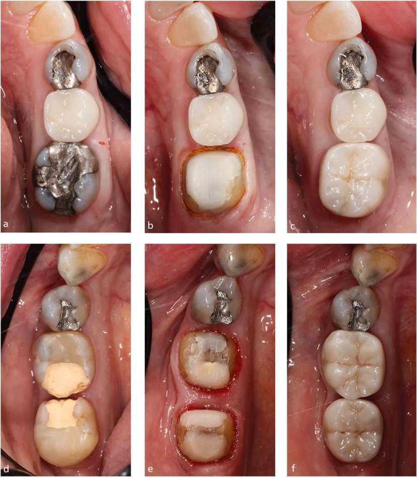
performed assessments of crown survival, marginal integrity, endodontic health, and periodontal health at the follow-up examinations.
Of the 73 crowns, 45 were replacement crowns and 28 were new crowns. Sixty-two teeth had impressions recorded at the same visit as the preparation. The mean follow-up was 62 months, with a range of 12 to 93 months. Eight teeth had complications, with 6 restorative or endodontic complications and 2 resulting in tooth extraction. One tooth suffered a fracture. Three crowns
had to be remade during the audit period, with 1 during the fit stage when the buccal wall fractured during try-in and 2 at later stages.
Thirty-seven teeth had root canal treatment before receiving a crown, with 1 having repeat root canal treatment after the crown had been placed. Thirty-six of the teeth had no previous root treatment and didn’t require subsequent root treatment during the audit period.
Caries developed at the margin of 1 tooth and was detected during a routine examination at 56 months. The patient chose to have the tooth extracted.
Mean follow-up time for radiographs was 55 months, with a range of 1 to 87 months. Alveolar bone level did not differ from previous views in 67 teeth. Two in the same patient with unstable periodontitis showed increased interproximal bone loss on the radiographs obtained after 69 months.
Seventy teeth had follow-up BPE scores, with periodontal pocket charting for teeth with pockets exceeding 3.5 mm. Seven of these patients had periodontal pockets recorded before or after receiving the vertical preparation. Patients showing increased pocketing underwent periodontal maintenance therapy, and periodontitis is stable in all but 1 patient.
Bleeding score was calculated for each tooth from records taken before and after vertical preparation for a crown. Comparison with a control tooth showed 14 teeth had more bleeding on the crowned tooth, 9 had less bleeding, and 46 teeth had the same level on crowned and control teeth. Overall, the mean bleeding scores for the crowned and control teeth were not different.
Positive outcomes were obtained for posterior teeth prepared with a vertical preparation for zirconia crowns over a mean follow-up period of 62 months.
Vertical preparation is now possible with zirconia knife-edge margins. Natural tooth structure is conserved compared to horizontal margin designs. Clinicians must be aware that vertical margin preparation is technique-sensitive, requiring special care when removing the crown bulbosity to ensure that no undercuts are left below the survey line. In addition, impressions can be challenging because there is no visible margin to inspect. The key to soft tissue management is placing the margin on the provisional crown correctly. Care must be taken to seat the restoration slowly and allow excess cement to flow out. Having a skilled technician who understands these concepts is important to achieve a stable margin and emergence profile.
Stack J, Millar B: Six-year analysis of posterior zirconia crowns with vertical margin preparations. Br Dent J 238:807-812, 2025
Reprints available from B Millar, Faculty of Dentistry, Oral and Craniofacial Sciences, King’s College London, UK; e-mail: brian.millar@kcl.ac.uk
Edentulism and partial edentulism are less common in Sweden than in the past, with today’s patients choosing not to have teeth extracted or retain a compromised tooth without investigating their options. A tooth with necrotic pulp can benefit from root canal treatment (RCT) before restoration with an indirect post and core (PC). The Swedish National Board of Health and Welfare recommends RCT because of its low costs in relation to the gained effect. Survival rates with this approach reach 86% to 93%. However, RCT is technically challenging and many general dental practitioners don’t have sufficient expertise to perform it. As a result, about 20% of the RCTs in molars have been extracted before treatment could be completed with a permanent root filling. Clinicians' preoperative tooth survival estimation should be carefully considered before beginning RCT. An alternative is tooth extraction and replacement by an implantsupported single crown (ISSC). These implant-supported restorations are more common now in Sweden and offer high survival rates. The downside involves the biological and technical complications and the loss of implants that can occur. The ISSC also has high initial costs, so the RCT approach appears more cost effective. Clinical decision analysis uses expected utility theory (EUT), in which the problem to be solved is turned into a model such as a decision tree. The model visualizes the different outcomes the situation can have. Outcome analysis includes both costs and probabilities, which usually come from published high-quality scientific literature. The costs and probabilities are used to calculate the expected costs for the outcomes, and the option with the lowest expected cost is considered the most costeffective. A decision analytic approach was used to compare the cost-effectiveness of RCT and PC versus extraction and an ISSC in a patient with a severely decayed first mandibular molar with a necrotic pulp in the context of Swedish general dental practice.
There was no actual patient in this model-based cost-effectiveness study. Costs were elicited from the Swedish Dental and Pharmaceutical Benefits Agency (TLV). Probabilities were derived from articles on tooth and implant survival and losses in Swedish general dentistry. Two decision trees were
developed, with decision tree I tracking an RCT and the intention to place a PC. Decision tree II tracked extraction of the tooth and installation of an implant (EI) to be finished with an ISSC. The outcome was controlled at 2 different time points— 1 year and 5 years postoperatively. Sensitivity analyses were done.
For decision tree 1, the probability of a 5-year survival of RCT plus PC was 70%. The corresponding probability for EI and ISSC was 98%. The cost for the various branches after choosing RCT varied from 15,860 SEK to 33,105 SEK. Expected cost for choosing RCT was 17,400 SEK, whereas the expected costs for choosing EI were 18,800 SEK.
Decision Tree II
Probability of a 5-year survival of RCT plus PC was 69%, with that for EI and ISCC being 98%. The RCT branch costs vary from 15,860 SEK to 54,290 SEK, whereas the cost for choosing RCT was 19,500 SEK and the cost of choosing EI was 18,800 SEK.
Sensitivity Analysis
Based on decision tree I, the lowest estimated cost for RCT was 15,860 SEK and the highest expected cost was 33,105 SEK. All the possible expected costs of RCT were placed on a line between these 2 points. The expected cost of ISSC was 18,800 SEK. For RCT to cost less than that, the probability value for RCT would have to be at least 83%.
Based on decision tree II, the lowest cost for RCT was 15,860 SEK, but the highest cost was 54,290 SEK. For RCT to be more cost-effective than ISSC, the probability value of RCT survival would have to be at least 93%.
From a cost-effectiveness perspective, the 2 approaches were nearly equivalent. However, the probability of tooth survival after RCT and an indirect restoration must be 83% to 93% to be more cost-effective than an extraction and ISSC.
Many assumptions and estimates are made to keep the analysis of the 2 approaches a simplified version of reality. Although many of these assumptions seem realistic, even a small deterioration in the conditions for RCT and PC and the resulting prognosis may change the expected cost and favor the use of extraction and ISSC as a more cost-effective treatment.
Savolainen N, Frisk F, Kvist T: Is root canal treatment and an indirect coronal restoration of a mandibular first molar costeffective compared to extraction and an implant-supported crown? A decision analytic approach. Acta Odontol Scand 84:95103, 2025
Reprints available from N Savolainen, Gislaved Folktandv�ard, Norra L� anggatan 25, Box 134, 332 38 Gislaved, Sweden; email: nikki.savolainen@rjl.se
TMDs are musculoskeletal conditions that affect the TMJ, masticatory system, and related tissues. The etiology of TMDs is unclear but factors that may contribute include occlusal conditions, trauma, emotional stress, deep source of pain, and parafunctional activity. Masticatory muscles or the TMJ can be treated with medical approaches, physiotherapy, surgical interventions, trigger point injections, acupuncture, occlusal splints (OSs), and Botox injections. The OS devices relieve pain and can address restricted mouth opening. Among the OS devices most often used are the Tanner apparatus, Fox apparatus, Michigan splint, centric relation apparatus, anterior reposition splints, and anterior bite plates. Botulinum toxin A (BTX) is a neurotoxin obtained from Clostridium botulinum that interrupts the neurotransmission of acetylcholine to the muscles and is delivered via an injection. It’s used to treat various medical conditions that involve muscle spasms and hyperactivity but is also used for cosmetic purposes. The effectiveness of the OS and BTX treatments postoperatively was investigated, noting changes in chronic pain conditions and the range of maximum mouth opening (MMO).
A review of the PubMed, Web of Science, and Scopus databases as well as the Google Scholar search engine identified 6 studies that offered information on the graded chronic pain scale (GCPS) scores and MMO parameters with OS and BTX treatments for TMD.
The publications that evaluated GCPS had follow-ups in the sixth month after treatment. Improvement criteria were based on the decrease in GCPS scores from levels II, III, and IV to levels 0 or 1. No pain or interference caused by TMD is present in level 0, and only low pain levels are seen in level 1. Level II has a high degree of pain but no interference, level III has moderate interference, and level IV has severe interference. The random effect model had an effect size of 0.673 in favor of OS. The GCPS scores of OS and BTX didn’t differ significantly.
Publications reporting MMO amounts included first-week, firstmonth, second-month, third-month, sixth-month, and twelfthmonth follow-ups. An effect size of 0.293 was found in favor of BTX but no significant difference in MMO was seen between the OS and BTX groups.
The amounts of MMO were significantly higher in the BTX group in studies focused on the first week and in the OS group in studies focused on the third month. No significant differences were noted for other follow-up periods.
More effective strategies may be formulated by recognizing how OS and BTX manage TMD symptoms. BTX provides better functional improvement in the short term and OS is more
effective in the long term. OS treatment reduces muscle hyperactivity and joint loading and may have a more gradual but longer lasting effect. Effectiveness may also change over time. Both OS and BTX had similar long-term effects on MMO.
OS applications and TBX injections provide similar results but work through different mechanisms and may function better in different situations. Applying OS during the night can be uncomfortable for patients. BTX may be faster and produce more pronounced effects, but it’s a more invasive method and side effects can occur in some patients. Factors to be considered when selecting a treatment modality include pain level, lifestyle, general health status, and tolerance to treatment. Treatment plans should take into account the patient’s condition and needs.
O IS ¸ isa� g, Atasoy H, Yıldız S: Comparison of the effects of occlusal splint and Botox injections on the amount of mouth opening and chronic pain in individuals with temporomandibular disorders: A systematic review and meta-analysis. Austral Dent J 70:132-140, 2025
Reprints available from O IS ¸ isa� g, Dept of Prosthodontics, Faculty of Dentistry, Afyonkarahisar Health Sciences Univ, Afyonkarahisar, Turkey; e-mail: ozer.isisag@afsu.edu.tr
Resin composite (RC) is often used for direct restorations because of its clinical versatility and adequate physical, mechanical, biological, and esthetic properties. Because RC direct restorations are technique dependent and sensitive, the skill of the operator is a significant factor in the success and survival of the restoration. Challenges to these restorations are achieving proper acid etching, dentin moisture control, complete solvent removal, correct adhesive system application, and the formation of a stable, homogeneous hybrid layer. Modern RCs have appropriate mechanical and biological properties for clinical use, but if there is high occlusal stress, restorations may fracture or wear over time. Long-term durability may be less than acceptable. Self-adhesive RCs may provide the needed clinical behavior to provide the desired aesthetic, functional, and biological outcomes. A comparison of the clinical performance of direct restorations using self-adhesive RCs with conventional or bulk-fill RCs in permanent teeth was done.
The Medline/PubMed, Web of Science, Scopus, Embase, LILACS, Cochrane Library, Base, Google Scholar, and OpenGray
databases were searched for randomized clinical trials that assess the clinical performance of direct restorations with selfadhesive RC and compare the conventional or bulk-fill RC restorations in permanent teeth. Twelve studies (15 reports) covering 356 participants (age 6 to 79 years) with 794 restorations were evaluated qualitatively and quantitatively. The follow-up period was from immediately after the restoration to 60 months later.
Of the 794 cavities evaluated, 396 were restored with conventional/bulk-fill RC and 398 were restored using selfadhesive RC. Curing times ranged from 20 to 40 sec and irradiance values were between 800 and 1250 mW/cm2.
Both the conventional/bulk-fill RC and self-adhesive RC groups have failures over time. The 3 distinct groups of failures were aesthetic failures including marginal staining and color stability; functional failures such as fracture/retention, marginal adaptation, and wear; and biological failures, which includes postoperative sensitivity and caries, erosion, and abrasion recurrence.
No significant differences were found between cavities restored with conventional/bulk-fill RC and those restored with selfadhesive RC. This meant that marginal staining outcome, color stability outcome, fracture/retention outcome, or marginal adaptation outcome did not differ significantly between the 2 groups. The wear outcome, postoperative sensitivity outcome, and recurrence of caries, erosion, or abrasion outcome could not be evaluated for effects estimates.
The self-adhesive RC direct restorations and conventional or bulk-fill RCs showed similar clinical performance over followup periods from 6 to 48 months.
The clinical performance of direct restorations placed with self-adhesive RCs is comparable to that of restorations placed with conventional/bulk-fill RCs over a range of follow-up periods, various cavity types or locations, and the outcomes assessed. Both materials degraded over time and suffered failures.
Besegato JF, Freire A, Zaniboni JF, et al: Clinical performance of self-adhesive resin composite direct restorations in permanent teeth: A systematic review and meta-analysis. Clin Oral Invest 29:375, 2025
Reprints available from JF Besegato, School of Dentistry, Federal Univ of Mato Grosso do Sul (UFMS), Campo Grande, MS, Brazil; e-mail: joao.besegato@ufms.br
The use of adhesive systems in dentistry was crucial in allowing a more conservative technique in restorative dentistry based on the chemical adhesion possible between the dental substrate and the restorative material. Adhesives are classified based on their application as etch-and-rinse (ER) or self-etch (SE) materials. Universal adhesives (UAs) are no-mix, single-bottle adhesives useful for ER, SE, and selective enamel etching (EE) bonding approaches. Some can also act as silane agents for ceramics and metals and in immediate dentin sealing. The clinically relevant indicators of possible failure in marginal seal and adhesion of resin composite restorations are retention, marginal staining, and postoperative sensitivity. Marginal staining can progress to failure of the seal that allows for bacterial access and secondary caries. Repair or replacement may be required, possibly causing an increased loss of healthy tooth structure. Postoperative sensitivity compromises patient comfort and may indicate either an adhesive or procedural failure. Each type of adhesive has a unique composition, and the clinical outcomes can be different. An investigation into whether a specific adhesive type or strategy achieves better restoration longevity and clinical practicality or is associated with less technical sensitivity was undertaken.
A review of the PubMed, Web of Science, Embase, Scopus, and Cochrane databases identified 15 randomized clinical trials (RCTs) appropriate for analysis. These RCTs assessed the clinical performance of different UAs applied in different modes to direct dental resin composite restorations and compared retention, marginal staining, and postoperative sensitivity with ER or SE materials. Follow-up ranged from 6 months to 4 years.
Five different ER and 4 different SE adhesives were used, along with 11 different resin composites. A meta-analysis of all 15 studies was done, with 14 assessing retention failure, 15 marginal staining, and 12 postoperative sensitivity. For retention failure and marginal staining, over follow-up periods from 6 to 48 months, 3 types of adhesives were used across 3 protocols. The use of UAs and control ER or SE adhesives showed no statistical differences in retention failure or marginal staining. Postoperative sensitivity was evaluated over follow-up periods from 6 to 36 months, with no significant differences observed between the 3 adhesive types and protocols.
UA systems had similar clinical performance to the ER and SE adhesives with respect to retention, marv ginal staining, and postoperative sensitivity for up to 4 years.
Dentists need to consider not only the adhesive's effectiveness but also its clinical practicality. Any of the 3 adhesive strategies or adhesive types can be effective. A good rule of thumb for professionals is to select the technique with which they are most proficient as long as all the choices are appropriate.
Patias MP, Fernandes-e-Silva P, Carreno NLV, et al: Comparative clinical performance of universal adhesives versus etch-and-rinse and self-etch adhesives: A meta-analysis. Clin Oral Invest 29:352, 2025
Reprints available from WLO Da Rosa, Dept of Restorative Dentistry, Federal Univ of Pelotas, Rua Gonçalves Chaves, 457, Rm 503, Pelotas 96015-560, RS, Brazil; darosa.wlo@gmail.com
A common procedure done in oral and maxillofacial surgery is lower third molar surgery. Often patients develop postoperative complications such as pain, trismus, swelling, and infection, which are of limited duration but still create discomfort and compromise daily living. Risk factors for developing inflammatory postoperative complications after lower third molar surgery can be related to the amount of plaque at the surgical site before and after surgery. Mechanical dental plaque removal may avoid the development of infectious or inflammatory complications and patient discomfort. Good oral hygiene before lower third molar surgery to prevent complications is not, however, supported by a significant level of evidence. The association between preoperative oral hygiene status and the occurrence of infectious or inflammatory complications, perceived pain, and edema after lower third molar surgery was investigated, with the goal of improving the success rate of this surgery.
The 222 patients were drawn from 3 studies of adults who had undergone lower third molar extraction. Full mouth plaque score (FMPS) was chosen as a parameter for oral hygiene status and was combined with demographics, anatomic factors, and surgical variables to serve as predictor variables. Study outcomes
were postoperative inflammatory complications, pain, swelling, and the use of antibiotics.
Continuous FMPS was significantly and independently related to the development of inflammatory complications and antibiotic use. Dichotomized FMPS was associated with antibiotic use only. FMPS was not independently related to postoperative pain, but showed some interaction with surgery. Thus the impact of high FMPS on the development of severe pain depended on the complexity and duration of the surgery. FMPS and postoperative swelling severity were both independently and strongly related. Continuous FMPS was also related to the use of antibiotics, but dichotomized FMPS wasn't. Overall, FMPS had moderate accuracy as a predictive variable for postoperative inflammation and the need for antibiotics.
Evaluating the preoperative FMPS may be a way to estimate the risk for causing patient discomfort and inflammatory complications after lower third molar surgery. Patients with high FMPS may need to have surgery rescheduled to allow time to perform added preoperative oral hygiene. This may be able to improve treatment outcomes and reduce the need for antibiotics.
Having significant bacteria present at the surgical site in patients scheduled for lower third molar surgery can increase the release of toxins and chemical mediators related to postoperative inflammation. Patients would be more likely to develop complications and require antibiotics to fight the infection. Using chlorhexidinebased rinses before extraction may reduce the incidence of alveolar osteitis by lowering the concentration of bacteria present. Professional oral hygiene and oral hygiene counseling may be an alternative, especially if long-term, complex surgery is anticipated.
Roberto P, Luisa LM, Stefano P: Is pre-operative full mouth plaque score related to inflammatory complications in lower third molar surgery? Cohort study. Clin Oral Invest 29:364, 2025
Reprints available from Roberto P, Dept of Odontostomatological and Maxillo Facial Sciences, Sapienza Univ of Rome, Rome, Italy; e-mail: roberto.pippi@uniroma1.it
Deep dental overbites are excessive vertical overlaps of maxillary incisors over mandibular incisors in centric occlusion. An overbite greater than 5 mm is considered severe. Both dental and skeletal etiologies can contribute to deep overbite malocclusion, creating an increased curve of Spee, elongated maxillary and mandibular incisors, excessive overjet, vertical facial type, and decreased gonial angle. Skeletal deep bite is caused by craniofacial skeletal discrepancies, whereas dental deep bite is caused by abnormal tooth eruption patterns. Correcting deep overbite is challenging but needed in orthodontic treatment to avoid sequelae such as TMJ disorders, gingival trauma, mandibular incisor wear, and impaired mastication. Treatment must incorporate the etiology of the malocclusion, patient age, growth potential, skeletal pattern, and aesthetics. Full-fixed appliance (FFA) treatment involves the use of an anterior bite plane plus posterior vertical elastics to facilitate bite opening and posterior teeth extrusion. A rectangular stainless steel arch wire with a reversed curve of Spee is recommended. The FFA approach has proved to be reliable in achieving complex tooth movements. Clear aligners (CAs) have advanced over the last 3 decades in their ability to treat mild cases of crowding, spacing, and open bite malocclusion. The evidence to support their effectiveness in treating severe dentoalveolar deep bites and skeletal deep bite cases is inconclusive. CA outcomes can be unpredictable, especially for discrepancies between planned and achieved outcomes in vertical and inclination movements. The available evidence on the
treatment of deep overbite malocclusion was reviewed, noting the effectiveness and accuracy achieved using CAs.
The electronic search was done in the PubMed, Embase via Elsevier, and Cochrane Central Register of Controlled Trials via Ovid as well as for grey literature through ProQuest Dissertations and Theses Global and the International Association for Dental Research (IADR) Abstract Archive. Eighteen studies on CA treatment for deep bite correction were selected, with 8 evaluating the effectiveness of CAs in treating deep bite, 8 studying the accuracy of deep bite correction with CAs, and 2 evaluating both outcomes.
The CA treatment duration ranged from 13.77 to 40 months. All of the studies measured dental overbite, with 8 using lateral cephalograms, 1 using digital models, and 1 using both. Three studies also assessed skeletal overbite changes with CA. Nine studies reported a duration of 1 to 2 weeks per aligner and 1 indicated a change was made every 3 to 4 days. Just 8 studies used precision bite ramps.
Bite depth reduction ranged from −0.09 to 4.07 mm. In 7 studies there was significant improvement in dental bite depth, but 3 reported no significant differences. The
treatment outcomes differed and were not appropriate for a meta-analysis.
Ten studies evaluated the predicted versus the achieved deep bite correction with CAs. The mean percent overbite reduction ranged from 27% to 48.88%. Differences in treatment protocols, patient responses, and possibly compliance were noted. This variability did not lend itself to meta-analysis.
The primary concerns with the findings dealt with confounding factors, participant selection, and deviations from intended interventions. CAs may be effective for mild to moderate deep bite correction but deep bite correction tended to be fraught with variations in the results. Most of the effectiveness studies failed to provide detailed information about the orthodontic treatment protocols used. There were areas of potential bias because variations in treatment techniques and mechanics influence outcomes and conceal the specific effects of the aligner system alone.
CAs were effective for mild to moderate dentoalveolar deep bites but their outcomes are unpredictable. Often the planned corrections fall short, which requires overcorrection or refinements to achieve the desired results. Currently CAs cannot be recommended as a method comparable to FFA. Future research is needed to provide more comprehensive insights regarding the efficacy and accuracy of CAs for deep bite correction. The full spectrum of dental and skeletal malocclusion severity should be addressed along with the impact of severity on treatment outcomes. Including control groups is essential to drawing meaningful conclusions.
Husain F, Riccardi J, Alletto L, et al: Effectiveness and accuracy of clear aligners in treatment of deep bite: A systematic review. Clin Oral Invest 29:306, 2025
Reprints available from F Husain, Dept of Developmental and Preventive Sciences, College of Dentistry, Kuwait Univ, PO Box 24923, Safat 13110, Kuwait; e-mail: fatemah.husain@ku.edu.kw
Dental implants are common choices to replace missing teeth and offer functional and aesthetic advantages. Implant length plays a critical role, with longer implants traditionally seen as offering better primary stability and enhanced success rates. However, advances in implant designs and surface characteristics have broadened the applications for short dental implants, especially for patients with limited bone height. Comparisons of long and short implants have suffered from inconsistencies in study design, patient populations, and outcome measures. The outcomes of currently available systematic reviews and metaanalyses were evaluated to determine the effect of implant length on failure or success rates for dental implants in natural bone.
The search was conducted in the Cochrane Database for Systematic Review, PubMed, Scopus, and Web of Science databases as well as WorldCat and Google Scholar for grey literature. Three systematic reviews were included in an umbrella review.
One review was designed to assess how implant length affects failure rate by comparing short implants (less than 10 mm) to long implants (10 mm or longer). The short implants had a significantly higher risk of failure than the long implants. There was a reduced possibility of implant failure with longer implants when implants of various lengths were compared. The duration of the follow-up period had no significant impact on risk ratio for implant failure between long and short implants. Followup for a maximum of 7 years showed an increase of 0.6 in risk ratio for every added month of follow-up. The conclusion was that while implant length is a significant factor in implant failure, it’s just one of many contributing factors. Patient’s overall health and treatment planning also play critical roles in the success of dental implants.
The second review examined the impact of implant length on survival rate for rough-surface dental implants placed in nonaugmented posterior regions in partially edentulous patients. The length of the dental implant had a marginally nonsignificant influence on survival rate. For each added 1 mm of length, the
survival rate was improved by 0.42 percentage points. In the maxilla, implant length had a statistically significant impact on survival rate. For every added 1 mm of length, survival rate improved by 0.68 percentage points. In the mandible, implant length had no significant impact on survival rate. The researchers’ conclusion was that when sufficient native bone is available, longer or regular-size dental implants are preferred over short implants in the posterior maxilla. In the posterior mandible, short implants can serve as a wise alternative.
In the third review, the goal was to investigate if there is a significant difference in survival between short (8 or 10 mm or less) and conventional rough-surface dental implants in patients who are totally or partially edentulous. The results indicated the survival rates between short and conventional roughsurface dental implants weren’t significantly different. Totally edentulous patients had risk ratios of 1.01 for short and 0.99 for conventional dental implants. For partially dentulous patients, the risk ratios were 0.99 for short and 0.99 for conventional dental implants. The conclusion was that short roughsurface dental implants are no less effective than conventional rough-surface dental implants for replacing missing teeth in totally or partially edentulous patients.
The 3 systematic reviews included in this umbrella review showed that short implants tended to have a higher risk of failure than long dental implants, especially in the posterior maxilla.
The choice of implant length relies on anatomic considerations, bone quality, the patient’s overall health, and the surface characteristics of the implant. It must be individualized for each patient. The current evidence is insufficient to establish optimal guidelines regarding the appropriate length for dental implants.
Haider Y, Mokhtar N, Roslan H, et al: Effect of implant length on the failure rates of dental implants: An umbrella review. Dent Hypotheses 16:27-31, 2025
Reprints available from N Mokhtar, Dental Simulation and Virtual Learning Research Excellence, Dept of Dental Science, Advanced Medical and Dental Inst, Universiti Sains Malaysia, Bertam, Pulau Pinang 13200, Malaysia; e-mail: norehanmokhtar@usm.my
Dental fear is a widespread concern worldwide, with painful injections one of the primary contributors. Inadequate pain control can lead both children and adults to avoid dental care, causing long-term issues that affect quality of life. Local anesthesia is critical to ensuring a painless experience and to alleviating anxiety for patients, but completely painless care is nearly impossible. Topical anesthesia, warming local anesthetic agents, buffering the local anesthetics, adjusting the injection rate, applying counter-irritation, using distraction techniques, and applying pressure to the injection site are some of the measures used to improve the patient’s experience. Cryotherapy is another method that can involve pre-cooling the injection site, result in vasoconstriction, inhibit nerve impulses, activate paininhibitory pathways, reduce edema, and slow the release of inflammatory mediators. It can also serve to distract patients. Various methods are used to administer cryotherapy. Ice packs can be applied to the injection site, a refrigerant can be sprayed onto a cotton applicator, and gel packs can be placed on the site. All of these methods have advantages and disadvantages. The
impact of cryotherapy on injection pain perception was assessed using the visual analogue scale (VAS) and Sound Eye Motor (SEM) measures.
A search was conducted in the MEDLINE, Web of Science, EMBASE, Scopus, and Cochrane’s CENTRAL databases. In addition, Google Scholar and ResearchGate were searched. Thirty-one randomized clinical trials (RCTs) were identified, covering 2470 subjects. Nineteen were split-mouth trials, 23 involved subjects under age 18 years, and 14 involved adults age 18 years or older.
The cryotherapy methods varied and included refrigerant spray on a cotton ball, ice packs, frozen capsules, ice cubes, cooled topical anesthesia, 20% benzocaine gel, and 5% lidocaine gel. Application time ranged from 5 sec to 4 minutes. Site of injection also varied, including nerve blocks or
infiltration in the upper or lower jaw and needle gauges from 24 to 31. The topical anesthesia was of different compositions and percentages, and 1 control group had no intervention to reduce injection pain. Other comparison substances or devices, such as honey, counter-irritation, vibration devices, and sucrose solutions, were also employed. The washout period for the split mouth studies varied, with 5 studies using 1 week or at least 1 week and 5 other studies using 2 weeks or at least 2 weeks. Three studies conducted both interventions in the same appointment using randomly assigned participants.
Twenty-seven studies with 2254 patients assessed the use of cryotherapy or topical anesthesia before the injection. The cryotherapy was significantly better than topical anesthesia in reducing injection pain in adult patients and in children. A statistically significant difference in VAS scores was noted between the 2 groups.
Thirteen studies included 1306 patients who were assigned to receive either cryotherapy or topical anesthesia before the injection. Cryotherapy made no significant difference from topical anesthesia in reducing injection pain in adult patients, but was significantly better in reducing pain for children. No significant differences were reported between the scores for adults and those for children.
Cooled topical anesthesia was compared in 3 studies (152 patients) to room temperature topical anesthesia before
injection. Cooled topical anesthesia provided significantly better pain reduction compared to room temperature topical anesthesia.
Cryotherapy proved to be a viable and accessible alternative to topical anesthesia in reducing injection pain in both adults and children. It appears to be a safe option and can also act as a distraction, enhancing its potential benefits.
Cryotherapy was an effective and possibly superior alternative to topical anesthesia. It’s accessible for pain management during intraoral local anesthesia injections. Pain perception is a subjective experience, so not all individuals will experience the same pain relief.
Saeedi P, Shirazi AS, Ghorbani M: The effectiveness of cryotherapy and cooled topical anesthesia compared with conventional topical anesthesia in alleviating intraoral injection pain: A systematic review and meta-analysis. Int J Paediatr Dent 35:655674, 2025
Reprints available from M Ghorbani, Dept of Pediatric Dentistry, Faculty of Dentistry, Mashhad Univ of Medical Sciences, Vakilabad Blvd, Mashhad, Iran; email: mahsaghorbanii@yahoo.com
Attention-deficit hyperactivity disorder (ADHD) is a common neurobehavioral disorder during childhood that can extend into adulthood. The primary symptoms of this disorder are inattention, hyperactivity, and impulsivity. The 3 subtypes are predominantly inattentive, predominantly hyperactive-impulsive, and a combined type. Often children with ADHD have trouble sitting still and exhibit short attention spans, resulting in impaired academic performance, well-being, and social relationships. Behavior management problems (BMP) are common in a dental setting, as is dental anxiety. Basic behavior management techniques (BMTs) can be insufficient to manage children with ADHD. Technologybased distraction techniques such as virtual reality (VR),
audiodistraction, and white noise can be added to BMTs to help reduce dental anxiety and improve behavior. The effectiveness of VR and white noise was compared to that of basic BMTs on dental anxiety and behavior in children with ADHD.
Forty-eight children age 7 to 10 years with ADHD and shown to have positive behavior participated. None had previous dental experience but all had at least 1 erupted first permanent molar indicated for fissure sealant and another molar with Class I/II dental caries that required a composite restoration. None had a history of pain or the use of preoperative analgesics. The children were divided into 3 groups of 16 children each,
with 1 group managed by BMTs plus VR, 1 by BMTs plus white noise, and the control group by basic BMTs with no adjunctive distraction tool. Each child made 3 visits, with the initial visit involving a thorough examination, oral prophylaxis with a rotary brush, and topical fluoride varnish application, plus oral hygiene instruction for the child and parent. The second visit included fissure sealant application and brushing, washing, drying, and partial isolation of the selected tooth using cotton rolls and high-volume evacuation. During the third visit, a composite restoration was placed with the child under local anesthesia. The children had topical anesthesia for 1 minute on dried mucosa followed by slow injection of articaine hydrochloride with epinephrine; rubber dam application; cavity preparation using rotary instruments and manual excavation; and light-cured composite restoration placement. The outcome measures used were the Faces Image Scale (FIS), heart rate (HR), and Venham’s Behaviour Rating Scale (VBRS).
Anxiety Assessment
The FIS scores between the 3 groups over the 3 visits did not differ significantly. A significant FIS decrease was noted in the VR, white noise, and control groups across the 3 visits.
A statistically significant difference in the mean HR was noted between the 3 groups at all visits. Mean HR was significantly lower in the white noise group compared to the control group in the first visit. At the second visit, the children in the white noise group had a significantly lower HR than those in the VR and control groups. Mean HR in the white noise group was significantly lower than that in the control group during the third visit. VR and control groups didn’t differ significantly from one another over all visits. Only the white noise group had a significant difference between mean HR at baseline, the first visit, and the third visit. Mean HR didn’t differ significant in the VR and control groups across all the visits.
Behavior Assessment
VBRS scores didn’t differ between the groups during the first and second visits, but in the third visit, the children in the VR and white noise groups had significantly better mean VBRS scores than the control group. During the third visit, about 44% of the children in the VR group and about 31% of the children in the white noise group demonstrated total cooperation, about 44% of the VR group and about 69% of the white noise group
showed mild protest, and about 13% of the VR group had a higher level of protest. Of the children in the control group, about 8% demonstrated reluctant behavior manifest as body movement. Compared to the control group, the children in the VR and white noise groups showed significant behavior improvement from baseline to the third visit.
VR, white noise, and basic BMTs reduced dental anxiety in these children with ADHD. However, white noise was associated with better physiologic parameters of dental anxiety than the other interventions. Distraction using VR and white noise coupled with basic BMTs achieved better behavior during restorative treatment than the basic BMTs alone.
Although the children with ADHD demonstrated improved behavior over the 3 visits to the dentist, the differences between the 3 groups weren’t statistically significant. Factors that could add to the interventions include familiarization with the dental procedures over the 3 visits and the presence of the child’s parents, even though this was limited to being passive observers. Both VR and white noise could prove beneficial in managing dental anxiety and behavior in children with ADHD. Further studies involving children with different subtypes of ADHD, in children with intellectual disabilities, and in those with negative behavior or a past dental history are needed. More immersive VR systems may also have added influence on the child’s behavior.
Aly NA, Abdelrahman AM, Omar TEI: Effectiveness of virtual reality glasses versus white noise on dental anxiety in children with attention-deficit hyperactivity disorder: A randomised controlled clinical trial. Int J Paediatr Dent 35:529-539, 2025
Reprints available from NA Aly, Pediatric Dentistry and Dental Public Health Dept, Faculty of Dentistry, Alexandria Univ, Champollion St, Azarita 21527, Alexandria, Egypt; e-mail: nahla.aly.dent@alexu.edu.eg
Traditionally, mature permanent teeth with irreversible pulpitis (IP) have undergone nonsurgical root canal therapy (RCT), which removes the entire pulp to prevent further infection and apical periodontitis. As a result, the immune defense mechanism and regenerative potential of the pulp are lost, along with fluid in the dentinal tubules. The preparation also diminishes the mechanical strength of the tooth structure. Pulpal disease that demonstrates no correlation between clinical symptoms and diagnostic data and the histological pulp status presents a dilemma, with no good way to understand the extent and severity of pulpal injury. It appears that the inflamed pulp in mature permanent teeth may possess a more robust potential for healing that was previously thought. Pulpotomy has been suggested as an alternative way to manage mature permanent teeth with IP that is less challenging. Success rates with pulpotomy have ranged from 92% to 100% after 1 year. With this approach, the long-term vitality of the dental pulp and the need for nonsurgical RCT is avoided. As a result, pulpotomy is gaining widespread acceptance to manage permanent teeth with evidence suggesting IP. Advancements in bioceramic materials have led to the development of iRoot BP Plus for direct pulp capping. This material is ready to use and has significant bioactivity, with superior results compared to calcium hydroxide. The clinical and radiographic outcomes of partial pulpotomy using iRoot BP Plus as the pulp capping agent on permanent molars were investigated.
A total of 88 patients age 6 to 15 years (average age 11.4 years) had 94 molars included in the study (Table 1). About 23% of the patients had spontaneous pain and the rest had severe lingering pain related to cold drinks. All underwent partial pulpotomy, then a 2-mm layer of iRoot BP Plus was applied to the pulp capping site, covered by a layer of glass ionomer cement. Total etching was used, then bonding material was applied. Patients were examined 1 month after treatment, with radiography and clinical evaluation after 6, 12, 18, and 24 months. Symptoms that were noted included postoperative pain, tenderness on percussion or palpation, gingival swelling, cold pulping test, and positive symptoms. Repairs were made to any restorations that required it. Radiographic assessment covered the hard tissue barrier, signs of periradicular or furcal pathology, and root
resorption. Follow-up lasted 0.2 to 59.4 months (average 15.1 months).
At the first follow-up, all teeth were clinically asymptomatic except 1 that evidenced spontaneous pain 2 weeks after partial pulpotomy and underwent RCT. The immediate success rate was 98.9%. Success rates at the 6-, 12-, and 24-month followup evaluations were 98.4%, 93.2%, and 89.7%, respectively.
RCT was performed in 4 patients who reported spontaneous pain. Survival rate was 95.2% after 2 years (Figure 1). After follow-up was completed, 6 patients had been declared failures and the success rate at that point was 93.6%. Factors that had no significant effect on success rates included gender, root maturity, caries, pain, and number of missing walls. No radiographic indications of periradicular bone or root resorption were observed in the teeth considered successes. No internal root resorption or
Table 1. Demographics and Clinical Characteristics and Their Effect on Overall Success Rate
Variables
Gender
Male
89.7% (26/29) .267
Female 94.9% (56/59)
Root maturity
Mature 96.3% (26/27) .443
Immature 92.5% (62/67)
Caries
Primary 94.4% (85/90) .235
Recurrent 75% (3/4)
Pain
Spontaneous pain 95.2% (20/21) .596
Severe lingering pain by cold testing 93.2% (68/73)
No. of missing walls
One 100% (30/30) .186
Two 88.9% (48/54)
Three 100% (9/9)
Four 100% (1/1)
(Courtesy of Zheng J, Ding M, Zhang S, et al: Outcomes of partial pulpotomy in permanent molars of children with irreversible pulpitis: A prospective cohort study. Int J Paediatr Dent 35:558-565, 2025.)
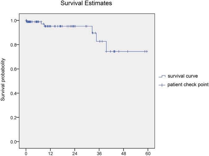
Figure 1. Survival estimates of teeth post-pulpotomy over time. (Courtesy of Zheng J, Ding M, Zhang S, et al: Outcomes of partial pulpotomy in permanent molars of children with irreversible pulpitis: A prospective cohort study. Int J Paediatr Dent 35:558-565, 2025.)
calcifications were noted in the pulp canal. The hard tissue bridge under the iRoot BP Plus layer was noted in all successful cases followed up for more than 6 months (Figure 2). None of the cases developed crown discoloration. Two composites had minor defects that required partial refilling, but all of the restorations were functional.
In pediatric patients whose permanent molars develop IP, partial pulpotomy offers a way to successfully preserve healthy pulp tissue. The immediate success rate is 98.9%, with a rate of 93.6% after several years. Pulpotomy is technically simpler and minimally invasive compared to apexification and RCT.
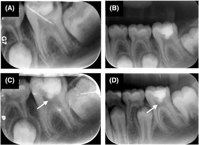
Figure 2. Lower left first molar in an 8-year-old male patient with clinical symptoms of irreversible pulpitis and immature roots. A, The preoperative periapical radiograph showed the presence of temporary restorative material from another clinic. B, The 8-day follow-up. C, The 3-month follow-up. The dentin bridge was well formed. D, The 2.7-year follow-up. The dentin bridge was thicker than before. The apical foramen was closed. (Arrow points to the dentin bridge.) (Courtesy of Zheng J, Ding M, Zhang S, et al: Outcomes of partial pulpotomy in permanent molars of children with irreversible pulpitis: A prospective cohort study. Int J Paediatr Dent 35:558-565, 2025.)
Performing partial pulpotomy is a viable alternative to apexificaiton and root canal therapy for vital, inflamed molars with IP in pediatric patients. The success rates are high for this approach and minimal invasiveness is possible.
Zheng J, Ding M, Zhang S, et al: Outcomes of partial pulpotomy in permanent molars of children with irreversible pulpitis: A prospective cohort study. Int J Paediatr Dent 35:558-565, 2025
Reprints available from Q Wen, 37A Xishiku St, Xicheng District, Beijing 100034, China; e-mail: wincky.ls@163.com
Depending on the time of day that we eat, our bodies process food differently. A new study from the German Institute of Human Nutrition Postdam-Rehbrucke indicates that our internal clocks and overall health are also important in managing glucose metabolism. The circadian system controls behavior and metabolism via a central clock located in our brain and other clocks in other organs. This system causes our metabolism to have daily variations in the processing of glucose and hormone release. Food intake is a signal that helps to align our internal clocks. When we eat out of sync with the natural light-dark cycle, these internal clocks can be disrupted, causing adverse metabolic effects.
The timing of food intake in relation to the individual’s biological rhythm can be evaluated through the gap between mealtime and the midpoint of sleep. This midpoint is the time halfway between falling asleep and waking up and indicates a person’s chronotype, which is whether he or she is an early bird or a night owl. The study used a twin registry and had 46 pairs of identical and fraternal twins as subjects. All had comprehensive metabolic assessments and had their chronotypes identified, then they recorded their food over 5 days. The study found that people who tended to eat their final meal later in the day and closer to bedtime had less effective glucose metabolism at the end of the day and lower insulin sensitivity.
Destini Moody, a registered dietitian not involved in the study, said, “While it’s well known that eating timing and meal composition is critical for controlling blood sugar and insulin action in those with diabetes, this study was performed on healthy individuals with normal pancreatic function and no other metabolic disturbances. Even without diabetes, those with metabolic syndrome should be careful about their level of insulin resistance, but none of the subjects fell under this category either given their BMI and waist circumference. The average age of subjects was also 32, which is relatively young.”
It’s been suggested that better regulating of meal times may help maintain metabolic health. Late eating has been linked to obesity and heart disease, but its exact effects on glucose metabolism and the role of genetics both remain unclear. Moody stated, “In general, blood sugar variations are a natural part of digestion and metabolism, and I would urge the public to refrain from stressing about the effects food may have on their blood sugar unless they have metabolic ailments like type 2 diabetes.” She also emphasized paying attention to how much fiber we ingest and how much comes from refined grains. “Foods with added sugars, that have been made with refined grains, and are generally low in fiber will raise blood sugar quickly and send it crashing back down due to a rebound effect. This tends to make people even hungrier and even cause fatigue, which is why you feel sleepy after having meals high in these types of carbs. Luckily, eating whole grains and high fiber fruits and vegetables along with lean protein can not only help mitigate this effect, but often results in a healthier diet overall that helps prevent the risk of chronic disease,” Moody explained.
[Cross PI: Eating Late in the Evening Could Make Blood Sugar Control Harder for the Body. Medical News Today, July 15, 2025]
An unusual side effect of GLP-1 drugs affects dental health, causing tooth decay, sensitivity, and tooth loss as a result of the drug producing dry mouth, vomiting, and low appetite. It’s essential that people using GLP-1 medications, which include semaglutide (Ozempic, Wegovy), tirzepatide (Mounjaro, Zepbound), liraglutide (Saxenda, Victoza), dulaglutide (Trulicity), and exenatide (Byetta, Bydureon BCise), take proper oral care to avoid these side effects and others that can result from rapid fat and muscle loss, which leaves the skin loose and looking aged.
Catrise Austin, a cosmetic dentist and author of Stop Diabetes Today, notes that dentists are seeing more patients who are taking medications such as Ozempic and Wegovy for diabetes and weight loss and these patients are experiencing unexpected oral side effects. This includes the sudden onset of dental decay, sensitivity, and tooth loss along with dry mouth, enamel erosion, inflamed gums, and, in severe cases, even cracked or loose teeth. Austin said, “Though anecdotal, these stories have gained traction as more users share alarming dental side effects online and in media reports.” Mechanisms that may contribute to the tooth issues include the following:
• Dry mouth (xerostomia)—Austin said, “Reduced salivary flow is one of the most common complaints I hear from patients on GLP-1 medications. Saliva is essential for neutralizing acids and protecting enamel. When it’s diminished, the risk of decay and gum disease rises sharply.”
• Nausea and vomiting—Nausea occurs in 16% to 20% of patients who use Ozempic and 5% to 9% have vomiting. Austin cautioned, “Repeated exposure to stomach acid can erode tooth enamel and increase sensitivity.”
• Malnutrition—Austin said, “Some patients experience appetite suppression to the point of nutrient deficiency, which can affect gum health and healing.” Vitamins C, D, and B complex play a crucial role in gum health.
• Reduced oral care—Some patients neglect their oral hygiene and flossing when they don’t feel well, which leads to accelerated tooth decay. For the most benefit, you should floss before brushing.
Ehsan Ali, board certified internal and geriatric medicine specialist, noted that “[Dental} health is tied to overall health. Poor oral hygiene can increase your risk for infections, inflammation, and even heart disease over time.” Just taking care to do a few easy things can help to protect against oral health problems. A quick list of preventive measures includes the following:
• Hydration is key—drink lots of water all day long to keep your mouth moist.
• Use a mouth rinse that promotes saliva production or chew sugar-free gum to keep your mouth hydrated.
• Don’t skip meals even if you aren’t feeling hungry.
• Eat a healthy, balanced diet that includes calcium and vitamin D for oral health. Don’t restrict calorie intake too severely. A healthy calorie deficit for most people is about 300 to 500 calories a day.
• Brush and floss regularly and avoid acidic or sugary snacks.
• If you’ve been vomiting, rinse your mouth out with water and baking soda before brushing to neutralize the stomach acid.
• Avoid alcohol, tobacco, and caffeine because they can exacerbate dry mouth.
The earlier you address problems, the easier it is to manage them and prevent permanent damage. Patients should seek help from the health care team that’s managing their weight loss if they develop persistent nausea or vomiting. They may need to adjust the dose of medication or add something to protect the stomach. It’s time to call the dentist if patients experience new sensitivity, bleeding gums, bad breath that won’t go away, or sudden changes such as loose teeth or breakage.
These medications offer powerful health benefits but patients must be proactive about addressing their side effects. They shouldn’t come at the cost of your teeth.
[Schimelpfening N: ‘Ozempic Teeth’ May Cause Tooth Decay, Other Dental Health Effects. Healthline, June 5, 2025]
PIERRE FAUCHARD AC ADEMY
An International Dental Honor Organization
Greetings from the International Headquarters!
We have experienced the middle of summer here in Rockville, MD and for those of you who have traveled to Washington, DC in the summer, you know that that means very hot, humid days, and the same holds true for Rockville which is nearby. Washington, DC was built next to the Potomac River on what was originally a swamp next to the Potomac River, and the low ground and proximity to Chesapeake Bay make this area steamy in the summer. Nonetheless, the work of the Pierre Fauchard Academy did not slow down! We continue to induct new Fellows around the world.
Our Fellows continue to have access to Dental Abstracts, which is published by Elsevier and features approximately fifty abstracts from key articles in dentistry in each issue of the journal. Dental Abstracts keeps dentists informed of developments and advances in general dentistry and its specialties in an easy-to-read, abstract format. The journal is available online with your PFA login information, or on the PFA website in the Member login area. PFA Fellows also have access to the Elsevier Patient Education library, which contains twenty-five easy-to-read patient education materials that are available in both English and Spanish on a variety of dental topics.
In addition, the Pierre Fauchard Academy recently received approval as a PACE CE Program Provider. As we move forward, all our online and in-person CE programs will be eligible for PACE credits. Our quarterly update, Dental World, is sent by email to all PFA Fellows around the world. We highlight activities of the PFA and news of the Academy.
Our work this year culminated with the 87th Meeting and Gala of the Pierre Fauchard Academy, which was held on October 25, 2025, at the Marriott Marquis Hotel in Washington, DC.
As part of our event this year, our Academy provided a morning Continuing Education program presented by Dr. Paul O’Dwyer. Dr. O’Dwyer is on faculty and

pursuing his PhD at the Royal College of Surgeons in Ireland, and our PFA Trustee for Europe. His presentation is titled ‘Emotional Intelligence in Dentistry – 5 Critical Skills That Take Dentists from Good to Great’.
As always, please honor your colleagues by nominating them for PFA Fellowship: https://fauchard. org/sponsor-nomination-for-newfellows/
I hope to see you at a PFA event in 2026.
Sincerely,
—Mr robert Cattoi, Executive Director of the Pierre Fauchard Academy
The Pierre Fauchard Academy publishes the Dental World as a service to its Fellows and it is distributed as part of the Dental Abstracts Journal.
Please send change of address information or subscription inquiries to the Central Office of the Pierre Fauchard Academy at:
Pierre Fauchard Academy 103 North Adams Street, Rockville, MD 20850.
E-mail: centraloffice@fauchard.org Phone: +1 (240) 658-8070
Submissions:
Please send to the Dental World Office for possible publication any editorials, or articles and photos about PFA Section activities, or Foundation grants and scholarship activities. Please contact the Dental World Editor for more information.
Dental World Office: Email: drfionacollins@gmail.com
Dental Abstracts Senior Publisher: Annie Zhao, Elsevier
Dental Abstracts Editor-In-Chief: Dr. Douglas B. Berkey
Dental Abstracts Journal Manager: Sangamithrai S, Elsevier
Dental World Editor: Dr. Fiona M. Collins
Dear Esteemed Fellows, Becoming a Fellow of the Pierre Fauchard Academy is an honor that recognizes leadership, service, and dedication to dentistry. Yet, as meaningful as it is to receive our certificate, key, or lapel pin, being inducted into PFA does not mark the end of the journey, it is only the beginning.
Our Academy thrives because of the contributions of Fellows who live its mission every day. It is not enough to simply wear the pin or display the certificate; we must actively participate in the life of the Academy, engaging in mentorship, contributing to our communities, and helping to advance the profession of dentistry. The honor of Fellowship carries with it a responsibility to lead, to inspire, and to build the future of our Academy.
A key part of this responsibility lies in nominating and sponsoring new candidates for Fellowship. Each of us was once invited into this distinguished community by someone who saw potential in us. Now, it is our turn to identify deserving colleagues, those who embody the values of service, leadership, and ethics, and bring them into the fold. By sponsoring new Fellows, we ensure that our PFA remains vibrant, diverse, and forward-looking.
Fellows are uniquely positioned to face challenges and embrace opportunities, from integrating new technologies and advancing patient care to mentoring the next generation of dental professionals. Our influence as Fellows extends well beyond our chapters; we can help shape policy, promote ethical practice, and contribute to the global conversation on the future of dentistry.
To fulfil our vision and achieve these goals, we must also support the work of the Pierre Fauchard Academy Leadership Foundation. Our contributions, whether financial or through service, help fund scholarships, humanitarian initiatives, and programs that prepare tomorrow’s dental leaders. By giving back, we not only honor the legacy of Pierre Fauchard but also invest in the future of the profession we love.
As we look to the next chapter of the Pierre Fauchard Academy, let us remember that being a Fellow is more than an honor, it is a calling. It is a commitment to serve, to lead, and to lift others as we climb. Together, through active participation, sponsorship, and support of our Academy’s mission, we can help ensure that PFA continues to be a beacon of excellence in dentistry for generations to come.
The future of PFA depends on us. Let’s build it together. Nominate a Fellow today.
PIERRE FAUCHARD ACADEMY www.fauchard.org
Naturally, the role of a Fellow extends beyond ceremonies and sponsorship. In a rapidly evolving dental landscape, PFA
Best regards, —Dr. Ghabi Kaspo, President-elect of the Pierre Fauchard Academy
Perhaps more than once we have each questioned why we belong to one organization or another. Could it be out of convenience, obligation, or conviction? Or is it something even deeper that drives us?
Here we can find an answer to this question by focusing on the sense or feeling of belonging, which is defined as the subjective identification that an individual experiences regarding a group, an organization, or a community. Throughout history, the sense of belonging has been crucial for the survival of the human species by fostering the ability to cooperate and form groups, which allowed people to face the risks and dangers of nature together.
Our sense of belonging is fundamental for personal and social develop-
ment. In the context of families, feeling accepted and valued reinforces our self-esteem and emotional security; and, when a member feels part of the emotional core of their family, they develop a solid foundation for relating positively to others. The theory of human needs, also known as Maslow’s pyramid, was formulated by the American psychologist Abraham Maslow (1908-1970). He highlighted the sense of belonging as an essential emotional need, placing it in his pyramid alongside other needs such as love and friendship.
The sense of belonging is also a fundamental element in the constitution of other human communities, whether that be nations, armies, work organizations, or sports teams. It involves individuals feeling comfortable, welcome, and accepted within their respective group, which then helps group members work together and prioritize collective well-

being over individual interests. For this reason, organizations and companies promote this sense of belonging – to strengthen commitment and collaboration among workers. Without a strong sense of belonging, coordination and cohesion within a group are affected.
We can affirm that being a Fellow of the Pierre Fauchard Academy represents that sense of belonging. Our Academy is an organization that recognizes and fosters collaboration and leadership in our profession and is based on higher values such as honor, integrity, and righteousness. Dear Fellows of the PFA, feel proud of your membership and always be willing to work together for the greater development of the Organization.
Sincerely,
—Dr. Luis FeLipe JiMenez squeLLa, International Vice President of the Pierre Fauchard Academy
A Special Thank You to members of the 1936 Club for their donations
CR & Marianne Anderegg (USA)
Lisa Bentley (Canada)
Patricia Blanton (USA)
Dan & Debbie Castagna (USA)
Robert & Roberta Cattoi (USA)
RK & Prema Chetty (USA)
Commerce Bank Denver (USA)
Jerry and Patricia Dushkin (Canada)
Theresa Gonzales (USA)
Hal Hale (USA)
Ghabi & Laila Kaspo (USA)
Cheri Newman (USA)
Paul & Ellen Obrock (USA)
Pankaj Patel (Africa)
Lucynda Raben (USA)
Karyn Stockwell (USA)
Raymund Tanaka (USA)
Scott & Sheri Waugh (USA)
Sir Nairn Wilson (Europe)
PFA New South Wales Section (Australasia)
PFA New Zealand Section (Australasia)
PFA Queensland Section (Australasia)
PFA South Australia Section (Australasia)
PFA Victoria Section (Australasia)
PFA Western Australia Section (Australasia)
PFA Canada in honor of Nancy Auyeung (Canada)
PFA Canada in honor of Barry Dolman (Canada)
PFA Canada in honor of Jerry Dushkin (Canada)
PFA Canada in honor of Trista Felty & Krist Chu (Canada)
PFA Canada in honor of Janet Leith (Canada)
PFA Canada in honor of Kevin Roach (Canada)
PFA Georgia Section (USA)
PFA Kansas Section (USA)
PFA Minnesota Section (USA)
PFA New York Section (USA)
PFA North Carolina Section (USA)
PFA Ohio Section (USA)
PFA Southern California Section (USA)
PFA Virginia Section (USA)
PFA Wisconsin Section (USA)
Thank You to our PFA Fellows who have donated funds in support of the PFA Leadership Foundation
Guy Acheson
Eva Ackley
Steven Albright
L. Craig Alford
Gary Altschuler
Charles Anderegg
CR Anderegg
Theodoros Aneziris
Leonard Anglis
James Arneson
L’Tanya Bailey
Lynne Barbour
Gordon Barkley
Todd Barrett
Henry Beson, Jr.
Lisa Bentley
Vinamra Bhasin
Abhiraj Bhatt
Catherine Bickley
William Bissell
Smauel Blanchard
Paul Bocciarelli
James Boyle
Tricia Bradley-Hess
Theodore Brasky
Joseph Breitman
Daniel Briskie
Steven Brock
Richard Brodoski
Steven Brown
Terry Buckheimer
Susan Cable
Tris John Carta
James Cassidy, Jr.
Daniel Castagna
McAllister Castelaz
Robert Cattoi
Lorraine Celis
Steve Chapman
Edward Chiera
Keith Climenhaga
R Cochrane
Allison Corbett
Robert Corns
Wayne Cottam
John Cottrell
Stanislaus Cowley, Jr.
Mark Crabtree
John Craig
Narisa Cramer
Roslyn Crisp
Paula Crum
Margaret Culotta-Norton
Roger Curry
Rony Elias Dagher
Steven Dater
George Davidson
Isaiah Davis
Brittany Dean
Larry Degroat
Colleen Delacy
Charles Dietrich
Catherine Dinka
John Dinka
Jeffrey Dodge
Richard Dunham
Michael Durbin
Wallace Edwards
Michael Eggnatz
Gregory Eissmann
Maxine Feinberg
Kristen Foley
Mariah Frazier
Robert Freitas II
Gary Frey
Brooke Fukuoka
Mark Funke
Louisa Gallegos
Paul Gamache
Kim Gardner
Maurice ‘Marty’ Garrett
Shauna Gilmore
Milton Glicksman
Sarah Godbold-Machowski
Christopher Gorecki
Albert Granger
Shannon Griffin
Steven Gusfa
Harold Haering
Hal Hale
Neil Hansen
Pamela Hanson
Jay Harris
Jess Harris
Thomas Harrison
Pete Hehli
Mark Heiss
Drew Hendrix
Ron Hill
Brian Hockenberger
Scott Hodges
Chirstina Honey
G Obert Hopper
Scott Houfek
Ronald Hsu
Hillarie Hudson
Joyce Huey
Mark Humenik
Adolphus Jackson
Bert Jacob
John Jansen
Paul Hansen
Jill Jenkins
Maritza Jenkins
Michael Jennings
Harold Jeter
D. Theodore Johnson
James Johnson
Karen Johnson
Terrence Jones
Ghabi Kaspo
Eric Katkow
Martha Ann Keels
Michael Keim
David Keller
Raymond Kessler
Timothy Kinzel
John Kois
Timothy Kosinski
Zachary Kouri
Kara Kramer
Woojin Kwon
Edward Laco
Karl Lange
Alfred Laporta
Mark Lawrence
Scott Leckie
Gabriela Lee
Peter Lemieux
Janice Liao
Todd Liston
William Lloyd
William Lobb
Stephen Lockwood
Keri Logan
Melanie Love
Clifford Lowdenback
Walter Machowski, Jr.
Robert Manga
Ted Mason
John McCambley
Patricia McConnell
Michael McCormick, Jr.
Thoams McDonald
Stephen McKenna
Ingrid McLellan
Alex Mellion
Zachary Mellion
Mark Mihalo
Glenn Miller
Rory Mortman
Frederick Mourad
Ned Murphy
Wallin Myers
Mike Mysinger
Nathan Niles
Terry Norris
Paul Obrock
Anne O’Day
Debra Palmer
Jennifer Peglow
Reuben Pelot, III
Christopher Pickel
Cassandra Pietrok
Dan Pitts
Joseph Platt
Gibbs Prevost, Jr.
Richard Raad
Elizabeth Ralstrom
Christopher Lee Rautenstrauch
Anne Richards
Amarjit Rihal
Lindsey Robinson
Harold Rogers
Kelly Roth
Brandon Scheer
Derek Schmidt
Joseph Schmidt
Keith Schneider
Amy Scriven
Samantha Shaver
Ted Sherwin
Adam Shisler
Myra Shivers
Marc Shlossman
Mark Shurett
Kerri Simpson
Jay Skolnick
Mark Small
Deena Holiman Smith
Robert Smith, Jr.
Susan Soderstrom
Mary Starsiak
Robert Stewart
Darryl Stich
Russell Street
Jeremy Suess
Kimberly Swanson
Andrea Taylor
Brenden Taylor
Joseph Thomas
Timothy Tremont
Ann Trivette
JD Troy
Michele Tulak-Gorecki
Claire Tyler
Calvin Utke
Craig Von Dongen
Whitam Van Meter, Jr.
Andrew Varga
Frank Vigliotti
Richard Voigt
Karl Woods
Craig Yarborough
The Bangladesh section of the Pierre Fauchard Academy held its first Scientific Session and Convocation on May 23rd, 2024, which was followed by a hands-on course and workshop the next day. This successful meeting was held at the Government Dental College, Bangalore, convened by Dr. Md. Shakhawat Hossein, and the Member Secretary for the event was Dr. Saima Akter. The event
was conducted by Dr. Chethan Chetty in his capacity as President of the Academy of General Dentistry, and five new Fellows were inducted into our Section and received their PFA certificates. Congratulations to our new Fellows!
Sincerely,
—Dr
rK Chetty, Section
Chair
for Bangladesh

The New York Section held its Annual Meeting on Friday, May 30th, 2025, at the Long Island Marriott Conference Center in Uniondale, NY. Dr. Jay Skolnick (Trustee for Region 2) welcomed our attendees and provided introductions.
Our guest speaker was Dr. Maxine Feinberg, Past President of the American Dental Association and a Fellow of the Pierre Fauchard Academy, the American College of Dentists, and the International College of Dentists. Dr. Feinberg, a Cranford, New Jersey based periodontist, was the first woman to serve as President of the New Jersey Dental Association and the third woman to serve as President of the American Dental Association. She is also a Past President of the New Jersey State Board of Dentistry, where she served for 9 years, and continues to serve as an Examin-
er for the ADEX dental licensure exams. In addition, Dr. Feinberg served as the American Dental Association’s Delegate to the FDI World Congress for 12 years. Dr. Feinberg is a Commissioner for the Council on Dental Accreditation, was Chair of CODA for 2023-2024, and currently sits on the Boards of the NJDDS/Donated Dental Services and the Gillette Hayden Foundations. Dr. Feinberg graduated from New York University College of Arts and Sciences in 1977 and New York University College of Dentistry in 1980. She later completed her Certificate in Periodontics in 1984.
The PFA New York Section was delighted to present its Distinguished Service Award to Dr. Amarilis Jacobo for her sustained high-level caring service to both the dental and local communities.
New York continued

Dr. Jacobo also served as Co-chair of the New York Section for many years.
Dr. Jacobo originates from the Dominican Republic and returned there to become a graduate student from La Universidad Central del Este School of Dentistry after which she returned to the U.S. and received her DDS from New York University School of Dentistry. Her years of involvement with organized dentistry began in 1993 as one of the founders of the Dominican Dental Association and around this time, she also joined the Bronx County Dental Society. She has been a Delegate/Alternate to the American Dental Association House of Delegates and is an alumna of the Institute for Diversity in Leadership. In 2016, she was President of the National Hispanic Dental Association and is currently Vice-President of the New York State Dental Association.
Dr. Jacobo serves as a member on multiple boards including the Hostos Community College Foundation, Doctor Nasry Michelen Foundation, and NY Section of the Hispanic Dental Association. She is a Trustee of SBH Health System and a member of the advisory board for Columbia School of Dental Medicine and el Colegio Dominicano de Odontologos
in the Dominican Republic. Her pride and joy was being able to open La Clinica Astin Jacobo in 2017 which she named after her father and that is located in a school named for him. This free dental clinic’s mission is to have a school campus free of cavities.
Together, Dr. Fabiola Milord (New York Section Co-chair) and Dr. Skolnick inducted our new Fellows. Congratulations to our new Fellows, Drs. Julie Connolly (New York), Thomas Connolly (New York), Ada Cooper (New York), Egidio Farone (New York), Mahnaz Fatahzadeh (Montvale), Sumir Gandhi (New York), Kathryn Rothas (Green Island), Gail Schupak (New York), Lynn Stacy (Lowville), Radhika Thakkar (Rochester), and Matthew Valerio (Churchville). We would also like to congratulate the recipients of the PFA New York Student Scholarships: Kyle Alfred (Columbia University), Nicole Israeli (Touro College of Dental Medicine), Michalle Lassiter (University at Buffalo School of Dental Medicine), Allyson Newman (Stony Brook University School of Dental Medicine) and Madusha Perera (New York University College of Dentistry).
TheNevada Section of the Pierre Fauchard Academy held its induction ceremony on July 24, 2025, where we inducted four new Fellows. We were honored that Dr. James Mah, Dean of the University of Nevada Las Vegas (UNLV) School of Dental Medicine, and
Dr. Charles Anderegg, PFA Region 5 Trustee, were in attendance. Their gracious presence truly elevated the event, which was a great success. Congratulations to our new Fellows, Drs. Karen Kucharski, Jyoti Mago, Sonal Shah, and Joanna Shen.
Vaping
Introduced approximately twenty years ago, the use of ecigarettes increased exponentially amid effective marketing, inadequate information, limited legislation, and controversy regarding the potential harms of vaping. Robust adoption and use rates were found globally for vaping, for example in North America and Europe, and peaked around 2018. At that time, in the US more than 1 in 4 high school students reported vaping in the prior 30 days. Since then, while still common, the percentage of youth and adults vaping has been declining. While in part due to increased information on vaping, depending on location, legislation has limited or banned the marketing and sale of vapes to youth or banned specific youth-friendly flavorings and cartridges; required submissions to countries’ regulatory authorities for approval to market vapes, restricted distribution, and instituted sales taxes.
When e-cigarettes were introduced, the potential short- and long-term effects of vaping were unknown, There was, however, significant concern over the ingredients in e-cigarettes and their by-products while vaping. Over time this has changed, and research is confirming the detrimental effects of vaping on systemic health, including cardiovascular disease, pulmonary disease, anxiety and impaired mental health, headaches, nausea, lower total sperm counts and chronic throat irritation. Associations for vaping and the risk of substance abuse (marijuana, and alcohol including binge drinking) have also been found.
Research and reviews on vaping have also provided insights and evidence on the harms to oral health, including dry mouth, oropharyngeal dryness, and the role of vaping in oral microbial dysbiosis and subsequently the potential risk for dental caries, periodontal disease, and other oral conditions. Recent data also indicates that ethnicity may influence the impact of vaping on oral health. Research continues and in the meantime dentists and their teams can advise patients against vaping and educate patients on the potential risks of vaping.
Select References for Further Reading
Centers for Disease Control and Prevention. Smoking and Tobacco Use. E-Cigarette Use Among Youth. https://www.cdc.gov/ tobacco/e-cigarettes/youth.html
Gentzke AS, Wang TW, Jamal A, Park-Lee E, Ren C, Cullen KA, Neff L. Tobacco Product Use Among Middle and High School Students — United States, 2020. Morb Mortal Wkly Rep 2020;69(50):1881-8. https://www.cdc.gov/mmwr/volumes/69/wr/ mm6950a1.htm? s_cid=mm6950a1_w.
Golder S, Hartwell G, Barnett LM, Nash SG, Petticrew M, Glover RE. Vaping and harm in young people: umbrella review. Tob Control. 2025 Aug 19:tc-2024-059219. doi: 10.1136/tc-2024-059219. Epub ahead of print.
Guo X, Hou L, Peng X, Tang F. The prevalence of xerostomia among e-cigarette or combustible tobacco users: A systematic review and meta-analysis. Tob Induc Dis. 2023 Feb 9;21:22. doi: 10.18332/tid/156676. Erratum in: Tob Induc Dis. 2023 Mar 27;21:46. doi: 10.18332/tid/161989.
He X, Meister M, Jeon J, Alqahtani S, Cushenan P, Weaver S, Luo R, Black M, Shannahan J, Wright C. Unveiling Oral Health Impacts of Vaping in African Americans through Untargeted Metabolomics and Proteomics. Environ Health (Wash). 2025 Apr 2;3(7):733-746. doi: 10.1021/envhealth.4c00276.
Iacob AM, Escobedo Martínez MF, Barbeito Castro E, Junquera Olay S, Olay García S, Junquera Gutiérrez LM. Effects of Vape Use on Oral Health: A Review of the Literature. Medicina (Kaunas). 2024 Feb 21;60(3):365. doi: 10.3390/medicina60030365.
Khattak O, Chaudhary FA, Sakoor A, Khattak MU, Ehsan A, Khan FH, Khalid AA, Siddiqui YD, Iqbal A, Issrani R. Oral health status, self-perceptions, and risk awareness among young adult users of electronic cigarettes in Pakistan. Tob Induc Dis. 2024 Nov 5;22. doi: 10.18332/tid/194963.
Liu X, Lu W, Liao S, Deng Z, Zhang Z, Liu Y, Lu W. Efficiency and adverse events of electronic cigarettes: A systematic review and meta-analysis (PRISMA-compliant article). Medicine (Baltimore) 2018;97(19):e0324. doi: 10.1097/MD.0000000000010324
Panariello B, Dias Panariello F, Misir A, Barboza EP. An Umbrella Review of E-Cigarettes’ Impact on Oral Microbiota and Biofilm Buildup. Pathogens. 2025 Jun 10;14(6):578. doi: 10.3390/pathogens14060578.
Rose JJ, Krishnan-Sarin S, Exil VJ et al. Cardiopulmonary Impact of Electronic Cigarettes and Vaping Products: A Scientific Statement From the American Heart Association. Circulation 2023;148(8):703728. doi: 10.1161/CIR.0000000000001160.
Tabnjh AK, Alizadehgharib S, Campus G, Lingström P. The effects of electronic smoking on dental caries and proinflammatory markers: a systematic review and meta-analysis. Front Oral Health. 2025 Apr 11;6:1569806. doi: 10.3389/froh.2025.1569806.
US E-cigarette Regulations – 50 State Review. June 15, 2025. https://www.publichealthlawcenter.org/resources/us-e-cigarette-regulations-50-state-review.
Our mission as Fellows in the Pierre Fauchard Academy is to recognize and develop outstanding leadership in our profession, internationally.
Central Office:
Robert Cattoi, Executive Director 103 North Adams Street, Rockville, MD 20850
E-mail: centraloffice@fauchard.org
Phone: +1 (240) 658-8070
Fax: +1 (240) 266-9188 www.fauchard.org
Cheryl Billingsley, President Virginia, USA
Ghabi Kaspo, President-elect, Michigan, USA
Pankaj Patel, Immediate Past President, Kenya
Luis Felipe Jimenez, Vice President, Chile
Robert Cattoi, Executive Director, Maryland, USA
REGION 1—Europe
Paul O’Dwyer, Ireland
REGION 4—Midwest USA
Kim Gardner, Ohio, USA
REGION 7—Latin America
Federico Perez Diez, Mexico
REGION 10—Central USA
Lucynda Raben, Kansas
REGION 2—Northeast USA Jay Skolnick, New York
REGION 5—Western USA CR Anderegg, Washington, USA
Dan Castagna, Treasurer, California, USA
Pankaj Patel, Secretary, Kenya
Dan Castagna, Treasurer, California
REGION 8—Australasia Deb Cockrell, Australia
REGION 11—Africa Tom Ochola, Kenya
REGION 3—Southeast USA Paul Obrock, Tennessee
REGION 6—Canada Lisa Bentley, Ontario
REGION 9—Asia
Ricardo R. Gatbonton, The Philippines
EDITOR—Dental World Fiona M. Collins, Georgia
Ghabi Kaspo, President, Michigan, USA
Robert Cattoi, Ex officio, Maryland, USA
1.Publication Title 2.Publication Number
7.Complete Mailing Address of Known Office of Publication (Not printer) (Street, city, county, state, and ZIP+4)
230 Park Ave Ste 800
New York NY 10169 91-44-4299-4077
8.Complete Mailing Address of Headquarters or General Business Office of Publisher (Not Printer) Elsevier INC
230 Park Ave Ste 800 New York NY 10169
9.Full Names and Complete Mailing Addresses of Publisher, Editor and Managing Editor (do not leave blank)
Publisher (Name and complete mailing address) Jane Ryley
Elsevier Inc. 3251 Riverport Lane
Maryland Heights, MO 63043
Editor (Name and complete mailing address)
Douglas B. Berkley, D.M.D., MPH, MS
7850 South Hudson Street
Centennial, CO 80122
Managing Editor (Name and complete mailing address) Elaine Steinborn
12853 Stoneridge Drive
and the exempt status for federal income tax purposes: X Has Not changed During Preceding 12 Months Has Changed During Preceding 12 Months (Publisher
PS Form 3526, July 2014