CS 3600 UPDATE - VERSION 3
BRACON Dental & Medical Products
INTELLIGENT ENOUGH TO IMPRESS A GENIUS
carestreamdental.com
www.bracon.co.uk
800.944.6365
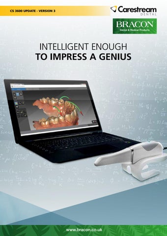
Introducing Our Latest Updates Carestream Dental is pleased to announce its latest developments to the CS 3600 family via the intraoral 3D acquisition software, version 3.0.16. Designed to improve the user experience, this update allows our end users to be up-to-date with the latest workflow advancements while enjoying significant feature enhancements.
An award-winning solution Since its launch in April 2016, the CS 3600 has been recognized as being a top new and innovative dental product. Additionally, the CS 3600 received a rating of “Excellent-Good” by Gordon J. Christensen Clinicians Report. It was also named a Clinicians Report “Best Product” of 2016.
New posterior tip In addition to the two current styles of tips—normal and side-oriented—we are now offering a third tip, which is optimized for posterior scanning. The new posterior tip offers the following advantages: • Features the shortest autoclavable tip height in the market • Provides access to difficult-to-reach areas for improved clinical outcomes • Improves patient comfort during the acquisition process • Is ideal for posterior scans, as short tip height improves access to the distal molar region
Tips autoclavable up to 60 cycles All three of our removable scanner tips are now autoclavable up to 60 cycles if you limit the exposure time at 134°C to not more than 4 minutes. Refer to the CS 3600 Family Safety, Regulatory, and Technical Specifications User Guide for additional details.
Optional posterior tip
Show and tell in full 3D HD color Significant improvements have been made to the color of the CS 3600 mesh, which now provides more vivid color and texture that better reflects reality. Additionally, self-adaptive hole filling automatically identifies holes and selects the appropriate anatomical color for the optimal aesthetic outcome.
Photograph
Full HD 3D
Monochromatic
CS 3600 V3 | 2
The mesh adjustment tool now allows you to adjust the brightness of the images as needed to suit different purposes, clinical settings and personal preferences. You can select from high, medium or low.
High
Medium
Low
Real-time scan quality check Reach expert scanning level quickly with live scan quality checks. A set of warnings provide feedback in real time, indicating areas of the scan that lack detail and require additional scanning to optimize clinical outcomes. Designed to provide realtime feedback, the warnings and guides help increase efficiency while reducing the need for repetitive scanning and image rendering. There are three forms of guidance in the software: 1. Yellow-colored highlights to alert you of gaps and indicate areas that require additional scanning for optimal detail. 2. Green-colored highlights to alert you of holes in the data set. 3. A green arrow-shaped indicator, which shows the ideal direction to re-scan missing information.
Green arrows
Green highlights
Yellow highlights and arrows
Note: In addition to the visible online scan quality warnings, the software will automatically remove unwanted soft tissue during the live scan process, providing much cleaner live and final images.
Workflow enhancements Significant enhancements have been made to the restorative, orthodontic and implant specialty workflows. The Select Acquisition Type window now has expanded capabilities. In addition to being able to select the orthodontic, restoration or implant workflow, you can now import data sets using either the workflow in which they were initially captured or any of the three workflows. This provides the ultimate flexibility for situations where you are performing different clinical applications or treatments on a single patient. Additionally, the restorative workflow now offers two options: perform a standard scan or a pre- and post- scan. The preand post- scan workflow allows you to scan the pre-scan the patient before the procedure, then cut away the prep area and re-scan the patient later.
New options in the Select Acquisition Type menu CS 3600 V3 | 3
Restorative workflow There are four primary changes to the restorative workflow, as indicated below: Pre- and Post-Scan Workflow: This evolutionary workflow allows you to cut zones that have changed and then rescan only the preparation area, eliminating duplication of effort while saving all the necessary patient information for a comprehensive patient record.
Pre and post scan view
Locking Tool for Multiple Teeth: Allows you to scan and lock up to six previously scanned preparation areas to prevent them from being updated by additional scanning. This feature can be used to lock retracted gingival tissue immediately after scanning, as the tissue might collapse and possibly degrade the scan area otherwise.
Scan of the preparation area
Preparation area locked to protect
Measurement Tool: Allows you to measure the distance between the tooth preparation and the opposing tooth. This can be used to help you determine if there is enough space for the restoration based on the desired thickness for your selected material.
Occlusal distance measurement
Close-up view of measurement
Undercut Check: In just one click, the software automatically detects undercuts based on the selected area, helping to ensure that your digital impression is optimal for the restoration. This helps to reduce back-and-forth communication with your lab regarding the quality of the impression. If the impression is not suitable, you can adjust the preparation and re-scan as needed.
Undercut check
Close-up view of undercut check
CS 3600 V3 | 4
Orthodontic workflow Quadrant Snapshot: Designed to replace the cumbersome and painful process associated with taking conventional orthodontic images, the quadrant snapshot automatically extracts up to five 2D intraoral images from the scanned data set. This results in a significantly more comfortable experience for the patient while saving you time, as there is no longer a need to use lip retractors or mirrors to obtain a full set of photographic records. Images include left buccal, right buccal, center, upper occlusal and lower occlusal, as well as a 5-in-1 composite image.
Orthodontic snapshot
Implant workflow Scanbody Selection Tool: The new scanbody selection tool helps to prevent the image mismatches that sometimes occur when using multiple scanbodies on a single case; since scanbodies are often uniform in shape, color and design, they can be difficult to scan. This tool allows you to select only the area where the scanbody is located, ensuring that the scanner only captures data for that individual scanbody. The scanner will only capture data in the area that is unlocked. This uniquely designed tool significantly improves the quality of complex restoration cases with multiple-scan bodies by allowing you to scan one scanbody at a time.
Implant locking
Close-up view of implant locking
All workflows Occlusion Mapping: Available for all three workflows, occlusion mapping displays a pressure map of the occlusal surface, helping you to evaluate the alignment of the patient’s bite. The color-coded map helps you quickly determine whether pressure is distributed evenly or if it is irregular.
Occlusion mapping
CS 3600 V3 | 5
OPTIONAL BASED ON REGIONAL NEED Introducing CS 3600 Access Carestream Dental is also pleased to announce the CS 3600 Access intraoral scanner, which delivers the same workflow flexibility, scan quality, accuracy and efficiency as the CS 3600, but with monochromatic scanning—offering an affordable entry into digital impression scanning. The CS 3600 Access is easily upgradeable to HD 3D color at any time for full spectrum digital scanning.
CS 3600 Access
BRACON Dental & Medical Products
Bracon Limited • Unit C • Swife Business Park • Burwash Road • Broad Oak • Heathfield • East Sussex TN21 8UP t: +44 (0)1580 817000 f: +44 (0)1580 819455 e: sales@bracon.co.uk w: bracon.co.uk
©Carestream Health, Inc. 2017. 16521 AL CS 3600 PN 1117
CS 3600 V3 | 6