



5 F r OM the P re S i D enthir O tA n A k A
7 F r OM the e D it O r -
Jiten D r A M A ngw A ni
8 Ankle Arthriti S
StA n DA r DSn etw O rk S A n D gir F t
Answering the question
‘Why do we need a Standard or Network?’


5 F r OM the P re S i D enthir O tA n A k A
7 F r OM the e D it O r -
Jiten D r A M A ngw A ni
8 Ankle Arthriti S
StA n DA r DSn etw O rk S A n D gir F t
Answering the question
‘Why do we need a Standard or Network?’
20 i ntern Ati O n A l F OO t & Ankle S PO rt S C O n S en S u S ( i FASC) At e SS k A 2026 -
A D vA n C ing i ntern Ati O n A l
C O ll ABO r Ati O n in e lite
Athlete C A re
Foot Print is the Bulletin of the British Orthopaedic Foot and Ankle Society
Edited by Professor Jitendra Mangwani. Associate Editors - Miss Verity Currall. Assistant Editors - Mr Munier Hossain, Mr Kailash Devalia
BOFAS
Company Registration Number: 01610419
Charity Number: 326114
General enquiries: administrator@bofas.org.uk
Disclaimer
12 the A M eri CA n Orth OPA e D i C F OO t & Ankle S OC iety (AOFAS) P r OF ile D
13 tO tA l tA lu S r e P l AC e M ent: Fr OM Aw A rene SS t O De C i S i O n M A king
Total talus replacement (TTR) has now emerged as a viable option in advanced talar pathology, yet many surgeons still have limited experience with it
16 24 h O ur S A r O un D the gl OB e Reflections on the 2026 Complications in Foot and Ankle Town Hall
18 nO vel MO ve M entD riven CO ntr O l
S y S te M FO r in vitro
FOO t A n D A nkle
te S ting D evel OP e D
At iMP eri A l C O llege
PAth OP hy S i O l O gy OF Frei B erg’ S D i S e AS e OF M etAtA r
Open Box Media & Communications
l Director Stuart.Walters@ob-mc.co.uk
l Director Sam.Skiller@ob-mc.co.uk
l Studio Manager Mark.Lamsdale@ob-mc.co.uk
l Production Matt.Hood@ob-mc.co.uk
l Advertising Sales Andrew.Hull@ob-mc.co.uk
32 F u S i O n FO ru MeMPO wering
PAtient S O n the r OAD t O A nkle F u S i O n
34 A nkle A rthriti S ACAD e M y return S t O new CAS tle
35 evi D en C e CO rner
37 gl OBA l FOO t A n D A nkle S u MM it 2026 in g OA, in D i A
38 h OBB ie S CO rner
Foot Print is produced on behalf of BOFAS by Open Box Media & Communications, Premier House, 13 St Pauls Square, Birmingham B3 1RB. T: 0121 200 7820.
For sales or design services, please discuss your requirements with a member of our team.
The articles and advertisements in this publication are the responsibility of the contributor or advertiser concerned. The publishers and Open Box Media and Communications Ltd and their respective employees, officers and agents accept no liability whatsoever for the consequences of any inaccurate or misleading data, opinions or statement or of any action taken as a result of any article in this publication.
We are committed to sustainable forest management and this publication is printed by Buxton Press who are certified to ISO14001:2015 Standards (Environmental Management System). Buxton prints only with 100% vegetable based inks and uses alcohol free printing solutions, eliminating volatile organic compounds as well as ozone damaging emissions.

Over the past few months, the overwhelming emotion I’ve felt is gratitude. Thank you for your support, kindness and encouragement. At times when life has felt uncertain, your messages and friendship have reminded me what a special community BOFAS truly is.
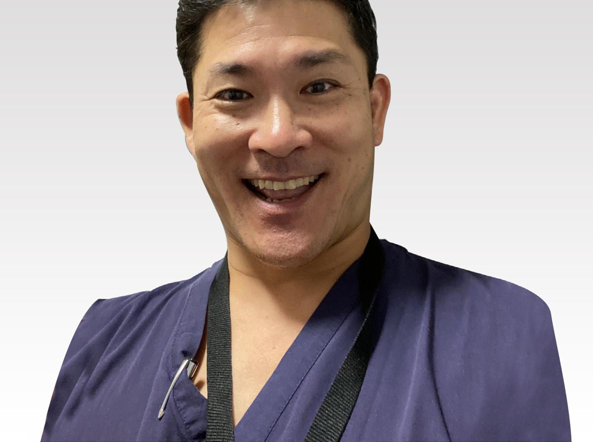

I’m particularly grateful to Callum, who has taken on the operational presidential duties and left me with the enjoyable part of the job. I couldn’t wish for a better group of colleagues in the Executive, Committees and, of course, Jo. They’re the people who quietly keep the BOFAS engine running every day.
I’m incredibly excited to share our plans for Congress 2026. All I’ll say for now is that it’ll be a little different. There will be some surprises, plenty of fun, and I hope, a few moments that will make us stop and think differently about our profession and ourselves.
As I write, I’m heading into surgery tomorrow and then focusing on recovery. Thank you again for everything.
I look forward to seeing you all in Newport in November. n

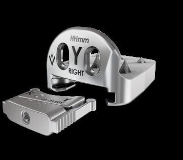
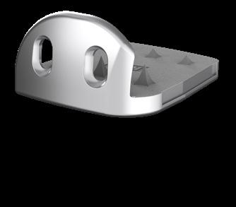
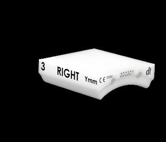
COMPONENTS
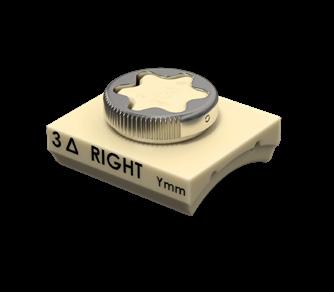
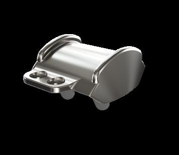
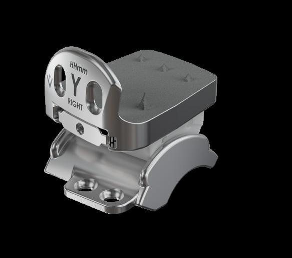
• Anatomic component design for natural load-sharing and reduced stress shielding.
• Semi-constrained fixed-bearing system built for both primary and revision ankle arthroplasty.
• Broad sizing: UHMWPE inlays in 4 thicknesses and two offsets plus full tibial/talar size offerings.
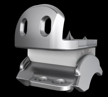
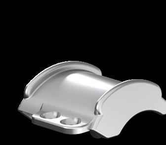
• 3-component, non-constrained mobile-bearing design offering four degrees of freedom.
• Anatomic components + UHMWPE insert sizes in 4 thicknesses for precise fit and reduced stress.
• Titanium plasma & hydroxyapatite coatings on tibial/talar implants for uncemented fixation.
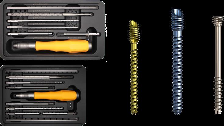
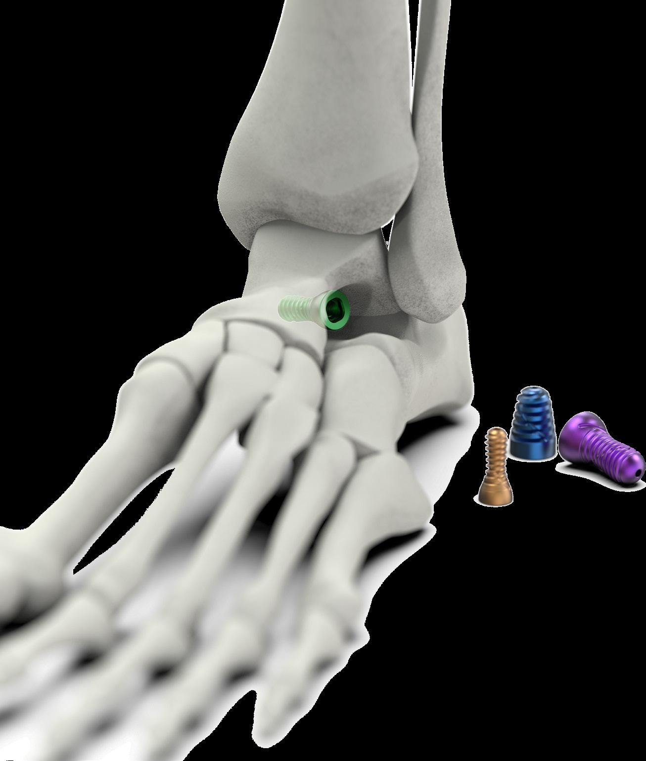
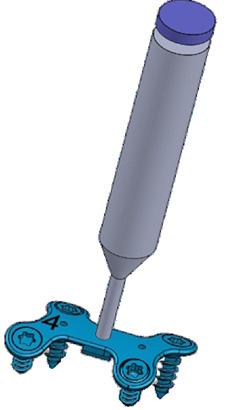
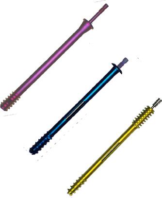
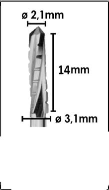
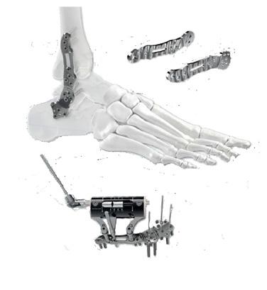
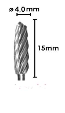
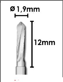
The VILEX MIS System provides comprehensive burr and titanium implant options for the minimally invasive, percutaneous fixation of the foot and ankle.
VILEX MIS Screws are sterile packed and offered in:
• 3.0mm and 4.0mm Diameter forefoot options and feature a 45O Chamfer Head to reduce soft tissue irritation.
• 4.6mm and 7.0mm Headless Mid and Hindfoot Screws.


Council Membership:
President: Hiro Tanaka
President Elect: Callum Clark
Immediate Past President: Robert Clayton
Honorary Secretary: Jitendra Mangwani
Treasurer: Dave Townshend
Media & Comms Director: Karan Malhotra
Education Committee Chair: Anna Chapman
Clinical Practice Committee Chair: Joel Humphrey
Scientific Committee Chair: Sarah Johnson-Lynn
Chief Operating Officer: Jo Millard
Trustee: Steve Hepple
Trustee: Steve Bendall
EFAS Representative: James Ritchie
EDI Lead: Lauren Thomson
Clinical Practice Committee Membership:
Chair: Joel Humphrey
Secretary: Ben Hickey
Caldicott Guardian: Jitendra Mangwani
Data Protection Officer: Jo Millard
Member: Tim Clough
Member: Shariff Hazarika
Member: Robbie Ray
Member: Peter Rosenfeld
Co-opted Registry Lead: Lyndon Mason
Co-opted Registry Lead: Ed Wood
Co-Opted: Andy Goldberg
Co-Opted: Nilesh Makwana
Education Committee Membership:
Chair: Anna Chapman
BOFAS Global Lead: Graham Chuter
Secretary: Maneesh Bhatia
Member: Jim Carmichael
Member: Verity Currall
Member: Yaser Ghani
Member: Annabel Hayward
Member: Nikki Kelsall
Member: Julie Anne Kohls
Member: May Labidi
Member: Nijil Vasukutty
Member: Krishna Vemulapalli
Member: Matthew Welck
Simulation Working Group: Togay Koç
MIS Working Group: Robbie Ray
BOTA Representative: Meg Baker
Co-Opted: Vivek Dhukaram
Media & Communications Committee Membership:
Chair: Karan Malhotra
Secretary: Togay Koç
Member: Edward Dawe
Member: Conrad Lee
Member: Joseph Ring
Webinar Fellow: Peter Cay
Scientific Committee Membership:
Chair: Sarah Johnson-Lynn
Secretary: Dev Mahadevan
Member: Kailash Devalia
Member: Toby Jennison
Member: Adrian Kendall
Co-Opted: James Ritchie
The summer issue of Foot Print celebrates the power of global collaboration, featuring articles from around the world.
Across the UK and beyond, we are witnessing remarkable progress in surgical techniques, implant technology, imaging, rehabilitation strategies, and data-driven outcome evaluation. However, familiar challenges such as rising demand, limited resources, workforce pressures, and the ongoing need to demonstrate value in all our efforts persist. These realities underscore the importance of collaboration, generating robust evidence, and openly exchanging ideas.
Advances in surgery, along with the sharing of diverse clinical perspectives, are essential for elevating patient care worldwide. In this issue, you will find a carefully curated selection of international articles, showcasing how foot and ankle teams across the globe address complex pathologies and offering new perspectives on both established challenges and emerging innovations.
It is very clear that no single region holds a monopoly on innovation and technological advances. Our partnership with international colleagues and foot and ankle societies ensures that we remain at the cutting edge of global practices. This cross-pollination of ideas accelerates our collective learning and refines our surgical outcomes.
The variety of articles featured in this issue encompasses a wide spectrum of topics in foot and ankle surgery and beyond. The Evidence Corner and Hobbies Corner remain very popular amongst the readership of this bulletin.
On behalf of the editorial board, I would like to express our sincere gratitude to all international contributors for generously sharing their ideas and expertise. We hope these pages inspire meaningful collaborative discussions within your clinical practice.
Jit Mangwani - Editor - Foot Print

Why do we need a Standard or network?
There are two recognised treatments for end stage ankle arthritis: Total Ankle Replacement (TAR) or Ankle Fusion (AF). Choosing the optimal one can be challenging due to patient factors, associated deformity, adjacent joint involvement, surgeon expertise and long term consequences of the definitive treatment. This is perhaps reflected in the significant variation in clinical practice.
In 2018, the National Joint Registry (NJR) showed an eight-fold variation in TAR activity by head of population by region1. In a more recent update, looking at Hospital Episode Statistic data (HES), the variation was starker, with some regions having a ratio of 0.98 TAR to 29.33 AF2. The Get It Right First Time (GIRFT) Programme, set up in 2015, has rightly challenged unwarranted variation, which can lead to poorer outcome and higher costs. A network model has shown benefits especially in sharing expertise, driving down costs and improving patient outcomes3. This model has been promoted by BOFAS4 and many of our colleagues have already set up functioning ankle arthritis networks5.
The number of TARs is increasing each year (as recorded in the NJR6). However, the majority of units, 80%, recorded less than ten TARs per year and the median number of TAR per consultant per year is four. A data linked study of the NJR data and the HES database were compared and showed that those units performing less than 20 TARs a year had a significantly higher odds revision rate7,13. Higher volumes in lower limb replacements have been shown to lead
Martin Raglan, Jim Carmichael, Joel Humphrey, Ian Sharpe, Nilesh Makwana, David Townshend
to better patient outcomes as assessed by revision, infection and mortality rates8-11. Whilst the evidence specific to ankle arthritis surgery is limited, we need to consider that there is likely to be a volume/outcome correlation and look for strategies to mitigate this.
The desire to improve patient outcomes, minimise variation and reduce costs has lead to development of formal networks in other arthroplasty such as hip, knee, shoulders and elbows. We hope that the recent publication of the BOFAS Ankle Arthritis Standards12 will assist colleagues to start the conversations necessary to develop and maintain ankle arthritis networks.
The British Orthopaedic Association introduced the first series of BOA standards for Trauma in 2008. These were known as “BOAST” and the aim was to improve the standard of care for trauma and provide a means to audit care and eliminate variation in practice. The scope was extended to include elective surgery and the acronym was modified to “BOA St” and specialty specific standards “SpecS”. The BOASt and SpecS are colour coded to help distinguish the two standards13
The BOASt document provides a set of standards that are auditable, reasonable and representative of current best practice. If evidence is contested or
poor, then a consensus group of experts can review and determine the standard. BOFAS formed an Ankle Arthritis Working group to review the literature and develop a set of standards. A consensus group of experts was invited from the BOFAS membership. Selection was based on equal representation of members throughout the United Kingdom and Northern Ireland, including large and small volume ankle replacement units, as well as foot and ankle units that do not undertake ankle replacements. Selection included an equal mix of senior and junior consultants and appropriate diversity, reflective of the BOFAS membership. The consensus group met in Nottingham in November 2023 and, following a lively debate and vote, consensus was agreed. The standard documents then went out to the entire BOFAS membership for consultation. Following feedback, the results were presented and ratified at the BOFAS annual congress, Belfast 2024. Finally, further discussion with the BOA standards committee crystallised the consensus draft documents into a single BOA SpecS document for Management of End Stage Ankle Arthritis12.The PJI document12 was also approved and had full BOA and specialist society support.
The ankle arthritis network is a polyaxial structure designed according to local resources and geography. The aim is to improve the pathways and outcomes for patients with ankle arthritis, by facilitating referrals and communication, sharing expertise, supporting peers, minimising clinical variation, providing a cost effective service and ensuring equal access to high quality care. The majority of foot and ankle units are already involved in local networks and the aim of
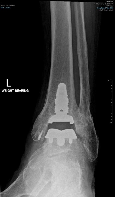
this standard was to provide additional structure and guidance, in order to help formalise this arrangement and ensure that resources are available to support surgeons in delivering and participating in a network.
All units should have access to a network for discussion of ankle arthritis suitable for Total Ankle Replacement (TAR) or Ankle Fusion (AF) in a collaborative setting to ensure sharing of expertise and equality of access for the optimal treatment for their patients. Surgeons who manage end stage ankle arthritis should be encouraged in attending a local network and this network should have administrative support and IT support, and outcomes need to be recorded to allow auditing and review of patient care.
Once joint preservation techniques have failed, TAR and AF are the recognised main treatments for end stage ankle arthritis. All units providing care for patients with end stage ankle arthritis should have access to and engage with the network defined above. There should be a nominated network lead. Units must submit data to a national joint registry (in England, NJR A1), with a strong recommendation to submit TAR and AF PROMS data to the BOFAS Outcomes Registry.
Patients undergoing TAR must share the local arthroplasty ring-fencing
orthopaedic guidelines and practices following GIRFT principles. There is strong recommendation that the core clinical team (surgeon, anaesthetist, scrub staff and operating department practitioners) are experienced and familiar with TAR surgery and its practices, including regional anaesthesia techniques.
Surgeons who wish to undertake TAR must have undergone training in ankle arthroplasty and have access to, and engage with, a network. Dual consultant operating is supported, especially in challenging cases or in gaining experience. These cases will count as one TAR for both consultants on the NJR but, if revised, they will count as 0.5 attributable revision for each consultant. TARs must have clinical and radiological follow up for the lifespan of the implant.
There is currently no consensus regarding surgeon minimum numbers, but there is a strong consensus that outcomes improve with volume. The networks should therefore support surgeons in achieving appropriate volume and improving patient outcomes.
This includes all patients in whom a revision ankle replacement is being considered. A revision is defined as any procedure generating a revision form for the relevant national joint registry (in England, NJR A2). The management of the failing TAR is technically challenging, and revision of the implants can be time consuming and expensive. All these cases should be discussed in a Network as defined above. Infection must always be considered and, if suspected, investigated as per the PJI Standard12. Units must submit data to a national joint registry
and it is strongly recommended to submit revision of TAR to the BOFAS Registry.
Units undertaking revision of failed TARs should have access to ring-fenced, dedicated elective beds in an orthopaedic ward following GIRFT principles. All revisions of TAR should be performed in a dedicated clean orthopaedic theatre. It is recommended that the core clinical team (surgeon, anaesthetist, scrub staff and operating department practitioners) are experienced and familiar with revision TAR surgery and its practices including regional anaesthesia techniques. Units undertaking revision of TAR should have access to support from local/regional plastic and vascular teams. Dual surgeon operating should be supported where surgeons are gaining experience or in complex cases. Provision should be made for long-term follow up until fusion or successful outcome (for revision to fusion) and annually (for revision to TAR).
Again, there is no consensus regarding surgeon minimum numbers, but there is a strong consensus that outcomes improve with volume. The networks are there to support surgeons in achieving appropriate volume and improving patient outcomes.
Peri-prosthetic joint infection (PJI) can be devastating for patients and expensive to treat. Definitive treatment pathways require coordinated, multidisciplinary (MDT) input from specialist infection treatment centres to achieve optimum patient outcomes. Initial management is considered in the relevant BOASt12 and the purpose of this standard is to advise on the definitive management. This standard was for all patients with primary or revision arthroplasty with suspected or confirmed PJI. There is consensus agreement that there was insufficient data for revision or primary ankle replacements and hence the standards would follow the same standards as for all joint arthroplasty. A national multicentre audit (the PRINTAR study) may provide additional information on PJI specifically in TAR. >>
A robust, well-defined pathway, coordinated by an MDT, should be in place for the timely investigation and management of all patients with suspected or confirmed PJI. Every unit managing PJIs must submit data to national databases (NJR, BAJIR) to monitor performance against national standards. The MDT should comprise a core of specialist arthroplasty surgeons, infectious disease physicians or microbiologists, specialist nurses and an MDT coordinator, and have administrative time reflected in job plans. Access to pharmacists, plastic surgeons, rehabilitation teams and allied specialties should be available, either locally or through a regional network.
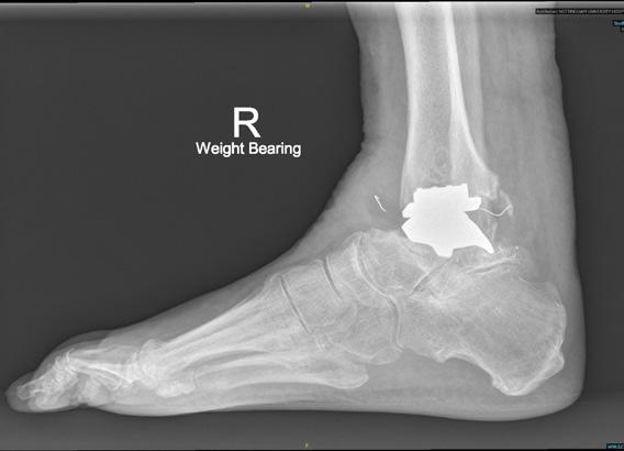
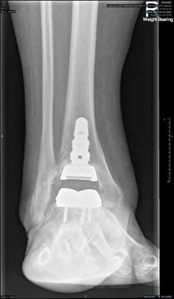
A bone scan is not routinely recommended, although newer nuclear imaging techniques with a strong negative predictive value may be useful in cases of diagnostic uncertainty. CT and MRI may also assist with evaluation.
In suspected infection, aspiration and biopsy should be undertaken in a clean operating theatre or radiology suite using aseptic technique, sterile pots and/ or blood culture bottles. Five separate surgical specimens are required, with clean instruments for each sample. PCR (r16s) analysis should be considered where extended culture does not identify organisms. Ideally antibiotics should not be administered prior to aspiration +/- biopsy except in cases of acute infection in a systemically unwell (septic) patient.
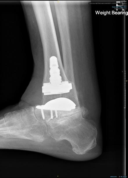
Arthroscopic washout and debridement is not recommended in the definitive treatment of PJI and should only be performed as a temporising / life-saving measure to reduce the septic load and allow patient optimisation prior to a formal Debridement Antibiotics and Implant Retention (DAIR) or revision procedure. DAIR may be indicated for acute infections in a well-fixed, well-functioning implant. It is contraindicated in the presence of a sinus, loose implant and infections caused by fungal, multi-drug resistant or atypical organisms, and caution is recommended in immunocompromised patients or those with multiple comorbidities.
Single or two-stage exchange arthroplasty should be discussed at the MDT. Clinical,
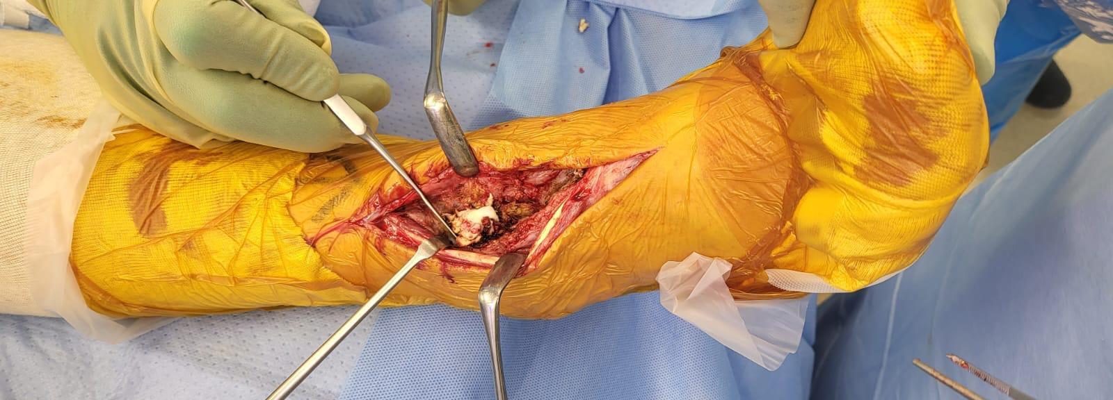
microbiological and reconstructive factors should guide decision-making. Single stage revision for infection is appropriate when pre-operative culture shows a single, antibiotic sensitive organism and where the soft tissue envelope is suitable. In cases of previous failed single stage, fungal, atypical or multi-drug resistant infection, or when there are negative pre- operative cultures, two-stage revision is recommended.
Reconstruction options for an ankle arthroplasty PJI include exchange to either a single or two stage ankle arthroplasty, or revision to a single or two stage ankle arthrodesis. Cases should be discussed in the PJI MDT and local ankle arthritis network12
This has been a forward step in attempting to reduce unwanted variation, improving equity of access and working towards improving clinical outcomes, which will benefit all our patients. A strength of these standards is the investment and support of the BOFAS membership.
Whilst no minimum threshold number could be established by evidence or consensus, the relationship between volume and outcome is acknowledged. Therefore, surgeons with a low volume practice should strongly consider whether they are the correct service to provide such care, although the potential risks of a low volume practice may be mitigated by engagement with a network.
The BOFAS Speciality Standards will be reviewed as new evidence emerges. n
References
1. https://reports.njrcentre.org.uk/ Portals/0/PDFdownloads/NJR%20 15th%20Annual%20Report%202018.pdf
2. https://www.bofas.org.uk/clinician/ meetings/past-congresses/ bofas-2021/bofas-2021-scientificpapers/meeting-abstract-viewer/ ankle-fusion-and-ankle-replacementvariations-in-surgical-practiceacross-england
3. Bloch B, Matar H, Berber R, Gray W, Briggs T, James P, Manktelow A; East Midlands Specialist Network. The impact of a revision arthropalsty network on patient outcomes. Bone Joint J 2023 Jun I;105-B(6):641-648
4. Bendall S, Halliwell P, Goldberg A, Robinson A. Ankle Arthritis Networking: Getting the right treatment to the right patient first time. Foot Ankle Surg. 2022 Feb;28(2):153-158
5. Carmichael J, Raglan M. Collaborative ankle arthritis networks. JTO; Vol 09;issue 4/pg54-56. Dec 2021.
6. https://reports.njrcentre.org.uk/ Portals/0/PDFdownloads/NJR%20 20th%20Annual%20Report%202023.pdf
7. Zaidi R, Macgregor A, Goldberg A. Quality measures for total ankle
replacement, 30- day readmisiion and reoperation rates within 1 year of surgery: a data linkage study using NJR data set. BMJ Open 2016: 6:e011332, doi: http://dx.doi. org/10.1136/bmjopen-2016-011332
8. Hughes et al. Effects of surgeon volume and hospital volume on quality of care in hospitals. Medical Care 1987 25 6 p489-503
9. Choudhury et al. A systematic review of the impact of volume of surgery and specialization on patient outcome. BJS 2007 94(2), 145–161.
10. Katz et al. Association between hospital and surgeon procedure volume and outcomes of total hip replacement in the United States Medicare population. JBJS 2001 83 11 p1622-29
11. Katz et al. Association of hospital and surgeon volume of total hip replacement with functional status and satisfaction three years following surgery. Arthritis Rheumatism 2003 48 2
12. https://www.boa.ac.uk/standardsguidance/boasts.html
13. Basque B, Bitterman A, Campbell K, Haughom B, Lin J, Lee S. Influence of surgeon volume on inpatient complications, costs and length of stay following total ankle arthropalsty. Foot Ankle Int 2016, doi: http://dx.doi. org/10.1177/1071100716664871
14. Monsell F, Trompeter A, Arseti N. Alterations in commissioning and construction of British Orthopaedic Association Standards. JTO; Vol 11; issue 02/ pg 26-28 June 2023.
The American Orthopaedic Foot & Ankle Society (AOFAS) is based in the United States and represents an international member community of 2,600 orthopaedic foot and ankle surgeons, fellows, residents, and advanced healthcare practitioners. Our mission is to improve lives through exceptional orthopaedic foot and ankle care.

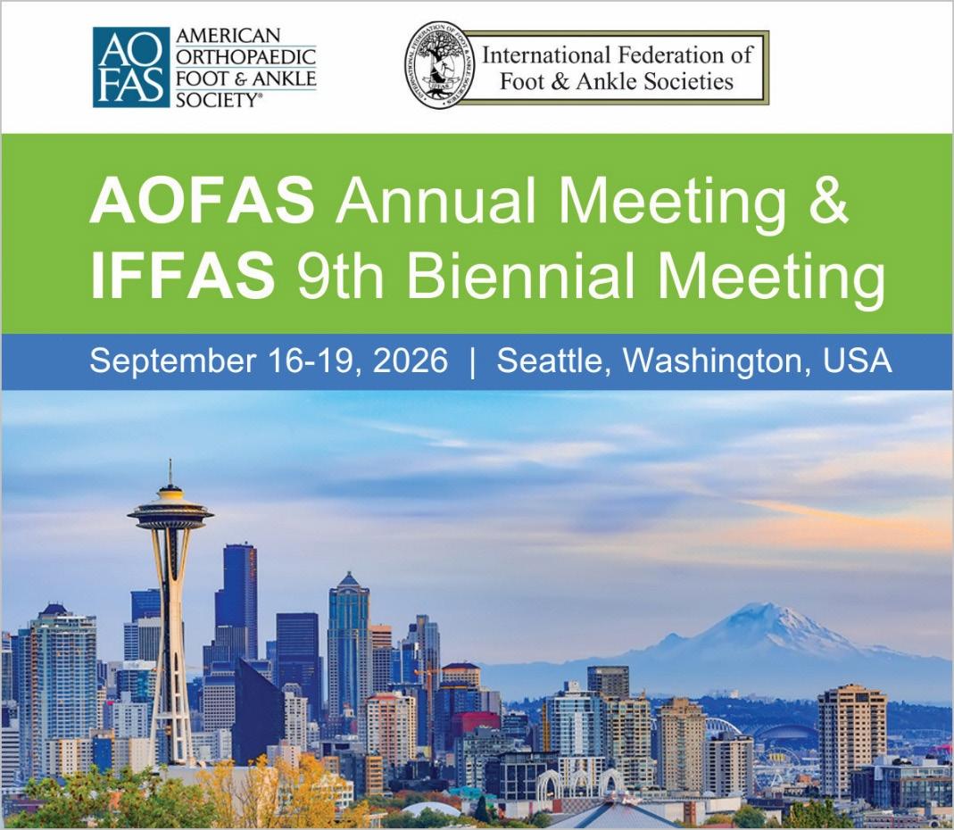
This year, we are honored and excited to host the International Federation of Foot & Ankle Societies (IFFAS) 9th Biennial Meeting. Held in conjunction with the AOFAS Annual Meeting, this outstanding educational event will bring together orthopaedic leaders and innovators from around the world for four days of learning, collaborating, and connecting. The AOFAS and IFFAS meeting will be held September 16-19, 2026, in Seattle, Washington, USA.
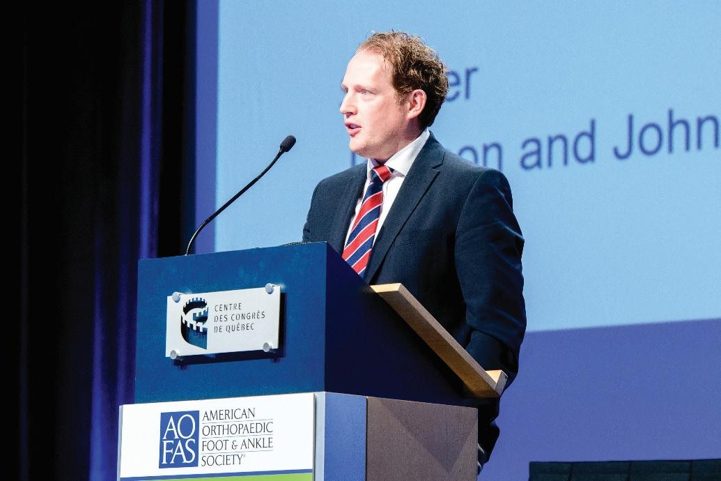
AOFAS and IFFAS 2026 will feature an impressive lineup of speakers, including BOFAS member Dr. Lyndon Mason, who will serve as the 2026 Kenneth A. Johnson International Speaker on Friday, September
Dr. Lyndon Mason, 2026 Kenneth A. Johnson International Speaker
18. Established in honor of AOFAS Past President Kenneth A. Johnson, MD, this keynote recognizes an individual from outside the United States who embodies Dr. Johnson’s dedication to education, collaborative outreach, and open exchange of ideas. Dr. Mason’s talk will focus on posterior malleolar fractures.
Last month, AOFAS held our first-ever Minimally Invasive Surgery (MIS) skills course. The course included didactic, Sawbones, and cadaveric lab training in foot and ankle MIS techniques, led by internationally renowned faculty. Planning is already underway for a second course in 2027. The in-person Sports Injuries of the Foot & Ankle Course will also return in 2027, April 8-10, in Austin, Texas, USA.
AOFAS values our strong partnership with BOFAS. In 2022, BOFAS served as the Guest Nation society at AOFAS Annual Meeting, bringing a UK perspective to a symposium on controversies in foot and ankle surgery. AOFAS and BOFAS also teamed up for three webinars in 2021-22 focusing on biologics, complex pilon fractures, and revision hallux revision surgery. More recently, our societies have enhanced our exchange of knowledge by inviting each other’s presidents to our annual conferences. We hope to grow this partnership by collaborating on new initiatives in the future, including joint humanitarian trips to countries in need of foot and ankle care. n
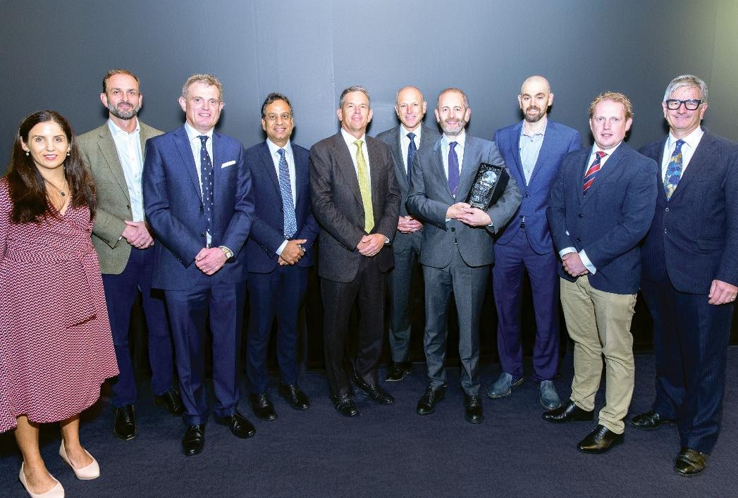
and BOFAS leadership in 2022
Total talus replacement (TTR) has now emerged as a viable option in advanced talar pathology, yet many surgeons still have limited experience with it. This article addresses key practical considerations including indications, durability, patient selection, and where TTR and TATTR (Total ankle and total talus replacement) fit within modern ankle reconstruction.
When the talus fails, the reconstructive options narrows rapidly. Advanced avascular necrosis (AVN), post-traumatic collapse, and severe talar deficiency in revision ankle surgery have traditionally led towards arthrodesis. Total talus replacement (TTR) has altered this landscape and is now increasingly recognised as a legitimate reconstructive option in carefully selected patients.
Although many surgeons are now familiar with the concept of TTR, fewer have substantial operative experience with it. The key practical questions therefore are: when should it be considered; what outcomes can be expected; how long will it last; when should the tibial side also be addressed and what advantages does TTR offer compared with fusion?
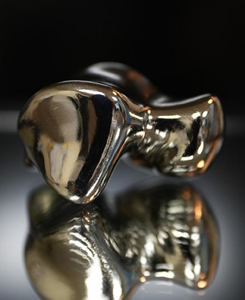
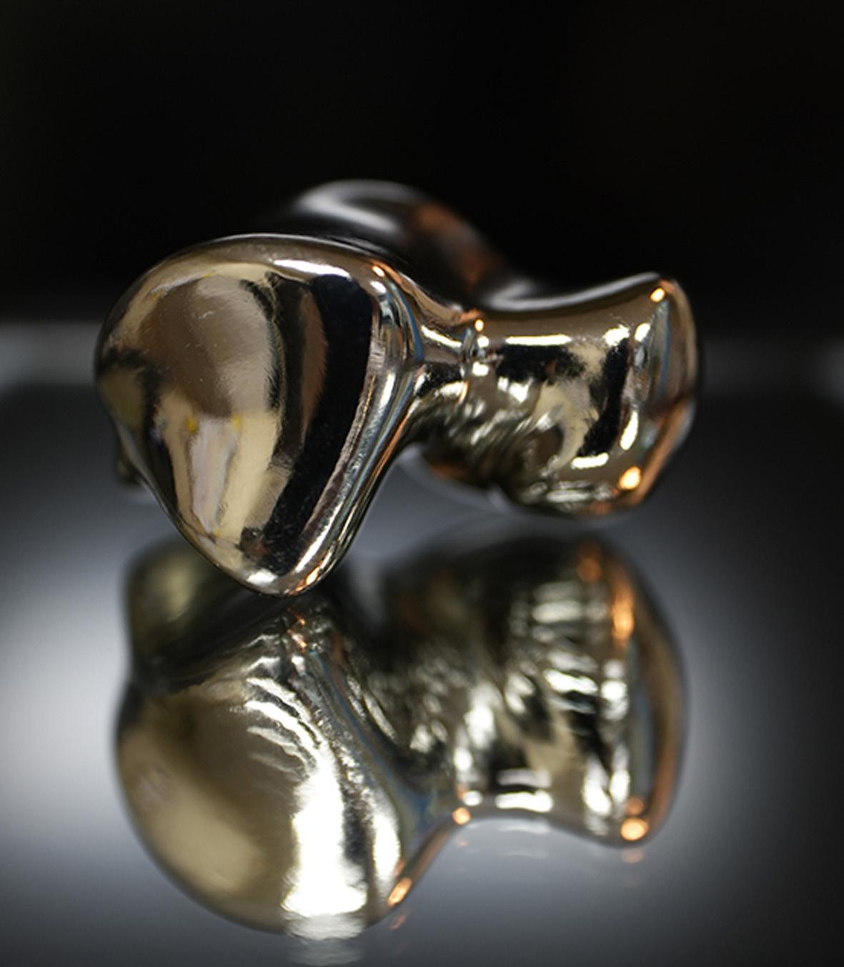
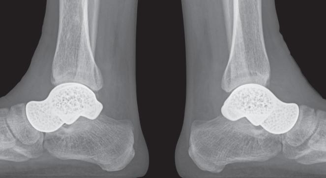
In our practice, more than 100 cases have been performed using custom 3D-printed reinforced pure titanium implants (Titalus, Cubelabs, South Korea) (Figure 1). In appropriately selected patients, outcomes can be highly favourable, with some patients achieving near normal day-to-day ankle function.
What does ttr actually solve?
The talus is a uniquely complex anatomical structure that is difficult to reconstruct. It is central to ankle and hindfoot biomechanics, yet is particularly vulnerable to collapse following trauma, AVN, or failed prior reconstruction. Once the talar body becomes unsalvageable, the problem is not simply bone loss, but the loss of a key articulating structure within the ankle–hindfoot complex.

Arthrodesis remains a reliable and widely accepted solution in many situations, providing pain relief and stability. However, this comes at the cost of joint motion, altered gait mechanics, and increased load transmission across adjacent joints.
TTR must be understood within this context. It is not a universal alternative to fusion, but rather a reconstructive option for patients in whom the talus cannot be salvaged and where preservation of motion remains clinically meaningful.
When should it be considered?
The primary indication for TTR is advanced talar AVN with intact tibial
plafond articulation. It is also useful in post-traumatic talar bone loss and in revision cases after failed total ankle replacement (TAR), particularly when the talus is no longer structurally viable.
The preservation of the adjacent joint cartilage is critical determinant of success. The TTR implant articulates with native joint surfaces and the durability of the reconstruction relies on the status of joint cartilage. Isolated TTR is most appropriate when tibial cartilage is preserved, and the joint space is maintained on weightbearing radiographs.
In contrast, when the tibial side demonstrates arthritic changes, isolated TTR may not be sufficient. In such cases, the tibial side should also be replaced, making combined total ankle-total talus replacement (TATTR) the more appropriate option.
Optimal outcome depends on careful patient selection. It performs best when the talus is the dominant pathology, alignment can be restored, and the soft tissue envelope is adequate.
is it simply a salvage procedure?
TTR is often described as a salvage procedure, which is accurate in the sense that it is typically considered when joint preservation is no longer realistic, and the standard options have narrowed. However, this terminology can be misleading if it implies only a compromised outcome.
“To T al T alus replacemen T (TT r ) has al T ered T h e landscape and is now increasingly recognised as a legi T ima T e recons T ruc T ive op T ion in carefully selec T ed pa T ien T s.”
A successful ankle arthrodesis is an excellent salvage procedure, but its endpoint is pain relief and stability at the cost of limited motion. In contrast, a successful TTR aims to restore the joint articulation, preserve normal gait mechanics, and maintain near normal physiological function.
Accordingly, TTR is better viewed as a reconstructive form of salvage rather than a last-resort procedure.
How good can the result be?
In our experience, TTR can produce excellent outcomes in appropriately selected patients. Pain relief is substantial, gait may normalise and functionally meaningful ankle motion is preserved. In some cases, patients are able to return to activities that would be challenging after arthrodesis.
There is often an assumption that TTR leads to inferior or limited outcome. In practice, this view is frequently overly pessimistic. When indications are appropriate, functional recovery can approach near-normal ankle function.
How long will it last?
Long-term outcome data remain limited as TTR continues to evolve. However, early- and mid-term results have been encouraging.
Durability depends on more than the implant itself. It relies on appropriate patient selection, as well as indication, alignment, soft tissue balance, and the condition of the adjacent cartilage. Where the tibial plafond is preserved, isolated TTR demonstrate acceptable longevity. It is less predictable when tibial articular surface is compromised, and TATTR is often more appropriate.
As with TAR, implant-specific factors are also likely to influence longevity. Total talus prostheses vary in material and design, with differences in mechanical and biological properties that may affect the long-term outcome.
What is the postoperative protocol?
Following TTR, range of motion exercises typically begin at 3–4 days once the soft tissue have stabilised. Full weight-bearing is usually allowed after one week. The ankle is maintained in a short-leg splint for approximately 6 weeks to support soft tissue healing in a neutral position. >>
The transition to regular footwear occurs between four to six weeks.
After TATTR, a more conservative protocol is used, with approximately six weeks of immobilisation in a cast or walker boot to allow tibial component integration.
Why does preserved motion matter?
The importance of motion preservation is best understood in a clinical context.
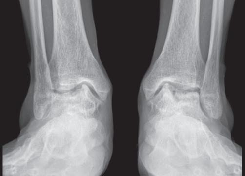
One patient in our practice was a pianist with bilateral talar AVN (Figure 2). Bilateral arthrodeses would have been a reasonable solution but would have significantly limited the ankle control required for piano performance. For this patient, motion preservation was central to required function. The patient underwent simultaneous bilateral TTR and returned to her previous level of performance within months.
This case illustrates that, in selected patients, preserved motion can make the difference between simple salvage and meaningful functional restoration.
What questions remain, and where might this lead?
Several important questions remain. These include the long-term interaction between prosthetic talar surface and native cartilage, the reliability of maintaining soft tissue balance, and future management strategies for failed reconstructions. Further refinement of indications and the accumulation of longer-term data will be essential.
Additional considerations include optimal implant material, the biomechanical significance of implant weight, appropriate sizing strategies, and the role of ligament reconstruction. The management of focal chondral defects in adjacent joint surfaces also requires further clarification.
Addressing these issues will determine future evolution and broader applicability of TTR and TATTR.
conclusion
TTR is now an established option in the management of advanced AVN, post talus fracture sequelae, and selected revision TAR cases. The key challenge is no longer awareness, but appropriate clinical judgement.
In carefully selected patients, TTR can deliver excellent functional outcomes. It does not replace arthrodesis but expands the available reconstructive options. When used for the right indication, it can restore a level of function that is often better than initially expected.
TTR should therefore be regarded not as a last resort alternative, but as a serious and evolving reconstructive strategy for complex ankle pathologies. n
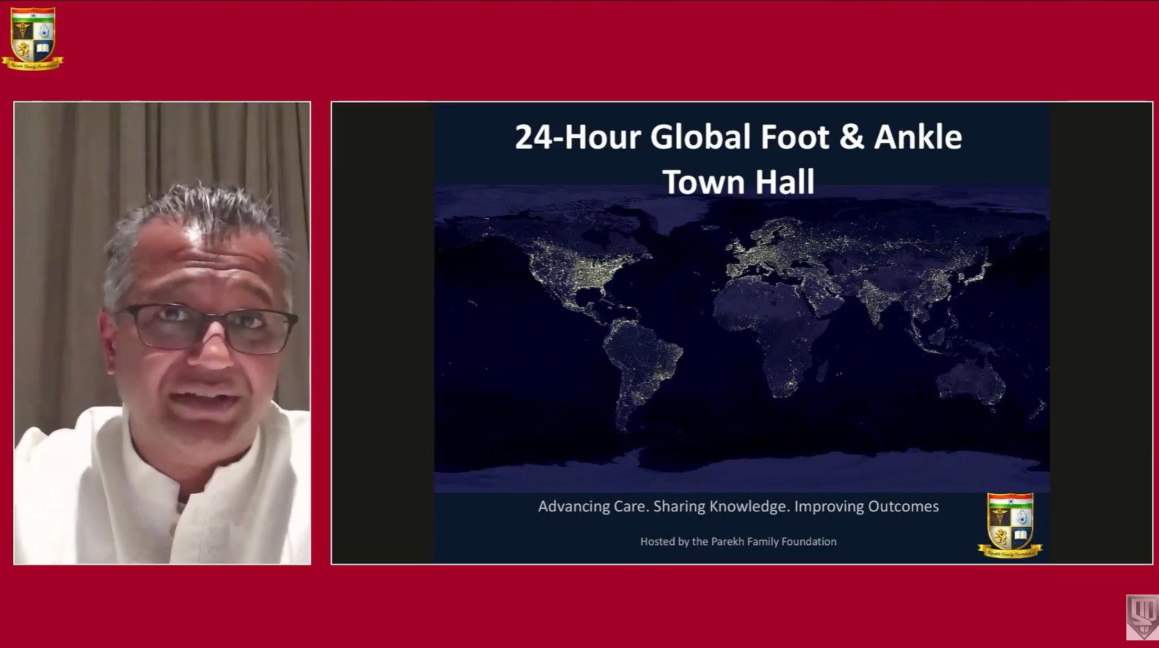
In April 2026, we hosted the annual 24-Hour Global Foot & Ankle Town Hall, an around-the-clock international educational event dedicated entirely to complications in foot and ankle surgery. What initially started as an ambitious idea — essentially asking whether surgeons around the world would truly stay engaged for an entire 24-hour educational experience — has rapidly evolved into one of the most collaborative and energetic global discussions in our specialty.
This year’s program featured more than 100 faculty members from across North America, South America, Europe, Asia, Africa, and Australia. Over the course of a full day and night, the meeting covered nearly every major area within foot and ankle surgery, including total ankle replacement, minimally invasive surgery, sports injuries, trauma, deformity correction, limb salvage, Achilles pathology, cavovarus and flatfoot
Selene G. Parekh MD, MBA Director, Foot & Ankle Fellowship - Rothman Orthopaedic Institute
as planned. The honest conversations surrounding complications, unexpected outcomes, difficult revisions, and lessons learned create a level of education and vulnerability that resonates deeply with surgeons at every stage of practice.

Professor of Orthopaedic Surgery -
Thomas
Jefferson University
Visiting Research CollaboratorPrinceton University
reconstruction, neglected trauma, and complex revision surgery.
What makes this year’s meeting unique is that it focuses entirely on complications, failures, difficult cases, and real-world decision making. In many traditional meetings, presenters naturally highlight successful outcomes and polished techniques. However, some of the most valuable lessons in surgery come from openly discussing the cases that did not go
One of the most rewarding aspects of the event has been watching surgeons openly share not only their technical successes, but also the mistakes, challenges, and difficult judgment calls which shaped their experience. Despite practising in different countries and healthcare systems, many of the challenges we face are remarkably similar: balancing innovation with evidence-based care, managing increasingly complex revision cases, navigating patient expectations, and maintaining humility while advancing technically demanding procedures.
The logistics behind coordinating a 24-hour global meeting are substantial. Faculty joined continuously across multiple time zones, often presenting at unusual hours from operating rooms, offices, airports, homes, and hospitals around the world. Behind the scenes, there was an enormous amount of co-ordination involving moderators, production teams, scheduling, streaming technology, social media engagement, and live troubleshooting. Yet somehow, the energy remained remarkably high throughout the entire event.
From a technological standpoint, one of the most exciting developments this year was the ability to integrate high-quality live streaming, audience interaction, social media engagement, and recorded educational content into a single global platform. It reinforced the idea that digital education is no longer supplemental — it is becoming central to how surgeons around the world learn, collaborate, and remain connected.
A particularly rewarding component of this year’s program was the strong participation from colleagues in the
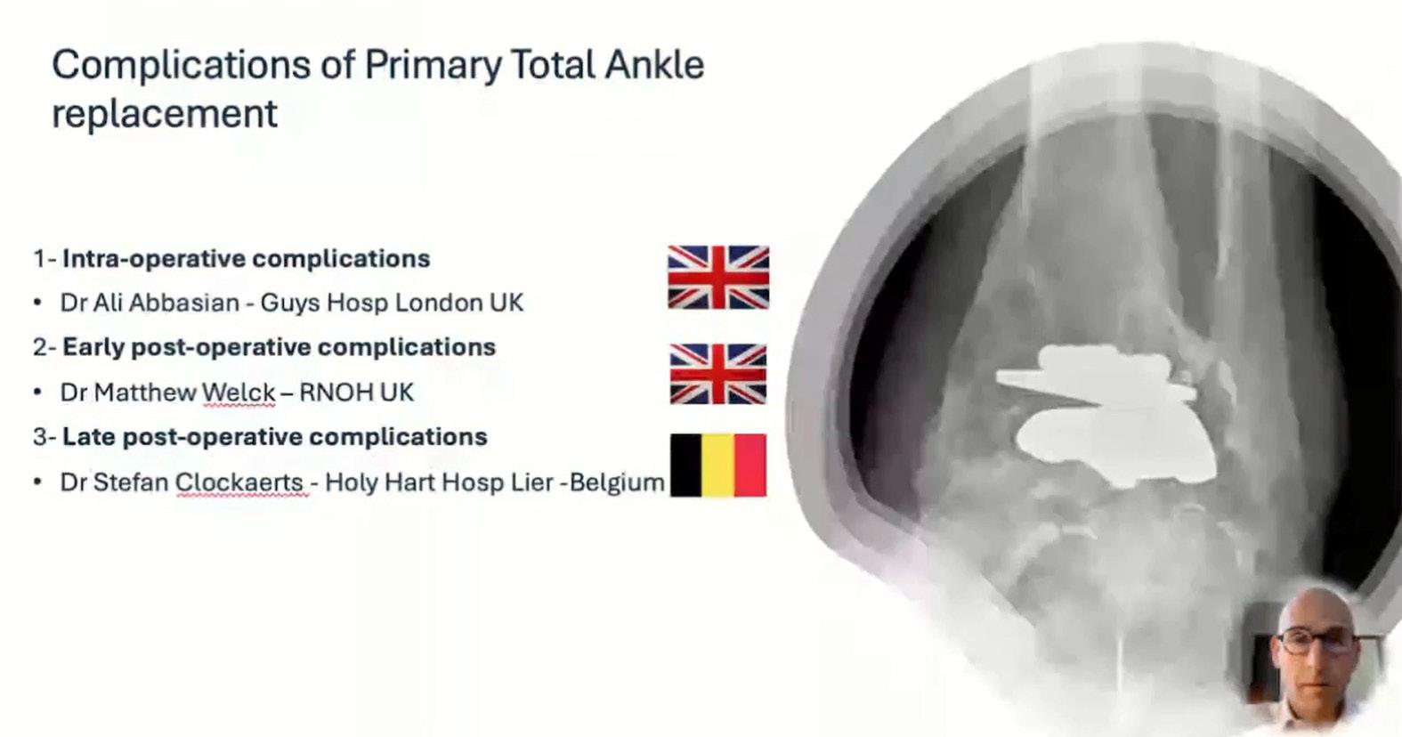
United Kingdom and Europe. We were fortunate to have exceptional sessions moderated and supported by Andy Goldberg and his faculty, including Ali Abbasian, Matthew Welck, Stefan Clockaerts, Kris Buedts, Joris Hermus, Mostafa Benyahia, Rick Brown, Sam Adams, Elena Samaila, Jim Carmichael, Diane Hildebrand, and Jit Mangwani, among many others. Their contributions elevated the scientific discussion significantly and reinforced the increasingly global nature of modern foot and ankle surgery.
The sessions focused on primary and revision total ankle replacement, total ankle-total talus reconstruction, and conversion of failed total ankles to fusion or below-knee amputation generated particularly thoughtful and nuanced discussion. These conversations highlighted not only the rapid evolution of total ankle surgery worldwide, but also the growing importance of complication management, patient selection, and revision strategies as the field matures.
From a personal perspective, the meeting has been incredibly rewarding — but also
humbling. What has gone especially well is the sense of community and collaboration that has developed around the conference. There is tremendous value in creating an environment where surgeons, who may otherwise never interact, can openly exchange ideas, discuss difficult cases, and learn from one another. The educational energy generated during the event has consistently exceeded expectations.
At the same time, every year provides opportunities for reflection and improvement. One of our biggest ongoing goals is improving accessibility and organisation of the educational content. With such a large amount of material presented over a 24-hour period, creating better systems for indexing, archiving, and distributing sessions remains an important priority. We are also continuing to expand audience interaction, polling, digital case discussions, and opportunities for trainees and younger surgeons to become more actively involved.
Perhaps most importantly, the meeting reinforced just how interconnected the global foot and ankle community has become. While we may practice
in different healthcare systems and environments, we all ultimately share the same goal: improving patient outcomes while continuing to advance the field through education, innovation, collaboration, and mentorship.
The 2026 Global Complications in Foot & Ankle Town Hall was supported by the Parekh Family Foundation and numerous faculty, moderators, industry partners and production teams who helped make the event possible. Recorded sessions are now being released weekly through our YouTube platform to allow continued access to the discussions and content.
As foot and ankle surgery continues to evolve, I believe these types of international collaborative educational initiatives will become increasingly important — not only for sharing knowledge, but also for strengthening relationships across our specialty worldwide.
And yes — we are already planning how to make next year’s meeting even bigger and better. n
The foot and ankle complex is a unique part of the human musculoskeletal system due to its adaptability and loadbearing capabilities. The complicated structures that make up the foot and ankle give it the ability to conform to different terrains, while supporting and stabilising the body. Understanding the complex internal interactions between the structures within the foot and ankle is important when developing surgical and rehabilitation protocols to rectify injuries and pathologies.
To measure and record movements and forces internal to the foot, invasive methodologies are needed, which are not practicable in vivo. As an
alternative, simulation approaches may be undertaken. There are two types of simulation, in vitro (using cadaveric specimens) and in silico (using computer models). In silico models have the limitation of making many assumptions regarding bone interactions and morphology, and the material properties of the structures of the foot. These may limit the translational potential of results to patients. In vitro simulations manipulate cadaveric foot and ankle specimens, maintaining anatomically correct interactions between the bones and soft tissue structures. The limitations of in vitro simulations are that trials are simulated at a reduced speed and force compared to the in vivo movements they replicate due to hardware limitations and the need to ensure that the cadaveric specimens can be used in as many trials as are needed for a study. Despite this, findings from in vitro simulation remain more translatable to patient applications. The assessment of joint interactions or muscle force synergies can be used to inform surgical procedures and rehabilitation protocols.

Angela Kedgley Reader in Orthopaedic Biomechanics, Department of Bioengineering, Imperial College London
Antony Crossman
Postdoctoral Researcher, Department of Bioengineering, Imperial College London
In vitro foot and ankle simulators that replicate gait have existed for almost 30 years; however, there has been little development in the way in which the muscles control the movement of the foot and ankle. Most previous simulators derived muscle forces from electromyography (EMG) recordings of the activation of the muscles in vivo. However, muscle activation is not commensurate to the force that the
muscle is producing. Additionally, the goal of in vitro simulation is to control the specimens to move in the most natural and realistic way possible, meaning that the aim of the system should mimic the instructions the brain sends to the muscles. The goal of the brain is to create a movement and not to achieve a specific force in each muscle; therefore, movement should lie at the heart of in vitro gait simulation.
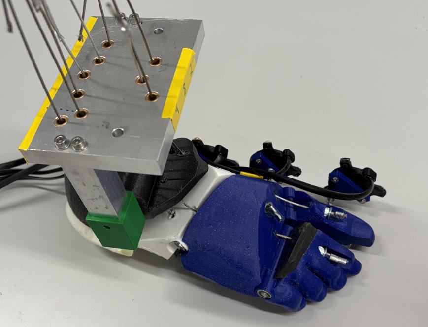
At Imperial College London, we have developed a novel movement-driven control system for in vitro foot and ankle testing. Movements recorded in vivo are input into the system; the minimum muscle forces needed to create the movements are calculated and used to move a specimen in real time.
This approach means that any movement may be simulated, not limiting the system to physiological
walking over flat ground. Enabling more complex real-world motions to be simulated may expose hidden limitations to surgical procedures. A unique set of muscle forces are calculated for each specimen; in this way, anatomical
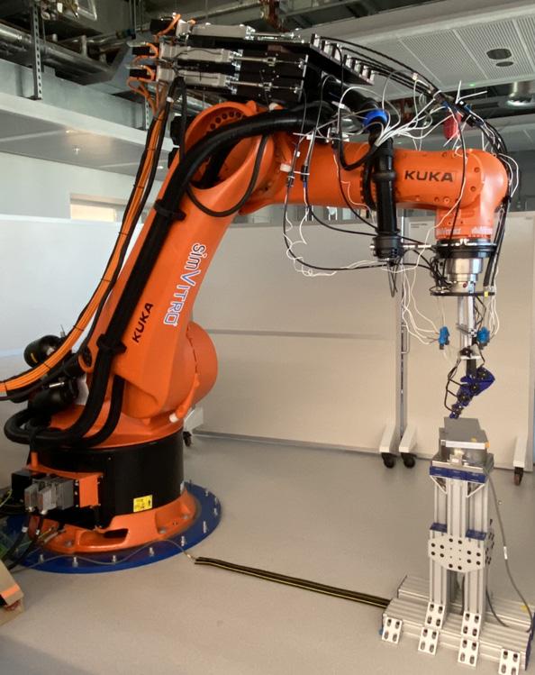
differences between specimens are taken into account. Muscles and joint forces for each condition tested – e.g., typical and intact, injured, simulated pathological, or surgically repaired –may be assessed. Changes in force can be used to inform surgical technique modifications, design of orthopaedic hardware, and rehabilitation protocols.
Our system is proven to accurately and repeatably recreate the movements of walking. We are now working to apply this tool to clinical questions and are open to collaboration ideas and proposals. Our aim is to help to improve clinical interventions, so that people can remain independently mobile and pain-free.
If you would be interested in getting involved with future projects, we would love to hear from you - please email: akedgley@imperial.ac.uk n

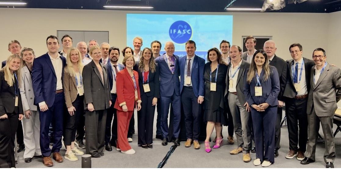
The International Foot & Ankle Sports Consensus (IFASC) group convened its third annual scientific meeting during the ESSKA Congress 2026 in Prague, Czech Republic, bringing together an international faculty of orthopaedic surgeons, sports medicine physicians, researchers, physiotherapists, and trainees dedicated to advancing evidence-based care for athletes with complex foot and ankle injuries.
Founded in 2024 by Dr. Arianna L. Gianakos, Director of IFASC, the initiative was established in response to the increasing need for structured international collaboration surrounding challenging injuries in elite athletes, particularly in areas where high-level


Arianna Gianakos Yale Medicine, Orthopedics, and Rehabilitation, New Haven, CT, USA
evidence remains limited and treatment algorithms continue to evolve. Since its inception, IFASC has grown into a multidisciplinary global collaborative involving experts from North America, Europe, South America, Asia, and Australia, with a shared mission of developing clinically meaningful, consensus-driven recommendations for sports-related foot and ankle pathology.
The group’s foundational work has focused on injuries frequently encountered in high-performance sport, including Lisfranc injuries, Jones fractures, and navicular stress injuries. Through the integration of systematic review methodology and formal expert consensus processes, IFASC has sought to generate practical guidance
surrounding diagnosis, operative and non-operative management, rehabilitation, biologic augmentation, and return-to-play decision-making. Multiple consensus statements developed through the collaborative have subsequently been accepted for publication in Knee Surgery, Sports Traumatology, Arthroscopy (KSSTA).
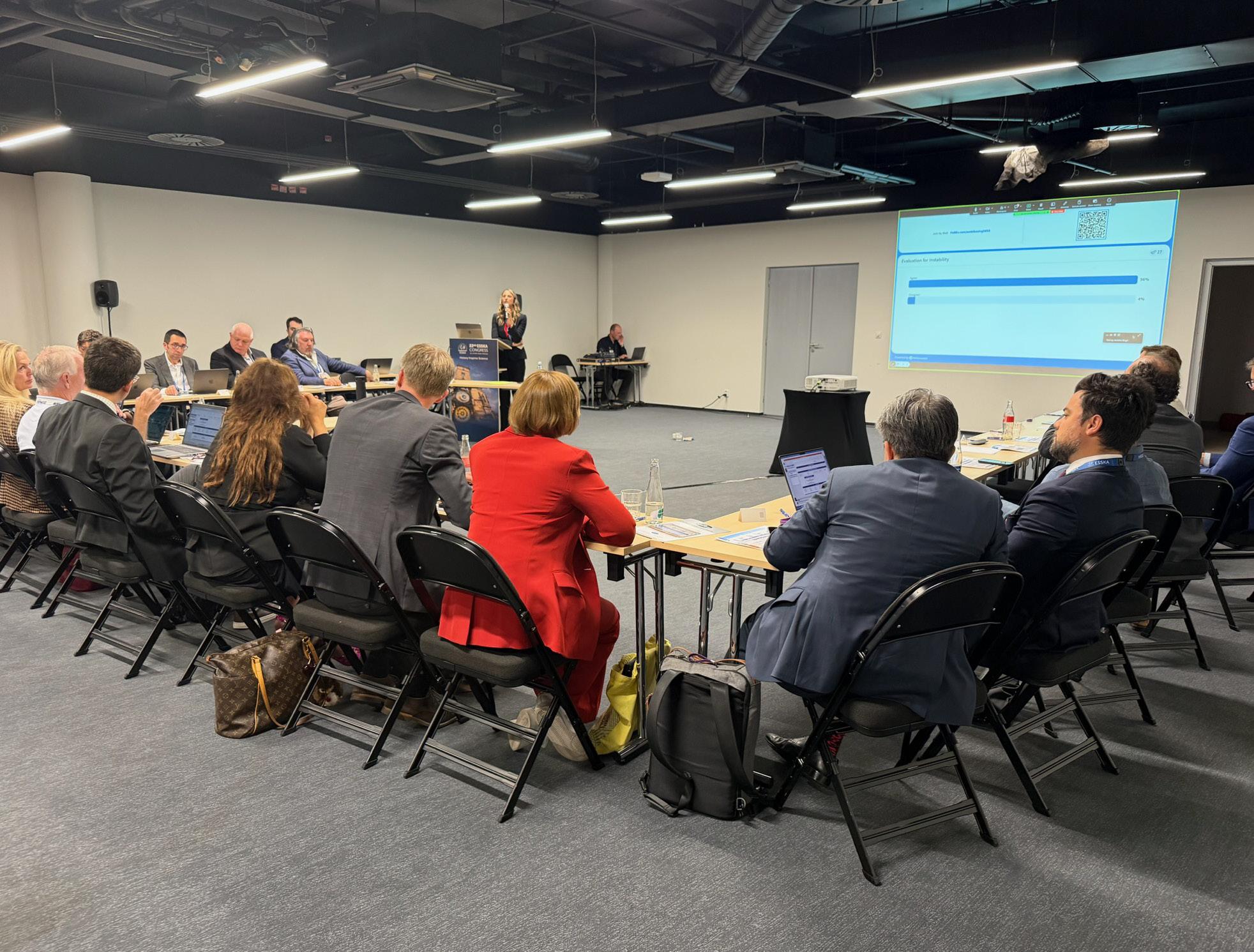
The 2026 ESSKA scientific session centered on syndesmotic (“high ankle”) injuries in elite athletes — a persistently challenging area within sports medicine given the variability in injury patterns, instability characteristics, treatment strategies, and return-to-sport timelines across athletic populations. International faculty discussions focused on contemporary approaches to diagnosis, imaging, instability classification, operative fixation, rehabilitation, and performance optimization following injury. Particular emphasis was placed on evolving concepts extending beyond traditional ligament-based classifications, including dynamic instability, injury severity stratification, sport-specific demands, and emerging technologies in management.
In addition to its scientific contributions, IFASC continues to prioritize mentorship and academic development as central components of its mission. The collaborative intentionally integrates residents, fellows, junior faculty, physiotherapists, and research
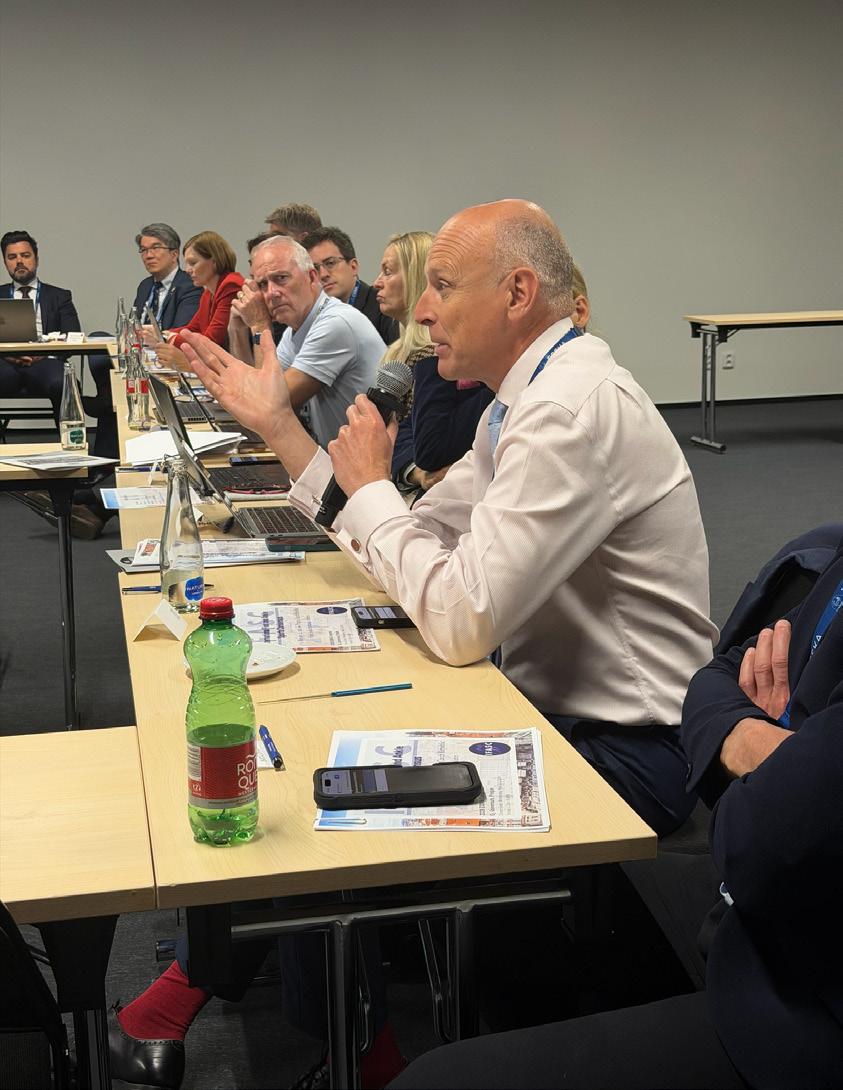
“The 2026 ess K a scien T ific session cen T ered on syndesmo T ic (“high an K le”) injuries in eli T e a T hle T es — a persis T en T ly challenging area wi T hin spor T s medicine.”
trainees into ongoing projects and consensus initiatives, fostering international academic engagement and multidisciplinary scholarship across varying stages of training and practice. This structure has facilitated not only meaningful academic productivity, but also the development of a growing global network committed to advancing athlete care through collaboration, education, and innovation.
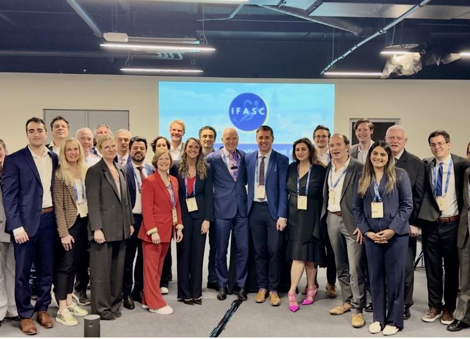
Alongside the scientific programme, IFASC hosted an international faculty and networking event in Prague, further strengthening relationships among collaborators from multiple continents and subspecialty backgrounds. The event reflected the broader ethos of the organization: combining rigorous
scientific collaboration with longterm international partnership and mentorship within the orthopaedic sports medicine community.
As IFASC continues to expand, future initiatives will focus on athlete-specific treatment algorithms, biologic and minimally invasive technologies, injury prevention strategies, and the development of further international consensus projects aimed at improving outcomes for athletes worldwide.
The IFASC group would like to sincerely thank ESSKA, its international faculty, collaborators, trainees, and industry partners for their continued support of this growing global initiative. n
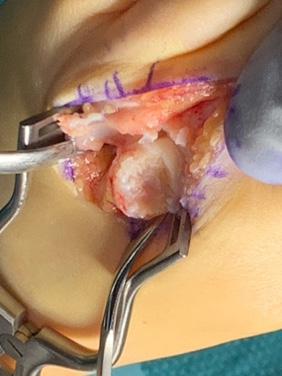
Osteochondral lesions (OCLs) of the metatarsophalangeal (MTP) joints represent a challenging clinical entity, frequently affecting young and active individuals. Among these, Freiberg’s disease—an osteonecrosis of the lesser metatarsal heads, first described in 1914—remains the most recognised pathology. The aetiology of these lesions is multifactorial, involving vascular compromise, repetitive microtrauma, biomechanical abnormalities, and anatomical predispositions. Progressive cartilage degeneration and subchondral bone collapse ultimately lead to joint dysfunction and chronic pain.
Freiberg’s infraction represents similar pathology to OCL of Talus involving the entire osteochondral unit and the subchondral bone. The outcome of any treatment therefore depends not only on a single preferred procedure but addressing the entire osteochondral unit improving the quality of the underlying subchondral bed for complete remodelling.
Recent advancements in biological reconstruction and joint-preserving surgical techniques, particularly Autologous Matrix-Induced Chondrogenesis (AMIC), have introduced promising strategies for restoring joint integrity. AMIC combines microfracturebased marrow stimulation with scaffold-assisted cartilage regeneration, aiming to promote neo-osteogenesis, chondrogenesis, and angiogenesis. The critical determinant of the outcome heavily relies on excising the necrotic bone and cartilage and replacing it with viable osteogenic and chondrogenic unit.
This article reviews the current understanding of the aetiology and
MBBS, MS (T&O), DNB (T&O), FRCS (T&O)
Consultant Foot and Ankle Surgeon Queen Elizabeth Hospital, Gateshead
pathophysiology of OCLs in the MTP joints, with a focus on Freiberg’s disease. It also explores the biological rationale and surgical principles underlying AMIC, emphasising its role in addressing both structural and functional deficits in osteochondral pathology.
The aetiology of OCLs in the MTP joints is multifactorial, with several contributing factors often coexisting. It predominantly affects adolescent females and patients during periods of rapid skeletal growth. Hormonal influences and activity patterns may also contribute, although these remain incompletely understood.
Certain anatomical variations increase the risk of OCLs:
• A relatively long second metatarsal
• A rigid second tarsometatarsal (TMT) joint
• Limited adaptability to load distribution
These structural factors in combination with Footwear-related compression produce high joint reaction forces and stress on the metatarsal head, predisposing it to injury.
Chronic mechanical overload can result in localised pressure necrosis, ischaemic changes and tissue death. The cumulative microtrauma results in subchondral bone fatigue, cartilage breakdown and progressive lesion formation. The vascular anatomy of the lesser metatarsals is relatively tenuous, making them susceptible to avascular necrosis leading to necrosis of subchondral bone, loss of structural support and secondary collapse of the osteochondral unit.
Continued loading leads to:
• Flattening or reversal of the normal convexity of the metatarsal head
• Chondral fissuring, delamination of cartilage and fragmentation of the articular surface
• Formation of osteophytes and loose bodies
• Loss of joint congruity
These changes ultimately result in persistent pain, reduced range of motion and functional impairment.
The most widely used classification system for Freiberg’s disease is Smillie’s classification, which describes progressive stages of the disease:
• Stage I: Subchondral fissure fracture
• Stage II: Early collapse of the dorsal metatarsal head
• Stage III: Depression of the central articular surface
• Stage IV: Advanced collapse with loose bodies
• Stage V: End-stage degenerative arthrosis
This classification guides treatment decisions and helps predict prognosis.
Biological Basis for AMic
Autologous Matrix-Induced Chondrogenesis (AMIC) is a one-step procedure designed to enhance cartilage repair by combining microfracture with a biological scaffold.
Key Components of AMIC
1. Microfracture (Nano-fracture)
• Penetration of subchondral bone
• Release of bone marrow elements
• Initiation of repair cascade
2. Scaffold Application (e.g., Chondrogide)
• Provides structural support
• Stabilises clot formation
• Promotes chondrogenic differentiation
3. Biological Augmentation
• Bone marrow aspirate
• Growth factors
• Tissue adhesives
Principles of AMic in MtP Joint reconstruction
The surgical philosophy underlying AMIC in MTP joints can be summarised into three key principles:
1. Radical Debridement
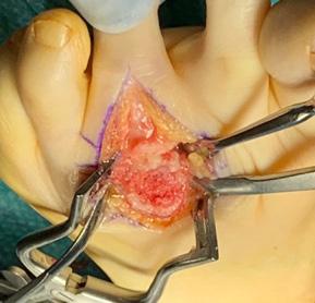
Surgeons must excise all unstable, necrotic cartilage and remove nonviable bone. Aim is to establish stable margins or contained cylinder to support the overlying scaffold. It must include preparation and microfracture of the recipient bed to promote osteointegration.
This step ensures a healthy biological environment and optimal conditions for regeneration.
2. Biological Regeneration
Reconstruction of osteochondral unit aims to stimulate:
• Neo-osteogenesis (Deposition of osteo-inductive cancellous bone in the subchondral bone to promote remodelling process)
• Neo-chondrogenesis (AMIC: cartilage regeneration via scaffold)
• Neo-angiogenesis (vascular ingrowth and bone marrow stimulation-BMS, through microfracture)
These processes collectively restore the viable osteochondral unit, capable of remodelling.
3. Mechanical Stability
Similar to OCL Talus reconstruction, stability is critical for successful healing of metatarsal lesions. The aim is to establish a concrete stabilisation of the scaffold which can resist delamination and displacement throughout the active and passive range of movements. This may need converting uncontained defects into contained ones to create a stable bone graft bed. >>
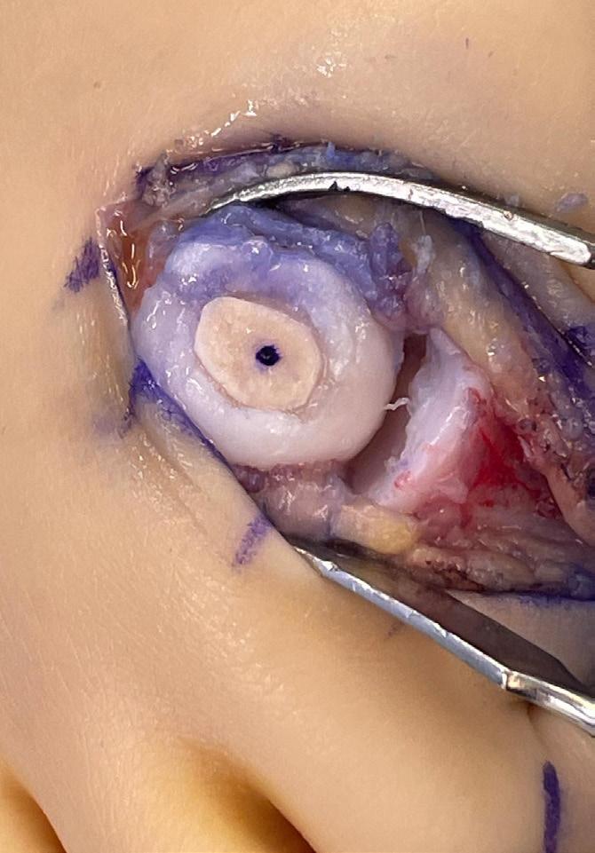
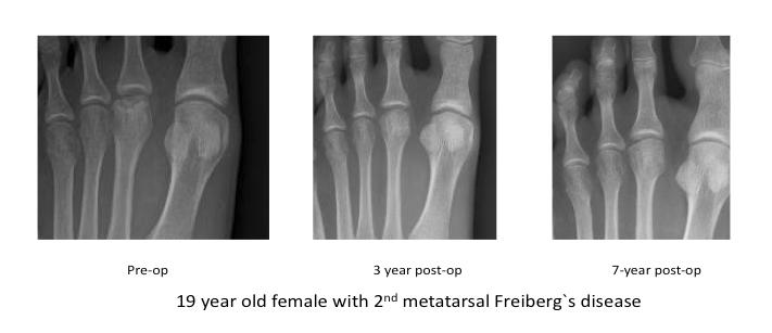
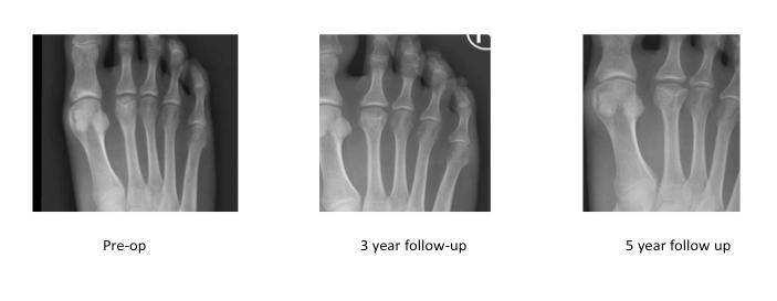
Additional stabilisation techniques include shaping the defect of the bed and the scaffold to a spherical defect to resist shear forces and use of tissue sealants with bone marrow aspirate augmentation.
The goal is to recreate a stable, biologically active environment conducive to tissue regeneration. This change has clinically important significance as it reinforces the new approach of regeneration through reconstruction rather than merely debridement and BMS. The evolution reflects a shift from traditional purely stimulatory intervention to more promising structural and biological
reconstruction. When the quality of subchondral bone is poor, debridement and BMS technique becomes less reliable leading to recurrence.
Osteochondral lesions of the MTP joints present unique challenges due to small joint size, high load-bearing demands and limited intrinsic healing capacity.
Traditional surgical approaches, such as debridement or osteotomy, often fail to restore normal joint anatomy. In contrast, AMIC with bone grafting addresses both structural defects (bone loss) and biological deficits (cartilage damage).
The success of AMIC depends heavily on precise surgical technique and restoration of remodelling capable osteochondral unit. While early results are promising, further studies are needed to compare AMIC with other cartilage repair techniques, evaluate long-term outcomes and define optimal rehabilitation protocols.
By integrating principles of biological regeneration and mechanical stability, AMIC offers a joint-preserving solution with encouraging long-term outcomes. As evidence continues to evolve, AMIC may become a cornerstone technique in the management of osteochondral lesions of the forefoot. n
Powered by SYNTHESTM
Mini Fragment Plating System
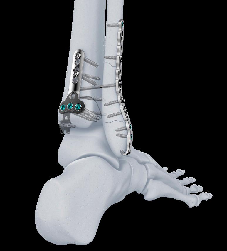
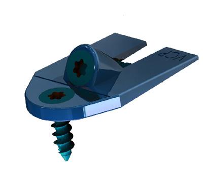
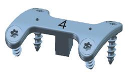
VOLT: Variable Angle Optimized Locking Technology is an innovative evolution in fracture management from Johnson & Johnson MedTech designed to improve stability, enhance performance, expand options, and increase efficiency
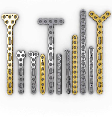
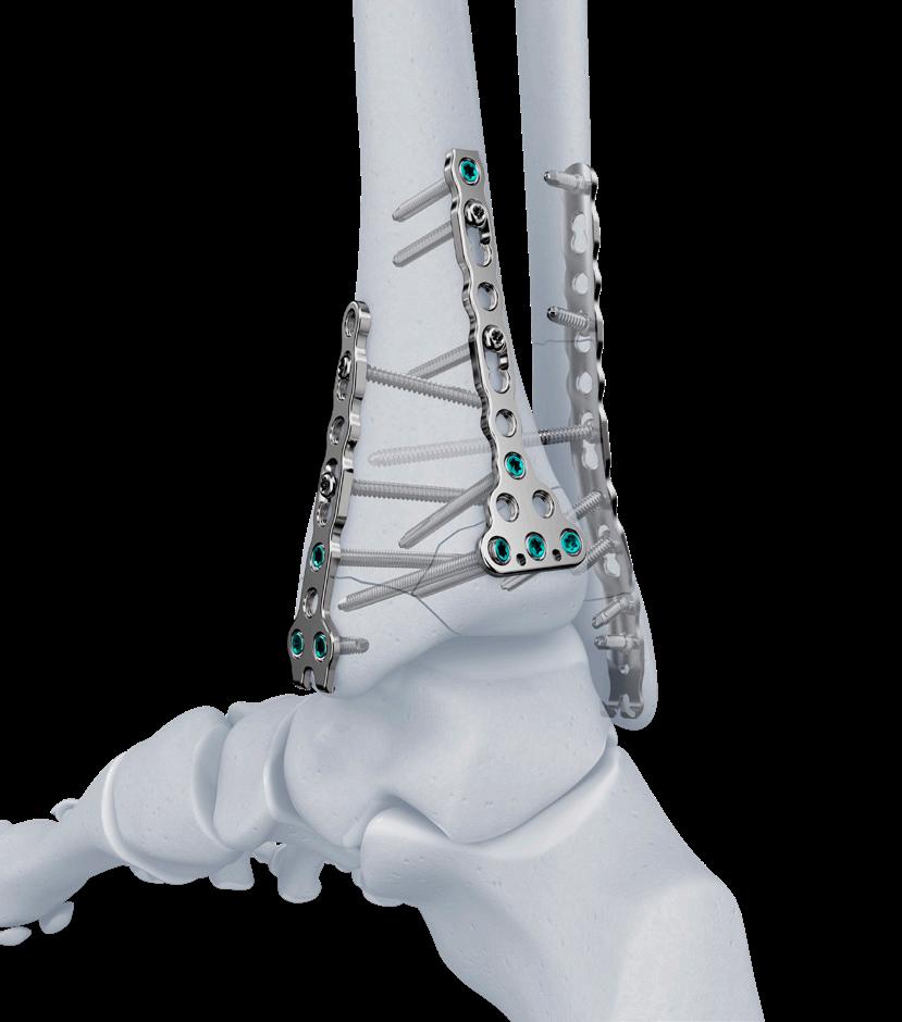
For additional details, refer to the following: https://www.jnjmedtech.com/en-US/product/volt-mini-fragmentplating-system or https://www.jnjmedtech.com/en-EMEA/product/volt-mini-fragmentplating-system Important


Powering the next step forward
Powering the next step forward


Osteochondral
Osteochondral lesions of the talus (OLT) represent a heterogeneous spectrum of pathology involving the articular cartilage, the osteochondral unit, and the subchondral bone. Contemporary operative management should therefore not be framed as the selection of a single preferred procedure, but rather as a structured decision process based on lesion morphology, biological reparability, mechanical environment, and prior treatment history. The updated recommendations of the “Working Group on Tissue Regeneration” of the German Orthopaedic Association have recently published a summary on the current evidence and suggested a treatment algorithm1. The critical determinants of treatment choice are lesion chronicity, the presence or absence of a viable osteochondral fragment, cartilage integrity, defect size, defect depth, cyst formation, and concomitant instability or malalignment.
A central conceptual advance in the current framework is the explicit integration of ankle mechanics into operative decisionmaking. Osteochondral reconstruction performed in the presence of residual instability or pathologic malalignment is biologically and biomechanically disadvantaged. The recommendation to identify and correct instability and malalignment is therefore not ancillary, but foundational. Persistent instability has been associated with inferior postoperative outcomes, and the available data support concomitant stabilisation when instability is present. Similarly, although the anklespecific literature on alignment correction is less robust than in other joints, the general principle of load normalisation before or during cartilage restoration is well established. In scientific terms, cartilage repair should be regarded as loaddependent tissue regeneration within a
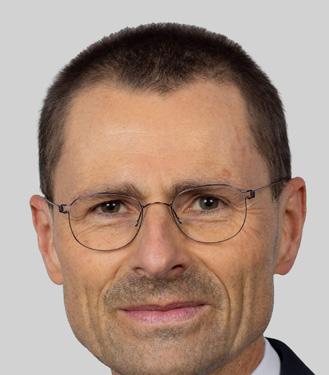
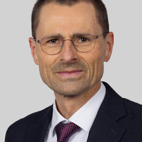
Prof. Markus Walther
Schön Klinik München Harlaching –FIFA Medical Centre of Excellence
Ludwig Maximilian University Munich, Klinik für Orthopädie und Unfallchirurgie
Justus Maximilian University Wuerzburg
joint-preserving construct. Figure 1 reflects this by placing evaluation and correction of malalignment or instability high in both the acute and chronic pathways, appropriately emphasising mechanical normalisation as an upstream therapeutic imperative.
In the acute traumatic setting, the presence of a sufficiently large and viable osteochondral fragment defines a distinct therapeutic category. When the fragment contains an adequate osseous component and is judged reparable, fixation remains the preferred tissue-preserving strategy. This offers the possibility of restoring native hyaline cartilage and the original subchondral architecture rather than substituting them with reparative tissue or a graft construct. The operative principles are those of osteochondral reduction and internal fixation: removal of interposed fibrous tissue, preparation and perforation of the recipient bed to promote osseous integration, and addition of bone graft where chronicity or bone loss has compromised the substrate. Fixation is not indicated for isolated chondral lesions without meaningful bony support, but in appropriately selected acute lesions, it remains the most anatomically restorative
intervention. Accordingly, the algorithm directs acute lesions with an osteochondral fragment and preserved cartilage toward fragment fixation.
This recommendation extends, with appropriate caution, to skeletally immature patients. In children and adolescents who fail conservative treatment, preservation of the native fragment should be considered when the lesion remains biologically plausible for fixation. The rationale is the same as in adults, namely preservation of tissue quality and restoration of the native osteochondral unit. Nevertheless, this indication should not be expanded indiscriminately. Fragment fixation is technically demanding, and access often necessitates a malleolar osteotomy, particularly for medial lesions. Furthermore, the viability and structural integrity of the fragment remain essential prerequisites.
When the articular surface is intact, retrograde techniques retain a limited but valid role. Retrograde drilling is best understood as a treatment for contained subchondral pathology beneath an intact cartilage cap. Its principal theoretical advantage is preservation of the articular surface while stimulating healing in the subchondral compartment. However, the current recommendations narrow its indication considerably. Cartilage integrity should not be inferred solely from magnetic resonance imaging, because radiologic assessment may overestimate the degree of surface preservation. Arthroscopic confirmation is therefore advisable whenever clinical uncertainty persists. More importantly, the available clinical results do not support the broad use of retrograde drilling, particularly in adults. Accordingly, retrograde drilling should be reserved for carefully selected lesions characterised by intact cartilage, absence of cystic degeneration, and a lesion morphology that does not justify surface violation or
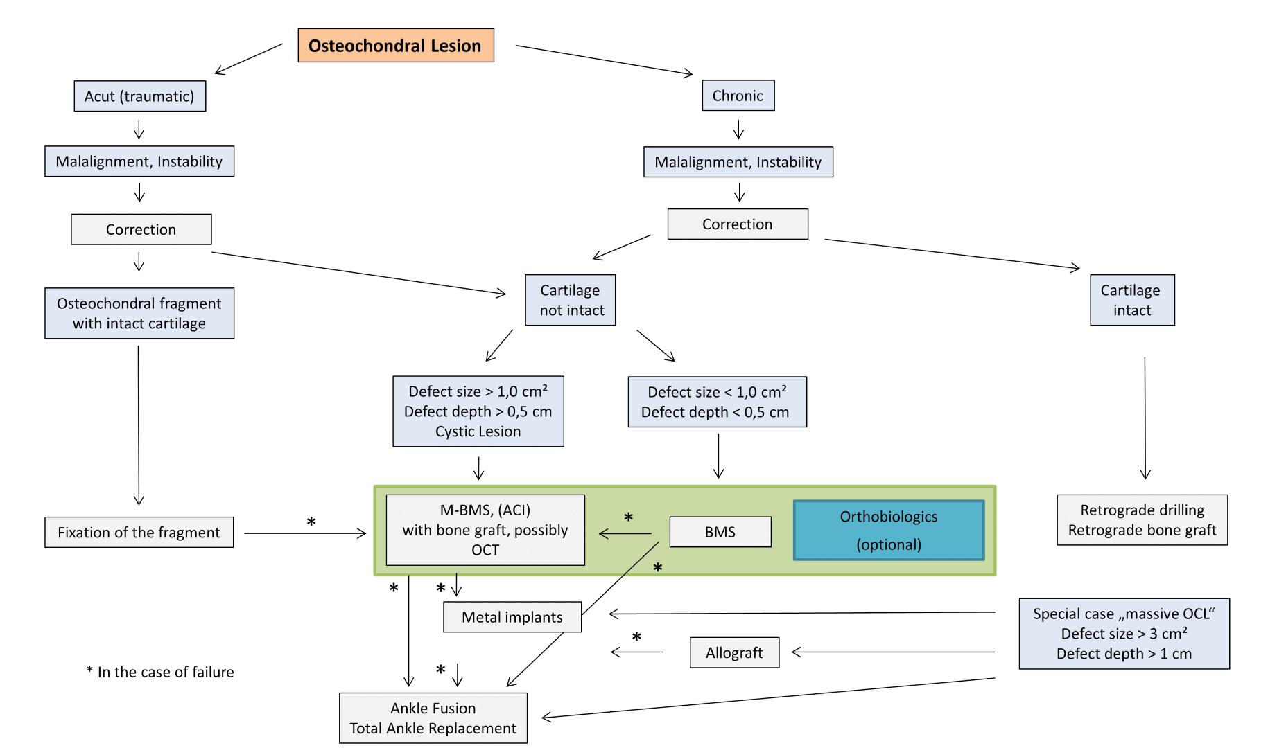
structural reconstruction. Once the cartilage is disrupted, alternative treatment concepts become more appropriate.
Retrograde bone grafting or retrograde application of bone substitute occupies a similarly restricted indication. This strategy is directed toward cystic subchondral defects beneath an intact articular surface and is intended to restore subchondral support while preserving the cartilage cap. While conceptually attractive, the recommendation remains guarded because lesion debridement through a limited retrograde tract is inherently constrained. Complete removal of cyst contents, fibrous tissue, or necrotic bone may be difficult to achieve, which introduces uncertainty regarding the biological quality of the prepared bed. The current framework, therefore allows retrograde grafting as a selected option, but not as a broadly endorsed solution. In practical terms, it is most defensible in lesions with preserved surface integrity and well-defined subchondral pathology.
Once the cartilage surface is no longer intact, debridement and bone marrow stimulation (BMS) remain central
to operative management. The recommendation is to excise unstable cartilage and small detached fragments, establish stable lesion margins, and perform marrow stimulation by microfracture or micro-drilling. There is no clear clinical superiority of one marrow stimulation technique over the other. BMS remains a rational first-line procedure because it is minimally invasive, reproducible, and clinically effective in properly selected lesions. At the same time, its limitations should be explicitly recognised. The repair tissue generated by marrow stimulation does not recapitulate native hyaline cartilage, and clinical durability decreases as the structural burden of the defect increases.
The most relevant threshold modification in the updated recommendations concerns the upper limit for isolated BMS. Whereas earlier frameworks commonly tolerated lesions approaching 1.5 cm², the current algorithm moves the practical boundary closer to 1.0 cm². This change has important clinical consequences because it reclassifies many previously “borderline” lesions as defects more appropriately managed with a reconstructive rather
than merely stimulatory strategy. The depth threshold of 0.5 cm reinforcing the principle that increasing structural deficiency reduces the suitability of isolated BMS. In scientific terms, lesion size and depth are no longer secondary modifiers of BMS success; they are primary determinants of whether BMS remains biologically and mechanically credible as a stand-alone intervention.
For lesions that are larger, deeper, or cystic, the current recommendations increasingly support matrix-associated bone marrow stimulation, frequently in combination with bone grafting. This evolution reflects a shift from purely stimulatory concepts toward structuralbiological reconstruction. The most robustly supported implementation is AMIC/M-BMS using a collagen I/III membrane, particularly ChondroGide. Where subchondral bone is deficient, the defect requires restoration of bony support; where the lesion is more extensive, marrow stimulation alone becomes less reliable, and stabilisation of the reparative milieu becomes desirable. In this setting, scaffold augmentation serves both as a containment strategy >>
and as a means of improving the local environment for tissue repair. For mediumsized and larger OLTs with disrupted cartilage and subchondral compromise, this has become one of the principal contemporary recommendations.
Osteochondral transplantation (OCT) remains a highly relevant option, especially after failed BMS. Its principal advantage is the transfer of mature osteochondral tissue, thereby offering immediate restoration of the osteochondral unit rather than reliance on fibrocartilaginous repair. In appropriately selected lesions, OCT can produce very good clinical results and remains one of the most effective biological salvage strategies. However, the recommendations appropriately temper this favourable view with recognition of donor-site morbidity and technical demands. Harvest-related morbidity at the knee is not negligible, and the procedure often requires a perpendicular access trajectory that may necessitate malleolar osteotomy. As such, OCT is best considered a potent but more invasive biological reconstruction method, particularly appropriate after failure of firstline marrow stimulation rather than as a universal initial strategy. Figure 1 captures this nuanced role by positioning OCT adjacent to the reconstructive pathway for larger lesions and within the broader failuremanagement context.
Autologous chondrocyte implantation (ACI) occupies a more equivocal place in the current treatment landscape. ACI can yield favorable clinical outcomes and may be viable in larger lesions. However, ACI does not provide clear value over acellular scaffold-based reconstruction in the talus. In the absence of demonstrated superiority, the disadvantages of ACI become more prominent: staged surgery, cartilage harvesting, cell expansion, increased cost, and logistical complexity. Consequently, ACI is not rejected, but neither is it advanced as the dominant reconstructive strategy in routine practice.
Adjunctive orthobiologic measures are incorporated into the algorithm in an intentionally subordinate fashion. The current recommendations do not treat platelet-rich plasma, marrow-derived
concentrates, or analogous biologic adjuncts as standalone therapies capable of substituting for structural reconstruction. Rather, they may be considered as optional additions to an already appropriate surgical concept. Biological adjuncts may improve the milieu of repair, but they do not negate the need to decide whether the lesion requires fixation, BMS, scaffold-supported reconstruction, transplantation, or salvage. Likewise, minced cartilage and adiposederived mesenchymal stem cell therapies remain investigational in the ankle. Their role is therefore supportive and potentially beneficial, but the evidence is still limited.
Several strategies are either no longer recommended or remain insufficiently validated for routine clinical use.
Anterograde transmalleolar drilling has effectively lost its place in contemporary management because of unfavourable clinical performance and procedurerelated morbidity.
Metallic resurfacing is assigned a distinctly salvage-oriented role. It is not presented as equivalent to biological reconstruction, but rather as an option for selected revision scenarios after failure of primary cartilage treatment, particularly when alternatives are narrowing toward arthrodesis or total ankle arthroplasty. Metal resurfacing may provide symptomatic improvement in some patients; outcomes remain inferior to successful biological restoration, and revision risk remains relevant. Metallic resurfacing should be understood as an intermediate salvage measure within a joint-preserving spectrum, not as a primary reconstructive recommendation for standard talar OCD lesions.
“Massive OCL” are defined as a lesion exceeding 3 cm² in area and 1 cm in depth. Such defects are qualitatively different from the more common OLT encountered in routine practice. They represent a level of structural loss that exceeds the practical reconstructive range of standard marrow stimulation–based or focal autograft-based strategies. For these lesions, the current framework directs treatment toward allograft-based reconstruction. Some lesions require restoration of a larger structural segment.
The current recommendations adopt a pragmatic stance toward medial malleolar osteotomy. Although osteotomy carries measurable risks, including displacement, step-off, and less favourable imaging features, its influence on final clinical outcome appears relatively modest compared with lesion characteristics, mechanical factors, and reconstructive choice. Osteotomy should therefore neither be pursued casually nor avoided at the cost of inadequate lesion management. Rather, it should be employed when required to obtain the access necessary for anatomically and biologically sound reconstruction.
In summary, the current talar OCD management algorithm is best characterised as a hierarchical, lesionspecific, and biomechanics-informed framework. Viable acute fragments should be fixed when possible. Intact cartilage lesions may be treated by selected retrograde techniques if morphology is appropriate. Isolated BMS remains valid, but primarily for truly small and relatively shallow defects. Larger, deeper, or cystic lesions should prompt earlier transition to scaffold-supported biological reconstruction, often with bone grafting. OCT remains an important second-line biological option, particularly after failed BMS. Orthobiologics are adjunctive. Metallic resurfacing, allograft reconstruction, arthrodesis, and total ankle replacement belong to salvage or extreme-defect pathways. Above all, instability and malalignment should be corrected whenever present. Figure 1 synthesises these principles effectively and should be read not merely as a flowchart, but as a concise expression of the contemporary operative philosophy for osteochondral lesions of the talus. n
Reference
1. Walther M, Gottschalk O, Aurich M. Operative management of osteochondral lesions of the talus: 2024 recommendations of the working group 'clinical tissue regeneration' of the German Society of Orthopedics and Traumatology (DGOU). EFORT Open Rev 2024; 9: 217–234. doi:10.1530/ EOR-23-0075
The treatment of symptomatic flexible flatfoot often combines hindfoot valgus and forefoot supination/varus repair (to restore medial arch). The medial sliding calcaneal osteotomy and also the Cotton osteotomy (Medial cuneiform opening wedge osteotomy) are two well-established components of this reconstruction.
In traditional practice, the opening wedge in the Cotton osteotomy is filled with structural bone graft (usually harvested from the iliac crest) or a synthetic bone substitute. In many settings, especially resource poor ones, synthetic grafts may not be available. Moreover, obtaining graft from faraway sites such as the iliac crest may increase operating time and donor-site morbidity. This article describes a simple surgical tip: using the bony step created during medial sliding calcaneal osteotomy as a local autograft source for the Cotton osteotomy.
Surgical concept
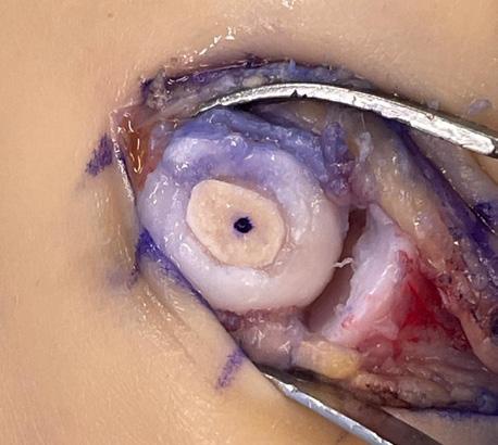
During medial sliding calcaneal osteotomy, the posterior calcaneal is translated medially. This maneuver frequently creates a lateral step or bony prominence at the osteotomy site.
Surprisingly, in our country, no step plate is available for the fixation of osteotomy steps. This step is used as a structural autograft to fill the gap of the opening wedge made during the Cotton osteotomy.
This approach allows the surgeon to utilize local bone that would otherwise be unused, eliminating the need for additional graft sources.
Surgical technique
Step 1
Medial Sliding Calcaneal Osteotomy
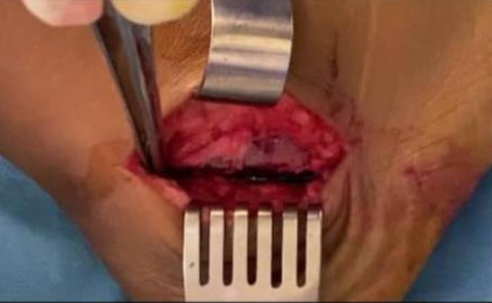
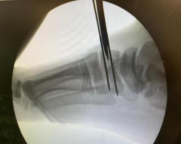
An oblique lateral incision is used to perform the medial sliding calcaneal osteotomy. To correct hindfoot valgus.
Dr Montaser Fatooh
Orthopedic and Trauma Senior Specialist, Foot and Ankle Unit, Bashaer University Hospital
Assistant Professor, Alneelain University-Sudan
The posterior calcaneal is translated medially to correct hindfoot valgus. After translation, a bony step remains laterally at the osteotomy interface.
Step 2
Harvesting the Local Autograft
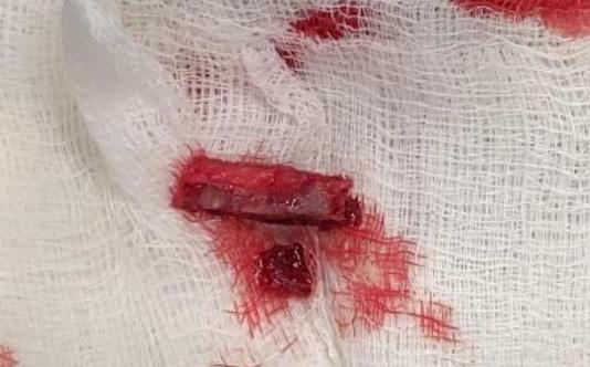
The osteotomy's lateral step is shaped and harvested as a corticocancellous graft. An osteotome or small saw can be used for this.
The graft is then shaped to the size of the wedge required for the Cotton osteotomy.
Step 3
Cotton Osteotomy
A dorsal opening wedge osteotomy is performed in the medial cuneiform. The harvested calcaneal graft is inserted into the osteotomy site to maintain the correction and restore the medial column alignment.
Step 4 Fixation
• The calcaneal osteotomy is fixed using cannulated screws.
• The Cotton osteotomy may be stabilized by the structural graft itself or with additional fixation if necessary.
Advantages of this technique
1. Avoids Synthetic Bone Grafts
In some countries or institutions,
synthetic bone substitutes are not available or are expensive. This technique eliminates the need for such materials.
2. Avoids Additional Donor Site Morbidity
Traditional autograft harvesting from the iliac crest or other sites can lead to:
• Additional postoperative pain
• Increased operative time
• Risk of complications
Using the calcaneal step avoids these issues.
3. Efficient Use of Local Bone
The technique recycles bone created during the primary osteotomy, maximizing available resources during surgery.
4. Reduced Postoperative Pain
Because no secondary donor site is required, patients may experience less postoperative pain compared with iliac crest graft harvesting.
5. Unavailability of a Step Plate
Due to the unavailability of a step plate in our setting, fixation following calcaneal osteotomy was performed using cannulated screws to stabilize the osteotomy site.
6. Cost-Effective
This approach is particularly valuable in resource-limited healthcare systems, where implant availability and bone substitutes may be limited.
Practical tips
• Ensure that the harvested graft is structurally strong enough to support the Cotton osteotomy wedge.
• Shape the graft carefully to maintain the desired medial column correction.
• Confirm forefoot alignment intraoperatively before final fixation.
conclusion
Using the bony step from medial sliding calcaneal osteotomy as a local autograft for Cotton osteotomy is a simple and practical technique. It allows surgeons to avoid synthetic graft materials and additional donor-site morbidity while maintaining effective correction of flatfoot deformity. This approach may be particularly useful in low-resource settings, but it can also serve as a costeffective alternative in standard practice. n
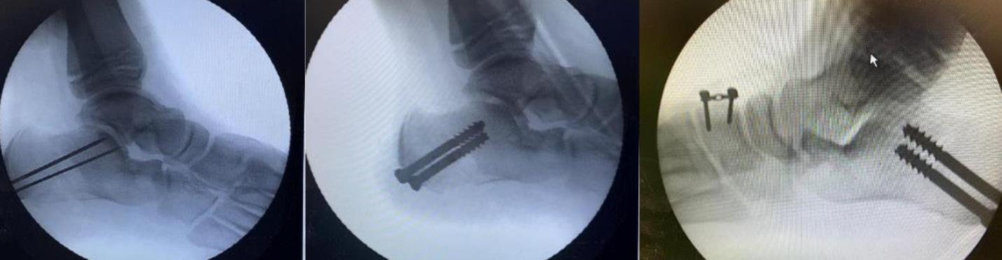
Consent is a process, and patients faced with major surgery often struggle to take in much of what was said during their clinic appointment. Furthermore, patients will often search the internet for information about procedures before they ever meet a doctor. They therefore commonly develop preconceived notions or have conflicting opinions about an operation about which they understand little, yet which has potentially life-changing consequences for them. Faced with this, the limited time of a clinic appointment is often insufficient for patients to make a fully informed decision about whether or not to have a procedure such as an ankle fusion.
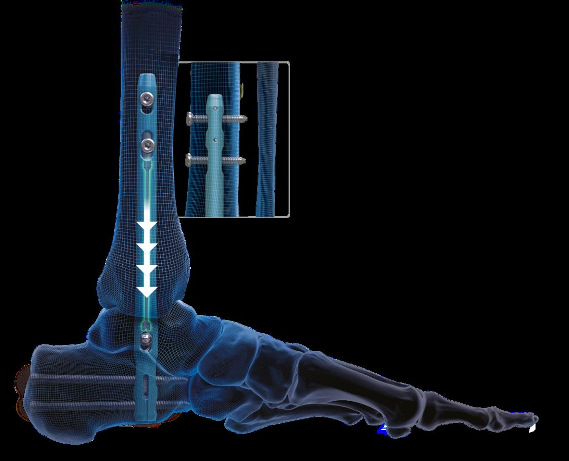
We can mitigate by handing out leaflets, showing patients videos, preparing detailed clinic letters, and scheduling follow-up appointments. However, this isn’t always enough: patients like to ask questions, yet in the clinic environment, many questions will go un-asked. Patients also love the power of anecdote – i.e. they like to talk to other patients.
At the RNOH foot and ankle unit, we feel strongly that shared decision-making is important; but this requires patients to be able to truly understand what is being said to them and frame it within the context of their lives. To achieve this, we must improve our patients' knowledge and understanding of their condition, discuss all relevant treatment options, discuss the risks and benefits and their relevant importance, and get buy-in from the patients. If we can achieve this, it results in greater patient satisfaction and adherence to treatment, an improved doctor-patient relationship and, perhaps most importantly, it empowers patients to make informed choices.
WHy AnKle ArtHritiS?
Ankle fusion is one of the biggest fusions we perform regularly, and ankle arthritis is encountered very commonly across most foot and ankle clinics. Decisionmaking is further complicated by the option of ankle replacement: many patients feel they would not do well with loss of movement, but perhaps this is because they don't really understand what a fusion actually entails. The concept of a fused joint is complex and requires a degree of anatomical and functional knowledge that patients are rarely given the time to develop.
In response to these challenges, the Fusion Forum was developed around fifteen years ago at the RNOH. It is a nonclinic forum where patients with ankle arthritis, considering an ankle fusion, are invited in a group to meet the team, receive information at a more relaxed pace, and where they can meet a guest patient with a previous ankle fusion.


Karan Malhotra Consultant Orthopaedic Foot & Ankle Surgeon
The forum is run by our clinical nurse specialists, and attended by other allied health professionals such as occupational and physiotherapists. The aim of the forum is to empower patients to make informed choices about surgery, in a setting that is less pressured than a clinic
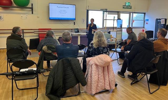
appointment, and gives them genuine time to digest information and ask questions. Lasting one hour, the session consists of a structured presentation reinforced with written information, followed by time for questioning, open discussion, and oneto-one support. Relatives can also attend, which is important, as these decisions are rarely made alone and home/family support is needed after an ankle fusion.
The session is deliberately broad, because the questions patients have are varied in scope.
• Anatomy: A patient cannot meaningfully consent to losing movement in a joint they don't understand.
• Treatment Options: Both operative and non-operative treatment options are discussed.
• On the Day of Surgery: The patient pathway is explained in full, including the anaesthetic and the surgical technique.
• Recovery: Weight-bearing status is a concern for many, and the therapy component includes practical advice on mobility, stairs, and personal care, including preparation and practice before the operation. Self-care, management of the home environment, transport, and family support are all discussed. Wound management, elevation, and anticoagulation are covered.
• Return to Mobility/Work: Life after a fusion - return to work, travel, sport, footwear, and driving. These are often the questions patients are most anxious about, and answering them honestly, including the timescales, is something patients find most useful.
• Health Education: Smoking cessation and weight management are addressed as practical steps which directly reduce the risk of post-operative complications; patients tend to respond to this framing far better in a peer setting than in a clinic room.
This session lends itself to being run by specialist nurses and other allied health professions; this also reflects how MDT care actually works. Patients are cared for pre-operatively almost exclusively by the surgical team, but, peri- and postoperatively, they are managed by nurses, physiotherapists, occupational therapists, and allied health professionals. However, they seldom meet the MDT prior to surgery. Having a genuine MDT input before major surgery is therefore not just beneficial, but a logical approach. The Forum runs as a charged clinic attracting its own tariff, which is an important point for any trust considering replication.
One of the aspects of the forum that patients value most is the previous Ankle Fusion patient. Each session includes a previously treated patient who has volunteered to share their experience. Attendees can watch them walk, which is often more instructive than anything we say or show them on a video. Seeing another patient with a fused ankle in motion and seeing that the person is getting on with their life does more for patient understanding than any diagram of a tibiotalar joint.
The questions are often not those patients typically ask their surgeons, but they are the ones which actually determine whether someone decides to go ahead. Meeting a patient who has been through
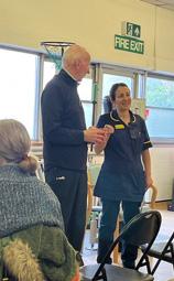
it, and who can speak to that experience without a clinical agenda, is something doctors simply cannot replicate.
We run this once a month on average and 150 patients attended during 2024 and 2025. Approximately 50% of attendees subsequently proceed to surgery. This is not a pathway designed to funnel patients towards surgery; it is one designed to help them decide what’s best for them. Patient feedback questionnaires are used for the development of the service, which has consistently helped alleviate anxiety and improve understanding ahead of a significant decision.
For those patients who struggle to make it to the forum, a virtual version is now available on the RNOH YouTube Channel, along with recorded sessions and patient testimonials. This extends the reach to patients who cannot attend in person, and to clinicians elsewhere who may be considering a similar model.
In a landscape where ankle arthritis networks and regional networks are increasingly part of how we organise care, the Fusion Forum highlights the important role allied health professionals play in the patient journey. Future plans include the development of a dedicated Ankle Arthritis Forum - a logical extension given the rapid changes in surgical treatment options for this condition, and the increasing complexity of the decisions patients face.
The model works because it is honest, unhurried, and supportive. The aim of the forum is to aid decision making, not to direct them towards surgery. It doesn't require a large budget - it requires a committed specialist nurse, a willing guest patient, a room, and the recognition that informed consent is a process. n
The Fusion Forum is led by Karen Alligan, Lead Foot and Ankle Nurse at the Royal National Orthopaedic Hospital, Stanmore.
The virtual fusion forum is available at www.youtube. com/watch?v=ovmAo_gmeN0

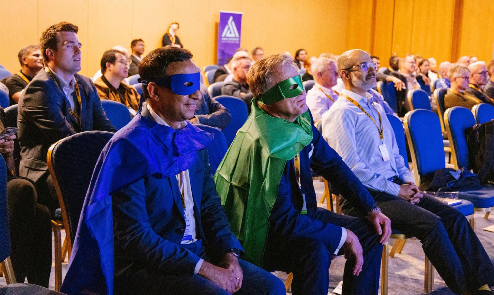
The Ankle Arthritis Academy returned to Newcastle-uponTyne’s iconic Quayside this June, bringing together more than 100 surgeons from across the UK and around the world. Faculty members from the UK, Europe, and Canada joined attendees who had travelled from as far afield as Scandinavia and Singapore for two days of lively discussion, expert lectures, and illustrative case-based debates.
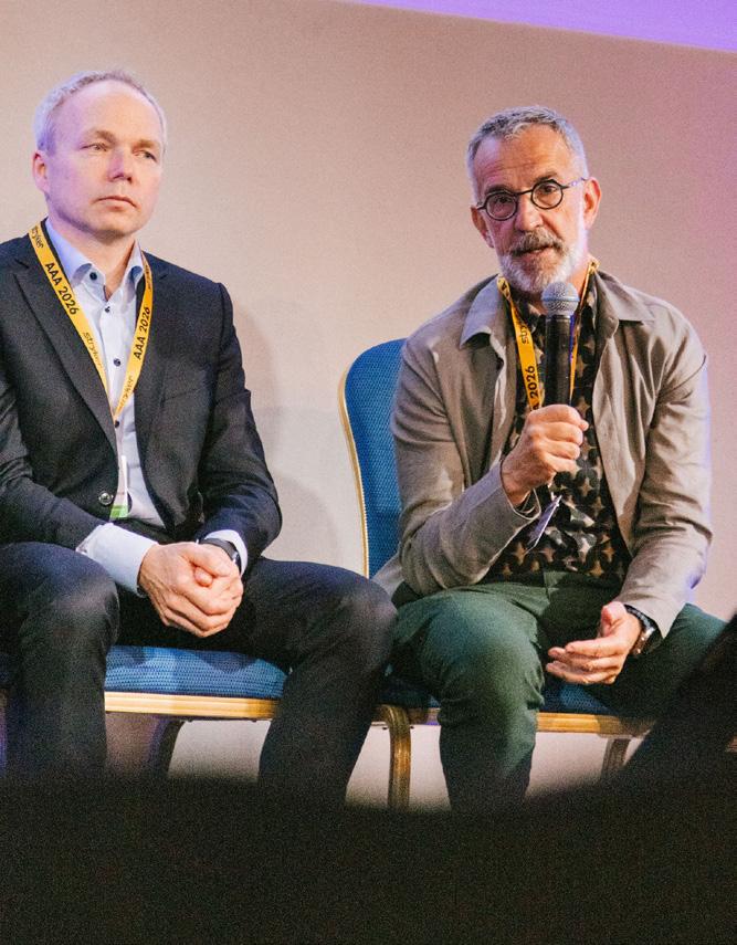
The meeting opened with a session on BOFAS Ankle Networks, an increasingly important topic as services continue to evolve in line with the Getting It Right First Time (GIRFT) agenda. Attention then turned to joint-preserving strategies, including grafting techniques for challenging large osteochondral defects and the role of distraction arthroplasty followed a broader discussion on the evidence currently available to guide clinical decisionmaking in 2026.
Delegates were also treated to practical insights into limb reconstruction, including a timely review of the osteotomy principles that many of us know we should remember but often need reminding of. This was followed by an excellent discussion on patient selection, planning, and surgical technique for supramalleolar osteotomy.
Further sessions covered complex fusion surgery, including long-term outcomes, valuable tips and strategies for managing complications when things do not go according to plan.
A particular highlight was a presentation by Clare Brockett, Professor of Bioengineering, who shared insights from the analysis of retrieved implants following earlier sessions on the choice of fixed- and mobile-bearing designs. A highlight of this year’s meeting was a spirited debate on the management of talar avascular necrosis (AVN).
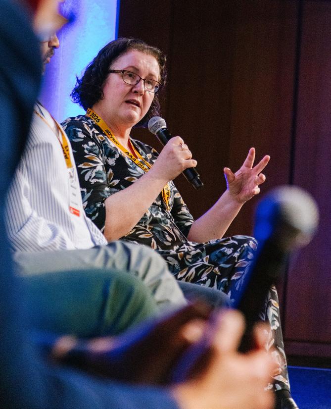
The second day followed an enjoyable evening on the banks of the River Tyne and focused on some of the most challenging problems in contemporary ankle arthroplasty. Topics included managing the learning curve in ankle arthroplasty, treatment of post-traumatic ankle arthritis, deformity correction, and strategies for dealing with complications and failed procedures. The session on prosthetic joint infection highlighted the importance of recognising that ankle arthroplasty cannot simply adopt treatment algorithms from hip and knee arthroplasty. Other discussions explored the management of peri-prosthetic cysts, including when to intervene with grafting and when observation remains appropriate, as well as the technical
challenges and encouraging outcomes associated with revision ankle arthroplasty.
The role of rotational alignment at the ankle, and its relationship to both the foot and proximal joints, continues to attract growing interest. Improved understanding of these interactions is increasingly influencing decision-making in both osteotomy and arthroplasty and was a recurring theme throughout the meeting.
The next Ankle Arthritis Academy will take place in June 2028.
debate case: What Would you do?
One of the meeting's thought-provoking cases involved a 54-year-old man with post-traumatic talar AVN. The talus was non-viable; the ankle and subtalar joints had significant degeneration.
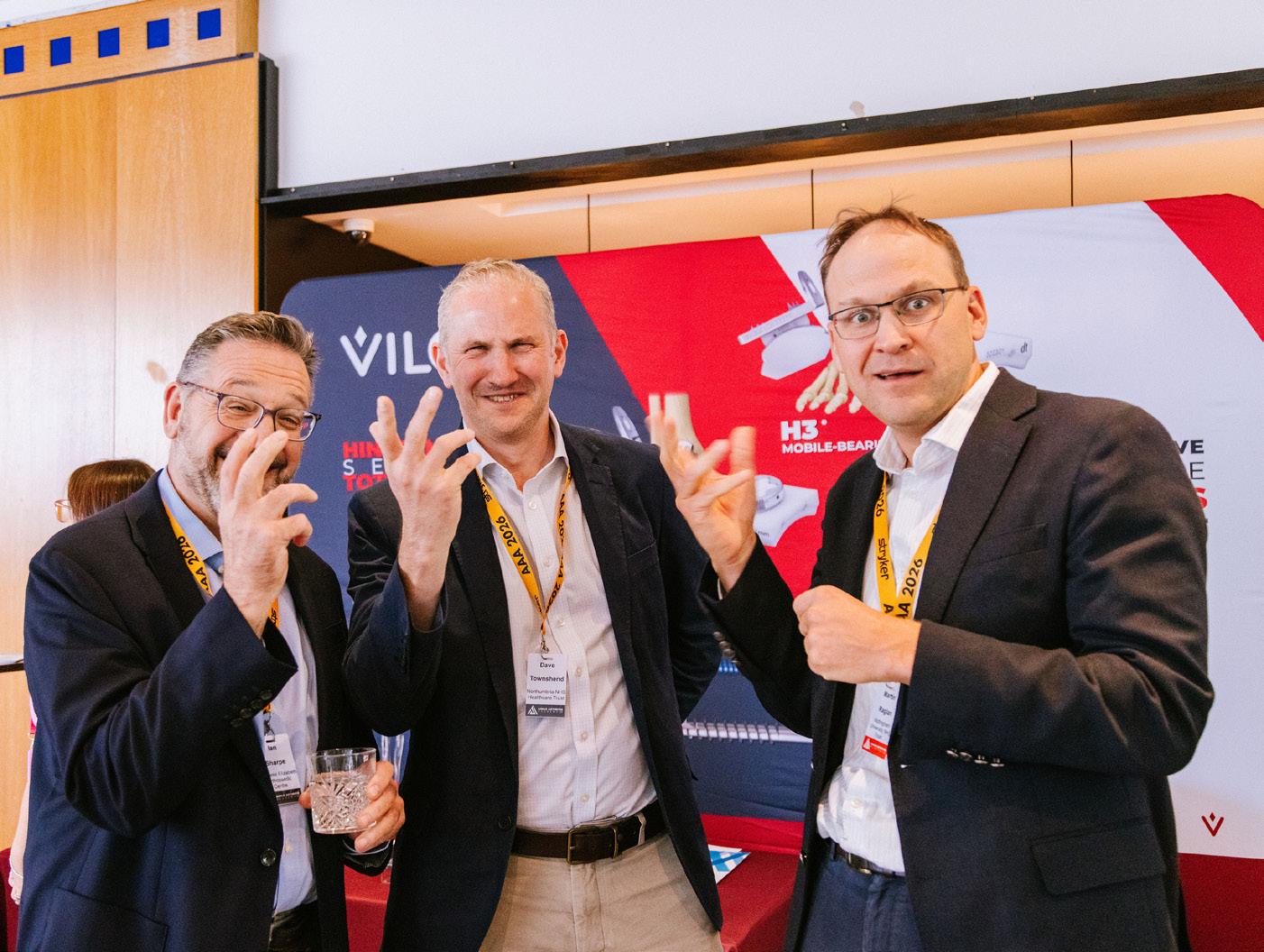
The panel debated three principal treatment options:
• Total talus replacement (with or without total ankle replacement)
• Custom 3D-printed implant fusion
• Impaction bone graft fusion
Opinion in the room was divided. With motion preservation increasingly achievable through advances in custom implants and total talus replacement, would you favour a reconstructive approach or does fusion remain the most reliable long-term solution in the setting of extensive talar collapse and adjacent joint arthritis. What would you do? n
Platelet-rich Plasma vs. Placebo injections in Achilles tendinopathy: A systematic review and Meta-analysis of randomized controlled trials
Authors: Assi, Ahmad; Hamyeh, Ali; Mohanna, Rami; Boutros, Marc; Boueri, Wissam and Sebaaly, Amer
Publication Date: 2026
Journal: Journal of Foot & Ankle Surgery
Abstract: Achilles tendinopathy (AT) is a prevalent overuse injury marked by pain, functional limitation, and structural tendon degeneration. Platelet-rich plasma (PRP) has emerged as a biological treatment alternative due to its regenerative potential. Nonetheless, its efficacy compared to placebo for AT remains unclear. This metaanalysis aimed to evaluate the effectiveness of PRP injections versus placebo in the treatment of AT.
Conclusion: This meta-analysis found no significant clinical or radiological benefit of PRP injections compared to placebo
in the treatment of Achilles tendinopathy. Larger, high-quality trials are needed to confirm these findings and standardize treatment protocols.
DOI: https://libkey.io/10.1053/j. jfas.2026.04.005
incidence, temporal trends, and Surgical Shift of Achilles tendon rupture: A Systematic review and Meta-analysis
Authors: Kotsifaki, Roula; Malliaras, Peter; Byron, Christopher; Marques, Joao and Korakakis, Vasileios
Publication Date: 2026
Journal: Sports Medicine
Abstract: Achilles tendon rupture is a severe injury affecting active and non-active populations, with considerable consequences on mobility, return to sport, and long-term function. Population-based incidence estimates provide important information for public health policy and resource allocation.


Conclusion: The incidence of Achilles tendon rupture has risen steadily over the last six decades, with the greatest burden observed among middle-aged physically active men. A majority of ruptures are sport related, highlighting the need for preventive interventions. Surgical treatment has declined since 2003, reflecting shifts toward conservative management. These findings underscore the importance of continued surveillance, targeted prevention, and individualized treatment strategies to reduce the growing burden of Achilles tendon rupture worldwide.
DOI: https://libkey.io/10.1007/ s40279-026-02397-5 >>
What outcomes matter to patients having surgical treatment for symptomatic hallux valgus?: A systematic review of qualitative studies
Authors: Sinha, Amil;Partha Sarathi, Celine Iswarya;Sedarous, Ramy and Fernandez, Miguel A.
Publication Date: Mar 01, 2026
Journal: Bone & Joint Journal 108-B(3), pp. 282–288
Abstract: Hallux valgus (HV) is a common deformity of the foot, for which various forms of surgical treatment are available. A recent think tank highlighted a lack of understanding about which key outcomes matter most to patients who undergo surgery for HV. The aim of this systematic review was to synthesize the existing evidence to establish whether commonly used outcome measures in foot and ankle surgery adequately reflect what is important to patients.
Conclusion: The Manchester-Oxford Foot Questionnaire was found to be the only outcome measure which specifically addresses these themes, while also including questions about the psychosocial impact. Cosmesis is underrepresented in the outcome measures. Further research is needed to explore the psychosocial factors which are important to patients so that they can be included within the measures which are used to assess outcome after the surgical treatment for HV.
DOI: https://libkey.io/10.1302/0301620X.108B3.BJJ-2025-0628.R1
early vs late Weight Bearing After Ankle Fracture Fixation: A Meta-analysis of randomized controlled trials
Authors: Egu, Chinedu; Akil, Hussein; Hakim, Rebecca Aida; Badhe, Neel; Essiet, Edidiong; Akintunde, Samuel; Woodmass, Michael; Obidigbo, Babajide; Afshari, Amir; Olowookere, Adedayo; Trigkilidas, Dionysios and Chuter, Graham
Publication Date: Feb 2026
Journal: Foot & Ankle International 47(2), pp. 174–183
Abstract: The optimal timing of weightbearing following surgical fixation of ankle fractures remains a topic of clinical debate. Although early weightbearing (EWB) may promote faster functional recovery, concerns about complication risks have limited its widespread adoption. This systematic review and meta-analysis aimed to evaluate the efficacy and safety of EWB compared with LWB in adults undergoing ankle fracture fixation, using evidence from high-quality randomized controlled trials (RCTs) published since 2000.
Conclusion: EWB after ankle fracture fixation leads to improved early functional outcomes and quicker return to work, with no clear increase in complications, in primarily lower-risk patients.
DOI: https://libkey. io/10.1177/10711007251392223
the Ankle recovery trial (Art): clinical outcomes and patient experience of a pragmatic multicentre rct comparing cast with removable boot for early mobilization after ankle fracture surgical fixation
Authors: Martin, Rachel; Docherty, Sharon; Heaslip, Vanessa; Allen, Helen; Tbaily, Lee W.; Hayward, Christopher J.; Marques, Elsa M. R. and Sangar, Anuraag Publication Date: Nov 10, 2025 Journal: Bone & Joint Open 6(11), pp. 1416–1424
Abstract: This study compares functional outcomes and patient experience between cast immobilization and early mobilization in a removable boot after ankle fracture fixation, with early weightbearing encouraged in both groups.
Conclusion: Patients managed in casts and boots had similar functional outcomes following ankle fracture fixation. Boots provided improved dorsiflexion and plantarflexion for some subgroups, but higher complication rates. Treatment modality decisions could therefore be informed by individual patient preference.
DOI: https://libkey.io/10.1302/26331462.611.BJO-2025-0108.R1 n
For recently published systematic reviews and meta-analyses in PubMed:
https://tinyurl.com/footprintSR
For recently published clinical trials:
https://tinyurl.com/footprinttrials
For recently published qualitative work:
https://tinyurl.com/footprintqual
If you have any feedback, please contact: pip.divall@uhl-tr.nhs.uk

The Global Foot & Ankle Summit (GFAS) 2026 concluded successfully in Goa, India, reaffirming India's growing prominence as a global destination for advanced foot and ankle surgical education and international academic collaboration.
Held from 27 February to 1 March 2026, the three-day summit, brought together some of the world's most renowned foot and ankle surgeons, researchers, educators, and healthcare professionals. With participation from more than 20 distinguished international faculty members and hundreds of delegates from India and abroad, GFAS 2026 emerged as one of the largest and most academically enriching foot and ankle conferences ever organised in the country. The meeting provided an exceptional platform for the exchange of scientific knowledge, clinical expertise, and innovative surgical techniques.
The summit was jointly led by its Course Chairs, Dr. Girish Motwani, Director of Foot & Ankle Services at Suyog Hospital, Nagpur, and Executive Committee
Member of the Indian Foot and Ankle Society (IFAS), and Dr. Rahul Upadhyay, Consultant Foot & Ankle Surgeon, New Delhi, and Executive Committee
Member of IFAS. Under their leadership, the conference delivered a world-class academic
program that attracted leading experts from across the globe. The event was efficiently coordinated by Dr Mahendra Kudchadkar, Local Organising Chairman, along with Dr Inderjit Singh and Dr Dhrumin Sangoi, who served as Organising secretaries.
The scientific theme of GFAS 2026, "Revisions and Complications in Foot and Ankle Surgery," focused on addressing some of the most challenging scenarios encountered in modern foot and ankle practice. Through interactive case discussions, expert panel deliberations, instructional lectures, debates, and hands-on workshops, participants gained valuable insights into the management of complex deformities, failed surgeries, ligament injuries, arthritic conditions, sports injuries, and revision procedures.

One of the most significant achievements of the summit was the conduct of India's first-ever Delphi Consensus Meeting on Acute Deltoid Ligament Injury. International experts collaborated to discuss key clinical controversies and work toward evidence-based recommendations aimed at standardising treatment approaches worldwide. The initiative is expected to contribute significantly to future scientific publications and clinical practice guidelines. GFAS 2026 also demonstrated its commitment to nurturing the next generation of specialists through a dedicated Rising Star Program, where promising young surgeons received opportunities for academic recognition, mentorship, and international exposure. In addition, multiple hands-on workshops enabled delegates to refine their surgical skills under the direct guidance of globally recognised faculty members.
Feedback from participants was overwhelmingly positive, with many describing GFAS 2026 as one of the best-organised and most scientifically relevant foot and ankle meetings they had attended. The quality of discussions, international faculty participation, and the focus on real-world clinical challenges were particularly praised.
Building on the tremendous success of the Goa meeting, the organising committee officially announced that the next Global Foot & Ankle Summit (GFAS) will be hosted in Nagpur, India, in February 2028. The forthcoming edition is expected to attract an even larger international faculty and feature advanced cadaveric workshops, cutting-edge technology demonstrations, collaborative research initiatives, and innovative educational formats. As GFAS continues to expand its global footprint, the summit remains committed to its mission of advancing foot and ankle surgery through excellence in education, research, innovation, and international cooperation. With Nagpur set to host the next chapter of this growing global movement, GFAS is poised to further strengthen India's position at the forefront of foot and ankle surgical excellence. n “ f eedbac K from par T icipan T s was overwhelmingly posi T ive, wi T h many describing gfas 2026 as one of T he bes Torganised and mos T scien T ifically relevan T foo T and an K le mee T ings T hey had a TT ended.”
If you have a hobby you would like to share with us, please email the editor at footprint_editor @bofas.org.uk
Complex surgery is often exciting and challenging but sometimes feels like a voyage into the unknown. However, it is strikingly similar to sailing offshore into the unknown. Both require prior planning, functioning in a team and always learning new skills.
For 16 years, around the longest day each June, we sail out of a new port for offshore adventure, following the wind and tides for a week as nautical wild rovers. A crew of 6 to 8 amateur sailors are crammed into

a 40-foot yacht This helps me to recharge my batteries of endurance and wellness. It first started after five years of life as a new Consultant, when my wife noticed I was being slowly ground down by the perpetual cycles of hospital deadlines. She reminded me of my former love of the sea and the perpetual rhythms of the ocean tides. The spark to launch the first adventure was a joint orthopaedic meeting of British and French paediatric orthopods in Normandy.
We boldly set sail, arriving off the D-Day beaches at dawn after a midchannel lightning storm, followed on the way home by more terrifying adventures in fog in the shipping lanes. None of us attended the orthopaedic lectures in France, but a passion was born!
The crew is a brotherhood of orthopaedic surgeons, a GP, and an ENT surgeon, interwoven with an architect, spook, engineer, bus driver and most usefully a psychologist, who has a lot of work to do! We have all learned new skills, kept night watches together, created delights in the galley and pretended not to be scarred in big waves and 51kt winds off the Fastnet. The reliance on each other at sea helps create the friendships for the rest of the year.
The passage plan requires details, such as tidal streams and water depths, which mimic the pre-operative planning for a revision hindfoot reconstruction. At sea, an error is the difference between gently gliding alongside a pontoon by a harbour pub or abruptly running aground and calling a lifeboat. Surgical attention to detail is required to sail thru a fishing area at night, where missing a red over white light may led you to be the catch in a fishing net.
For me this is my complete escape from the grinding pressures of waiting times, staff shortages and clinical decision making. I return with my batteries recharged to face these once more. Once safety ashore, in the first Monday morning theatre list there are definite symptoms of a lingering PTSD –post traumatic sailing delight.
Rick Brown n
DUROLANE hyaluronic acid (HA) therapy:
• Provides longer-lasting OA knee pain relief versus intraarticular steroid*1
• DUROLANE has a half-life of 30 days in the human joint with a single-injection treatment regimen.2
• HA may, with repeated courses of single-injection treatment, delay the need for joint replacement surgery3


UK/IR
Email: cc@BioventusGlobal.com
UK toll free: 0800 05 16 384

Fax: 0207 147 1430 IR toll free: 00800 02 04 06 08 and 00800 03 05 07 09 Fax: 0165 39103 Germany
Tel toll free: 0800 - 183 37 35 Fax: 032 - 22 10 94 056
*Based on OMERACT-OARSI responder rate at 26 weeks vs methylprednisolone acetate (MPA).
References: 1. Leighton R, Åkermark C, Therrien R, et al. NASHA hyaluronic acid vs methylprednisolone for knee osteoarthritis: a prospective, multi-centre, randomized, non-inferiority trial. Osteoarthritis Cartilage. 2014;22(1):17-25. doi:10.1016/j.joca.2013.10.009 2. Edsman K, Hjelm R, Lärkner H, et al. Intra-articular duration of Durolane™ after single injection into the rabbit knee. Cartilage. 2011;2(4)384-8. doi:10.1177/1947603511400184 3. Romero Jurado M, Enrique Fidalgo A, Rodríquez Villar V, Mar Medina J, Soler López B. Factors related with the time to surgery in waiting-list patients for knee prostheses. Reumatol Clin. 2013;9(3):148-55. doi:10.1016/j.reuma.2012.09.003
Summary of Indications for Use: DUROLANE (3 mL): Argentina, Australia,† Brazil, Chile, Colombia, EU,† India, New Zealand,† Russia, Switzerland,† Turkey,† United Arab Emirates: Symptomatic treatment of mild to moderate knee or hip osteoarthritis. In addition, DUROLANE has been approved in Australia, EU and New Zealand for the symptomatic treatment associated with mild to moderate osteoarthritis pain in the ankle, shoulder, elbow wrist, ingers, and toes.
Mexico: Symptomatic treatment of mild to moderate knee osteoarthritis.
Taiwan: Treatment of pain in OA of the knee in patients who have failed to respond adequately to conservative non-pharmacologic therapy and simple analgesics, e.g., acetaminophen.
Canada:† Symptomatic treatment of mild to moderate knee or hip osteoarthritis. In addition, DUROLANE has been licenced for the symptomatic treatment associated with mild to moderate osteoarthritis pain in the ankle, fingers and toes.
Summary of risks: You should not use DUROLANE if you have infections or skin disease at the injection site. DUROLANE has not been tested in pregnant or lactating women, or children. Risks can include transient pain, swelling and/or stiffness at the injection site.
†DUROLANE is also indicated for pain following joint arthroscopy in the presence of osteoarthritis within 3 months of the procedure. Full prescribing information can be found in product labeling, or at DUROLANE.com

Scan here to register for the event
Sports Medicine
Friday 14 August 2026 | 18:30 - 21:30
Brewerkz One Fullerton
1 Fullerton Rd, #01-01 One Fullerton, Singapore 049213
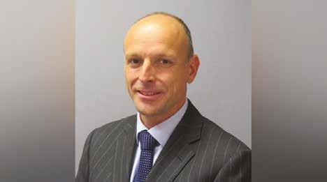
London, UK
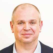

Queensland, Australia
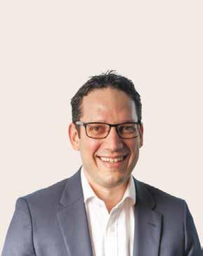
Perth, Australia

Join us for the Foot and Ankle Innovation Evening, a dynamic event showcasing the latest advancements in sports medicine technology and techniques. This exclusive gathering will feature expert-led case discussions and engaging panel discussions focusing on cutting-edge solutions for Foot and Ankle procedures.