13 minute read
Cervical Oesophageal Rupture in a Pregnant Mare Shaan Mocke, Gareth Fitch & JoAnna Faircloth


CERVICAL OESOPHAGEAL RUPTURE IN A PREGNANT MARE.
Shaan Mocke S.Mocke@massey.ac.nz
Gareth Fitch leefitch@cityu.edu.hk
JoAnna Faircloth Dr.joannafaircloth@gmail.com
SUMMARY
Successful resolution of a case of oesophageal rupture with Horner syndrome in a late-term mare was achieved with surgery, insertion of a temporary indwelling oesophagostomy tube and enteral feeding.
Figure 1: Soft tissue swellings seen on left neck at first visit.
SIGNALMENT AND HISTORY
A 7-year-old standardbred broodmare was reported by the owner to be dull and had separated herself from the broodmare herd. She was primiparous and due to foal in 6 weeks’ time. She was initially examined in the field, and was quiet, but still alert and responsive. On physical examination the mare was mildly tachycardic, other parameters were within normal limits, including her temperature.
She had several soft-tissue swellings located on the lateroventral aspect of the caudal third of her neck (Figure 1) which were warm to the touch. There was no evidence of external trauma. She had been observed drinking and ate grass that was offered without hesitation. Phenylbutazone was administered intravenously at a dose rate of 4.4 mg/ kg and dimethyl sulfoxide applied topically. The owner was instructed to keep her in a yard overnight and monitor comfort, water intake and appetite. The following day, clinical signs worsened. The swellings had increased in size, and the owner reported that the mare had nasal discharge and an unusual sweating pattern around her head.
EXAMINATION FINDINGS
On presentation at the clinic, she was more lethargic than the day before, there was bilateral green nasal discharge, moderate tachycardia, 55bpm, and pyrexia, 38.90C. There was increased swelling and diffuse cellulitis in the ventral aspect of the caudal third of the neck. The swelling was painful on palpation; however, the mare was able to move her neck normally. The sweating pattern round her head was unilateral (left side only) and was combined with a left-sided ptosis (upper eyelid droop), and left-sided muzzle droop, indicative of Horner syndrome (Figure 2).

Figure 2: Left unilateral sweating of the face with ptosis, consistent with Horner syndrome.
The blood results from the first day revealed leucocytosis, due to a moderate neutrophilia [16.4 x 10⁹ /L] with a left shift [8% band neutrophils]. There was a moderate increase of fibrinogen [50 g/L] and SAA was increased [1278.6 µg/ml]
An endoscopic examination was performed, and she had a left-sided laryngeal hemiplegia (grade 3.1 on the Havemeyer scale), and some pooling of food in the oesophagus. There was also a section of the oesophagus, ~3 cm in length, that was compressed halfway down the neck. The oesophagus itself appeared normal oral and aboral to the area of compression.
Ultrasound of the neck showed significant swelling of subcutaneous tissue along with hyperechoic material outside the lumen of the oesophagus. The jugular vein was normal.
Cervical radiographs were taken before and after feeding of (Iohexol™), The radiographs indicated that there was food material outside of the oesophageal lumen, i.e. an oesophageal tear (Figure 3)

Figure 3: radiograph showing gas and food material in the soft tissues adjacent to the oesophagus.
TREATMENT PLAN
Based on the findings, the mare was diagnosed with cervical oesophageal rupture, presumed to be secondary to an oesophageal obstruction. Both the mare and foal had commercial and sentimental value and the owner was interested in saving both the mare and foal. A guarded prognosis was given for both short and long-term survival. It was decided to perform surgery to allow lavage of the contaminated tissues and place an oesophagostomy tube for feeding for sufficient time to allow the oesophagus to heal by second intention. The procedure was to be performed under general anaesthesia.

Figure 4: Oesophagostomy tube in place.
SURGICAL PROCEDURE
Prior to induction of anaesthesia, a nasogastric tube was passed down the oesophagus to assist with the surgical approach. An incision was made using a ventrolateral approach, distal to site of rupture. A 10cm long incision was made through the skin. The sternocephalicus and brachiocephalicus muscles were separated, and the deep cervical fascia incised to expose the oesophagus. Two longitudinal tears were noted. A large amount of food material had dissected through the fascial planes but did not extend into the mediastinum and thoracic cavity. The food material was removed by lavaging the area with 1:40 dilute povidone iodine solution delivered via a stomach pump and a small nasogastric tube. To allow passage of a feeding tube into the oesophagus distal to the tear, a smaller bore nasogastric tube was placed inside a modified, shortened standard diameter nasogastric tube. The smaller tube was then passed into the distal oesophagus as a guide and then the larger tube passed over this into the stomach, the smaller tube was withdrawn. A Chinese Finger Trap suture was used to secure it in place during recovery.
Broad spectrum antimicrobial therapy was commenced with the mare receiving 22 mg/kg intravenous sodium penicillin three times daily, 6.6 mg/kg intravenous gentamicin once daily and 15 mg/kg enteral metronidazole three times daily. For pain and inflammation, she received flunixin meglumine at doses of 0.5-1.1 mg/kg at 8-12 hourly intervals, either intravenously or orally.
Omeprazole was administered initially at 4 mg/kg PO, reducing to 2 mg/kg after 2 weeks plus ranitidine at 6.6 mg/ kg PO for the first week. Altrogenest (Regumate™, MSD Animal Health) was administered at 22 mg/kg to help maintain pregnancy. Penicillin and gentamicin were discontinued after 9 days and metronidazole was discontinued after 14 days based on bloodwork results.
Feed was enteral only administered via the oesophagostomy tube (Figure 4) and each feed consisted of a slurry of high protein feed given five times daily. The composition of each feed for the first two days was: 100 g of a protein and energy supplement (Rebuild™, Prydes, Aus), 600 g of EasiSport™ (Prydes, Aus), 40 g NaCl, 50 mL rice bran oil and vitamin E as Nano-E™ (Kentucky Equine Research, KY, USA). The feed was pre-soaked in warm water for a minimum of one hour prior to feeding. After two days the Rebuild™ was increased to 250 g and the EasiSport™ to 700 g, and the NaCl was decreased to 25g and the rice bran oil to 15 mL. The total volume per feed was not to exceed 7 L, which included additional water administered to flush the tube. The feeding times were spaced 3-4 hours apart. Prior to feeding the mare would be checked for any reflux, if any was present this feed would be missed until the next scheduled feed. Blood electrolytes were monitored for the first few days (i-STAT Handheld, Abbot) to allow compensation for salivary loss.
Every seven days a gastroscope was passed to assess healing of the oesophagus. The cavity surrounding the oesophagus reduced with formation of granulation tissue until there was a functional oesophageal lumen. The feeding regimen was maintained for a total of three weeks at which time the oesophagostomy tube was removed. The oesophagostomy site surrounded by granulation tissue was allowed to close by second intention. The mare could graze and was able to swallow normally with only a small amount of grass-tinged saliva exiting the site. At this stage she was fed 100 g of Rebuild™, 600 g of EasiSport and 200 g of Biomare (Prydes, Aus) and 10 mL rice bran oil per feed, five times daily. Salt was reduced to 20 g NaCl in two of the feeds only as there was reduced salivary loss.
The mare was discharged to a local stud farm one week prior to her foaling date, with a body condition score of 6/9. Discharge instructions included grass and soaked feed diet only with no hay or haylage, a separate paddock and access to a salt lick.
OUTCOME
The oesophagostomy site granulated in within several days. She did not have any problems eating and was not attended for choke. There was some concern over swelling at site, but this appeared to be associated with superficial scar tissue. Two weeks after discharge, the mare produced a healthy foal (Figure 5). Both mare and foal were reported to be doing well and the mare has subsequently produced another foal.
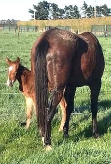
Figure 5: Mare with foal, both doing well.
DISCUSSION
Disorders of the oesophagus include stricture, obstruction, external mass, neoplasia, diverticulum formation and rupture (Bedezkoya 2012, Stick 2012). Oesophageal obstruction (choke) is commonly seen in equine practice with clinical signs such as distress, dysphagia, food material at the nares, coughing, ptyalism (saliva), and palpable lumps along the left neck (Bezdekova2012, Archer 2013).
Oesophageal rupture is an uncommonly reported disease with a poor prognosis with non-surgical management (Whitfield‐Cargile 2013, Craig 1990). There are reports of cases of cervical oesophageal rupture managed successfully following surgical intervention and alimentation via an oesophageal tube placed via the wound or separate oesophagostomy or naso-oesophageal tube (Whitfield-Cargile et al. 2013). Rupture can occur secondary to long-standing obstruction, repeated or aggressive nasogastric tube passage, foreign body perforation, external trauma such as a kick to the neck, or extension of infection from surrounding structures (Bezdekova 2012). Obstruction can cause pressure necrosis to esophageal mucosa which can rupture, with ingesta leaking into the soft tissues of the neck causing severe infection.
Contrast radiography was used to confirm that an oesophageal rupture had occurred. Iodinated contrast medium (Iohexol™), was chosen instead of barium as the latter is contraindicated in suspected perforations as it is irritant to extra oesophageal tissues (Koskinen 2015).
Secure closure of a ruptured or perforated defect is usually possible only in patients operated on shortly after (< 12 h) perforation has occurred (Stick 2012). In early cases, when esophageal tissues are too damaged to hold sutures or when infection or contamination with ingesta has already occurred, some means of draining the esophageal contents to the outside must be provided (Stick 2012).
For much of the cervical portion, the oesophagus is accompanied by the recurrent laryngeal nerves and by the left common carotid artery and its accompanying vagosympathetic trunk in the carotid sheath. It receives its innervation from the cervical sympathetic supply and from the vagus nerves, including the recurrent laryngeal branches; the vagal supply being the most important for motility.
Trauma, peri-jugular injections, abscesses and cellulitis involving the cervical sympathetic trunk can result in Horner syndrome. The classic signs of Horner syndrome are unilateral ptosis (upper eyelid droop and lowered eyelash angle) and miosis, along with ipsilateral facial sweating, and must be distinguished from facial paralysis (Palumbo 2011). Laryngeal hemiplegia will only accompany Horner syndrome if the recurrent laryngeal nerve is affected also (Palumbo 2011). Perhaps most commonly, Horner syndrome is caused by an inadvertent extra-jugular injection and will usually have a good prognosis if the offending compound is not too irritant. The mare in this report had not received any intravenous injections prior to being seen, so this was an unlikely cause of the nerve damage that was almost certainly due to the inflammation of the nerve due to leakage of ingesta and saliva from the rupture site.
Formation of a stoma caudal to the rupture site has been advocated to avoid delayed healing at the site of the rupture (Whitfield-Cargile 2013, Stick 2012), but in this case the rupture occurred at a distal cervical location making creation of a separate incision site caudal to this difficult. The indwelling tube was placed through the distal portion of the oesophagus at the site of the rupture which has been reported to not have any deleterious effect on healing (Leus and Rasmussen 2021, Whitfield-Cargile et al. 2013). Placement of the tube in a standing horse has been described, but int this case general anaesthesia was chosen due to the degree of contamination. It could be argued that a standing procedure would be indicated in a late term mare. It is preferable to leave the tube in place for 7-10 days until granulation forms around the tube to form a fistula (Craig 1989; Freeman 1989; Risnes and Mair 2003; Whitfield-Cargile et al. 2013). In cases of enteral feeding via a nasogastric tube, the tube is left in place until healing is sufficient to allow removal. The tube can be removed between feedings, but once granulation is established the stoma can contract very rapidly making passing of the tube complicated and potentially lead to trauma. In the case of this mare, the tube was left in place for three weeks. In cases of dysphagia due to guttural pouch mycosis, a nasogastric tube has been maintained for much longer periods with no apparent complications. Reported complications include accidental tube removal, oesophageal irritation, development of strictures or diverticula at the site, recurrence of the obstruction, postoperative infection, laryngeal hemiplegia and even carotid artery rupture (Waguespack 2007, Risnes and Mair 2003). The stoma generally heals spontaneously after the tube is removed, with fistula formation being rare (Whitfield-Cragile et al. 2013, Stick 1981).
The neutralising effect of alkaline saliva is lost initially as significant portion of saliva exits the stoma and it is likely that the indwelling tube passed through the cardiac sphincter, although this was not confirmed in this case, which could potentially lead to reflux of gastric contents into the oesophagus. In an attempt to reduce the likelihood of reflux oesophagitis and gastric or oesophageal ulcer formation anti-ulcer medication was used initially. Positioning of the tube within the stomach as apposed to the distal oesophagus reduces the likelihood of dislodgement of the tube (Stick 1981).
Enteral feeding via an oesophagostomy tube in a Quarter horse with an oesophageal rupture has been described in detail (Read et al. 2002), but this is the first report of complete enteral feeding via an oesophagostomy tube in a late gestation mare. Complete enteral feeding via an oesophageal tube is relatively intensive if body condition is to be maintained because it requires feeding at least 5 times a day over a period of many days. The diet in this case was based on estimation of the body weight and energy requirements according to published data (Anderson 2011). This case report demonstrates that it is possible to maintain a mare in late gestation with gastric feeding alone for three weeks and maintain sufficient body condition for tissue repair, and to maintain the pregnancy through until parturition and lactation. The diet was formulated from commercially available feeds supplemented with oil and electrolytes and preparation of the feed is straightforward in that it does not require maceration or blending. No patch was created to cover the stoma during healing as there was minimal leakage of food or water and so it was not deemed necessary. The mare did not have problems with recurrent obstruction and so endoscopic examination of the oesophagus was not performed. Thus, the possibility of formation of a degree of stricture or of a traction diverticulum remained (Whitfield-Cargile et al. 2013), although it was assumed that if there were such complications, there would have been clinical signs during recovery. This mare was fortunate that the food had not dissected distally through the fascial planes and into the thoracic cavity. If there had been infiltration of feed into the mediastinum no attempt would have been made to save the mare due to the poor prognosis for survival.
This case serves to illustrate the clinical signs, diagnosis and management of a cervical oesophageal rupture and to add to the number of cases successfully treated with surgical management.