i
The Murdoch Institute for Research into Birth Defects Limited
ANNUAL REPORT 1987 «
*
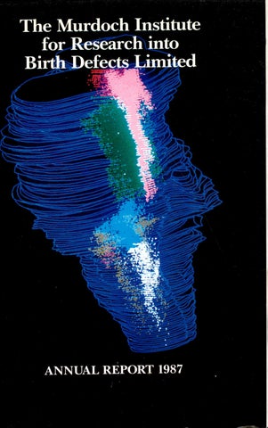
Cover Human brainstem in which the serotonergic neurons are demonstrated by immunocytochemical staining, using the PH8 antibody. Computer assisted three-dimensional reconstruction. Photograph courtesy of Dr. I. Tbrk, School of Anatomy, The University of New South Wales.
1
I
The Murdoch Institute for Research into Birth Defects
1
j II
.a
J
Royal Children’s Hospital, Flemington Road, Parkville 3052.
Ml
The Murdoch Institute combines research into the causes of birth defects with the provision of top-quality counselling and diagnostic services. Few other Institutes in the world combine these two important roles so effectively. The development of the Institute has depended upon the support of generous benefactors and of governments. To continue its progress towards the reduction of suffering due to birth defects, the Institute needs the support of many more generous people. Birth defects are abnormalities and diseases due to causes which are present at birth. Two percent of babies — 5000 a year in Australia have serious birth defects. The burden on these children, their families and the community is immense. Genetic defects are paramount among the causes of birth defects. The Murdoch Institute is one of the better known among the research organisations around the world focussing attention on genetic diseases. All this effort is necessary because there are over 3000 genetic diseases which must be understood and conquered, one at a time.
■j
I
I
'if Genetic services diagnosis and counselling are sought by thousands of Victorian couples each year. The services which we have developed over many years are being recognised and expanded as part of a new agreement between the Victorian Government, the Institute, the Royal Children’s Hospital and other collaborating hospitals. This will give Victoria an arrangement which will be the envy of most other countries around the world. Nowhere else is such a systematic network of clinical services so closely integrated with a major research establishment. The establishment of the Institute in 1985 was made possible by the generosity of the Murdoch family, the late Sir Jack Brockhoff, the Miller family and other donors. The Scobie and Claire Mackinnon Trust and the Helen M. Schutt Trust have now joined the select group of our principal supporters. Our development would not have been possible without the support of the Australian Government, through the National Health and Medical Research Council, and the Victorian Government, through the Department of Health with the encouragement of the Department of Community Services. The Murdoch Institute illustrates what can be achieved by co-operation between private and public resources. Its further progress towards its goal of reducing the impact of birth defects depends on further support from Government and from generous private and corporate supporters.
Research in the Institute concentrates on — genetic diseases which cause copper deficiency or copper toxicity — phenylketonuria and related conditions which cause brain damage, now preventable because of our advancing knowledge — genetic diseases which limit the energy supplies to cells, especially in muscle and brain — methods of diagnosing genetic diseases by laboratory tests and using computerised information systems. The Institute is renowned internationally for discoveries in these subjects. Within Australia the standing of its research was acknowledged in 1986 when it became one of the five Institutes awarded Block Grant funding by the National Health and Medical Research Council.
Training of clinical geneticists is another important role of the Institute which has trained most of those now practicing this specialty in Australia. Four young doctors from other States are currently training in the Institute.
5(
■ii -
CHAIRMAN’S REPORT
INDEX Chairman’s Report Board of the Murdoch Institute Finance Committee Director’s Report Murdoch Institute Lectures, 1987 Visiting Scientists on Sabbatical Leave Staff Involvement in Australian Scientific Community Activities Editorial Boards Overseas and Interstate Visits, Lectures and Seminars by Institute Staff Postgraduate Degrees Awarded Research Collaborations Staff List List of Publications Helen M. Schutt Trust The Scobie and Claire Mackinnon Trust Trace Elements and Human Health — The Scobie and Claire Mackinnon Research Group Possum Clinical Genetic Services Staff Profiles — Trace Element Research Detailed Project Reports Donations to the Murdoch Institute, 1987 Statements of Accounts
5 7 12 14 17 17 ■.
'
17 18 18 19 20 21 22 26 27
28 40 41 42 45 64 65
S
I
fcii
I
Mr. Neil Walford
The outstanding public event of the Institute for 1987, the Opening Dinner held in Eebruary, was described in the 1986 Annual Report. Our Annual General Meeting on May 6 was brief and formal, with invitations issued to Members only. In 1988 and future years we will be holding a different type of Annual Meeting at which the Director and some members of the staff will discuss the work of the Institute. We look forward to seeing a number of our friends and supporters at our Annual Meeting on May 30. The Institute is very fortunate in that it continues to attract major supporters. Elsewhere in this Report details are given of the wonderful new undertakings of the Scobie and Claire Mackinnon Trust and the Helen MacPherson Schutt Trust. Members of the Board look forward to developing and maintaining a close link with the Trustees and hope that they will derive considerable satisfaction from following the research of the Institute in general and especially the projects in those specific sections of the Institute which their grants will be supporting. The Board wishes to record its special gratitude to the members of the Einance Committee and its investment advisors. County Australia, who have
guided our affairs during a difficult year. No doubt the share market crash has had an effect on the value of all invested funds. With well managed funds such as ours, it is a reasonable expectation that these paper losses will be recovered in the future. Anyway, we believe that we have fared relatively well. Moveover, the important reality for the Institute and its supporters is that we have intact in our capital fund an amount equivalent to the total of all donations received to date, despite having expended $3 million on the work of the Institute over the last 2 years. The original aim of our fundraising was to expand our research to approximately double that conducted in 1983. The immediate acquisition of a corpus of $10.5 million would have made it possible to expand to this extent and to sustain that expansion in the future with only minimal additional fundraising provided there was also a substantial increase in our funding from the National Health and Medical Research Council and other granting agencies. Our scientists have achieved the increase in research grants which we hoped for and our donors have promised sums which are very close to our original target, but the spread of payments over 5-10 years reduced the
7
effective cash flow considerably. We are further indebted to the Murdoch family who have recognised this effect upon our long-term cash flow and have very generously advanced their payments ahead of schedule. Indeed, we have received in three years $4.75 million of the $5 million which was promised over 5 years. We are very pleased that the Victorian Government has recognised the importance of genetic services in the prevention of birth defects and has allocated additional support through the Health Department in 1987/88 and has anticipated further additional support in 1988/89. We are proud that the Victorian Clinical Genetics Service will be established as a subsidiary of the Murdoch Institute and see this centralisation as rational and efficient. The Board wishes to express particular gratitude to Mr. David White, Minister for Health and to Mrs. Caroline Hogg, for her support as Minister for Community Services at the time of these negotiations. The Board has taken on a substantial task of finding the funds necessary to locate the new Service on the 10th Floor of the Royal Children’s Hospital, but believes in this type of partnership of private and corporate donors with the Government and is confident that our friends and supporters will rally to make this important new venture a great success.
BOARD OF THE MURDOCH INSTITUTE Mr. N. Walford B.Com., F.C.A. Mr. Neil Walford was elected the first Chairman of the Board in June 1986 following the incorporation of the Institute. He has been a chartered accountant, footwear manufacturer, stockbroker and Company Director. He is a former Chairman of Repco Corporation, Costain Australia, Actrol, a former Commissioner of the State Bank and a former partner in Ord Minnett. He is also currently Chairman of Electrolux Pty. Ltd. and a director of various public and private companies. Mr. N. Walford
Mr. L.G. Cox B.Com., A.A.S.A., F.S.I.A. Mr. Laurence Cox is the Vice-Chairman of the Board of the Institute and the Chairman of the Finance Committee. He is a Director of the Potter Partners’ Group of Companies and has been involved in the securities industry since graduating from Melbourne University in 1959. Mr. Cox has wide experience in both the domestic and international financial markets and he is responsible for his company’s joint venture with S.G. Warburg PLC as well as various other activities. He is Chairman of the Australian Stock Exchange (Melbourne) Limited and a director of the Australian Stock Exchange Limited. His involvement with the Murdoch Institute began some six years ago when he helped form a small group to assist in the initial fundraising program.
I am fortunate indeed in having the assistance of a Board of Directors made up of a number of very able, experienced and dedicated men and women. I am grateful to them for their contribution during the year. The Board congratulates our Director, Professor David Danks, and his staff, on a very productive and successful 1987 year.
Mr. L.G. Cox
Dr. G.L. Barnes M.D., Ch.B., E.R.A.C.P. Dr. Graeme Barnes is the Board member nominated by the Royal Children’s Hospital. He is Director of Gastroenterology, a clinical department with strong research interests. Originally from New Zealand where he obtained his medical degree in 1965, he was appointed to his present position at the Hospital in 1975. His major research interest is in the development of a rotavirus vaccine to prevent severe gastroenteritis in children.
Dr. G.L. Barnes
Mrs. J. Calvert-Jones
Professor G.J. Fraenkel
Mrs. Janet Calvert-Jones represents the Murdoch family on the Board of the Institute. She was a Foundation member of the Advisory Council for Children with Impaired Hearing (Vic.) and has been its Chairman since 1973. Mrs. Calvert-Jones is also a Director of Cruden Investments Pty. Ltd. and a Director of the Herald and Weekly Times Limited. She has recently been appointed to the Council of the University of Melbourne.
A.M., M.A., B.M., M.Ch., Hon. M.D., F.R.C.S., F.R.A.C.S., F.R.A.C.M.A., Hon. F.F.A.R.A.C.S. Professor Gustav Fraenkel was educated at Perse School, Cambridge, and the University of Oxford where he graduated in Medicine in 1943. In 1958 he was appointed the Professor of Surgery, University of Otago, New Zealand. Professor Fraenkel was the Foundation Dean of the School of Medicine at Flinders University South Australia, 1970-1984. He is the Co-ordinator of Research and Chief Executive, Royal Children’s Hospital Research Foundation (appointed 1985), and represents the Foundation on the Board of the Institute.
Mrs. Janet Calvert-Jones
Professor G.J. Fraenkel
Df. B.R. Catchlove M.B., B.S., F.R.A.C.P., F.R.A.C.M.A., F.H.A.
Mr. W.H. Hodgson
Dr. Barry Catchlove is the Chief Executive of the Royal Children’s Hospital. Dr. Catchlove graduated in medicine from Sydney University and is a Fellow of both the Royal Australasian College of Physicians and the Royal Australian College of Medical Administrators. He came to the Royal Children’s Hospital in 1981 following a period as the Director of Medical Services and Deputy Chief Executive Officer of the Royal North Shore Hospital, N.S.W. Dr. Catchlove is also Secretary of the Royal Children’s Hospital Research Foundation.
Mr. Bill Hodgson is the Deputy Managing Director of the National Australia Bank Limited. He is also the Chairman of the Australian Resources Development Bank and of Carrington Confirmers Limited. Mr. Hodgson has had a long career with the Bank, joining the then National Bank of Australasia Limited in Perth, Western Australia, in 1945. He has held senior appointments since 1971 in London, Victoria and Queensland. In April 1981 Mr. Hodgson was appointed General Manager, Corporate and International Banking and after the restructuring associated with the National Australia Bank merger he became General Manager, Corporate Banking. He was appointed Deputy Managing Director in January 1986. He is also a Director of the National Heart Foundation.
Dr. B.R. Catchlove
I
.!
Mr. J.A. Fitzgerald Mr. John Fitzgerald has been Managing Director of International Public Relations Pty. Ltd., Australia’s largest public relations company, since 1982. This followed a period of 28 years in newspapers, the last 5 of which were spent as Editor of the Herald, Melbourne. Mr. Fitzgerald is corporate affairs advisor to some of Australia’s largest corporations.
Mrs. J. Lewisohn B.A. Mrs. Penny Lewisohn is a member of the Committee of Management of the Royal Children’s Hospital and represents the Hospital on the Board of the Institute. She has been a member of the Committee of Management since 1980 and is currently a Vice-President. From 1970-1973 she worked with the Rural Finance Commission administering the Commonwealth Government Rural Reconstruction Scheme. She has been one of the three non-producer members of the Victorian Egg Marketing Board since 1981.
Mr. J.A. Fitzgerald
1%
ma
I tl '"li
^
:i
J Mrs. Penny Lewisohn
! 10
.»:li !
J
1
r-; i Dame Patricia Mackinnon
Dame Patricia Mackinnon
Professor G.B. Ryan
D.B.E.
M.D., B.S., Ph.D., F.R.C.P.A., F.R.A.C.P.
Dame Patricia has had a long and distinguished association with the Royal Children’s Hospital. She joined the Committee of Management in 1948 and served as President from 1965-1979. She was appointed a member of the Board of the Royal Children’s Hospital Research Foundation in 1965 becoming Chairman in 1967, a position she held until her retirement in 1985. Throughout her association with the Hospital and the Foundation she has been an untiring advocate for the support and growth of research. She is a Patron of the Royal Children’s Hospital Auxiliary, and the Royal Children’s Hospital Pied Pipers and an Honorary Life Member of the Royal Children’s Hospital Volunteer Service and the Uncle Bob’s Club. Dame Patricia is also a member of the Victorian Council of the Child Accident Prevention Foundation of Australia.
Professor Graeme Ryan is Dean of the Faculty of Medicine, University of Melbourne. A medical graduate of the University of Melbourne, he was appointed Professor of Anatomy in 1978. His major research interests, still based in the Department of Anatomy, are concerned with kidney structure, function and disease. He became Deputy Dean of the Faculty of Medicine in 1980 and was appointed Dean in 1986. He was appointed Vice-Chairman of the Academic Board and Pro Vice-Chancellor of the University of Melbourne in 1987. He is a member of the Medical Research Committee of the NH & MRC, the Council of the University of Melbourne, the Australian Medical Council, the Zoological Board of Victoria, the Boards of the Howard Florey, Baker, Walter and Fliza Hall, and Ludwig Institutes, the Mental Health Research Institute, the Victoria Institute of Forensic Pathology, and the Potter Foundation.
Mr. J.S. Guest A.M., O.B.F., V.R.D., B.Sc., M.B., B.S., F.R.C.S., F.R.A.C.S. Mr. James Guest is a distinguished Melbourne surgeon. He held the appointment of Honorary Surgeon Alfred Hospital 1952-1976, and has been Consultant Surgeon there since 1976. He was a member of the Board of Management of the Alfred Hospital from 1970-1976. He is a director of the Jack Brockhoff Foundation. Mr. Giiest has served as a Member of the Board of the Peter MacCallum Cancer Institute since 1967 and from 1983 has been its Chairman.
The Scientific Director (Professor D.M. Danks) and the Deputy Scientific Director (Dr. R.G.H. Cotton) are also members of the Board of the Institute.
Mr. J.S. Guest
Professor P.D. Phelan M.D., B.S., B.Sc., F.R.A.C.P.
Professor P.D. Phelan
Professor Peter Phelan is the Stevenson Professor of Paediatrics at the University of Melbourne and a distinguished thoracic physician. He graduated in medicine and science at the University of Queensland, and obtained his M.D. for studies on obstructive airway disease in infancy. He came to Melbourne in 1964 and spent some time as a Research Fellow in the Clinical Research Unit at the Royal Children’s Hospital before taking up a Postdoctoral Fellowship at Harvard University. He was the Director of the Department of Thoracic Medicine at the Royal Children’s Hospital from 1974-1983 when he was appointed to the Chair of Paediatrics.
!
i
■|
J
Professor G.B. Ryan
13
12
Mr. G.E. Heeley
FINANCE COMMITTEE
B.Ec., F.A.S.A.
Chairman: Mr. L.G. Cox Mr. C.P. Abbott
Mr. Geoff Heeley is the Executive General Manager Finance of BHP. He has been with the financial area of the Company since joining in 1956 as a cadet. He is a Director of North West Shelf Development Pty. Ltd., Woodside Petroleum Ltd., Woodside Oil Ltd., Mid Eastern Oil Ltd., Woodside Petroleum Development Pty. Ltd., and a number of BHP subsidiary companies.
L.L.B., C.P.A. Mr. Charles Abbott has been a partner in the firm of solicitors, Blake & Riggall since 1970 (Blake Dawson Waldron as of 1st March 1988). Mr. Abbott’s expertise lies in banking and finance. He is a Director of Norwich Winterthur Insurance (Australia) Limited Group, Norwich Finance Holdings Limited and Norwich Union Life Australia Limited. Mr. G.E. Heeley
Mr. C.P. Abbott
Mr. D.E. Meiklejohn B.Com., F.A.S.A., C.P.A., A.A.U.Q.
Mr. D.T. Craig A.C.A.(N.Z.), A.A.I.B.
Mr. D.T. Craig
Mr. David Craig is the Managing Director of Esanda Finance Corporation Limited the wholly owned finance company subsidiary of ANZ Banking Group Limited. Mr. Craig joined the ANZ Bank in 1955 at Temuka, New Zealand. He has held a number of senior appointments in the ANZ Bank which have included Senior Manager, Customer Investments (1977); Controller AHQ (1979); General Manager, Management Services (1983); Executive Director, Grindlays Bank pic (1984); General Manager, Finance, AHQ (1985); Director, Americas and Pacific Basin (1986). He is an associate chartered accountant (New Zealand) and an associate of the Australian Institute of Bankers.
Mr. David Meiklejohn is an Executive Director of Amcor Limited and is General Manager — Commercial of the Group. In addition Mr. Meiklejohn is a Director of New Zealand Forest Products Ltd., Kimberly-Clarke Australia Pty. Ltd., all Amcor’s major operating subsidiaries and an alternate Director of Mayne Nickless Limited. Mr. Meiklejohn is involved with a number of outside business associations and has been a member of the executive committee of the Australian-New Zealand Business Council for some years. Mr. D.E. Meiklejohn
Mr. F.D. Ryan F.C.S. Mr. Fergus Ryan is the Managing Partner of the Melbourne office of Arthur Andersen & Co. He is a Fellow of the Institute of Chartered Accountants in Australia and a member of the Australian Society of Accountants. Mr. Ryan is Chairman of the Graduate Careers Council of Australia, a Council member of the Royal Melbourne Institute of Technology, and a Trustee of the Committee of Economic Development in Australia (CEDA). He has a specialist interest in international business which is reflected in his membership of the Pacific Basin Economic Council and the Australia-Japan Business Co-operation Committee.
Mr. P.J. Griffin B.Com., A.S.I.A. Mr. Peter Griffin is the Chief Executive of Rothschild Australia Asset Management Limited. He is also a Director of N.M. Rothschild and Sons (Australia) Pty. Ltd. and N.M. Rothschild Asset Management Limited (London). Mr. Griffin was formerly a founding partner and Chairman of the Executive Committee of the stockbroking firm, McIntosh Griffin Hamson, (now associated with Hoare Govett).
ir
!1,
Mr. P.J. Griffin
Mr. F.D. Ryan
15
14
The present Government has been more sympathetic than any previous Government to our aim of preventing and treating birth defects. The former Minister for Health, Mr. Tom Roper, provided some additional support for these services, but the strongest support has come from the present Minister for Health, Mr. David White and also from the former Minister for Community Services, Mrs. Caroline Hogg. We are particularly grateful to these two Ministers and to key members of their respective Departments — Dr. Patricia Wilkinson and Dr. Stephen Duckett of the Health Department and Dr. Joan Graystone, Mr. Brian Butterworth and Dr. Michael Steer of Community Services Victoria. Mr. Walford and I spent an enjoyable afternoon with the two Ministers when they visited the Institute in April, 1987. They showed a keen interest in our research and great enthusiasm about the benefits which a more efficient genetic service would offer the people of Victoria. I also want to record our appreciation of the support given by Dr. Catchlove and the Committee of Management of the Royal Children’s Hospital in advocating the improvement of genetic services.
DIRECTOR’S REPORT .. .................. ..
iwSiteSiaiil Professor David Danks
Most of the major news items in 1987 are discussed in greater detail in separate sections of this report, but each deserves a brief mention in this overview of the year’s activities. The formal establishment of the Victorian Clinical Genetics Service, anticipated early in 1988, will represent the culmination of negotiations which began in 1974 when I proposed to the Health Commission of the time “a network of Genetic Services” in Victoria. It took six years to obtain any financial support for clinical genetics and the support which was finally provided was paid through the Royal Children’s Hospital, establishing the Department of Genetics within the hospital, rather than to an independent service. An Expert Co-Ordinating Committee on Genetic Services was set up to advise about further development of diagnostic and counselling services located in the Royal Children’s Hospital, Royal Women’s Hospital and Queen Victoria Medical Centre.
The new arrangements will bring together the major components of a modern genetic service (newborn metabolic screening tests, clinical diagnostic work, cytogenetic, biochemical and DNA laboratory tests, and genetic counselling) and facilitate collaboration with our research scientists. Close integration of research and clinical practice is essential in a rapidly advancing subject like genetics. Our clinical group lost a good friend in December 1987 when Clive Roxborough died after a long illness. He was one of the early members of the Uncle Bob’s Club. With his wife, Joan, he established a trust fund within the Uncle Bob’s Club. In 1984 they decided to donate the accumulated interest to the development of a computerised record system for our clinical service and to commit future interest to its maintenance. This system is now in full use and is very important in maintaining the efficiency of our service work. We all extend our sincere sympathy to his wife Joan. We are proud to have two further major supporters of the Institute whose names will be commemorated permanently in our daily activities. Our trace element research group is now named in honour of Scobie and Claire Mackinnon. The work of this group is featured at length in this report. Young post doctoral scientists form the life blood of most successful research institutes and we are delighted to have the Helen MacPherson Schutt Post Doctoral Fellowship now available to attract
bright young people to work in the Institute for two or three years after the completion their Ph.D. studies. It is a pleasure to announce that Sue Forrest will join the Institute as the first Helen MacPherson Schutt Fellow in July 1988. We congratulate Dr. Choo on his promotion to Research Fellow level by the N.H. & M.R.C. Appointments to this level are guarded jealously and the N.H. & M.R.C. has a special Fellowships Committee whose task it is to ensure that only well established independent scientists are promoted to this level. Two years have now elapsed since Harry McArdle joined our Trace Element group. He brought many new ideas to our research and his work has been proceeding very satisfactorily. He has been re-appointed following a successful review of his initial period in the Institute. It is our practice to review the appointments of all of our scientists at regular intervals. Malgorzata Schmidt joined the Institute in June, to lead work in Cytogenetics, both research and the work of our Cytogenetics Laboratory. Dr. Schmidt graduated in medicine from the University of Poznan in Poland and then immediately specialised in cytogenetics, obtaining her Ph.D. in this discipline. After supervising a cytogenetics laboratory for several years she took up a Fellowship at Johns Hopkins Medical School in Baltimore, U.S.A. and worked there for three years studying the factors which control the inactivation of one of the X-chromosomes in the cells of normal females. She plans to continue this line of research in Melbourne. We have already come to know her as a person with great energy and enthusiasm. Our cytogeneticists have responded warmly to this enthusiasm. She and Margaret Leversha, who controls the day to day work of the laboratory, are developing a good partnership. We were pleased to welcome Dr. Tomiko Hokama back to the clinical group in May. She spent some time with us on an informal basis several years ago while her husband was training in paediatric surgery within the hospital. On this occasion she has returned to undertake training in clinical genetics over a period of 10 months supported by a Japanese scholarship. During 1987 Mrs. Anne Ellis handed over most of her responsibilities in the Research Foundation to Mrs. Christine Chow and became full time Business Manager of the Institute. We are delighted to have the whole of Anne’s considerable skills available to us. One unfortunate consequence of this change was the departure of Mrs. Sue Hirst, whose special role in fund-raising has been absorbed into Anne’s responsibilities. Sue’s charm
and ability were appreciated by Institute staff and our supporters. In recent years we have enjoyed having a number of overseas visitors on sabbatical leave. This year it was the turn of two of our senior staff members to go away on sabbaticals. Dick Cotton and Garry Brown both took their leave in Oxford, although in different Departments. Dick worked from September 1986 to September 1987 in a molecular genetics group in the Department of Biochemistry and developed a new method of detecting the mutations in genes which are responsible for genetic diseases. Although modern molecular genetics has allowed us to isolate many of the genes involved in genetic diseases, the methods available for identifying the subtle changes which cause disease have been very cumbersome. We are excited about the new method that Dick has developed. The technique of nuclear magnetic resonance (NMR) spectroscopy has made it possible to observe some metabolic reactions as they occur in the tissues of a living animal or person without all the artificial disturbances that are involved in the more traditional methods of analysing tissue extracts or cultured cells. Of course, such techniques are not quite as simple as they sound and considerable difficulties have been experienced in building NMR instruments into which a human limb, or a whole small animal, can be placed and in interpreting the results that are obtained. The cost of the equipment is very high (millions of dollars). One of the leaders in this work is in Oxford and Garry Brown has spent a very productive period in his research group from March 1987 to January 1988. In 1988 we will be pleased to welcome a number of young post doctoral scientists in addition to Sue Forrest, who has been mentioned already. They will work in molecular genetics and protein chemistry. Unfortunately Dr. Marie Dziadek, who had established an experimental embryology research group within the institute, felt isolated from other embryologists and did not find it as easy as we had hoped to use interaction with scientists from different backgrounds as a substitute for collaboration with like-minded embryologists. An opportunity arose for her to become assistant director of the Centre for Early Human Development at Monash University and she moved there in October 1987. We are not hurrying to look for another embryologist to replace her. It may to take some time to find a suitable person. In the meantime we prefer to use the space made available by Marie’s departure for other purposes. We will intensify our search for another embryologist when new space becomes available.
17
We were sorry to lose Dr. Samantha Wake when she and her husband moved to Sydney, but thank her for her contribution as a Ph.D. student and in setting up our DNA diagnostic work since completion of her studies. Dr. Pamela Dry has taken over this work. Space is the limiting determinant of our expansion at the moment. Delays in the Hospital’s building programme are of great concern to us and we do hope that 1988 will bring more rapid progress. Additional space for the Institute will become available only after new buildings have been erected and other Departments occupying parts of the 10th floor have moved into the new buildings. Of course the most important thing about 1987 has been the progress of our research. A number of important projects have moved ahead in ways that are very encouraging. The isolation of the gene coding for dihydropteridine reductase, the enzyme at fault in a rare type of PKU, was reported the last year but we have now located the gene on chromosome 4 and have used the gene probe in prenatal diagnosis for one family from Sydney. One major project, involves the study of an enzyme called pyruvate dehydrogenase, which serves a critical role at the point of metabolic interchange between fats, carbohydrates and proteins. It is essential for the generation of energy from these nutrients. The control systems governing this enzyme are more subtle and sophisticated than most others in the body. The enzyme itself is complex, being composed of four major subunits and two additional interacting proteins. Garry Brown and his colleagues isolated the most important of the subunits (Ela) and Henrik Dahl has cloned the gene coding for this subunit. Kiyoshi Hayasaka and Pam Dry were working towards isolation of the EjP and E2 subunits, but other groups overseas have now isolated these, so we expect to be exchanging gene probes with them to obtain the full set of probes needed for our work. Our work will focus on the systems which control the activity of the enzyme. Progress with the analysis of the sheep metallothionein gene has been particularly pleasing and is dealt with in more detail in the section on our Trace Element group. We are very pleased to have established a collaboration with Dr. Harold Rauch from Massachusetts in studying the very interesting toxic milk mutant mouse which appears to have much in common with Wilson disease in humans. This year we present a detailed account of our research on trace elements written for our lay
readers. Brief reports of all projects, written in a slightly more technical style, are found later in the Report. The success of an organisation like this Institute depends upon the skills and efforts of many people — scientists and doctors, research assistants and other laboratory support staff, administrative staff (Anne Ellis and Laboratory Manager Barry Holt) and secretaries. I appreciate the support of all these people. We are all grateful for the wise counsel of Board members, especially Mr. Neil Walford (Chairman), Mr. Laurie Cox (Deputy Chairman). We approach 1988 with great confidence, excited to have so many new young post-doctoral scientists, pleased to have Dick Cotton and Garry Brown back and enthusiastic about the potential of all our major projects.
MURDOCH INSTITUTE LECTURES, 1987 Dr. M. Brandon, Department of Veterinary Predinical Sdences, University of Melbourne. Biochemical characterization of MHC molecules of sheep. D. L. Selwood, Department of Zoology, LaTrobe University. Cleavage in marsupials; An analysis of development in vitro using micromanipulation. Professor M. Hynes, Department of Genetics, University of Melbourne. Molecular analysis of gene regulation in fungi.
VISITING SCIENTISTS ON SABBATICAL LEAVE Dr. Nigel Brown, Visiting Fellow in the Department of Genetics, University of Melbourne for the period August 1987 —July 1988. Dr. Brown is a distinguished molecular biologist currently holding a Royal Society Senior Research Fellowship and is a member of the Department of Biochemistry, University of Bristol. His major work has been on the molecular basis of mercury resistance in micro organisms. Work on copper transport and resistance mechanism in the breakdown of Escherichia coli has expanded with his arrival.
Dr. S. Corey, Molecular Biology Unit, Walter and Eliza Hall Institute. Transgenic mice as a tool for studying oncogenes.
STAFF INVOLVEMENT IN AUSTRALIAN SCIENTIFIC COMMUNITY ACTIVITIES
Dr. G. Baldwin, Ludwig Institute, Melbourne. Gastrin binding proteins.
PROFESSOR D.M. DANKS
Dr. J. Pilbrow, Department of Physics, Monash University. Electron spin resonance in biological and biomedical sciences. Professor L. Harrison, Burnet Clinical Research Unit, Walter and Eliza Hall Institute. Mechanisms of P-cell destruction in type H diabetes. Dr. N. Williams, Department of Physiology, University of Melbourne. Regulation of platelet production in megakaryocytes. Dr. J. Graves, Department of Genetics, LaTrobe University. Mammalian sex chromosome evolution. Dr. A. Dunn, Ludwig Institute, Melbourne. Growth factors in normal and neoplastic haemopoiesis.
Member, Recombinant DNA Monitoring Committee and Scientific Sub-committee, Department of Industry, Technology and Commerce. Member, Scientific Council and Research Grants Committee, Adelaide Children’s Hospital. Member, Committee of Review, Children’s Medical Research Foundation, Princess Margaret Hospital, Perth. Member, Appointment Committee, Chair of Human Genetics, John Curtin School of Medical Research, Canberra. Co-opted Member, Medical Research Ethics Committee, National Health and Medical Research Council. Chairman, Expert Co-ordinating Committee on Genetic Services, Health Department, Victoria. Member, Congenital Malformations Subcommittee, Consultative Council on Obstetric and Paediatric Mortality and Morbidity, Health Department, Victoria. Chairman, Neonatal Metabolic Screening JointCommittee, Human Genetics Society of Australasia and Australian College of Paediatrics. Member, National Health and Medical Research Council Regional Grants Interviewing Committee.
18
DR. J.G. ROGERS Chairman, Victorian Branch, Human Genetics Society of Australasia. Member, Paediatric Examination Committee, Royal Australasian College of Physicians. Member, Congenital Malformations Subcommittee, Australian Drug Evaluation Committee. Convenor, Clinical Genetics Subcommittee, Human Genetics Society of Australasia. Member, Expert Co-ordinating Committee on Genetics Services, Health Department, Victoria. Member, Committee of National Association of Loss and Grief, Victoria. DR L.J. SHEFFIELD Member, Australian Ionising Radiation Advisory Council. Member, Expert Co-ordinating Committee on Genetic Services, Health Department, Victoria. Member, Congenital Malformations Subcommittee, Consultative Council on Obstetric and Paediatric Mortality and Morbidity, Health Department, Victoria. Chairperson, Prenatal Diagnosis Committee, Human Genetics Society of Australasia. Convenor, Working Party on Genetic Counselling, Human Genetics Society of Australasia. Member of Council, Human Genetics Society of Australasia.
EDITORIAL BOARDS PROFESSOR D.M. DANKS American Journal of Medical Genetics Birth Defects Encyclopedia Brain Dysfunction European Journal of Pediatrics Genomics Journal of Trace Elements in Medicine Journal of Trace Elements and Electrolytes in Health and Disease Prenatal Diagnosis DR. R.G.H. COTTON Pteridines
19
OVERSEAS AND INTERSTATE VISITS, LECTURES AND SEMINARS BY INSTITUTE STAFF DR. G. BROWN Conferences: Society for the Study of Inborn Errors of Metabolism ^— Sheffield, U.K. The Human Gene Map — Paris. Oxford Regional Clinical Biochemistry meeting. Oxford Duchenne Muscular Dystrophy Conference, Invited Seminars: Department of Clinical Biochemistry, Wilhelmina Kinderziekenhuis, Utrecht. Department of Clinical Biochemistry, Rikshospital, University of Oslo. Department of Clinical Biochemistry, Ullevaz Hospital, Oslo. Department of Biochemistry, University of Newcastle Upon Tyne. Institute for Child Health, Great Ormond Street, London. Medical Research Council Radiobiology Unit, Harwell. PROFESSOR D.M. DANKS Fourth International Congress of Inborn Errors of Metabolism, Sendai, Japan — Future prospects in PKU. University of Kyoto, Japan — Menkes disease: current knowledge. Trace Element Metabolism in Man and Animals (TEMA-6), Asilomar, U.S.A. — Metallothionein and ceruloplasmin genes; Zinc, copper and metallothionein mRNA in sheep liver during development; The role of albumin in copper uptake by hepatocytes and fibroblasts. Australian Academy of Social Sciences, Melbourne — Impact of Molecular Genetics upon Medicine. DR. M. DZIADEK Australian and New Zealand Society for Cell Biology, Auckland, New Zealand — The possible role of nidogeri degradation in basement membrane remodelling.
DR. H. McARDLE Australian Biochemical Society, Perth, W.A. — Copper uptake by hepatocytes and fibroblasts. Moredun Research Institute, Edinburgh — Copper transport mechanisms in hepatocytes. Rowett Research Institute, Aberdeen — A comparison of uptake mechanisms for trace metals. University of St. Andrews — Membrane transport processes. Department of Paediatrics, Monash University — Iron transport in the placenta. DR. H.-H.M. DAHL Human Genetics Society of Australasia, Rotorua, New Zealand — Progress in analysis of malignant phenylalanaemia; Isolation of the pyruvate dehydrogenase Ela gene. MRS. R. BROWN Human chromosome meeting, Paris — Mapping the dihydropteridine reductase gene to human chromosome 4. DR. J. CAMAKARIS 6th International Symposium on Trace Elements in Man and Animals, Asilomar, U.S.A. Genetics Department, University of Hawaii. Scripps Research Institute, La Jolla. DR. R.G.H. COTTON Genetics Department, University of Oxford — The biochemical genetics of the hyperphenylalanines including PKU. MRC Molecular Development Unit, London — Recent progress in purification of a factor which retards the differentiation of embryonal carcinoma cells. Wellcome Research Laboratories, Kent, England — Analysis of phenylalanine hydroxylase using monoclonal antibodies. International Days of Pediatrics, Turin, Italy — The molecular defect in dihydropteridine reductase deficiency. First Workshop on Unconjugated Pterins and Related Biogenic Amines, Flims, Switzerland — Molecular analysis of human dihydropteridine reductase and dihydropterin reductase deficiency. Department of Biochemistry, University of Bergen, Norway — Molecular biology of phenylalanine hydroxylase. Department of Molecular Sciences, University of Aston, England — Molecular aspects of dihydropteridine reductase and its deficiency. Laboratory of Cellular and Molecular Neurology, Gif sur Yvette, France — Molecular studies on phenylalanine hydroxylase.
Department of Genetics, Guys Hospital, London — Molecular aspects of dihydropteridine reductase and its deficiency. Lafayette Clinic, Detroit — Molecular analysis of human dihydropteridine reductase and dihydropterin reductase deficiency. Department of Pharmacology, St. Jude Children’s Research Hospital, Memphis — Molecular studies of dihydropteridine reductase — cloning, sequencing and patient studies. Department of Biochemistry, Medical College of Ohio — Immunochemical analysis of phenylalanine hydroxylase. Department of Biochemistry, College of Science, King Saud University, Saudi Arabia. Use of monoclonal antibodies in the study of structure/function relationships of proteins. Molecular biology of dihydropteridine reductase and its deficiency: Patient, protein and nucleic acid studies. Molecular biology of phenylalanine hydroxylase and phenylketonuria. Immunochemical analysis of phenylalanine hydroxylase. DR. L.J. SHEFFIELD , American Society of Human Genetics, Philadelphia — A genetic study of 64 patients with the Robin malformation complex. Human Genetics Society of Australasia, Rotorua, New Zealand — The role of the non medical genetic counsellor within a co-ordinated genetic service; Current issues in prenatal diagnosis.
POSTGRADUATE DEGREES AWARDED Doctor of Philosophy L. Jones — Keratin defects in genetic skin disorders. T.W. Stevenson — Involvement of metallothionein gene regulation and inherited disorders of trace element metabolism. S. Wake — Regulation of metallothionein gene expression.
Molecular Genetics Projects
RESEARCH COLLABORATIONS PKU Related Projects
41'
ii!i
Professor L.B. Geffen and Dr. W.W. Blessing, Flinders Medical Centre — Serotinergic neurones in human pons and midbrain using PH8 antibody. Dr. W.L.F. Armarego, John Curtin School of Medical Research — Structure of DHPR. Dr. A. Doskeland and Dr. T. Flatmark, Department of Biochemistry, University of Bergen — Monoclonal antibodies in the analysis of function of phenylalanine hydroxylase. Professor J. Freisheim, Department of Biochemistry, Ohio — Reaction of pterin anti idiotype antibodies with folate requiring enzymes. Dr. A. Ponzone and Dr. O. Guardamagna, Institute of Clinical Paediatrics, Turin — Pterin administration to BH4 deficient patients and study of their defect. Dr. N. Isaacs and Dr. J. Orbel, St. Vincent’s Institute of Medical Research — Crystal structure of DHPR and antibody to the phosphopeptide epitope of phenylalanine hydroxylase. Dr. F. Morgan, St. Vincent’s Institute of Medical Research — Peptides of phenylalanine hydroxylase. Dr. K. Murray and Dr. E. Bakschi, Department of Chemistry, Monash University — Mechanism of the phenylalanine hydroxylase reaction. Dr. M.E. Molliver, Johns Hopkins University Study of the effect of drugs of abuse on serotinergic projections using antibody PH8. Dr. J.D. McDonald and Dr. V.C. Bode, Kansas State University — Study of a tetrahydrobiopterin deficient mouse.
Copper Projects Dr. H. Rauch, Department of Zoology, University of Massachusetts, Amherst, Mass. U.S.A. — Studies of toxic milk mice. Professor A. Sargeson, Department of Chemistry, A.N.U. — The effect of chelators on copper uptake. Dr. A. Wild, Lincoln Institute of Health Sciences — The effect of electromagnetic fields on fibroblast growth. Dr. G. Legge, Department of Physics, University of Melbourne — Protein microprobe analysis of copper in tissues. Dr. B.T.O. Lee and Dr. M. Black, Department of Genetics, University of Melbourne — enetic and biochemical analysis of copper transport in E.coli. Professor J. Howell, Department of Veterinary Pathology, Murdoch University, W.A. — Metallotnionein studies in the sheep. Dr. K. Ward, Dr. J. Murray and Dr. C. Mancarrow, Division of Animal Production, Sydney — Tissue specific expression of sheep metallothionein genes.
g
!.
Dr. G. Yeoh and Dr. E. Edkins, Elniversity of W.A. — DNA and chromadn changes in phenylalanine hydroxylase gene. Professor G.G. Brownlee, Department of Pathology, Oxford University — Molecular cloning of the human fragile-X DNA. Drs. T. Meitinger, Y. Boyd and E Graig, Genetics Laboratory, Department of Biochemistry, Oxford University — Gharacterisation of cloned DNA sequences from a Cosmid library derived from a fragile-X chromosome.
Embryology Professor G. Schreiber and Mr. T. Thomas, Department of Biochemistry, University of Melbourne — Expression of transthyretin in developing rat brains.
Clinical/Epidemiology Projects Mr. R. Batagol, Pharmacy Department, Royal Women’s Hospital — Risks of drugs taken during pregnancy. Dr. J. Mulley and Mrs. S. White, Adelaide Children’s Hospital — Statistical interpretation of results of DNA diagnostic tests. Dr. S. Sherman, New York — Genetics of Haemophilia A. Dr. A. Poulos, Adelaide Children’s Hospital — Chondrodysplasia punctata. Dr., J. Lloyd, Mrs. J. Braun and Mr. B. Duncan, Royal Adelaide Hospital — Genetics of Haemophilia A. Dr. Seegolene Ayme, Institute National de la Sante et de la Recherche Medicale, Marseille — Further development of POSSUM data system. Dr. Michael Baraitser and Dr. Robin Winter, London Dysmorphology Data Base, U.K. — Further development of POSSUM data system.
Metabolism and Enzymology Drs. S. Frostick, D. Taylor, G. Hogan and G. Radda, Department of Biochemistry, Oxford University — In vivo NMR spectroscopy in genetic disorders of energy metabolism.
Cytogenetics Dr. L. Pilarski, Walter and Eliza Hall Institute, Melbourne — Involvement of the X chromosome in human T cell differentiation. Dr. J. Graves, Department of Genetics, LaTrobe University — Mechanisms of the X chromosome inactivation in mammals. Dr. J.L. Mandel, INSERM, Strasbourg — Fragile site Xq27.
STAFF LIST Administration: David Danks, M.D., B.S., F.R.A.C.P. — Scientific Director, 1962Dick Cotton, B.Ag.Sci., Ph.D., D.Sc. — Deputy Scientific Director, 1968-70, 1973Anne Ellis, B.Sc., B.Bus.(Acc.) — Business Manager, 1975Barry Holt, B.App.Sci.(M.T.), A.A.I.M.L.S. — Laboratory Manager, 1972Sue Hirst — Fundraising Assistant (P/T), 1985-1987 Anneke Veenstra, Cert.App.Sci.(M.Lab.), Ass.Dip.App.Sci.(Media Production) — Graphic Designer/Photographer, 1985-
Scientists (Senior): Dick Cotton, B.Ag.Sci., Ph.D., D.Sc. — 1968-70, 1973- Protein Chemistry Jim Camakaris, B.Sc., Ph.D., 1975- Trace Element K.H. Choo, B.Sc.(Hons.), Ph.D., 1979-80; 1984DNA Julian Mercer, B.Sc.(Hons.), Ph.D., 1979- DNA/ Trace Element Garry Brown, M.B., B.S., Ph.D., 1980- Enzymology Henrik Dahl, Ph.D., 1984- DNA Marie Dziadek, B.Sc.(Hons.), D.Phil.(Oxon.), 19851987 Embryology Harry McArdle, B.Sc.(Hons.), Ph.D., 1985- Trace Element Malgorzata Schmidt, M.D., Ph.D., 1987Cytogenetics
Scientific Officers and Research Assistants: Ian Jennings, B.Sc., 1975- Protein Chemistry Andrew Grimes, B.App.Sci., 1975- DNA Denise Kirby, B.Sc.(Hons.), 1976- Enzymology Kerry Fowler, B.App.Sci.(M.L.T.), 1976- Cell Culture Leigh Ackland, M.Sc., 1978- Trace Element Ruth Brown, M.Sc., 1980- Cell Culture Neil Francis, B.App.Sci.(M.L.S.), 1979-1987 Trace Element Wendy Russell, B.App.Sci.(M.L.T.), 1981- Protein Chemistry Elizabeth Earle, A.A.I.M.L.S., 1982- Cytogenetics Gay Filby, B.Sc., B.A., 1982- DNA Jane Halliday, B.Sc.(Hons.), 1981- Clinical Robyn McCaskill, B.App.Sci.(M.L.T.), 1981Enzymology Wendy McGarry, B.App.Sci.(Applied Biology), 1984- DNA Judy Dodge, B.Sc.(Hons.), M.Sc., 1985- Clinical Peter Gatehouse, 1985-1987 Cell Culture Richard Clements, B.Sc., 1986-1987 Embryology Sharon Gross, B.Sc., Grad.Dip.Diet., 1986- Trace Element Kathy Mitrangas, B.Sc.(Hons.), 1986-1987 Embryology Haley Vogel, 1986- Trace Element/DNA Sue Hunt, B.Sc.(Hons.), 1978-1987 Enzymology Peter Wajngarten, B.App.Sci.(M.L.S.), 1984-1987 Cell Culture
Rohan Farrell, B.Sc.(Hons.), 1987- Trace Element Leigh Faulds, 1987- Gell Culture Peter Kyriakou, B.Sc.(Hons.), 1987- Trace Element Marcela Potenza, 1987 Cell Culture Jenny Smith, B.Sc.(Hons.), 1987- DNA Rosa De Fazio, 1987- Cell Culture
Postdoctoral Fellows: Samantha Wake, B.Sc.(Hons.), 1986-1987 DNA/ Clinical Kiyoshi Hayasaka, M.D., Ph.D., 1986- Enzymology
Students: Ph.D. Janet Patten, B.Sc.(Hons.), 1982-1987 Les Jones, B.Sc.(Hons.), 1980-1987 Bryce Vissel, B.Pharm.(Hons.), 1986John Christodoulou, M.B., B.S., 1986-
Clinical Genetics (Staff): David Danks, M.D., B.S., F.R.A.C.P., 1962John Rogers, M.B., B.S., D.C.H., F.R.A.C.P., 1976Agnes Bankier, M.B., B.S., F.R.A.C.P., 1983Les Sheffield, B.Med.Sci., M.B., B.S., M.Sci., D.C.H., F.R.A.C.P., 1985-
Clinical Fellows: Jenny McGill, M.B., B.S., 1986Jim McGill, M.B., B.S., 1985Tomiko Hokama, M.D., 1987John Christodoulou, M.B., B.S., 1986-
Scientific Officer
DNA Diagnosis
Pam Dry, B.Sc.(Hons.), Dip.Ed., Ph.D. 198
Cytogeneticists: Margaret Leversha, B.Sc.(Hons.), 1978Sue Dale, B.Sc.(Hons.), 1978-83, 1985Desiree Dusart, B.App.Sci., 1983Paula Stoddart, 1986-1987 Rhonda Hutchinson, M.Sc., 1986Vida Petrovic, B.Sc., 1986Lucille Voullaire, M.Sc., 1985Julie Roberts, B.Sc., 1986Dean Foster, B.Sc. 1987Anthony Hudson, B.Sc., 1987 Michael O’Rourke, B.Sc., 1987Louise Hills, B.Sc., 1987-
Co-ordinator, Genetics: Ann Glynn, B.S.W., 1985-
Secretaries: Lorraine White, 1971-77; 1984- (P/T) Sue Tomkins 1977Margaret Turnbull 1981Sharon Grosvenor 1985Julie Taylor 1986-1987 Jo Wells 1983- (P/T) Debbie Davis, 1987-
LIST OF PUBLICATIONS 1987 In press previous reports, now published [il
ALDRED, A.R., GRIMES, A., SCHREIBER, G. and MERCER, J.E.B. — Rat ceruloplasmin: Molecular cloning and gene expression in liver, choroid plexus, yolk sac, placenta and testis. J. Biol. Chem. 262:2875-2878 (1987). BATEMAN, J.F., CHAN, D., WALKER, I.D., ROGERS, J.G. and COLE, W.G. — Lethal perinatal osteogenesis imperfecta due to the substitution of arginine for glycine at residue 391 of the al(l) chain of type I collagen. J. Biol. Chem. 262:70217027 (1987). BOARD, P.G. and WEBB, G.C. — Isolation of a cDNA clone and localization of human glutathionine S-transferase 2 genes to chromosome band 6pl2. Proc. Natl. Acad. Sci. USA 84:23772381 (1987). BROWN, G.K., HUNT, S.M., MITCHELL, D.K. and DANKS, D.M. — Profound neurological illness, relieved by protein restriction, in a baby with a transient disturbance in the metabolism of ingested isoleucine. Eur. J. Pediat. 146:365 (1987). CHOO, K.H., BROWN, R., WEBB, G., CRAIG, I.W. and FILBY, R.G. — Genomic organisation of human centromeric alpha satellite DNA: characterisation of a chromosome 17 alpha satellite sequence. DNA J. Mol. Biol. 6:297-305 (1987). COULSON, B.S., FOWLER, K.J., WHITE, J.R. and COTTON, R.G.H. — Non-neutralizing monoclonal antibodies to a trypsin-sensitive site on the major glycoprotein of rotavirus which discriminate between virus serotypes. Arch. Virol. 93:199-211 (1987). DAHL, H.-H.M., HUNT, S.M., HUTCHISON, W.M. and BROWN, G.K. — The human pyruvate dehydrogenase complex: isolation of cDNA clones for the Ela subunit, sequence analysis and characterisation of the mRNA. J. Biol. Chem. 262:7398-7403 (1987). DAHL, H.-H.M., HUTCHISON, W., McADAM, W., WAKE, S., MORGAN, F.J. and COTTON, R.G.H. — Human dihydropteridine reductase: characterisation of a cDNA clone and its use in analysis of patients with dihydropteridine reductase deficiency. Nucleic Acids Res. 15:1921-1932 (1987). D’SOUZA, S.E. and MERCER, J.E.B. — Antithrombin III mRNA in adult rat liver and kidney and in rat liver during development. Biochem. Biophys. Res. Commun. 142:417-421 (1987).
FONG, L.V., WRAITH, J.E., CHOW, C.W. and MENAHEM, S. — Endocardial fibroelastosis occurring in the Maroteaux-Lamy syndrome. Clin. Cardiol. 10:362-364 (1987).
detectable with a phenylalanine hydroxylase cDNA probe. Pamily typing for PKU by linked Hindlll RPLP. Clin. Genet. 29:491-495 (1986). VOULLAIRE, L.E., WEBB, G.C. and LEVERSHA, M.A. — Chromosome deletion at 1 lq23 in an abnormal child from a family with inherited fragility at llq23. Hum. Genet 76:202-204 (1987).
GIBBS, R.A., CAMAKARIS, J., HODGSON, G.S. and MARTIN, R.E. — Molecular characterization of ‘^^I decay and X-ray-induced HPRT mutants in CHO cells. Int. J. Radiat. Biol. 51:193-199 (1987).
Published and accepted for publication since 1986 report
HAAN, E.A., JENNINGS, EG., CUELLO, A.C., NAKATA, H., EUJISAWA, H., CHOW, C.W., KUSHINSKY, R., BRITTINGHAM, J. and COTTON, R.G.H. — A monoclonal antibody recognising all three aromatic amino acid hydroxylases allows identification of serotonergic neurons in human brain. Brain Res. 426:19-27 (1987).
ACKLAND, M.L., DANKS, D.M. and McARDLE, H.J. — Studies on the mechanism of zinc uptake by human fibroblasts. J. Cellular Physiol. 31:123-129 (1987). ARMAREGO, W.L.F., TAGUCHI, H., COTTON, R.G.H., BATTISTON, S. and LEONG, L. — Lipophilic 5,6,7,8 tetrahydropterin substrates for phenylalanine hydroxylase (monkey brain), tryptophan hydroxylase (rat brain) and tyrosine hydroxylase (rat brain). Europ. J. Med. Chem. 22:283-292 (1987).
HAAN, E.A., SCHOLEM, R.D., PITT, J.J., WRAITH, J.E. and BROWN, G.K. —- Episodes of severe metabolic acidosis in a patient with 3methylglutaconic aciduria. Eur. J. Pediat. 146:484488 (1987). HETHERINGTON, E.L., SORKY, P.J. and CAMAKARIS, J. — The preparation of high specific activity copper-64 for medical diagnosis. App. Radiat. Isot. 37:1242-1243 (1986).
BANKIER, A. and HAAN, E. — Birth Defects and their prevention, in Child and Adolescent Health for Practitioners, ed. N. Buchanan, Williams and Wilkins and Assoc. Pty Ltd, Sydney, Australia: 1-13 (1987).
LIM, B.C., McARDLE, H.J. and MORGAN, E.H. — Transferrin-receptor interaction and iron uptake by reticulocytes of vertebrate animals — a comparative study. J. Comp. Physiol. B 157:363-373 (1987).
BANKIER, A., HAAN, E. and BIRRELL, R. — Familial occurrence of Brachmann de Lange syndrome. Amer. J. Med. Genet. 25:163-165 (1986). BANKIER, A., McGILL, J.J., DANKS, J.A., McGILL, J.A. and DANKS, D.M. — Dysmorphology — problems in nomenclature. J. Clin. Dysmorphol. (in press).
LOESCH, D.Z., HAY, D.A., SUTHERLAND, G.R., HALLIDAY, J., JUDGE, C. and WEBB, G.C. — Phenotypic variation in male-transmitted fragile X: Genetic inferences. Am. J. Med. Genet. 27:401-417 (1987).
BANKIER, A., SHEFFIELD, L.J. and DANKS, D.M. — Renal ultrasound examination of parents in dominantly inherited renal adysplasia — a note of caution. Am. J. Med. Genet, (in press).
MANN, J.E. AND LOVELL-BADGE, R.H. — The development of XO gynogenetic mouse embryos. Development. 99:411-416 (1987). SCHNIEKE, A., DZIADEK, M., BATEMAN, J., MASCARA, T., HARBERS, K., GELINAS, R. and JAENISCH, R. — Introduction of the human pro al(l) collagen gene into pro al(l)-deficient Mov-13 mouse cells leads to formation of functional mousehuman hybrid type I collagen. Proc. Natl. Acad. Sci. USA 84:764-768 (1987). SMITH, S.C., McADAM, W.J., KEMP, B.E., MORGAN, P.J. and COTTON, R.G.H. — A monoclonal antibody to the phosphorylated form of phenylalanine hydroxylase. Biochem. J. 244:625631 (1987). SPEER, A., DAHL, H.-H., REISS, O., COBET, G., HANKE, R., COTTON, R.G.H. and COUTELLE, Ch. — Typing of families with classical phenylketonuria using three alleles of the Hindlll linked restriction fragment polymorphism.
f
I
BROWN, G.K., HAAN, E.A., KIRBY, D.M., SCHOLEM, R.D., WRAITH, J.E., ROGERS, J.G. AND DANKS, D.M. — Cerebral lactic acidosis. Eur. J. Pediat. (in press). BROWN, R.M. and DAHL, H.-H.M. — Localisation of the human dihydropteridine reductase gene to band pi5.3 of chromosome 4 by in situ hydridisation. Genomics 1:67-70 (1987). BROWN, G.K., SCHOLEM, R.D., HUNT, S.M., HARRISON, J.R. and POLLARD, A.C. — Hyperammonaemia and lactic acidosis in a patient with pyruvate dehydrogenase deficiency. J. Inher. Metab. Dis. (in press). CAMAKARIS, J. — Copper absorption, transport and storage, in The Metabolism of Copper in Animals and Man, eds. J.McC. Howell and J.M. Gawthorne, C.R.C. Press Inc., U.S.A. (in press).
CHOO, K.H., RAPHAEL, K., McADAM, W. and PETERSON, M.G. — Expression of active human blood clotting factor IX in transgenic mice. Nature 15:871-874 (1987). CHRISTODOULOU, J., HALL, R.K., MENAHEM, S., HOPKINS, I.J. and ROGERS, J. — Genetic and clinical features of the syndrome of progressive neurological deterioration and amelogenesis imperfecta. J. Med. Genet, (in press). CLARK, R.D., DONNAI, D., ROGERS, J., COOPER, J and BARAITSER, M. — Proteus syndrome: An expanded phenotype. Am. J. Med. Genet. 27:99-177 (1987). COTTON, R.G.H., JENNINGS, EG., McADAM, W.J., HUTCHISON, W.M. and H.-H.M. DAHL. — The molecular defect in dihydropteridine reductase deficiency, in International Days of Paediatrics (in press). COULSON, B.S., TURSI, J.M., McADAM, W.J. and BISHOP, R.E. — Derivation of neutralizing monoclonal antibodies to human rotaviruses and evidence that an immunodominant neutralization site is shared between serotypes 1 and 3. Virology 154:302-312 (1986). DAHL, H.-H.M. — Prenatal diagnosis of genetic defects using recombinant DNA techniques. Biology in Action, (in press). DAHL, H.-H.M., WAKE, S., COTTON, R.G.H. and DANKS, D.M. — The use of restriction fragment length polymorphisms in prenatal diagnosis of dihydropteridine reductase deficiency. J. Med. Genet, (in press). DANKS, D.M. — Acute neonatal illness in inborn errors of metabolism, in Paediatric Emergencies, Second edition, ed. J.A. Black, Butterworth and Co. Publishers Ltd., England: 708-715 (1987). DANKS, D.M. — Copper deficiency in Menkes’ disease and in infants, in The Metabolism of Copper in Animals and Man, eds. J.McC. Howell and J.M. Gawthorne, C.R.G. Press, U.S.A. (1987). DANKS, D.M. — Copper deficiency in humans, in Annual Review of Nutrition, ed. R.E. Olsen, Annual Reviews Inc. Vol.8 (in press). DANKS, D.M. — Disorders of copper metabolism, in The Principles and Practice of Medical Genetics. Second edition, eds. A.E.H. Emery and D.L. Rimoin, Churchill Livingstone, England (in press). DANKS, D.M. — Genetic counselling, in Clinical Paediatric Surgery — Diagnosis and Management. Eourth edition, ed. P.G. Jones, Blackwell Scientific Publications (1987). DANKS, D.M. — Hereditary disorders of copper metabolism in Wilson’s disease (hepatolenticular degeneration) and Menkes’ (steely-hair) disease, in
The Metabolic Basis of Inherited Disease. Sixth edition, eds. G.R. Scriver, A.L. Beaudet, W.L. Sly and D. Valle, McGraw-Hill Company, U.S.A. (in press). BANKS, D.M. — Prenatal diagnosis, in Fetal and Neonatal Neurology and Neurosurgery, eds. M.L Leverne, M.J. Bennett and J. Punt, Churchill Livingstone, London (in press). DANKS, D.M. and COTTON, R.G.H. — Future developments in phenylketonuria. Proceedings of 4th International Congress of Inborn Errors of Metabolism, Sendai, Japan, Enzyme 38:296-301 (1987). .ii:;
DANKS, D.M. and MERCER, J.F.B. — Metallothionein and caeruloplasmin genes. Trace Element Metabolism in Man and Animals (TEMA 6)(in press). DANKS, D.M. and WRAITH, J.E. — Prenatal diagnosis, in Fetal and Neonatal Neurology and Neurosurgery, eds. M.L Leverne, M.J. Bennett and J. Punt, Churchill Livingstone, London (in press). DANKS, D.M., WRAITH, J.E. and BROWN, G.K. — Inborn errors of metabolism, in Fetal and Neonatal Neurology and Neurosurgery, eds. M.L Leverne, M.J. Bennett and J. Punt, Churchill Livingstone, London (in press).
i!;.
DANKS, D.M. — DNA and clinical medicine. Aust. N.Zi J. Med. (in press). DZIADEK, M., CLEMENTS, R., MITRANGAS, K., REITER, H. and FOWLER, K. — Analysis of degradation of the basement membrane protein nidogen, using a specific monoclonal antibody. Eur. J. Biochem. (in press). DZIADEK, M., TIMPL, R. and JAENISCH, J. — Collagen synthesis by cell lines derived from Mov13 mouse embryos which have a lethal mutation in the collagen al(l) gene. Biochem. J. 244:375-379 (1987). FINGER, J.M., MERCER, J.F.B., COTTON, R.G.H. and DANKS, D.M. — Stability of protein and mRNA in human postmortem liver — analysis by two-dimensional gel electrophoresis. Clin. Chim. Act. 170:209-218 (1987). FIRGAIRA, F.A., COTTON, R.G.H., JENNINGS, LG. and DANKS, D.M. — Use of a naphthoquinone adsorbent for the isolation of human dihydropteridine reductase, in Methods in Enzymology: ed. S. Kaufman, Academic Press Inc. (Orlando) 142:116-126 (1987). FUNG, W.-P., THOMAS, T., DICKSON, P.W., ALDRED, A.R., MILLAND, J., DZIADEK, M., POWER, B., HUDSON P. and SCHREIBER, G. — Structure and expression of the rat transthyretin (Prealbumin) gene. J. Biol. Chem. (in press).
GEORGIOU, G., ROBERTON, D., PEREIRA, A., SCALETTI, B., KANNOURAKIS, G., HOSKING, C. and FOWLER, K. — A human complement fixing monoclonal antihuman lymphocyte antibody of rat origin. Immunol. & Cell Biol, (in press). HAAN, E.A., KIRBY, D.M., TADA, K., HAYASAKA, K. and BANKS, D.M. — Difficulties in assessing the effect of strychnine on the outcome of non-ketotic hyperglycinaemia. Observations on sisters with a mild T-protein defect. Eur. J. Paediatr. 145:267-270 (1986). HAAN, E.A., MULLEY, J.C., GIDEON, A.K., SHEFFIELD, L.J. and SUTHERLAND, G.R. — Presymptomatic testing for myotonic dystrophy using linked DNA marker Apoc2. Med. J. Aust. (in press). HALLIDAY, G.M., LI, Y.W., JOH, Y.W., COTTON, R.G.H., HOWE, P.R.C., GEFFEN, L.B. and BLESSING, W.W. — Distribution of monoamine-synthesizing neurons in the human medulla oblongata. J. Comp. Neurol, (in press). HALLIDAY, J.L., BROWN, G.K. and DANKS, D.M. — Is mild deficiency of mitochondrial malonyl CoA decarboxylase a risk factor for hyperlipidaemia? Biochem. Med. (in press). HARRIS, T., MULLER, B., COTTON, R.G.H., BORRI VOLTATTORNI, C. and BELL, C. — Dopaminergic and noradrenergic sympathetic nerves of the dog have different dopa decarboxylase activities. Neuroscience Letters 65:155-160 (1986). HAYASAKA, K., TADA, K., FUEKI, N., NAKAMURA, Y., NYHAN, W.L., SCHMIDT, K., PACKMAN, S., SEASHORE, M.R., HAAN, E., DANKS, D.M. and SCHUTGENS, R.B.H. — Non ketotic hyperglycinemia: Analyses of the glycine cleavage system in typical and atypical cases. J. Pediat. 110:873-877 (1987). HERD, S.M., CAMAKARIS, J., CHRISTOFFERSON, R., WOOKEY, P. and DANKS, D.M. Uptake and efflux of copper-64 in Menkes’ disease and normal continuous lymphoid cell lines. Biochem. J. 247:341-347 (1987). JONES, L.N., FOWLER, K.J., MARSHALL, R.C. and ACKLAND, M.L. — Studies of development of human hair shaft cells in vitro. J. Invest. Dermatol, (in press). JONES, C.L., GEORGIOU, G., FOWLER, K.J., WAJNGARTEN, P.I. and ROBERTON, D.M. — Purification of polymeric immunoglobulin from cell culture supernatant by affinity chromatography using secretory component. J. Immunol. Meth. 104:237-243 (1987).
JONES, L.N. and POPE, F.M. — Isolation of intermediate filament assemblies from human hair follicles. J. Cell Biol. 101:1569-1577 (1985). KEITH, G.C., WEBB, G.C. AND ROGERS, J.G. — Absence of a lateral rectus muscle associated with duplication of 7p34 to 7q36. J. Med. Genet, (in press). KOOPMAN, P., THOMAS, C., FOWLER, K.J., McARDLE, H.J. and COTTON, R.G.H. — A differentiation defective Con A resistant variant of a pluripotent embryonal carcinoma cell line. Differentiation 34:216-221 (1987). LYNCH, B.C., PITT, D.B., MADDISON, T.G., BANKS, D.M. and WRAITH, J.E. — Maternal phenylketonuria: successful outcome in four pregnancies treated prior to conception. Eur. J. Pediat. (in press).
PONZONE, A., GUARDAMAGNA, O., FERRARIS, S., BRACCO, G. and COTTON, R.G.H. — Clinical and metabolic expression of dihydropteridine reductase deficiency with different mutations. Arch. Dis. Child (in press). ROGERS, J. and BANKS, D. — Congenital cutis laxa with retardation of growth and motor development: a recessive disorder of connective tissue with male lethality. Clin. Genet, (in press). SHEFFIELD, L.J., BANKS, D.M. and MAYNE, V.M. Osebold-Remondini syndrome vs chondrodysplasia punctata. Am. J. Med. Genet, (in press). SHEFFIELD, L.J., REISS, J.A., STROHM, K. and GILDING, M. — A genetic follow-up study of 64 patients with the Pierre Robin complex. Am. J. Med. Genet. 28:25-36 (1987).
McARDLE, H.J., GUTHRIE, J.R., ACKLAND, M.L. and BANKS, D.M. — Albumin has no role in the uptake of copper by human fibroblasts. J. Inorg. Biochem. 31:123-131 (1987).
SHERMAN, S.L., TURNER, G., SHEFFIELD, L., LAING, S. and ROBINSON, H. — Investigation of the twinning rate in families with the fragile X syndrome. Am. J. Med. Genet, (in press).
McARDLE, H.J. and TYSOE, J. — The effect of nicotine on transferrin binding and iron uptake by cultured rat placenta. J. Cell Physiol, (in press).
SILLENCE, D.O., KOZLOWSKI, K., ROGERS, J.G., SPRAGUE, P.L., CULLITY, G.J. and OSBORN, R.A. — Atelosteogenesis: evidence for heterogeneity. Pediatr. Radiol. 17:112-118 (1987).
McDonald, J.D., cotton, r.g.h., jennings, L, McADAM, W., LEDLEY, F., WOO, S.L.C. and BODE, V.C. — The biochemical defect of the hph1 mouse mutant is a deficiency of GTPcyclohydrolase activity. J. Neurochem. (in press). MULLEY, J.C., HAAN, E.A., SHEFFIELD, L.J. and SUTHERLAND, G.R. — Recombination between Duchenne muscular dystrophy and intragenic markers in multigeneration families. Human Genet, (in press). MULLEY, J.C., GEDEON, A.U., HAAN, E.A., SHEFFIELD, L.J., WHITE, S.J., BATES, L.J., ROBERTSON, E.F. and SUTHERLAND, G.R. — Application of DNA probes to carrier detection and prenatal diagnosis of Duchenne (and Becker) muscular dystrophy. Aust. Paed. J. (in press). PITT, D.B. — Kyphomelic dysplasia versus femoral hypoplasia — unusual facies syndrome. Amer. J. Med. Genet. 24:365 (1986). POLLITT, R.J., CROMBY, C.H., MANNING, N.J. and BROWN, G.K. — Urinary organic acids in succinic semialdehyde deficiency: evidence of Poxidation of 4-hydroxybutyrate, interaction of succinic semialdehyde with pyruvate dehydrogenase and possible secondary inhibition of p-oxidation. J. Inber. Metab. Dis. (in press). PONZONE, A., GUARDAMAGNA, O., FERRARIS, S., BRAGCO, G. and GOTTON, R.G.H. — Screening for malignant phenylketonuria. Lancet 1:512 (1987).
SRIVASTAVA, G., BORTHWICK, LA., MAGUIRE, D.J., ELFERINK, C.J., MERCER, J.F.B., MAY, B.K. and ELLIOTT, W.H. — Heme regulation of 5-aminolevulinate synthase mRNA in different rat tissues and during development. J. Biol. Chem. (in press). VISSELL, B. and CHOO, K.H. — Human alpha satellite DNA — concensus sequence and conserved regions. Nuc. Acids Res. 15:6751-6752 (1987). WEBB, G.C., EARLE, M.E., MERRITT, C. and BOARD, P.G. — Localization of human ai acid glycoprotein genes to 9p31-9p34.L Cytogenet. Cell Genet. 6:297-305 (1987). WRAITH, J.E., BANKIER, A., CHOW, C.W., BANKS, D.M. and SARDHARWALLA, LB. — Geleophysic dysplasia. Am. J. Med. Genet, (in press). WRAITH, J.E., BANKS, D.M. AND ROGERS, J.G. — Mild Sanfilippo syndrome: a further case of hyperactivity and behavioural disturbances. Med. J. Aust. 147:450-451 (1987). WRAITH, J.E., ROGERS, J.G. and BANKS, D.M. — The mucopolysaccharidoses. Aust. Paed. J. 23:329-334 (1987).
27
HELEN M. SCHUTT TRUST The Helen M. Schutt Trust was established by the Will of the late Helen MacPherson Schutt who died on 19 April 1951, having directed that the income from her estate should be paid in perpetuity at the discretion of the Trustees to charitable institutions situated in Victoria. The family wealth which made this charitable bequest possible originated from the early grazing interests of her grandfather, John MacPherson, and from the successful timber and other business interests of her father, Robert Smith. Mrs. Schutt’s grandfather, John MacPherson, arrived in Sydney with his new wife Helen by the sailing ship “Triton” (399 tons) on 28 October 1825. Emigrating from Skye, Inverness-shire, Scotland, he was to become one of Australia’s most successful graziers, and occupy a prominent position amongst the ]5ioneers of the colony. His first grant of land (made it is believed as a reward for his part in the capture of a bushranger) was at Limestone Plains, the site of the Eederal Capital, Canberra. He was the first resident landholder at Canberra, and his wife was one of the first, if not the first, white woman to reside in what is now the Federal Capital Territory. The property was named “Springbank” and was situated at Canbury (Canberra), Limestone Plains on the north side of the Molonglo River. A further grant of land was obtained near Canbury and in 1836 additional acreage was purchased by auction at five shillings per acre. He retained this property at Canberra when he later moved to Victoria. In December 1841, he took up 25,000 acres near Casterton which he named “Springbank” Station, and held it for ten years. The next year the family settled at Moonee Ponds, now a suburb of Melbourne. The property by which the family was most widely known was Nerrin Nerrin, near Strentham, in the Portland Bay district. It was taken up by John MacPherson in August 1846, and consisted of 52,027 acres and was capable of running 40,000 sheep. The name Nerrin Nerrin is aboriginal and means “many waters” referring to the lakes within its boundaries. Lakes Oonah, Challicum, Nerrin and MacPherson. This property eventually became freehold and with additional purchase, its area was increased to 62,000 acres, said to then be the largest station in Victoria. He later acquired other properties in Victoria. During their latter years, Mr. and Mrs. MacPherson resided at Helena House, Nicholson Street, Fitzroy, Melbourne. There his wife died in 1872, and John on 9 April 1875. They raised a family of four sons and seven daughters, the youngest of whom was Jane Priscilla MacPherson (born 15 January 1847), the mother of
Helen MacPherson Schutt. She married Robert Smith, a timber merchant on 20 February 1873 at Melbourne. He was born at Darnick, Melrose, Scotland in 1835, a son of John Smith, a partner in the firm of Messrs John and Thomas Smith, Architects and Builders of Melrose. Robert had emigrated to Melbourne in 1856 and was later joined by his elder brother William. They were engaged in importing timber from Scotland for some years. William Smith married Christina Elizabeth MacPherson, a sister of Jane Priscilla. Shortly after their marriage Robert and Jane Smith returned temporarily to his family in Scotland where their only child, Helen MacPherson Smith, was born at Darnick, Melrose on the 17 April 1874. She was baptized at Melrose on 22 May 1874. At age 27 years, Helen MacPherson Smith married William John Schutt, a Barrister and Solicitor, later a Judge of the Supreme Court of Victoria, at the Presbyterian Church, Toorak, on 11 December 1901. Less than three years later her father died at “Aberfeldie”, Toorak on 17 June 1904, aged 69. Her mother died on 2 December 1914. Mrs. Schutt travelled regularly to Europe. She had no children and after her husband’s death on 30 November 1933, she spent more and more of her time in France, and was living in Cannes when France was invaded, during World War 11. Evacuating quickly to Switzerland, she remained there throughout the war, returning to Erance when peace was declared. She resided at the Hotel Majestic, Cannes, and died there on 19 April 1951. It was her wish that her residuary estate should benefit charities in the State of Victoria, the origin of her wealth through the efforts of her grandfather and father. The current Trustees are Messrs Darvell M. Hutchinson (Chairman), J. Barry Hutchins and David C. Petley. The broad objective of the Trustees is to make grants which will aid or support worthy programmes in the fields of education, public health, medical research, general cultural activities and social welfare, particularly for disabled and aged persons. The Helen M. Schutt Postdoctoral Fellowship will be used to encourage able young Victorian scientists to work in the Institute, especially to return to Victoria from overseas, or to allow them to gain overseas experience before taking up a career in Victoria.
THE SCOBIE AND CLAIRE MACKINNON TRUST The Scobie and Claire Mackinnon Trust was established by the Will of the late Donald John Scobie Mackinnon, a pastoralist of “Mooramong”, Skipton, Victoria, who directed his Trustees, Partners of the legal firm of Blake and Riggall, to apply the funds to a variety of charitable purposes among which the welfare and health of children and of animals are prominent. These instructions maintained the pattern of the charitable interests which Mr. and Mrs. Mackinnon had demonstrated during their lives. Donald John Scobie Mackinnon was a member of a family important in the early development of the colony of Victoria. Descended from Lauchlan Mackinnon of Corry and Letterfern, Isle of Skye, Scotland (1772-1828), the first to arrive in Melbourne in 1838, was Lauchlan, the son of his eldest daughter Anne, who married her cousin the Rev. John Mackinnon of Skye in 1815. After an adventurous early life in Australia, he joined Edward Wilson in 1852 as a proprietor of the “Argus” newspaper. Lauchlan was the uncle of L.K.S. Mackinnon, father of Donald John Scobie Mackinnon. L.K.S. Mackinnon came to Melbourne in the 1880s. He was senior partner in the law firm of Blake and Riggall, had extensive interests in pastoral properties in Queensland and Victoria, and was Chairman of the Victoria Racing Club. His name is still very well known to Australians who are interested in horse-racing — the L.K.S. Mackinnon Stakes is one of the prestige events of the Melbourne Spring Racing Calendar. D.J.S. Mackinnon was born in Melbourne on 25 March 1906 and was educated at Geelong Church of England Grammar School and Cambridge University. On returning to Australia at the completion of his education he settled on “Mooramong” Station, Skipton and established himself as a successful pastoralist, a career which he continued until his death at the age of 68 years, on 22 December 1974. He was a keen follower of horse-racing and owned a number of racehorses. In 1937 he married Miss Claire Adams an American lady who was one of Hollywood’s silent Movie Stars of the 1920s. She had starred in many films including five Zane Grey pictures (four with Tom Mix). She also starred with other great actors such as Lon Chaney, Milton Sills, Wallace Beery, Edward Everard Horton and Adolph Menjou. Mrs. Mackinnon had an affinity with animals and acted in many of the “Rin Tin Tin” pictures. She was a connoisseur of jewellery and built up a notable
jewellery collection starting from her “Hollywood days”. Mrs. Mackinnon died in 1978. On her death, part of the “Mooramong” Station was sold and the remainder passed into the ownership of the National Trust (Victoria) with the aid of a substantial legacy under her Will. It is proposed that her memory be perpetuated in a Flora and Fauna Park adjoining “Mooramong”. The generous support promised to the Institute by the Trustees of the Scobie and Claire Mackinnon Trust will be used to sustain and expand the research of the Trace Elements Research Group which is described in some detail elsewhere. The results of this research are increasing the understanding of the role of copper in the health of humans and of animals and will bring specific benefits to the health of children and of sheep in the future. We believe that this work would be of great interest to Donald John Scobie Mackinnon, were he alive today.
TRACE ELEMENTS AND HUMAN HEALTH — THE SCOBIE AND CLAIRE MACKINNON RESEARCH GROUP A baby boy with short stubble on his scalp fails to sit up or to take interest in his parents, has convulsions and dies of a brain haemorrhage at 12 months. A boy in another family has multiple hernias and repeated urinary infections. Another couple have a baby who developed a severe ulcerating rash around his mouth soon after he is weaned from the breast and it spreads all over his body; he loses weight and suffers chronic diarrhoea. A white mouse is born to a dam with patchy pale and dark pigmentation, is never able to walk normally, begins to twitch and dies at 15 days. Two dams from different strains of mice each produce healthy looking pups who grow poorly, become very feeble and die. The pups in one litter developed a skin rash and those in the other litter lost their normal pigmentation. All these disorders are the result of genetic defects causing trace element deficiencies. The babies and pups with skin lesions lacked zinc. All the others suffered copper deficiency. The genetic defects were in the babies or pups in the first four examples, but in the dams in the last two. Many more examples could be given.
What are essential trace elements? Fifteen chemical elements are generally accepted as essential trace elements in iron, zinc, copper, manganese, nickel, cobalt, molybdenum, selenium, chromium, iodine, fluorine, tin, silicon, vanadium and arsenic. These elements are distinguished by their low concentrations in the tissues and by the fact that they are essential nutrients, at least in animals, and probably in humans. Deficiencies of each element cause specific symptoms.
If?
B
P-
Also present in trace amounts in body tissues are a number of other elements which serve no known useful function and can be toxic when present in excess — aluminium, cadmium, mercury and lead are the most important of these.
I
Discovery of essential trace elements and determination of their functions has occurred mainly over the last 60 years and has depended upon the development of instruments capable of measuring accurately the small amounts of these elements present in living tissues. Australians have played a very important part in this branch of science. Some of the initial discoveries about the effects of copper and cobalt deficiency were made in sheep in Australia. Names like Hedley Marsden and Eric Underwood are world famous. The latter was the doyen of experts on trace elements until his death in 1981 shortly before a world congress was held in Perth in his honour. In 1952 Dr. Alan Walsh of CSIRO invented atomic absorption spectrometry, a technique which has remained the principal method of measuring trace element concentrations ever since.
- i;
affecting the availability of trace elements in animals or humans have been discovered and the study of these mutations is playing an important part in advancing our knowledge of the normal functions of trace elements. Our work on trace elements focuses principally on copper, although we are doing some work on zinc and we keep a close watch on the progress of research on all trace elements. We have two main objectives — to discover the basic faults in copper transport in the various genetic diseases of copper availability in humans and to learn the full details of the transport of copper within and between cells in the human body. Many of the experiments that are needed to gain this knowledge cannot be undertaken in humans, so our work involves studies of several animal species, most notably mice and sheep with some experiments in rats and dogs. The reason for the selection of these species will become clear as the description of our work proceeds.
4
... . '>
Eric Underwood
Alan Walsh
Most of the symptoms of copper deficiency can be predicted from the functions of copper in body. One of the more obvious effects of copper deficiency is best seen in black sheep, whose wool loses its pigment and its crimp. Some farmers keep one black sheep in each flock as an indicator of copper deficiency. Reduction in the density of bone (osteoporosis) is another effect of copper deficiency in most species. Degeneration of the walls of arteries is a prominent feature in pigs, rats and
Hayley Vogel using the atomic absorption spectrometer
The demonstration, in 1935, of toxic accumulation of copper in a hereditary disease (Wilson disease) was an important landmark in the study of trace elements. Thirty seven years later, in 1972, we described a second genetic defect in copper transport (Menkes disease) in which the problem was deficiency rather than excess. This finding settled many years of controversy about the occurrence of copper deficiency in humans. Over the last 15 years a number of other defects
Most people know that iron and zinc are essential for human health. Many are aware that copper is also necessary. Most would be surprised to know that arsenic is an essential trace element. Its presence in the list emphasises the fact that many trace elements are toxic, if taken in excess. Our principal interest is in copper. Both deficiency and toxic excess of copper are causes of human disease. Most of the human body is composed of those elements which form the large molecules (proteins, fats, carbohydrates) of living tissues. The elements concerned are carbon, hydrogen, oxygen, nitrogen, sulphur and phosphorus. The remaining major elements (calcium, potassium, sodium, chlorine and
wool and yet another enzyme is required for the formation of normal pigment in skin, hair and wool.
magnesium) all form charged particles or ions in body fluids and confer specific active functions upon proteins or act as messengers. Most of the trace elements play similar roles, but in fewer or less abundant proteins. They are found in combination with proteins or other molecules to which they give a specific active function.
From bottom upwards: A copper deficient sheep, a control sheep, and a control ewe lamb aged about seven weeks ■“•A"
m
W
1
* «
i
The biological role of copper and the importance of copper transport Copper is an essential nutrient because it is a component of at least seven important proteins in the human body. Most of these proteins are enzymes (biological catalysts which speed up chemical reactions in the body). The copper is essential for the catalytic activity of each enzyme. One of these enzymes is essential for the production of energy in cells. Another is needed to bind together the fibrous and elastic proteins which occur in bones, ligaments, skin and the walls of arteries. Another enzyme is important in forming some of the neurotransmitters (chemical signal molecules in the brain). Another is necessary for the assembly of proteins in human hair or sheep’s
Si:
I
Ui
I'S
ay . I
Si
4.'.
¥ ’ /
M »
i
m
. T
Copper deficient wool
■' .1
'I 11
--i mm
poultry. Skin and joints become lax and stretch abnormally easily. Anaemia is seen in most species, but the precise explanation is not known. Degenerative brain disease is observed in some species (e.g. sheep), especially if copper deficiency starts in utero.
Copper from Intestine
Copper distribution in liver cells
objectives of understanding the genetic diseases and understanding the normal transport processes must be carried forward together. When excessive amounts of copper accumulate in the body, the liver is the organ that is first affected. Sometimes the severity of damage to the liver can be lethal before any other tissues become involved. In other patients, in whom the accumulation of copper occurs more slowly, the liver may not be damaged so severely and toxic effects are later seen in the brain and kidneys. Distribution of copper in the body
It will be apparent that the body needs an efficient method of delivering copper to the various cells which need the element. These delivery systems need to be very precise because the copper must be delivered to the specific site within each cell where the copper enzyme is made. Copper is also a very toxic element and must be kept bound to proteins and other large molecules wherever it goes within the body. We expect that we will eventually find a whole family of copper transport proteins involved in taking copper from the food in the intestine to specific sites within each cell in the body and carrying it out of the body again when it is no longer needed. Proteins which can mop up (bind and detoxify) excess copper would be anticipated in cells. Each of the genetic diseases affecting the amount of copper in the body will turn out to be caused by a specific fault in one or other of these copper transport proteins. There will be as many diseases as there are proteins involved in copper transport. This is why our parallel
We know that most of the copper that is absorbed from the food is taken up by the liver and much of this copper is then released into the blood stream attached to a particular copper protein called caeruloplasmin. We are not really sure whether caeruloplasmin carries copper from the liver to other tissues, or whether it is merely a copper enzyme that needs to be in the bloodstream. Perhaps it serves both these functions. Eventually, copper that is no longer required in the body is excreted by the liver in the bile. The amount of copper absorbed from the food each day balances the amount that is excreted in the bile. Very little copper is excreted in the urine.
effects of copper deficiency in humans. In 1912 Dr. Kinnear Wilson described an hereditary condition involving progressive and lethal disturbance of movement and co-ordination in middle-aged adults, who also had liver disease. This condition has been known as Wilson disease ever since and the role of copper toxicity in producing its symptoms was recognised during the 1930s. Even today we know only that there is a defect in the excretion of copper from the liver in the bile and also in the transfer of copper to the caeruloplasmin, the copper-containing protein that is so abundant in the blood. The symptoms of the disease are those already described as the symptoms of copper poisoning. In children we see liver damage rather than brain disease. Unfortunately many of the patients are first seen with such severe and acute liver damage that they die in the initial illness. This is very sad, because the outlook for patients who are diagnosed when they are not so ill has been very good ever since the introduction of a drug called penicillamine 30 years ago. It binds copper and increases the excretion in the urine.
gene was clearly passed on by unaffected females in these families. When we observed osteoporosis and severe abnormalities in the major arteries of the body we realised that all of the symptoms could be explained by copper deficiency and were soon able to demonstrate that this was the cause. Other groups around the world quickly confirmed these findings and there has been a surge of interest in copper deficiency in humans since that time. Soon after the observations in Menkes disease, reports appeared describing babies and some older patients with copper deficiency occurring because solutions used for long-term intravenous nutrition did not contain enough copper. During the 1960s doctors had learned to administer complete nutrition intravenously and to keep patients alive and in good health for many years without eating any food at all. Under these circumstances the patients became totally dependent upon the inclusion of appropriate amounts of essential trace
The existence of copper deficiency in humans was debated rather heatedly for many years. Most researchers were looking for copper deficiency in patients with anaemia which could not be explained by other causes. Eventually a group of doctors from Johns Hopkins Hospital demonstrated that the anaemia present in some malnourished children in Peru could be corrected only when copper was administered. This report appeared in 1966. In 1972 we described an extreme degree of copper deficiency in patients with Menkes disease, an inherited disorder due to a defect in a gene on the X-chromosome. Ten years earlier Dr. John Menkes had described a number of families in which several boys had died by the age of one or two years, after failing to grow or develop satisfactorily. They had abnormal hair and were profoundly retarded. The Menkes disease
When we try to fill in the details that are left out of this simple description, the deficiencies in our knowledge become very obvious. It is to these deficiencies that our research is directed.
Copper and human health When talking of copper and human health, it is best to start by talking of the toxic effects of copper because these have been recognised longer than the
Characteristic copper deposits seen in the eye in Wilson disease
Menkes disease
elements in the nutrient solutions. Deficiencies of zinc, copper, chromium and selenium were all observed before the levels of these elements in the solutions were adjusted correctly. Patients with Menkes disease show the full range of the effects that one would expect if the known copper functions were disturbed. Neurological degeneration with particular severity of muscle inco-ordination, severe arterial degeneration due to defects in the elastic and fibrous proteins of the arterial wall, looseness of skin and joints, and osteoporosis are all striking features. It is interesting that anaemia, seen in nutritional copper deficiency in all animal species and in humans, has not been described in Menkes disease or in a similar genetic disorder which was subsequently recognised in mice. Conversely, a number of the features which occur in Menkes disease have not been seen in nutritional copper deficiency in humans. This is more easily explained by tbe greater degree and duration of copper deficiency in Menkes disease and because the deficiency commences in utero. We soon showed that the copper deficiency in Menkes disease was a consequence of defective absorption of copper from the food. We hoped that administration of copper by injection would cure the problem, but this was not the case. The problem is much more complicated. Copper that is injected into the bloodstream accumulates in an unusable form in many of the body tissues and only a small amount of it reaches the copper enzymes. Clearly there is a defect in one of the copper transport processes within most body cells. It is frustrating that we have still not identified the particular transport process at fault. About 10 years ago doctors in Sydney referred to us a patient who turned out to have a very mild variant of Menkes disease and one similar patient has since been identified in the United States. More recently, another disturbance of copper distribution has been identified in children and young adults who have skin, joint and bone disturbances like those seen in Menkes disease, but have not developed brain damage. This is called the occipital horn syndrome because there is a bony lump at the back of the skull. As in Menkes disease the gene concerned is on the X-chromosome. We do not know yet whether this is a very mild form of Menkes disease or whether there are several different genes on the X-chromosome which are concerned in transporting copper within cells. Until now we have been discussing the consequence of severe copper deficiency. If there are some patients who receive much less copper than they need, there must be others whose intake of copper is just a little below that necessary for good health. These patients might suffer anaemia.
t
The thiomolybdates which form in the rumen of sheep and block the copper intake can be turned to good use in treating copper toxicity. Pure tetrathiomolybdate given by injection is able to bind the toxic copper in the blood stream very tightly and it is then excreted in a harmless form in the urine over many weeks. This treatment has been developed by Professor John Howell and Dr. Jeff Gawthorne in Perth, with whom we have collaborated for several years. Their experience suggested that tetrathiomolybdate might be useful in the treatment of those patients with Wilson disease who come to hospital acutely ill with liver failure and generally die. The usual copper binding drug, penicillamine, acts too slowly to be useful in this situation.
Mild variant of Menkes disease
Alternatively, osteoporosis might be expected, or mild deficiency may contribute to the development of arterial disease in adults. Osteoporosis is one of the commonest of all disorders in postmenopausal women and, to a lesser degree, in older males. Until we know more about the forms of copper present in different cells of the body, we will not be able to assess accurately the significance of mild copper deficiency. It is possible that mild copper deficiency may turn out to be important in some of the very common disorders of middle-aged and elderly humans.
Copper and sheep It is for studies of copper deficiency and excess in sheep that Australian scientists are particularly famous. Pastures in some areas of Australia are copper deficient because the antiquity of our continent has allowed some elements to be leached out of the soils. However, this is not the only cause of copper deficiency in sheep. In many instances copper deficiency is brought about by one of the interactions that occurs between trace elements. Molybdenum is found in quite high concentrations in some grasses and especially in spring clovers. When sheep or cattle eat a large amount of molybdenum it combines with sulphur in the rumen to form complexes called thiomolybdates which bind copper and prevent its absorption. When copper deficiency occurs in grazing animals such as sheep or cattle it is usual to supplement the copper intake. Sometimes this is done by giving a drench of copper, but this is a tedious method and various depot injections are
used, using copper in a form which will be absorbed over many months. When these practices were begun farmers and veterinarians began to encounter another problem — copper toxicity. Research has shown that the excretion of copper from the liver into the bile is much less efficient in sheep than in humans or rats. Some argue that this has been an adaptation to a low copper intake during evolution. Whatever its cause sheep are very susceptible to copper poisoning which causes liver damage with crises during which copper is released from the liver, breaks down red blood cells and damages further liver cells. Sheep given excess copper resemble humans with Wilson disease, in many respects. We hope to learn more about Wilson disease from our studies in sheep as well as learning to prevent copper toxicosis in sheep.
I
i
We have used tetrathiomolybdate in four patients with acute liver failure due to Wilson disease and are very encouraged by its effects. In the first two patients we waited until very late in the course of the illness because the substance had never before been given to human patients. We observed encouraging changes in the levels of copper in the blood, but the patients did not survive. We nearly succeeded in the third and fourth patients. In each patient we have learned more about the best method of using this powerful new method of treatment. We hope that next time we will achieve complete control of the copper which is causing the destruction of liver cells in this disease.
of genetic mutations are well documented in mice. Among these are two groups of mutations which affect copper utilisation. Mice with the rather picturesque names of mottled, brindled, and blotchy, have disturbances of copper transport which are very similar to those seen in Menkes disease. (These words describe the patchy loss of pigment in the fur of the female mice carrying the defective gene; affected males are white.) To be more precise the defects seen in the brindled mouse resemble very closely those seen in typical Menkes disease and the milder disorders seen in the blotchy mouse are very similar to those observed in the occipital horn syndrome. Mouse geneticists have regarded blotchy and brindled as different alterations of tbe same Xchromosomal gene, but it is possible that they could be mutations of different genes which are very close together on the X-chromosome, each coding for a different copper transport protein. We have used these mice extensively in our research on Menkes disease. In fact our research has moved back and forward between mice and humans, depending upon the type of experiment we wanted to perform. The distribution of copper to different body tissues can be studied in mice, but not in humans. On the other hand, human cells grow better and for a longer time in culture than mouse cells and it is difficult to take enough blood from a mouse to study blood cells, so studies of this type have been conducted mainly in humans. The mice give us an opportunity to try experimental forms of treatment which could not be offered to humans. For instance, we were surprised to find that brindled mice can be kept alive and well by injections of copper started at 7 days of age, but that treatment started on day 10 is not successful. There may be a critical stage in the development of the brain (the brindled mice die of brain damage) before which the copper must be administered. We then wondered whether the same problem may be true in humans and may explain the disappointing results of copper injection treatment in Menkes
Copper in rats and mice The rat has always been the experimental animal preferred by physiologists and biochemists, particularly because its size is convenient for most experiments. More work has been done on copper transport in rats than in any other species. Unfortunately, very few genetic defects have been documented in rats. On the other hand, hundreds
Blotchy mouse
a
*
, ''b
Brindled mice disease even when it is commenced a few days after birth. (The opportunity to treat as early as this may arise in families who have already had one child with Menkes disease). The mouse is born much less mature in brain development than the human and a seven day old mouse is about equivalent to a human fetus of 30-35 weeks gestation in regard to brain development. We therefore thought that treatment starting at 30-35 weeks gestation might be more successful. Recently we had the opportunity to test this idea when a woman who had previous children affected by Menkes disease asked for prenatal diagnosis and then decided not to go ahead with termination of the pregnancy when the test showed that she was carrying an affected male. She asked whether we had any ideas of methods of treatment which might be worth trying, so, after careful discussion, we arranged for the baby to be delivered at 35 weeks gestation and we started copper treatment on the second day of life. This little boy is now 14 months old and is making pleasing progress, although it is too soon to be sure that the treatment has been more effective than treatment given after delivery at full-term.
enhance its action. The problem with experiments of this type is that the methods used to interfere with an enzyme reaction are never completely specific to that enzyme and generally have some effect on other enzymes. This complicates the interpretation of the results.
Another strain of mice called toxic milk is of great interest to us because these animals seem to have a disorder which is very similar to Wilson disease. We have recently begun a collaboration with Dr. Harold Rauch of the University of Massachusetts in Amherst, Massachusetts, and we are very excited about the prospects of this research. The name toxic milk arises because the pups of a female mouse with this condition all die in the first few weeks after birth if they are fed by their own mother, but grow up healthy if they are fostered to another dam. The milk of the affected dam contains very little copper and the pups die of copper deficiency. Further study of the dam revealed liver damage and other features similar to those of Wilson disease and these effects are also found in some of the “brothers” of such dams.
When genes are damaged by mutations only the enzyme which is normally made by that gene is affected and its function is altered in a very specific way. The “experiments of nature” seen in genetic disorders are therefore much better experiments than those which the scientist conducts in test-tubes. These comments apply to the use of mutations in all types of biochemical and physiological research and not just to the study of trace elements. Indeed, a great deal of the exciting progress in biochemistry and physiology over the last 30 years has depended upon the study of genetic mutants in bacteria, yeast, fruit flies, mice and especially in humans.
The initial experiments that we are undertaking are looking at the more basic aspects of the disturbance in copper transport in this strain of mice, but later it would be very interesting to use these mice to work out the best way of administering tetrathiomolybdate in the treatment of crises in patients with Wilson disease. They would offer a much more convenient experimental system for evaluating this treatment than the sheep with copper toxicosis.
Although the best experiments of nature are those in which one is able to compare two animals of the same species which differ only by a mutation in one particular gene, one can also make use of the rather more complicated differences that may exist between different strains of the same species or between different species.
Some specific copper transport projects Uptake of copper into mouse cells Dr. Harry McArdle, Ms. Sharon Gross and Mrs. Janet Guthrie have studied the uptake of copper into fibroblastic cells grown from mouse skin and into liver cells, testing whether the copper is taken up from albumin, a very abundant serum protein which is known to have a specific site for binding copper, or from amino acids. The preparation of isolated liver cells which can be kept alive in culture for 24 hours is a difficult technique which was perfected in rats in a research group in Perth where both Harry McArdle and Sharon Gross worked previously. The small size of the mouse makes it much more difficult to apply this technique, but the technical problems have been overcome and good results have been obtained. The studies show that the copper which is bound to albumin in the plasma is transferred to amino acids, especially histidine, before it is taken into the liver cell. The results with fibroblasts are more difficult to interpret. They seem to exclude both albumin and amino acids as the donor of copper and leave
In our work we have made most extensive use of the human and mouse mutants (Menkes disease in humans and brindled and blotchy mutants in the mouse), but we are also obtaining some very interesting data by comparing humans with sheep, mice and rats. The work with sheep brings us into collaborations with veterinary scientists at Murdoch University in Perth (Professor John Howell, Dr. Jeff Gawthorne, Mr. Paul Gill) and molecular geneticists in CSIRO in Sydney (Dr. Kevin Ward and his colleagues).
The value of mutations in the study of normal processes All research into normal biochemical and physiological processes involves disturbing the balance of a system in some carefully controlled way and then observing the way the body restores the balance. For instance, a load of a particular chemical might be given and one might observe how the body restores the level of the chemical to normal. Once the specific enzymes involved in the process are identified, the researcher tries to block the function of the enzyme in some way or to
at****.
1 i
Normal mouse and brindled littermate
Mouse hepatocytes in culture
Good results have been obtained so far starting copper treatment with a child with Menkes disease at 35 weeks gestation
1 37
ij
us uncertain about the normal mode of uptake. These cells are constituents of the connective tissues of the body and have a substantial requirement for copper which is essential for the formation of the fibrous and elastic proteins. Dr. Jim Camakaris and Mrs. Leffie Paul (and previously Ms. Sue Herd, a PhD student) have been studying the same process in human white blood cells in culture and have obtained rather different results. These cells do seem to take up copper from histidine. We had expected that white blood cells and fibroblasts would behave similarly and that both would be different from liver cells. Sue Herd made an extensive study of the uptake of copper into cultured white blood cells from normal persons and from patients with Menkes disease and showed that the mutation in Menkes disease does not interfere with the uptake process. Its effect must be on some later step in the transport of copper in the cells.
Attachment of copper to intracellular proteins We regard these studies as of critical importance and are delighted with a number of technical improvements that have made it possible to identify the proteins to which copper is bound. In all our studies, we use a radioactive isotope of copper to trace the movement of copper atoms from outside the cell to inside the cell or from one protein to another within the cell. Most elements have radioactive isotopes and each isotope loses its radioactivity at a specific rate which cannot be altered. Unfortunately, the copper isotope loses half its radioactivity every twelve hours. This makes it very difficult to follow copper atoms for very long in any experiment. The first technical improvement was in the production of the isotope. Scientists at the Atomic Energy Commission can now provide us with an isotope that has a higher level of radioactivity. This allows us to extend our experiments by 24-48 hours. The second improvement has involved the use of a system of separating proteins called FPLC (Fast Protein Liquid Chromatography), which allows us to separate proteins from one another more rapidly than previous systems. The third technique, developed by Jim Camakaris and Mr. Rohan Farrell, allows us to identify the proteins which bind copper after they have been separated from one another by FPLC and other analytical methods. Using these methods we are now able to trace the movement of copper atoms from one protein to another within the cells over one or two days. Jim and Rohan are studying the livers of mice killed at varying times after injections of radioactive
copper. Harry and Sharon are conducting similar experiments in mouse hepatocytes in culture. Already, these experiments are showing some interesting differences between normal mice and brindled mice and we look forward to exciting results in 1988. These experiments will also help us understand the roles of the known copper-binding proteins, metallothionein and caeruloplasmin.
their results with those of groups in the U.S. who have studied mouse and human genes are interesting. Mice and rats each have only two metallothionein genes coding for the two very closely related proteins which have been known for some time in these species. Humans and the sheep have a large number of metallothionein genes. In neither species have all of the genes yet been isolated, but each has at least four actively functioning genes. The reason for these differences between species is not yet clear.
Metallothionein The metallothioneins are unusual small proteins discovered because they have a great capacity to bind and detoxify heavy metals like cadmium and mercury. They protect organisms against the toxic effects of these metals. They appear to play a quite different role in the transport of zinc, acting as a temporary depot, holding zinc available for zincrequiring enzymes within cells. At the moment it is not quite clear which of these roles metallothioneins play in regard to copper, but they certainly bind copper very strongly.
Originally we isolated the metallothionein gene because we thought that it might be the site of the basic defect in Menkes disease. It is now clear that none of the known mouse or human copper diseases is due to a defect in the genes coding for metallothioneins. One of these days we may be able to breed a mouse without functional metallothionein genes and see what disease this produces.
Over the last ten years the genes which code for metallothioneins have been studied extensively in a number of species and a great deal is now known about the factors which control production of metallothioneins. Dr. Julian Mercer isolated the rat metallothionein genes and later he and Mr. Greg Peterson isolated the sheep genes. Comparisons of
I
j
We decided to study the sheep metallothionein genes because we thought that they might tell us something about the inefficiency of copper excretion through the liver which is found in this species. Julian Mercer, assisted by Ms. Jenny Smith and Mr. Andrew Grimes, has found that the genes coding for metallothionein are more sensitive to stimulation by zinc in the sheep and it is just possible that the accumulation of copper in the sheep liver will ultimately be explained by some disturbance of the uptake of zinc by the fetal sheep. Our plans for 1988 include assessment of this hypothesis. In all species zinc induces the production of metallothionein more strongly than copper, yet copper binds to metallothionein much more strongly than zinc. Consequently, predosing with zinc will make tissues take up much more copper than they would otherwise hold. This is seen in the intestine. Administration of large doses of zinc can block the absorption of copper and cause copper deficiency. Zinc can be used in the treatment of Wilson disease. There is also reason to be concerned that the current fad of consuming zinc supplements bought from “healthfood” shops may cause copper deficiency.
Caeruloplasmin
Correlation between zinc and metallothionein mRNA in adult sheep
It is strange that the two most abundant copper proteins — metallothionein and caeruloplasmin — are very poorly understood. Metallothionein is made in large quantities in the liver and binds large amounts of copper, holding it in the liver cells. Caeruloplasmin is also made in the liver, but is released into the bloodstream. About 90% of all of the copper present in the blood plasma is in caeruloplasmin. Because it is present in large
quantities in the plasma and because each molecule of caeruloplasmin carries 6 atoms of copper, many scientists have assumed that it must transport copper from the liver to other tissues. There is some evidence in favour of a transport role, but other information argues against this role. In the laboratory, caeruloplasmin has enzymic activity against a number of substances which are of importance in the body. It can alter iron atoms, making them more easily released from tissues where they are stored. It can inactivate a number of messenger molecules (neurotransmitters and related substances) which are released into the plasma and need to be destroyed quickly after they have done their job. Perhaps all of these functions are carried out by one protein. Perhaps one of them is overwhelmingly important and the others are trivial in life, or perhaps there is some major function which has not yet been discovered. We came to the conclusion that more knowledge about the role of caeruloplasmin is essential if we are to understand the transport of copper. It seemed necessary to isolate the gene and examine the factors which control the production of the protein. Julian Mercer and Andrew Grimes isolated the human caeruloplasmin gene at about the same time as another group in Texas. They subsequently isolated the rat caeruloplasmin gene and have done some interesting work on its synthesis in conjunction with scientists at the Department of Biochemistry at Melbourne University. We were surprised to find high levels of production in one region of the brain, as well as in the liver. Most recently, Andrew has isolated the mouse caeruloplasmin gene. He is about to use this gene probe plus the mouse metallothionein gene probe to analyse the samples which have been supplied to us by Dr. Rauch from his toxic milk mice. Since slow incorporation of copper into caeruloplasmin is such a striking feature of Wilson disease, we will be most interested to see how the caeruloplasmin gene is controlled in the toxic milk mouse compared with normal mice. Over the years a number of doctors have proposed that Wilson disease is basically a genetic defect of caeruloplasmin. There were many arguments against this hypothesis, but it has now been dismissed completely by the results obtained in studying the gene which codes for caeruloplasmin.
Practical research on Menkes disease Some years ago we and others demonstrated that copper accumulates to an abnormally high level in cells cultured from the skin of patients with Menkes disease and in cultured white blood cells. These findings enable us to diagnose Menkes
disease prenatally and to investigate women in affected families to determine which are carriers. The prenatal diagnostic test of affected males is very reliable, but the test for carrier females does not recognise all of those women who are carriers. Since Menkes disease is such a devastating disease, most women who are definitely carriers of the gene, and some family members in whom we have not been able to completely disprove this possibility, want to have prenatal diagnostic tests and to terminate the pregnancy if an affected male should be diagnosed. We provide a prenatal diagnostic service for all centres in Australasia and for some centres in the United States. A group in Gopenhagen performs tests for most other centres in Europe and some in the U.S. Our experimental treatment starting at 35 weeks gestation has been mentioned already. If the opportunity to carry out this type of treatment should present again we would probably recommend intrauterine administration of copper starting at 30 weeks gestation. We did not feel justified in recommending it in this family because we did not have a good enough indication of the dose that should be administered. However, the experience in this patient has given us this information. Of course, such treatment would be suggested only if a couple decided that they did not wish to terminate the pregnancy and expressed a wish to try some form of treatment which might work better than the previous methods. This circumstance will arise very rarely, so we are not expecting to make rapid progress in this matter.
Practical treatment of Wilson disease The use of tetrathiomolybdate in patients presenting in acute liver failure has been mentioned already. This method of treatment has considerable potential and may allow us to save a number of children who have previously died in these episodes. Although we have yet to save a child with this regime, we came very close to success in the last patient and believe that we may be able to refine the treatment further in the next patient we treat. In some centres, liver transplantation has been undertaken in these patients, but it is rarely possible to arrange this procedure in the few days of the illness when it could be effective. Less than half of the patients treated by transplantation have survived the procedure. In the long term it would be preferable to find an effective medical treatment for these patients because the long-term prognosis of a patient who has received a transplanted liver is not as good as the prognosis of a patient with Wilson disease who has been treated medically. There are many patients alive and well today who
have been on treatment with penicillamine for over 25 years. Our colleagues in the Department of Veterinary Pathology at Murdoch University in Perth (Professor John Howell and Dr. Jeff Gawthorne) have been working with tetrathiomolybdate in the treatment of copper toxicosis of sheep for many years and are currently performing a number of additional studies to look at the regimes of treatment which are appropriate for humans. We hope that it will also be possible to undertake similar studies in toxic milk mice in due course. Studies in mouse liver cells in culture will also be valuable, for we will then all be able to look in some detail at the way in which the tetrathiomolybdate interacts with the copper within the liver cell. In fact Harry McArdle and Sharon Gross have been performing studies of this type already with penicillamine and with two new copper binding chemicals synthesised by Dr. Alan Sargeson of the Australian National University in Ganberra. These studies have shown that penicillamine binds copper very weakly but the two new drugs bind it very strongly. It will be interesting to see how tetrathiomolybdate compares with these two new drugs.
Genetic studies of copper transport in other systems The work described so far has concentrated upon naturally occurring mutations in humans and mice or on genetic differences between different species of animals. It is possible to isolate cells with gene mutations which alter copper transport by growing cells in culture media that have either very high or very low levels of copper, looking for mutations which make the cells more resistant to the toxic effects of copper or ones which enable the cell to take up copper more efficiently and survive in low concentrations which would be lethal to normal cells. The type of cell which can be cultured from the ovaries of Ghinese hamsters (GHO cells) has properties which are particularly suitable for this type of study. Mrs. Janet Patten, a PhD student working with Jim Gamakaris, isolated a number of cell lines which are very resistant to the effects of copper toxicity. She made some progress in analysing the basic defects in these cells and this work will continue on, using some of our new methods of identifying the proteins in the cells. Great strides have been made in the understanding of a number of metabolic processes by studying mutants of the microorganism E. coli. Jim Gamakaris and Dr. Barry Lee of the Department of Genetics at the University of
Melbourne have isolated mutant bacteria which have enhanced resistance to copper or increased sensitivity to its toxic effects. Much of this work was done by a PhD student, Mr. Duncan Rouch, but now a postdoctoral fellow Dr. Suzanne Rogers is carrying on this work and we look forward to having a fairly complete description of the processes of copper transport in E. coli within the next few years. While we would not expect the transport system in E. coli to be identical to that in bumans, we would expect some of the proteins involved in transport may be found in both bacteria and mammals, and that the range of processes involved would be quite similar. This whole activity, which we are now proud to call the Scobie and Claire Mackinnon Trace Element Research Group (see separate section of this report), involves three groups who work in different laboratories, but collaborate very closely together. The individuals are introduced in yet another section of this report. Dr. Jim Camakaris and his colleagues (Mrs. Leffie Paul, Mr. Rohan Farrell, Mrs. Janet Patten) and Dr. Barry Lee and Dr. Suzanne Rogers work in the Department of Genetics at the University of Melbourne. Originally Jim worked in the Genetics Research Unit (the forerunner of the Murdoch Institute) and we have always held joint research grants from the NH & MRC. His work is still largely funded by our NH & MRC Block Grant and we certainly regard his group as an integral part of the Murdoch Institute. Dr. Julian Mercer leads the molecular genetic aspects of the work, cloning and studying the metallothionein and caeruloplasmin genes. Mr. Andrew Grimes and Ms. Jenny Smith work with him. Previously Ms. Samantha Wake and Mr. Greg Peterson contributed to this work as PhD students. Dr. Harry McArdle has joined us most recently, coming from Scotland via a four year postdoctoral period in a group in Perth working on iron transport. He, Ms. Sharon Gross, Mr. Peter Kyriakou and Mrs. Leigh Ackland work on copper transport in mouse liver cells and in cultured fibroblasts. Our work on copper has been boosted by three visitors on sabbatical leave. Dr. David Hunt, the mouse geneticist who found the disturbance in copper in the brindled mouse, was here in 1983/84. Dr. Ed Harris, from Texas, spent 1984/85 with us and Mrs. Janet Guthrie worked with Harry McArdle in 1986. It is the ambition of our trace element group that in five or ten years time we will be able to describe in full detail the processes of copper transport from the entry of the copper into the intestinal cell to its excretion in the bile and that we will be able to explain the defects present in
Menkes disease and Wilson disease. We expect that several more diseases of copper transport will be discovered in the intervening years and we would hope that it will be easier to identify the basic defects in these diseases because we will have a much more complete knowledge of the normal processes. During these years we would also expect to make some progress in looking at the effects of mild chronic copper deficiency. In the process we expect to learn quite a lot about copper transport in the sheep and hope that this may also contribute to the health and wealth of the Australian community.
I:; 41
POSSUM
have co-operated by supplying photographs of cases under their care.
POSSUM, Pictures of Standard Syndromes and Unknown Malformations, is a system designed to assist clinicians in the diagnosis of birth defect syndromes. Over 1200 birth defect syndromes are known, each identifiable because of a characteristic array of functional and physical abnormalities. Down syndrome, familiar to most people, is the prototype. In most of these conditions, facial and/or bodily appearance provide important clues to the diagnosis. The process involved in correct recognition of a syndrome is like that which we all use when we recognise a friend or relative in a crowd. Recognition is instantaneous and it is difficult to describe the process to someone else. However, provision of a few clues and a good photograph might enable another person also to recognise your friend/relative.
Marketing of POSSUM commenced in April 1987 and by the end of the year 60 systems were operating around the world. We regard this as a satisfactory start to the distribution of the system considering the inevitable scepticism of doctors about a new product and a few initial problems which further delayed the acceptance of the system. Differences in the basic electronics of video equipment between the United States and the rest of the world delayed release and we lost some credibility because we failed to meet the date that we had originally announced. Learning from this experience, we are taking great care about the dates that we announce for releases of updates during 1988. We were surprised to learn that it is really quite difficult to purchase videodisc players in the United States. This difficulty deterred some potential purchasers, but we now know suppliers in most parts of the country. Finally, our partnership with a commercial organisation caused some strange reactions among doctors, who point out that other systems on the market are backed by government or non-profit organisations. This reaction is rather unusual when one considers that all medical books, and nearly all medical and scientific journals, are produced and distributed by commercial publishers, not by non-profit organisations.
POSSUM uses a microcomputer to store information about those features of over 1200 syndromes which can be described in simple words to gain access to 18,000 photographs of patients with the various syndromes stored on a videodisc. A doctor can use the data base on the computer to remind him of syndromes which he should consider when assessing a new patient. Then he can look at the photographs on the videodisc to remind him of the characteristic appearance associated with each syndrome. The system operates very rapidly. Even the slowest steps, the search of the data base and the search of the 18,000 photographs, take no more than 4 seconds. POSSUM is designed to act as an aid to the memory of the clinician and not as a substitute for his clinical judgment. In designing the system we have assumed that the well trained clinician has much better judgment than any computer. Our system is aimed to make up for the frailties of the memory of the clinician, rather than to supplant his role in making clinical decisions. This is an important point, for others who have developed similar systems have attempted to train the computer to make these judgments. We consider this aspect of the design of our system to be one of its great strengths. Its other special strengths are speed and the inclusion of photographs. No other system has photographs. POSSUM is the product of collaboration between the Murdoch Institute, the Royal Children’s Hospital and Computer Power Export Pty. Ltd. Dr. S. Ayrhe of INSERM, Marseilles, Erance, is collaborating in its development, using data from her Gendiag system and we have also included data from the London Dysmorphology Database. Professor David Sillence and Dr. Kazomir Kozlowski of Sydney have assisted in upgrading the sections on bone dysplasias. Hundreds of individual clinicians around the world
on individual conditions and in the references cited for further reading, plus features of syndromes described over the last 12 months. Version 2 will follow at the end of 1988 and will include 3000 additional photographs on videodisc as well as further update of the data on the computer. We are greatly encouraged by contacts we have had from one or two medical journals asking us to conduct searches of POSSUM before they accept for publication papers which are claiming to be describing new syndromes. This seems an important first step towards our ultimate goal of seeing POSSUM accepted as the world’s bench mark in syndrome classification, analogous to the Catalogue of Inherited Diseases which is published in both hard copy and computer form by Professor Victor McKusick from Johns Hopkins University. In March Computer Power Export presented a copy of POSSUM to the Singapore Government in the person of Mr. J.F. Conceicao, High Commissioner for Singapore. Later in the year a copy was donated to Dr. Wilson Lo of the Peking Union Medical College in Beijing. Earlier in the year the system was displayed to the Vice-Governor of Jiangsu Province, China, during an official visit to the Royal Children’s Hospital.
CLINICAL GENETIC SERVICES
For all of these reasons, sales were initially slow, but we are pleased with the build up of purchases over the latter months of the year, especially since POSSUM was displayed at the March of Dimes — Birth Defects Meeting in Minneapolis and at the Annual Meeting of the American Society of Human Genetics in San Diego.
In the 1986 Annual Report we described the history of clinical genetics services within the Royal Children’s Hospital and throughout Victoria. It was argued that “a network of genetic services” servicing all hospitals in Victoria would be desirable and that the establishment of the Murdoch Institute made it logical to attach this genetic service to the Institute rather than the Royal Children’s Hospital.
Work towards version 1.5 is well advanced. This version will be released in March 1988 and will include a considerable increase in the database on the computer, improvements in the commentaries
It is a great pleasure to announce that the Minister for Health has accepted a proposal from the Institute and the Hospital regarding the reorganisation of genetic services and that money was allocated in the 1987/88 Victorian Budget to expand the services. The Minister has indicated that additional funds should be available in the 1988/89 budget to complete the proposed expansion. These decisions followed a visit to the Institute by Mr. David White and by Mrs. Caroline Hogg, the then Minister for Community Services. Both Ministers took a very lively interest in the research work which was shown to them and agreed with our arguments about the importance of preventing birth defects. The formal arrangements for the establishment of the new service should be completed early in 1988. It will be known as the Victorian Clinical
Presentation of POSSUM to Mr. J.F. Gonceicao, High Commissioner for Singapore
i
Genetics Service and will be set up under the Companies Act as a wholly owned subsidiary of the Murdoch Institute. It will be recognised by the Health Department for support from the Hospitals endowment fund. Clinical geneticists, cytogeneticists and support staff currently employed by the Royal Children’s Hospital in the Department of Genetics will be transferred to the new Service, as will the staff of the Neonatal Metabolic Screening Laboratory at Mont Park Pathology Centre and the scientists of the Murdoch Institute who have set up DNA diagnostic tests. Eventually, all of these activities will be housed together with the Institute’s research staff on the 10th Eloor of the Royal Children’s Hospital building. To make this possible the Hospital will make the area currently occupied by Ward 10 West available in addition to the space already promised to the Institute. Private endowment is being sought to underwrite the cost of the extra space. It will be several years before this space is available. In the meantime, the groups making up the new Genetics Service will remain scattered, some on the 10th Eloor, some on the 4th Eloor and some in the Mont Park Pathology Centre. With these new arrangements, it should be possible to develop a really top quality genetics service in Victoria. Very few other places in the world have such extensive genetics services dealing with patients of all ages working in such close collaboration with a substantial research institute studying basic aspects of medical genetics. The advantages of the flow of information in both directions between researchers and clinicians will be very great. The most important reason for the new arrangement is that genetic counselling and assistance in the diagnosis of genetic disorders is important, not only in paediatric diseases and in obstetrics practice, but also in many diseases of adult life. Modern genetic techniques are becoming important in diagnosis and counselling of many adults with progressive brain or muscle diseases, with kidney diseases and with many forms of cancer. The only way of providing the assistance needed in each hospital in Melbourne, and in rural centres, is to have a pool of well-trained clinical geneticists available to assist in these various hospitals. Progress is being made already in the development of collaborations with nephrologists, gastroenterologists and neurologists at the Royal Melbourne Hospital and with neurologists at St. Vincent’s Hospital. We expect to make arrangements with other hospitals during the coming year and look forward with great enthusiasm to the new partnerships which will develop.
"I
t
STAFF PROFILES — TRACE ELEMENT RESEARCH Jim Camakaris Jim has had a long association with our research, going back to 1967 and 1968, when he worked in our laboratory as a student during the summer vacations. He went on to complete a PhD in microbial genetics under Professor Pittard at the University of Melbourne and then undertook postdoctoral studies for two years with Dr. Don Metcalf at the Cancer Research Unit in the Walter and Eliza Hall Institute, where he purified and analysed a blood cell growth factor called colony stimulating factor, which has now come into great prominence in research on leukaemia and on bone marrow function.
cells with cells from patients with Menkes disease or from mice affected by one of the mottled mutations. He has also extended the use of mutations to include those selected in cultured cell lines and mutations affecting copper transport in bacteria. His work has always been included in our NH & MRC applications and has been supported by our Program Grant and Block Grant. Jim Camakaris has become well known around the world for his work on trace elements. He has been invited to lecture at several international conferences and to contribute chapters to books on the subject.
In 1975 Jim returned to the Genetics Research Unit as a Research Lellow supported by an NH & MRC grant to study the disturbances of copper transport in Menkes disease and the mottled mouse mutants. In 1979 he was appointed Lecturer in Human Genetics in the Department of Genetics at the University of Melbourne. This change brought him a considerable teaching load, but also the advantage of having a number of postgraduate ' students to work with him. His close collaboration in our overall research activity on copper transport has continued without interruption ever since. He was promoted to Senior Lecturer in 1983. Over recent years, Jim Gamakaris’ work, and that of his research students, has focused particularly upon studies which can be carried out in cultured cells using direct comparison of normal
He also worked on phenylalanine hydroxylase, looking at the different forms of the "enzyme present in rats. Most recently he has turned his attention to the genes coding for caeruloplasmin, another copper protein of great interest. Julian enjoys teaching and has supervised several BSc Honours and PhD students. He has collaborated with Jim Camakaris in teaching as well as in research in the Department of Genetics, and has organised our seminar programme for several years.
Harry McArdle Julian Mercer Julian joined the Genetics Research Unit in 1979, coming from the strong molecular genetics group in the Department of Biochemistry at Adelaide University. His undergraduate training was at the Australian National University, and included a strong emphasis on organic chemistry, as well as in biochemistry. During his PhD work in Adelaide, he focused on the molecular mechanisms of protein synthesis in bacteria. This was followed by work on gene regulation in the Molecular Biology Unit at A.N.U. and a further period of postdoctoral experience in the prestigious Laboratory of Molecular Biology at Cambridge University.
Jim Camakaris
trace element transport which we all find so interesting.
Initially, we asked Julian to use his background in organic chemistry to supervise some aspects of our work on metabolic diseases, especially the diagnosis of these diseases by analysis of the chemical constituents of blood and urine. At the same time, we asked him to set up recombinant DNA techniques. This was a large amount to ask of one man and it was not until Garry Brown arrived in 1980 to take over the work on metabolism that Julian had enough time to give to the DNA work, which has expanded rapidly in the subsequent years. He chose metallothionein as the first gene to isolate because there were technical reasons to think that its isolation might be relatively simple, and because this protein was of interest for our work on copper transport. This proved a fortunate choice, for the metallothionein gene has become one of the models for studying the mechanisms which control genes. Several groups of molecular geneticists have studied this system, but no-one has been in a better position than Julian to set these results in perspective among the processes of trace element transport and storage. He has become progressively more interested in the problems of copper transport and is now strongly committed to using molecular approaches to resolve the questions about
Harry joined the Institute only in 1985, but he has already had a considerable influence, because he brings a broad knowledge of physiology and because of his strong personality and great energy. Harry was trained as a physiologist at the University of St. Andrews, where he also undertook PhD studies, working with muscle enzymes. After a brief period of postdoctoral work in the same Department, he moved to the Department of Physiology in the University of Western Australia as a Postdoctoral Lellow. There he worked with Professor Evan Morgan, studying iron transport, especially in the placenta. This formed a particularly suitable background to the work we wanted him to do in Melbourne. We wanted someone who would bring an appropriate degree of scepticism to the study of copper transport, questioning the things that are taken for granted by people who have worked on copper transport for many years and looking at the subject through eyes which have been trained on a system which is better understood, as is true of iron transport.
have progressed well during the two years that he has been with us. He has also played a strong role in the overall development of the Institute, taking a lively interest in all of our projects and always having some good ideas to feed into discussion.
Andrew Grimes Andrew joined the Genetics Research Unit in 1975 as a Trainee Technologist, working initially in the Protein Chemistry Laboratory, but more recently in the DNA Laboratory, where he has played an important part in the isolation of metallothionein and caeruloplasmin genes, working with Julian Mercer. He has recently developed an improved and simplified method of measuring the amount of a specific messenger RNA in a tissue sample, which has speeded up many of our studies on gene expression. This is one of many contributions he has made to the improvement of techniques in the laboratory and he plays an important role in training new students and staff in DNA techniques. He has also contributed good ideas to the plans for copper projects. Over the years he has accepted an increasing level of responsibility for laboratory organisation and he has acted as Laboratory Manager during periods of leave for Barry Holt.
Sharon Gross Sharon came to the Institute from the Department of Physiology at the University of Western Australia, where Harry McArdle was able to see the quality of her work. When a vacancy arose for a Research Assistant in the Trace Element Group, Sharon’s background of experience in trace element work set her well above the other applicants. She is particularly skillful in the delicate
We have benefited from the new approaches that Harry has brought to our work and his studies
'sl
smsi0, Andrew Grimes and Julian Mercer
III
Left to right: Hayley Vogel, Harry McArdle, Peter Kyriakou, Sharon Gross, Leigh Ackland
Jenny Smith technique of mouse liver cell culture, a procedure which she has now taught to Peter Kyriakou, the most recent addition to the staff of the Trace Element Group.
Leigh Ackland Leigh has worked in the Genetics Research Unit and Institute since 1978, although her work has been interrupted by time spent rearing a family. Originally she worked in the Tissue Culture Laboratory, then she became the tissue culture specialist in the trace element work, and most recently she has been examining the processes of uptake of zinc into cultured fibroblasts, as the only member of the group working with zinc rather than copper. She is a cheery and co-operative person who is always willing to help out with any problem that arises.
transport. Rowan developed some new methods of studying copper binding proteins in liver during his BSc Honours project and has gone on improving these methods.
DETAILED PROJECT REPORTS
Neil Francis and Hayley Vogel
THE SCOBIE AND CLAIRE MACKINNON TRACE ELEMENT RESEARCH GROUP
Neil provided an expert service in trace element analysis for the group from 1979 until he resigned late in 1987. A meticulous individual, he set himself and others a high standard of precision in this work. During the latter years he developed a very strong interest in computers and computer programming and spent his last 12 months in the Institute assisting in the choice and installation of our computer system. For a time it seemed that he might make a new career as our computer expert, but this did not work out quite satisfactorily and he decided to go into consultant practice in the computer industry. In order for Neil to spend time with the computers, Hayley Vogel was recruited to spend part of her time performing trace element analyses. She did so well in this role that she has been able to take over the analytical work since Neil’s departure.
I
The general strategy of our work on trace elements is described in detail elsewhere in this report. This section provides brief statements about individual projects. There are three main streams of work which overlap and intertwine. Dr. Camakaris has a particular interest in the analysis of mutations affecting copper transport. Dr. McArdle has concentrated on the mechanisms of uptake of copper into normal cells and its movement within and between cells using hepatocytes (liver cells) and fibroblasts. Liver cells seem to control copper distribution in the body to some extent and fibroblasts need copper to produce the proteins they secrete to form connective tissues. Dr. Mercer’s approach involves isolating genes coding for copper binding proteins and analysing their control and function.
Of course, Hayley, who has not yet completed her Bachelor of Applied Science in Medical Laboratory Technology, needs some guidance in her work, and we are most fortunate that Mr. Max Amos from Chisholm Institute, an expert in trace element analysis and with the instruments used for this purpose, has agreed to take on a consultant role guiding Hayley.
Leffie Paul Leffie worked for three years with Jim Camakaris in the Department of Genetics showing great facility in the studies of copper transport in cultured cells and also a good ability to manage the overall activities of Jim’s laboratory at the University — a task which is not easy when there are a number of students who are inexperienced with laboratory techniques. Sadly, she leaves at the end of 1987 to take up a position nearer to her home at Latrobe University.
Jenny Smith and Rowan Farrell Jenny Smith and Rowan Farrell each undertook BSc Honours studies in the Trace Element Group in 1986 and stayed on to assist in the research. Jenny has studied the relationship between zinc and metallothionein levels in sheep liver and the consequence of these changes upon copper
D.M. Banks, J. Camakaris, H.J. McArdle, J.F.B. Mercer
1
The hepatocytes make caeruloplasmin, the major copper protein of plasma. Andrew Grimes and Julian Mercer have isolated cDNA to the mouse caeruloplasmin gene and we are studying how copper uptake alters the expression of caeruloplasmin and of metallothionein.
Uptake of copper into human fibroblasts in culture H.J. McArdle, M.L. Ackland We have been continuing our efforts to identify the carrier system for copper into fibroblasts. Our previous work examined the possibility that albumin, metallothionein or the histidyl tripeptide could be carriers and have, we believe, excluded them as donors of copper to these cells. We found that histidine inhibited uptake. Further analysis of the influence of histidine has shown that its inhibitory effects on copper uptake occur only at concentrations above 10 pM. Below this concentration, it appears to have little effect on the rate of uptake of copper. Most of the copper taken up by the cells under these conditions appears in the metallothionein peak. At first we considered this an indication that the uptake process was a non-physiological one. More recent findings suggest that this may represent a normal pathway of uptake.
Metabolism of copper by mouse hepatocytes in culture
Zinc uptake by fibroblasts
H.J. McArdle, S.M. Gross, P. Kyriakou
M.L. Ackland, H.J. McArdle
Over the past year we have made good progress in our understanding of the mechanisms of copper uptake by these cells. We have shown that there is a specific carrier mechanism involved, which binds copper-histidine and then transports the copper, but not the histidine, into the cell. The process does not appear to require metabolic energy and is not entirely specific for a histidine copper-complex; that is, other amino acids can also mediate the uptake process. Of the other trace elements, only zinc can compete for uptake with copper, and even then only to a very limited extent.
Zinc is another trace metal of considerable physiological importance and we have been studying the uptake of this metal by fibroblasts. The results suggest that there is a carrier system, but we have not yet clearly identified the mechanism whereby the system operates. There appears to be a very large number of zinc binding proteins on the surface of the cell, all with a similar affinity for the metal and/or its ligand, but we do not yet know if all the binding proteins are actually involved in the uptake process. We also do not know which of the possible serum ligands for zinc are involved in the uptake. These problems are currently being studied.
Once inside the cell, the copper binds to proteins of at least three different molecular weights. One of these corresponds to the same molecular weight as metallothionein, although we do not know if it is the same protein. The other two are only poorly characterised at present. The sequence in which the copper binds to the proteins is also being investigated. Preliminary results indicate that the copper is not transferred from the low molecular weight protein to the other proteins. This remains to be confirmed, however. These studies compliment those of Jim Gamakaris and Rohan Farrell in intact mouse liver.
The effect of chelators on copper uptake S.M. Gross, H.J. McArdle with A. Sargeson (Dept, of Chemistry, A.N.U.). In conjunction with Professor Alan Sargeson of the ANU, we are investigating the effects that different chelators have on copper uptake. Penicillamine, a chelator which is commonly used in Wilson disease therapy, is presumed to act
'
I'i;
47
by binding copper in liver cells and increasing efflux of the metal from the cells. Our data suggests that this is not the case. Penicillamine does not alter copper uptake or efflux in hepatocytes so that the effect in Wilson disease would seem to be due to some other aspect of the chelator-copper interaction. In contrast, chelators synthesised by Professor Sargeson do block uptake and also increase the release of copper from the cells. The difference can possibly be explained by the different structures of the compounds. Penicillamine has a similar structure to amino acids and, as discussed above, the copper transport system is not very particular about the amino acid presenting the copper to it. The Sar and Diamsar chelators form a three dimensional “cage” around the copper and presumably hide it from the transporter.
/
/
\
\
/
SAR Three dimensional “cage" around copper formed by the Sar chelator Using these latter chelators, we have been able to identify different pools of copper within the cell. The major pool is a readily exchangeable one, related to the “metallothionein” peak described. A minor, unexchangeable pool is labelled more slowly.
Copper uptake by cultured continuous lymphoid cell lines (CLC’s)
MORMAI I IVFRS- IQ- lUlFCTinM
CPtl 2SOO
J. Camakaris, L. Paul. Copper uptake by hepatocytes is stimulated by L-histidine whereas in fibroblasts low concentrations of L-histidine above 10 |xM inhibit Cu uptake. Using 30 minute uptake periods at 37°C in Hank’s balanced salt solution it was found that ®^Cu uptake by CLC’s was only inhibited when the histidine concentration was greater than 200 |xM. The addition of 10% foetal calf serum caused marked inhibition whilst the tripeptide gly-his-lys had no significant effect on Cu uptake. Previous studies had shown that the major Cu complex detected by EPR spectroscopy in culture medium is the Cu-histidine complex. Experiments were also carried out at 4°C where binding to putative cell membrane receptors for Cu (or Cu complexes) would be expected to occur without significant translocation of Cu into the cell. The findings at 4°C suggest that free Cu competes with the Cu(his)2 complex for binding, and equivalent amounts of ^‘‘Cu were found to be associated with cells whether free Cu or Cu-(his)2 was used. These data suggest that free Cu rather than Cu-(his)2 binds to the cell surface of CLC’s and in the presence of Cu(his)2 ligand exchange would occur. Previous studies have shown that the protonophore CCCP markedly stimulates uptake into CLC’s. EPR studies have shown that CCCP does not form a primary coordination complex with Cu in culture medium. As CCCP is capable of interacting with sulfhydryl groups, the effects of sulfhydryl group compounds were investigated. Mercaptoethanol, dithiothreitol, and L-cysteine all stimulated ®^Cu uptake into CLC’s whilst Nethyl-maleimide (which binds strongly to sulfhydryl groups) inhibited ®^Cu uptake. CCCP could overcome this inhibition. This data implicates the involvement of sulfhydryl group(s) in Cu uptake by CLC’s.
Analysis of copper distribution amongst Cu-binding proteins in cultured cells and mouse liver J. Camakaris, R. Farrell, D.M. Danks. Most evidence suggests that the Menkes and mottled mutations cause a “block” in the intracellular transport of copper. Past studies by our group and other groups on Cu-binding proteins in normal and mutant cells and tissues have been limited by the low specific activity and short half life of the ®^Cu available, and the poor resolution and slow fractionation achieved by conventional gel filtration methods.
2250 2000
1750 tsoo 1250
KXX) 750SOO-
250
\ 30
4>
60
A
B
C
D
FRACTION
im
NORMAL LIVERS: IB HR IMJECTION CPfl
90000
Analysis of proteins in ^‘*Cu peaks from FPLC columns by SDS polyacrylamide gel electrophoresis
80000 70000
the proteins followed by probing with ®^Cu under conditions where non-specific binding of ®'^Cu is minimal.
60000
soooo 40000 30000
I
Copper-resistant variants of cultured Chinese Hamster Ovary (CHO) cells.
20000 10000
J. Patton, J. Camakaris, T. Trung. 20
30
40
SO
60
70
60
FRACTKM
Separation by FPLC gel permeation chromatography of ^'‘Culabelled proteins in 1 OOfiOOg mouse liver supernatants Using high specific activity ®^Cu produced by a new method developed by our colleagues at the Australian Atomic Energy Laboratories, and the rapid high resolution fractionation possible with FPLC gel permeation chromatography we have been able to examine ^'^Cu distribution amongst Cu binding proteins after relatively short intervals of exposure of cells to ®^Cu or in liver cells following injection of mice. It was found that after 30 minutes the major ®'^Cu binding peak in both mouse liver and cultured continuous lymphoid cells is a metallothionein (MT) like protein. At later times the proportion of ®^Cu on MT-like proteins decreases whilst the proportion increases of other higher molecular weight Cu-binding proteins. The presence of high amounts of MT-like proteins due to pre-induction with Cu or as a consequence of the Menkes and mottled mutations results in a greater proportion of ®^Cu being bound to MT-like proteins. A technique has been developed for analysis of Cu binding proteins derived from FPLC Cu binding peaks. This involves “Western” blotting of
Isolation of mutant cultured CHO cells is facilitated by the functional hemizygosity of these cells at a number of loci. Copper resistant variants of CHO cells have been isolated using a variety of selection protocols. One of the variants which was isolated by growing cells in medium containing step-wise increments in copper concentration was found to have a significantly reduced initial rate of Cu uptake (Vmax). The reduced Vmax could account for the reduced accumulation of Cu which is the basis of the Cu resistance. The variant was found to possess an extra chromosome which may be associated with the resistance phenotype. Two other copper-resistant variants were isolated by selection of cells which survived repeated exposure to toxic concentrations of copper when not actively growing. In one of these variants Cu accumulation is decreased and the resistance to Cu is also expressed when the cells are growing. The second variant is not resistant to Cu when growing actively, and shows a parodoxical increased Cu accumulation, which cannot be attributed to increased amounts of metallothionein. Indeed CHO-Kl cells (parental cells from which variants were derived) produce barely detectable levels of metallothionein. Analysis of ®‘‘Cu binding proteins by gel filtration suggests that Cu is accumulating in
'I
1 48
CHO-KI
r 11 (t) i
!
Evolutionary interactions of pCo were investigated using Southern hybridisation analysis. Significant cross-hybridisation was found between pCo and a DNA fragment from the E.coli chromosome and also an 870 base-pair region from within regulatory regions of the bacteriophage lambda.
Developmental profile of CP mRNA in normal, copper-deficient and mottled mutant mice G. Mitropoulis, J. Camakaris, J. F.B. Mercer To further clarify the involvement of copper in CP gene expression, we studied the hepatic concentration of CP mRNA in developing mice born to dams fed a copper-deficient or copperadequate diet. There was no difference in the mRNA levels between the two groups and the profile resembled that found in rats. Therefore even severe copper-deficiency has no effect on the normal developmental expression of this gene.
10
3
SIMM* I I • 12
15
14
13
18
17
%
19
20
SD-CL3
The mottled mouse mutants have an X-linked disorder which results in altered copper metabolism. The affected males are copper deficient despite an accumulation of copper in certain tissues (eg. kidney and gut). Hepatic concentrations of copper are low, as in the nutritionally copper-deficient mouse. We compared the CP mRNA levels in brindled mice (the more severely affected mutant) with normal litter mates and found an identical pattern. The blotchy mutants (less severely affected) also had identical CP mRNA profiles to their normal sibs, however, the profile differed from the brindled. This difference is not related to the mutation but could be due to differences in developmental profile between strains of mice. The CBA mouse, which is closely related to the blotchy strain, appears to have a similar profile to the latter mouse.
%
1
23456789
10
n I f M»*' ‘ I 12
13
14
15
16
17
18
19
20
21
G-banded karyotype of wild type cultured CHO cells (CHO-Kl) and copper resistant variants (SD-CL3)
protein(s) present in the void volume peak which may be a consequence of sequestration of Cu on the cell membrane.
Genetic and biochemical analysis of copper transport in Escherichia coli J. Camakaris with B.T.O. Lee, M. Black (Genetics Department, University of Melbourne) Copper resistance in E.coli is conferred by the plasmid PRJ1004 from which two copper-resistance determinants which have been cloned. The main determinant, pCo, mediates resistance to Cu by enhancing Cu efflux from cells, resulting in reduced Cu accumulation in the cells. Cu-sensitive mutants have been isolated previously and these have defects in different steps in copper transport and utilisation. It has been found that jbCo-mediated resistance is not expressed in two of the copper-sensitive mutants indicating a requirement for at least two chromosomal gene products in the expression of resistance — one of these is known to be involved in Cu uptake and the other in Cu efflux.
STUDIES ON CAERULOPLASMIN (CP) CP is a blue copper-containing protein found in plasma and it is estimated that 90% of the copper found in blood is bound to this protein. The function of CP is still uncertain. Of particular interest to us is the possibility that it may be a copper transport protein. In the following molecular studies we have been addressing the question of whether hepatic Cu levels regulate CP mRNA. The overall conclusion is that the CP gene is not regulated by hepatic copper levels, but that the enzyme activity of CP does depend upon an adequate supply of this metal.
Ceruloplasmin mRNA and copper treatment in rats J.F.B. Mercer, A. Grimes
Developmental profile of CP mRNA in the rat J.F.B. Mercer, A. Grimes Studies in developing humans and rats show that plasma caeruloplasmin is low prior to birth but increases rapidly in the postnatal period. We have analysed the level of CP mRNA as a function of hepatic copper in the developing rat liver. The profile of CP mRNA is closely paralleled by the enzyme activity in plasma (determined by others), suggesting the protein concentration is being controlled by the amount of mRNA. The mRNA concentration, however, rises without significant change in hepatic copper in the first 10 days postnatally and does not respond to the transient rise of hepatic copper around day 15. Thus it appears that hepatic copper does not induce the mRNA, and the gene is presumably controlled by other developmental signals.
i.
Earlier studies on the effect on CP mRNA of copper chloride treatment of rats was complicated by the effect of starvation (rats were routinely starved for 18-24h prior to RNA isolation). Starvation was found to reduce the CP mRNA levels to about 25% of normal. To investigate whether this was due to dietary restriction of copper intake or some other effect of food restriction, one group of rats was starved for 24h with copper-supplemented drinking water available. This treatment did not prevent the fall in the CP mRNA concentration.
STUDIES OF METALLOTHIONEIN (MT) Metallothioneins are small proteins which have a great capacity for binding metals like zinc, copper, cadmium and mercury. It is clear that they serve a detoxifying function for the latter two toxic metals. Most evidence points to an active role in the distribution of zinc within cells, perhaps acting as a short term store for this essential metal. They may serve both storage and detoxification roles for copper, but evidence available to date is inconclusive. In past years we have isolated the genes coding for metallothioneins from rats and sheep and others have isolated the genes from mice and humans. Rats and mice make two slightly different metallothioneins whereas humans and sheep have at least four different forms.
Metallothionein studies in the sheep J.F.B. Mercer, J. Smith with J.McC Howell (Murdoch University) We have continued our analysis of the expression of the metallothionein (MT) genes in sheep. These studies have shown that foetal sheep have very high levels of MT mRNA in the liver. Although the hepatic concentrations of MT mRNA are lower in adult sheep they are still much higher than found in adult rats. There is a good correlation between hepatic zinc concentration and MT mRNA, but even in copper-poisoned sheep, which have very high hepatic copper concentrations, there is no correlation of copper concentration with MT mRNA. To further understand these remarkable inter species differences it became necessary to measure the MT protein levels. A mercury binding assay was used. Comparison of MT mRNA levels with MT levels revealed a big difference. In early gestation the MT level in sheep liver was 3 to 7 times the
adult sheep
Metallothionein levels in the liver of developing sheep
highest level found in foetal rat liver, whereas the MT mRNA level was 30 times higher in foetal sheep. In adult sheep liver very little MT could be detected even though the MT mRNA levels were considerably higher than in rats. One possible explanation might be a rapid turnover of MT in sheep. Such a difference might have a major effect on copper metabolism.
Tissue specific expression of sheep metallothionein genes J.F.B. Mercer, J. Smith with K. Ward (CSIRO, Sydney) RNA was extracted from various sheep tissues and the quantity of each MT mRNA was estimated by Northern blots. MT mRNA was detected in liver, kidney, ovary and brain. No MT mRNA was found in lung, intestines, heart, skeletal muscle or spleen. There was a clear indication of differential regulation of the genes between tissues, eg. MT-la mRNA levels were highest in liver and ovary, but low in kidney, whereas MT-lc mRNA levels were high in liver and kidney, but low in ovary. This could indicate that the different metallothioneins may have tissue-specific functions.
Transgenic sheep containing a metallothionein promoter growth hormone construct J.F.B. Mercer with K. Ward, J. Murray, C. Mancarrow (CSIRO) We have isolated the promoter region of the sheep MT-la gene and have supplied it to the CSIRO group who linked it to a sheep growth hormone gene. The hybrid gene was injected into fertilized sheep eggs, which were then implanted in foster mothers. Some lambs were born that had incorporated the hybrid gene. These animals are producing enormous amounts of sheep growth hormone (300-1,000 times normal), and the most interesting observation which is related to our work is that the sheep MT promoter is far more sensitive to induction by zinc in the sheep than when it is present in transgenic mice. This is entirely in accord with our findings of high levels of MT mRNA in sheep, and provides independent confirmation that there is something unusual about the regulation of zinc and metallothionein levels in sheep. As yet the transgenic sheep have not shown increased growth rate despite their high growth hormone levels.
A rapid micro method for mRNA measurement A. Grimes, H.J. McArdle, J.F.B. Mercer
— 23.6 MT II
-
#
9.6
MT Ic —
6.6
MT la —
4.3
tiSi*
i
J
2.3 MT lb —
Northern blot of adult sheep tissues
2.0
For our standard mRNA measurements, RNA must be isolated from the tissue samples. This is time consuming and it is difficult to obtain a satisfactory yield from less than 50mg of tissue. For future studies we need to determine CP and MT mRNA levels in cultured hepatocytes, and this requires analysis of many small samples. In addition, we would like to be able to analyse liver biopsy samples from patients with Wilson and Menkes diseases, using samples of less than 10 mg. We have developed a method in which the tissue is dissolved in the protein denaturant, guanidine hydrochloride, and applied directly to nitrocellulose, where it hybridizes satisfactorily with the mRNA specific probe. This technique is rapid, and good results have been obtained with liver biopsy samples. The technique is being applied to the analysis of mRNA in developing mouse embryos (with M. Dziadek).
t
Metallothionein and caeruloplasmin in toxic milk mice
OLIVE MILLER PROTEIN CHEMISTRY GROUP
J.F.B. Mercer, A. Grimes, H. Vogel with H. Rauch (University of Massachussets, Amherst, U.S.A.)
R.G.H. Cotton
The toxic milk mutant mouse has a severe disturbance of copper transport which causes copper accumulation in the liver, gradually damaging the liver cells. The most serious effects are upon the pups of females with this recessively inherited defect. They die of copper deficiency if fed by their mother, but survive if fostered to a normal dam. The milk of the affected female is copper deficienct. Despite the different clinical effects, the changes in copper transport resemble Wilson disease and this may be the murine equivalent. We are collaborating with Dr. Rauch, who discovered this strain of mice, to analyse the parts played by metallothionein and caeruloplasmin in this condition.
Elemental analysis of mouse tissues and of cultured fibroblasts using the protein microprobe J. Camakaris, L. Paul, H.J. McArdle, D.M. Danks with G. Allan, B. Kirby, G. Legge (School of Physics, University of Melbourne) Two separate projects of two Ph.D. students of the Department of Physics are using the very sensitive protein microprobe developed by Dr. Legge. One project is aiming to analyse the copper content of single cultured fibroblasts. Women heterozygous for the Menkes disease mutation would be expected to have a mixture of cells with normal levels of copper and cells with several times normal levels. This technique could provide a more reliable method of detecting heterozygotes. The other project involves analysis of the distribution of the excess copper in the intestinal mucosa and kidneys of brindled mice. Good progress has been made with the studies of kidneys which have shown that the copper accumulation is confined to certain regions of the kidney tubules and is not seen in the glomeruli. Copper loaded normal mice are being studied as controls.
Despite the absence of Dr. Cotton on sabbatical leave for most of the year, the main programme directed at the definition of the structural components of phenylalanine hydroxylase and dihydropteridine reductase responsible for their activity progressed well during the year. The principal substrates of phenylalanine hydroxylase are phenylalanine and tetrahydrobiopterin. The sites at which the enzyme binds these compounds are of special interest. We now have convincing data that we have isolated an anti-idiotype monoclonal antibody which recognises the tetrahydrobiopterin (BH4) binding site. This is a powerful tool for further study of the enzyme. Monoclonal antibodies purified previously have been put to good use in our own studies and by collaborators.
Active site of phenylalanine hydroxylase I.G. Jennings, R.G.H. Cotton. We have characterized in detail one of a series of monoclonal antibodies developed by the anti idiotype strategy. (This strategy allows specific selection of antibodies directed to the substrate binding site of an enzyme). The most convincing evidence that this antibody is directed to the BH4 binding site of the enzyme is the finding that the antibody binding is inhibited by a pterin competitive inhibitor and the fact that the antibody has heen found to bind to other pterin requiring enzymes, for example, tyrosine and tryptophan hydroxylase, dihydropteridine reductase and dihydrofolate reductase. Use of synthetic peptides and enzyme digests has allowed the position of binding of this antibody to be narrowed to 24 amino acids of the human enzyme sequence.
Analysis of phenylalanine hydroxylase by monoclonal antibodies R.G.H. Cotton, I.G. Jennings, K. Fowler with F. Morgan, N. Isaacs (St. Vincent’s Institute of Medical Research), B. Kemp (Repatriation Hospital). Our own analysis of our first series of eleven antibodies is virtually complete. Antibody PH8 which also binds to tryptophan hydroxylase and to tyrosine hydroxylase (under some conditions) has proven valuable in the study of the human brain. It is now being used to map human serotinergic neurones (which contain tryptophan hydroxylase) in normal and disease states. Dr. I. Tork, University of
i!
New South Wales, has mapped these neurones for the first time in the human brain stem. More recently Drs. Geffen and Blessing at Flinders Medical Centre have been identifying these neurones in human medulla oblongata. We have identified the region of the phenylalanine hydroxylase sequence recognised by PH8 and have shown that this sequence is almost completely conserved in tyrosine hydroxylase. We expect to find the same sequence conserved in tryptophan hydroxylase, but the complete sequence is not yet available. Some progress has been made by Dr. Parniak in Montreal in the characterisation of antibody PHI. He postulates that the binding site of the antibody is created by the coming together of four subunits of the enzyme during activation of the enzyme, a process initiated by the presence of phenylalanine. A new collaboration which has started with Dr. Doskeland and Professor Flatmark in Bergen aims to characterise in detail all our antibodies which stimulate or inhibit enzyme activity. Refinement of the crystal structure of the active fragment (Fab) of the PH7 antibody, which recognises the phosphopeptide section of the enzyme, has continued at the St. Vincent’s Medical Research Centre.
Structure of dihydropteridine reductase W. Russell, H.-H.M. Dahl, R.G.H. .Cotton with J. Orbel, N. Isaacs (St. Vincent’s Institute of Medical Research). The isolation and sequencing of the cDNA for human DHPR and prediction of the aminoacid sequence were reported last year and the extension of this work is described in the report of the DNA projects. The cDNA sequence was confirmed by the laboratory of Dr. Savio Woo in Texas several months later. We aim to study the crystal structure of the enzyme, but only small crystals have been obtained and little data has been collected. Purification from human liver is laborious. We hope to produce the quantity of pure enzyme required by expressing the gene in bacteria in a future collaboration with Dr. Armarego in Canberra.
Study of mutant dihydropteridine reductase W. Russell, H.-H.M. Dahl, R.G.H. Cotton, A. Ponzone, O. Guardamagna (Institute of Clinical Paediatrics, Turin). We have studied the heterogeneity of the defect in DHPR in patients and at the protein and nucleic acid levels. Some patients respond to a BH4 load test with a lowering of the serum phenylalanine level, but others do not. To date we have found that responders lack detectable DHPR protein while non-responders have mutant protein in their cells. These findings are to be investigated further. At the nucleic acid level a study of twelve patients found no gene rearrangements which could explain their disease but one patient produced no DHPR messenger RNA. Prenatal diagnosis of DHPR deficiency was undertaken twice this year applying both enzyme assay and linkage analysis of DNA to chorionic villi.
A mouse with tetrahydrobiopterin deficiency EG. Jennings, W. Russell, R.G.H. Cotton Several years ago Drs. V. Bode and I. MacDonald of Kansas produced a strain of mouse (hph-1) with a high serum phenylalanine level. They sought our collaboration in defining the metabolic defect. After showing the levels of phenylalanine hydroxylase and DHPR to be normal, analysis of pterin levels suggested a defect in the first step in the synthesis of BH4. Assay of the enzyme concerned, GTP cyclohydrolase, confirmed this as the site of the defect. These animals will be useful for the study of BH4 deficiency and of deficiency in the synthesis of neurotransmitters derived from tyrosine and tryptophan.
DNA RESEARCH H.-H.M. Dahl, J.F.B. Mercer, K.H. Choo When this group started recombinant DNA techniques were new and exciting, required some special facilities and were perceived by some to represent a new and separate area of science. However, it was always apparent that they should be integrated with existing techniques like protein chemistry and enzymology in the pursuit of new knowledge rather than standing as a separate discipline. Dr. Dahl’s interests are closely integrated with those of Dr. Brown (enzymology group) and Dr. Cotton (protein chemistry group). Dr. Mercer has become deeply involved in the work on trace elements and his molecular studies are reported in that section. Dr. Choo’s work has focused upon the function of unusual DNA sequences found near chromosomal centromeres and upon the factors which influence the integration of foreign DNA into chromosomes and the expression of the gene product.
Cloning and analysis of the human pyruvate dehydrogenase subunit genes H.-H.M. Dahl, K. Hayasaka, P. Dry, S.M. Hunt, W.M. Hutchison, R. Brown, R. McCaskill, G.K. Brown. The pyruvate dehydrogenase (PDH) complex, which converts pyruvate to acetyl CoA in the mitochondrion, is one of the major enzyme systems involved in the regulation of energy metabolism. The intact complex has a molecular weight of 7 x 10® and consists of multiple copies of three enzyme activities, designated El, E2 and E3. The El enzyme is composed of a and |3 subunits. Defects of the PDH complex are the single most common cause of primary lactic acidosis and, in most cases, the a subunit of the El component is the site of the mutation. Using affinity purified antibodies to the Ela subunit, we isolated cDNA clones from a human liver cDNA library, confirming their identity by comparison of the predicted amino acid sequence with sequences previously published. N-terminal amino acid analysis of the purified subunit has enabled us to identify the start of the mature protein and to define the primary structure of the mitochondrial import sequence.
Over the years several groups of metabolic diseases have been selected for detailed study — the various forms of phenylketonuria, and diseases caused by inadequacies in energy production in cells, especially defects in the enzyme complex named pyruvate dehydrogenase (PDH). Last year we reported isolation of the genes coding for dihydropteridine reductase (DHPR) (an enzyme defective in some cases of malignant hyperphenylalanineamia) and the PDH Ela subunit (the enzyme defective most often in primary lactic acidosis). Further analysis of these genes has been undertaken this year.
During 1987 we endeavoured to use the same strategy to isolate the genes coding for the El|3 and E2 subunits without success. Since other laboratories have now isolated these genes we have decided to concentrate our efforts on the Ela subunit, collaborating with the other groups to integrate results.
Over the last 2 years we have established a small group using DNA analysis for diagnosis of genetic diseases in families affected by severe genetic disorders. This work was started by Dr. Samantha Wake, but has been continued by Dr. Pamela Dry since Samantha moved to Sydney. Tests for several important genetic diseases are now available to our patients.
Northern blot analysis has demonstrated two classes of PDH Ela mRNA (1.6 and 3.3kb), which are present in all tissues. These appear to result from differential poly-adenylation. Preliminary studies of mRNA levels in patients with severe PDH deficiency have revealed considerable heterogeneity suggesting that a wide range of mutations can be responsible for enzyme defects.
We have also been looking at ways of improving the methods of diagnosing major chromosome abnormalities such as Down’s syndrome, which is caused by having three chromosome 21 instead of the normal two. We have devised a technique using DNA probes that has the potential to be easier, faster and cheaper than the present cytogenetic methods.
Further analysis of the gene structure, its regulation and expression is in progress.
Cloning and analysis of the human dihydropteridine reductase gene. H.-H.M. Dahl, W.M. Hutchison, R. Brown, W. Russell, S. Wake, R.G.H. Cotton. Dihydropteridine reductase (DHPR) maintains the supply of tetrahydrobiopterin (BH4) the cofactor for phenylalanine, tyrosine and tryptophan hydroxylases. Lack of BH4 causes a malignant form of PKU which is very difficult to treat successfully. In 1986 we isolated a cDNA clone for DHPR. This
<l!i
clone was shown to contain the complete protein coding region and we were able to deduce the amino acid sequence of the protein. Analysis of RNA and DNA from patients showed that the mutations causing DHPR deficiency are heterogeneous, but did not allow immediate definition of these mutations. Multiple restriction enzyme fragment length polymorphisms (RFLPs) have been detected with the cDNA probe, including one MspI, one Hindi, two Avail, and two Ncul polymorphisms. These have proven useful in prenatal diagnosis in families with DHPR deficiency. The DHPR gene had been assigned to human chromosome 4 by the use of somatic cell hybrids. We have further mapped the gene to band 4pl5.3 using the cDNA probe for in situ hybridisations to chromosomes from lymphocytes. High resolution analysis of metaphase chromosomes from 41 cells suggests that the DHPR gene is localised on subband 4pl5.31. The distal part of the p-arm of chromosome 4 has attracted considerable interest, especially because the locus for Huntington’s disease is found there. However, the DHPR gene is not close enough to the gene concerned in Huntington’s disease to be useful in presymptomatic diagnosis of that disease.
Detection of chromosome monosomy and trisomy by DNA hybridisation analysis. H.-H.M. Dahl, K.H. Choo, D.M. Danks. The standard methods of cytogenetics have been developed and improved for the last thirty years so that loss or gain of relatively small segments of individual human chromosomes or rearrangements between chromosomes can now be identified reliably. These methods are time consuming and therefore costly, but are essential when searching for an underlying chromosome defect in abnormal babies or when offering prenatal diagnosis to the carriers of balanced chromosome rearrangements which predispose to unbalanced defects. In making prenatal diagnosis of chromosome abnormalities available to older women, the primary objective is to identify trisomy 21, and also to identify those other autosomM trisomies (13 and 18) which are relatively frequent, cause severe disability, and are age dependent on their occurrence. When standard cytogenetic procedures are used for this purpose, aneuploidy of X and Y chromosomes, a wide variety of normal variations within human chromosomes and harmless balanced translocations are also identified. The cost of the basic cytogenetic technique is one of the factors limiting the application of prenatal diagnosis to older women. This cost is increased by the work involved in
evaluating the rearrangements and minor variants which are recognised.
similar with respect to our genetic material, there are also small and usually unimportant differences between individuals. These differences sometimes affect restriction enzyme cleavage sites (restriction fragment length polymorphisms, RFLPs) and can be detected by Southern blot analysis if a suitable DNA probe exists. For a probe to be useful it should preferably be the gene in question itself or map very close to the gene. We have set up testing procedures for a number of severe genetic disorders, including Duchenne and Becker muscular dystropy, cystic fibrosis, hemophilia A and B, the phenylketonurias, myotonic dystrophy and Huntington’s disease.
With the assistance of a research grant from the Department of Community Services, we have developed a novel way of determining the number of individual chromosomes in human cells. The method involves the simultaneous hybridisation of two chromosome 21 and specific DNA probes and probes specific to other chromosomes (differently labelled) to human chromosomal DNA. Initially a single chromosome 21 probe and a single probe specific to another chromosome were employed, but the signals produced were too weak. Mixtures of chromosome specific probes proved more satisfactory (see below).
RFLP analysis involves tracing the defective gene through the family. Consequently its application may be thwarted by unavailability of key family members, because there is only a single affected individual or by other chance factors determining the genetic makeup of the individuals wishing to be tested. Errors may be introduced by genetic recombination. None the less we can often offer prenatal diagnosis at an early stage of pregnancy (usually on CVS samples). Genetic counselling of affected families is of course of utmost importance before and after having the test performed.
Isolation of chromosome 21 unique sequences for use in detecting trisomy 21 K.H. Choo, G. Eilby, E. Earle, R. Brown We isolated 50 unique sequences (totalling approx. 80kb) from a human chromosome 21 library (ID code LA21 NSOl). These sequences were independently assigned to chromosome 21 using a mouse-human somatic cell hybrid line. Use of these unique clones as a mixture of probes for in situ hybridisation to human metaphase chromosomes demonstrated strong signals on chromosome 21. Detectable amounts of non-specific background grains were seen and these proved unacceptable when the probes were applied to interphase cell nuclei. Using alpha satellite DNA probes specific to chromosome 17 and to the Xchromosome we were able to achieve good signal to background ratios with both metaphase and interphase cells. Unfortunately, no alpha-satellite sequences unique to chromosome-21 have been isolated. We therefore believe that a larger number of chromosome-21 sequences will be required if trisomy 21 is to be detected satisfactorily in interphase cells by in situ hybridisation. This should become possible as more and more chromosome 21 specific sequences are being isolated by different workers and pooled.
Application of restriction fragment length polymorphisms (RFLPs) in prenatal diagnosis H.-H.M. Dahl, P. Dry, S. Wake, J. Dodge, L.J. Sheffield, D.M. Danks. Recombinant DNA technology has enabled us to analyse the human chromosomal DNA in some detail. This has shown that, although we are all very
Molecular cloning of the human fragile-X DNA K.H. Choo, R. Brown, G. Filby, E. Earle with T. Meitinger, Y. Boyd and I. Craig (Genetics Laboratory, Department of Biochemistry, Oxford) and G.G. Brownlee (Department of Pathology, Oxford).
r
Our aim is to isolate the mouse/human junction DNA sequence from a somatic cell hybrid which carries, as its only human chromosome, the Xchromosome which has been translocated onto a mouse chromosome with the break point in the region of the fragile-X site. This hybrid has been shown to carry the gene for factor IX, but not that for factor VIII or the region recognised by stl4.1 probes. These facts are in keeping with our interpretation of the rearrangement. A cosmid library prepared from this hybrid has been screened with total human genomic DNA followed by total mouse genomic DNA. More than 10 mouse/ human double-positive clones were identified. Most of these failed to show the characteristics of the true mouse/human junction sequence, but some are still being analysed. Southern blot analysis of one clone reveals that it is present in two copies on the Xq arm. The distance between these two loci and linkage to other gene loci in the region is being determined by pulsed-field gel electrophoresis, which allows the separation of large fragments of DNA (1-2 megabases).
Molecular characterisation of human centromeric repetitive DNA K.H. Choo, B. Vissel, E. Earle, R. Brown, G. Eilby Recent studies by ourselves and others have identified a family of repetitive (alpha satellite) DNA sequences which appear to be specific for the centromeric region of individual human chromosomes. Sequences for chromosomes X and 17 have already been isolated and partially characterised. We are now concentrating our efforts on similar sequences found on the acrocentric chromosomes (chromosomes 13, 14, 15, 21 & 22). Our specific interests are in the molecular evolution of these sequences and their potential involvement in Robertsonian translocations, which occur rather frequently between certain pairs of acrocentric chromosomes (13/14 and 14/21).
Homologous recombination at the centromeric repetitive DNA region B. Vissel, K.H. Ghoo We are exploring the possibility of using the centromeric alpha satellite DNA for the study of homologous recombination between genes introduced into cells and the host genome. Mouse satellite DNAs will be used since these constitute approximately 10% of the genome and the individual members are well conserved in sequence composition. A vector has already been constructed which carries a representative member of the mouse satellite DNA, and experiments are underway to test the use of this vector to increase the frequency of recombination into genomic repetitive sequences. These studies are aimed at eliciting the role of repetitive DNA (ie high genomic copy number) in the mechanism of gene targeting. They may also provide an alternative approach of introducing foreign genes into what appears to be a “innocuous” region of the genome.
DNA and chromatin changes in phenylalanine hydroxylase gene J. Mercer, H.-H.M. Dahl, R.G.H. Cotton, E. Edkins, G. Yeoh (University of Western Australia). We have provided our collaborators in Western Australia with a doned DNA fragment from the 5^ end of the rat phenylalanine hydroxylase gene (putative promoter region). They have been looking for changes in the gene which may be correlated with activity. Methylation sites around this area have been assessed and no difference in Hpall site methylation was found between expressing (liver, kidney) and non-expressing (brain, spleen) tissues.
Ij
or during development, suggesting that no dramatic alteration in methylation of the gene accompanies activity. Analysis of DNAase hypersensitive sites is underway but results are inconclusive at present.
Gene mapping by in situ hybridisation of DNA probes to chromosomes R. Brown, E. Earle, H.-H.M. Dahl, K.H. Choo. DNA probes hybridise to the specific sites in chromosomes which contain the complimentary sequence. A probe specific to a particular gene will hybridise to the locus encoding that gene. The problem is to amplify the signal created by the hybridisation sufficiently to be visible. Sometimes this can be achieved relatively easily, but not always. During 1987 localisation of the dihydropteridine gene to 4pl5.3 was achieved with relative ease, but mapping of the Ela subunit of pyruvate dehydrogenase has proved very difficult. The alpha satellite sequences of the centromeric regions are easy targets because they are repeated so many times over in these regions. However, the similarity of sequences present on different chromosomes makes it difficult to find probes which distinguish one chromosome from another. The use of a mixture of single copy probes to “paint” a chromosome has also been explored.
All these projects are reported in more detail under other headings.
METABOLISM AND ENZYMOLOGY
Last year we described a young boy who presented with various metabolic abnormalities including lactic acidosis, hypertriglyceridaemia, severe neutropenia and deficiencies of the urea cycle intermediates. We have since recognised that many of these abnormalities are consistent with Glycogen Storage Disease Type lb, and he has been commenced on appropriate treatment. However, the low levels of urea cycle intermediates and his dramatic response to administration of the urea cycle amino acid, citrulline, still remain a mystery. We shall continue to study this aspect of his disease.
G.K. Brown
Cytochrome oxidase K. Hayasaka, D.M. Kirby, G.K. Brown. Cytochrome oxidase deficiency is the most commonly recognised defect of the electron transport chain in man. However, a variety of clinical manifestations is encountered and specific genetic defects of individual subunits of the complex are rarely defined. In the past year, we have studied two patients with Leigh’s encephalopathy due to cytochrome oxidase deficiency. A systemic defect in cytochrome oxidase was identified by enzyme assay, the estimation of cytochrome content and immunochemical analysis. By pulse-chase experiments, all subunits were shown to be synthesized in the fibroblasts of the patients, however, a decrease in the amount of subunit HI and accelerated breakdown of cytochrome oxidase were observed. The patients seem to have a defect in making a stable assembly of this enzyme. The primary site of the defect is still unknown.
Among the patients with inborn errors of metabolism we have identified this year are a young boy with a mild variant of ornithine transcarbamylase deficiency, who is responding well to dietary treatment; a severe case of citrillinaemia; teenage siblings with pyridoxine responsive homocystinuria, one of whom presented with a stroke; a boy with tyrosinaemia type II, and two patients with rare amino acid transport disorders, one with lysinuric protein intolerance, thought to be a defect of dibasic amino acid transport, and one with Hartnup disease, a defect of neutral amino acid transport.
Pyruvate dehydrogenase
In vivo NMR spectroscopy in genetic disorders of energy metabolism
R.D. McCaskill, D.M. Kirby, G.K. Brown. Study of the structural and functional aspects of PDH complex has continued this year mainly by examining the enzyme in patients with PDH deficiency. We have identified three new patients with decreased activity of the PDH complex, and have undertaken prenatal diagnoses in two of the families of these patients. Immunochemical analysis of the PDH complex from these patients has revealed a marked decrease in the amount of the Ela subunit in one of the patients. Further analysis of the mutations will be undertaken using the cDNA probe which we now have available.
G.K. Brown with S. Frostick, D. Taylor, G. Hogan and G. Radda (Department of Biochemistry, Oxford). Many genetic disorders which affect cellular energy metabolism have been well characterised in vitro using cultured cells and tissues from patients. However, it is usually extremely difficult to assess the level of energy metabolism in vivo and to monitor the response to treatment in the patients themselves.
New inborn errors of metabolism J.J. McGill, D.M. Kirby, R.D. McCaskill, K. Hayasaka, G.K. Brown, D.M. Danks with H. Croll, J. Pitt, D. Mitchell, P. Vervaart (Department of Clinical Biochemistry). In conjunction with the Metabolic Screening Laboratory in the Department of Clinical Biochemistry, we have identified and investigated a variety of inborn errors of metabolism. This year we have identified another patient with sulphite oxidase deficiency. This patient was diagnosed at 6 months of age, so we are in a good position to obtain valuable information about the course of this extremely rare disease.
r
One approach to this problem is the use of in vivo '“•0 ^'P NMR spectroscopy to study the levels of high energy phosphate intermediates in tissues such as liver, brain and skeletal muscle. The technique offers particular advantages when applied to skeletal muscle as alterations in the concentrations of these compounds can be followed over short time intervals during controlled periods of contraction and relaxation. We have been using this method to study abnormalities in energy metabolism in patients with mitochondrial myopathies and muscular dystrophies. We have also developed probes to study resting muscle metabolite levels in mice to exploit some of the mutant mouse models of the in vivo findings with biochemical analysis of the muscle tissue extracted immediately after the NMR study.
In patients with muscular dystrophy and in mice with the mdx mutation, comparable results are obtained. Even at rest there are demonstrable changes in the NMR “visible” phosphate compounds, inorganic phosphate, phosphocreatine (PCr) and ATP, with a significant decrease in the PCr/ATP ratio and an increase in the relative amount of inorganic phosphate. When the patients perform a standard exercise protocol, the rate of depletion of PCr is considerably greater than in normal controls. In patients with mitochondrial myopathies, there are similar changes at rest and on exercise in those most severely affected. However, there is also a significant delay in the recovery of pre-exercise phosphocreatine levels during relaxation. These changes are of sufficient magnitude to allow assessment in vivo of the efficacy of a variety of possible therapeutic measures.
Malonyl CoA decarboxylase J. Christodoulou, G.K. Brown. It has been suggested from in vitro experiments that the accumulation of malonyl CoA can interfere with a number of important intracellular enzyme systems. In the normal individual the mitochondrial enzyme malonyl CoA decarboxylase prevents the potentially toxic accumulation of malonyl CoA. Preliminary results suggest that we are close to obtaining total purification of the enzyme derived from bovine liver. The purified enzyme will be used to generate antibodies which will be utilized to scan available human cDNA libraries to isolate the gene for the enzyme. Using fibroblast cell lines from our two patients with a deficiency of this enzyme we will be in a position to study the kinetic properties and biological significance of the enzyme. We should also be in a position to more accurately define the basic genetic defects in our two patients. We hope that our studies will provide valuable information about the regulation of mitochondrial catabolic pathways.
EXPERIMENTAL EMBRYOLOGY M. Dziadek
Monoclonal antibodies to basement membrane components R. Clements, K. Mitrangas, M. Dziadek We have continued to produce monoclonal antibodies against two basement membrane glycoproteins, laminin and nidogen, with the intention of using them as specific probes to study the structural heterogeneity of basement membranes, and to monitor changes in basement membranes during remodelling. The lamininnidogen complex was purified from the mouse EHS tumor, and used to immunize rats. Thirteen hybridoma cell lines have been cloned. These have not yet been characterised, but several appear interesting. One antibody appears to recognise the EHS tumor and many adult mouse basement membranes using immunofluorescence, but not embryonic basement membranes. Another antibody recognises the B subunit of laminin, and binds predominantly to cellular structures rather than basement membranes in tissue sections. These antibodies will be analysed by immunoprecipitations, immunoblotting and immunofluorescence to determine whether they are useful probes for our further studies.
Studies of basement membrane degradation M. Dziadek, K. Mitrangas Last year we reported the production and characterisation of a monoclonal antibody which recognises a small, soluble fragment of nidogen. We used this antibody to demonstrate heterogeneity in basement membranes within a given tissue (eg. placenta, kidney, eye) and in different tissues, in the concentration of enzyme (trypsin, thermolysin) and digestion time to solubilise the nidogen fragment from the matrix. In general, nidogen in the EHS tumor matrix and in embryonic basement membranes appeared more soluble and more degradable than in most adult tissues. We extended these studies using polyclonal antibodies to laminin, collagen IV and heparan sulfate proteoglycan, and also found tissue differences in the solubility and susceptibility to degradation of these components. Nidogen proved most susceptible to degradation. Embryonic cells appear to be in contact with a highly soluble, degradable matrix, and their interaction with this matrix is presumably different to adult cell-matrix interactions.
Cell-matrix interactions K. Mitrangas, M. Dziadek It is well known that the polarity and differentiated function of epithelial cells are dependent on their interaction with an extracellular matrix. We aim to study the role of specific matrix components on gene expression by visceral yolk sac epithelial cells. These cells synthesise several plasma proteins in vivo, including alphafetoprotein, transferrin, transthyretin and caeruloplasmin. We can assay the level of expression of these genes both at the mRNA and protein levels. We have separated visceral yolk sac epithelia from the underlying mesoderm layer, and cultured epithelial cell sheets or single cells on native collagen gels. We were initially interested to determine whether these cells synthesise a basement membrane at the cell-collagen interface. Immunofiuorescent studies show that no basement membrane matrix can be detected even after prolonged culture. We are now progressing to analyse gene expression on the collagen gel alone, using in situ hybridisation and immunofiuorescent assays, and will then modify the collagen matrix by addition of basement membrane components.
Expression of basement membrane gene products M. Dziadek, A. Grimes, J.E.B. Mercer In order to study the function of basement membrane components by experimentally modifying the basement membrane structure in specific ways, it is important to analyse the factors responsible for the normal assembly of basement membranes. Our previous immunofluorescence and biosynthetic studies indicated that mesenchymal cells rather than epithelial cells may be responsible for synthesis of basement membrane components. We are now proceeding to identify which tissue layers synthesise mRNA for collagen IV and laminin, using cDNA and oligonucleotide probes.
Dot-blot hybridisation of cytoplasmic extracts from dissected tissues clearly shows that mesenchymal cells from the visceral yolk sac, salivary gland and lung primordia contain collagen IV mRNA. However, the data from epithelial cell extracts is less conclusive. We plan to use in situ hybridisation to localise mRNAs to specific cells within developing tissues.
Production and characterisation of embryo-derived stem cell lines K. Eowler, M. Dziadek, K. Mitrangas, R. Clements We have continued our efforts to produce totipotent embryo-derived stem cell lines (ES cells) from mouse blastocysts for purposes of genetic manipulation (insertion mutagenesis and gene transfection). Eight cell lines have so far been isolated, of which only two were of a normal diploid karyotype. We assessed the ability of the cell lines to form differentiated embryoid bodies in vitro, and found that all eight cell lines produced large quantities of basement membrane matrix. The protein synthetic profile in each case was identical to that of parietal endoderm cells which synthesise Reichert’s membrane in the embryo. We conclude that differentiation of the ES cells occurred at some stage during the establishment of these lines. These cells could however be a valuable in vitro source of the basement membrane matrix, which would be a viable alternative to transplantation of the EHS tumor in mice. Hence, despite our lack of success in production of the totipotent ES cells which we wanted, these cell lines will prove useful for our basement membrane research projects. In future we will have to direct our efforts to maintaining embryo-derived cells in an undifferentiated state.
li
iiiiiift
Immunofiuorescent staining of sections of (L) the EHS tumour, (centre) embryo-derived cell line aggregate and (R) Reichert’s membrane with a monoclonal antibody against the basement membrane component nidogen.
“In situ” hybridization using a ^^P-labelled oligonucleotide probe to detect transthyretin mRNA. Specific hybridization to cells of the presumptive choroid plexus epithelium (E) is shown on a section of 12+ day fetal rat hind brain. Adjacent neural tissue (N) and mesenchymal tissue (M) show only background hybridization.
Expression of transthretin in developing rat brains T. Thomas, G. Schreiber (Department of Biochemistry, University of Melbourne), P. Hudson, B. Power (CSIRO Division of Protein Chemistry) and M. Dziadek We have used an oligonucleotide probe to detect transthyretin mRNA by in situ hybridisation to sections of the developing rat brain. Transthyretin mRNA could be detected specifically in choroid plexus epithelial cells at very early stages of their differentiation from the neural tissue of the brain. The in situ hybridisation technique was found to be as sensitive as dot-blot hybridisation of cytoplasmic extracts of dissected tissues using cDNA probes. Further studies are planned to analyse the factors which are involved in the differentiation of choroid plexus epithelial cells, looking in particular at cell cell and cell-matrix interactions.
CYTOGENETICS M. Schmidt
Search for specific DNA sequences that underlie the process of the X chromosome nactivation in early development of mammals. M. Schmidt, D. Dusart, M. Leversha, K.J. Eowler, V. Petrovic, R. Hutchinson, S. Dale, J. Roberts, D. Foster, M. O’Rourke, L. Voullaire, E. Earle, H.-H.M. Dahl, K.H. Choo with I. Jack (Virus Laboratories). The available data suggests that the mammalian X chromosome contains numerous genes that play a crucial role in cell commitment during differentiation, and that must undergo inactivation when present in more copies than one. Neither the products, nor the precise location of these genes are known, which has made them inaccessible to studies. We are in a special position to approach these sequences directly, since we have found a deletion Xq26-27, that interferes with a proper inactivation of the X chromosome in B cells and in fibroblasts. This deletion narrows the area of interest significantly, and we are at present screening numerous single copy anonymous genomic probes for location within the deleted region. The probes that map there will be further tested for transcription in differentiating B cells and fibroblasts. The sequence from that region that would be transcribed specifically in B cells, or fibroblasts, in relation to their differentiation, is likely to represent a gene, whose expression is
60
involved in cell commitment and in inactivation of the extra copies of the X chromosome in early embryogenesis.
Involvement of the X chromosome in human T cell differentiation. M. Schmidt, D. Dusart, M. Leversha, with L. Pilarski (Walter and Eliza Hall Institute). Neonatal thymus can serve as a source of differentiating cells for studies of the X chromosome inactivation. Immature cells from this organ divide spontaneously in vitro and thus replication of the sex chromosomes can be followed, and compared to that characteristic of the mature, PHA-responding T cells. Possible intermediate stages between the early and late replication of the X chromosome would provide direct evidence for a multistep progression of the X inactivation process during cell differentiation. They would also indirectly indicate which regions of the human X chromosome are likely to contain the sequences taking part in T cell differentiation. Thymocytes can also serve as a useful source of mRNA from the above sequences, and thus, fractionated, T3negative, immature thymocytes will be stored for future studies on differentiation-related transcription from specific regions of the X chromosome.
Cell culture collection
EPIDEMIOLOGY
KJ. Fowler, M.M. Potenza, P. Gatehouse, P.I. Wajngarten, L. Faulds, R. de Fazio.
L.J. Sheffield
During 1987, approximately 30 fibroblast cell lines from patients and their family members were sent to specialist laboratories both interstate and overseas for further testing and diagnosis. Cell cultures were established on a number of chorionic villi and amniotic fluid samples for prenatal diagnosis of inherited diseases such as lactic acidosis, DHPR deficiency and Duchenne mucular dystrophy. Previously frozen fibroblast cell lines from family members were thawed and grown to aid with the prenatal diagnosis. Our collection of cell lines from patients with rare genetic disorders continues to expand.
(Left) Normal fibroblasts; (Centre) Normal fibroblasts plus chlorpromazine 19 hours; (Right) Normal fibroblasts plus colchicine 19 hours.
Chlorpromazine as an agent inducing metaphase arrest in fibroblast, amniotic and chorionic villus cell lines K.J. Fowler, R. Hutchinson, E. Earle.
I ‘ A
I
A
The inactive (1) and the active (A) X chromosome from a B cell of a normal female (left) and from a B cell of the patient with the deletion Xq26-27 (right), stained with the BrdU-HoechstGiemsa technique, to visualize the latest replicating regions (dark). The aberrant chromosome, deleted where arrowed, is active in 90% of B cells. The structurally normal inactive X chromosome of the patient replicates abnormally early at the region corresponding to the deletion, which suggests activity of this monosomic portion of the chromosome. The interpretation of these findings is that the deleted region contains sequences whose transcription is indispensable in B cells, and whose presence is necessary for a proper inactivation of the X chromosome in these cells.
The addition of chlorpromazine to skin fibroblast, amniotic fluid and chorionic villus cell lines produces an even greater increase in the number of cells in metaphase than the colchicine treatment conventionally used in cytogenetics laboratories. To date the quality of the chromosome preparations is not quite as good as with colchicine. The technique appears to have a particular advantage when synchronised growth of cells is desired, especially because the viability of cells for further culture after metaphase arrest is better with this agent.
The clinical, radiological and biochemical features of chondrodysplasia punctata. L.J. Sheffield, J. Halliday, F. Jensen with J. Walker (R.C.H. Craniofacial Unit) and A. Poulos (Adelaide Children’s Hospital). We are currently reviewing patients with this condition to try to develop a better classification. The radiographs of the 120 patients locally known with this condition have been reviewed and reclassified. This series of cases is much larger than any other reported. The findings have been entered on computer for easy recall. About 35 patients have been recalled for examinations and assessment. There is collaboration with the Craniofacial Unit of the Royal Children’s Hospital in documenting facial and skull shape. Our results are confirming the biochemical changes reported in the rhizomelic type of chondrodysplasia punctata and excluding similar biochemical changes in the other types. A probable new sub-group of chondrodysplasia punctata has been identified and new information is being developed in the clinical and radiological classification of the condition.
Study of prescription drugs taken during pregnancy in the causation of birth defects
New strategies to prevent birth defects. L.J. Sheffield, J. Halliday, J. Dodge, D.M. Danks, P. Dry. This group is continually evaluating new methods that may be used for diagnosis of birth defects, especially by prenatal diagnosis. We have continued to develop the way that we evaluate and report results of DNA diagnosis in patients to ensure that the results are available for those cases requiring it in a form that is easily understood by the patient and referring doctor. We have used the medical literature to look at screening programmes during pregnancy for neural tube defects and Down’s syndrome and considered various methods of doing this such as blood testing or ultrasound screening. As new methods are published we constantly evaluate them to see if they are applicable to our situation.
Genetics of haemophilia A L.J. Sheffield with S. Sherman (New York), J. Lloyd, J. Braun, B. Duncan (Royal Adelaide Hospital). The field work of this study was carried out in Adelaide. Computer analysis being carried out in New York is at present investigating the origin of the mutation when there is not a family history of previous cases. We have established an estimate of genetic fitness for this disease which is useful for calculating risks for isolated cases.
L.J. Sheffield, J. Dodge with R. Batagol (R.W.H.) A pilot study has commenced to investigate the accuracy of the computerised prescription pharmacy records at the Royal Women’s Hospital for pregnant patients. Eventually we aim to develop a system that can monitor drugs during pregnancy and correlate these with birth defects. The second activity of this research group is to completely revise and bring up to date Mr. Batagol’s book entitled “Reference Guide on Drugs During Pregnancy”. This book is being totally rewritten in a new format that will enable practitioners to evaluate the risk of a birth defect for the commonly used drugs that a pregnant woman might take in pregnancy.
A study of the genetic causes of hearing loss J.J. McGill, D.M. Danks A large proportion of hearing loss is genetic in origin and is usually not associated with any other clinical abnormalities. The genetic forms of hearing loss are frequently not recognised until a sibling is born with a similar hearing loss or until an offspring has a similar hearing loss to the parent. This study aims to try and identify the inherited forms of hearing loss by looking at patterns of a combination of several different audiological tests, all of which measure slightly different aspects of the auditory pathway. To date 300 children with hearing loss who were born in 1973-74 have been seen and examined and audiology tests are being performed on selected individuals. The children were contacted with the help of the Counselling, Guidance and Clinical Services Branch of the Department of Education.
The study is also looking at another aspect of inherited hearing loss, namely those individuals who are the first in their family with a form of hearing loss which can he passed to future generations. For this aspect of the study individuals with a hearing loss who were born in the 1950s are being contacted to see how many of them have hearing impaired children. At this stage the task of tracing current addresses of the cohort who have been identified through records at the National Acoustic Laboratory is underway. The study is being conducted with the collaboration of Helen Paton and Eddie Keir of the Department of Audiology and Louise Creati and Cliff Hosking of the Department of Immunology, Royal Children’s Hospital.
Studies of clinical syndromes T. Hokama, J. Christodoulou, J.G. Rogers The cause of Williams syndrome, elfin facial appearance with aortic stenosis and with retardation in most patients, is still not known. Excessive intrauterine exposure to calcium or to vitamin D has been blamed and others have suggested dominant inheritance. Diagnosis of the condition in one of non-identical twins (taken along with past descriptions in both twins in identical twinning) supports dominant inheritance. Congenital bowing of both femurs, with or without bowing of other bones, is a rare condition accompanied by other physical abnormalities in some cases. Satisfactory classification of these conditions has not been achieved. Observations we have made in a brother and sister whose limbs have straightened progressively with growth will help in understanding the underlying disturbance of bone growth. The connection between deficiency of tooth enamel (amelogenesis) and abnormal brain development is not clear, but these two findings go together in an uncommon inherited disease. Poor dental hygiene is frequent in severely retarded children and it is easy to fail to notice the special features of the teeth in this condition.
f
DONATIONS TO THE MURDOCH INSTITUTE 1987 $ Cruden Investments 2,500,000.00 The Jack Brockhoff Foundation Ltd. 250,000.00 Scobie & Claire Mackinnon Trust 100,000.00 The Miller Foundation Ltd. 50,000.00 Anonymous 30,060.00 National Australia Bank Ltd. 15,000.00 Mr. Richard Green 10,000.00 Mrs. Marie Louise Griffin 10,000.00 Percy Baxter Charitable Fund 10,000.00 J.B. Were & Son Charitable Fund 8,000.00 Mrs. P.S. Walford 5,000.00 Mr. R.N. Walford 5,000.00 Repco Ltd. 5,000.00 Rothschild Australia Ltd. 5,000.00 The Ian Potter Foundation 5,000.00 Arthur Andersen 8c Co. Foundation 4,500.00 Uncle Bob’s Club 4,100.00 The Morris Family Trust 4,000.00 Coles Myer Ltd 3,750.00 Professor Danks 3,633.01 Mrs. D. Simpson 3,000.00 Mobil Oil Australia Ltd. 2,500.00 Estate of M.K.A. Bell 1,500.00 In memory of Danielle Spata 1,141.00 Amcor Limited 1,000.00 Mr. & Mrs. E. Baillieu 1,000.00 Mr. J.M. Gooch 1,000.00 Linfox Transport Group 1,000.00 The Shell Company of Australia 1,000.00 Wade High School 563.00 Brambles Industries Ltd. 500.00 Cadbury Schweppes Pty. Ltd. 500.00 Leighton Holdings Ltd. 500.00 W.G. Grace Australia Limited 500.00 Mr. L. Cox 400.00 McMullin Unit Trust 350.00 Little People’s Association of Australia 300.00 Mr. & Mrs. S.E. Gooley 300.00 Dr. Cotton 226.27 Mrs. G.A. Grimwade 200.00 Mr. 8c Mrs. L. Barbieri 150.00 Mt. Eliza North Primary School 130.44 Mr. P. Clarke 112.00 Arthur Robinson & Hedderwicks 100.00 Mrs. E. Oakes 100.00 Mrs. J. Calvert-Jones 100.00 Mrs. P. Law 100.00 Sir Robert Southey 100.00 In memory of David Moore 70.00 Mr. Kevin Richardson 50.00 Mr. P.E. Woods 50.00 Mr. 8c Mrs. J.A. Watson 35.00 Mr. E. Arnel 30.00
The Murdoch Institute for Research into Birth Defects Limited
Mr. E. Sahin Mr. 8c Mrs. S.A. Auteri Mr. & Mrs. R.M. Eoster
30.00 15.00 10.00 $3,046,705.72
Equipment donations Beckman Instruments (Australia) Pty Ltd Spectrophotometer Eirmware
Canberra Packard Pty Ltd Packard Pico Xte Microcomputer Packard 1700 Sample Processor
$ 1,000
$ 4,500 4,200
Statement of Income and Expenditure for year ended 31 December 1987 i
INCOME Grants — Royal Children’s Hospital — NHMRC — Other Donations Interest Income — Other
1987 $
1986 $
175,000 762,554 55,345 3,046,706 1,421,780 41,288
230,000 394,127
TOTAL INCOME EXPENDITURE Salaries and Wages Payroll On Costs Lab Consumables Equipment and Eurnishings Equipment Maintenance Travel Library Audit Fee (no other benefits were received by the Auditors) Central Service and Administration Costs TOTAL EXPENDITURE SURPLUS FOR PERIOD
$
1,596,023 643,341 5,502,673
2,863,491
1,256,516 116,220 288,965 222,456 26,780 31,493 26,768 2,750
1,128,835 37,504 263,612 288,808 18,526 53,048 13,400 2,350
148,149
81,429 2,120,097 3,382,576
1,887,512 975,979
67
The Murdoch Institute for Research into Birth Defects Limited
The Murdoch Institute for Research into Birth Defects Limited
Balance Sheet at 31 December 1987
Consolidated Statement of Sources and Applications of Funds Year ended 31 December 1987 NOTES
1987
1986
53,617 453 7,628,921
27,867 4,587,895
TOTAL CURRENT ASSETS TOTAL NON-CURRENT ASSETS
7,628,991
4,615,762
TOTAL ASSETS
7,682,991
4,615,762
305,323 84,649
624,455 64,718 41,169 730,342
CURRENT ASSETS Cash Accrued Income Investments
CURRENT LIABILITIES Sundry Creditor Accrued Expenses Grants in Advance TOTAL CURRENT LIABILITIES
2
3 4 5
389,972
NOTES SOURCES OF FUNDS FUNDS FROM OPERATIONS Inflow of funds Outflow of funds
6
FUNDS FROM OPERATIONS
94,464
69,441
TOTAL NON-CURRENT LIABILITIES
94,464
69,441
TOTAL LIABILITIES
484,436
799,783
NET ASSETS
7,198,555
3,815,979
MEMBERSHIP FUNDS
7,198,555
3,815,979
5,502,673 2,109,064 3,393,609
REDUCTION IN ASSETS Current assets Non-current assets INCREASE IN LIABILITIES Current liabilities Non-current liabilities
7 7
TOTAL SOURCES OF FUNDS NON-CURRENT LIABILITIES Provision for Long Service Leave
1987
APPLICATION OF FUNDS INCREASE IN ASSETS Current assets Non-Current assets REDUCTION IN LIABILITIES Current liabilities Non-current liabilities TOTAL APPLICATIONS OF FUNDS
As the Institute was incorporated on 20 May 1986, no comparative figures are shown.
i
i
19,931 13,990 3,427,530
8
3,067,229
8
360,301
3,427,530
68
The Murdoch Institute for Research into Birth Defects Limited Notes to and forming part of the accounts Year ended 31 December 1987 1. STATEMENT OE ACCOUNTING POLICIES The significant accounting policies used in the preparation of these financial statements are as follows: 1.1 Basis of preparation of the financial statements The financial statements have been prepared in accordance with the historical cost accounting convention. Applicable Approved Accounting Standards and Australian , Accounting Standards have been adopted as the basis for preparing the financial statements. Accounting policies are consistent with those applied in the previous year. The disclosure requirements of the revised Schedule 7 as in force on 1 October 1986 have been adopted for the first time. This has resulted in the reclassification of certain comparative information. 1.2 Investments Investments are stated at cost. No provision has been made for diminution in value of investments as the directors believe the difference between cost and market value is not a permanent difference. 1.3 Equipment The Murdoch Institute adopts the policy of writing off plant and equipment acquired for research and development activities in the year of purchase, agairjst profit. This is a consistent policy with similar research institutions. As no future benefit will be derived from this plant and equipment, the prudent approach under AAS 13 is to write off the asset 100% in the year of purchase. This accounting treatment is consistent with that of the prior year. The amount of the write-off was $222,456. 1.4 Employee Benefits Provisions for employee benefits, which include long service leave, sick leave, holiday pay and other benefits are computed to cover expected entitlements at balance date. Contributions to employee superannuation funds are charged against operating profit.
69
2. INVESTMENTS At Cost Shares — listed on a prescribed stock exchange — unlisted Government Bonds — listed on a prescribed stock exchange Other investments Total Market Value of Investments Shares Government Bonds Other Investments
1987
1986
7. SOURCES OE EUNDS REDUCTION IN ASSETS 1) Current Assets 'i
3,152,787 16,000
2,027,197 457,567
3,168,787
2,484,764
1,537,076 2,923,058
940,992 1,162,139
7,628,921
4,587,895
2,552,701 1,605,479 3,251,477
3,101,967 972,147 1,435,619
7,409,657
5,509,733
3. SUNDRY CREDITOR Royal Children’s Hospital 305,323 624,455 This is an suspense account which is used for payments to creditors. The hospital pays the creditors on behalf of the Institute. 4. ACCRUED EXPENSES 33,453 29,199 Salaries and Wages 51,196 35,519 Holiday Pay
84,649
FUNDS FROM OPERATIONS
1986
The Murdoch Institute for Research into Birth Defects Limited
2) Non-current Assets INCREASE IN LIABILITIES 3) Current liabilities — Accrued Expenses 4) Non-current liabilities — Long Service Leave Transfer 8. APPLICATION OF FUNDS INCREASE IN ASSETS 1) Current Assets — Cash at Bank — Investments — Accrued Income
Directors’ Report The directors have pleasure in submitting their report for the year ended 31 December 1987.
19,931
1. DIRECTORS The names and relevant details of the directors of the company in office at the date of this report are set out below:
13,990
Names and Qualifications Dr. G.L. Barnes, M.D., Ch.B., F.R.A.C.P. Dr. Barnes is the Director of the Department of Gastroenterology, Royal Children’s Hospital. He represents the Hospital on the Institute’s Board.
25,750 3,041,026 453
Mrs. J. Calvert-Jones Mrs. Janet Calvert-Jones was a Foundation member of the Advisory Council for Children with Impaired Hearing (Vic.) and has been its Chairman since 1973. Mrs. Calvert Jones is also a Director of Cruden Investments Pty. Ltd. and a Director of the Herald and Weekly Times Limited. She has recently been appointed to the Council of the University of Melbourne.
3,067,229 2) Non-Current Assets
1
REDUCTION IN LIABILITIES 3) Current Liabilities — Sundry Creditors — Grants in Advance
64,718
319,132 41,169 360,301
Dr. B.R. Catchlove, M.B., B.S., F.R.A.C.P., F.R.A.C.M.A., F.H.A. Dr. Catchlove is the Chief Executive of the Royal Children’s Hospital. Dr. Catchlove came to the Royal Children’s Hospital in 1981 following a period as the Director of Medical Services and Deputy Chief Executive of the Royal North Shore Hospital, N.S.W. Dr. Catchlove is also Secretary of the Royal Children’s Hospital Research Foundation.
4) Non-Current Liabilities
5. GRANTS IN ADVANCE These grants were received in 1986 for projects commencing in 1987. During 1987, the monies were transferred into the Grants Received Account. 6. FUNDS FROM 1987 OPERATIONS Grant — Royal Children’s Hospital Grant — other Income — other (Computer Power; bank interest) Donations Interest from Investments
1987
9. REMUNERATION OF DIRECTORS REMUNERATION Amounts received or due and receivable from the company by directors of the company.
175,000 817,899
41,288 3,046,706 1,421,780 5,502,673 •i
Number of directors whose remuneration was within the following bands $80,000-85,000. SUPERANNUATION BENEFITS Superannuation contributions paid in respect of directors. The directors believe that the provision of full particulars would be unreasonable.
81,019
80,031
NO.
NO.
1
1
7,486
3,967
Dr. R.G.H. Cotton, B.Ag.Sci., Ph.D., D.Sc. Dr. Cotton is Deputy Scientific Director of the Institute. An agricultural science graduate from Melbourne University, he developed a special interest in biochemistry and has since followed a career in medical research. Mr. L.G. Cox, B.Com., A.A.S.A., F.S.I.A. Mr. Laurence Cox is Vice-Chairman of the Board of the Institute and the Chairman of the Finance Committee. He is a Director of the Potter Partners’ Group of Companies, Chairman of the Australian Stock Exchange (Melbourne) Limited and a director of the Australian Stock Exchange Limited. Professor D.M. Danks, M.D., B.S., F.R.A.C.P. Professor Danks has been the Scientific Director of the Institute from its incorporation, having been Head of the Genetics Research Unit from
70
71
which it evolved since 1973. In 1975 he was appointed the Stevenson Professor of Paediatrics of the University of Melbourne, transferring to the Chair of Paediatric Research in 1983 Mr. J.A. Fitzgerald Mr. Fitzgerald has been Managing Director of International Public Relations Pty. Ltd., Australia’s largest public relations company, since 1982. This followed a period of 28 years in newspapers, the last 5 of which were spent as Editor of the Herald, Melbourne. Mr. Fitzgerald is corporate affairs advisor to some of Australia’s largest corporations. Professor G.J. Fraenkel, A.M., M.A., B.M., M.Ch., Hon. M.D., F.R.C.S., F.R.A.C.S., F.R.A.C.M.A., Hon. F.F.A.R.A.C.S. Professor Fraenkel was the Foundation Dean of the School of Medicine at Flinders University South Australia, 1970-1984. He is the Co ordinator of Research and Chief Executive, Royal Children’s Hospital Research Foundation (appointed 1985), and represents the Foundation on the Board of the Institute. Mr. J.S. Guest, A.M., O.B.E., V.R.D., B.Sc., M.B., B.S., F.R.C.S., F.R.A.C.S. Mr. Guest is a distinguished Melbourne surgeon. He held the appointment of Honorary Surgeon Alfred Hospital 1952-1976, and has been Consultant Surgeon there since 1976. He was a member of the Board of Management of the Alfred Hospital from 1970-1976. He is a director of the Jack Brockhoff Foundation.' Mr. Guest has served as a Member of the Board of the Peter MacCallum Cancer Institute since 1967 and from 1983 has been its Chairman. Mr. W.H. Hodgson Mr. Hodgson has been Deputy Managing Director of the National Australia Bank Limited since 1986. He is the Chairman of the Australian Resources Development Bank and of Carrington Confirmers Limited, and a Director of the National Heart Foundation. Mrs. P.M. Lewisohn, B.A. Mrs. Lewisohn is a member of the Committee of Management of the Royal Children’s Hospital and represents the Hospital on the Board of the Institute. She has been one of the three non producer members of the Victorian Egg Marketing Board since 1981. Dame Patricia Mackinnon, D.B.E. Dame Patricia Mackinnon was President of the Committee of Management of the Royal Children’s Hospital from 1965-1979. She was appointed a member of the Board of the Royal Children’s Hospital Research Foundation in 1965 becoming Chairman in 1967, a position she held
until 1985. Dame Patricia is also a member of the Victorian Council of the Child Accident Prevention Foundation of Australia. Professor P.D. Phelan, B.Sc., M.D., B.S., F.R.A.C.P. Professor Phelan is the Stevenson Professor of Paediatrics at the University of Melbourne and a distinguished thoracic physician. He was the Director of the Department of Thoracic Medicine at the Royal Children’s Hospital from 1974-1983 when he was appointed to the Chair of Paediatrics. Professor G.B. Ryan, M.D., B.S., Ph.D., F.R.C.P.A., F.R.A.C.P. Professor Ryan is Dean of the Faculty of Medicine, Vice-Chairman of the Academic Board and Pro Vice-Chancellor of the University of Melbourne. He is Chairman of the NH & MRC Grants Committee and a member of the Medical Research Committee and Council of the NH & MRC. Professor Ryan is also a member of Council of the University of Melbourne, and of the Boards of the Howard Florey, Baker, Walter and Eliza and Ludwig Institutes. Mr. N. Walford, B.Com., F.C.A. Mr. Walford was elected the first Chairman of the Board in June 1986 following the incorporation of the Institute. He has been a chartered accountant, footwear manufacturer, stockbroker and Company Director. He is a former Chairman of Repco Corporation, Costain Australia, Actrol, a former Commissioner of the State Bank and a former partner in Ord Minnett. He is also currently Chairman of Electrolux Pty. Ltd. and a director of various public and private companies. 2. At the date of this report, and since the date of the previous report, no director has declared any interest in any contract or proposed contract with the company. 3. The principal activities of the Institute during the course of the financial year were to promote and undertake medical research into the understanding, prevention and treatment of birth defects. 4. The net surplus of the Institute for the financial year was $3,382,576. 5. In the opinion of the Directors the results of the Institute’s operations during the financial year were not substantially affected by any item, transaction, or event of a material and unusual nature.
6. At the date of this report: (a) The Directors are not aware of any circumstances that would render the values attributed to current assets in the accounts misleading. (b) No charge on the assets of the Institute exists that has arisen since the end of the financial year and secures the liability of any other person. (c) No contingent liability has arisen since the end of the financial year. (d) The Directors are not aware of any circumstances not otherwise dealt with in the report or accounts that would render any amount stated in the accounts misleading. 7. No contingent or other liability has become enforceable or is likely to become enforceable, within the period of twelve months after the end of the financial year, that, in the opinion of the Directors will or may substantially affect the ability of the Institute to meet its obligations when they fall due. 8. Since the end of the previous financial year, no director of the company has received or become entitled to receive a benefit other than a benefit included in the aggregate amount of directors’ remuneration shown in the accounts by reason of a contract made by the company or a related corporation with a director or with a firm of which he is a member, or with a company in which he has a substantial financial interest.
Auditor’s Report to the Members of The Murdoch Institute for Research into Birth Defects Limited We have audited the accompanying accounts being the Balance Sheet, Statement of Income and Expenditure, Statement of Sources and Applications of Funds, Notes 1 to 9, and Directors’ Statement thereon in accordance with Australian Auditing Standards. In our opinion, the accounts are properly drawn up in accordance with the provisions of the Companies (Victoria) Code so as to give a true and fair view of: (i) the state of affairs of the Institute at 31 December 1987 and of the surplus of the Institute for the year ended on that date; (ii) the other matters required by Section 269 of that Code to be dealt with in the accounts; and are in accordance with Applicable Approved Accounting Standards and Australian Accounting Standards.
TOUCHE ROSS & CO.
By Order of the Board
DAVID M. DANES (Director)
COX (Director) Melbourne 11th May, 1988.
B. JAMIESON - PARTNER Chartered Accountants Melbourne 11th May, 1988.
72
Statement by Directors 1. In the opinion of the directors of the Murdoch Institute for Research into Birth Defects Limited: (a) The accompanying Statement of Income and Expenditure is drawn up so as to give a true and fair view of the surplus of the Institute for the year ended 31 December, 1987. (b) The accompanying Balance Sheet is drawn up so as to give a true and fair view of the state of affairs of the Institute as at 31 December, 1987. (c) As at the date of this statement, there are reasonable grounds to believe that the Institute will be able to pay its debts as and when they fall due. 2. The Institute’s accounts have been made out in accordance with Applicable Approved Accounting Standards and Australian Accounting Standards.
!i
By Order of the Board
DAVID M. DANKS (Director)
COX (Director) Melbourne 11th May, 1988.
Printed by Tudor Frintery, Brunswick
/
1 i