MORAN EYE CENTER
MORAN EYE CENTER
July 1, 2018 – June 30, 2019
Ophthalmic Imaging Procedures: 21,652
Includes: Optical Coherence Tomography (OCT); HD OCT; OCT Angiography (OCTA); Autofluorescence; Color Fundus Photography; Infrared Imaging; Fluorescein Angiography; Indocyanine Green Angiography; Slit Lamp; External Facial and Strabismus Photography; Blue-Light Reflectance; Specular Microscopy; Multiphoton Microscopy; Fluorescence Lifetime Imaging Ophthalmoscopy.
Top 5 Ophthalmic Imaging Procedures
HD OCT: 11,424
OCT: 3,373
Color Fundus: 2,975
Autofluorescence: 1,443
Fluorescein Angiography: 723
Ophthalmic Ultrasound: 1,770
Electrophysiology: 511
Includes: Visual Evoked Potential (VEP); Multifocal VEP; Electroretinogram (ERG); Full-Field ERG; Multifocal ERG; Auditory Brainstem Response; Electrooculogram (EOG); Multifocal EOG.
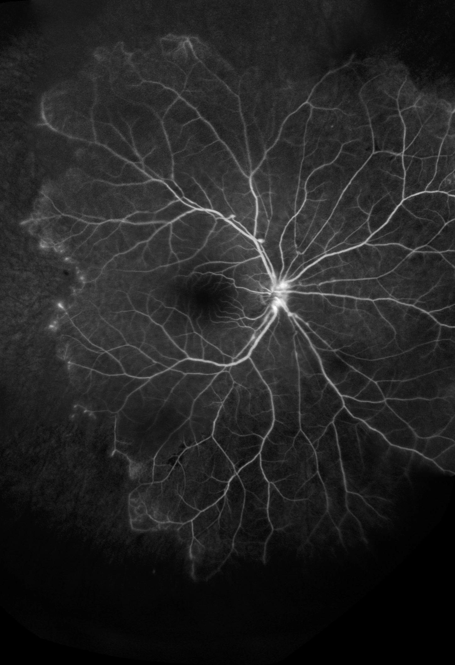
ABOUT THE COVER PHOTO:
James Gilman, CRA, FOPS, Moran’s Ophthalmic Imaging project administrator, has seen thousands of images over his nearly 40-year career. But about once a month, he says, the screen of a digital imaging system presents a new challenge—an image he’s never seen before. That was the case in the cover photo, captured by Gilman using a Heidelberg Spectralis imaging camera with a 105-degree lens. Gilman’s imaging verified a diagnosis of idiopathic retinal vasculitis-aneurysms-neuroretinitis syndrome, a rare genetic condition known as IRVAN. “That’s the best part of my job, seeing something I’ve never seen before,” says Gilman. “When you see something new, that diagnosis then becomes part of your vocabulary of recognition.”
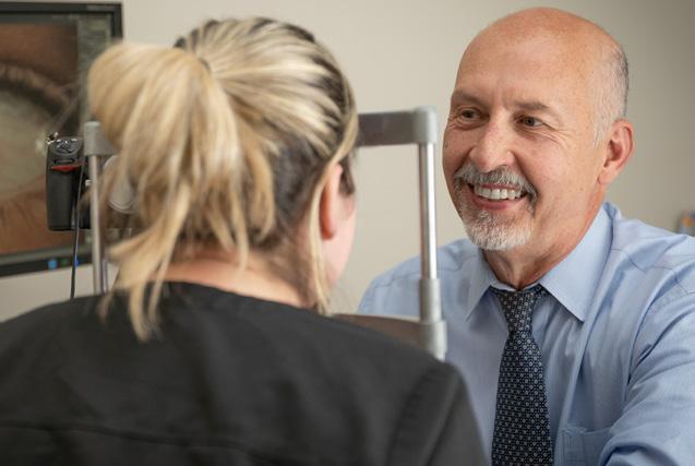
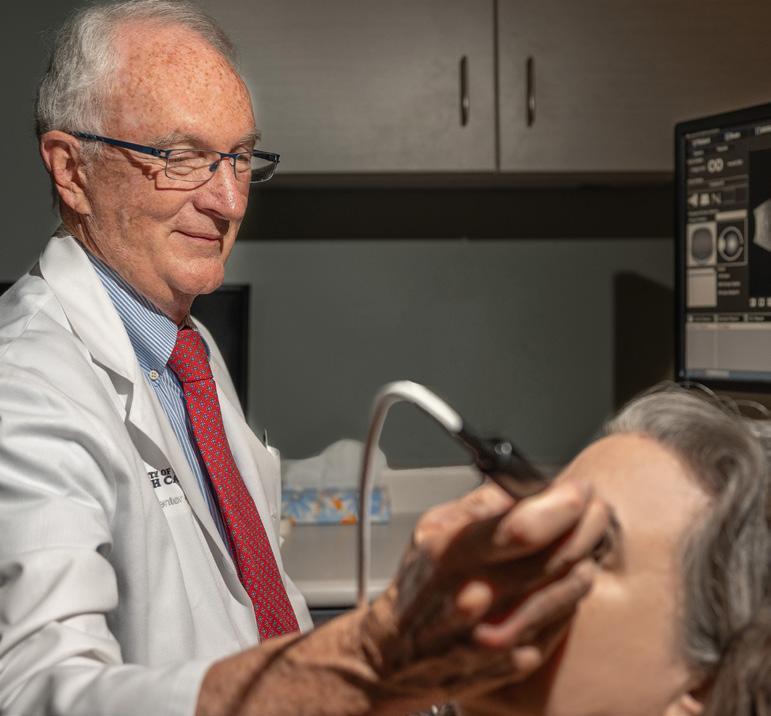
James Gilman, CRA, FOPS, project administrator of Moran’s Ophthalmic Imaging, performs a test using slit lamp imaging.

At the John A. Moran Eye Center at the University of Utah, we strive for excellence in everything we do to provide the best care possible for our patients—especially those facing complex eye conditions and diseases.
In so many cases, the right combination of imaging technology and medical expertise can save or improve vision. That’s why we pair cutting-edge imaging with exceptional physicians and staff who can leverage the increasingly detailed amounts of information these technologies provide.
World-class retinal specialists like Michael P. Teske, MD, Moran’s director of Vitreoretinal Diseases and Surgery, are now using optical coherence tomography angiography (OCTA), one of the latest modalities in our array of imaging options, as part of their everyday practice. OCTA is noninvasive, cost-effective, and produces amazingly sharp 3D images of the retinal and choroidal vascular systems.
More conventional clinical technologies, like ophthalmic ultrasound, in the hands of physicians like Roger P. Harrie, MD, and electrophysiology, under the direction of Donnell J. Creel, PhD, are used to tease out the best diagnosis and treatment paths for complicated, and at times mysterious, cases.
And we have so much to look forward to as the field develops.
Moran physician-scientist Paul S. Bernstein, MD, PhD, is investigating the potential uses of one of the most exciting new imaging technologies I have seen to date: fluorescence lifetime imaging ophthalmoscopy (FLIO). His work has already uncovered much of FLIO’s enormous potential for clinical use in the early diagnosis of disease. It has also led to several world-first discoveries, playing an important role in identifying the first causal gene for a rare retinal disease, macular telangiectasia.
With the addition of Steffen Schmitz-Valckenberg, MD, to the faculty in 2020, Moran will build a state-of-the-art ophthalmic reading center to evaluate images from research sites around the world and undoubtedly expand the role of imaging in research.
For this edition of Clinical Focus, I've asked some of our leading clinicians and researchers to highlight the role of imaging and electrophysiology in the care of our patients. I hope you will find their perspectives enlightening as we consider how new technologies are shaping and improving the practice of ophthalmology.
Sincerely,

Randall J Olson, MD Professor and Chair, Department of Ophthalmology and Visual Sciences, University of Utah CEO, John A. Moran Eye Center
By Judith E.A.Warner, MD; Marissa Larochelle, MD; and Donnell J. Creel, PhD
A woman in her mid-20s was seen for a new gray spot in the vision of her left eye in 2019. She had experienced an episode of optic neuritis of the left eye three years earlier, which resolved without treatment. She had refractive surgery in 2018. She described four days of bright flashes in the vision, off to the left in the left eye, and a flickering, with variable intensity depending on ambient lighting. She was able to see the photopsia with her eyes closed. For two days, she noticed a gray spot in the same area, which seemed to be enlarging. There was mild pain/soreness of the eye, slightly worse with movement. The rest of her vision was clear. She had no neurologic symptoms.
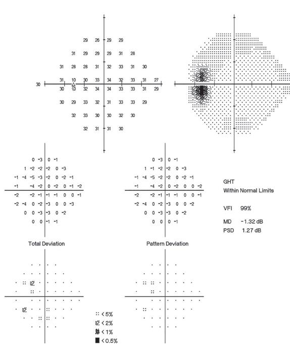
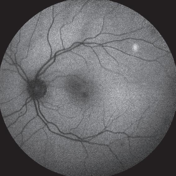
On examination, her vision was 20/20 in both eyes. Color vision was normal. She had no relative afferent pupillary defect. Stereopsis was 9/9. Critical flicker fusion was normal in both eyes. Humphrey visual field (HVF) testing showed an enlarged physiologic blind spot in the left eye. She noticed that during visual field testing, there was a blue hue around her scotoma. Fundus examination showed a trace of optic nerve edema left eye (OS), rare vitreous cell, and a
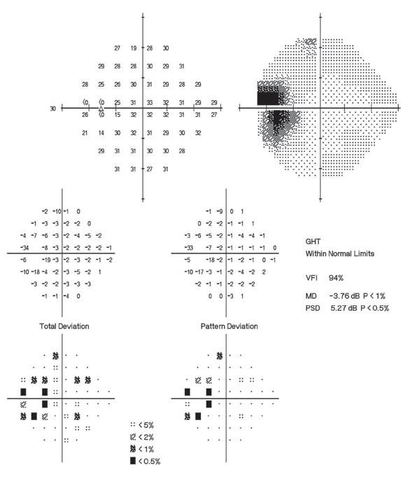
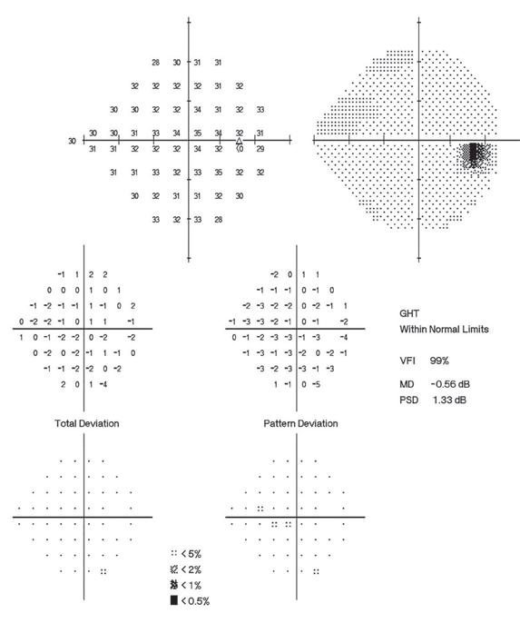
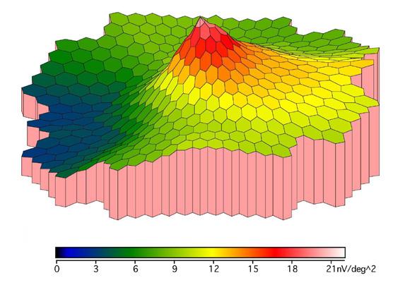
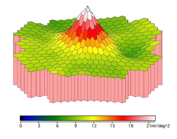
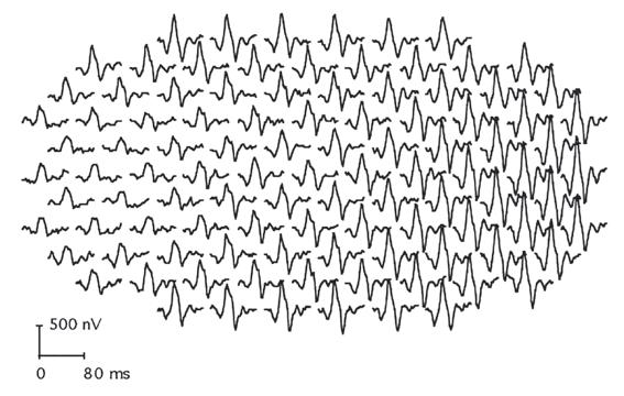
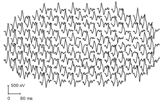
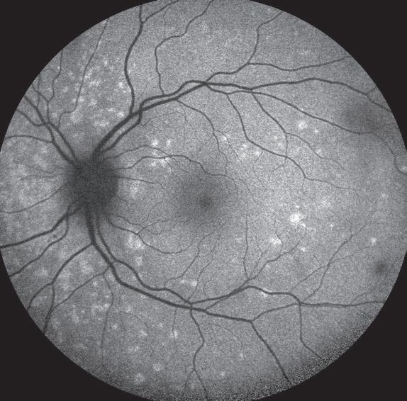
Figure 1. Humphrey visual field (HVF) testing, multifocal electroretinogram (mfERG), and fundus autofluorescence (FAF) clinical tests helped diagnose a case of multiple evanescent white dot syndrome. HVF (top) showed an enlarged physiologic blind spot in the left eye. The blue area in the mfERG (second from top, left) indicated a dysfunctional portion of the retina around the blind spot. FAF (bottom, left) showed marked hyperfluorescent spots in the area of the blind spot in the left eye.
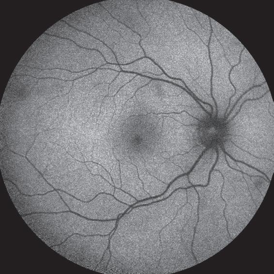
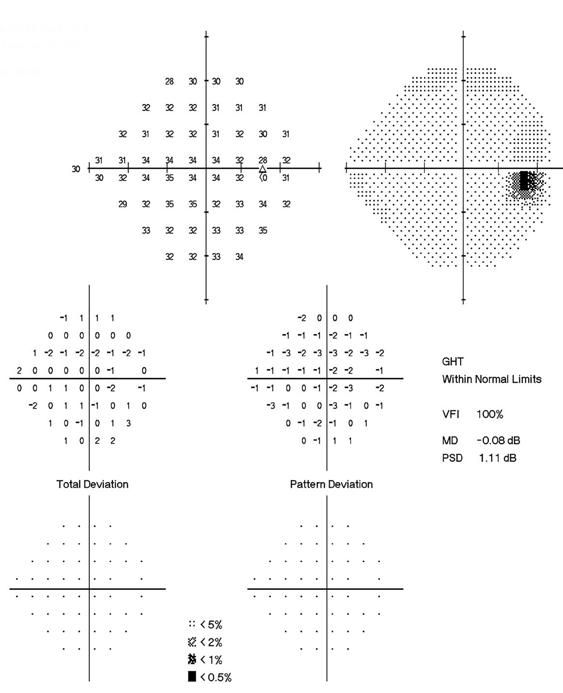
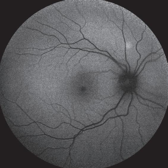
2. Over a few months, the patient’s symptoms and findings resolved without treatment, as is expected with a self-limited condition like MEWDS.
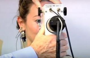
Moran’s electrophysiology eye testing service has been directed for 40 years by Donnell J. Creel, PhD, who is a leader in the field and actively involved in research to improve electrophysiological testing.
normal appearing retina (Figure 1). Multifocal electroretinography (mfERG) showed depression of responses in the region of the enlarged physiologic blind spot indicated by blue area OS. The right eye was normal. Fundus autofluorescence (FAF) showed marked hyperfluorescent spots in the area of the blind spot in the left eye. The right eye was normal (Figure 1).
She was diagnosed with multiple evanescent white dot syndrome (MEWDS), a rare unilateral, self-limited condition characterized by multiple yellow-white retinal lesions that primarily affects women aged 14 to 47. Since there is no diagnostic laboratory test for MEWDS, electrophysiology and clinical imaging tests are essential for diagnosis.
Over the next few months, her symptoms resolved without treatment (Figure 2).



Electrophysiology technology is similar to an electrocardiogram (ECG) in that it shows the health of the retina, similar to how an ECG reflects the health of the heart muscles. It is key to diagnosing and following the progression of disorders affecting the retina.
For more on Moran’s electrophysiology eye testing service, visit https:// healthcare.utah.edu/moran/ electrophysiology.php#electrooculogram.
Educational resources for clinicians are also available at webvision.med.utah. edu, including Dr. Creel’s widely viewed internet chapters on visual evoked potential and electroretinogram (ERG) procedures. His video on multifocal ERG, the most recent advance in ERG technology, is available on the NeuroOphthalmology Virtual Education Library (NOVEL) at novel.utah.edu.
By Rachel Patel, MD; Albert T. Vitale, MD; Donnell J. Creel, PhD; and Kathleen B. Digre, MD

Dr. Digre specializes in neuro-ophthalmology and evaluates and treats complex visual complaints, which can be due to the optic nerve or brain disease.

Vitale directs Moran’s Uveitis Division and is one of the few ophthalmologists in the Mountain West specializing in the diagnosis and treatment of uveitis and other infections and inflammatory diseases of the eye.

A 67-year-old woman was referred to neuro-ophthalmology for visual disturbances that she had noticed for the last five years. She reported continuous movement like a ceiling fan in her peripheral vision. Sometimes she would see smoke swirling around her, lights seemed to flicker, and she had prominent floaters. She had progressive difficulty with adaptation to dim-light conditions. She rarely had headaches. She had been seen by several ophthalmologists and neurologists and had received the diagnosis of visual snow, migraine with aura, or migraine symptoms.
Her examination showed a normal visual acuity and normal visual fields to confrontation. Her slit lamp examination showed old vitreous cells. Her neurological examination was normal. Her visual field showed peripheral losses in both eyes (Figure 1), and her optical coherence tomography (OCT) showed bilateral granular disruption of the ellipsoid zone that spared the fovea (Figure 2).
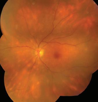
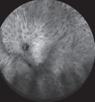
Because of the problem with poor dark adaptation, we obtained a full-field electroretinogram (ffERG) (Figure 3), which demonstrated evidence of poor rod function but normal cone function. The ffERG abnormalities in combination with her vitreous and retinal findings prompted a referral to our uveitis colleagues. Her dilated examination revealed subtle creamy yellow spots in the superior macula and periphery bilaterally, most prominently nasal to the optic discs (Figure 4). Indocyanine green angiography (ICG) showed numerous hypofluorescent spots in the mid- and farperipheral retina (Figure 4). Based on her clinical appearance and multimodal imaging, a diagnosis of birdshot chorioretinopathy was made. This diagnosis was further suggested by HLA-A29 positivity, and by a normal basic laboratory workup and chest X-ray to exclude alternative conditions such as sarcoidosis and lymphoma.
Birdshot chorioretinopathy is an uncommon chronic bilateral posterior uveitis, named for the characteristic cream-colored retinal spots scattered in a “birdshot” pattern. While nearly all patients with birdshot chorioretinopathy are HLA-A29 positive, routine testing is not fruitful as the haplotype is present in approximately 7
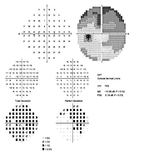
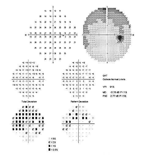
Figure 1 Visual field tests show peripheral visual field abnormalities.
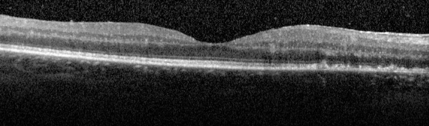
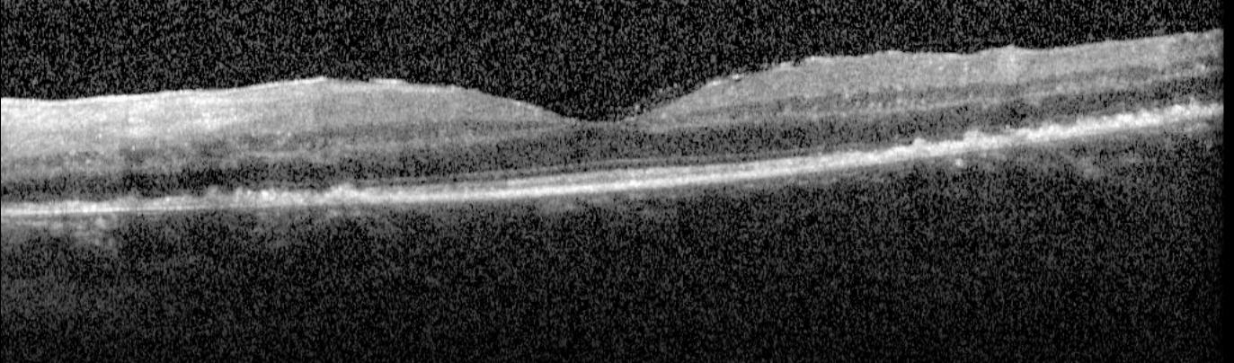
Figure 2. Macular OCT demonstrates granular disruption of the ellipsoid zone with sparing of the fovea in both eyes.
Figure 3
Electroretinography is suggestive of birdshot chorioretinopathy. Scotopic dim blue and red flash ERGs are reduced in amplitude 30 percent or more. The patient’s photopic b-wave times are delayed several standard deviations to 35 and 37 msec.
percent of the general population. Birdshot chorioretinopathy can be treated with immunosuppression. Monitoring for disease activity and initiating appropriate therapy is paramount; without treatment, disease progression may be insidious and result in irreversible vision loss.
Visual snow is a syndrome of positive visual phenomena in the visual field. While many such patients have had migraine, the constant presence of the visual disturbances differentiates it from migraine aura. The cause of visual snow is thought to be a visual processing disorder in the brain. It is considered a benign condition, and treatments are generally not successful. Most people, after learning of the diagnosis, develop the ability to ignore their symptoms.
As in patients with visual snow, patients with birdshot chorioretinopathy can have visual disturbances despite excellent visual acuity. However, there are differentiating features of birdshot chorioretinopathy that distinguish it from visual snow, including older age of onset, delay in dark adaptation (in contrast to nyctalopia alone), and absence of constant tiny flickering dots in the visual field. Birdshot chorioretinopathy tends to be a slowly progressive disease that can result in visual field loss and eventual loss of central visual acuity (often due to macular edema); therefore, recognition of the signs that distinguish birdshot chorioretinopathy from more benign etiologies of visual phenomena is critical.
Figure 4. A color montage shows creamy yellow ovoid spots in the mid- and far-periphery, while late indocyanine green angiography demonstrates even more hypofluorescent spots.
W hile visual symptoms such as floaters and headaches are very common, not all visual symptoms are attributable to migraine or visual snow. Signs that may indicate that a uveitic or retinal pathology may be contributing include vitritis, focal visual field losses, and particularly electrophysiologic abnormalities. Patients with atypical symptoms or presentation may benefit from a multimodal imaging approach to determine the underlying etiology
By Srav Vegunta, MD; and Mary Elizabeth Hartnett, MD

Dr. Hartnett specializes in pediatric and adult retinal diseases and surgery and is founder and director of Moran’s Pediatric Retina Center, as well as PI of a laboratory focused on conditions including retinopathy of prematurity.

Vegunta is a thirdyear resident at the Moran Eye Center.
A one-year-old girl was referred to the pediatric retina clinic by her pediatric ophthalmologist for evaluation of likely persistent fetal vasculature syndrome (PFVS), formerly known as persistent hyperplastic primary vitreous. She was born full-term with normal intrauterine development but had congenital motor nystagmus and amblyopia of the left eye, myopia of both eyes, and bilateral sensorineural hearing loss. There was no family history of eye or hearing problems. In the clinic, she had normal external appearance of the eyes and was able to fix and follow. Her glasses prescription was -4.25 +1.50 x 090 in the right eye and -4.75 sphere in the left eye. There appeared to be a retinal stalk emanating from the optic disc in her left eye, and the right retina grossly appeared normal. The examination was limited due to poor cooperation and a blonde fundus in both eyes. An exam under anesthesia was performed.
The patient had a normal anterior segment in the right eye. The left eye had a temporal and inferior retrolental membrane abutting the posterior lens capsule from a stalk of fibrovascular and retinal tissue extending from the optic nerve (Figure 1). The retina of the right eye had peripheral avascularity and whitish tissue at the junction between the vascular and avascular retina (Figure 2).
Imaging was performed on both eyes. Optical coherence tomography (OCT) demonstrated retained inner layers of the retina at the fovea of the right eye and no obvious fovea of the left eye (Figure 3). A fluorescein angiogram (FA) demonstrated peripheral avascular retina in the right eye with leakage from temporal neovascularization. Evidence of retinal vessels was seen in the stalk of the left eye.
The examination under anesthesia and imaging provided insight into the diagnosis and management for both eyes. First, eyes appeared about the same size, whereas in PFVS one eye can be smaller, but not always. Also, PFVS often presents with an inferonasal location in relationship to the lens capsule, whereas in our patient, the location was inferotemporal. The peripheral avascular retina in the right eye was bilateral, so the diagnosis was changed from PFVS to familial exudative vitreoretinopathy (FEVR). Because of leakage in the right eye, laser was performed to the peripheral avascular retinal area to reduce the risk of tractional retinal detachment that can occur with FEVR. Because of the evidence that the retina was pulled into
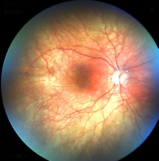
Figure 1. A fundus photograph of the right eye shows peripheral avascular retina. The optic nerve was normal in color but appears pale due to camera flash in this photo.
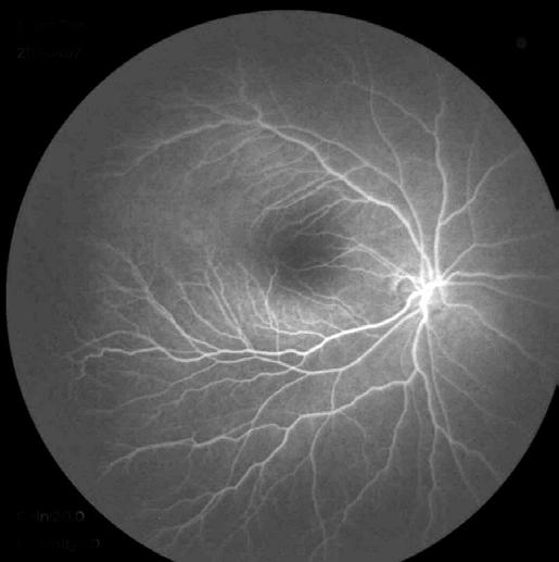
2. FA of the right eye late phase demonstrates peripheral avascular retina, seen to the left of the image.
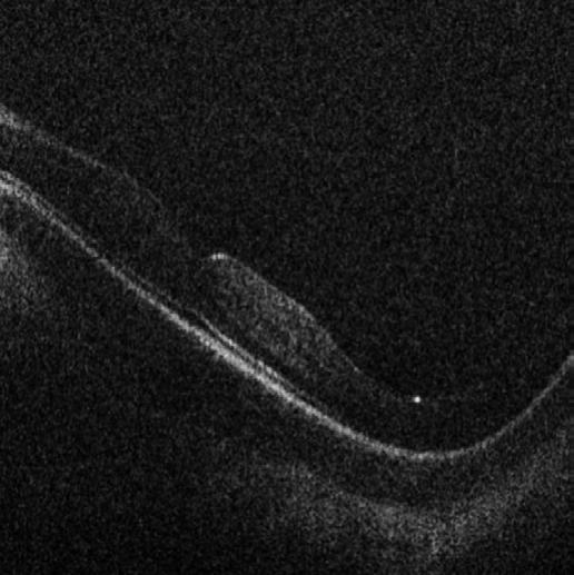
3.
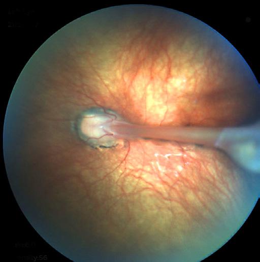
A fundus photograph of the left eye demonstrates a stalk of retinal tissue from the pallid optic nerve to the anterior retina. The peripheral retina was avascular, noted during fluorescein angiography (FA).
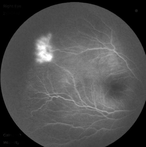
Another FA of the right eye late phase also demonstrates peripheral leakage from a temporal vascular frond.
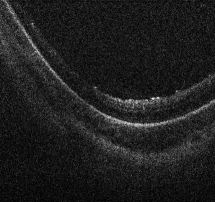
OCT during an exam under anesthesia of the left eye demonstrates no fovea. It is possible the foveal was incorporated into the stalk.
the stalk abutting the posterior lens capsule in the left eye, no surgical intervention was recommended. Surgery carries the risk of causing a retinal break, with limited prognosis for macular reattachment or vision. Although FEVR can be associated with nystagmus, a future evaluation for albinism will be considered for our patient given her nystagmus, foveal hypoplasia, and blonde fundus.
FEVR was first described in 1969 by Criswick and Schepens as an inherited condition of wide variability. The condition may be associated with exudation and retinal detachment or intravitreal neovascularization and tractional retinal detachments. The presentation classically has been described as one of bilateral incomplete vascularization of the peripheral retina in an infant that was born full-term. However, even within the same individual there can be variability in phenotype, including in our patient in whom there was a stalk of tissue. This exam finding led to the initial diagnosis of a unilateral process, PFVS. It is important to consider FEVR when a patient appears with seemingly unilateral PFVS. FEVR can also have a “normal” fellow eye, but more often a fluorescein angiogram can detect peripheral avascular retina. The prevalence and incidence of FEVR are unknown, but genetic variants can be detected in about 50 percent of patients, some in the Wnt signaling pathway (FZD4, LRP5, NDP, TSPAN12) as well as ZNF408, KIF11, and CTNNB1.
Genetic counseling is helpful, as some FEVR genotypes have associated systemic findings, including osteoporosis with LRP5 variants.
By Roger P. Harrie, MD
A 78-year-old man was referred for an ultrasound of his right eye to evaluate a small choroidal nevus. He also mentioned almost total loss of vision in his left eye over the past several days. His ophthalmologist attributed this to a central retinal artery occlusion secondary to atrial fibrillation.
His past medical history was significant for hypertension (Rx Losartan and Triamterene-hydrochlorothiazide), coronary artery disease, hyperlipidemia (Rx Atorvastin), hypothyroidism (Rx Levothyroxine), and osteoarthritis (Rx acetaminophen). Asymptomatic atrial fibrillation had recently been discovered on a pre-op evaluation for rotator cuff surgery. A few weeks later, he noted hazy vision in his right eye with lightning streaks. A workup included a brain MRI, which showed chronic microvascular changes, a carotid ultrasound and echocardiogram, both with normal results. He had been started on Eliquis for presumed emboli due to his atrial fibrillation, with resolution of his visual symptoms.
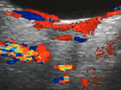
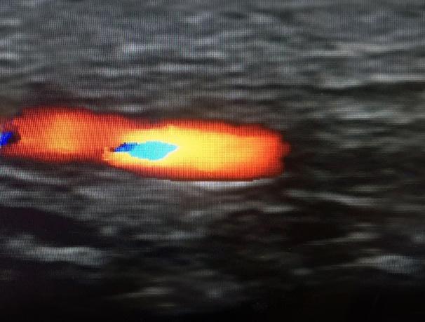
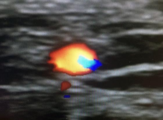
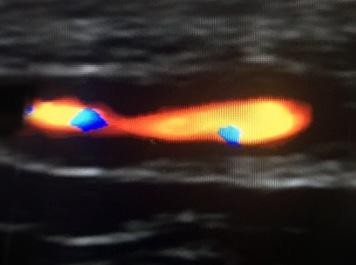
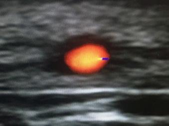
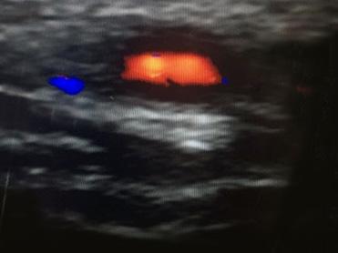
During the performance of the ultrasound for the fundus lesion, the patient mentioned the recent onset of severe headaches, temporal scalp tenderness, and pain on chewing, which necessitated changing his diet to soft foods and liquids. Because of this history, an examination was performed, which revealed vision OD of 20/50-2 and OS of light perception with a 4+ afferent pupil defect. Intraocular pressures were normal, and the anterior segment examination was unremarkable except for bilateral pseudophakia. Fundus exam was normal OD, but OS showed optic disc pallor and vascular attenuation. We performed an orbital color Doppler study, which showed a "dead" orbit OS consistent with giant cell arteritis (GCA) (Figure 1). We ordered an erythrocyte sedimentation rate (ESR), which was borderline elevated at 35mm, and a c-reactive protein (CRP), which was highly elevated at 81.
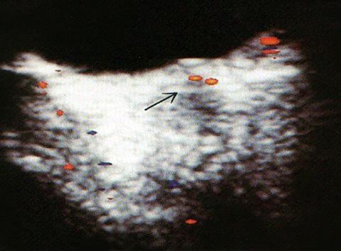
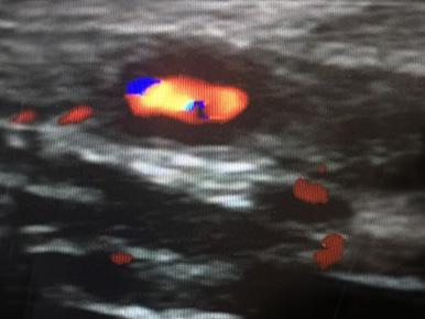
We scheduled the patient for a temporal artery biopsy, and he accepted an invitation to participate in a study at the Moran Eye Center using color Doppler to evaluate the temporal artery (Figure 2). This demonstrated a positive “halo” sign, which was suggestive of inflammatory edema of the temporal arteries.
We referred the patient to the emergency room in his hometown, where he received one gram of intravenous methylprednisolone for three days. A temporal artery biopsy was performed two days later and was positive for GCA. He was started on 60 mg of oral
Prednisone per day, with a tapering dosage to be monitored by his primary care physician. He felt his right eye improved slightly, but there was no change in his left eye. This case demonstrates the importance of the clinician remaining open to alternative diagnostic possibilities when evaluating a patient.
Clinical Tria l: Color Duplex Sonography in Evaluating Giant Cell Arteritis
Temporal artery biopsy is the gold standard in the diagnosis of giant cell arteritis (GCA), but color Doppler is being studied as a non-invasive alternative in a new clinical trial at the Moran Eye Center.
W hile temporal artery biopsy has a high specificity in diagnosis, it lacks sensitivity and carries potential complications that come with undergoing a surgical procedure.
Scheduling the procedure can also delay or end the treatment with high-dose corticosteroids, which have frequent and impactful side effects.
Recently, there have been efforts to find a more accessible, less invasive, less costly, and more rapid diagnostic tool. Color duplex sonography (CDS) of the temporal arteries has emerged as such a diagnostic method.
Temporal artery biopsy in cases of GCA reveals inflammation of the vessel wall in a predictable pattern. Similarly, CDS can provide a visual representation of vessel wall edema, referred to as a “halo,” throughout the length of the vessel. CDS also can eliminate the concern for negative temporal artery biopsy results due to “skip lesions.”
Nevertheless, evidence and large population studies are still inadequate to propel CDS as a mainstay of evaluation and diagnosis in GCA. More patients are needed to support further and justify this method of evaluation. With more evidence, CDS could become an integral part of diagnosis in GCA.
— Michael Burrow, MD
Moran is seeking about 100 patients for its study, “Use of Color Duplex Sonography in the Evaluation of Giant Cell Arteritis.” Email michael.burrow@hsc.utah.edu for more information.

Dr. Marx specializes in pediatric and adult oculoplastic and reconstructive surgery, with a particular interest in orbital tumors, eyelid and orbital reconstruction, and congenital disorders.
By Douglas P. Marx, MD
Patient-specific orbital implants are revolutionizing the results we can provide for patients who have experienced significant trauma to the eye sockets. Such trauma often leads to double vision that is very debilitating, as well as significant visual deformities to the eye sockets. We are utilizing patient-specific implants at the Moran Eye Center to provide the best quality care for our patients.
A man in his 30s was brought to the Emergency Department at the University of Utah following a gunshot wound to the face. A computed tomography (CT) scan of the face revealed multiple facial fractures, including eye socket fractures involving the medial walls, orbital floors, and lateral walls. The facial fractures were repaired by our Otolaryngology colleagues with excellent results. Because of the significant amount of trauma to the left eye socket, the patient was noted to have a large disparity between the positioning of the two eyes that was very bothersome to him. Likewise, this asymmetry led to significant double vision, making it difficult for him to perform routine daily activities.
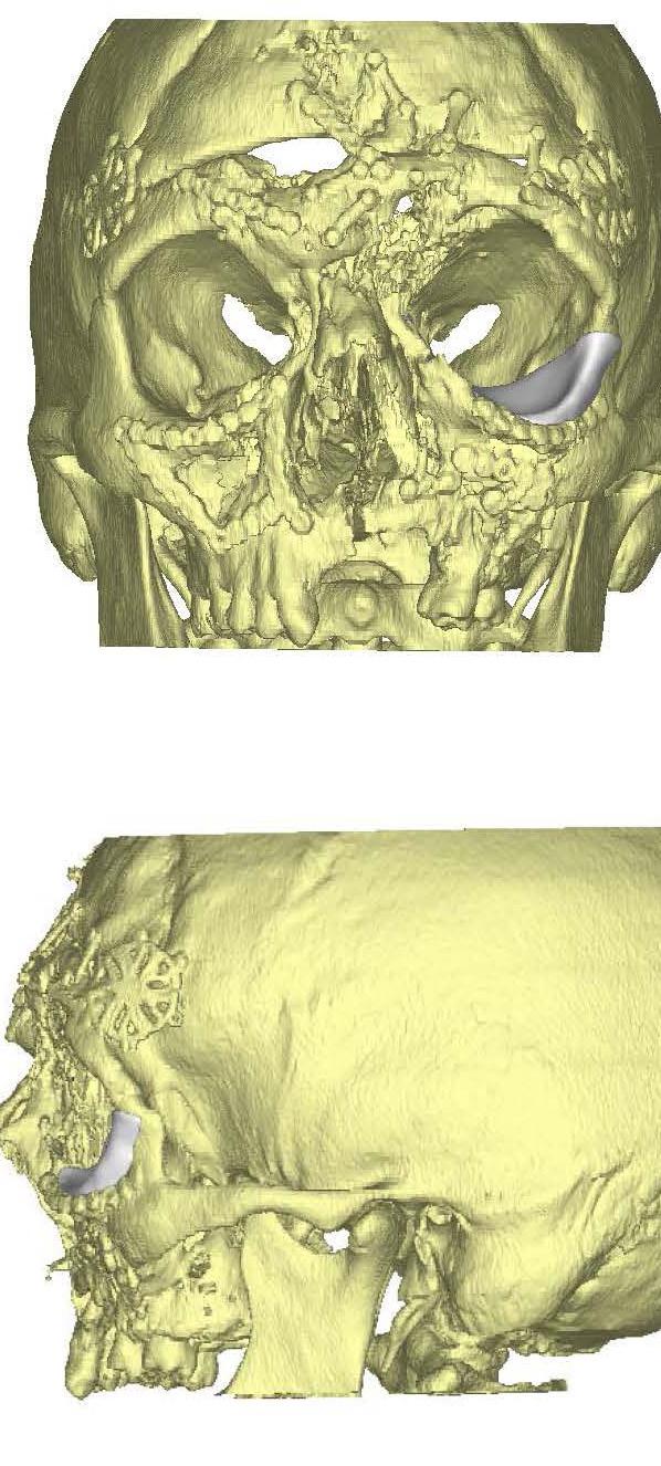
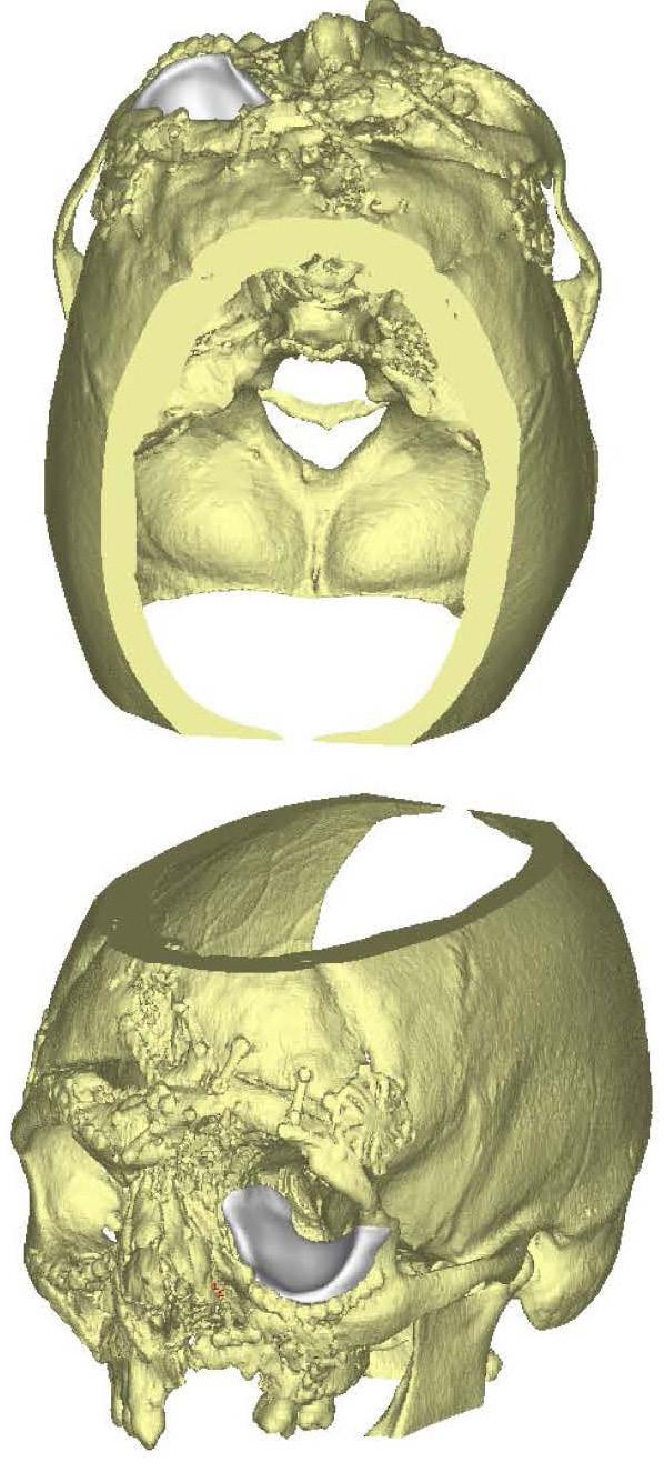
Using the CT images of the patient's eye sockets, a medical device company created a customized eye socket implant using computer-assisted design and computer-assisted manufacturing.
The patient underwent surgery to place the custom implant into his left eye socket. Following surgery, the positioning of his left eye was noted to be substantially better and comparable to his right side. His double vision resolved, and his ability to perform daily functions has substantially improved.
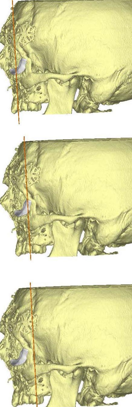
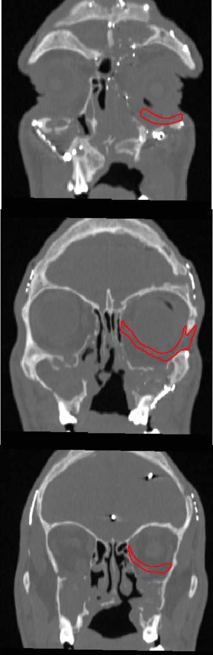
Reconstruction of complex orbital and periorbital deformities can be extremely challenging. Many implants exist that can help repair the eye sockets, but many of these implants fall short in producing optimal results. This report demonstrates the significant contribution patient-specific implants have created in eye socket reconstruction. Because of the multidisciplinary approach that we take at the Moran Eye Center, we are able to provide the best possible care for patients, utilizing the most up-to-date resources and technologies. Although many patients will not require a customized implant for their repair, this addition to the previously available technologies provides one more important piece to achieving optimal results.

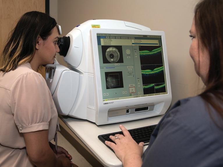
Retinal specialist Michael P. Teske, MD, has been an early adopter of optical coherence tomography angiography (OCTA), a developing technology that produces detailed 3D images of the retinal and choroidal vascular systems without the use of dyes. We asked him to share how he’s using OCTA and the benefits this advanced imaging provides for patients.
How does OCTA differ from other imaging modalities you typically use?
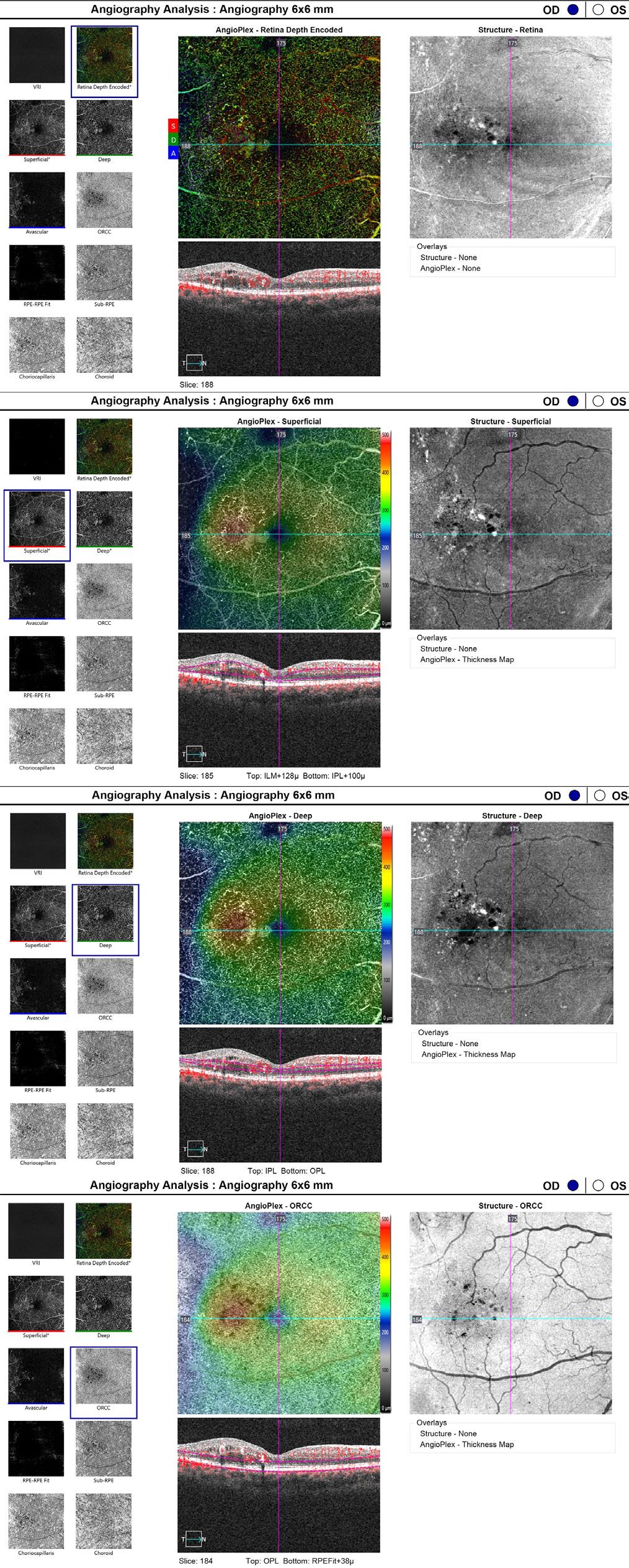
What OCTA is doing is looking at microvasculature and blood flow in the retina, as well as to the optic nerve. We have imaged the vessels in the retina for many years, but it’s always involved more invasive and time-consuming studies. We have to use fluorescein or indocyanine green angiography, which both involve intravenous dyes. There’s a little more risk involved; some people get allergic reactions to the dyes. It's also time consuming, taking 20-30 minutes, since patients need an IV. OCTA is noninvasive and takes just a few minutes, and doctors can look at the microvasculature in a 3D fashion. These kinds of images are not attainable in standard angiography.
For what types of cases are you finding OCTA analysis most useful?
Most of what we treat in the retina these days—diabetic retinopathy, macular degeneration, post vein occlusions, macular telangiectasia—are things that alter the blood flow or blood vessels of the retina, so imaging the blood vessels is a very important part of what we do. OCTA enables us to do this imaging quickly and noninvasively, which is easier on patients and a fraction of the costs of other tests. The two things I use it for most are macular degeneration and diabetes. With macular degeneration, OCTA helps me detect if there is leakage, and if the vessels regressed or are still present. This helps me decide if I need to continue treatment or watch the vessels more closely. In diabetes, it really helps us look at what’s happening even before there are any vision changes on a clinical exam. You’ll start to see changes in little capillaries very early using OCTA.
How do you see OCTA fitting into your practice in the future?
We’re still learning about it. We’re still imaging different types of diseases. For example, we’re in the early stages of how it will be used in glaucoma. The quality and resolution are getting better and better. With things like diabetic retinopathy, when you want to look at the blood vessels in the macula, OCTA has become the standard angiogram in just a couple of years.
The downside is if you want to look at the peripheral part of the retina, it’s still not doable. OCTA also doesn’t work for some patients. OCTA detects blood vessels by the motion of the blood cells through the blood vessels, so patients have to hold relatively still. If they are moving, the machine detects that as motion, and it can give you an artifact. Some patients have tremors or can’t hold their eyes still, and you might not get a very good study.
For now, it’s sort of revolutionized the way we look at circulation in the retina, to be able to do it very quickly and very easily, inexpensively, non-invasively. Is it a game-changer?
Not yet, but neither was OCT when it first came out. Now there’s barely a patient I see that I don’t use OCT. I would guess as the machines are upgraded and become more common over the next few years, we’ll see OCTA technology used more and more and more.
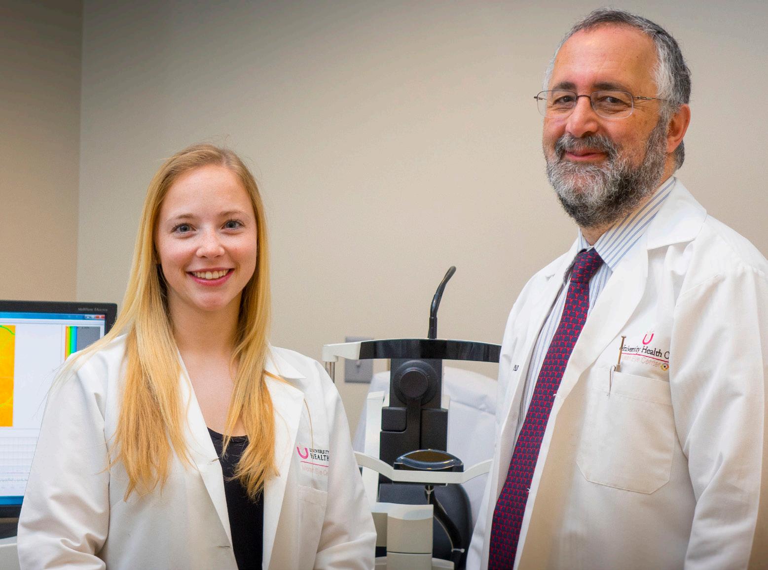
Moran’s Lydia Sauer, MD, and Paul S. Bernstein, MD, PhD, work with a cutting-edge, non-invasive imaging tool known as FLIO.
Using the only imaging modality of its type in the U.S., Moran physician-scientists are working to revolutionize how clinicians diagnose and treat diseases
By Lydia Sauer, MD; and Paul S. Bernstein, MD, PhD
The imaging of the human eye has long guided clinical care, helping ophthalmologists better understand and diagnose pathologies and diseases of the eye.
The retina was first viewed in living patients by German physician and physicist Hermann von Helmholtz in 1851, who used fundoscopy to see the inside of the eye. Since then, beginning with delicate hand drawings, a variety of modalities have been developed. From fundus photography to advanced imaging technologies, they all give detailed information on the microstructure of the human eye.
Commonly used techniques are optical coherence tomography, which investigates the reflectance of different layers within the eye, and fluorescence-based imaging modalities, which give information about the molecules in the eye. Fluorescent molecules within the eye, known as fluorophores, can absorb and emit light to different extents. Some fluorophores absorb more light; these appear dark on images. Other fluorophores absorb less light; these appear bright on images.
Harvard scientist Francois C. Delori and coworkers spent considerable effort describing the fluorescence of the human retina and established how fluorophores are altered in diseases. The strongest fluorescence is emitted from lipofuscin, the dominant fluorophore within the retina.1 Accumulation of lipofuscin and other strongly fluorescent fluorophores can be seen in diseases such as age-related macular degeneration. Overall, changes in the fluorophores lead to altered light signals, which are emitted from the eye, and subsequently, ophthalmologists can detect these as disease-related changes.
A promising but not yet widely established fluorescencebased imaging method is fluorescence lifetime imaging ophthalmoscopy (FLIO), which was invented by German scientist Dietrich Schweitzer and coworkers in 2002.2 In contrast to conventional fluorescence imaging, FLIO investigates a different property of the fluorescence, the fluorescence lifetime. This parameter tells us how long the fluorophores glow, which is independent of the strength of the individual fluorescence. Fluorophores that emit only weak amounts of light can be detected with this novel method.3 As some diseases start with very small changes, conventional fluorescence imaging may not always detect these tiny changes. FLIO, however, can do so, making it helpful in the early detection of diseases. It also helps to understand how eye diseases will progress over time.
A variety of retinal diseases have been investigated with FLIO, and many studies showed the advantages of FLIO technology over other imaging modalities.4, 5 The Moran Eye Center's FLIO, a prototype manufactured by Heidelberg Engineering in Germany, is the only device of its kind in the United States.
One interesting example of the advantages of FLIO relates to an inherited macular dystrophy called macular telangiectasia type 2 (MacTel). Initially, MacTel was thought to be a rare disease, but recent studies argue for a much higher prevalence than initially assumed.6 Early clinical trials of an implant that is aimed to stop disease progression showed promising results, which means that treatment
for this disease might soon be available. It is, therefore, crucial to correctly diagnose this disease at an early stage for the success of the treatment.7 Many findings that lead to the diagnosis of MacTel, such as retinal cysts, para-foveal crystals, and even low macular pigment values, can be absent in early stages of the disease.
There is a need for a reliable imaging modality that can detect changes at early stages. Our research has shown FLIO to be an excellent option 8 Not only can FLIO detect retinal changes in MacTel once the disease has manifested, but FLIO has also been shown to detect alterations in individuals before typical retinal damages occur.9 As an example, we investigated one family with three siblings, in which the youngest, diagnosed at age 21, had severe MacTel. We found the genetic mutation in this patient.10 His two sisters (aged 26 and 28) showed completely healthy eye exams, although one carried the genetic mutation. Despite the healthy clinical exam, FLIO already shows MacTel-related changes in this woman, which may lead to an earlier treatment that might preserve her vision.
FLIO is a promising new technology that holds the potential to revolutionize how clinicians diagnose and treat diseases. By detecting changes before damages are manifest, FLIO may be a beneficial tool for clinical care in the near future.
Dr. Bernstein specializes in vitreoretinal diseases and surgery, retinal biochemistry, and macular and retinal degeneration. He directs clinical research and serves as associate director of research at the Moran Eye Center.
Dr. Sauer is a research associate in the Bernstein Lab who specializes in FLIO.
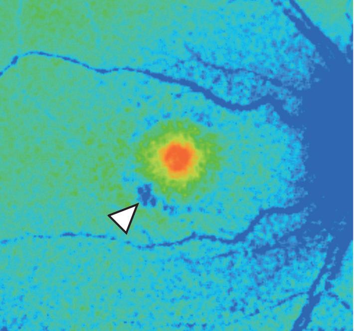
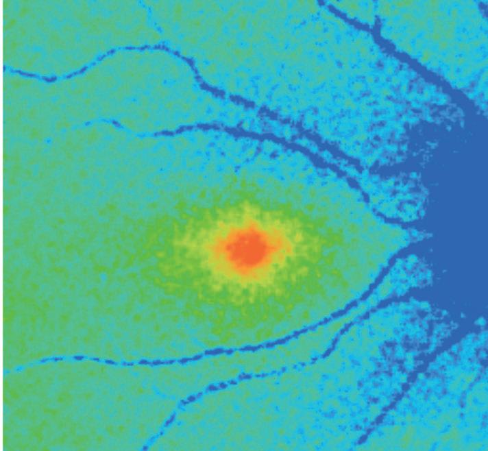
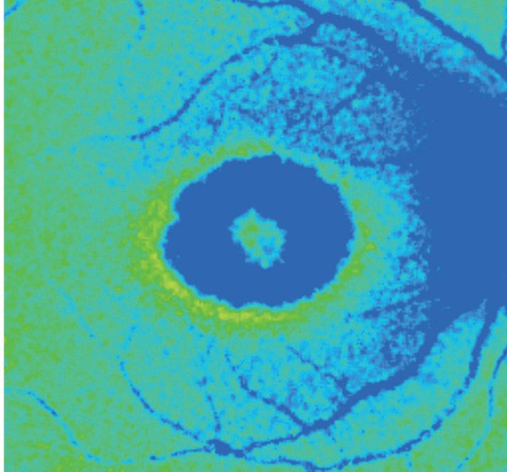
These fluorescence lifetime imaging ophthalmoscopy (FLIO) images show a family with MacTel. The youngest sibling (left, male, 22,) shows advanced-stage MacTel, highlighted by the blue center indicating the disease. One sibling, despite normal clinical exam findings, also shows FLIO alterations indicative of MacTel (middle, female, 26), whereas the other sibling has a completely healthy FLIO (right, female, 28). FLIO has been shown to be effective in detecting early stages of MacTel before retinal damage has occurred.
1. Delori FC, Dorey CK, Staurenghi G, Arend O, Goger DG, Weiter JJ. In vivo fluorescence of the ocular fundus exhibits retinal pigment epithelium lipofuscin characteristics. Investigative Ophthalmology & Visual Science 1995;36:718-729.
2. Schweitzer D, Kolb A, Hammer M, Anders R. [Time-correlated measurement of autofluorescence. A method to detect metabolic changes in the fundus]. Der Ophthalmologe: Zeitschrift der Deutschen Ophthalmologischen Gesellschaft 2002;99:774-779.
3. Lakowicz JR. Principles of Fluorescence Spectroscopy: Springer; 2006.
4. Dysli C, Wolf S, Berezin MY, Sauer L, Hammer M, Zinkernagel MS. Fluorescence lifetime imaging ophthalmoscopy. Progress in Retinal and Eye Research 2017;60:120-143.
5. Sauer L, Andersen KM, Dysli C, Zinkernagel MS, Bernstein PS, Hammer M. Review of clinical approaches in fluorescence lifetime imaging ophthalmoscopy. Journal of Biomedical Optics 2018;23:1-20.
6. Charbel Issa P, Gillies MC, Chew EY, et al. Macular telangiectasia type 2. Progress in Retinal and Eye Research 2013;34:49-77.
7. Chew EY, Clemons TE, Peto T, et al. Ciliary neurotrophic factor for macular telangiectasia type 2: results from a phase 1 safety trial. American Journal of Ophthalmology 2015;159:659-666.
8. Sauer L, Gensure RH, Hammer M, Bernstein PS. Fluorescence Lifetime Imaging Ophthalmoscopy: A Novel Way to Assess Macular Telangiectasia Type 2. Ophthalmology Retina 2018;2:587-598.
9. Sauer L, Vitale AS, Andersen KM, Hart B, Bernstein PS. Fluorescence Lifetime Imaging Ophthalmoscopy (FLIO) Patterns in Clinically Unaffected Children of Macular Telangiectasia Type 2 (MacTel) Patients. Retina 2019. 10. Gantner ML, Eade K, Wallace M, Handzlik MK, Fallon R, Trombley J, Bonelli R, Giles S, Harkins-Perry S, Heeren TFC, Sauer L, Ideguchi Y, Baldini M, Scheppke L, Dorrell MI, Kitano M, Hart BJ, Cai C, Nagasaki T, Badur MG, Okada M, Woods SM, Egan C, Gillies M, Guymer R, Eichler F, Bahlo M, Fruttiger M, Allikmets R, Bernstein PS, Metallo CM, Friedlander M. Serine and Lipid Metabolism in Macular Disease and Peripheral Neuropathy. The New England Journal of Medicine. 2019 Oct. 10;381(15):1422-1433.
CLINICAL TRIALS 2019
Nearly 90 clinical trials and studies are underway at the Moran Eye Center.
CATARACT
Multi-Center GATT Retrospective Review
PI: Craig J. Chaya, MD
Clinical Study of the ARTISAN Aphakia Lens for the Correction of Aphakia in Adults
PI: Alan S. Crandall, MD
The Safety and Effectiveness of the Hydrus Aqueous Implant for Lowering Intraocular Pressure in Glaucoma Patients Undergoing Cataract Surgery, A Prospective, Multicenter, Randomized, Controlled Clinical Trial (Hydrus 4 Study)
PI: Alan S. Crandall, MD
Evaluation of Resources Required for Measurement of Cataract Surgery Outcomes
PI: Jeff Pettey, MD
Multivariate Analysis of Variables Affecting Resident Performed Cataract Surgery Complication Rates
PI: Jeff Pettey, MD
Relationship Between Geriatric Patient Age and Refractive Outcomes in Cataract Surgery
PI: Jeff Pettey, MD
CORNEA
Survey of Post-Operative Pain in Photorefractive Keratectomy when Using Topical Versus Oral Non-Steroidal Anti-Inflammatory Drugs
PI: Mark D. Mifflin, MD
Outcomes of Topography-Guided Laser-Assisted in Situ Keratomileusis (LASIK) and Photorefractive Keratectomy (PRK) Compared to Wavefront Optimized LASIK and PRK
PI: Mark D. Mifflin, MD
Incidence of Chronic Dry Eye Following Photorefractive Keratectomy and Laser in Situ Keratomileusis
PI: Mark D. Mifflin, MD
A Randomized, Double-Masked, Placebo-Controlled Study for Determining the Safety of Processed Amniotic Fluid (pAF) Drops after Photorefractive Keratectomy
PI: Mark D. Mifflin, MD
Causes of Death and Co-Morbidities among Individuals with Keratoconus
PI: Randall J Olson, MD
GENERAL
National Ophthalmic Genotyping Network, Stage 1 - Creation of Repository for Inherited Ophthalmic Diseases
PI: Paul S. Bernstein, MD, PhD
Patterns of Blindness in the Navajo Nation: A Retrospective Study
PI: Craig J. Chaya, MD
Assessing Efforts to Raise Awareness about OCT
PI: Mary Elizabeth Hartnett, MD
Focus Groups on Assistive Technology for Visual Impairment
PI: Lisa Ord, PhD, LCSW
Solar Eclipse Retinopathy: What were the after-effects of the "Great American Eclipse"?
PI: Jeff Pettey, MD
Assessing a Professionalism Mentor in Academic Medical Departments
PI: Jeff Pettey, MD
Visual Field Screening using a Mobile Device
PI: Jeff Pettey, MD
Tracking Outcomes in Moran Outreach Programs
PI: Jeff Pettey, MD
Prevalence of Ophthalmic Disease in a Salt Lake City Homeless Population.
PI: Jeff Pettey, MD
Sight Outcomes Research (SOURCE): Multicenter Ophthalmology EHR Repository
PI: Jeff Pettey, MD
Machine Learning Algorithm for Electroretinography Sorting
PI: Akbar Shakoor, MD
GLAUCOMA
Open-Angle Glaucoma after Vitrectomy: A Retrospective Review
PI: Alan S. Crandall, MD
Decision Support for Glaucoma Care Protocol
PI: Alan S. Crandall, MD
Virtual Reality Functional Testing to Screen for Early-Stage Glaucoma
PI: Randall J Olson, MD
Pseudoexfoliation and Co-Morbidities
PI: Barbara M. Wirostko, MD
NEURO-OPHTHALMOLOGY
A ssessment of Photophobia in Moran Eye Center Patients
PI: Kathleen B. Digre, MD
Retrospective Review of Orbital Pseudotumors
PI: Kathleen B. Digre, MD
Evaluation of Optic Neuropathies with Imaging
PI: Kathleen B. Digre, MD
Retrospective Review of Primary and Secondary Causes of Pseudotumor Cerebri
PI: Kathleen B. Digre, MD
Causes of Eye Pain
PI: Kathleen B. Digre, MD
Visual Quality of Life Migraine Study
PI: Kathleen B. Digre, MD
SCA7 Chart Review
PI: Kathleen B. Digre, MD
Multi-Center Study of Medical Therapy v. Medical Therapy Plus Optic Nerve Sheath Fenestration or Stereotactic Ventriculoperitoneal Cerebrospinal Fluid Shunting in Patients with Idiopathic Intracranial Hypertension
PI: Kathleen B. Digre, MD
A Phase 2/3, Randomized, Double-Masked, Sham-Controlled Trial of QPI-1007 Delivered by Single or Multi-Dose Intravitreal Injection(s) to Subjects with Acute Non-Arteritis Anterior Ischemic Optic Neuropathy (NAION)
PI: Bradley J. Katz, MD, PhD
Genetic Initiative in Neuro-Ophthalmic Conditions (GINOC)— Idiopathic Intracranial Hypertension (IIH)
PI: Judith E.A. Warner, MD
PEDIATRIC OPHTHALMOLOGY
Effect of Dermatological Pulsed Dye Laser Therapy on Intraocular Pressure in Children with Facial Port Wine Stains
PI: Craig J. Chaya, MD
Clinical Study of the ARTISAN Aphakia Lens for the Correction of Aphakia in Children
PI: Alan S. Crandall, MD
Genetic Associations in Preterm Infants at Risk of Retinopathy of Prematurity
PI: Mary Elizabeth Hartnett, MD
Preeclampsia and Retinopathy of Prematurity
PI: Mary Elizabeth Hartnett, MD
Genetics of Pediatric Retinal Disorders
PI: Mary Elizabeth Hartnett, MD
Genetic Polymorphisms Associated with Retinopathy of Prematurity Severity among Premature Infants
PI: Mary Elizabeth Hartnett, MD
ROP1: Phase 1 Trial of Bevacizumab Treatment for Severe Retinopathy of Prematurity
PI: Mary Elizabeth Hartnett, MD
ROP2Y: Two-year Follow Up after Treatment of Severe Retinopathy of Prematurity
PI: Mary Elizabeth Hartnett, MD
Collaborative Retrospective Reviews of Rare Pediatric Ophthalmic Diseases
PI: Mary Elizabeth Hartnett, MD
Bedside OCT Assessment of Hypoxic Ischemic Encephalopathy
PI: Mary Elizabeth Hartnett, MD
Functional Vision Loss and Childhood Trauma
PI: Bradley J. Katz, MD, PhD
Analysis of Genetic Variant and Treatment Based Variations in Infants at Risk for Retinopathy of Prematurity (ROP)
PI: Leah Owen, MD, PhD
Clinical and Molecular Relationship between Maternal Pre-eclampsia and Retinopathy of Prematurity
PI: Leah Owen, MD, PhD
Analysis of Clinical Intraocular Lens Outcomes in a Pediatric and Adult Population
PI: Leah Owen, MD, PhD
Pediatric Cataract Surgery Outcomes Registry
PI: Leah Owen, MD, PhD
Building Capacity for Pediatric Eye Care in the Navajo Nation
PI: Leah Owen, MD, PhD
RETINA
Natural History Observation and Registry Study of Macular Telangiectasia Type 2: The MacTel Study
PI: Paul S. Bernstein, MD, PhD
Utah Center for MacTel Genetics: A Sub-Study of Subjects Enrolled in “The Macular Telangiectasia Project” at the University of Utah
PI: Paul S. Bernstein, MD, PhD
Macular Pigment Measurements in Eye & Other Tissues
PI: Paul S. Bernstein, MD, PhD
A Phase 2 Multicenter, Double-Masked, Randomized, PlaceboControlled Study to Investigate the Long Term Safety, Tolerability, Pharmacokinetics and Effects of ALK-001 on the Progression of Stargardt Disease
PI: Paul S. Bernstein, MD, PhD
Safety and Efficacy of Abicipar Pegol (AGN-150998) in Patients With Neovascular Age-related Macular Degeneration: SEQUOIA Study
PI: Paul S. Bernstein, MD, PhD
4D Molecular Therapeutics Protocol Number: 4D-CHM-001NH-0001—A Multicenter Prospective Observational "Natural History" Study in Patients with Choroideremia
PI: Paul S. Bernstein, MD, PhD
Rate of Progression in USH2A Related Retinal Degeneration (RUSH2A)
PI: Paul S. Bernstein, MD, PhD
NTMT-03: A Phase III Multicenter Randomized, Sham Controlled, Study to Determine the Safety and Efficacy of Renexus in Macular Telangiectasia type 2
PI: Paul S. Bernstein, MD, PhD
OPH2003: A Phase 2b Randomized, Double-Masked, Controlled Trial to Assess the Safety and Efficacy of Intravitreous Administration of Zimura (Anti-C5 Aptamer) in Subjects with Geographic Atrophy Secondary to Dry Age-Related Macular Degeneration
PI: Paul S. Bernstein, MD, PhD
Detection of Hydroxychloroquine Levels Using Fluorescence Lifetime Imaging
PI: Paul S. Bernstein, MD, PhD
OPH2005: A Phase 2b Randomized, Double-masked, Controlled Trial to Establish the Safety and Efficacy of Zimura (Complement C5 Inhibitor) Compared to Sham in Subjects with Autosomal Recessive Stargardt Disease
PI: Paul S. Bernstein, MD, PhD
MyRetina Tracker Genetic Testing Study
PI: Paul S. Bernstein, MD, PhD
Phase 3 Multi-Center Study to Evaluate Efficacy and Safety of Intravitreal APL-2 Therapy in Patients with Geographic Atrophy Secondary to Age-Related Macular Degeneration
PI: Paul S. Bernstein, MD, PhD
Natural History of the Progression of X-Linked Retinitis Pigmentosa
PI: Paul S. Bernstein, MD, PhD
Carotenoid Supplementation During Pregnancy
PI: Paul S. Bernstein, MD, PhD
SeaSTAR: Phase 3 Study Comparing Efficacy and Safety of Emixustat Hydrochloride with Placebo for the Treatment of Macular Atrophy Secondary to Stargardt Disease
PI: Paul S. Bernstein, MD, PhD
Phase 1 Study of Safety, Tolerability and Preliminary Efficacy of Intravitreal 4D-110 in Patients with Choroideremia
PI: Paul S. Bernstein, MD, PhD
Retrospective Review of Complications of X-Linked Retinoschisis Especially Causes of Vitreous Hemorrhage
PI: Mary Elizabeth Hartnett, MD
Coats' Longitudinal Fellow Eye Study
PI: Mary Elizabeth Hartnett, MD
Phase 3 Study of Efficacy and Safety of Intravitreal Aflibercept Injection in Patients with Moderately Severe to Severe Nonproliferative Diabetic Retinopathy
PI: Rachael Jacoby, MD
Phase 3 Multi-Center Study to Evaluate Efficacy and Safety of RO6867461 in Patients with Diabetic Macular Edema
PI: Rachael Jacoby, MD
Zoster Eye Disease Study (ZEDS): A Multi-Center, Randomized, Clinical Trial of Suppressive Valacyclovir for One Year in Participants with an Episode of Herpes Zoster Ophthalmicus in the Year Prior to Enrollment
PI: Amy Lin, MD
Retinal Disease Burden: Prevalence and Associated Risk Factors in Mwanza, Tanzania
PI: Akbar Shakoor, MD
Stem Cell Research on Usher Syndrome
PI: Jun Yang, PhD
Clinical phenotypes of inherited retinal disease
PI: Marielle Young, MD
UVEITIS
Oral Steroids in the Treatment of Bacterial Endophthalmitis
PI: Akbar Shakoor, MD
Vitreous Biopsy Analysis
PI: Akbar Shakoor, MD
Macular Edema Ranibizumab v. Intravitreal Anti-inflammatory Therapy (MERIT) Trial
PI: Albert T. Vitale, MD
Gilead: A Phase 2, Randomized, Placebo-Controlled Trial Evaluating the Efficacy and Safety of Filgotinib in Subjects with Active Non-Infectious Uveitis
PI: Albert T. Vitale, MD
Center for Uveitis Research and Education (CURE) Consortium
PI: Albert T. Vitale, MD
Adalimumab vs. Conventional Immunosuppression for Uveitis (ADVISE) Trial
PI: Albert T. Vitale, MD
Use of Color Duplex Sonography in the Evaluation of Giant Cell Arteritis
Above, FL-41 thin-film coated glasses. Below, FL-41 rose-tinted lenses.
PI: Alison Crum, MD
Growth Factors in Vitreoretinal Diseases
PI: Mary Elizabeth Hartnett, MD
Adalimumab in Juvenile Idiopathic Arthritis-Associated Uveitis Stopping Trial (ADJUST)
PI: Albert T. Vitale, MD
Refer a Patient 801-213-2001
https://physicians.utah.edu/
Moran CORE
Clinical Ophthalmology Resource for Education https://morancore.utah.edu/ NOVEL
Neuro-Ophthalmology Virtual Education Library https://novel.utah.edu/ WEBVISION
The Organization of the Retina and Visual System https://webvision.med.utah.edu/ CME Information https://medicine.utah.edu/cme/



Subscribe to the monthly Moran Update email by sending your email address to moran.info@hsc.utah.edu
Since its inception in 1972, the Utah Lions Eye Bank (ULEB) has facilitated the gift of sight worldwide. An extension of the Moran Eye Center, ULEB is a 501(c)(3) nonprofit organization dedicated to the mission of sight restoration and preservation through transplant, research, and education. Through the relationship with Moran, ULEB can access industry-leading facilities and support to ensure the organization remains on the cutting edge of advancements in eye banking well into the future. ULEB is uniquely positioned to provide the gift of sight because of its unparalleled access to the donation community and strong partnerships with organ and tissue banking colleagues.
Highly skilled technical staff work around the clock to ensure that every donation is not only safe and of the highest quality, but used in a manner consistent with sight restoration and conservation. ULEB is one of only seven eye banks in the country licensed to offer Patient Ready DMEK® (Descemet Membrane Endothelial Keratoplasty) and is capable of meeting a variety of tissue specifications for transplant surgeons.
In the fiscal year 2019, ULEB is proud to have provided nearly $500,000 in goodwill tissue through the Gratis Tissue Program. Working with local, domestic, and international surgeons, ULEB helps to provide transplant tissue for patients in need throughout the world.
To find out more about ULEB, and how to establish partnerships to help facilitate healing through the generosity of others, visit https://healthcare.utah.edu/moran/utah-lions-eye-bank/ or contact Wade McEntire, director of operations and business development, at 801-581-2039.
Utah Lions Eye Bank by the numbers in FY 2019:
Processing success rates over 96% for DMEK and over 95% for DSAEK.
Nearly 60% of tissues provided the gift of sight to individuals outside of Utah.
46% of tissues processed were used for penetrating keratoplasty.
45% of tissues processed were used for endothelial keratoplasty, and 9% were used for anterior lamellar keratoplasty or other grafting procedures.
Over 800 tissues were provided for research and medical education.
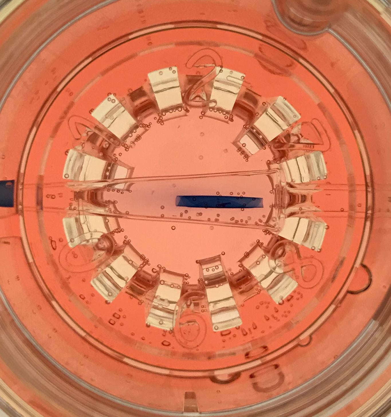
Tissue carrier (Straiko modified Jones tube) is securely held in corneal viewing chamber.
Patented Endothelial Delivery System (EDS™) ensures no vials and an easy access chamber.
Scrolled DMEK graft is stored in 20ml of Optisol-GS with no isolation by plugs or caps.
Saves time and resources in the OR.
Pre-punched with a Moria trephine, grafts available in 7.5, 7.75, and 8.0mm, requiring no trephine in the OR.
Grafts are pre-stained, requiring no trypan blue in the OR.
Truly Patient Ready, simply attach the carrier to syringe and transplant.
All Patient Ready DMEK® grafts are evaluated in accordance with EBAA Medical Standards.
Slit lamp examination and specular images are acquired with the graft scrolled inside the tissue carrier after all processing is complete.
The John A. Moran Eye Center at the University of Utah is the largest ophthalmology clinical care and research facility in the Intermountain West, with more than 60 faculty members, 10 satellite clinics, and 15 research labs.
Physicians provide comprehensive care in nearly all ophthalmic subspecialties, making Moran a major referral center for complex cases. Services include:
Cataracts
Cornea & External Eye Disease
Electrophysiology
Emergency Care
Glaucoma
LASIK and Vision Correction Surgery
Neuro-Ophthalmology
Oculoplastic and Facial
Plastic Surgery
Optometry
Patient Support Program for Patients with Vision Loss
Pediatric Ophthalmology
Pediatric Retina
Retinal Diseases
Strabismus
Ultrasound
Uveitis
We’re proud and honored to share the news that a panel of our peers—chairpersons and directors of academic programs across the country—voted the John A. Moran Eye Center at the University of Utah No. 8 in the nation—and that’s not all.