Chapter 01: Orientation to the Structure and Imaging of the Central Nervous System
Haines: Fundamental Neuroscience for Basic and ClinicalApplications, 6th Edition
Test Bank
MULTIPLE CHOICE
1. A 66-year-old man presents with paralysis of the upper and lower extremities on the right and paralysis of most movement of the left eye. Which of the following represents the best “localizing sign” in this patient?
a. Paralysis of the right upper extremity
b. Paralysis of the right lower extremity
c. Paralysis of upper and lower extremities
d. Paralysis of most eye movements on the left
e. Sensory deficits on the right side
ANS: D
Long tract signs, such as motor and sensory losses of the extremities, could indicate a lesion at many different levels of the neuraxis. However, these tracts pass through the midbrain. Significant deficits of eye movement in this patient indicate a lesion of the oculomotor nerve, the nucleus of which is located in the midbrain and the root of which exits the midbrain. In fact, the corticospinal tract is quite close to the root of the oculomotor nerve in anteromedial areas of the mesencephalon.
2. A patient presents to the emergency department with a sensory deficit involving the same side of the face and body. This would most likely suggest a lesion in which of the following structures?
a. Cerebral hemisphere
b. Brainstem
c. Cervical spinal cord
d. Lumbosacral spinal cord
e. Peripheral nerve roots
ANS: A
In general, sensory and/or motor deficits located on the same side of the face and body signify damage to a cerebral hemisphere. Lesions of the brainstem usually result in alternating deficits or crossed deficits (face/head one side, body opposite side), whereas damage to the spinal cord will result in deficits of the body below the lesion, with sparing of the head and/or face.
3. A 19-year-old man presents with motor deficits on one side of the body and sensory deficits
Copyright© 2026 Elsevier, Inc.All Rights are reserved, including those for text and data mining,AI training, and similar technologies.
on the other side of the body after an automobile collision. It is most likely that the lesion causing these deficits is located in which of the following parts of the nervous system?
a. Spinal cord
b. Medulla
c. Pons
d. Midbrain
e. Telencephalon
ANS: A
A lesion in the spinal cord results in deficits of the body only (no cranial nerve deficits) and may result in a motor deficit on one side of the body and a sensory deficit (pain and temperature loss) on the opposite side of the body.
4. A physician taps the patellar tendon of a 22-year-old woman and notes that the resulting reflex (knee jerk) is less active than normal. Which of the following most accurately describes this reflex?
a. Areflexia
b. Hyporeflexia
c. Hyperreflexia
d. Crossed deficit
e. Localizing sign
ANS: B
When a reflex is depressed or less active than normal, it is hyporeflexia. Areflexia refers to a completely absent reflex, and hyperreflexia refers to an excessively active reflex.
5. A 71-year-old man presents to the emergency department with a severe headache, nausea, and a decreased level of consciousness. The emergency department physician suspects an acute subarachnoid hemorrhage. Which of the following provides the best diagnostic evidence in this case?
a. X-ray
b. CT
c. MRI, T1-weighted image
d. MRI, T2-weighted image
e. PET scan
ANS: B
With CT, acute blood (generally less than 3 hours after a hemorrhage) appears white relative to the gray color of the brain and the black color of normal cerebrospinal fluid.
6. A 16-year-old boy is examined in the emergency department within 45 minutes of a motorcycle collision. The attending physician suspects a potential skull fracture and intracranial hemorrhage. Which of the following would the physician most likely order?
a. MRI, T1-weighted image
b. MRI, T2-weighted image
c. X-ray
d. CT
e. Brain biopsy
ANS: D
CT is especially useful in visualizing bone (it appears very white) and blood in an acute time frame (it also appears white to very white). Because CT of the brain only takes a few minutes to complete, it is particularly useful in evaluating traumatic brain injuries and allows rapid visualization of hematomas, contusions, and fractures.
7. A 21-year-old woman presents with a small tumor (confirmed by MRI) that has resulted in a change in her eating habits and sexual behavior. She now eats excessively and has become promiscuous. Which of the following represents the most likely location of this lesion?
a. Telencephalon
b. Hypothalamus
c. Midbrain
d. Pons
e. Medulla
ANS: B
The hypothalamus is a comparatively small part of the thalamus that is especially concerned with autonomic (visceromotor) functions. (The thalamus is also commonly called the diencephalon.) These functions are very important and include sexual functions, feeding behavior, regulation of body temperature, hormone production, and influence of many visceral centers in the brainstem and spinal cord.
8. A 1-year-old child is brought to the pediatric neurologist. Examination of this infant reveals a somnolent patient with an apparent mass in the parietal-occipital area of the skull. The neurologist orders the MRI shown here. Which of the following most accurately describes this image?
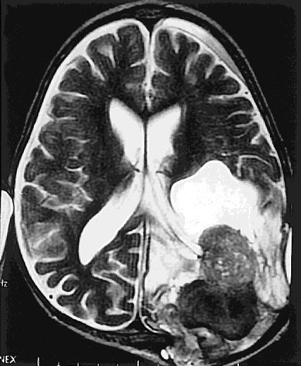
From Haines DE, Lancon JA. Review of Neuroscience. Philadelphia: Churchill Livingstone; 2003.
a. X-ray
b. CT
c. CT, bone window
d. MRI, T1-weighted image
e. MRI, T2-weighted image
ANS: E
In the T2-weighted image, bone is very dark, white matter and gray matter of the brain are shades of gray, cerebrospinal fluid is white, and fat is light gray.