42 minute read
The Laboratory

Medical research news from around the world
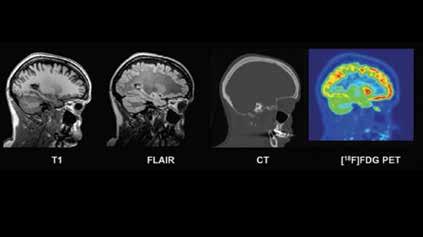
Figure 1: Example of co-registered T1 MRI, FLAIR MRI, CDT and [18F] FDG PET images (sagittal plane) for one subject of the database
New database of healthy adult human brain PET, MRI and CT images is now available for research
A new multi-modal database of healthy adult human brain scans has recently been made available for research.
The acquisition of imaging data can be a costly and logistically difficult process, including gaining participants’ consent for acquiring and disseminating their data. The fact that many countries restrict the use of ionising radiation in healthy controls adds to the complexity of neuroimaging research projects. Imaging database sharing plays a key role in the reduction of research costs and radiation exposure.
The CERMEP-IDB-MRXFDG database , a collaboration between King’s College London & Guy’s and St Thomas’ PET Centre at the School of Biomedical Engineering & Imaging Sciences, CERMEP and Neurodis Foundation Lyon, is a collection of PET, CT, and MR images, which allows for quantitative analyses and methodology development in neuroimaging.
Professor Alexander Hammers, Head of PET Centre and one of the senior authors of the study said: “There are quite a few databases of MR images of the brain, but there is very limited choice for brain PET (FDG) databases, especially for younger adults whom we regularly scan with FDG PET/ CT as part of their epilepsy surgery workup. Ours is the first I am aware of which is published with the explicit aim of making it available to others.”
The age range of subjects – between 23 and 65 - is reflective of participants in research studies at imaging centres on conditions such as epilepsy, movement disorders, multiple sclerosis and disorders of consciousness, allowing statistical comparison to a database of healthy controls.
The database is stored in three different formats: DICOM (data not processed), NIFTI (multimodal images coregistered to PET subject space) and NIFTI normalised (images normalised to the Montreal Neurological Institute (MNI) coordinate system), enabling a high level of interoperability.
The database contains PET images, CT data and two different MR sequences, enabling machine learning of the relationship between modalities and the synthesis of missing modalities. A potential future application is in MR-based attenuation correction for simultaneous PET-MR scanners. • Interested researchers can request access to the CERMEP-IDB-MRXFDG database via a short form which can be obtained by emailing the lead author, Dr Inés Mérida: merida@cermep.fr • doi: https://doi.org/10.1101/2020. 12.15.422636
Tuberculosis: New biomarker indicates individual treatment duration
When can tuberculosis therapy be stopped without risk of relapse? Doctors are faced with this question time and again, because the lack of detection of the tuberculosis pathogen Mycobacterium tuberculosis is no guarantee for a permanent cure of the lung infection. Patients who respond to the standard therapy may be out of treatment after six months. But for resistant cases, more than 18 months of treatment duration is currently advised.
“This is a very long time for those affected, who often have to take more than four antibiotics every day and suffer from side effects”, explained Prof. Dr. Christoph Lange, Clinical Director at the Research Center Borstel and director of the study, conducted at the German Center for Infection Research (DZIF) in cooperation with the German Center for Lung Research (DZL). “We urgently need a biomarker that enables the implementation of an individualised treatment duration,” he emphasises. After all, not every patient needs so long to recover.
Since the absence of bacteria in the sputum does not justify a safe stop in therapy, the team around Christoph Lange set out to find alternative biomarkers in the patient. In collaboration with international tuberculosis centres, on the basis of patient cohorts a model for the end of therapy could be developed that is based on an RNA determination in the blood. From many thousands of genes, 22 have been identified whose activity correlates with the course of the disease.
“The production of RNA of these 22 genes in human blood can tell us whether the patient is cured,” PD Dr Jan Heyckendorf from the FZ Borstel said. Together with Maja Reimann and Dr Sebastian Marwitz, he is the lead author of the study. “It is an RNA signature from 22 genes

identified on two cohorts and validated on another three cohorts,” he added. “No other published transcriptom marker shows comparable properties so far.”
To identify this individual biomarker, the scientists within the DZIF have established five different patient cohorts. In all cases, these were adults who had contracted pulmonary TB, partly from non-resistant, partly from resistant forms. In addition to cohorts in Germany, patients in Bucharest (Romania) were also included, where the DZIF supports a study centre.
“The individualisation of the treatment duration is an important milestone on the road to precision medicine for tuberculosis,” Christoph Lange said. Even without progression values, one could risk to end a patient’s treatment on the basis of this RNA determination. As a next step, the researchers are planning a prospective study at the DZIF. The aim is for patients in one study arm to receive treatment for as long as the biomarker suggests, while patients in the other arm receive treatment for as long as the national tuberculosis programme recommends. The scientists then want to see whether the biomarker makes a shorter treatment duration possible. The team around Dr Lange is confident.
“Hopefully, it will then be possible for patients with multidrug-resistant tuberculosis to save about one-third of treatment on average,” said Dr Lange.
Reference:
http://dx.doi.org/10.1183/13993003.03492-2020
Digital health tracking tools help individuals lose weight, study finds
Digital health tools, such as diet-tracking apps, increase engagement in weight loss programs, helping users shed pounds, according to a new study.
Weight loss advice is exasperating. Eat breakfast. Don’t eat breakfast. No fats. Lots of fats. Run long distances. Exercise hard in spurts.
A new study led by a Stanford Medicine researcher makes at least one thing clear: No matter which weight loss tactic you choose, you’re typically more successful if you track your progress with digital health tools.
According to the study, the closer people track their weight-loss efforts with things like smart watches, digital scales and dietmonitoring websites, the more weight they tend to lose.
“We’ve seen this rise of digital health tools in the last decade, and they provide a great way for people to access interventions to better their health,” said Michele Patel, PhD, postdoctoral scholar at the Stanford Prevention Research Center. “We’re also starting to see that more weight loss programs are trending toward digital tools, too. But exactly what is being used, how it’s being used and the impact it has on the user has never been systematically studied on a large scale.”
The analysis also revealed that individuals who tracked their diet or physical activity digitally were more engaged, meaning they were more consistently active in using their digital tools, than those who tracked their behavior through more traditional means, such as handwritten records of exercise routines or calorie intake. In the end, it all comes back to goal-setting and consistency, said Patel, and digital tools can help facilitate both.
A paper detailing the analysis was published online Feb. 24 in Obesity.
Digital is convenient Patel and her team compared nearly 40 different studies on weight-loss monitoring that were conducted between 2009 and 2019. In each study, participants tracked their behaviours, such as calorie and nutrient consumption, the number of daily bites they took and their physical activity, with digital tools. Three-quarters of the time, those who used digital tools more frequently to monitor themselves lost more weight than those who self-monitored less frequently with digital tools, Patel and her colleagues found.
The finding makes sense, Patel said. Tracking allows us to be aware of what we’re eating, how much we’re moving and how our body weight fluctuates on the scale each day. People can leverage this monitoring feedback to make changes to their daily behaviours. Investigating the grams of nutrients and calories for every meal is burdensome for most people. Digital tools make calorie counting and nutrient tracking easier, she said. Diet-monitoring websites and apps already “know” information about the foods users log – such as the grams of carbs versus fat in a waffle. Some


apps even allow users to take a picture of their meal and upload it; the app does the rest of the calorie-counting work.
Digital tools may also ramp up engagement by tapping into one’s competitive side, as some apps allow for networking or provide visual cues indicating how close one is to reaching a goal – for example, how close one is to completing a colourful circle representing step count.
What’s more, the study showed that inperson weight loss coaches weren’t necessary for people to stay engaged in weight loss programs. “I think that’s promising for individuals who are seeking to lose weight on their own,” Patel said.
The other important takeaway, she said, is that it didn’t matter what individuals monitored. They could track weight loss, calories or exercise. As long as they did it with digital tools, the more they monitored, the slimmer they became.
Patel plans to dig in deeper to the phenomenon, looking at exactly which behaviour – weight tracking, physical activity tracking, or calorie monitoring – seems to generate the most weight loss. She also plans to specifically recruit people from racial and ethnic minority groups to measure the impact of digital monitoring on weight loss, as these groups are often underrepresented in weight-loss programs.
Deep brain stimulation prevents epileptic seizures in mouse model
Epileptic activity originating from one or more diseased brain regions in the temporal lobe is difficult to contain. Many patients with so-called temporal lobe epilepsy often do not respond to treatment with anti-epileptic drugs, and the affected brain areas must therefore be surgically removed. Unfortunately, this procedure only gives seizure freedom to about one third of patients, so the development of alternative therapeutic approaches is of great importance.
Scientists led by neurobiologist Prof. Dr Carola Haas, head of the research group at the Department of Neurosurgery at Medical Centre, University of Freiburg and the BrainLinks-BrainTools research center, have investigated a new therapeutic approach to prevent epileptic seizures in temporal lobe epilepsy. They showed in mice that low-frequency stimulation of specific brain areas could completely stop epileptic activity. Instead of using electric current, the researchers stimulated the cells with light. To do this, they had previously introduced a light-sensitive molecule into the cells that allows particularly precise stimulation. They published the results in December 2020 in the scientific journal elife.
“As soon as we stimulated the brain region with a frequency of one hertz, the epileptic seizures disappeared. This effect was stable over several weeks,” Prof. Haas said. Habituation, which can occur with drug therapy, did not take place. The brain region was stimulated for one hour daily.
In temporal lobe epilepsy, the hippocampus is often pathologically altered and usually represents the so-called focus of epileptic activity. Previous studies have used precise genetic labelling techniques to map the fibre system and its synaptic contacts between the temporal lobe and hippocampus, which are typically preserved in temporal lobe epilepsy. The researchers used this fibre system to manipulate hippocampal activity in a specific and temporally precise manner using light-dependent proteins. Measuring brain waves showed that rhythmic activation of the diseased hippocampus at a low frequency of one hertz suppressed epileptic activity and prevented it from spreading.
Haas and her colleagues demonstrated that the anti-epileptic effect is largely due to the repeated activation of surviving granule cells in the seizure focus. Single cell studies confirmed the assumption that the granule cells are less excitable due to the stimulation, making the epileptic seizure less likely to spread.
“It’s also possible that we have a widespread network effect because the stimulation can spread through the hippocampal circuitry,” Prof. Haas said.
In the future, the team, along with the medical physics department at the Medical Center - University of Freiburg, would like to use magnetic resonance imaging to observe the entire brain during stimulation. This technique could be used to identify additional brain regions that are affected by the stimulation. Corresponding findings on these could provide information on how they are connected and what further consequences stimulation has. • doi: https://doi.org/10.7554/eLife.54518
Massive new genetic sequencing data helps rectify underrepresentation of minority participants in genomic studies
Researchers at the University of Maryland School of Medicine (UMSOM) and their colleagues published a new analysis in the journal Nature from genetic sequencing data of more than 53,000 individuals, primarily from minority populations. The early analysis, part of a large-scale program funded by the National Heart, Lung, and Blood Institute, examines one of the largest and most diverse data sets of high-quality whole genome sequencing, which makes up a person’s DNA. It provides new genetic insights into heart, lung, blood, and sleep

disorders and how these conditions impact people with diverse racial and ethnic backgrounds, who are often underrepresented in genetic studies.
The program, called Trans-Omics for Precision Medicine (TOPMed), seeks to understand the genetic variations that occur among individuals both in nuclear families and in populations from diverse ethnicities residing on different continents. The project’s ultimate goal is to improve the diagnosis, treatment, and prevention of the most common conditions that lead to disability or death.
“We have already identified some surprising new insights,” said study corresponding author Timothy O’Connor, PhD, Associate Professor of Medicine & Endocrinology at the Institute for Genome Sciences (IGS) at UMSOM. For example, the team identified more than 400 million genetic variations, but 97 percent of them are extremely rare, occurring in less than one percent of the population.
“Most of the time, these variants mean nothing,” said Dr O’Connor, “but they can provide a new understanding of mutational processes and recent human evolutionary history.”
The TOPMed team includes more than 180 researchers from leading institutions in genomics worldwide, who have been compiling huge datasets in systematic and defined ways to increase knowledge about diversity in genetic studies. Since its launch in 2014, the TOPMed investigators have begun adding whole genome sequencing and “omics” analysis (which includes a study of genetic and molecular profiles like proteins) to research studies in order to better understand how variations affect different organ systems giving rise to disease in, for example, the heart and lungs.
Causal genetic variants In the new Nature paper, the researchers pointed out that the program “aims to identify causal genetic variants and how they interact with the environment, to characterize disease and its molecular subtypes, to understand differences in disease across diverse ancestries, and to establish a foundation for personalized disease prediction, prevention, diagnosis, and treatment.” Braxton Mitchell, PhD, Professor of Medicine at UMSOM, and Jeffrey O’Connell, PhD, Associate Professor of Medicine at UMSOM, were co-authors on this paper.
TOPMed is the largest sequencing project to date and has identified over 400 million gene variants with an overarching mission of understanding global genetic diversity. Since joining the TOPMed program in 2016, UMSOM researchers have published valuable new insights on genetic diversity, including sequencing data from the initial flagship paper on the first 53,831 TOPMed samples.
Increasing diversity
The increasing diversity of the population samples will help investigators learn more about how specific diseases impact different ethnic populations around the world. In addition, the group has established uniform standards for sequencing performed on a massive scale. The standards maximize the integrity of the data as the large group of international researchers use uniform methods as they continue to add other “omics” methods for analysis such as the study of metabolic differences.
“This is a major effort to rectify the
underrepresentation of minority participants in genomic studies and tracks with a broader mission within the School of Medicine to increase diversity in clinical trials,” said E. Albert Reece, MD, PhD, MBA, Executive Vice President for Medical Affairs, UM Baltimore, the John Z. and Akiko K. Bowers Distinguished Professor and Dean, University of Maryland School of Medicine. “This hopefully will move the genomics field closer to extending personalized medicine for all patients.”
Cashell Jaquish, Ph.D., an NHLBI program officer for TOPMed and a corresponding author on the Nature paper, agrees. “The NHLBI’s TOPMed program is a huge resource for the scientific commusion of a diversity of sampling, which will be invaluable to the international group in learning more about the diseases impacting these populations. Because of the vast sample sizes and the longitudinal scope of many of the population samples, the investigators were able to demonstrate that the rare variants represent recent and potentially deleterious changes that can impact protein function, gene expression, or other biologically important elements.
nity. We didn’t really know what genomic variation looked like in diverse groups until now. This new study represents truly historic findings, and we look forward to continued research studies in this area as we move toward personalized medicine.”
In addition to enabling detailed analysis of the combined genomic and health data for sequenced samples, TOPMed has enhanced the analyses of genotyped samples through a new reference panel that now includes over 97,000 individuals. The TOPMed imputation reference panel is publicly available for review and input of new genetic data by researchers.
The first stage of the data release in the Nature study demonstrated a greater inclu-
Reference:
Sequencing of 53,831 diverse genomes from the NHLBI TOPMed Program. Nature. 10 February 2021. doi: https://doi.org/10.1038/ s41586-021-03205-y

A cure for severe diabetes type 2?
Long-term study shows gastrointestinal surgery patients stay diabetes-free for 10 years
The results of a randomized clinical trial with the longest follow up to date show that metabolic surgery is more effective than medications and lifestyle interventions in the long-term control of severe type 2 diabetes.
The study, published January 22, 2021 in The Lancet[1], also shows that over one-third of surgically-treated patients remained diabetes-free throughout the 10-year period of the trial. This demonstrates, in the context of the most rigorous type of clinical investigation, that a “cure” for type 2 diabetes can be achieved.
Researchers from King’s and the Fondazione Policlinico Universitario Agostino Gemelli IRCCS, Rome, Italy report the 10-year outcomes of a trial that compared metabolic surgery with conventional medical and lifestyle interventions in patients with type 2 diabetes.
The study involved 60 patients with advanced type 2 diabetes and treated at a major academic hospital in Rome, Italy. The patients randomly underwent drugs plus lifestyle interventions or metabolic surgery (gastric bypass or biliopancreatic diversion). At the start of the study, all patients had severe disease, with poorly controlled blood sugar levels and more than five years history of diabetes.
The results of the study show that 37.5% of surgically-treated patients were able to maintain non-diabetic glycaemia without need for diabetes medication – a condition referred to as diabetes remission – for the duration of the 10-year study period. In 2009, American Diabetes Association defined “cure” of diabetes as a continued state of disease remission for more than five years.
Professor Francesco Rubino, senior author of the report and Chair of Bariatric and Metabolic Surgery at King’s College London and a consultant surgeon at King’s College Hospital in London said: “The findings from this study provide the most robust scientific evidence yet that fullblown type 2 diabetes is a curable disease, not inevitably progressive and irreversible. In addition to represent a major advance in the treatment of diabetes, metabolic surgery is our best lead to the elusive cause of the disease.”
Diabetes-related complications
Compared to conventional medical treatment, surgery also resulted in better overall metabolic control, lower cardiovascular risk, better kidney function and quality of life. Notably, patients treated surgically had a significant lower incidence of diabetes-related complications, including cardiac, renal, and neurological adverse events. Metabolic surgery also reduced medication usage, including drugs for diabetes, high blood pressure and dyslipidaemia.
The study investigated the early and longterm safety of the different intervention strategies. Patients who underwent biliopancreatic diversion had more incidences of serious adverse events, including events associated to both disease and intervention, compared to subjects in both other groups. Patients treated by conventional medical therapy had significantly higher incidence of serious adverse events compared to patients who underwent surgery by Roux-en-Y gastric bypass.
Professor Geltrude Mingrone, first author of the report, Professor of Medicine at the Catholic University of Rome and a Professor of Diabetes and Nutrition at King’s College London said: “These data corroborate the notion that surgery can be a cost-effective approach to treating type 2 diabetes. The evidence is now more than compelling that metabolic surgery should be considered as a main therapeutic option for the treatment of patients with severe type 2 diabetes and obesity.”
Previous studies had shown that bariatric or weight loss surgery can induce longterm remission of diabetes in patients with very severe obesity; however, most patients who undergo traditional weight loss surgery have typically mild or recentonset diabetes. This trial shows the potential curative effect of metabolic surgery for patients with severe disease.
The findings from this study provide the most robust scientific evidence yet that fullblown type 2 diabetes is a curable disease, not inevitably progressive and irreversible. – Professor Francesco Rubino, senior author of the report and Chair of Bariatric and Metabolic Surgery at King’s College London
Reference:
[1] Metabolic surgery versus conventional medical therapy in patients with type 2 diabetes: 10-year follow-up of an open-label, single-centre, randomised controlled trial. The Lancet. 23 January 2021. doi: https://doi.org/10.1016/ S0140-6736(20)32649-0
CapaCare Protect
Antiviral paint for healthcare facilities
Middle East Health speaks to Martin Rosocha, Managing Director at Caparol Arabia about their CapaCare Protect antiviral paint for healthcare facilities.
Middle East Health: Can you tell us a bit about CapaCare Protect?
Martin Rosocha: When the global pandemic hit us last year, our expert solutions team put a lot of effort and worked nonstop to join the fight against Coronavirus. This is the reason why we launched “CapaCare Protect”, an innovative and sustainable solution that provides better protection against harmful micro-organisms across the Gulf region. The launch of this product aligns with our company’s ethos which is to constantly look for innovative, quality and sustainable solutions to provide comfortable and healthy living. CapaCare Protect is a premium antiviral paint suited for homes, hospitals, clinics and day-care centers.
CapaCare Protect is a premium quality Interior Emulsion developed with SILVERbac technology which uses active silver ions to inhibit the growth of bacteria and viruses on the painted surface which decreases the spread of contagious diseases. It has kill rate of 99.9% against a host of microbes including gram +ve and gram -ve bacteria and virus. This paint enhances indoor comfort as it has zero odor with zero VOC.
The levels of available silver in the paint has been demonstrated to reduce bacterial numbers by greater than 3 logs (99.9% reduction) using a range of American and Japanese International Testing Standards which is considered to have excellent antiviral activity.
Along with viruses and bacteria, CapaCare Protect showed resistance to mould and fungus as well contamination which would allow reduced cleaning to be undertaken and any related complications in the healthcare environment which would lead to respiratory illness do to mould spores released into the air form contaminated walls in areas of damp.

MEH: How did you carry out testing of the product?
MR: CapaCare Protect is the only paint in the region that has shown 99.9% kill rate against the coronavirus. Tests were conducted according to American and Japanese standards in two independent ISO 17025 laboratories in the United States and were approved and accredited by International Antimicrobial Council (IAC).
MEH: How is CapaCare being applied in the region, and for which sectors is it the most suitable and advantageous?
MR: CapaCare Protect is manufactured in our production plant in the UAE and has already been significantly used on various projects including hospitals, commercial, residential areas and schools. We received solid demand from the market for this product which is on the rise and its production is expected to continue growing.
CapaCare Protect is washable, easy to clean and maintain and is highly recommended for hospitals, schools, malls, restaurants and all living spaces. For instance, it has been approved for DAFZA (Dubai Airport Free Zone authority), Gems School, Saudi German Hospital, Dubai London Hospital, Rashid Center of Disability, Sofitel JBR and many other highprofile projects across the Middle East.
MEH: Can you tell us about your future
plans in the Middle East?
MR: We want to play a key role in sup-
Martin Rosocha, Managing Director at Caparol Arabia
porting the positive development of the region and reflect governmental transition towards a green economy. We are seeing positive shifts in the market with more and more developers keen to make projects more efficient and sustainable. There’s a real opportunity to help them improve the performance and safety of their buildings and enhance the experience of residents with our innovative solutions.
As a green company, we also want to continue championing indoor air quality and offer our VOC-free paints which can be complementary to a healthier lifestyle and play a central role in the protection of our environment and well-being. Our motto is: “Everyone should be living and working in healthy, well-designed, efficient and sustainably constructed buildings.”
On a regional level, we are also planning to expand our innovative products to tailor to the demand of the market.
Covid-19 Update
Damage to the heart found in more than half of Covid-19 patients discharged from hospital
The European Society of Cardiology is reporting that around 50% of patients who have been hospitalised with severe Covid-19 and who show raised levels of a protein called troponin have damage to their hearts. The injury was detected by magnetic resonance imaging (MRI) scans at least a month after discharge, according to new findings published in the European Heart Journal [1] .
Damage includes inflammation of the heart muscle (myocarditis), scarring or death of heart tissue (infarction), restricted blood supply to the heart (ischaemia) and combinations of all three.
The study of 148 patients from six acute hospitals in London is the largest study to date to investigate convalescing Covid-19 patients who had raised troponin levels indicating a possible problem with the heart.
Troponin is released into the blood when the heart muscle is injured. Raised levels can occur when an artery becomes blocked or there is inflammation of the heart. Many patients who are hospitalised with Covid-19 have raised troponin levels during the critical illness phase, when the body mounts an exaggerated immune response to the infection. Troponin levels were elevated in all the patients in this study who were then followed up with MRI scans of the heart after discharge in order to understand the causes and extent of the damage.
Professor Marianna Fontana, professor of cardiology at University College London (UK), who led the research with Dr Graham Cole, a consultant cardiologist at Imperial College London, said: “Raised troponin levels are associated with worse outcomes in Covid-19 patients. Patients with severe Covid-19 disease often have preexisting heart-related health problems including diabetes, raised blood pressure and obesity. During severe Covid-19 infection, however, the heart may also be directly affected. Unpicking how the heart can become damaged is difficult, but MRI scans of the heart can identify different patterns of injury, which may enable us to make more accurate diagnoses and to target treatments more effectively.”
The researchers investigated Covid-19 patients discharged up until June 2020 from six hospitals across three NHS London trusts: Royal Free London NHS Foundation Trust, Imperial College Healthcare NHS Trust and University College London Hospital NHS Foundation Trust. Patients who had abnormal troponin levels were offered an MRI scan of the heart after discharge and were compared with those from a control group of patients who had not had Covid-19, as well as from 40 healthy volunteers.
“The recovering Covid-19 patients had been very ill; all required hospitalisation and all had troponin elevation, with around one in three having been on a ventilator in the intensive care unit,” said Prof. Fontana.
“We found evidence of high rates of heart muscle injury that could be seen on the scans a month or two after discharge. Whilst some of this may have been pre-existing, MRI scanning shows that some were new, and likely caused by Covid-19. Importantly, the pattern of damage to the heart was variable, suggesting that the heart is at risk of different types of injury. While we detected only a small amount of ongoing injury, we saw injury to the heart that was present even when the heart’s pumping function was not impaired and might not have been picked up by other techniques. In the most severe cases, there are concerns that this injury may increase the risks of heart failure in the

MRI scan of damaged heart. Blue means reduced blood flow, orange is good blood flow. In this figure the inferior part of the heart shows dark blue, so the myocardial blood flow is very reduced and the black and white angiography, which looks directly at the blood vessels, shows that the vessel which supplies the blood to this part of the heart is occluded. The 3 coloured images are 3 different slices of the heart: the basal the mid and the apical slice.
Covid-19 Update
future, but more work is needed to investigate this further.”
The function of the heart’s left ventricle, the chamber that is responsible for pumping oxygenated blood to all parts of the body, was normal in 89% of the 148 patients but scarring or injury to the heart muscle was present in 80 patients (54%). The pattern of tissue scarring or injury originated from inflammation in 39 patients (26%), ischaemic heart disease, which includes infarction or ischaemia, in 32 patients (22%), or both in nine patients (6%). Twelve patients (8%) appeared to have ongoing heart inflammation.
Prof. Fontana said: “Injury relating to inflammation and scarring of the heart is common in Covid-19 patients with troponin elevation discharged from hospital,
A new study looking at how Covid-19 affects people with asthma provides reassurance that having the condition doesn’t increase the risk of severe illness or death from the virus.
George Institute for Global Health researchers in Australia analysed data from 57 studies with an overall sample size of 587,280. Almost 350,000 people in the pool had been infected with Covid-19 from Asia, Europe, and North and South America and found they had similar proportions of asthma to the general population.
The results, published in the peer-reviewed Journal of Asthma, show that just over seven in every 100 people who tested positive for Covid-19 also had asthma, compared to just over eight in 100 in the general population having the condition. They also showed that people with asthma had a 14 percent lower risk of acquiring Covid-19 and were significantly less likely to be hospitalized with the virus.
There was no apparent difference in the risk of death from Covid-19 in people with asthma compared to those without.
Head of The Institute’s Respiratory pens to people who are not hospitalised with Covid, or those who are hospitalised but without elevated troponin. The findings indicate potential ways to identify patients at higher or lower risk and suggest potential strategies that may improve outcomes. More work is needed, and MRI scans of the heart have shown how useful it is in investigating patients with troponin elevation,” concluded Prof. Fontana.
but is of limited extent and has little consequence for the heart’s function.
“These findings give us two opportunities: firstly, to find ways of preventing the injury in the first place, and from some of the patterns we have seen, blood clotting may be playing a role, for which we have potential treatments. Secondly, detecting the consequences of injury during convalescence may identify subjects who would benefit from specific supporting drug treatments to protect heart function over time.”
The findings of the study are limited by the nature of patient selection and included only those who survived a coronavirus infection that required hospital admission.
“The convalescent patients in this study had severe Covid-19 disease and our results say nothing about what hap-
Reference
[1] “Patterns of myocardial injury in recovered troponin-positive Covid-19 patients assessed by cardiovascular magnetic resonance”, by Tushar Kotecha et al. European Heart Journal. doi: https://doi.org/10.1093/eurheartj/ ehab075
Study shows asthmatics have no higher risk of dying from Covid
Program, co-author Professor Christine Jenkins said that while the reasons for these findings weren’t clear, there were some possible explanations - such as some inhalers perhaps limiting the virus’ ability to attach to the lungs.
“Chemical receptors in the lungs that the virus binds to are less active in people with a particular type of asthma and some studies suggest that inhaled corticosteroids - commonly used to treat asthma - can reduce their activity even further,” she said.
“Also, initial uncertainty about the impact of asthma on Covid-19 may have caused anxiety among patients and caregivers leading them to be more vigilant about preventing infection.”
Lead author Dr Anthony Sunjaya added that while this study provides some reassurance about the risks of exposure to Covid-19 in people with asthma, doctors and researchers were still learning about the effects of the virus.
“While we showed that people with asthma do not seem to have a higher risk of infection with Covid-19 compared to those without asthma and have similar outcomes, we need further research to better understand how the virus affects those with asthma,” he said.
When the Covid-19 pandemic first spread across the world concerns were raised that people with asthma might be at a higher risk of becoming infected, or of becoming sicker or even dying.
Previous findings have shown that people with chronic respiratory conditions like asthma were reported to be at greater risk during the Middle East Respiratory Syndrome (MERS) outbreak, caused by a virus with a similar structure.
“Respiratory infections like those caused by coronaviruses can exacerbate asthma symptoms and corticosteroid treatment may increase susceptibility to Covid-19 infection and its severity,” Dr Sunjaya said.
However this study using the best evidence available on the risk of infection, severe illness – requiring admission to ICU and/or ventilator use – and death from Covid-19 in people with asthma finds “no significant difference” of people with asthma being at higher risk.

Covid-19 Update
Lab study suggests blood group A poses higher risk for Covid-19 infection
As researchers around the world work to identify and address risk factors for severe Covid-19, there is additional evidence that certain blood types could be associated with greater risk of contracting the disease. A new Blood Advances study details one of the first laboratory studies to suggest that SARS-CoV-2, the virus that causes Covid-19, is particularly attracted to the blood group A antigen found on respiratory cells.
In the study, researchers assessed a protein on the surface of the SARS-CoV-2 virus called the receptor binding domain, or RBD. The RBD is the part of the virus that attaches to the host cells, so it is an important research target for understanding how infection occurs. The team assessed synthetic blood group antigens on respiratory and red blood cells found in blood group A, B, and O individuals, and analysed how the SARS-CoV-2 RBD interacted with each unique blood type. They discovered that the RBD had a strong preference for binding to blood group A found on respiratory cells. It did not display a preference for blood group A red blood cells, or other blood groups found on respiratory or red cells. The capacity of the RBD to preferentially recognize and attach to the blood type A antigen found in the lungs of blood type A individuals may provide insight into the potential link between blood group A and Covid-19 infection.
“It is interesting that the viral RBD only really prefers the type of blood group A antigens that are on respiratory cells, which are presumably how the virus is entering most patients and infecting them,” said study author Sean R. Stowell, MD, PhD, of Brigham and Women’s Hospital. “Blood type is a challenge because it is inherited and not something we can change. But if we can better understand how the virus interacts with blood groups in people, we may be able to find new medicines or methods of prevention.”
Based on their observations, the team sought to determine whether a similar binding preference existed for the RBD of SARSCoV, the virus that causes severe acute respiratory syndrome (SARS). Although the makeup of the virus differs, the SARS-CoV RBD exhibited the same preference to bind to the group A antigens on respiratory cells.
Dr Stowell and his team emphasized that their findings alone could not fully describe or predict how coronaviruses like SARS-CoV-2 and SARS-CoV would affect patients of various blood types. “Our observation is not the only mechanism responsible for what we are seeing clinically, but it could explain some of the influence of blood type on Covid-19 infection.”
While further research is needed to understand that influence, the paper adds to findings from earlier Blood Advances studies suggesting a possible link between blood type and Covid-19 susceptibility and severity.
Covid-19 infection in pregnancy not linked with still birth or baby death
Covid-19 infection in pregnancy is not associated with stillbirth or early neonatal death, according to a new study.
However the research, from over 4000 pregnant women with suspected or confirmed Covid-19, also found women who had a positive test were more likely to have a premature birth.
The research, led by scientists from Imperial College London and published in the journal Ultrasound in Obstetrics and Gynecology, used data from the UK and the USA.
The study team looked at data from 4004 pregnant women who had suspected or confirmed Covid-19. Of these women, 1606 were from the UK, from a data registry called PAN-Covid, while 2398 were from the US, from the American Academy of Pediatrics SONPM data registry.
PAN-Covid was funded by the Medical Research Council, UK National Institute for Health Research and the NIHR Imperial Biomedical Research Centre.
All the women gave birth between January-August 2020.
The research found that no babies died from Covid-19 in the study. There was also no increase in risk of stillbirth or low birth weight.
However, both the UK and US data suggested a higher risk of pre-term birth (defined as birth before 37 weeks).
In the UK data, 12 per cent of women with suspected or confirmed Covid-19 had a preterm delivery – 60 per cent higher than the national average rate of 7.5 per cent. In the US data, 15.7 per cent of women had a pre-term birth, 57 per cent higher than the US national average of 10 per cent.
The study team say part of this association may be due to doctors deciding to deliver the baby early due to concerns about the effect of Covid-19 infection on mother and baby. The rate of spontaneous pre-term birth was lower than expected.
Professor Christoph Lees, senior author of the study from Imperial’s Department of Metabolism, Digestion and Reproduction, said: “The finding that Covid-19 infection does not increase the risk of stillbirth or baby death is reassuring. However, a suspected or confirmed Covid-19 diagnosis was linked to a higher risk of preterm birth, and it isn’t entirely clear why.”
Dr Ed Mullins, co-author from Imperial’s Department of Metabolism, Digestion and Reproduction, added: “This study supports the prioritisation of vaccination for women who are pregnant or who plan to become pregnant, and existing measures that protect women in pregnancy from infection, in order to reduce pre-term birth.”
CleanSpace Technology
Australian respirator manufacturer finds itself at forefront of COVID-19 crisis

CleanSpace Technology, an Australian company that designs and manufactures nextgeneration respirators, has found itself at the forefront of the COVID-19 pandemic.
The proprietary technology, at the heart of all CleanSpace Respirators, was designed by ex-ResMed biomedical engineers. ResMed is a world leader in CPAP devices. The engineers had a vision to make respirators that delivered high level protection in an easy to use and comfortable system. The company has been successfully protecting workers in a wide range of sectors for the past ten years.
Until CleanSpace, the technology for masks had not changed for 30 years. Traditional devices were typically uncomfortable, hot and provided low protection.
“Our technology was seen as game-changer, and still is. One of the main reasons people go unprotected is because of low compliance. If masks are uncomfortable or not quick and easy to put on then it simply doesn’t get used,” said Alex Birrell, CleanSpace Technology CEO. “CleanSpace is unique, it’s a Powered Air Purifying Respirator (PAPR) without the heavy and cumbersome belt and hoses associated with PAPRs. Its simplicity with fresh air on the face, makes it far preferable to the N95 disposable [mask].”
The clear silicon mask is comfortable and soft and allows for easy communication. CleanSpace Respirators are operated using a simple one-button smart system. These features combined mean healthcare workers are more likely to wear them for a full shift.
Compared to disposable masks, CleanSpace Respirators offer more protection and are more economical as the cost of replacing disposables stacks up. Disposable masks are well-known for causing fogging and discomfort, leading to low compliance.
CleanSpace HALO Following the Ebola outbreak in 2014, the WHO and CDC put out a call for light weight highly protective PAPRs specifically designed for healthcare workers. CleanSpace Technology were confident, with their medical device background and a commercially proven technology, they could develop a healthcare respirator. Thirty percent lighter, smaller and more ergonomic than its predecessors, CleanSpace HALO was the first respirator to be designed in consultation with healthcare.
Well before the COVID-19 crisis, CleanSpace HALO was protecting the lives of thousands of frontline healthcare workers globally. Since the outbreak, this unique system has become the ‘standard of care’ for protection of high-risk healthcare teams in anaesthetics, surgery and general care. The advantages of reusable systems with superior protection and a secure supply chain make this Australian manufacturer an attractive and reliable vendor for many Australian and international hospitals.
Since COVID-19, with the rapid depletion of disposable masks and reliance on lean supply chains, hospitals were suddenly desperately seeking alternatives to disposable masks.
“The need is very real and very urgent, we have had to quickly adapt our manufacturing to respond to the need,” added Dr Birrell.
CleanSpace HALO is designed specifically for the healthcare, pharmaceutical and laboratory sectors and are being used to protect the lives of thousands of frontline healthcare workers globally.
Smart technology for respiratory protection CleanSpace HALO is a PAPR system that houses smart technology in a revolutionary, compact design. CleanSpace delivers the highest protection in healthcare while being comfortable, quick to fit and easily integrated into any setting. • High Protection P3/TM3 99.95% • Reusable and cost-effective • Lightweight 400g/0.9lb • No belts or hoses • Simple and fast to put on • IP rated 66 water tolerant • More information: https://cleanspacetechnology.com/health/
CleanSpace STERI-PLUS exhalation filter for source control The world’s first PAPR exhalation valve filter is utilised for source control in sterile settings where filtering of the wearer’s exhaled air is required. • Filtration efficiency 99% for particles 0.3µm and above • Approved for use with CleanSpace respirators & CleanSpace half masks • Reusable case is compatible with standard disinfection/sterilization protocols • Easy to fit. Easy to clean • Cost-Effective • Learn more:
Flyer: https://cleanspacetechnology.com/ health/resources/
Video: https://cleanspacetechnology.com/ health/steri-plus-exhalation-filter/
CleanSpace Technology, an Australian company based in Sydney, assists in product training, fit testing and instructions on maintenance and care with attentive customer support. • Contact CleanSpace Technology to request a demonstration sales@cleanspacetechnology.com www.cleanspacetechnology.com
Becton Dickinson
Maintaining safe IV infusion therapy during the COVID-19 pandemic
Smart pumps with Dose Error Reduction Systems (DERS) reduce the risk of medication error, but the requirement for strict isolation of large numbers of patients during the COVID-19 pandemic has made maintaining the Rights of IV medication administration increasingly difficult.
Right Maintenance of continuous critical short half-life infusions (CSHLI), such as Noradrenaline or Glyceryl Trinitrate is also vital as any prolonged interruption of CSHLI delivery could be fatal, and nursing staff must respond promptly to any infusion alarm if serious cardiovascular events are to be avoided. Centralised monitoring of infusions can significantly reduce nurse reaction times to CSHLI alarms.
To reduce nursing time inside SARSCoV-2 patient rooms we can use long extension lines that allow the patient’s pumps to remain outside of the isolation room. Running the IV line under the door and across the room’s floor, with taping to prevent tripping or dislodgment, is not ideal but provides protection of the line. However, the technique may cause issues of pressure gradient changes affecting occlusion alarms, and accumulation of air in the line due to the low level of the line in relation to the pump and the patient.
Long lines increase siphonage in the case of large bore lines and increase downstream pressure when microbore lines are used. It is important to maintain the recommended height of the infusion bag above a large volume pump (this is usually 50 centimetres) and any unnecessary resistance in the downstream line should be reduced by limiting the number of extension-set additions whilst achieving a safe working distance, and infusing through as large an IV catheter as possible. Priming of long extension lines can be undertaken by gravity, but it is often easier to control the prime by using the pump. Downstream occlusion pressure limits may need to be increased to avoid nuisance alarms, particularly at higher rates with narrow tubing. This can be done by bedside-users, but with wireless-connected smart pumps changes to default pressure alarms configurations can be made centrally and distributed rapidly via the network to all pumps.
Studies on the cleaning of long-lines and their materials suggest that wiping a PVC extension set 2-3 times daily with 70% isopropyl alcohol solution has minimal impact on the line’s function and performance (i.e. there will be no weakening leading to excess kinking or excessive compliance in the line). It is therefore expected that PVC IV extension sets would still deliver their critical function with minimal risk to clinician or patient.
For intermittent infusions nurses should consider priming long extension-sets with the medication rather than with normalsaline or dextrose, to facilitate prompt delivery. Post-medication flushes should be given at the same rate as the medication, the pump’s ‘restore’ function can help achieve this.
Appropriate cleaning and decontamination of pumps between patients, and on a regular basis, is a both vital component of pandemic planning, as well as being central to any ‘standard’ infection control plan. Selection of infusion pumps is a factor here. There should be no difficult to access areas that can harbour contaminant and that cannot be exposed to disinfectant material. This includes plunger grips on syringe pumps and line or cartridge loading spaces on large volume pumps. Furthermore, the pump’s body must be not be degraded by cleaning products that can fight SARS-CoV-2. New polymers released in the last few years by some pump manufacturers have considerably broadened the cleaning products that can be used without fear of damage to the device.

Becton Dickinson
Article supplied by Clinical Resource Consultants, Medication Management Solutions, Eastern Europe, Middle East & Africa. Becton Dickinson.
Greiner Bio-One
Glucose stabilisation right from the beginning

VACUETTE FC Mix Tube from Greiner Bio-One provides effective glycolysis inhibition for precise determination of in vivo blood sugar content
Plasma glucose levels are essential for the evaluation of diabetes mellitus as well as gestational diabetes. Diabetes mellitus is one of the most common metabolic disorders in the world. The breakdown of glucose (glycolysis) in venous blood samples is of great significance in pre-analytics, particularly in relation to the diagnosis of diabetes mellitus and gestational diabetes.
Greiner Bio-One has a solution in the form of the VACUETTE® FC Mix Tube. This special additive mixture not only reduces the pH value and blocks the pH-dependent enzymes that would be active in the initial stage of the glycolysis cascade. The VACUETTE FC Mix Tube from Greiner Bio-One can also stabilise the sample immediately after collection for up to 48 hours.
The time from collection until separation of plasma and cells, temperature as well as cell count strongly affect glucose levels possibly leading to false low results. Unfortunately, fluoride alone is not able to stabilise the real in-vivo glucose level completely.
VACUETTE FC Mix Tubes are citrated and therefore can help to prevent the initial loss of glucose within the first few hours from collection until fluoride shows its effect. Buffered Na2EDTA, citric acid, sodium citrate and sodium fluoride are used to decrease the pH and block the pH dependent enzymes, which would be active in the initial stage of the glycolysis cascade.
The shatter-proof tube is made of polyethylene terephthalate (PET). PET is important for the stability of the vacuum. The safety cap is particularly easy to open and allows for hygienic working. The transparent plastic label provides an optimum view of the tube contents.
The powder additive in the VACUETTE FC Mix Tube has no dilution effect. There is no need to take a conversion factor into consideration. Inverting ten times ensures that the tube additive is completely dissolved and well mixed with the sample.
Should the tubes be stored longer than 24 hours at room temperature, samples should be centrifuged after blood collection. Centrifuged aliquots from FC Mix Tubes can be stored for up to 48 hours at room temperature. Tubes should be centrifuged within 20 minutes after blood collection. Cooling of the samples is also suitable for 48 hours glucose stabilisation. • For more information, visit: www.gbo.com

