South African Journal of Ga stroenterology and Hepatology
Vol 01 No 01
31 JU ly to 3 AUGUS t 2025
COASTLANDS UMHLANGA HOTEL & CONVENTION CENTRE
DURBAN
DiScovER NEw HoRizoNS iN GAStRoENtERoloGy At SAGES coNGRESS 2025
Set in the vibrant coastal town of Umhlanga, Durban, the SAGES Congress 2025 offers you the perfect blend of world-class knowledge and scenic inspiration. Engage with leaders in gastroenterology, participate in insightful discussions, and gain practical tools to enhance your practice.
Whether you’re seeking fresh perspectives, new collaborations, or simply the chance to connect with colleagues in a stunning coastal setting, this congress promises an unforgettable experience.
Seize the opportunity to advance your expertise and make meaningful contributions to the future of gastroenterology.
EDITORIAL TEAM
Prof. Martin Brand
University of Pretoria (South Africa)
Prof. Mark Sonderup
University of Cape Town (South Africa)
Prof. Adam D Mahomed
University of the Witwatersrand (South Africa)
ADVERTISING QUERIES
Cheryl Stulting
Mobile: 082 894 8119 or Office: 012 664 7460
E-mail: cheryl@medpharm.co.za
PUBLISHED BY
Medical and Pharmaceutical Publications (Pty) Ltd, trading as:
Medpharm Publications, Ground Floor, Centurion Wine & Art Centre, 123 Amkor Road, Lyttelton Manor PO Box 14804, Lyttelton, 0157
Tel: (012) 664-7460, Fax: (012) 664-6276
E-mail: info@medpharm.co.za www.medpharm.co.za
of
• Factors associated with knowledge of viral hepatitis B and C during a hepatitis awareness campaign in 2021 in Togo
EDITORIAL
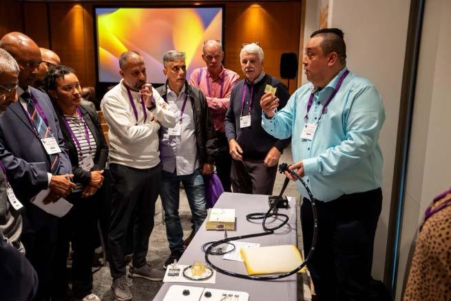
It seems like it was only a few days ago that we were all gathered at the Cape Town ICC for the IHBPA congress and its parallel SAGES and African Viral Hepatitis Convention programmes; but that was May and we are now in September. Still intoxicated by the high-flying speakers with their state-of-the-art topics, cutting edge research outcomes and plans, and just plain formidable academia; the realities of working in public and private South African healthcare (gastroenterology) can be challenging at times.
In the public sector we continue to face significant resource constraints which make practising adequate gastroenterology an administrative as well as a clinically onerous task; in private the challenges are different but real, in many cases the funders
dictating what is and is not available for patients, thereby forcing treatment plans that may not necessarily be optimal for the patient. A doctoring fatigue is evident across the country, which is scarcely alleviated by the government of national unity…. Where will the national health insurance plans drive our profession?
Taking all of this into account, and the fact that this is only the second issue of our new societal journal, this issue goes back to basics. We have a few reviews of common topics, a case study and some research papers. As we begin to refashion a footprint, we would very much appreciate feedback, as the content, for both this issue and the May 2024 edition, is very different. What do you as readers wish to see in your (our) journal?
Martin Brand
Dear members
SAGES has no further involvement in the Discovery Health biologics approval process. This relationship was terminated by mutual agreement at the end of August 2024.
MESSAGE FROM THE PRESIDENT
As you are all too aware, Discovery Health has embarked on extensive cost-cutting measures at the beginning of this year. As a society, we were most impacted by the access to biologics for our IBD patients. Furthermore, IBD patients were subjected to a one-sided treatment algorithm. Discovery Health paid little respect to clinical appropriateness and scientific treatment guidelines recommended by us and other international societies. Co-payments have become the order of the day and the many patients that can’t afford these additional payments go without medication.
After numerous meetings, discussions and deliberations no common ground could be reached unfortunately. However, I must admit that the relationship with Discovery Health was cordial and accommodating on all levels. They were approachable and always available for discussion of issues of mutual concern.
More of the same is on the cards for the near future. Discussions are afoot for limiting endoscopy utilisation. Failing to reach consensus on this issue, Discovery Health will implement its own interpretation of how and when scopes are indicated.
As a society, we will continue to engage all role-players to advance the cause of gastroenterology. We will fight for issues important to the well-being of our patients and access to equitable healthcare.
Happy reading
Ernst Fredericks SAGES President
High energy: 2.0 kcal/ml
High protein: 9.5 g/100 ml
Low carbohydrate
330 ml (bigger size)
Rich source of omega-3 fatty acids
TGF-β2 to help decrease inflammation
Glutamine and arginine – to protect the intestinal mucosa
MCT for better absorption
Nutritionally complete, ready-to-drink
Various flavours
Suitable for diabetics
Gluten and lactose-free
Halaal CONDITION-SPECIFIC CLINICAL NUTRITION FOR IBD-RELATED MALNUTRITION
Think differently about your IBD patients ` nutritional therapy! Think condition-specific!
SOUTH AFRICA
South African Journal of Gastroenterology and Hepatology. 2024;01(2):40-42 https://doi.org/10.36303/SAJGH.0933
Background: Eosinophilic oesophagitis (EoE) is a chronic inflammatory disease of the oesophagus which often presents in younger children as food refusal and failure to thrive, and in older children as food impaction and dysphagia. The goal of this study was to highlight some characteristics of the presence of EoE in African children who presented for upper gastrointestinal (GIT) endoscopy at the Korle-Bu Teaching Hospital in Accra, Ghana.
Cases: This is a case series of four children seen at a paediatric gastroenterology clinic with abdominal pain, vomiting and food impaction. They had personal and family histories of atopy. Uppe GIT endoscopy and oesophageal biopsies showed eosinophil count of greater than 15. They all had a remarkable response to the treatment of EoE.
Conclusion: EoE was diagnosed in Ghanaian children although the exact prevalence is not known. There is the need to create awareness among clinicians in Africa for early diagnosis and optimal care of this condition.
Keywords: eosinophil, oesophagitis, endoscopy, children
Introduction
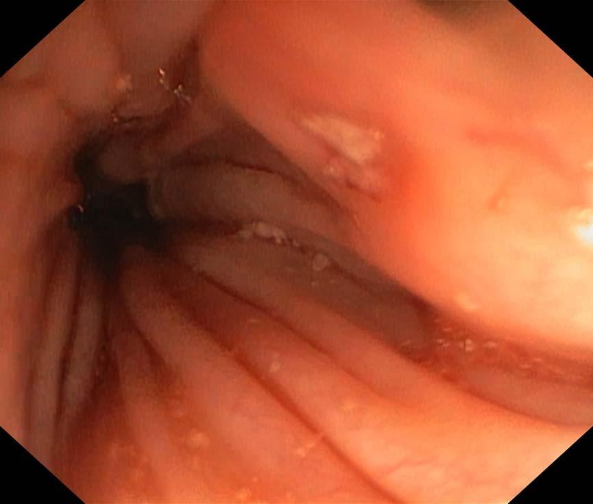
Eosinophilic oesophagitis (EoE) is a chronic inflammatory disease of the oesophagus, characterised by clinical features of oesophageal dysfunction in the setting of immune and antigenmediated eosinophilic infiltration of ≥ 15 eosinophils per high power field of the oesophageal mucosa.1 EoE is a cause of significant upper gastrointestinal (GIT) morbidity and high cost of healthcare.2 In untreated cases, it may not lead to mortality or the risk of malignancy, but it leads to poor quality of life.3 Eosinophils are normally found in the mucosa of the GIT tract, except for the oesophagus.4 The presence of eosinophils in the oesophagus is not exclusively due to EoE, but may also be found in conditions like gastro-oesophageal reflux disease, Barrett’s oesophagus, Crohn’s disease, vasculitis, and some infections and medicine use. It is essential to distinguish EoE from other causes of oesophageal eosinophilia during the patient’s evaluation.5 EoE affects both children and adults with a mean age at diagnosis of between 5.4 to 9.6 years in children.3 The commonest clinical presentation is dysphagia and food impaction in older children and adolescents.6 In younger children, food refusal and failure to thrive predominate in presentation. Other symptoms include abdominal pain, vomiting and diarrhoea.7 At endoscopy, some oesophageal abnormalities seen are longitudinal furrowing, friability, oedema, longitudinal shearing, whitish exudates, narrow oesophagus, felinisation, and transient or fixed rings.8 A normal-appearing oesophagus at endoscopy does not rule out the diagnosis of EoE.7 Children with EoE often have other associated allergic diseases and/or a family history of atopy.9 Males are more affected than females with a risk ratio of 3:1.10 This study highlighted the presence of EoE in Ghanaian children.
Case descriptions
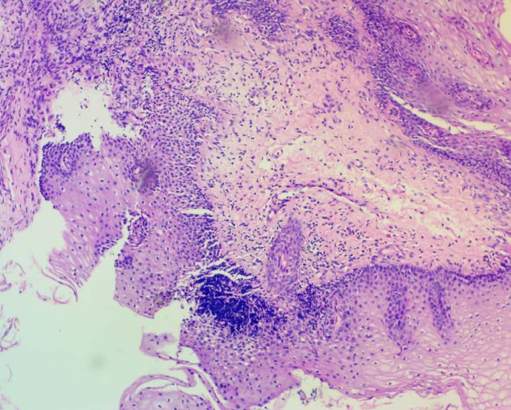
This is a case series of four children diagnosed with EoE at the gastroenterology clinic at the Korle Bu Teaching Hospital. Upper GIT endoscopy was done for all the children and biopsies were taken from the lower, middle and upper oesophagus as well as from stomach and duodenum. The biopsies were processed routinely into formalin fixed paraffin embedded blocks, slides were prepared from thin sections (3 um), stained with haematoxylin and eosin. The stained slides were examined microscopically for the presence of eosinophils and other histological features of EoE. The clinical presentations are shown in Table I.
Case 1: A 12-year-old adolescent with recurrent abdominal pain of 13 months was referred for upper GIT endoscopy. He was managed with triple therapy based on positive stool Helicobacter pylori antigen test. He had intermittent asthma, for which he was managed with inhaled short-acting beta agonist and the last episode was three years ago. He has both personal and family history of atopy. No abnormality was noted at endoscopy, but biopsies were taken based on personal and family history of asthma and atopy. Histology confirmed the diagnosis of EoE and he was managed with dietary elimination, which resolved the abdominal pain.
Case 2: This case involves a 7-year-old male child with seven months history of poor growth, difficulty swallowing, and poor appetite. He had episodes of food impaction a month prior to presentation and this was relieved by straightening his neck and hitting his chest to bring comfort. He was previously
Table I: Characteristics of patients
Indication for endoscopy
Duration of symptoms (months)
History of asthma
History of other atopic diseases
Family history of asthma and or atopy
Endoscopy findings
Treatment given before endoscopy
Treatment given after diagnosis of EoE
Abdominal pain
Abdominal pain, vomiting, food impaction and failure to thrive
Vomiting and dysphagia
Normal Longitudinal furrows, oedema (Figure 1) Normal
Triple therapy
Dietary elimination
managed with a proton pump inhibitor (PPI), which produced no improvement. He had asthma which was intermittent with allergic conjunctivitis and eczema. Upper GIT endoscopy showed oesophageal oedema and longitudinal furrows (Figure 1). Biopsies confirmed the diagnosis of EoE. He was managed with systemic steroid for two weeks and then changed to topical steroid. He improved dramatically with resolution of the symptoms and weight gain. He was lost to follow-up for six months after which he reported again with food impaction and severe odynophagia. A barium swallow revealed oesophageal stenosis. This was managed with four sessions of oesophageal dilation and oesophageal steroid (40 mg of methylprednisolone) injections together with dietary elimination. This led to improvement of the symptoms and he is currently on topical steroid.
Case 3: An 8-year-old female was referred with reported upper GIT endoscopy indicating hiatus hernia. She also suffered from daily vomiting and dysphagia. She was given antacid and PPI but the symptoms did not improve. Allergic rhinitis and conjunctivitis and a family history of atopy were also present. She had a repeat upper GIT endoscopy with biopsies taken from the oesophagus, stomach and duodenum. Histology reports confirmed EoE. She was managed with topical steroid.
Case 4: An 11-year-old male adolescent with well-controlled asthma was referred to our unit with a four-year history of abdominal pain and vomiting. Pain was generalised, dull and interfered with activities, but not sleep. Vomiting was usually unprovoked and not associated with any time of day, but usually occurred after meals. He also had functional constipation, which was managed with low molecular weight polyethylene glycol. He had completed triple therapy for Helicobacter pylori before referral. An endoscopy confirmed the diagnosis of EoE. He was managed with dietary elimination.
Proton pump inhibitor
Diet, systemic and topical steroid, dilation
Proton pump inhibitor
Abdominal pain, vomiting
Triple therapy
Topical steroid Dietary elimination
Figure 1: Oesophageal appearance. Longitudinal or linear furrows of the oesophagus with exudates
Figure 2: Histology of oesophageal biopsy Histology of the oesophagus showing < 15 eosinophils per high power
Discussion
EoE is an emerging GIT condition worth noting by clinicians especially in the paediatric population. The only case series available were a case report from South Africa11 and one from Nigeria.12 Our study highlighted four Ghanaian children with EoE with varied clinical characteristics.
Clinical presentation of EoE varies depending on the age of the patient and severity of disease. EoE can present in all age groups from infancy to old age. Patients from this series were all of school going age and able to articulate their symptoms very well. Their clinical presentation included vomiting, vague abdominal pain, failure to thrive, dysphagia and food impaction. Severe disease with features of oesophageal stenosis was seen in one of the patients. EoE is commonly seen in male patients and in patients with atopic disease such as food allergy, asthma, and allergic rhinitis. Seventy-five per cent (3/4) of the patients in this series were males and they all had personal and family histories of atopic disease which included allergic rhinitis, conjunctivitis, eczema and asthma. The duration of symptoms in this series was between seven months to two years, which is in keeping with the duration found in the literature of 1.2 to 3.5 years.5
Diagnosis of EoE requires a high index of suspicion and specialised expertise. During upper GIT endoscopy, biopsies are taken from the upper and lower oesophagus regardless of the endoscopicy appearance of the oesophagus. Biopsies are taken from the stomach and duodenum to exclude the presence of eosinophils and other differential diagnosis in other parts of the upper GIT.6
EoE is not well known or diagnosed in Africa due to the low number of gastroenterologists and the need for a high index of suspicion coupled with knowledge of exactly what to do at endoscopy, and finally to communicate with the histopathologist on what the differential diagnosis is. Knowing and doing, all of these will improve the diagnostic yield and hence influence treatment. Typical endoscopic features are: oesophageal rings, a thickened, pale mucosa with linear furrows and white exudates and, less frequently, narrowing of the calibre of the oesophagus. A normal oesophagus at endoscopy does not exclude the diagnosis of EoE. Only one patient had classic endoscopic features (Figure 1) of EoE whilst all of them had histological features. This strengthens the need to take biopsies from the oesophagus for confirmation of the diagnosis even when the endoscopy appears normal. Paediatric gastroenterologists have a role in alerting pathologists when sending out biopsy specimens from such patients for review, and pathologists who receive such specimens should actively look out for signs indicative of EoE.12
The mainstay of treatment is dietary elimination, steroid and oesophageal dilation in severe disease with stenosis. Patients in this series were exposed to all the types of treatment based on
the severity of disease. All the patients in this series responded well to the treatment available locally.
Conclusion
EoE is a chronic inflammatory condition of the oesophagus that presents in children with features not only limited to the oesophagus. It is a masquerade of all kinds of extra-intestinal diseases. Although it is more common among Caucasians, African children can also be affected. There is the need to create awareness among clinicians in sub-Saharan Africa of the disease entity, as its treatment may be inappropriate. Further research including patients from other referral sites is needed to estimate the burden and associated characteristics of EoE in Ghanaian children.
Acknowledgement
To Dr Lawrence Edusei of the Department of Pathology and Pathology Without Borders for the photograph of the histology.
Conflict of interest
None declared by the authors.
Funding source
None.
Ethical approval
Informed consent was obtained from the caregivers and assent from the patients. All ethical issues have been considered to protect the patients’ rights.
References
1. Sutton AG, Mir S, Steiner MJ. Esophagitis: allergic and eosinophilic. Pediatr Rev. 2015;36(8):375-376. https://doi.org/10.1542/pir.36.8.375
2. Cavalli E, Brusaferro A, Pieri ES, et al. Eosinophilic esophagitis in children: doubts and future perspectives. J Transl Med. 2019;17(1):262. https://doi.org/10.1186/ s12967-019-2014-0
3. Lucendo AJ, Arias-González L, Molina-Infante J, Arias Á. Systematic review: health-related quality of life in children and adults with eosinophilic oesophagitis-measure instruments and determinant factors. Aliment Pharmacol Ther. 2017;46:401-9. https://doi.org/10.1111/apt.14194
4. Visaggi P, Savarino E, Sciume G, et al. Eosinophilic esophagitis: clinical, endoscopic, histologic, and therapeutic differences and similarities between children and adults. Therap Adv Gastroenterol. 2021:14:1756284820980860. https://doi.org/10.1177/1756284820980860
5. Shaheen NJ, Mukkada V, Eichinger CS, et al. Natural history of eosinophilic esophagitis: a systematic review of epidemiology and disease course. Dis Esophagus. 2018;31(8):doy015. https://doi.org/10.1093/dote/doy015
6. Soon IS, Butzner JD, Kaplan GG, de Bruyn JC: Incidence and prevalence of eosinophilic esophagitis in children. J Pediatr Gastroenterol Nutr. 2013;57:72-80. https://doi.org/10.1097/MPG.0b013e318291fee2
7. Papadopoulou A, Koletzko S, Heuschkel R, et al. Management guidelines of eosinophilic esophagitis in childhood. J Pediatr Gastroenterol Nutr. 2014;58(1):107-118. https://doi.org/10.1097/MPG.0b013e3182a80be1
8. De Matteis A, Pagliaro G, Corleto VD, et al. Eosinophilic esophagitis in children: clinical findings and diagnostic approach. Curr Pediatr Rev. 2020;16(3): 206-214. https://doi.org/10.2174/1573396315666191004110549
9. Hopp RJ. Eosinophilic esophagitis in pediatrics: The worst of all possible allergy worlds? J Allergy (Cairo). 2012;2012:179658. https://doi. org/10.1155/2012/179658
10. Iuliano S, Minelli R, Vincenzi F, et al. Eosinophilic esophagitis in pediatric age, state of the art and review of the literature. Acta Biomed. 2018;89(8-S):20-26.
11. Levin M, Motala C. Eosionphilic oesophagitis in Cape Town, South Africa. Clinical and Translational Allergy. 2011; 1(Suppl 1). https://doi. org/10.1186/2045-7022-1-S1-P26
12. Ikobah JM, Ikwuagwu E, Okpabio I, et al. Eosinophilic oesophagitis in a Nigerian adolescent: a case report. Pan African Medical Journal. 2024;47(3). https://doi. org/10.11604/pamj.2024.47.3.36280
South African Journal of Gastroenterology and Hepatology. 2024;01(2):43-44 https://doi.org/10.36303/SAJGH.0930
Potent antiretroviral therapy (ART) has been associated with a significant reduction in morbidity and mortality among patients with human immunodeficiency virus (HIV). However, individuals with advanced immunosuppression are still at risk for hepatobiliary complications, such as acalculous cholecystitis and acquired immunodeficiency syndrome (AIDS)-related cholangiopathy.1
Additionally, patients on ART may experience adverse events related to the treatment, such as lactic acidosis, hepatic steatosis, and drug-induced hepatotoxicity. Many of these patients are also co-infected with hepatitis B or C viruses due to shared transmission routes.
As HIV patients live longer, the incidence of HIV-related malignancies has increased relative to the general population. Cancer-related deaths now account for a growing proportion of overall deaths among individuals with HIV, including a notable rise in hepatocellular carcinoma associated with viral hepatitis.2
The initial evaluation of HIV patients presenting with symptoms or signs of hepatobiliary disease should focus on determining the presence of intrahepatic and/or extrahepatic disease. The clinical presentation will guide the selection of appropriate laboratory tests and radiographic studies.
Right upper quadrant pain, with or without jaundice, can indicate various conditions, including HIV cholangiopathy, papillary stenosis, acalculous cholecystitis, cholelithiasis/ choledocholithiasis, or drug-induced liver injury. While choledocholithiasis may present similarly, it usually results in a less pronounced elevation in alkaline phosphatase levels and greater aminotransferase levels.
Non-alcoholic fatty liver disease
Similar to the general population, the prevalence of non-alcoholic fatty liver disease (NAFLD) has risen significantly among HIV patients, with studies indicating incidence rates exceeding 30%. NAFLD is now the most common form of liver disease in this group. HIV patients may be at a higher risk for hepatic steatosis compared to those without HIV.3,4 Furthermore, patients with HIV may experience liver injury induced by antiretroviral drugs or medications used to manage related conditions. Studies have demonstrated that HIV patients with chronic viral hepatitis and baseline elevations in aminotransferases are at a higher risk of
ART-induced hepatotoxicity compared to those with HIV who do not have hepatitis B or C.5
Gallstone disease
Symptoms of gallbladder stones typically appear three to four years after starting an antiretroviral drug, with a range from one to 90 months. Complications associated with gallbladder stones include cholecystitis in 11 patients, cholangitis in one patient, and acute pancreatitis in four patients. Gallstone disease in subSaharan African populations is rarely investigated. Collectively, some studies suggest that HIV patients on ART exhibit altered hepatic regulation of cholesterol-metabolizing genes, leading to reduced cholesterol scavenging and increased cholesterol efflux.6 Atazanavir has been associated with complicated cholelithiasis. In a study including 14 HIV patients on an atazanavir-based antiretroviral regimen, eight individuals had significant concentrations of atazanavir detected in their biliary stones.7
The incidence of metabolic disorders in settings with a high prevalence of HIV is a significant burden in developing countries. HIV infection and ART can alter cholesterol and fat metabolism, potentially leading to complications such as cholelithiasis. Among black South African women living with HIV, the age at onset of gallstone disease was significantly lower, and fewer women were obese compared to HIV-uninfected women with gallstone disease. These findings differ from gallstone risk factors observed in other populations and HIV-uninfected black South African women. This discrepancy may be attributed to the metabolic changes induced by HIV itself or the long-term use of ART. Larger cohort studies are needed to understand better the role of HIV and ART in the development of cholestatic diseases.8
Cholangiopathy
HIV cholangiopathy is a condition marked by biliary obstruction due to infection-associated strictures of the biliary tract. It is typically observed in patients with a CD4 count below 100 cells/ µL. Before the introduction of effective ART, HIV cholangiopathy was found in a quarter of AIDS patients. The incidence of this condition has decreased with improved HIV treatment, but the current rate remains uncertain.
Liver cirrhosis
HIV infection accelerates the progression of hepatitis C virus (HCV)-related liver fibrosis, with lower CD4 counts being
associated with a faster rate of fibrosis progression. It has also been reported that hepatocellular carcinoma (HCC) tends to develop at a younger age and after a shorter duration of HCV infection in patients co-infected with HIV and HCV compared to those with HCV infection alone. This suggests that HIV infection may accelerate liver fibrosis and carcinogenesis, particularly in co-infected patients with CD4 counts below 200 cells/mm³.9
High prevalence rates of liver cirrhosis and periportal fibrosis have been observed among adults co-infected with HIV and hepatitis B virus (HBV). In one of the first HCC screening initiatives in subSaharan Africa, 2% of patients co-infected with HIV and HBV had significant liver lesions. Additionally, one-quarter of the screened patients showed findings suggestive of schistosomiasis-induced liver damage.10
Chronic pancreatitis
HIV-infected patients with chronic pancreatitis, compared to non-infected patients with the same condition, tend to be significantly younger, even though they share similar risk factors. This suggests that HIV infection may contribute to an earlier onset of chronic pancreatitis in the presence of established risk factors. Chronic pancreatitis should be considered in the differential diagnosis for patients with persistent non-infective diarrhoea or steatorrhoea. Notably, fewer HIV-infected patients with chronic pancreatitis had diabetes mellitus, despite the duration of chronic pancreatitis being similar between the two groups. This difference might indicate a distinct pathological process affecting the islet cells in HIV-infected patients compared to non-infected individuals.11
Conclusion
HIV-endemic populations exhibit a higher incidence of metabolic disorders, as both HIV and its treatment are associated with altered lipid and cholesterol metabolism. Patients with HIV and severe immunosuppression are at risk for HIV cholangiopathy and opportunistic infections, which can lead to significant hepatobiliary diseases. Additionally, due to shared routes of transmission, individuals with HIV are also at increased risk for hepatitis B and C infections. These infections are significant causes of morbidity and mortality, often leading to complications such as decompensated cirrhosis and hepatocellular carcinoma.
Patients with HIV, chronic viral hepatitis, and baseline elevations in aminotransferases are at a higher risk of developing ARTinduced hepatotoxicity compared to those with HIV without hepatitis B or C. Moreover, HIV infection appears to accelerate liver fibrosis and carcinogenesis, resulting in hepatocellular carcinoma developing at a younger age and after a shorter duration of HCV infection in patients co-infected with HIV and HCV compared to those with HCV alone.
HIV and ART increase the risk for many hepatobiliary and pancreatic pathologies, such as acalculous cholecystitis, cholelithiasis, choledocholithiasis, HIV cholangiopathy, papillary strictures, chronic pancreatitis, drug-induced liver injury, and malignancies. While ART has extended the lifespan of patients with HIV, it has also led to a higher prevalence of morbidity and mortality due to HIV-related malignancies compared to those who are not infected with HIV.
References
1. Keaveny AP, Karasik MS. Hepatobiliary and pancreatic infections in AIDS: Part II. AIDS Patient Care STDS. 1998;12:451. https://doi.org/10.1089/apc.1998.12.451
2. Gjærde LI, Shepherd L, Jablonowska E, et al. Trends in incidences and risk factors for hepatocellular carcinoma and other liver events in HIV and hepatitis C virus-coinfected individuals from 2001 to 2014: A multicohort study. Clin Infect Dis. 2016;63:821. https://doi.org/10.1093/cid/ciw380
3. Gaslightwala I, Bini EJ. Impact of human immunodeficiency virus infection on the prevalence and severity of steatosis in patients with chronic hepatitis C virus infection. J Hepatol. 2006;44:1026. https://doi.org/10.1016/j.jhep.2006.02.009
4. Rafiq N, Stepanova M, Lam B, et al. Predictors of chronic liver disease in individuals with human immunodeficiency virus infection. Ann Hepatol. 2013;13:60. https://doi.org/10.1016/S1665-2681(19)30905-6
5. Soriano V, Puoti M, Garcia-Gascó P, et al. Antiretroviral drugs and liver injury. AIDS. 2008;22:1. https://doi.org/10.1097/QAD.0b013e3282f0e2fd
6. Kinoo SM, Chuturgoon AA, Singh B, Nagiah S. Hepatic expression of cholesterol regulating genes favor increased circulating low-density lipoprotein in HIV-infected patients with gallstone disease: a preliminary study. BMC Infectious Diseases. (2021)21:294. https://doi.org/10.1186/s12879-021-05977-0
7. Rakotondravelo S, Poinsignon Y, Borsa-Lebas F, et al. Complicated atazanavirassociated cholelithiasis: a report of 14 cases. Clin Infect Dis. 2012;55:1270. https://doi.org/10.1093/cid/cis620
8. Kinoo SM, Nagiah S, Chuturgoon A, Singh B. Symptomatic gallstones and HIV in black South African women: Changing trends of gallstone disease? S Afr J HIV Med. 2021;22(1):a1208. https://doi.org/10.4102/sajhivmed.v22i1.1208
9. Uchinoa K, Tateishia R, Nakagawa H, et al. Uninodular combined hepatocellular and cholangiocarcinoma with multiple non-neoplastic hypervascular lesions appearing in the liver of a patient with HIV and HCV coinfection. Journal of Clinical Virology. 2013;57:173-177. https://doi.org/10.1016/j.jcv.2013.01.012
10. Riebensahma C, Chitundub H, Muula G, et al. Screening for hepatocellular carcinoma among adults with HIV/HBV co-infection in Zambia: a pilot study. International Journal of Infectious Diseases. 2022;116:391-396. https://doi. org/10.1016/j.ijid.2021.12.338
11. Brand M. Characterization of chronic calcific pancreatitis in the presence of HIV infection: Comparison with uninfected patients. The South African Gastroenterology Review. 2022;(2).
South African Journal of Gastroenterology and Hepatology. 2024;01(2):45-51 https://doi.org/10.36303/SAJGH.1033
Open Access article distributed under the terms of the Creative Commons License [CC BY-NC 3.0] http://creativecommons.org/licenses/by-nc/3.0
Objective: The study aimed to identify the factors associated with good knowledge of viral hepatitis B and C.
Methods: A cross-sectional study with prospective data collection was conducted from 27 to 28 July 2021 (two days) on World Hepatitis Day in 2021.
Results: The study included 577 respondents. The average age of the respondents was 36.68 ± 13.09 years. There were 299 men (51.82%) with a 1.08 sex ratio. The proportion of respondents with good overall knowledge was 58.23% for viral hepatitis B and 45.22% for viral hepatitis C. The factors associated with good overall knowledge of viral hepatitis B were gender, education level, and having once heard about the hepatitis B virus (HBV) during a blood donation. The factors associated with good knowledge of the hepatitis C virus (HCV) were education level and having once heard about HCV during an awareness campaign.
Conclusion: The respondents in this study did not have adequate information about viral hepatitis B and C. To inform more people, it is important to intensify awareness campaigns.
Keywords: knowledge, viral hepatitis B and C, awareness
Introduction
Viral hepatitis is a public health issue.1 There are several virus types with hepatic tropism; the most well-known are the alphabetic viruses (A, B, C, D, E, and G). Hepatitis B and C viruses are responsible for a transition to chronicity, with the possibility of complications, such as cirrhosis or liver cancer.2 According to World Health Organization (WHO) estimates, there are 240 million cases of chronic hepatitis B virus (HBV) infection and 71 million cases of hepatitis C virus (HCV) infection worldwide.1 Sub-Saharan Africa is the most affected by HBV.3
Viral hepatitis and its complications are a leading cause of death worldwide. In 2015, more than one million people died of viral hepatitis, mainly from cirrhosis and primary liver cancer.1 Viral hepatitis is an enormous psychological and financial burden in endemic regions, particularly in low-income countries.4 Prevention remains the best strategy for combating the spread of these viruses. Accordingly, sufficient knowledge and appropriate attitudes towards these infections are the cornerstones of prevention.
In Togo, numerous studies have examined knowledge about viral hepatitis B and C, but none have examined the factors associated with this knowledge.5,6 Therefore, this study sought to describe respondents’ level of knowledge about viral hepatitis B and C and identify the factors associated with good knowledge of viral hepatitis B and C.
Methods
Study framework
The study was conducted at the Campus Teaching Hospital of Lomé during an awareness campaign on viral hepatitis B and C in 2021 as part of World Hepatitis Day.
Study population
This was a cross-sectional study. The study population consisted of all individuals of either sex, aged 15 years and above, who presented at the screening site. Anyone aged 15 years or above who gave informed verbal consent was included in the study. Anyone with an unusable survey form was excluded from the study.
Study materials and process
A questionnaire was used. Individual counselling was given to all those present. Following informed consent at the end of the interview, a trained interviewer completed the questionnaire.
Data collection
Data were collected using a pre-established survey form. The data was entered using EpiData software version 3.1. The parameters studied were sociodemographic data, history and lifestyle, and data on the knowledge of hepatitis B and C viruses (such as the sources of information on viral hepatitis B and C, cause, primarily affected organ, means of transmission, symptoms, complications, and possible treatment and cure in Togo). For each question asked, a score of one point was given if the respondent gave the correct answer on knowledge of viral hepatitis B and C. There were seven questions. A point was awarded if the respondent could give the following answers to the questions:
• Cause of hepatitis B and C? Viruses.
• Organ most affected by HBV and HCV? Liver.
• Means of transmission? For HBV, blood, sexual, and motherchild. For HCV, blood and sexual.
• Symptoms of viral hepatitis B and C? Jaundice, fatigue, and fever.
• Complications of viral hepatitis B and C? Cirrhosis and cancer.
• Possible to treat viral hepatitis B and C? Yes.
• Possible to cure viral hepatitis B and C in Togo? Yes.
For each question about viral hepatitis B and C, a point was not awarded if the respondent gave the following answers:
• Cause of hepatitis B and C? Answers other than the viruses.
• Organ most affected by HBV and HCV? Answers other than the liver.
• Means of transmission? For HBV, a single answer between blood, sexual, mother-child, or other answers than these. For HCV, other answers than blood and sexual transmission.
• Symptoms of viral hepatitis B and C? Answers other than jaundice, fatigue, and fever.
• Complications of viral hepatitis B and C? Other answers than cirrhosis and cancer.
• Possible to treat viral hepatitis B and C? No.
• Possible to cure viral hepatitis B and C? No.
Operational definition
The respondents’ overall knowledge was considered good if they could give at least five correct answers to the seven questions. Their overall knowledge was considered poor if they gave fewer than five correct answers to the seven questions.
Statistical analysis
All data were recorded in a database designed using EpiData version 3.1. Statistical analysis was done using Stata software version 14.2. Statistical analysis included a descriptive section of the population and a comparative section according to the studied variables. For descriptive analysis, results were expressed as headcount and percentage for qualitative variables or means and standard deviations for quantitative variables. For comparative analysis, Pearson’s chi-square or Fisher’s exact tests were used for qualitative variables and t-test for quantitative variables.
Logistic regression was used to identify the factors associated with the level of knowledge about hepatitis B and C. For each initial model, variables showing a p < 0.2 in univariate analysis were used as explanatory variables. A top-down, stepby-step procedure was used to select the final model. This involved progressively removing the least significant variables (p > 0.05). At each step, we confirmed no confounding between the removed variable and those remaining in the model by checking for changes in the odds ratios (tolerated variation: 20%) or radical changes in their degrees of significance and the overall significance statistic of the model.
Ethical considerations
The hospital’s management agreed to conduct the study, and the respondents gave informed verbal consent. The questionnaire
was designed to be submitted and processed by a computer. Thus, each file had a unique number identifying the report to preserve anonymity.
Results
Global data
Of the 635 respondents who participated in the campaign, 577 were aged 15 years or over.
Data on knowledge of viral hepatitis B and C existence
Of the 577 people surveyed, 486 had heard of HBV, and 429 had heard of HCV.
Sociodemographic characteristics
The average age of the 577 respondents was 36.68 ± 13.09 years, with extremes of 15 and 77 years. The most represented age group was 25–35 years (26.17%). There were 299 men (51.82%) with a 1.08 sex ratio (Table I).
Information resources on viral hepatitis B and C
Among those respondents who had once heard of viral hepatitis B and C, the hospital was their source of information on hepatitis B (46.50%) and C (47.09%) (Table II).
Table I: Study population characteristics
Distribution of respondents according to their knowledge of the possibility of treatment and
Data on knowledge of viral hepatitis B and C
The virus was the cause cited by 315 respondents (64.81%) for viral hepatitis B and 276 respondents (64.34%) for viral hepatitis C. The liver was the affected organ cited by 387 respondents (79.63%) for viral hepatitis B and 322 respondents (75.06%) for viral hepatitis C. The blood route was the means of transmission cited by 316 respondents (65.02%) for HBV and 266 respondents (62%) for HCV. For HBV, the sexual and mother-to-child transmissions were cited by 280 (57.61%) and 168 (34.57%) respondents, respectively.
Fatigue was the symptom cited by 267 respondents (54.94%) for viral hepatitis B, and 226 respondents (52.68%) for viral hepatitis C. Jaundice was cited by 185 respondents (38.07%) for viral hepatitis B and 168 (39.16%) for hepatitis C. Complications cited
by respondents were liver cancer (n = 474, 97.53%) and cirrhosis (n = 285, 58.64%) for viral hepatitis B, and cirrhosis (n = 235, 54.78%) and liver cancer (n = 172, 40.10%) for viral hepatitis C.
The possibility of treating viral hepatitis B in Togo was known to 417 respondents (85.80%), and curative treatment was possible according to 399 respondents (82.10%). Viral hepatitis C could be treated and cured in Togo, according to 339 (79.02%) and 338 (78.79%) respondents, respectively (Table III)(Figure 1).
Respondents’ knowledge of the causes and organs affected by viral hepatitis B and C was good; however, complications were poorly understood (Table IV). Overall, 58.23% of respondents had good knowledge of HBV, and 45.22% had good knowledge of HCV.
Table II: Distribution of respondents according to information sources on viral hepatitis B and C
Figure 1: Distribution of respondents according to their knowledge of the causes of viral hepatitis B and C
Table III:
Table IV: Knowledge level of viral hepatitis B and C
Factors associated with knowledge of viral hepatitis B and C during a hepatitis awareness campaign in 2021 in Togo
Table V: Factors associated with good knowledge of viral hepatitis B
Factors associated with knowledge of viral hepatitis B and C during a hepatitis awareness campaign in 2021 in Togo
Table VI: Factors associated with good knowledge of viral hepatitis C
Factors associated with knowledge of viral hepatitis B and C during a hepatitis awareness campaign in 2021 in Togo
Factors associated with overall knowledge of viral hepatitis B and C
In multivariate analysis, male gender, university education, and having heard about the viral HBV during a blood donation were associated with good overall knowledge of viral hepatitis B (Table V). In multivariate analysis, good knowledge of viral hepatitis C was associated with university education and having heard of hepatitis C during an awareness campaign (Table VI).
Discussion
During this study, 577 people aged 15 years and over were surveyed, of whom 486 had heard of HBV and 429 had heard of HCV. Among those who had heard of HBV, 58.23% had good overall knowledge. Ahmad et al.7 reported similar results. However, these results differ from those of Frambo et al.,8 who reported that only 16% of the study population had a good knowledge of viral hepatitis B in their study. Although similar or better than those described in the literature, these results are still insufficient.
Togo is a country in an endemic viral hepatitis B and C zone, and knowledge of these diseases needs to be improved among the population. Of those respondents who had heard of HCV, 45.22% had good overall knowledge. Yakoubou et al.6 reported low knowledge of viral hepatitis C among their patients in 2018, while Joukar et al.9 reported that healthcare workers’ knowledge of viral hepatitis C was satisfactory. The fight against viral hepatitis, which is still little-known in our communities, should involve disseminating the right information about these diseases to all sections of the national population.
Male gender, university education, and having heard about HBV during a blood donation were factors significantly associated with good knowledge of HBV. University education and having heard about HCV during awareness-raising were factors significantly associated with knowledge of HCV. This shows that education programmes, blood donation counselling, and awareness campaigns are essential to disseminate information about viral hepatitis and improve knowledge of the disease. Therefore, it is important to include a section on viral hepatitis in education programmes from primary school onwards and increase awareness campaigns.
Brouard et al.10 reported that good knowledge of HBV transmission modes was found more often in people with a tertiary qualification (compared to those with no qualifications). Our results are also comparable to those of Lohouès-Kouacou et al.,11 who reported that among the factors influencing knowledge of hepatitis B, education level was associated with good knowledge of hepatitis B. In the study by Mongo-Onkouo et al.,12 they found that individuals with a higher education level had better knowledge of the existence of a treatment and vaccine against HBV, and adopted a better attitude towards the pathology. In their study, Niangoran et al.13 reported that having undergone specific training in viral hepatitis outside the academic curriculum, having been screened for viral hepatitis,
and having people with the condition in their entourage were factors associated with good knowledge of viral hepatitis B and C.
In India, Mukherjee et al.14 found that knowledge of viral hepatitis B and C was associated with higher education and income levels. They speculated that participants in their study with a lower socioeconomic status may have less access to information about HBV and HCV, including fewer opportunities to visit healthcare facilities for such information. The cost of medical care, facility remoteness, and the lack of transportation could be reasons for these patients’ lack of knowledge.
Healthcare providers can raise awareness to improve knowledge about viral hepatitis. However, they are also at the core of the population’s lack of knowledge. Studies have reported that patients’ inability to understand medical jargon, the healthcare staff’s unfriendliness, patients’ fear of being verbally abused by doctors and paramedical staff, and anxiety are major obstacles to effective communication between patients and healthcare staff.15,16
Conclusion
Respondents’ knowledge of HBV was associated with gender, education level, and having heard about HBV once during a blood donation, and knowledge of HCV was associated with education level and having heard about HCV once during an awareness campaign. These results call for measures to inform the public through awareness-raising sessions in the media and other communication channels, counselling during consultations and blood donations, and including viral hepatitis B and C information in school programmes from primary school onwards.
1. who.int [Internet]. Global hepatitis report 2024: action for access in low- and middle-income countries. Geneva: World Health Organization; 2024. Available from: https://www.who.int/publications/b/68511.
2. Sawadogo A, Dib N, Calès P. Pathophysiology of cirrhosis and its complications. Réanimation. 2007;16(7-8):55762. French. https://doi.org/10.1016/j. reaurg.2007.09.001.
3. Patassi A, Benaboud S, Landoh DE, et al. Hepatitis B infection in HIV-1-infected patients receiving highly active antiretroviral therapy in Lomé, Togo: prevalence and molecular consequences. S Afr Med J. 2016;106(6):6349. https://doi. org/10.7196/SAMJ.2016.v106i6.10312
4. Pourette D, Enel C. Representations and disease experience of hepatitis B by sub-Saharan patients in Côte d’Ivoire and France. Sante Publique. 2014;26(6):86978. French. https://doi.org/10.3917/spub.146.0869
5. Lawson-Ananissoh LM, Bouglouga O, Bagny A, et al. Knowledge of hepatitis B virus among outpatients consulting in the hepatogastroenterology department at the Lomé Campus University Hospital. Med Sante Trop. 2015;25(3):31922. French. https://doi.org/10.1684/mst.2015.0489
6. Yakoubou RE-H, Lawson-Ananissoh LM, Bouglouga O, et al. Evaluation of knowledge on viral hepatitis C in the University Hospital Centre Campus of Lomé. J Rech Sci Univ Lomé. 2020;22(3):75762. French.
7. Ahmad A, Sann LM, Rahman HA. Factors associated with knowledge, attitude and practice related to hepatitis B and C among international students of Universiti Putra Malaysia. BMC Public Health. 2016;16(611). https://doi. org/10.1186/s12889-016-3188-5
8. Frambo AAB, Atashili J, Fon PN, Ndumbe PM. Prevalence of HBsAg and knowledge about hepatitis B in pregnancy in the Buea Health District, Cameroon: a cross-sectional study. BMC Res Notes. 2014;7(394). https://doi. org/10.1186/1756-0500-7-394
9. Joukar F, Mansour-Ghanaei F, Soati F, Meskinkhoda P. Knowledge levels and attitudes of health care professionals toward patients with hepatitis C infection. World J Gastroenterol. 2012;18(18):2238-44. https://doi.org/10.3748/wjg.v18. i18.2238
10. Brouard C, Gautier A, Saboni L, et al. Hepatitis B knowledge, perceptions and practices in the French general population: the room for improvement. BMC Public Health. 2013;13(576). https://doi.org/10.1186/1471-2458-13-576
11. Lohouès-Kouacou M-J, Assi C, Ouattara A, et al. Hepatitis B knowledge among secondary school students in Côte d’Ivoire. Sante Publique. 2013;25(2):22732. French. https://doi.org/10.3917/spub.132.0227
12. Mongo-Onkouo A, Itoua-Ngaporo NA, Apendi CPA, et al. Knowledge, attitudes and practices of persons infected with the hepatitis B virus on their own disease. Open J Gastroenterol. 2022;12(4):11927. https://doi.org/10.4236/ ojgas.2022.124012
Factors associated with knowledge of viral hepatitis B and C during a hepatitis awareness campaign in 2021 in Togo http://www.sajgh.co.za
13. Niangoran S, Inghels M, Kouassi AK, et al. Knowledge of doctors on viral hepatitis B and C in Côte d’Ivoire and associated factors: the urgency of intensifying the training of health personnel in a context of a generalised epidemic [Internet]. Dakar: 10th Francophone Conference on HIV and Viral Hepatitis; 2020. French. Available from: https://hal.science/hal-04120921/. Accessed 3 April 2024.
14. Mukherjee PS, Dutta E, Das DK, Neogi S, Sarkar A. Knowledge about hepatitis B and hepatitis C virus infection and consequences: a cross-sectional assessment of baseline knowledge among infected patients in West Bengal, India. Hepatol Med Policy. 2017;2(6). https://doi.org/10.1186/s41124-016-0014-8.
15. Ha JF, Longnecker N. Doctor-patient communication: a review. Ochsner J. 2010;10(1):3843.
16. Basu S, Andrews J, Kishore S, Panjabi R, Stuckler D. Comparative performance of private and public healthcare systems in low- and middle-income countries: a systematic review. PLoS Med. 2012;9(6):e1001244. https://doi.org/10.1371/ journal.pmed.1001244
South African Journal of Gastroenterology and Hepatology. 2024;01(2):52-54 https://doi.org/10.36303/SAJGH.0929
Open Access article distributed under the terms of the Creative Commons License [CC BY-NC 3.0] http://creativecommons.org/licenses/by-nc/3.0
A clinical revision of gastro-oesophageal reflux disease (GORD)
S Nkomo
Upper Gastro-enterology Fellow, Steve Biko Academic Hospital, University of Pretoria, South Africa
Gastro-oesophageal reflux disease (GORD) is a common problem in the adult population with an increasing prevalence of 30–40%. There is geographical variation but increasing prevalence across all populations with North America and Europe leading the burden of the disease.1,2 Most humans are predisposed to at least one reflux episode per week of which the majority will be symptomatic, but not require any intervention. The Montreal Consensus Group defines the condition as when stomach refluxate causes troublesome symptoms and/or complications.3,4
The presentation may be typical, due to direct injury of the oesophaeeal mucosa, with the cardinal symptoms of heartburn, epigastric/retrosternal pain, and water brash which is described by patients as a sour taste in the mouth.2 There are also many atypical – also labelled extra-oesophageal –symptoms which are usually because of micro-aspiration and vagal mediated events, which mimic other diseases and therefor require investigation.2,4 Atypical symptoms can be grouped according to ear, nose and throat (ENT) (laryngitis, sinusitis, otitis, vocal cord ulcers and granulomas), pulmonary (asthma, recurrent pneumonia, chronic cough, interstitial fibrosis), cardiac (chest pain and sinus arrhythmias), and other (dental caries, halitosis and tongue ulcers).4,6
There are alarm signs or red flags which should prompt a clinician to further investigate a patient presenting with GORD. These include age more than 50 years, symptoms present for > five years, no improvement of symptoms on proton pump inhibitor (PPI) treatment, significant weight loss, persistent vomiting, iron deficiency anaemia, and dysphagia/odynophagia with or without a choking sensation.6 At minimum, if a patient has any of these symptoms, an upper endoscopy is mandatory.
Mechanism of reflux disease, in a nutshell
Most symptomatic patients have no anatomical abnormality. Their symptoms are because of a failure of endogenous mechanisms designed to prevent reflux. The lower oesophageal sphincter (LES) has sling/clasp fibres, which are arranged in an oblique fashion, with contraction resulting in a mechanical barrier.7 The sphincter also has circular muscle fibres which are in constant tonic contraction, thereby increasing the pressure at the oesophago-gastric junction.7 The length of the intra-abdominal oesophagus is very important because of the difference in
intra-abdominal and mediastinal pressure.7 Furthermore, the stomach’s angle of His acts as a mechanical barrier to reflux. The most common anatomical abnormalities are hiatal hernias which disrupt these mechanisms, thereby predisposing patients to GORD.8
There are four types of hiatus hernia according to the Hill’s classification:
• Type 1 called a sliding hiatal hernia: the most common type whereby the gastro-oesophageal junction slides cranially into the mediastinum.
• Type 2 called a rolling hiatal hernia (a true para-oesophageal hernia): the fundus of the stomach herniates alongside the left side of the oesophagus with the gastro-oesophageal junction maintaining its normal anatomical position.
• Type 3 is a combination of Types 1 and 2.
• Type 4 can be any of the preceding three types but includes the simultaneous herniation of any intra-abdominal organs (i.e. colon, spleen, small bowel, and, rarely, left lobe of the liver).7,8’11
Risk factors for GORD
The list is extensive, the following being some of the more common factors:
• Presence of a hiatus hernia, regardless of type.
• Diet, typically consisting of large, fatty, heavy meals.
• Obesity, especially when associated with obstructive sleep apnoea.
• Smoking.
• Ingestion of alcohol and/or caffeine.
• Pregnancy.
• Chronic medication use, some commonly prescribed ones include, amongst others, calcium channel blockers, theophylline and corticosteroids.
• Advancing age.
Diagnosis
GORD diagnosis starts with a typical history and must be excluded by an atypical history. However, it must be noted that there is poor correlation of symptoms to objective findings which makes it difficult to diagnose and widens the spectrum of the disease.1 The Lyon Consensus guidelines subdivided the spectrum into
Conclusive evidence for pathologic reflux
Borderline or inconclusive evidence
Adjunctive or supportive evidence*
Evidence against pathologic reflux
Unproven GORD
Endoscopy, wireless pH study, 24 hour pH or pH-impedance, HRM off therapy
Endoscopy
LA grades B, C & D oesophagitis
Biopsy proven Barrett’s mucosa
Peptic oesophageal stricture
LA grades A oesophagitis
Hiatus hernia
Histopathologic scoring system
Electron microscopy of biopsies
AET > 6% on 24 hour studies
AET > 6% on > 2 days on wireless studies
AET 4-6% on 24 hour studies
AET 4-6% on > 2 days on wireless studies
Total reflux episodes 40–80 days
Reflux-symptom associated
Total reflux episodes < 40/day
> 2 500 Ω
AET < 4% each day of study** Total reflux episodes < 40/day
> 2 500 Ω
* factors that increase confidence for presence of pathologic reflux when evidence is otherwise borderline or inconclusive ** wireless pH monitoring < 4% on all days; pH-impedance: all criteria should be met
Figure 1: Oesophageal tests to diagnose GORD based on the updated Lyon consensus
LA grade = Los Angeles classification, AET = acid exposure time, SAP = reflux-symptom association, MNBI = mean nocturnal baseline impedance, PSPWI = post reflux swallow-induced peristaltic wave index
four groups based on objective clinical evaluation.9, The three recommended tests include endoscopy, 24-hour ambulatory Ph monitoring with/without impedance, and high-resolution manometry.9’ The use of a barium swallow in diagnosis is limited but may be used as an adjunct. In our setting, upper endoscopy is the first-line investigation followed by judicious use of Ph manometry and manometry.
Keeping in mind of the GORD differential diagnosis, amongst others includes acute coronary artery syndrome, oesophageal and gastric malignancy, peptic ulcer disease, non-ulcer dyspepsia, achalasia, eosinophilic oesophagitis, oesophageal diverticula, gastroparesis and rumination syndrome. Hence, it is important that the workup includes basic investigations to exclude these diagnoses.
Management
There are three pillars for the management of this disease. Most patients will resolve their symptoms with lifestyle and dietary modifications, some will require medication, and the minority will qualify for surgical intervention.
1. Lifestyle and diet modification. Patients should be counselled to:
◦ Lose weight and exercise regularly.
◦ Avoid eating 1–2 hours before bedtime.
◦ Eat frequent smaller meal portions.
◦ Avoid large fatty meals.
◦ Avoid tight-fitting clothes.
◦ Stop smoking and excessive alcohol ingestion.
◦ Avoid caffeine, dark chocolate and mint.
◦ Modify medication following discussion with the prescribing doctor.
◦ Elevate bed or pillows when sleeping.2
2. Medical therapy
◦ PPIs, once daily, are the mainstay of treatment (esomeprazole, pantoprazole, omeprazole, lansoprazole, dexlansoprazole, etc).
◦ Twice-daily PPI is more effective for patients who don’t respond to single daily PPI.
◦ The American Gastroenterological Association (AGA) recommends PPI for eight weeks and symptom reassessment thereafter.
◦ In those who respond to PPI therapy, tapering the degree of acid suppression to a minimum is the next step.
◦ H2-receptor antagonists if there is a contraindication for PPIs (cimetidine, ranitidine, loratadine).
◦ H. pylori eradication, if indicated.2,5
3. Surgical management
Indications for surgery
◦ Failure of appropriate medical therapy.
◦ Young patients unwilling to take lifelong medication.
◦ Structurally defective LES and hiatal hernias.
◦ Complications despite adequate medical therapy e.g. severe oesophagitis, stricture and Barrett’s with volume reflux symptoms.2,5
– Patients with atypical symptoms should be thoroughly investigated.
– Stricture should be dilated to > 50 Fr bougie and dysphagia resolved before anti-reflux procedure is performed.
– Barrett’s should be thoroughly assessed for dysplasia and adenocarcinoma before anti-reflux.
Studies have shown there are no differences in terms of symptom resolution and recurrence for the two above operations. Therefore both are considered to be the gold standard.1,2,10
Procedures no longer routinely performed include Laparoscopic Dor fundoplication (180° anterior wrap, Watson’s and Thal fundoplications).
Patients with a hiatus hernia should have their hernia repaired before anti-reflux operation.
Common morbidities following anti-reflux procedure
◦ Inability to belch or vomit (all patients).
◦ Dysphagia (25% of these patients will require balloon dilatation).
◦ Gas-bloat syndrome (affects 19% of patients).
◦ Increased flatulence.
◦ Recurrence of symptoms (10–15% after 10 years will require repeat surgery).5
Take home messages
• Presence of red flag symptoms must prompt a clinician to further investigate a patient presenting with GORD with at least an upper endoscopy. These include age more than 50 years, symptoms present for > five years, no improvement of symptoms on PPI treatment, significant weight loss, persistent vomiting, iron deficiency anaemia, and dysphagia/odynophagia.
• The majority of patients with symptoms of GORD will respond to lifestyle and dietary modifications.
• Empiric therapy is currently the most specific tool to determine whether reflux disease is playing a significant role in a patient’s extra-oesophageal symptoms.
• In patients unresponsive to PPI therapy, causes other than GORD should be sought.
References
1. Slater, B.J., Collings, A., Dirks, R. et al. Multi-society consensus conference and guideline on the treatment of gastroesophageal reflux disease (GERD). Surg Endosc 37, 781–806 (2023). https://doi.org/10.1007/s00464-022-09817-3
2. Sandhu DS, Fass R (2018) Current trends in the management of gastroesophageal reflux disease. Gut Liver 12:7–16
3. Ronkainen J, Agréus L. Epidemiology of reflux symptoms and GORD. Best Pract Res Clin Gastroenterol. 2013;27:325-337. https://doi.org/10.1016/j. bpg.2013.06.008
4. Hungin, A. P. S., Molloy-Bland, M., & Scarpignato, C. (2019). Revisiting Montreal: New Insights into Symptoms and Their Causes, and Implications for the Future of GERD. The American journal of gastroenterology, 114(3), 414–421. https://doi. org/10.1038/s41395-018-0287-1
5. Maret-Ouda J, Markar SR, Lagergren J. Gastroesophageal reflux disease: a review. JAMA. 2020;324(24):2536-47. https://doi.org/10.1001/jama.2020.21360
6. Vakil N, van Zanten SV, Kahrilas P, Dent J, Jones R. The Montreal definition and classification of gastroesophageal reflux disease: a global evidencebased consensus. Am J Gastroenterol. 2006;101:1900-1920. https://doi. org/10.1111/j.1572-0241.2006.00630.x
7. Rosen RD, Winters R. Physiology, Lower Esophageal Sphincter. [Updated 2023 Mar 17]. In: StatPearls [Internet]. Treasure Island (FL): StatPearls Publishing; 2024 Jan-. Available from: https://www.ncbi.nlm.nih.gov/books/NBK557452/
8. Kahrilas PJ, Kim HC, Pandolfino JE. Approaches to the diagnosis and grading of hiatal hernia. Best Pract Res Clin Gastroenterol. 2008;22:601-616. https://doi. org/10.1016/j.bpg.2007.12.007
9. Matteo G, Brigida B, Vincenzo S, et al. The Lyon consensus, does it differ from previous one? J Neurogastroenterol Motil. 2020;26(3):311-321. https://doi. org/10.5056/jnm20046
10. SAGES The Definitions Document: A Reference for Use of SAGES Guidelines. sages.org/publications/guidelines/guidelines-for-the-surgical |-treatment-of-gastroesophageal-reflux-gerd
11. Kavic SM, Segan RD, George IM, et al. Classification of hiatal hernias using dynamic three-dimensional reconstruction. Surgical Innovation. 2006;13:49-52. https://doi.org/10.1177/155335060601300108
12. Gyawali CP, Yadlapati R, Fass R, et al. Updates to the modern diagnosis of GERD: Lyon consensus 2.0. Gut. 2024;73:361-371. https://doi.org/10.1136/ gutjnl-2023-330616
South African Journal of Gastroenterology and Hepatology. 2024;01(2):55-57 https://doi.org/10.36303/SAJGH.0934
Open Access article distributed under the terms of the Creative Commons License [CC BY-NC 3.0] http://creativecommons.org/licenses/by-nc/3.0
A discourse of non-alcoholic steatohepatitis
M Ahmed
HPB Surgery Consultant Steve Biko Academic Hospital, University of Pretoria, South Africa
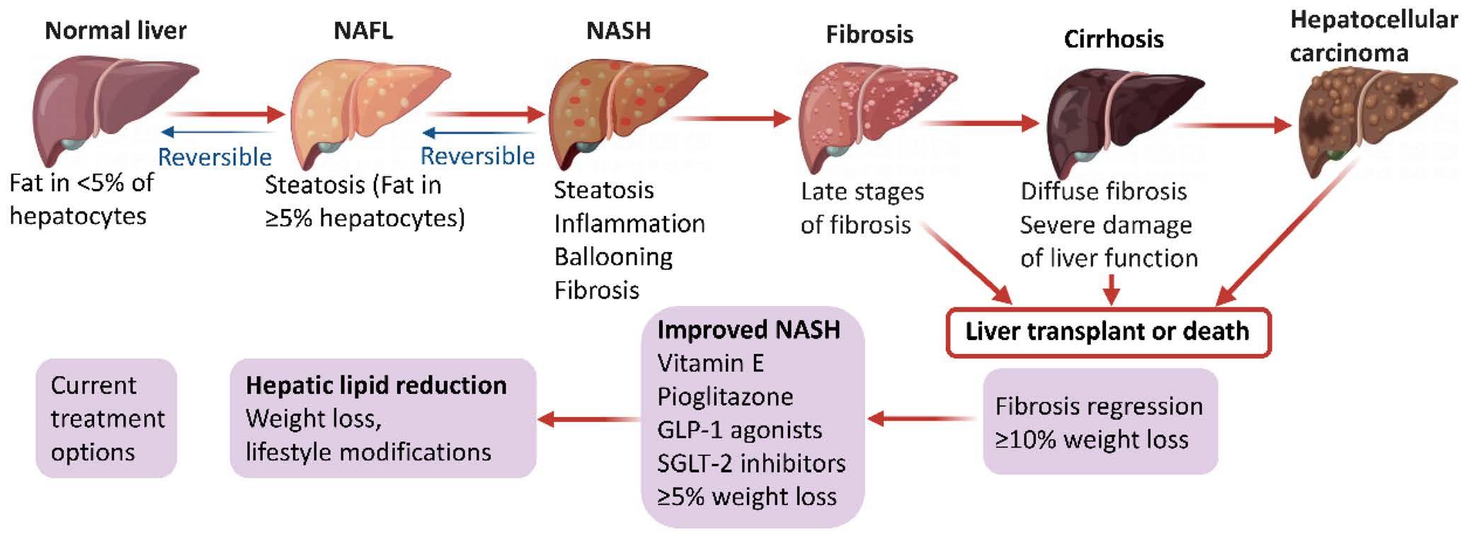
Non-alcoholic fatty liver disease (NAFLD) is a major cause of chronic liver disease and a worldwide public health concern with a prevalence of 25%.1 The highest recorded prevalence (32%) is found in the Middle East, followed by South America (31%), Asia (27%), the United States of America (USA) (24%), Europe (23%) and Africa (14%).2 By 2030, 400 million people worldwide will be affected by NAFLD in Europe, the USA, and Asia. The incidence of the disease in China has recently exceeded that of the USA and Europe.3 China will have the greatest growth in NAFLD patients worldwide by 2030, with 314.58 million individuals affected.4 A term recently developed, metabolic dysfunction-associated fatty liver disease (MAFLD), highlights diagnostic characteristics, such as obesity, type II diabetes, and insulin resistance, which are favourable for the development of this disease.
Non-alcoholic fatty liver disease
NAFLD is mostly indicated by excessive fat deposition in hepatocytes without excessive alcohol use. NAFLD is diagnosed using a variety of clinical and pathological signs. By using imaging or histology, steatosis in NAFLD can be distinguished as 5% of liver fat.5,6 From simple steatosis to steatohepatitis (NASH), a group of illnesses known as NAFLD combines fatty liver with parenchymal damage (apoptosis and ballooning, localised necrosis, lobular/portal inflammation, and varying degrees of fibrosis).4,5,7 When NAFLD progresses from simple steatosis to non-alcoholic steatosis (NASH), a complex interplay of factors, including metabolic anomalies, lipo-toxicity, intestinal dysbiosis, oxidative stress, and hepatic necrosis, comes into play. These factors together encourage the growth of chronic inflammation, which maintains tissue damage and parenchyma cell.7
NASH is currently the primary cause of liver cirrhosis and the second most common indication for liver transplantation in the USA due to the exponential rise in global obesity.4 It is not surprising that NAFLD is the fastest-growing cause of hepatocellular carcinoma (HCC) globally. Over 50% of cases are detected in individuals between the ages of 50 and 70. Men are more likely than women to have NASH, and those with diabetes, obesity, dyslipidaemia, etc. and hypertension are also more likely to have NASH. The frequency of NASH has grown by around 100%. It is believed that 2–3% of adult Americans suffer from NASH.8,9
Hepatocellular carcinoma (HCC)
Currently, HCC accounts for most of the primary liver cancers (PLC), accounting for approximately 75% of total cases,10 and is the third leading cause of cancer-related mortality worldwide, being responsible for more than 800 000 deaths annually.11 Incidence rates have been rising in many countries globally, but particularly rapidly in Western Europe, Australasia, and North America. In the United Kingdom (UK), PLC has been amongst the cancers with the most rapid rate of growth in both incidence and mortality in recent decades and is projected to be the important driver of future liver cancer trends worldwide.12 Survival is poor for HCC with five-year relative survival estimates below 10%.13 Most HCC cases arise in the setting of chronic liver disease, such as chronic hepatitis B virus (HBV) infection, chronic hepatitis C virus (HCV) infection, alcoholic steatohepatitis (ASH), and now more with NASH. With the current obesity pandemic, and the introduction of direct-acting antivirals for the treatment of HCV, NASH is becoming the leading cause of HCC.14
The poor prognoses observed in HCC associated with NASH may be attributed to patients at risk of HCC not receiving a complete diagnosis. Tissue histology indicates that 25% of NASH-related HCCs have no underlying cirrhosis; this has also been referred to in other studies as the development of HCC in non-cirrhotic NASH.8 Individuals with NASH, even those with screeningdetected HCC, have a poorer survival rate than individuals with other underlying liver disorders. This might be due to the high prevalence of extrahepatic metabolic problems that render surgical intervention unfeasible, such as chronic renal disease and cardiovascular disease.15
Diagnosis and surveillance
The development of reliable and practical diagnostic tools is critical to the early detection and effective treatment of NAFLD. Blood biomarkers that can accurately diagnose and stage NAFLD at any stage of the illness are still unknown. Combining prognostic and diagnostic biomarkers would be ideal for identifying high-risk situations and therapeutic benefits.6
The most reliable method for diagnosing NASH is still liver biopsy, but this is limited by cost, procedure morbidity, and inaccurate sampling. Non-invasive tests like magnetic resonance elastography (MRE), ultrasound-based transient elastography (FibroScan), and marker-based predictive assessment panels like the Fibrosis-4 Index (FIB-4)16 and the NAFLD fibrosis score
(NFS) have contributed to the improvement of the diagnostic approach for NAFLD.
Random liver biopsy of patients with NAFLD revealed NASH in 6.67% of cases. 59.10% of NAFLD patients with a clinical justification for a liver biopsy had NASH.9 Additionally, as they have a significantly higher risk of developing cirrhosis and HCC, patients with NAFLD and elevated liver enzymes should be constantly monitored.17
The HCC surveillance technique includes testing for tumour markers (alpha-fetoprotein, or AFP) and abdominal ultrasonography (US) every three to six months. Intervals of three to four months are recommended for monitoring the extremerisk group. This routine monitoring strategy could be used with dynamic computed tomography (CT) or magnetic resonance imaging (MRI) for patients who are especially high-risk. New nodular lesions are differentially diagnosed by US using dynamic CT and dynamic MRI with contrast. If there is a prolonged rise of ≥ 200 ng/mL AFP or ≥ 15% of the AFP-L3 fraction, dynamic CT/ MRI should be studied even if no tumour is seen in the US.16
Treatment
According to the European Association for the Study of the Liver guidelines,18 pharmacological therapy should be commenced in patients with either:
• Progressive NASH (bridging fibrosis and cirrhosis).
• Early-stage NASH with high risk for disease progression (increased serum ALT, presence of metabolic syndrome and diabetes mellitus, age > 50 years), or
• Active NASH with high necro-inflammatory activities.
Although a single drug therapy is not currently accessible, there are several different approaches to address the disease, including weight loss, lifestyle changes, and surgical and pharmacological therapy. Reversing alcohol and/or tobacco usage, increasing physical activity, and changing lifestyle are examples of combination therapy that may be beneficial. Those individuals who lost at least 5% of their body weight showed
some improvement.6 International standards have strongly advocated aiming for a weight loss of at least 10% to achieve optimum benefits.
The most recent American Association for Study of Liver Diseases guidelines recommend vitamin E therapy for patients without diabetes mellitus and pioglitazone medication for individuals with diabetes and NASH. This pharmaceutical treatment includes pioglitazone, peroxisome proliferator-activated receptor γ (PPARγ), vitamin E, and semaglutide. Among the approved medications are ertugliflozin, dapagliflozin, empagliflozin, and canagliflozin. Clinical trials with pioglitazone and empagliflozin combination therapy (in process) and evoglipin monotherapy (completed) have produced encouraging findings. A recent meta-analysis found that the combination of pioglitazone with Roux-en-Y gastric bypass surgery has a stronger positive effect on NAFLD activity.6
The role of metabolic surgery for NAFLD and NASH is still being defined. The two most commonly performed procedures include a laparoscopic Roux-en-Y gastric bypass (RYGB) and a laparoscopic sleeve gastrectomy (LSG), both of which have been shown to have an impact on NAFLD. RYGB has been shown to significantly reduce NAFLD activity scores, steatosis, inflammation and liver ballooning during a one-year postoperative observation period;19 and LSG reduces activation of hepatic progenitor cells, hepatic stellated cells and macrophages thereby improving the NAFLD activity score and decreasing liver fibrosis.20 Of note, a minority of studies have shown worsening NAFLD following bariatric procedure, the mechanism for this having not been identified.21,22
Conclusion
NAFLD is likely to surpass all other causes of end-stage liver disease in the coming decades. Since there aren’t any particular, non-invasive markers for NAFLD, liver biopsy is now the gold standard for diagnosing and staging the condition. Although a number of drugs are in the early stages of development, there is currently no approved pharmacological therapy specifically for
non-alcoholic fast-fat liver disease. Metabolic surgery is proving to be an effective treatment option and may soon feature more prominently in guidelines.
References
1. Younossi ZM, Koenig AB, Abdelatif D, et al. Global epidemiology of nonalcoholic fatty liver disease-Meta‐analytic assessment of prevalence, incidence, and outcomes. Hepatology. 2016;64(1):73-84. https://doi.org/10.1002/hep.28431
2. Majumdar A, Tsochatzis EA. Changing trends of liver transplantation and mortality from non-alcoholic fatty liver disease. Metabolism. 2020;111(Supplement). https://doi.org/10.1016/j.metabol.2020.154291
3. Estes C, Anstee QM, Arias-Loste MT, et al. Modeling NAFLD disease burden in China, France, Germany, Italy, Japan, Spain, United Kingdom, and United States for the period 2016-2030. Journal of Hepatology. 2018;69(4):896-904. https://doi. org/10.1016/j.jhep.2018.05.036
4. Liao Y, Wang L, Liu F, et al. Emerging trends and hotspots in metabolic dysfunction-associated fatty liver disease (MAFLD) research from 2012 to 2021: A bibliometric analysis. Frontiers in Endocrinology. 2023;14. https://doi. org/10.3389/fendo.2023.1078149
5. Paternostro R, Sieghart W, Trauner M, Pinter M. Cancer and hepatic steatosis. ESMO Open. 2021;6(4). https://doi.org/10.1016/j.esmoop.2021.100185
6. Yin X, Guo X, Liu Z, Wang J. Advances in the diagnosis and treatment of non-alcoholic fatty liver disease. International Journal of Molecular Sciences. 2023;24(3):2844. https://doi.org/10.3390/ijms24032844.
7. Cannito S, Dianzani U, Parola M, Albano E, Sutti S. Inflammatory processes involved in NASH-related hepatocellular carcinoma. Bioscience Reports. 2023;43(1). https://doi.org/10.1042/BSR20221271.
8. Neuschwander-Tetri BA, Caldwell SH. Nonalcoholic steatohepatitis: summary of an AASLD Single Topic Conference. Hepatology (Baltimore, Md). 2003;37(5):1202-19. https://doi.org/10.1053/jhep.2003.50193
9. Hamid O, Eltelbany A, Mohammed A, et al. The epidemiology of non-alcoholic steatohepatitis (NASH) in the United States between 2010-2020: a populationbased study. Annals of Hepatology. 2022;27(5). https://doi.org/10.1016/j. aohep.2022.100727
10. Altekruse SF, Devesa SS, Dickie LA, McGlynn KA, Kleiner DE. Histological classification of liver and intrahepatic bile duct cancers in SEER registries. Journal of Registry Management. 2011;38(4):201-5.
11. Sung H, Ferlay J, Siegel RL, et al. Global Cancer Statistics 2020: GLOBOCAN Estimates of Incidence and Mortality Worldwide for 36 Cancers in 185 Countries. CA: A Cancer Journal For Clinicians. 2021;71(3):209-49. https://doi.org/10.3322/ caac.21660.
12. Petrick JL, Florio AA, Znaor A, et al. International trends in hepatocellular carcinoma incidence, 1978-2012. International Journal of Cancer. 2020;147(2):317-30. https://doi.org/10.1002/ijc.32723
13. Burton A, Tataru D, Driver RJ, et al. Primary liver cancer in the UK: Incidence, incidence-based mortality, and survival by subtype, sex, and nation. JHEP Reports. 2021;3(2). https://doi.org/10.1016/j.jhepr.2021.100232
14. Qin J, Higashi T, Nakagawa S, et al. Steatohepatitic variant of hepatocellular carcinoma is associated with both alcoholic- and non-alcoholic steatohepatitis: A study of two cohorts with molecular insights. American Journal of Surgical Pathology. 2020;44(10):1406-12. https://doi.org/10.1097/ PAS.0000000000001533
15. Hassan I, Gane E. Improving survival in patients with hepatocellular carcinoma related to chronic hepatitis C and B but not in those related to non-alcoholic steatohepatitis or alcoholic liver disease: a 20-year experience from a national programme. Internal Medicine Journal. 2019;49(11):1405-11. https://doi. org/10.1111/imj.14304
16. Hasegawa K, Takemura N, Yamashita T, et al. Clinical practice guidelines for hepatocellular carcinoma: The Japan Society of Hepatology 2021 version (5th JSH-HCC Guidelines). Hepatology Research: The Official Journal of the Japan Society of Hepatology. 2023;53(5):383-90. https://doi.org/10.1111/hepr.13892.
17. Huang Y-H, Chan C, Lee H-W, et al. Influence of nonalcoholic fatty liver disease with increased liver enzyme levels on the risk of cirrhosis and hepatocellular carcinoma. Clinical Gastroenterology and Hepatology. 2023;21(4):960-9. https:// doi.org/10.1016/j.cgh.2022.01.046
18. European Association for the Study of the Liver (EASL) European Association for the Study of Diabetes (EASD); European Association for the Study of Obesity (EASO). EASL-EASD-EASO Clinical practice guidelines for the management of non-alcoholic fatty liver disease. J Hepatol. 2016;64:1388-1402. https://doi. org/10.1016/j.jhep.2015.11.004
19. Pedersen JS, Rygg MO, Serizawa RR, et al. Effects of Roux-en-Y gastric bypass and sleeve gastrectomy on non-alcoholic fatty liver disease: a 12-month follow-up study with paired liver biopsies. J Clin Med. 2021;10:3783. https://doi. org/10.3390/jcm10173783
20. Nobili V, Carpino G, De Peppo F, et al. Laparoscopic sleeve gastrectomy improves non-alcoholic fatty liver disease-related liver damage in adolescents by reshaping cellular interactions and hepatic adipocytokine production. J Pediatr. 2018;194:100-8. https://doi.org/10.1016/j.jpeds.2017.10.036
21. Lee Y, Doumouras AG, Yu J, et al. Complete resolution of non-alcoholic fatty liver disease after bariatric surgery: a systematic review and meta-analysis. Clin Gastroenterol Hepatol. 2019;17:1040-60. https://doi.org/10.1016/j. cgh.2018.10.017
22. Mathurin P, Hollebecque A, Arnalsteen L, et al. Prospective study of the long-term effects of bariatric surgery on liver injury in patients without advanced disease. Gastroenterology 2009;137:532-40. https://doi.org/10.1053/j. gastro.2009.04.052
South African Journal of Gastroenterology and Hepatology. 2024;01(2):58-59 https://doi.org/10.36303/SAJGH.0932
Open Access article distributed under the terms of the Creative Commons License [CC BY-NC 3.0] http://creativecommons.org/licenses/by-nc/3.0
Short clinical case: the indeterminate biliary stricture
M Brand
Head of HPB Surgery, Steve Biko Academic Hospital, University of Pretoria, South Africa
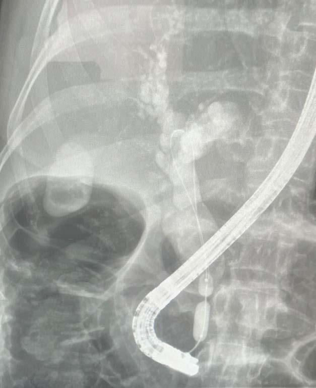
A 59-year-old male presented with vague symptoms of right upper quadrant pain. He provided a history of having undergone three endoscopic retrograde cholangiopancreatographies (ERCPs) in the last 14 months, the last being eight months prior to his presentation. He stated that he had had his first onset of jaundice 14 months ago and the doctor had placed a stent to relieve an obstruction. He was not sure what the investigations were, but he said the doctor told him not to worry, all was OK.
Apart for the one episode of jaundice, he had no medical history and took no chronic medications. He had no previous surgical history, no relevant family history, there was no atopy or autoimmune disease in the family, no inflammatory bowel, and no known liver disorders. He had no known allergies.
Clinical examination revealed a healthy patient, no signs of weight loss, no jaundice, no anaemia or lymph adenopathy. His abdomen was scaphoid, soft, non-tender, no masses were palpable.
Discussion with the doctor who had performed the ERCPs disclosed that their opinion was that of a benign distal common bile duct stricture and their plan was for serial dilatations. Their
opinion was based on the appearance of the stricture, described as a smooth, tight stricture with no shouldering. Two brush cytologies had both come back with normal biliary epithelial cells. The patient’s clinical history was unremarkable.
Blood investigations repeated on two occasions following the first ERCP and stenting indicated a normal full blood count and differential; normal ESR; liver function test had normalised; CA19-9 was 0 units/milliliter and CEA was 1.2 ng/ml. The doctor supplied an ERCP image from the third ERCP, noting the dumbbell shape of the hurricane balloon. A fully covered selfexpanding metal stent was placed at this procedure. No further investigations had been requested.
We performed an endoscopic ultrasound (EUS) which demonstrated a mass around the distal common bile duct and two enlarged periductal lymph nodes. Core biopsy of both the mass as well as lymph node came back as adenocarcinoma. A staging triphasic abdominal CT scan confirmed the EUS findings; the lesion was resectable and there was no metastatic disease.
A pancreaticoduodenectomy was performed and the histology came back as a distal CBD cholangiocarcinoma, T2, N1, Mx, Stage IIB.
The patient has been referred for chemotherapy.
Discussion
This is an example of an initially diagnosed indeterminate stricture believed to be benign, which was, however, a cholangiocarcinoma.
Taking into consideration that this patient had no identifiable risk factors for a benign biliary stricture, and together with his age, further investigations to determine whether it truly was a benign stricture should have been undertaken. Not uncommon traps are the return of normal biliary epithelial cells with brush cytology at ERCP. The sensitivity is approximately 60% with a negative predictive value of 50%,1 and much lower after a stent has been present across the stricture. The second trap in this case was the CA19-9 value of 0 units/milliliter, effectively undetectable. This should be interpreted as the patient being unable to produce CA19-9, which is seen in 10–15% of the general population,2 and hence cannot be used for diagnostic reassurance or follow-up purposes.
Short clinical case: the indeterminate biliary stricture
These cases are not uncommon when working in gastroenterological practice. In our practice, given this scenario, we would perform an EUS with biopsy of the bile duct stricture and adjacent lymph nodes. If this were negative, we would consider performing an elective hepaticojejunostomy, but at the time of surgery, before commencing the biliary bypass, we would – at the biliary dochotomy site – place a spyglass down to the stricture, biopsy it and await the results of the intraoperative pathology. If benign, we would proceed with the bypass; if malignant we would proceed to a pancreaticoduodenectomy. If EUS is not available, we would place a percutaneous transhepatic biliary drain (PTBD) and follow this with a spyglass examination and biopsy via the PTBD.
A review of this topic is Biliary Strictures: diagnostic considerations and approach. Singh, Gelrud and Agarwal.
Take home message
• Endoscopic therapy is a safe and reliable first-line management option for most extra-hepatic biliary strictures.
• Endoscopists must always keep the possibility of a cholangiocarcinoma in the back of their minds, especially if there are no other risk factors for biliary strictures.
• Beware of negative investigations, always correlate these with their efficacy and the patient’s pathology.
References
1. Kobayashi M, Ryozawa S, Araki R, et al. Investigation of factors affecting the sensitivity of bile duct brush cytology. Intern Med. 2019;58(3):329-35. https://doi. org/10.2169/internalmedicine.1551-18
2. Parra-Robert M, Santos VM, Canis SM, et al. Relationship between CA 19.9 and the Lewis Phenotype: options to improve diagnostic efficiency. Anticancer Res. 2018;38(10):5883-8. https://doi.org/10.21873/anticanres.12931
SAGES STATEMENT ON THE CRISIS FACING INFLAMMATORY BOWEL DISEASE (IBD) PATIENTS IN SOUTH AFRICA
Adopted following a meeting at the SAGES Congress, CTICC, 18th May 2024
1. Autoimmune diseases are increasing globally and it is estimated nearly 5% of humans live with autoimmune or inflammatory diseases such as Crohn’s disease and ulcerative colitis. In South Africa, the IBD Africa Registry has seen an exponential increase in IBD incidence over the past 50 years. The vast majority of patients do not die from autoimmune diseases, so compounding prevalence results in an everincreasing number of patients requiring care.
2. Evidence for the early introduction of advanced therapies (biological drugs and small molecules) to prevent permanent gut damage and disability in IBD is now well established.
3. South African IBD patients are fortunate in that we are one of the few countries on the continent to have access to advanced IBD therapies. In addition, a legal framework, enshrined in the Medical Schemes Act 131 of 1998, enables access to these therapies provided this is clinically appropriate.
4. In the public sector, advanced therapies such as anti-TNFs are listed on the EDL but funding restraints limit their use for the treatment of IBD.
5. Despite drug availability in the public and private sectors, overwhelming clinical evidence of efficacy and the legal imperative to support therapy, very few patients receive these treatments.
6. Certain funds refuse to support any advanced IBD therapies unless patients upgrade to the highest, often unaffordable, scheme plans without exceptions.
7. Other funders have a short-sighted approach to funding advanced therapies for IBD. Treatment algorithms are not based on scientific evidence, do not appreciate the clinical heterogeneity of IBD and are adjusted annually based on prevailing financial circumstances. This rigid and short-term approach to managing a lifelong, chronic and incurable disease is illogical and fails to appreciate the long-term health costs and loss of QALYs of such a strategy. The administrative burden imposed by annual changes to scheme rules is particularly onerous.
8. Clinicians treating IBD find themselves in a conflicted situation. On the one hand trying to uphold ethical principles of beneficence and non-maleficence, while on the other hand being forced to make treatment decisions at the discretion of third parties.
To clinicians
We encourage doctors to avoid wasteful healthcare expenditure on ineffective IBD therapies, particularly unnecessary combination therapies and to limit unnecessary endoscopy and cross-sectional imaging procedures
To funders
We caution that ignoring the needs of the increasing number of patients with IBD and other autoimmune diseases is reckless, and by doing so, the problem will not vanish but reappear in the future with a far greater financial burden to your scheme.
We request schemes look at alternative reimbursement models that would allow for cost-effective, and consistent treatment of IBD with advanced therapies.
We encourage engagement with all relevant stakeholders to reach consensus on the funding of advanced IBD therapies.
To the pharmaceutical industry
We appeal to keep drug prices affordable and consistent with our socioeconomic environment and to work with schemes to find alternative reimbursement models that allow timeous and appropriate access to advanced therapies.
To the Council of Medical Schemes
We appeal to allow for the setting of precedent when it comes to adjudicating the merits of advanced therapies in IBD. Your current process is heavily biased toward funders in that:
• Patients who are suffering with chronic ill health do not have the physical or emotional resilience to embark on the arduous CMS process
• Funders obfuscate the issues using medical and legal jargon beyond the comprehension of most patients.
• The ability to delay and appeal rulings by funders frequently leads to the patient resigning from their scheme and switching to another scheme in order to access appropriate therapy before the conclusion of their CMS case.
Drafted by:
South African Gastroenterology Society, Inflammatory Bowel Disease Africa, Autoimmune Alliance of South Africa
SAGES AT IHPBA 2024 PRE-CONFERENCE ENDOSCOPY WORKSHOP
The need for endoscopy training/workshops
Endoscopy is a vital diagnostic and therapeutic tool to ensure optimal patient care, accurate diagnoses, and effective treatment. Its effectiveness, however, is dependent on the quality of the procedure as defined by key performance indicators. Providing quality care is dependent on training and the standardisation of techniques based on evidence-based best practices. Unfortunately, access to training in therapeutic endoscopy is limited in South Africa with no standardisation. SAGES has recognised the need to improve quality in everyday endoscopy. As such, an endoscopy workshop was held prior to the start of the IHPBA/SAGES 2024 conference.
Workshop faculty
The focus of the workshop was on common everyday endoscopic challenges including establishing core competencies in principles of basic polypectomy as well as basic ERCP cannulation. The workshop featured an international guest speaker, Dr David Tate, a Belgium-based interventional endoscopist who trained under the tutorage of Professor Michael Bourke in Sydney, Australia. Dr. Tate is a cofounder of the Ghent International Endoscopy Quality Symposium (GIEQS). He has published widely in the field of endoscopy and is one of the leads for the ESGE (European Society of Gastrointestinal Endoscopy) academy. Additional faculty members included Professor Sandie Thomson, Dr Ernst Fredericks, Dr Vikash Lala, Dr Imraan Sardiwalla and Dr Didintle Mokgoko. Furthermore, the workshop included a number of industry representatives whose contributions and support were greatly appreciated.
Workshop overview
The workshop opened with a welcome by the current SAGES president Dr Ernst Fredericks. This was followed by the opening keynote lecture by Dr Tate who provided a comprehensive and practical overview of the ESGE curriculum for training in endoscopic mucosal resection (EMR) in the colon. Dr Tate was the lead author for this curriculum and was therefore well-suited to deliver the keynote address. His lecture highlighted critical steps in EMR which are often overlooked or not considered at all. This was an eye opener and emphasised the need for training to achieve quality and safety for enhanced patient outcomes. This was followed by the following small group breakaway stations which afforded participant interaction and hands on training:
• Lesion assessment (colonic polyps) – Dr David Tate
• Hot and cold snare technique – Dr Vikash Lala
• Know your accessories – snares, loops, clips, electrosurgical units – Dr Didintle Mokgoko and Prof Sandie Thomson
• ERCP cannulation basics – Dr Imraan Sardiwalla
The ability to recognise and assess colonic polyps is a vital tool for any endoscopist performing colonoscopy. It is a fundamental starting point prior to any sort of endoscopic resection. Correctly assessing lesions ensures that the right treatment modalities are offered to the right patients, thus optimising outcomes. While a number of classification systems exist to assist in assessing colonic polyps, these systems are often complex and difficult to remember in everyday practice. Dr. Tate provided a simplified validated means to assess polyps using basic terminology (regular, regularly irregular and irregularly irregular) eliminating the need to remember more complex systems. He subsequently reinforced the utility of this classification system through real life video-based cases. His deconstructed approach ensured participant interest and participation.
The hot and cold snare technique station hosted by Dr. Lala aimed to provide practical advice on selecting appropriate snares. The mechanics, advantages and disadvantages of varying snares were discussed. The station focused on seldomly thought principles by providing a deconstructed approach on how to actually manipulate a snare in order to achieve optimal placement and improve tissue capture. In addition, the correct technique to open a snare, mark a snare, close a snare and safety checks prior to resection were discussed and practically demonstrated. These techniques are seldom the focus of workshops however are an essential part of everyday colonoscopy. Video based cases and demonstrations with actual snares were used to demonstrate these core concepts.
Any task is made easier and safer with the correct equipment, and this is especially true in the field of interventional endoscopy. One of the most common tasks for any endoscopist is the basic polypectomy. Once an endoscopist accurately and confidently assesses a mucosal lesion and decides to proceed with polypectomy, the next crucial step is selecting the appropriate resection method and the right polypectomy tools and accessories. Dr. Didintle Mokgoko and Prof. Sandie Thomson led the “Know your accessories” station during the workshop. They began by demonstrating the difference between dedicated
cold and hot polypectomy snares, with an emphasis on the types of cold snares available locally. A dedicated cold snare is far superior to the “hot snare, used cold” approach. It is a critical tool in an endoscopy unit for resecting small (<10mm) nonpedunculated polyps, offering lower complication rates, such as post-polypectomy bleeding and perforation.
Proper application and deployment of an endoloop (or polyloop) during the resection of a pedunculated polyp can prevent both immediate and delayed post-polypectomy bleeding. This procedure requires skill and seamless communication between the endoscopist and the assistant, both of whom should be familiar with the endoloop technique. To ensure successful placement of the loop on the polyp stalk, the following steps should be followed: positioning the loop over the polyp and onto the stalk, tightening the loop, monitoring the polyp for colour changes while avoiding excessive pressure that might prematurely transect the stalk, and finally, the correct deployment of the loop. Participants were given the opportunity to practice these steps by deploying the endoloop on model pedunculated polyps repeatedly.
Prof. Sandie Thomson also presented on the correct use of an electrocautery unit during polypectomy, highlighting the recommended settings for basic procedures. Several endoscopic haemostatic devices were demonstrated at this station including various through the scope clips. The highlight of this station was the demonstration of the correct use and application of the “over-the-scope clip,” an invaluable tool for treating GI bleeding that extends beyond the scope of polypectomy.
The ERCP station allowed participants to use the latest endoscopes and accessories to cannulate chicken hearts placed in a simulator. This provided an opportunity to simulate the changes required in scope handling to reach the ampulla. It also allowed the participants to experience real time what different changes to the wheels of the endoscope do to the tip of scope in relation to ampulla. The chicken hearts provided similar tactile feedback to real ampullary cannulation and also allowed participants to simulate performing a sphincterotomy. Overall, the feedback post the session was very positive and I do feel that this training will greatly assist any participants who subsequently go on to performing ERCP in a structured training programme.
In closing, SAGES wishes to express its gratitude to all the participants who made the workshop a success. The overwhelming interest shown, evident by a bookedout event, is testament to the need to develop quality endoscopy in South Africa. This can only be attained through training and upskilling an endeavor to which SAGES is committed. Furthermore, the generous and eager support offered by industry deserves acknowledgement. The sponsored equipment (Marcus Medical, Olympus/Surgical innovations, First Medical, electrosurgical units supplied by Obsidian) allowed for an interactive, practical workshop while travel assistance by Boston Scientific made for a memorable
experience by allowing interaction with Dr Tate, a world leader in endoscopy.
We look forward to hosting similar events in the future.
Vikash Lala
Didintle Mokgoko
Imraan Sardiwalla
Participant testimonial
As a gastroenterology fellow, I found the recent endoscopy workshop at the SAGES annual conference to be an invaluable and inspiring experience. The highlight was undoubtedly meeting Dr. David Tate, the founder of GIEQS, who has been a tremendous source of inspiration for me in the field of endoscopy. Dr. Tate kicked off the session with an illuminating lecture on piecemeal EMR for large non-pedunculated colorectal polyps (LNPCP), setting a high standard for the hands-on training that followed.
The workshop was thoughtfully divided into four stations, each offering unique insights and practical skills. We began by honing our ERCP cannulation techniques through a practical demonstration and hands-on practice on a chicken heart. Dr. Tate then delivered an engaging lecture on the accurate assessment of colonic polyps. This was followed by a practical and insightful short presentation by Dr. Vikash Lala on the correct usage of snares, emphasising the critical importance of selecting the appropriate size for effective polyp removal. The final station was a host of practical demonstrations on important skills required in the endoscopy room. I was particularly impressed by Dr. Mokgoko’s excellent presentation on the correct placement of endoloops for pedunculated polyp removal. I really enjoyed the opportunity to practice deploying an ovesco clip, outside the context of an acute GIT bleed.
Overall, this workshop provided a well-rounded and immersive learning experience, combining expert lectures with practical application. The opportunity to learn directly from pioneers like Dr. Tate and other esteemed colleagues has not only enhanced my clinical skills but also reinvigorated my passion for endoscopy. This experience will undoubtedly have a lasting impact on my clinical practice and professional development.
Dr Nasief van der Schyff, Gastroenterology Fellow at UCT/Groote Schuur Hospital
David Tate’s keynote lecture
Sponsors: Boston Scientific, Marcus Medical, Olympus/Surgical innovations, First Medical Electrosurgical units supplied by Obsidian
Hot and cold snare technique station - Vikash Lala
ERCP station - Imraan Sardiwalla
Know your accessories - Sandie Thomson
David Tate and Vikash Lala
Know your accessories - Didintle Mokgoko
THE 1 ST INTERNATIONAL CONFERENCE OF THE AFRICAN HELICOBACTER AND MICROBIOTA STUDY GROUP
The inaugural International Conference of the African Helicobacter and Microbiota Study Group (AHMSG) took place at the President Hotel in Bantry Bay, Cape Town, from August 16–17, 2024. This event marked a significant milestone for the group, which first convened virtually in 2021 during the COVID-19 pandemic, followed by an in-person launch meeting in Lagos, Nigeria, in 2022.
AHMSG has 16 board members representing 10 African countries: Nigeria, South Africa, Cameroon, Tanzania, Kenya, Senegal, Zambia, Namibia, the Democratic Republic of Congo, and Morocco. Our primary goal is to unify and consolidate efforts to address the challenges posed by Helicobacter pylori infections across Africa, offering solutions tailored to the unique needs of the continent. The group also includes over 90 members, extending our representation to 18 countries.
Professor Stella Smith, from the Nigerian Medical Institute (NIMR), is the founder of the AHMSG and served as the first president of the Group. In Lagos, we drafted a constitution that mandates a biannual rotation of the conference location among member countries, as well as the presidency and secretary-general roles. Cape Town was chosen to host the first conference in 2024.
The conference was truly international yet firmly grounded on African soil. We were honoured by the support of esteemed members of the European Helicobacter and Microbiota Study Group (EHMSG), including keynote speaker Professor Peter Malfertheiner, as well as prominent global experts on Helicobacter
pylori. The first day began with a welcome from Prof. Smith, followed by video footage from our 2022 launch in Nigeria. Goodwill messages were shared by Prof. Chris Kassienedis (founder of GHASSA), Dr. Ernst Fredericks (president of SAGES), and Prof. Anthony Okoh from the African Academy of Sciences.
The keynote address was followed by insightful presentations from international experts such as Prof. Christian Schulz, Prof. Marcis Leja, Prof. Christian Abet, and Prof. Yoshio Yamaoka, all of whom were present in person. Two speakers, Prof. Gianluca Ianiro and Prof. Naima Lahbabi-Amrani, delivered their talks virtually. A notable focus was placed on the microbiome, highlighted by Prof. Ianiro’s talk on “Why African Microbiomes Matter” and a presentation by Prof. Thulani Makhalanyane from Stellenbosch University on the significance of establishing a microbiota consortium. We were also honoured to have national expert Prof. Reid Ally, who delivered a masterful lecture on the complexities of managing Helicobacter pylori within the African context. Another talk from a national was by Mr Ebrahim Hoosein, a microbiologist who spoke on the diagnostic modalities for Heliobacter pylori. The remaining talks over both days were given by the AHMSG board members.
The second day began with oral presentations and concluded with a lively debate on the existence (or lack thereof) of the “African enigma,” which sparked robust discussion. The conference wrapped up with awards and prizes, after which Prof.
Smith officially handed over the presidency to me—a role I will serve in for two years until our next meeting in Zambia in 2026.
For a first conference, both in-person and virtual attendance exceeded expectations, especially for such a specialised event. The success of this meeting is largely thanks to the months of preparation; here I wish to specifically acknowledge the significant contributions of Prof Smith, whose leadership was exemplary. Her experience, tireless efforts and support were a great resource to make this conference a reality. We also enjoyed the invaluable support of our international faculty, and expert logistical management by Bini Seale and Juliet Desilla from
Cornucopia Communications. Indeed this meeting would not be possible without the generosity of all our sponsors.
This was a thoroughly enjoyable and intellectually stimulating event. I look forward to many more years of growth, productivity, and the generation of vital data that can influence policy, guidelines, and outcomes for the vulnerable populations we serve across Africa.
To join as a member and peruse the work of the AHMSG please visit our website: https://ahmsg-africa.org/
Mashiko Setshedi
AFRICAN VIRAL HEPATITIS CONVENTION 16–18 MAY 2024
Advocating for the awareness and elimination of viral hepatitis in Africa hosted by GHASSA under the auspices of the Gastroenterology Foundation of SSA in conjunction with the IHPBA meeting 15–18 May 2024, Cape Town, South Africa.
Viral hepatitis and liver cancer represent a major public health burden in Africa. It is important that Africa, which has one of the greatest burdens of liver disease, responds to the 2016 World Health Organization call, for the elimination of viral hepatitis as a public health threat, by 2030. This cannot be achieved without further capacity and infrastructure development on our continent. An African Viral Hepatitis Convention focusing solely on this public health threat, will be hosted by GHASSA (The Gastroenterology and Hepatology Association of SSA) in conjunction with the IHPBA meeting in Cape Town in 2024. The convention will provide an opportunity for the formation of national and international networks of hepatologists, gastroenterologists, infectious disease specialists and other clinicians, virologists, public health professionals, community health organisations, activists, as well as policy makers and Industry to strive towards the elimination of viral hepatitis and liver cancer in Africa. Hepatitis B viral infection (HBV) rates in Africa largely reflect a failure of maternal and child healthcare programmes to prevent HBV mother-child transmission and early childhood acquisition. HBV is entirely vaccine preventable and only 25% of sub-Saharan African (SSA) countries (12 of 47) have implemented hepatitis B birth dose vaccination. This warrants urgent and immediate legislation. Access to curative directacting antiviral agents for chronic hepatitis C remains limited in many SSA countries and we need to learn from the highly successful HCV elimination programmes in Egypt and Rwanda. It is important that we urgently raise awareness of the extent of liver disease in Africa, both nationally and internationally. We need to learn from the lessons learnt in addressing HIV/AIDS, malaria and tuberculosis and other communicable diseases, using the capacity and infrastructure developed over the years, in order to increase the profile and management of viral hepatitis and liver disease in Africa. Advocacy in viral hepatitis is gaining momentum globally and the successful patient advocacy
programmes in Africa need to be expanded across all regions of Africa to raise the awareness of the importance of preventing hepatitis B mother-child transmission and to give individuals infected with viral hepatitis a voice to demand care. Advocacy groups play a critical role in overcoming the barriers of stigma associated with viral hepatitis and influencing policy-makers, National Departments of Health, and Industry on their obligatory role in providing affordable and accessible diagnostics and therapeutics. What viral hepatitis in Africa requires is a “Nkosi moment”. Nkosi Johnson, a young fellow, who in 2000, at the first AIDS Conference held in Africa in Durban, South Africa captured the audience and world’s attention to the plight of those living and dying from HIV/AIDS in Africa. This became a landmark and seismic shift in the acceptance that access to HIV care was required for all and catapulted the issue of HIV/AIDS in SSA onto the global stage. Viral hepatitis in Africa too needs a seismic shift and more specifically a place on the global health funding arena.
Chris Kassianides, Mark Sonderup & Wendy Spearman (Convention Chairs)
Full report available on www.gastrofoundation.co.za
Wendy Spearman, Anna Kramvis, Mark Sonderup, Chris Kassianides, Geoff Dusheiko
ASSA SAGES AT IHPBA 2024
Prizes awarded
SAGES: Prize Winners
Best Oral 1st prize
Mico Price, University of the Free State
Title: The dietician’s role in the preoperative management for patients undergoing laparoscopic surgery of the upper GIT
Best Oral 2nd prize
Rafaela Hamada Juca, Elaine Ataide State University of Campinas, Brazil
Title: Multidrug resistant infections in liver transplant donors –epidemiology and impact on transplantation outcome
Best oral 3rd prize
Humphrey Kamuzinzi, University of Pretoria
Title: The association between serum fructosamine and random spot urine fructose levels with the severity of non-alcoholic fatty liver disease – an analytical cross-sectional study
Gastro Foundation prize for a fellow
Yonela Qubekile, University of KwaZulu-Natal
Title: Point-of-care immunochemical faecal occult blood testing and symptom-based clinical prediction models to promote earlier diagnosis of colorectal cancer in South Africa.
ASSA: Abstract Prize Winners
1st prize winner:
Dr Matthias Scriba
Title: Predicting full thickness necrosis in adult acute corrosive ingestion injuries in a sub-Saharan African setting
2nd prize winner:
Dr Ilna Conradie
Title: The landscape of thyroid cancer in South Africa: a multiinstitutional review